Article

Cognitive Recovery of Young Males in Thermoneutral Indoor Environments: Effects of Sleep Restrictions

1
School of Environmental and Municipal Engineering, Qingdao University of Technology, Qingdao 266520, China
2
Department of Science and Technology, Hunan Automotive Engineering Vocational University, Zhuzhou 412001, China
3
School of Engineering and Built Environment, Griffith University, Southport, QLD 4222, Australia
4
School of Creative Science and Engineering, Waseda University, Shinjuku City 169-8050, Japan
*
Author to whom correspondence should be addressed.
Buildings 2025, 15(17), 3021; https://doi.org/10.3390/buildings15173021 (registering DOI)
Submission received: 9 July 2025 / Revised: 15 August 2025 / Accepted: 22 August 2025 / Published: 25 August 2025

Abstract

To explore effects of sleep restriction (SR) on next-morning cognitive recovery of young males under thermoneutral environments, three SR conditions, namely the mild (ending at 01:20), moderate (ending at 02:30) and severe sleep restriction (ending at 03:40), were carried out on participants in a thermoneutral environment. During experiments, the subjective sleepiness, perceived workload, and thermal sensation were surveyed. Electrocardiogram (ECG) data were recoded continuously to conduct the heart rate variability (HRV) analysis. In addition, the Deary–Liewald task (including the Simple Response Time task and the Choice Response Time task), Stroop task and Corsi Block task were completed. Results revealed significant increases in sleepiness and perceived workloads during SR. In addition, mean heart rate reduced significantly during moderate (ΔHR = −9.48, p < 0.05) and severe SRs (ΔHR = −9.69, p < 0.01), although it returned to the baseline level in the next morning. The root mean square of successive differences (RMSSD) was elevated during all SRs (Mild SR ΔRMSSD = 27.34, p < 0.05; Moderate SR ΔRMSSD = 33.06, p < 0.01; Severe SR ΔRMSSD = 24.61, p < 0.05) but reduced to baseline the next morning. Furthermore, the sustained attention (SRT) and selective attention performances (CRT) were impaired significantly under moderate (SRT ΔPI = −0.59, p < 0.05; CRT ΔPI = −0.24, p < 0.05) and severe SR (SRT ΔPI = −0.39, p < 0.05; CRT ΔPI = −0.42, p < 0.01). However, the sustained attention performance was restored the next morning even after severe SR, whereas the selective attention performance remained impaired (ΔPI = −0.36, p < 0.01). Significant reductions were observed in the Stroop task performance only after the severe SR (ΔPI = −0.17, p < 0.05), while short-term memory was slightly affected either during or after all SRs (p > 0.05). The overall cognitive performance reduced significantly after the moderate and severe SRs (Moderate SR ΔOPI = −0.30, p < 0.05; Severe SR ΔOPI = −0.40, p < 0.05), even in the next morning. Findings suggest that cognitive impairments caused by mild and moderate SRs could be partially recovered the next morning, while severe SR produced significant impairments in complex cognitive functions, potentially linked to parasympathetic dysregulation and failure of prefrontal compensatory mechanisms. Preliminary findings from this study offer initial implications for cognitive preservation strategies in office environments after night-time overwork.

1. Introduction

In order to meet the escalating needs for career development, working overtime at night has become common in modern cities, particularly in some Asian countries where overworking late into the night is quite common. It was reported that China has emerged as one of the countries with the longest working hours globally [1]. In 2018, full-time employees in China worked 46.5 h per week [2]. The ‘996 work culture’ of China has been widespread in recent years. Similarly, Japan has long been plagued by a culture of excessive working hours. It was disclosed in the 2022 White Paper titled “Measures to Prevent Karoshi” that more than 80 h of overtime are worked monthly by one out of ten Japanese workers, while one in five workers is identified as being at risk of karoshi, a Japanese term for “death by overwork”, with risks including heart stroke, heart attack, and stress-induced suicides [3]. This work pattern, with reduced nocturnal sleep, has led to progressive sleep deprivation, thereby imposing additional health burdens and significantly undermining the public health and overall well-being [4]. For night-time overworking people, these health risks could be largely attributable to impairments in neurocognitive functions by sleep restrictions, e.g., the vulnerability of the prefrontal cortex (PFC) to sleep deprivation [5], as well as the cognitive resources loss due to sleep deprivation (as claimed by Self-Regulatory Resource Theory), finally leading to reduced performance [6]. Some cognitive functions were reported to be significantly impaired by prolonged sleep restriction [7,8]. These negative effects would impair the subsequent working performance and productivity the next morning. Despite these documented negative consequences, it is notable that no established sleep health guidelines or workload management frameworks has been developed for overtime workers. Therefore, the effects of different sleep restrictions on subsequent morning cognitive performance are considered critical for investigation.
Studies have been conducted from the neurophysiological perspective to elucidate the differential effects of the sleep restrictions and acute sleep deprivation on cognitive functions. A prevailing consensus has been reached, which claimed that the glucose metabolic rate of the prefrontal cortex (PFC) was reduced by sleep deprivation, significantly impairing PFC-mediated regulation of attentional control, decision-making, and emotional modulation [5]. This was accompanied by diminished working memory capacity and compromised executive functioning, ultimately resulting in elevated misconduction during work. Notably, the attentional system is particularly vulnerable [9,10,11,12,13,14,15,16]. This is evidenced by significant increases in reaction times and error rates on tasks like the Psychomotor Vigilance Task (PVT) [13,14], reduced interference control (e.g., elevated Stroop errors) [9,17,18,19,20], and impaired performance on tasks requiring divided attention [21,22]. Critically, some impairments, including reduced Stroop accuracy, may be sustained even after sleep recovery [15].
Furthermore, declines in working memory capacity have been regarded as one of the reasons for the impairments of human cognitive performance. Working memory is a fundamental cognitive system with limited capacity that can temporarily retain information and process it [23]. It has been reported that sleep deprivation would reduce the working memory capacity, resulting in prolonged response time, reduced accuracy, or both, in behavioural tasks [24]. Consequently, the ability to manage complex tasks and information was significantly compromised, ultimately diminishing work performance. For example, the critical thinking and working memory scores were reported to be reduced during standardized testing after just a few days of sleep deprivation in a month-long investigation [25]. Significant performance impairments, particularly in accuracy under high cognitive load (e.g., 3-back), were observed in tasks like the n-back after sleep restriction [26]. Similarly, Ma [27] found that during sleep deprivation, the response time increased across tasks, while the accuracy reduced only under high loads. This outcome was attributed to reduced rates of core information accumulation and increased variability in accumulation efficiency.
Apart from attentional performance and working memory capacity, the impairment of executive function is another crucial factor of the attenuated cognitive performance by sleep restrictions. Executive functions include basic cognitive processes such as attentional control, inhibitory control, working memory, and cognitive flexibility [28]. The impairment of executive function resulting from sleep deprivation is primarily evidenced by decision-making bias and difficulties in task-switching during executive tasks. For instance, in the Iowa Gambling Task (IGT), high-risk gambling choices were preferentially selected by sleep-deprived subjects, accompanied by diminished risk–reward evaluation capacities [29,30]. Similarly, in the context of dynamic cognitive tasks, including the Mosaic Task (MT) and The Columbia Card Task cold version (CCTc), research revealed that sleep deprivation over a period of five nights resulted in an elevation of automatic reflexive impulses and risk-taking behaviours [31]. Task-switching challenges under sleep deprivation were further demonstrated through elevated rule-switching error rates in the Wisconsin Card Sorting Test (WCST) [32,33]. Notably, these deficits were reversed following one night of recovery sleep [34].
However, performance in specific cognitive tasks has been shown to remain unaffected by sleep restriction in certain studies. For instance, a multi-day sleep restriction protocol was conducted by Finelli [35], in which stable performance in semantic association tests was observed, and similar findings been observed in other investigations [36,37,38,39]. This finding was interpreted as the evidence that specific cognitive domains may exhibit immunity to sleep loss. The underlying mechanisms for such resilience have been hypothesized by scholars to involve subjective efforts or external rewards [40]. Additionally, differential effects of sleep deprivation on executive function components have been reported. While non-executive cognitive aspects (e.g., task execution efficiency) were impaired, executive functions such as working memory scanning efficiency and interference resistance remained unaffected [41]. Phoneme-speech fluency performance was also preserved, contradicting the notion of executive functions’ universal vulnerability to sleep loss. These findings challenged the prevailing view that attention-regulated cognitive processes were pervasively disrupted by sleep deprivation. Consequently, academic consensus on sleep restriction’s cognitive impacts still needs further investigation.
Conclusively, previous studies elucidated the effects of sleep deprivation on human cognitive functions by examining three crucial aspects: attentional processes, working memory capacity, and executive function, and proposed several significant findings and conclusions. However, current studies regarding the effects of sleep deprivation/restriction on cognitive performances were predominantly carried out by researchers in medicine, psychology, and behavioural science. Consequently, exploration of underlying mechanisms was their priority, while the real scenarios of late-night overwork still needed more specific investigations. For instance, the duration of the overtime work was not well designed in previous studies, and neither were the physical environmental factors like CO2, lighting, acoustics, etc. Moreover, very few studies focused on the different changes in cognitive performances the following morning. Most crucially, there was a lack of exploration on how sleep-restriction durations affected different types of cognitive functions.
In light of the gaps above, three sleep-restriction conditions were devised in a thermoneutral environment, during which tasks with commonly used cognitive functions in overtime work were administered, including those requiring attentional control, inhibitory control, and working memory. During these tasks, subjective perceptions related to cognitive performance were continuously collected through the Stanford Sleepiness Scale (SSS) and workload assessments. Participants’ electrocardiogram (ECG) data were recorded and heart rate variability (HRV) analyses were conducted, and variations in next-morning cognitive performance were systematically examined.
Compared to previous studies, the advantages of this work lie in the design of different sleep-restriction conditions based on real-world late-night overwork scenarios. In the meantime, the physical environmental variables (including the lighting and acoustic condition, and the CO2 centration) were also well designed, to simulate the real office working settings. With these two special designs, the findings could provide more reliable results that would fit the situation in night-time office work well. The novelty of this work lies in the fact that tasks with critical cognitive functions during office work were selected, and the details of their recovery have been scrutinized. This approach enables the identification of how sleep restrictions impact next-morning cognitive performance of young males within realistic contexts. As a pilot study focused exclusively on young males, this investigation is aimed at minimizing sex-related confounders. Findings of this study offer preliminary insights that may inform the optimization of working schedules, refining task management, and maintaining mental productivity in frequent overtime work.
What has to be clarified is that we had finished another piece of work, from the same experimental framework, prior to this study. They had shared methodology and experimental design. However, they differed in objectives and findings, completely. This study focused on the morning recovery of the cognitive performances after sleep restrictions, while the former one emphasized the cognitive changes during three sleep restrictions. The prior study revealed that different cognitive performances kept reducing during three sleep restrictions, as well as the overall cognitive performance, accompanied by several performance fluctuations. These findings initiated another question, regarding how the next-morning cognitive performance would change after these three different sleep durations, which formed the main research question that was addressed by this study.

2. Materials and Methods

2.1. Brief Information About Participants

Eight male participants were recruited in this study. The reason for including only males in this study lay in the fact that the sex difference is a known confounding factor. In order to better observe the cognitive recovery, this confounding factor was therefore excluded, considering that the sample size was not big enough [42]. The mean age of participants was 24 ± 1.3 years, with mean height and weight of 174.4 ± 7.3 cm and 75.4 ± 6.5 kg, respectively. In addition, all participants reported no history of smoking, alcohol abuse, cardiovascular diseases, psychological/mental disorders, or psychotropic medication use. Additionally, normal or corrected-to-normal vision was confirmed by all participants.
One week before the experiment, participants were asked to maintain consistent sleeping schedules, with their bedtime restricted to 23:30–00:30, to standardize circadian rhythms. Compliance was verified via daily sleep diaries, thereby reducing additional variability arising from irregular schedules on experimental days. During the experiment, consumptions of alcohol, caffeine, or stimulants were prohibited. Furthermore, emotional and mood stability was also emphasized, to reduce confounding effects. Participants would be asked to report their affect status before each experiment. Finally, signed informed consents were obtained from all participants, after they understood the experiment protocols well and agreed to join. The experimental procedures were performed in compliance with relevant laws, and were approved by the Ethics Committee of the affiliation with the reference number QDU-HEC-2025327.

2.2. Experimental Conditions and Materials

The experiment was carried out in a climatic chamber located in the Shibei Campus of Qingdao University of Technology. Environmental parameters, including air temperature and relative humidity, were maintained at the thermoneutral condition (24 ± 0.5 °C, 55 ± 5%, respectively), as recommended by ISO 7730 [43]. The chamber had the dimensions of 5000 mm × 3000 mm × 2600 mm (L × W × H), with a waiting room and an experimental room, as shown in Figure 1. Other environmental parameters were set up, according to office environments recommendations by related standards. Specifically, the illuminance of the lighting devices was fixed at 500 lx [44], the air velocity near these desks was controlled at 0.1 m/s [45], and the indoor CO2 concentration was controlled at approximately 700 ppm [46]. To mitigate individual differences in thermal sensation, participants were permitted to adjust their clothing flexibly, to maintain thermoneutrality. Consequently, the confounding effects of lighting condition, air movement, CO2 concentration, and thermal comfort could be eliminated well.
The office environment parameters (lighting, CO2, and air velocity) were selected to simulate the actual working conditions where night-time overtime typically occurs, ensuring ecological validity for studying cognitive recovery after work-induced sleep restriction. During the subsequent sleep phase, environmental conditions were maintained for comfort, but were not office-specific.
Multi-disciplinary methods were adopted in this study, including subjective scales, ECG, and behavioral tests. Thermal Sensation Vote (TSV), Stanford Sleepiness Scale (SSS), and NASA Task Load Index (NASA-TLX) scale were administered to assess the thermal sensation, sleepiness, and perceived workload during sleep restrictions. All scales were completed and recorded electronically in the PsyToolkit platform [47]. In addition, electrocardiogram (ECG) data were acquired using the Polar H10 chest strap (Polar Electro Oy, Kempele, Finland), which operated at a sampling rate of 1 kHz with an accuracy of ±1 ms. Time-domain HRV metrics based on the ECG data were obtained using the Kubios HRV Standard software (version 3.5.0, Kubios Oy, Kuopio, Finland), including mean heart rate (Mean HR) and root mean square of successive differences (RMSSDs). Reasons for choosing the above measurements are shown in Table 1.

2.3. Cognitive Tasks

The Deary–Liewald task, Stroop task, and Corsi Block task were selected in this study. The reasons for selecting them lay in that they had included the fundamental and common cognitive functions of the human body during mental work, including attention, working memory, inhibition, and executive function.
All these cognitive tasks were designed on the online open access platform PsytoolKit [47]. The results could also be recorded online. The corresponding functions and duration of these tasks are provided in Table 2.
Upon completion of these tasks via the platform, reaction time (RT), accuracy, and maximum memory span were automatically recorded as the raw data. In accordance with the Psychomotor Vigilance Task (PVT) evaluation criteria [57], responses exceeding 500 ms were classified as lapses. Specifically, for the Simple and Choice Reaction Time tasks, accuracy was calculated as the proportion of correct responses with RTs below 500 ms. Since the speed-accuracy ‘trade-off’ was not the primary focus of this study, a Performance Index (PI) was introduced by geometrically weighting the accuracy and response time, with equal weights (0.5) assigned to each parameter. This PI provides a comprehensive assessment of cognitive task performance. The formula for calculating the PI can be found in previous literature [58].

2.4. Experimental Procedures

The study was conducted strictly in accordance with the Helsinki Declaration, to ensure the protection of participants’ rights. All participants were required to complete all three sleep-restriction conditions, but they were free to quit the experiment at any time.
Three sleep-restriction conditions with different durations were designed: specifically, the mild (Condition 1, ending at 01:20), moderate (Condition 2, ending at 02:30) and severe (Condition 3, ending at 03:40) sleep restriction. Regarding the classification of mild, moderate, and severe sleep restrictions, while some academic debate remains, the design of this study was supported by the existing literature. The 4.7 h of sleep per night was defined as severe sleep restriction by reference [59], and even longer sleep durations were employed in the mild and moderate sleep-restriction protocols by reference [60]. Two participants were engaged in each experiment. A minimum interval of seven days was required between two sleep-restriction conditions, to ensure the full elimination of effects of the former one. In addition, the time and date displayed on the displays of computers were concealed, to make participants unaware of the time of the undergoing experiment. Therefore, they did not know the specific sleep-restriction condition until the completion. Additionally, participants were required to alternately complete cognitive tasks and watch the documentary video ‘Aerial China’ during sleep restriction periods. This combined protocol was designed to simulate multidimensional resource-allocation demands in real-world overtime work. The standardized documentary stimulus was selected because its continuous narrative flow requires sustained attentional engagement, comparable to routine monitoring operations (e.g., quality control surveillance), while its passive nature minimizes movement artifacts in physiological recordings. Critically, alternating active cognitive tasks enables precise titration of competing resource dimensions, including perceptual, attentional, and working memory systems, thus generating cumulative fatigue through controlled resource competition, as described in multidimensional workload models [61].
The experiment proceeded as follows. On the experimental day, participants were required to arrive at the climate chamber by 22:00. Within 15 min of arrival, heart rate straps were attached. Participants then entered the climatic chamber for a 30 min thermal adaptation, during which these cognitive tasks were practiced, to mitigate any ‘learning effect’ that might occur during formal experiments [62]. Following adaptation, cognitive tests, subjective scales and ECG recording were administered from 22:45 to 23:00, results of which were used as the baseline data in this study. At 23:00, participants started to watch a 20 min documentary video, followed by a 15 min cognitive testing stage and scale surveys. The order of these three cognitive tasks within each testing stage was randomized, to eliminate the order effect. Subsequent intervals repeated the documentary video watching, cognitive testing, and survey, until the end of each sleep-restriction condition (01:20 for Condition 1, 02:30 for Condition 2, or 03:40 for Condition 3). Upon completion, participants slept in the chamber till awakened by experimenters at 08:20 the next morning. During the sleeping, the environmental variables were well controlled. In the next morning, after a 10 min wakefulness period, final cognitive tests and scale surveys were conducted at 08:30. The detailed procedures are illustrated in Figure 2.

2.5. Data Analysis

ECG data acquired during sleep restrictions were processed using ECG Viewer. Prior to analysis, a ‘filtering of the power frequency of 50 Hz’ was applied, to eliminate alternating current interference. Thereafter, ECG data were segmented into intervals corresponding to cognitive testing stages in the experiments. These segmented data were subsequently imported into Kubios HRV software for heart rate variability (HRV) analysis, generating task-specific HRV metrics.
Statistical analyses were conducted with SPSS 27 and Origin 2023. The sample size (n = 8) was determined by G*Power 3.1 for repeated-measures ANOVA (within-subject design), with the α of 0.05, the power of 0.8, the effect size of 0.7 from pre-experimental results, and the measurement times of 3. The results showed that the sample size of 8 could meet the requirements of statistical analyses. Prior to statistical analysis, outliers were identified and removed from raw datasets. Thereafter, the normality (Shapiro–Wilk test) and homogeneity of variance (Levene’s test) were examined for each variable, under all experimental conditions. For datasets satisfying normality and homoscedasticity assumptions (p > 0.05), repeated-measures ANOVA with Bonferroni-corrected pairwise comparisons were conducted. Nonparametric Friedman and Wilcoxon signed-rank tests were applied to non-normally distributed or heteroscedastic data. The significance level for all statistical analyses was set as 0.05.

3. Results

This section includes changes in results from subjective scales, HRV, and cognitive performance in sleep restrictions, and the variations observed the next morning.

3.1. Results of Subjective Surveys

3.1.1. TSV

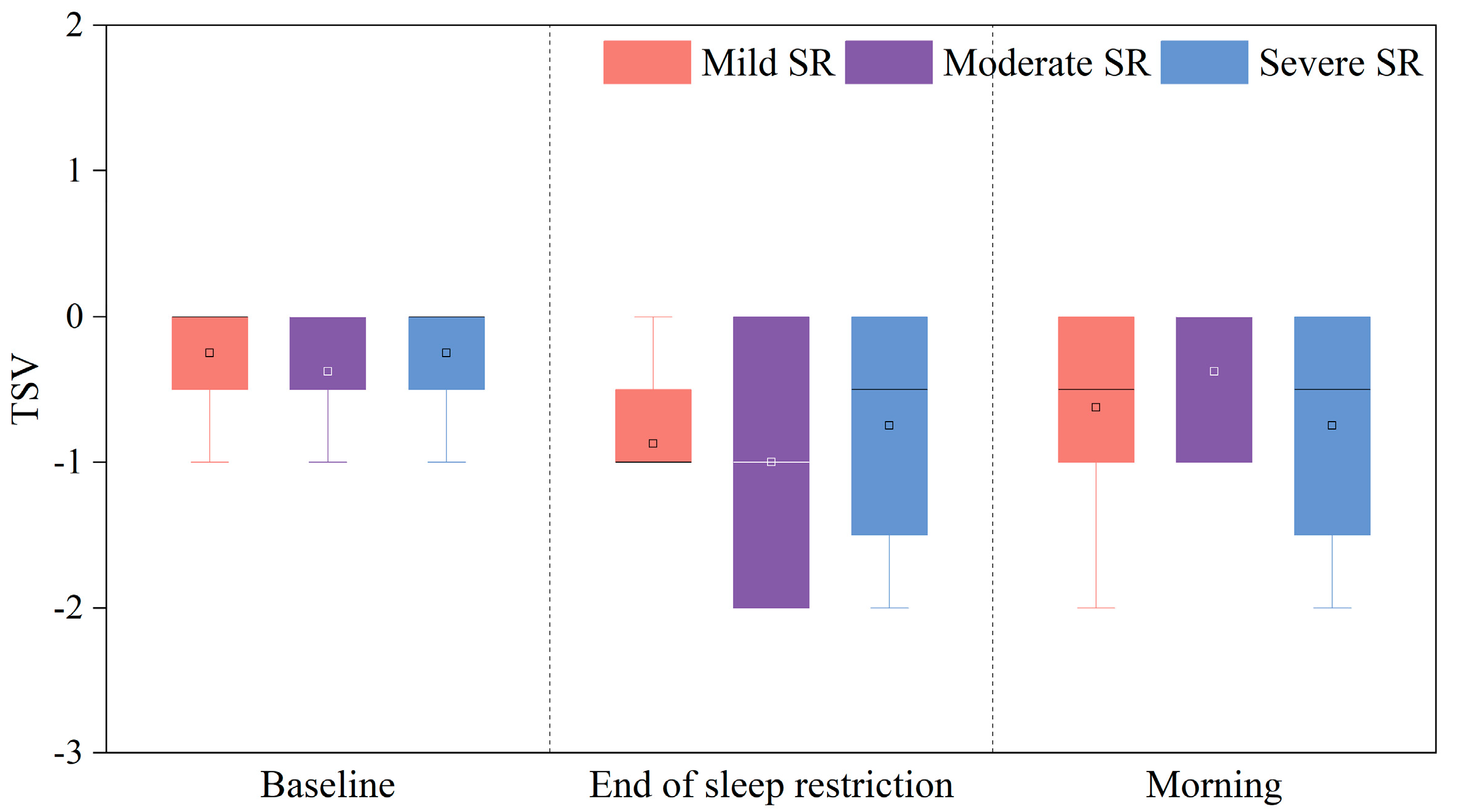
Thermal sensation vote (TSV) scores under these three sleep-restriction conditions are illustrated in Figure 3. As shown by Figure 3, TSV scores were predominantly distributed within the ‘slightly cool’ range (−1 < TSV < 0) for all conditions, with no statistically significant differences observed among conditions (p > 0.05). This finding confirmed that cognitive performance changes during all three sleep restrictions were not produced by thermal environments.

3.1.2. Sleepiness and Perceived Workload

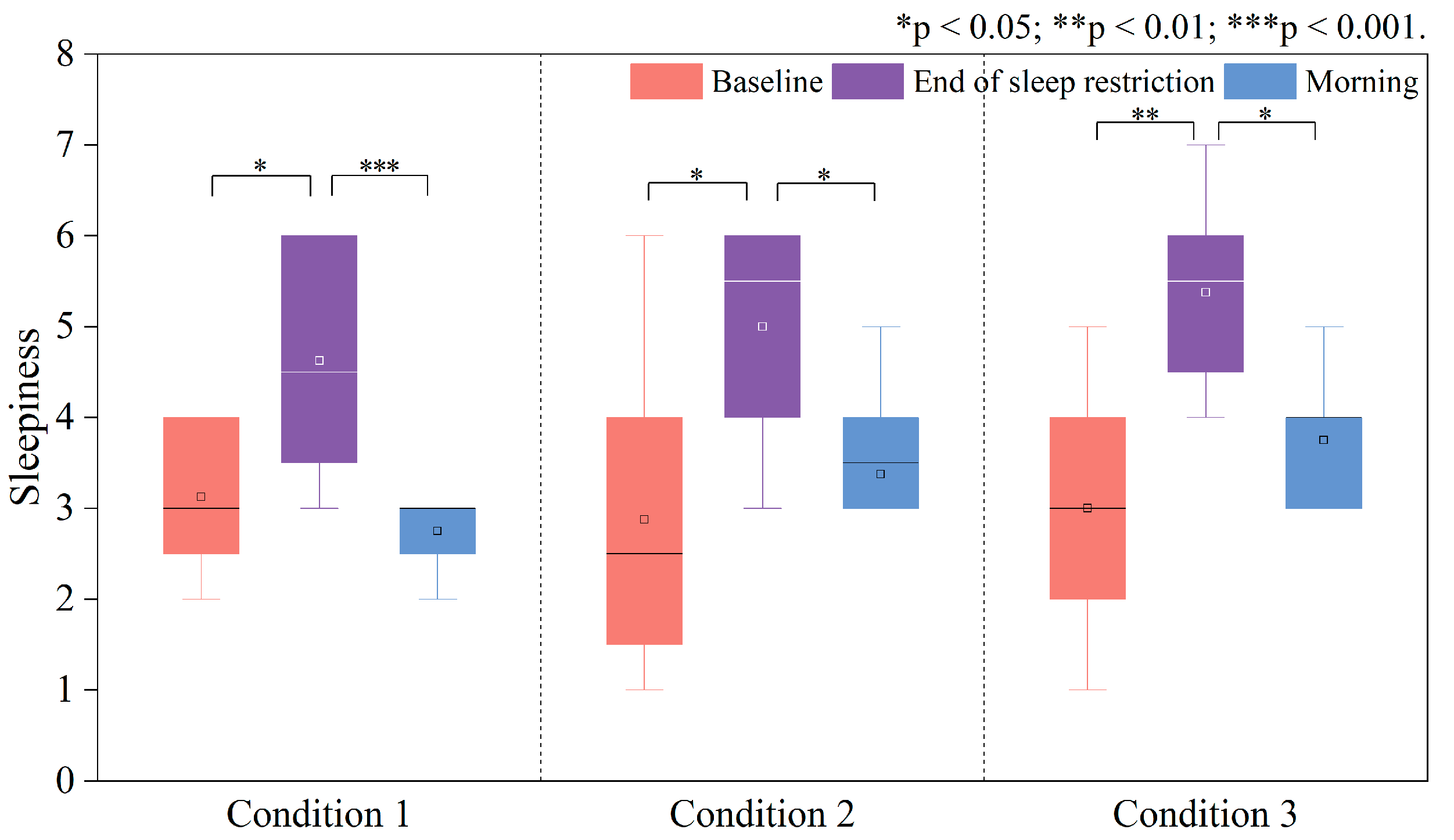
Changes in subjective sleepiness under and after three sleep-restriction conditions are presented in Figure 4. The subjective sleepiness increased significantly from baseline to the end of each sleep restriction (p < 0.05), with greater escalation observed under longer sleep-restriction durations (Condition 3). Following the recovery sleep, morning assessments revealed reductions in subjective sleepiness, which were confirmed to be significantly lower than at the end of each sleep restriction (p < 0.05). The most pronounced recovery (or sleepiness reduction) was observed in Condition 1 (mild restriction, p < 0.001), whereas Condition 3 (severe restriction, p < 0.05) showed a less effective recovery.
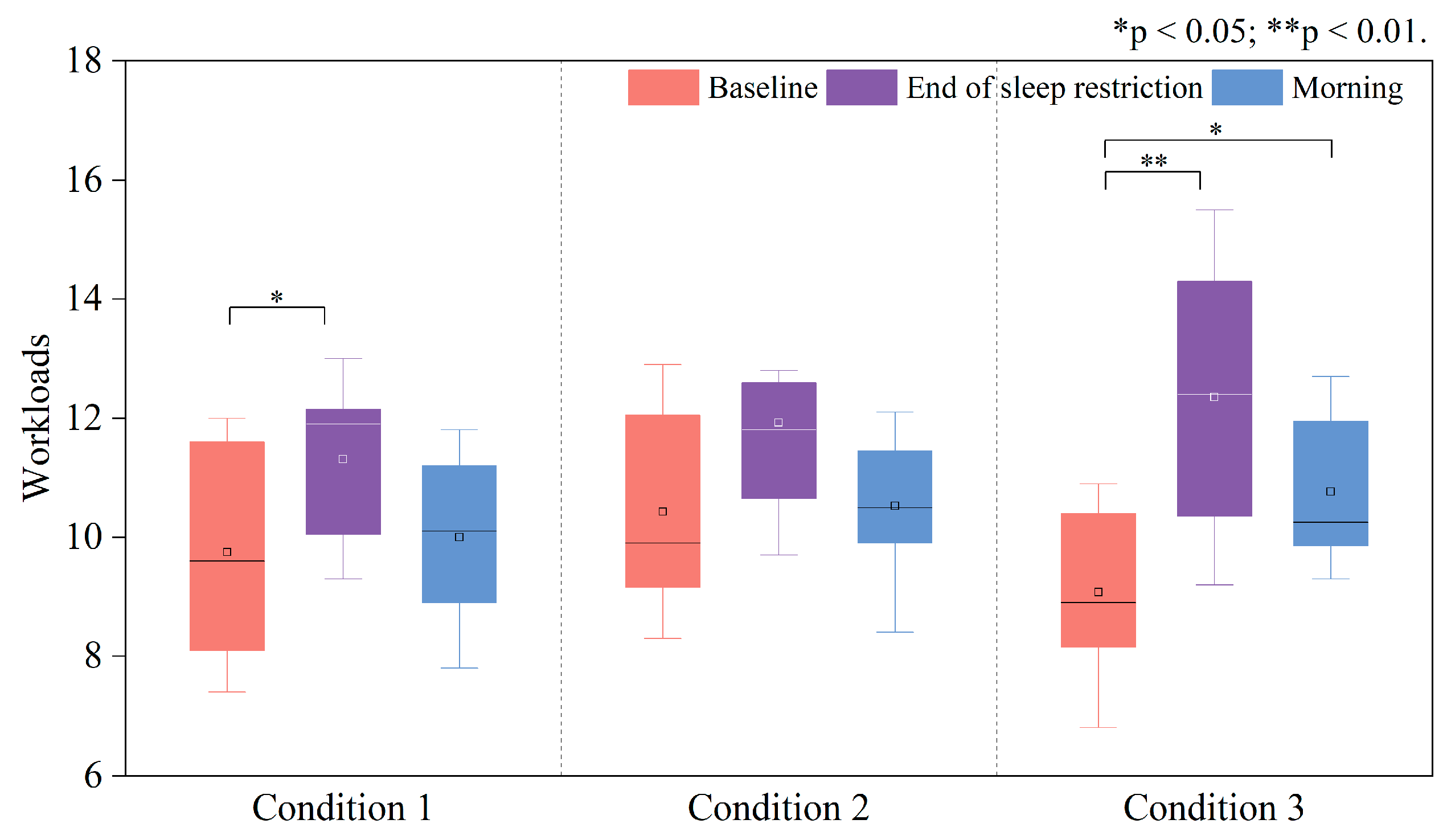
Variations in perceived workload for all three sleep-restriction conditions are illustrated in Figure 5. At the end of each sleep restriction, significant increases in perceived workload relative to baseline were detected only in Conditions 1 (p < 0.05) and 3 (p < 0.01), while Condition 2 just had a slightly upward pattern (p > 0.05). In morning assessments, perceived workload reduced for all conditions compared to the level at the end of each restriction (p > 0.05). Specifically, a significant difference was observed between baseline and morning assessments in Condition 3 (p < 0.05). Notably, shorter sleep restrictions corresponded to lower perceived workloads in the morning.

3.2. Changes in HRV Time-Domain Indices

Figure 6 and Figure 7 present the variations in mean heart rate (HR) and the HRV time-domain index RMSSD for three sleep-restriction conditions. Overall, as shown in Figure 6, a downward pattern of the mean HR was observed during all sleep-restriction conditions compared to baseline levels. In Condition 1, mean HR reduced from the baseline level 71.06 beats/min to 65.18 beats/min, although no significant differences were observed (p > 0.05, Cohen’s d = 3.84). In contrast, Conditions 2 and 3 showed significant reductions in mean HR, from 69.99 beats/min and 71.80 beats/min to 60.51 beats/min (ΔHR = −9.48, p < 0.05, Cohen’s d = 3.09) and 62.11 beats/min (ΔHR = −9.69, p < 0.01, Cohen’s d = 3.88), respectively.
The next morning, mean HR for all conditions returned to levels close to or slightly over baseline levels (Cohen’s dMild = 3.54, Cohen’s dModerate = 11.79, Cohen’s dSevere = 4.46). Conditions 1 and 2 showed less increase, while Condition 3 had a significantly higher mean HR than at end of its sleep restriction (ΔHR = 13.96, p < 0.01, Cohen’s d = 4.64). These results indicated a negative correlation between sleep-restriction duration and mean HR in sleep restrictions.
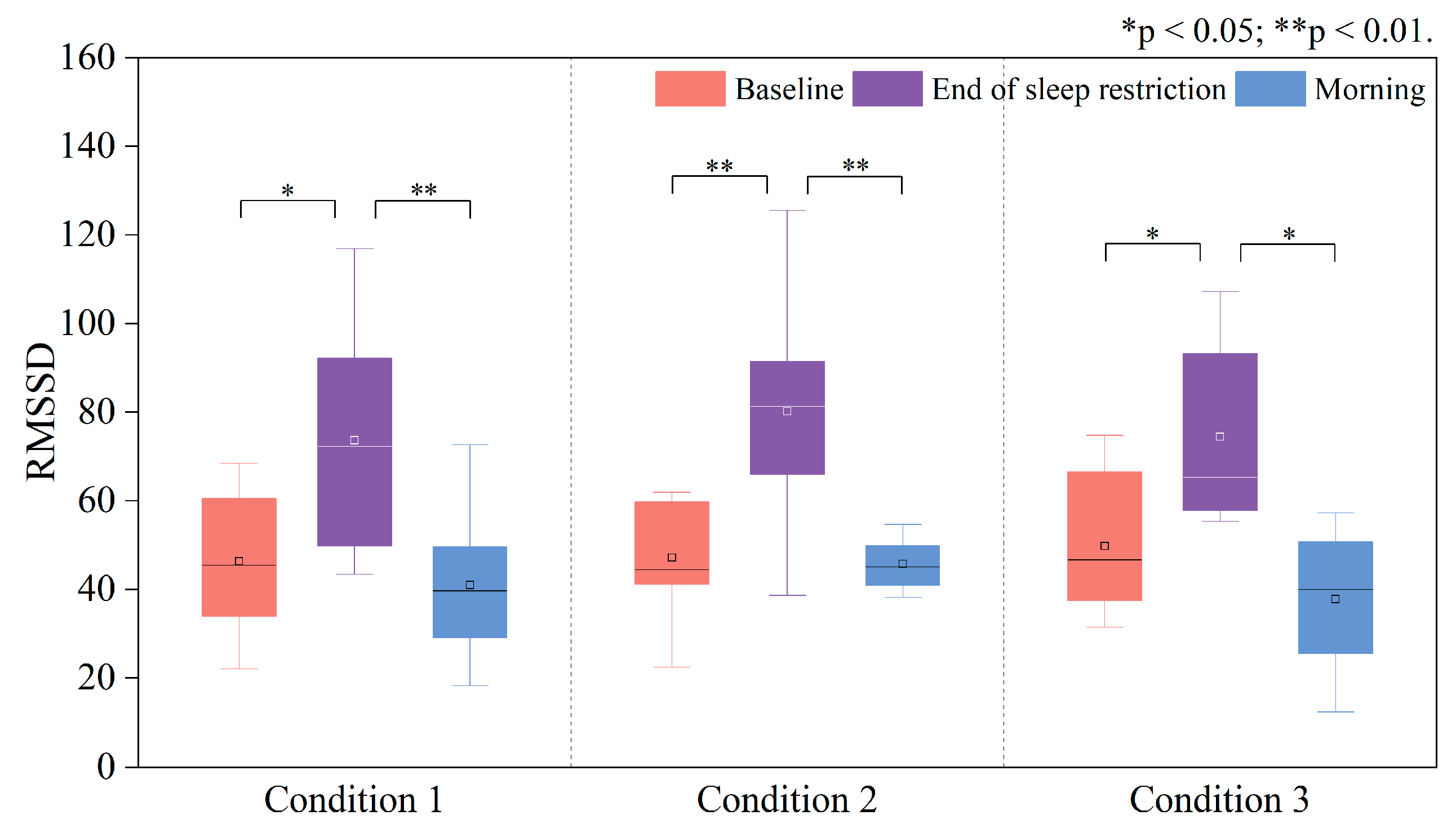
Changes in RMSSD are given in Figure 7. Results indicated that at the end of each sleep restriction RMSSD increased significantly, compared to baseline levels, under all three sleep-restriction conditions. Conditions 1 and 3 exhibited similar elevation patterns (Condition 1 ΔRMSSD = 27.34, p < 0.05, Cohen’s d = 22.11; Condition 3 ΔRMSSD = 24.61, p < 0.05, Cohen’s d = 16.29), whereas Condition 2 showed a more pronounced increase in RMSSD, relative to baseline (ΔRMSSD = 33.06, p < 0.01, Cohen’s d = 15.88). The next morning, RMSSD for all sleep-restriction conditions reduced significantly, compared with the levels at the end of sleep restrictions (Condition 1 ΔRMSSD = −32.65, p < 0.01, Cohen’s d = 20.73; Condition 2 ΔRMSSD = −34.48, p < 0.01, Cohen’s d = 33.81; Condition 3 ΔRMSSD = −36.63, p < 0.05, Cohen’s d = 18.61).

3.3. Variations in Performance Index (PI) Under Three Sleep-Restriction Conditions

3.3.1. Changes in the PI of the Deary–Liewald Task

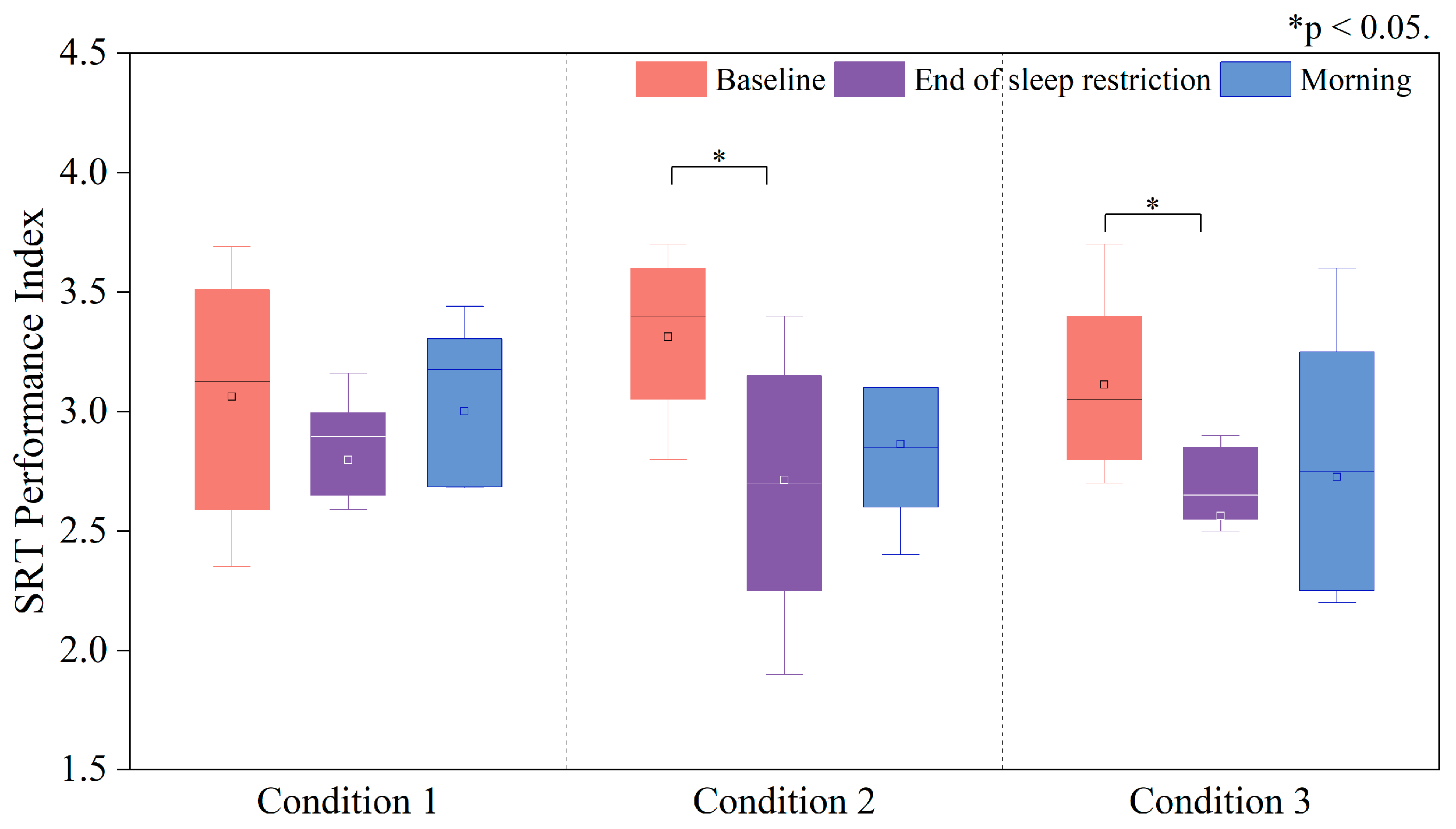
The Deary–Liewald task comprises the Simple Response Time (SRT) task and the Choice Response Time (CRT) task, which assess sustained attention and selective attention, respectively. Variations in the Performance Index (PI) of the SRT task under these three sleep-restriction conditions are illustrated in Figure 8.
PI was reduced at the end of each sleep restriction for all three sleep-restriction conditions, compared with baseline levels, but significant reductions in PI were only observed in Conditions 2 and 3 (Condition 2 ΔPI = −0.59, p < 0.05, Cohen’s d = 0.60; Condition 3 ΔPI = −0.39, p < 0.05, Cohen’s d = 0.63). Notably, longer sleep restriction durations correlated with lower PI values at the end of each sleep-restriction condition.
Moreover, PI increased the next morning for all sleep-restriction conditions compared with the levels at the end of each sleep restriction, but no statistical significance was observed. Meanwhile, the PI the next morning also showed no significant differences compared with the baseline levels (p > 0.05). Notably, the mean PI in Conditions 2 and 3 was slightly lower than that in Condition 1, though this difference was not statistically significant (p > 0.05, Cohen’s dMild = 0.43, Cohen’s dModerate = 0.43 and Cohen’s dSevere = 0.42 respectively).
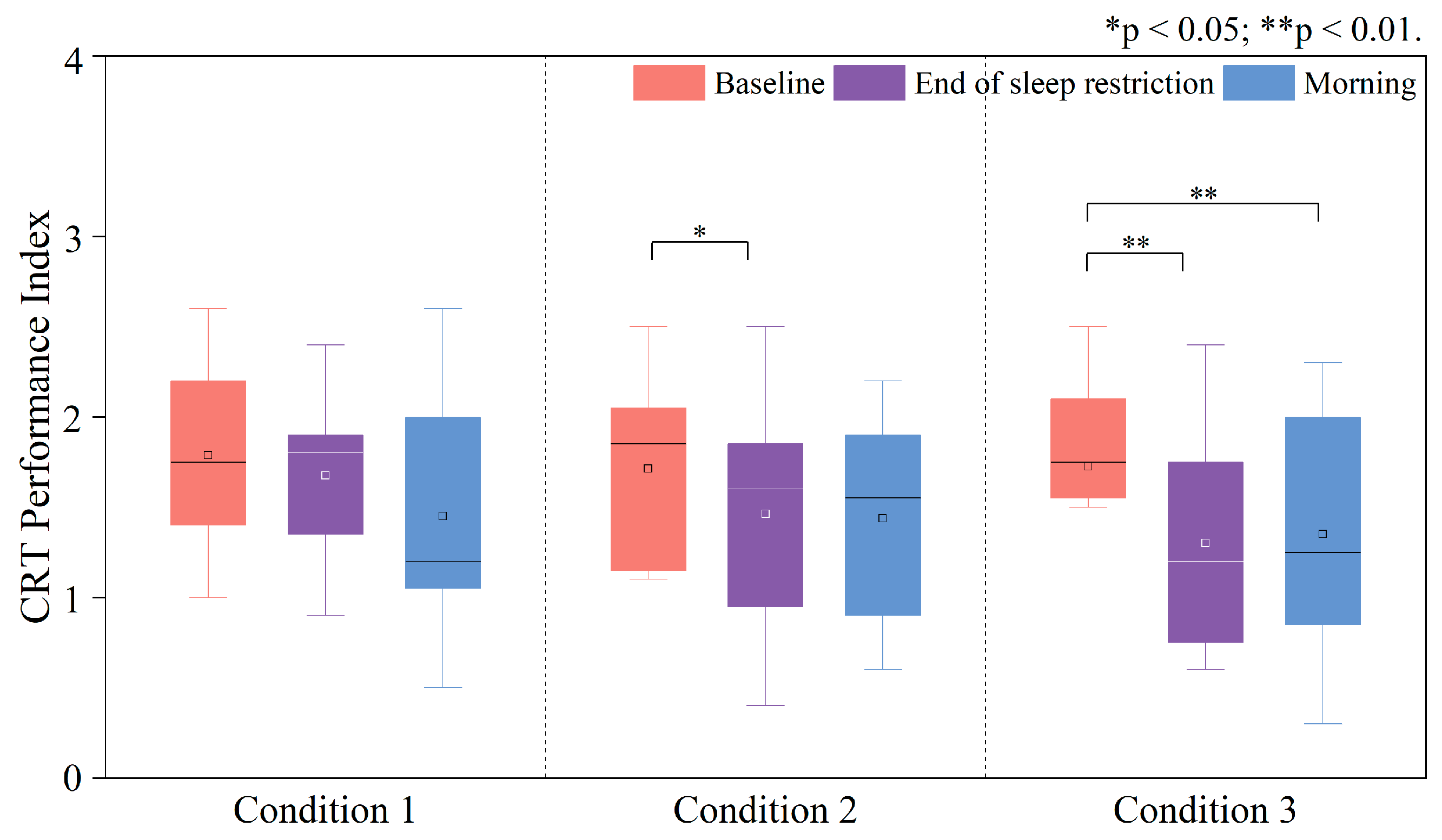
Changes in PI of the Choice Response Time (CRT) task under and after these three sleep-restriction conditions are illustrated in Figure 9. Similar to SRT performance changes, PI was reduced at the end of each sleep restriction, compared with the baseline levels, and statistically significant declines were observed in Conditions 2 (ΔPI = −0.24, p < 0.05, Cohen’s d = 0.24) and 3 (ΔPI = −0.42, p < 0.01, Cohen’s d = 0.42). However, PI in the morning changed differently. In Condition 1, the mean PI reduced more than that in Condition 2 and 3, compared with the levels at the end of each sleep restriction. However, no significant differences were observed for all three sleep-restriction conditions (p > 0.05, Cohen’s dMild = 0.42, Cohen’s dModerate = 0.61 and Cohen’s dSevere = 0.49, respectively). For Condition 3, significant reduction in morning PI was observed compared with the baseline level (ΔPI = −0.36, p < 0.01, Cohen’s d = 0.34).

3.3.2. Changes in the PI of the Stroop Task

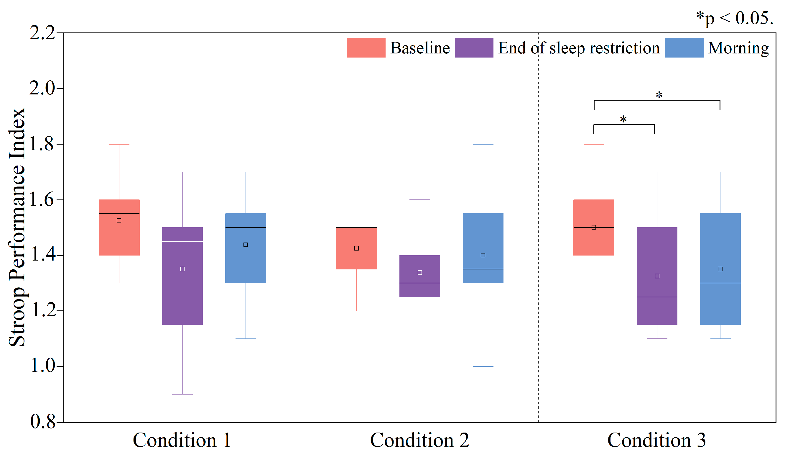
The Stroop task is a measure of selective attention, inhibitory control, working memory, and executive function. Variations in PI of the Stroop task under and after these three sleep-restriction conditions are illustrated in Figure 10. As depicted in Figure 10, the mean PI at the end of each sleep restriction was reduced, compared with the baseline levels, for all three conditions. However, statistically significant reduction was observed only in Condition 3 (ΔPI = −0.17, p < 0.05, Cohen’s d = 0.14).
The next morning, the mean PI in all three sleep restrictions did not rise much, compared with that at the end of each condition (p > 0.05, Cohen’s dMild = 0.20, Cohen’s dModerate = 0.23 and Cohen’s dSevere = 0.07, respectively). However, compared with the baseline levels, only the PI in Condition 3 was significantly lower (ΔPI = −0.11, p < 0.05, Cohen’s d = 0.15), which implied that the prolonged sleep restriction might produce severe performance declines in the Stroop task the next morning.

3.3.3. Changes in the PI of the Corsi Block Task

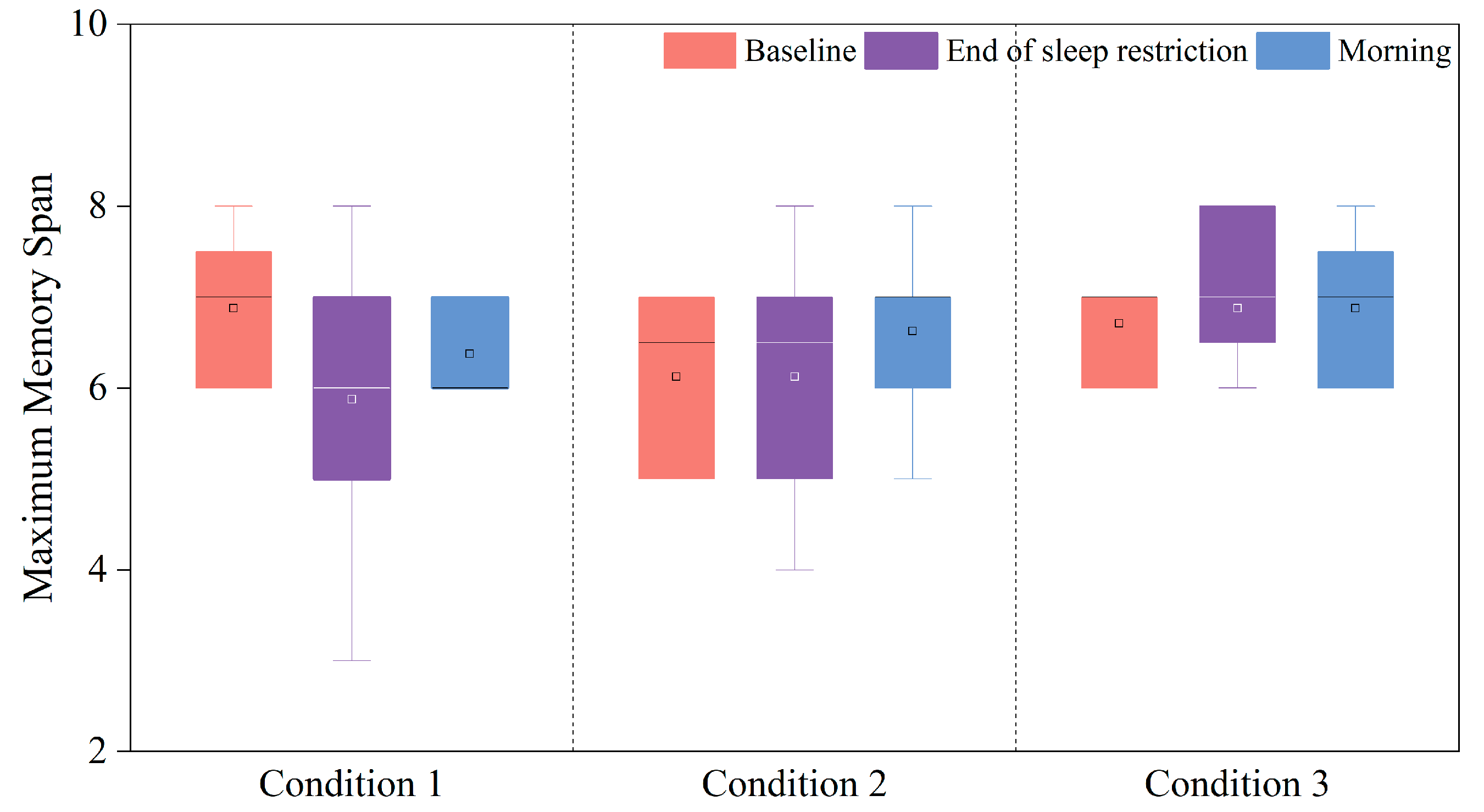
Spatial working memory and short-term memory are measured by the Corsi Block task. Changes in the maximum memory span during the Corsi Block task under these three sleep-restriction conditions are presented in Figure 11. It was found that changes in maximum numbers of the task showed divergent changes for all three sleep-restriction conditions. No similar pattern was observed. Meanwhile, no statistical significance was observed for all these changes in all three conditions.

3.3.4. Changes in the Overall Performance Index

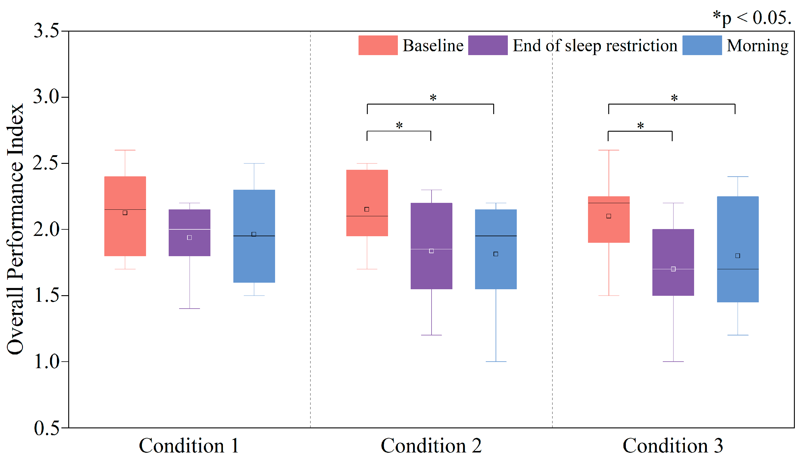
The Overall Performance Index (OPI) was calculated as the arithmetic mean of the PI derived from the Simple Response Time (SRT), Choice Response Time (CRT), and Stroop tasks. The Corsi Block task was excluded from the analysis, because its outcome could not be integrated into arithmetic averaging, and its performances under all three conditions failed to fall into a similar pattern. The OPI represented a macro evaluation of participants’ cognitive performance during and after these sleep restrictions.
Variations in the OPI under and after these three sleep-restriction conditions are illustrated in Figure 12. In Condition 1, no statistically significant changes in the Overall Performance Index (OPI) were observed from baseline to the end of the restriction, or the next morning (all comparisons, p > 0.05). Although the OPI was numerically lower than baseline at the end of the restriction and numerically higher than the end-of-restriction level in the morning, these differences did not reach statistical significance. For Conditions 2 and 3, significant reductions in OPI were observed at the end of each condition (Condition 2 ΔOPI = −0.30, p < 0.05, Cohen’s d = 0.26; Condition 3 ΔOPI = −0.40, p < 0.05, Cohen’s d = 0.33), compared with the baseline levels. However, the morning mean OPI rose slightly in Condition 3 (p > 0.05, Cohen’s d = 0.23), and did not change much in Condition 2 (p > 0.05, Cohen’s d = 0.39), compared with that at the end of each condition. In addition, it was found that the morning mean OPI in Condition 2 and 3 was significantly lower than the baseline levels (Condition 2 ΔOPI = −0.33, p < 0.05, Cohen’s d = 0.15; Condition 3 ΔOPI = −0.30, p < 0.05, Cohen’s d = 0.25).

4. Discussion

Changes in cognitive performances the next morning due to three different sleep restrictions (mild, moderate, and severe) were explored in this study, by means of subjective scales, ECG recording, and cognitive tests.
It was found that the mean subjective sleepiness and the RMSSD both showed inverted-U patterns during and after these sleep restrictions. It meant that the subjective sleepiness increased during sleep restrictions, but was reduced to the baseline level the next morning. Changes in mean RMSSD followed this pattern. RMSSD is a measure of the parasympathetic nervous activities, increases in which may indicate the rise of the parasympathetic tone [63]. Consequently, participants in this study would experience relaxation and sleepiness during the sleep restrictions, but returned to the baseline levels the next morning. The changes in RMSSD in this study were in accordance with previous findings by Chua, who reported a positive correlation between RMSSDs (Root Mean Square of Successive Differences) and sleepiness, following acute sleep deprivation [64].
However, it is not sufficient to prove that the cognitive performance could return to the baseline level. Changes in mean heart rate and perceived workload provided something different. The mean heart rate during these three sleep restrictions showed U-shaped patterns. However, no significant differences were observed in mild sleep restriction. In moderate and severe sleep restrictions, the mean heart rate reduced significantly at the end of the sleep restrictions, then returned to the baseline levels the next morning. This was in accordance with the changes in mean RMSSD. Higher RMSSD corresponded to lower sympathetic tone and thus lower heart rate, as indicated by a few references [63,65,66]. Meanwhile, low heart rate might also correspond to low mental arousal level [67]. Consequently, during the moderate and severe sleep restrictions, participants had significant lower arousal levels at the end of sleep restrictions, which corresponded to lower cognitive performance. However, it is necessary to consider potential confounding factors of HRV metrics, such as diurnal variations, physical activity, mental stress, and individual health conditions [68]. In this study, these variables were controlled by standardizing the physical environments and testing conditions (as we stated in the Section 2). However, the mental states were usually hard to control, which might act as a confounder of RMSSD in this study.
Although the mean heart rate rose back to the baseline levels, it was again hard to conclude that the cognitive performance in the morning was well recovered, since the results from NASA-TLX scale gave some different messages. The perceived total workload measured by the NASA-TLX scale showed a quasi-inverted-U pattern during and after sleep restrictions. However, significant changes were only observed in severe sleep restriction (Figure 5). Specifically, the mean perceived workload at the end of the sleep restriction and in the morning were both significantly higher than the baseline level, which meant that participants felt that it was harder to cope with workloads at the end of the sleep restriction, and they even failed to recover to the baseline level in the morning. Consequently, it could be concluded that the cognitive performance might have suffered more from severe sleep restriction than the mild and moderate ones, although the RMSSD and subjective sleepiness results showed that the autonomous balance and sleepiness had been well recovered.
Crucially, our physiological and cognitive data jointly suggest that autonomic recovery may not fully translate to functional cognitive restoration. While RMSSD and heart rate, which were used as a biomarker of the cognitive performance in a few studies [53,69,70] returned to baseline levels by next morning across all conditions (Figure 6 and Figure 7), selective attention and executive functions remained impaired after severe sleep restriction (Figure 9 and Figure 10). This dissociation implies that parasympathetic normalization (indexed by RMSSD recovery) is necessary, but insufficient for restoring higher-order cognition. We propose a non-exclusive mechanism: prefrontal (PFC) vulnerability to sleep loss might not fully resolve alongside autonomic recovery. This is tentatively supported by sustained Stroop deficits, though other contributing factors cannot be excluded.
The most interesting part of this study was the way in which different cognitive functions were affected by different sleep restrictions. We understand that there have been some studies scrutinizing the performances of different cognitive tasks during sleep deprivation/loss, but this study gave the specific scenario of overwork during the night in office environments. Similarly, this study revealed again that different cognitive functions were affected differently by sleep restrictions, as some previous studies had revealed [10,14,21,24,28,29]. However, this study revealed the recovery of these cognitive declines the next morning, to offer some implications for real applications.
Specifically, the sustained attention represented by the Simple Response Time task (SRT) did not suffer from significant attenuation. However, as the sleep restriction extended to moderate and severe conditions, the sustained attention showed no statistically significant difference from baseline the next morning. In addition, the selective attention represented by the Choice Response Time task (CRT) was not compromised significantly during mild sleep restriction. However, in moderate and severe sleep restrictions, the selective attention reduced significantly at the end of the sleep restrictions. Moreover, in moderate sleep restriction, it could then recover to the baseline level the next morning, but it failed to rise back to baseline level in severe sleep restriction. Furthermore, important cognitive functions of participants, including the working memory, executive function, and inhibitory control, which were represented by the Stroop task, were significantly compromised by the severe sleep restriction, and they failed to recover to the baseline levels in the next morning. On the contrary, these cognitive functions did not suffer significant attenuations in the mild and severe sleep restrictions, and they could also rise back to baseline level in the morning. Finally, the overall cognitive function of participants showed no significant changes in the mild sleep restriction. However, the moderate and severe sleep restrictions produced significant reduction in overall cognitive function, and it could not return to baseline level the next morning. Findings are summarized in Table 3.
Table 3 reveals that the cognitive functions of the human body were affected differently by sleep restrictions. It indicates that selective attention and working memory, executive function, and inhibitory control showed no statistically significant difference from baseline after mild and moderate sleep restrictions, while they could not rise back to baseline levels after the severe sleep restriction. On the other hand, sustained attention and short-term memory would return to baseline levels after all three sleep restrictions.
The reason for the changes in selective attention lay in the fact that the SRT task was a measure of response to a single stimulus of the human body [71]. As a cognitively undemanding task requiring minimal resources, SRT performance showed no statistically significant difference from baseline levels the next morning. In addition, a study by Diekelmann [72] also claimed that tasks with low cognitive loads could be maintained at acceptable performances through the several neuro-compensations during sleep loss. This might also be another reason.
The reason for the insignificant changes in the performance of the Corsi Block task could be ascribed to the features of this task per se. The Corsi Block task is a measure of the working memory and short-term memory, and involves the coding process and memory of spatial information [73]. A study by Krause [74] claimed that attenuations in memory coding due to sleep deprivations could be compensated by the mediation of the arousal network, through the improvements of connectivity among the hippocampus, the brain stem, and the thalamus. Consequently, the performance of the Corsi Block task did not show significant reduction in any of these three sleep restrictions.
The performances of the Stroop task, which included several higher-order cognitive functions, were compromised more significantly than other tasks. It complied with a few previous studies [9,15,20]. E.g., a meta-analysis claimed short-term sleep deprivations less than 48 h had limited influence on short-term memory, but compromised the complex cognitive tasks significantly (like working memory) [75]. However, it could also be observed that cognitive functions like working memory, executive function, and inhibitory control worked more stably in mild and moderate sleep restriction than the sustained attention and selective attention did. It meant that performances of more complex task were maintained than simpler ones, which contradicted previous findings. This could be interpreted by a study by Chee and Choo, who stated that prefrontal and thalamic activation might be associated with compensatory adaptations of the human brain, and that performances of more complex tasks were better conserved than simpler ones, after sleep loss [76].
There were several limitations of this study. As a pilot study in young males, the small sampling size was the major one, as well as the single-sex participants, Notably, gender-specific differences in sleep regulation and cognitive responses to sleep restriction have been widely documented, with sex hormones such as estrogen and progesterone in women exerting significant effects on sleep architecture and hormonal rhythms (e.g., cortisol and leptin fluctuations), which t may further modulate cognitive performance [6]. Future studies could include more participants and must incorporate female participants, considering factors such as menstrual cycles, to systematically compare gender-specific cognitive performance related to sleep restrictions. Furthermore, reliance solely on subjective reporting via sleep diaries for the week prior to the experiment is insufficient, and objective sleep monitoring such as actigraphy should be incorporated in future experiments. In addition, the documentary video was used during these experiments. However, in real night-time overwork, the task loads might not be as stable as the video provided. Therefore, some other tasks, like typing, calculation, and even multi tasks, should be considered, to better simulate the real workload in overtime work. Finally, the circadian rhythm was not included in the discussions. Further analysis is needed in future works.

5. Conclusions

In young males, changes in next-morning cognitive performances of different tasks were investigated experimentally in this study, under three sleep-restriction conditions. The main findings of this pilot study are summarized below.
The mild sleep restriction (ended at 01:20) produced no significant impairments in the performances of the sustained attention (SRT), selective attention (CRT), executive function, working memory, inhibitory control (Stroop task), and short-term memory (Corsi Block task), with performances remaining at baseline levels the next morning.
The moderate sleep restriction (ended at 02:30) led to significant declines in the performances of sustained attention (ΔPI = −0.59, p < 0.05) and selective attention (ΔPI = −0.24, p < 0.05) in sleep discrepancies, but they were both restored to baseline levels the next morning (no statistically significant difference from baseline was observed the next morning). Notably, no significant changes in the performances of the executive function, working memory, inhibitory control, and spatial working memory were observed, both in and after the moderate sleep restriction.
In contrast, the severe sleep restriction (ended at 03:40) resulted in substantial impairments of the performances of selective attention (CRT ΔPI = −0.42, p < 0.01), working memory, executive function, and inhibitory control (Stroop ΔPI = −0.17, p < 0.05), and they failed to rise back to the baseline levels the next morning. This implied that complex cognitive functions were more vulnerable to sleep restriction, even till the next morning.
Finally, the overall cognitive performance reduced significantly under moderate (ΔOPI = −0.30, p < 0.05) and severe sleep restriction (ΔOPI = −0.40, p < 0.05), and it failed to rise back to baseline level the next morning.
As a male-specific pilot study, these findings suggest potential implications for contexts involving night-time overwork (e.g., medical or transportation sectors); however, they should be interpreted as hypothesis-generating, rather than prescriptive. Future research with larger cohorts and diverse populations is essential, to validate these observations before practical application.

Author Contributions

H.Z.: conceptualization, funding acquisition, investigation, methodology, writing—review and editing; D.Y. (Duo Yang): data curation, formal analysis, investigation, validation, writing—original draft; Q.L.: data curation, investigation, writing—original draft; D.Y. (Da Yuan): investigation, writing—review and editing; F.Z.: conceptualization, writing—review and editing; M.U.: methodology; L.M.: investigation. All authors have read and agreed to the published version of the manuscript.

Funding

This work was supported by the Natural Science Foundation of Hunan Province [Grant Number 2023JJ60218] and the Natural Science Foundation of Shandong Province [Grant Number ZR2022ME061] as well as the General Project of the Department of Education of Hunan Province [Grant Number 22C1007].

Institutional Review Board Statement

The study was conducted in accordance with the Declaration of Helsinki, and approved by the Institutional Review Board (or Ethics Committee) of Qingdao University (protocol code QDU-HEC-2025327 and date of approval [March 2025]).

Informed Consent Statement

Informed consent was obtained from all subjects involved in the study.

Data Availability Statement

The original contributions presented in the study are included in the article; further inquiries can be directed to the corresponding author.

Acknowledgments

The authors thank all participants for their engagement in this study.

Conflicts of Interest

The authors declare no conflicts of interest.

References

  1. Cui, J. Research review on overtime issues. Mod. Manag. 2018, 8, 387–394. [Google Scholar] [CrossRef]
  2. Ministry of Human Resources and Social Security (MoHRSS). China Labor Statistical Yearbook 2019; China Statistics Press: Beijing, China, 2019; p. 140. [Google Scholar]
  3. Ministry of Health, Labour and Welfare. The 2022 White Paper on Measures to Prevent Karoshi; Ministry of Health, Labour and Welfare: Tokyo, Japan, 2025.
  4. Dembe, A.E.; Erickson, J.B.; Delbos, R.G.; Banks, S.M. The impact of overtime and long work hours on occupational injuries and illnesses: New evidence from the United States. Occup. Environ. Med. 2005, 62, 588–597. [Google Scholar] [CrossRef]
  5. Walker, M.P. Cognitive consequences of sleep and sleep loss. Sleep Med. 2008, 9, S29–S34. [Google Scholar] [CrossRef]
  6. Christian, M.S.; Ellis, A.P. Examining the effects of sleep deprivation on workplace deviance: A self-regulatory perspective. Acad. Manag. J. 2011, 54, 913–934. [Google Scholar] [CrossRef]
  7. Hennecke, E.; Lange, D.; Steenbergen, F.; Fronczek-Poncelet, J.; Elmenhorst, D.; Bauer, A.; Aeschbach, D.; Elmenhorst, E.M. Adverse interaction effects of chronic and acute sleep deficits on spatial working memory but not on verbal working memory or declarative memory. J. Sleep Res. 2021, 30, e13225. [Google Scholar] [CrossRef] [PubMed]
  8. Andersen, M.L.; Pires, G.N.; Tufik, S. The Impact of Sleep: From Ancient Rituals to Modern Challenges. Sleep Sci. 2024, 17, e203–e207. [Google Scholar] [CrossRef] [PubMed]
  9. García, A.; Del Angel, J.; Borrani, J.; Ramirez, C.; Valdez, P. Sleep deprivation effects on basic cognitive processes: Which components of attention, working memory, and executive functions are more susceptible to the lack of sleep? Sleep Sci. 2021, 14, 107. [Google Scholar]
  10. Killgore, W.D. Effects of sleep deprivation on cognition. Prog. Brain Res. 2010, 185, 105–129. [Google Scholar]
  11. Chen, Y.; Pan, L.; Ma, N. Altered effective connectivity of thalamus with vigilance impairments after sleep deprivation. J. Sleep Res. 2022, 31, e13693. [Google Scholar] [CrossRef] [PubMed]
  12. Hudson, A.N.; Van Dongen, H.P.; Honn, K.A. Sleep deprivation, vigilant attention, and brain function: A review. Neuropsychopharmacology 2020, 45, 21–30. [Google Scholar] [CrossRef]
  13. Banks, S.; Jones, C.W.; McCauley, M.E.; Dorrian, J.; Basner, M.; Maislin, G.; Van Dongen, H.P.; Dinges, D.F. Long-term influence of sleep/wake history on the dynamic neurobehavioural response to sustained sleep restriction. J. Sleep Res. 2024, 33, e14117. [Google Scholar] [CrossRef]
  14. Albuquerque, P.M.; Franco, C.M.R.; Rocha-Filho, P.A.S. Assessing the impact of sleep restriction on the attention and executive functions of medical students: A prospective cohort study. Acta Neurol. Belg. 2023, 123, 1421–1427. [Google Scholar] [CrossRef]
  15. Ochab, J.K.; Szwed, J.; Oleś, K.; Bereś, A.; Chialvo, D.R.; Domagalik, A.; Fąfrowicz, M.; Ogińska, H.; Gudowska-Nowak, E.; Marek, T. Observing changes in human functioning during induced sleep deficiency and recovery periods. PLoS ONE 2021, 16, e0255771. [Google Scholar] [CrossRef]
  16. Skorucak, J.; Arbon, E.L.; Dijk, D.-J.; Achermann, P. Response to chronic sleep restriction, extension, and subsequent total sleep deprivation in humans: Adaptation or preserved sleep homeostasis? Sleep 2018, 41, zsy078. [Google Scholar] [CrossRef]
  17. Wiggins, E.; Mottarella, M.; Good, K.; Eggleston, S.; Stevens, C. 24-h sleep deprivation impairs early attentional modulation of neural processing: An event-related brain potential study. Neurosci. Lett. 2018, 677, 32–36. [Google Scholar] [CrossRef] [PubMed]
  18. Lim, J.; Tan, J.C.; Parimal, S.; Dinges, D.F.; Chee, M.W. Sleep deprivation impairs object-selective attention: A view from the ventral visual cortex. PLoS ONE 2010, 5, e9087. [Google Scholar] [CrossRef]
  19. Song, T.; Xu, L.; Peng, Z.; Wang, L.; Dai, C.; Xu, M.; Shao, Y.; Wang, Y.; Li, S. Total sleep deprivation impairs visual selective attention and triggers a compensatory effect: Evidence from event-related potentials. Cogn. Neurodyn. 2023, 17, 621–631. [Google Scholar] [CrossRef] [PubMed]
  20. Cain, S.W.; Silva, E.J.; Chang, A.-M.; Ronda, J.M.; Duffy, J.F. One night of sleep deprivation affects reaction time, but not interference or facilitation in a Stroop task. Brain Cogn. 2011, 76, 37–42. [Google Scholar] [CrossRef] [PubMed]
  21. Jackson, M.L.; Hughes, M.E.; Croft, R.J.; Howard, M.E.; Crewther, D.; Kennedy, G.A.; Owens, K.; Pierce, R.J.; O’Donoghue, F.J.; Johnston, P. The effect of sleep deprivation on BOLD activity elicited by a divided attention task. Brain Imaging Behav. 2011, 5, 97–108. [Google Scholar] [CrossRef]
  22. Chua, E.C.-P.; Fang, E.; Gooley, J.J. Effects of total sleep deprivation on divided attention performance. PLoS ONE 2017, 12, e0187098. [Google Scholar] [CrossRef]
  23. Buchweitz, A. Models of working memory: Mechanisms of active maintenance and executive control (1999). Ilha Desterro 2002, 43, 193–200. [Google Scholar]
  24. Frenda, S.J.; Fenn, K.M. Sleep less, think worse: The effect of sleep deprivation on working memory. J. Appl. Res. Mem. Cogn. 2016, 5, 463–469. [Google Scholar] [CrossRef]
  25. Gohar, A.; Adams, A.; Gertner, E.; Sackett-Lundeen, L.; Heitz, R.; Engle, R.; Haus, E.; Bijwadia, J. Working memory capacity is decreased in sleep-deprived internal medicine residents. J. Clin. Sleep Med. 2009, 5, 191–197. [Google Scholar] [CrossRef]
  26. Mathew, G.; Strayer, S.; Ness, K.; Buxton, O.; Chang, A. Vulnerability to sleep restriction is associated with decreased working memory performance. Sleep 2020, 43 (Suppl. 1), A34–A35. [Google Scholar] [CrossRef]
  27. Li, J.; Cao, Y.; Ou, S.; Jiang, T.; Wang, L.; Ma, N. The effect of total sleep deprivation on working memory: Evidence from diffusion model. Sleep 2024, 47, zsae006. [Google Scholar] [CrossRef]
  28. Miyake, A.; Friedman, N.P. The nature and organization of individual differences in executive functions: Four general conclusions. Curr. Dir. Psychol. Sci. 2012, 21, 8–14. [Google Scholar] [CrossRef]
  29. Latibeaudiere, A.; Butler, S.; Owens, M. Decision-making and performance in the Iowa Gambling Task: Recent ERP findings and clinical implications. Front. Psychol. 2025, 16, 1492471. [Google Scholar] [CrossRef] [PubMed]
  30. Lei, L.; Renlai, Z. Effect of 72 h of sleep deprivation on the Iowa Gambling Task. Nöropsikiyatri Arş. 2016, 53, 357. [Google Scholar]
  31. Salfi, F.; Lauriola, M.; Tempesta, D.; Calanna, P.; Socci, V.; De Gennaro, L.; Ferrara, M. Effects of total and partial sleep deprivation on reflection impulsivity and risk-taking in deliberative decision-making. Nat. Sci. Sleep 2020, 12, 309–324. [Google Scholar] [CrossRef]
  32. Herscovitch, J.; Stuss, D.; Broughton, R. Changes in cognitive processing following short-term cumulative partial sleep deprivation and recovery oversleeping. J. Clin. Exp. Neuropsychol. 1980, 2, 301–319. [Google Scholar] [CrossRef]
  33. Khazaie, H.; Tahmasian, M.; Ghadami, M.R.; Safaei, H.; Ekhtiari, H.; Samadzadeh, S.; Schwebel, D.C.; Russo, M.B. The effects of chronic partial sleep deprivation on cognitive functions of medical residents. Iran. J. Psychiatry 2010, 5, 74–79. [Google Scholar] [PubMed]
  34. Couyoumdjian, A.; Sdoia, S.; Tempesta, D.; Curcio, G.; Rastellini, E.; De Gennaro, L.; Ferrara, M. The effects of sleep and sleep deprivation on task-switching performance. J. Sleep Res. 2010, 19, 64–70. [Google Scholar] [CrossRef] [PubMed]
  35. Finelli, L.; Baumann, H.; Borbély, A.; Achermann, P. Dual electroencephalogram markers of human sleep homeostasis: Correlation between theta activity in waking and slow-wave activity in sleep. Neuroscience 2000, 101, 523–529. [Google Scholar] [CrossRef]
  36. Habeck, C.; Rakitin, B.C.; Moeller, J.; Scarmeas, N.; Zarahn, E.; Brown, T.; Stern, Y. An event-related fMRI study of the neurobehavioral impact of sleep deprivation on performance of a delayed-match-to-sample task. Cogn. Brain Res. 2004, 18, 306–321. [Google Scholar] [CrossRef]
  37. Harrison, Y.; Horne, J.A. The impact of sleep deprivation on decision making: A review. J. Exp. Psychol. Appl. 2000, 6, 236–249. [Google Scholar] [CrossRef] [PubMed]
  38. Harrison, Y.; Horne, J.A. One night of sleep loss impairs innovative thinking and flexible decision making. Organ. Behav. Hum. Decis. Process. 1999, 78, 128–145. [Google Scholar] [CrossRef] [PubMed]
  39. Harrison, Y.; Horne, J. Sleep loss impairs short and novel language tasks having a prefrontal focus. J. Sleep Res. 1998, 7, 95–100. [Google Scholar] [CrossRef]
  40. Massar, S.A.; Lim, J.; Huettel, S.A. Sleep deprivation, effort allocation and performance. Prog. Brain Res. 2019, 246, 1–26. [Google Scholar]
  41. Tucker, A.M.; Whitney, P.; Belenky, G.; Hinson, J.M.; Van Dongen, H.P. Effects of sleep deprivation on dissociated components of executive functioning. Sleep 2010, 33, 47–57. [Google Scholar] [CrossRef]
  42. Kheloui, S.; Jacmin-Park, S.; Larocque, O.; Kerr, P.; Rossi, M.; Cartier, L.; Juster, R.P. Sex/gender differences in cognitive abilities. Neurosci. Biobehav. Rev. 2023, 152, 105333. [Google Scholar] [CrossRef]
  43. ISO 7730:2005; Ergonomics of the Thermal Environment—Analytical Determination and Interpretation of Thermal Comfort Using Calculation of the PMV and PPD Indices and Local Thermal Comfort Criteria. ISO: Geneva, Switzerland, 2005.
  44. ISO/CIE 8995-1:2025; Light and Lighting—Lighting of Work Places—Part 1: Indoor. ISO: Geneva, Switzerland, 2025.
  45. ANSI/ASHRAE Standard 55-2023; Thermal Environmental Conditions for Human Occupancy. ASHRAE: Atlanta, GA, USA, 2023.
  46. ISO 17772-1:2017; Energy Performance of Buildings—Indoor Environmental Quality—Part 1: Indoor Environmental Input Parameters for the Design and Assessment of Energy Performance of Buildings. ISO: Geneva, Switzerland, 2017.
  47. Stoet, G. PsyToolkit: A novel web-based method for running online questionnaires and reaction-time experiments. Teach. Psychol. 2017, 44, 24–31. [Google Scholar] [CrossRef]
  48. Koelblen, B.; Psikuta, A.; Bogdan, A.; Annaheim, S.; Rossi, R. Thermal sensation models: A systematic comparison. Indoor Air 2017, 27, 680–689. [Google Scholar] [CrossRef] [PubMed]
  49. Hoddes, E.; Zarcone, V.; Smythe, H.; Phillips, R.; Dement, W.C. Quantification of sleepiness: A new approach. Psychophysiology 1973, 10, 431–436. [Google Scholar] [CrossRef] [PubMed]
  50. Hart, S.G.; Staveland, L.E. Development of NASA-TLX (Task Load Index): Results of empirical and theoretical research. In Advances in Psychology; Elsevier: Amsterdam, The Netherlands, 1988; Volume 52, pp. 139–183. [Google Scholar]
  51. Herlambang, M.B.; Cnossen, F.; Taatgen, N.A. The effects of intrinsic motivation on mental fatigue. PLoS ONE 2021, 16, e0243754. [Google Scholar] [CrossRef]
  52. Cheng, S.-Y.; Hsu, H.-T. Mental fatigue measurement using EEG. In Risk Management Trends; IntechOpen: London, UK, 2011. [Google Scholar]
  53. Forte, G.; Favieri, F.; Casagrande, M. Heart rate variability and cognitive function: A systematic review. Front. Neurosci. 2019, 13, 710. [Google Scholar] [CrossRef]
  54. Deary, I.J.; Liewald, D.; Nissan, J. A free, easy-to-use, computer-based simple and four-choice reaction time programme: The Deary-Liewald reaction time task. Behav. Res. Methods 2011, 43, 258–268. [Google Scholar] [CrossRef] [PubMed]
  55. Golden, C. Stroop, Test de Colores y Palabras; Tea Ediciones: Madrid, Spain, 1994. [Google Scholar]
  56. Corsi, P.M. Human Memory and the Medial Temporal Region of the Brain; McGill University: Montreal, QC, Canada, 1972. [Google Scholar]
  57. Whitney, P.; Hinson, J.M.; Satterfield, B.C.; Grant, D.A.; Honn, K.A.; Van Dongen, H.P. Sleep deprivation diminishes attentional control effectiveness and impairs flexible adaptation to changing conditions. Sci. Rep. 2017, 7, 16020. [Google Scholar] [CrossRef]
  58. Lan, L.; Wargocki, P.; Lian, Z. Quantitative measurement of productivity loss due to thermal discomfort. Energy Build. 2011, 43, 1057–1062. [Google Scholar] [CrossRef]
  59. Paech, G.M.; Ferguson, S.A.; Sargent, C.; Kennaway, D.J.; Roach, G.D. The Relative Contributions of the Homeostatic and Circadian Processes to Sleep Regulation under Conditions of Severe Sleep Restriction. Sleep 2012, 35, 941–948. [Google Scholar] [CrossRef]
  60. Kontou, T.G.; Roach, G.D.; Sargent, C. Mild to moderate sleep restriction does not affect the cortisol awakening response in healthy adult males. Clocks Sleep 2022, 4, 722–734. [Google Scholar] [CrossRef]
  61. Wickens, C.D. Multiple resources and performance prediction. Theor. Issues Ergon. Sci. 2002, 3, 159–177. [Google Scholar] [CrossRef]
  62. Middleton, E.L.; Schwartz, M.F.; Dell, G.S.; Brecher, A. Learning from errors: Exploration of the monitoring learning effect. Cognition 2022, 224, 105057. [Google Scholar] [CrossRef]
  63. Shaffer, F.; Ginsberg, J.P. An overview of heart rate variability metrics and norms. Front. Public Health 2017, 5, 258. [Google Scholar]
  64. Chua, E.C.; Tan, W.Q.; Yeo, S.C.; Lau, P.; Lee, I.; Mien, I.H.; Puvanendran, K.; Gooley, J.J. Heart rate variability can be used to estimate sleepiness-related decrements in psychomotor vigilance during total sleep deprivation. Sleep 2012, 35, 325–334. [Google Scholar] [CrossRef]
  65. Laborde, S.; Mosley, E.; Thayer, J.F. Heart rate variability and cardiac vagal tone in psychophysiological research—Recommendations for experiment planning, data analysis, and data reporting. Front. Psychol. 2017, 8, 213. [Google Scholar] [CrossRef]
  66. Camm, A.J.; Malik, M.; Bigger, J.T.; Breithardt, G.; Cerutti, S.; Cohen, R.J.; Coumel, P.; Fallen, E.L.; Kennedy, H.L.; Kleiger, R.E.; et al. Heart rate variability: Standards of measurement, physiological interpretation and clinical use. Task Force of the European Society of Cardiology and the North American Society of Pacing and Electrophysiology. Circulation 1996, 93, 1043–1065. [Google Scholar]
  67. Azarbarzin, A.; Ostrowski, M.; Hanly, P.; Younes, M. Relationship between arousal intensity and heart rate response to arousal. Sleep 2014, 37, 645–653. [Google Scholar] [CrossRef]
  68. Srinivasan, A.G.; Smith, S.S.; Pattinson, A.L.; Mann, D.; Sullivan, K.; Salmon, P.; Soleimanlooa, S.S. Heart rate variability as an indicator of fatigue: A structural equation model approach. Transp. Res. Part F Psychol. Behaiour 2024, 103, 420–429. [Google Scholar] [CrossRef]
  69. Zhu, H.; Yang, D.; Liao, Q.; Ukai, M.; Zhang, F.; Hu, S. Cognitive performances during sleep restrictions in thermoneutral indoor environments. Energy Build. 2025, 346, 116163. [Google Scholar] [CrossRef]
  70. Zhu, H.; Wang, Y.; Wang, Y.; Su, H.; Yu, C.W. Cognitive performance in the heat: A prediction with heart rate variability (HRV) time-domain indices. Indoor Built Environ. 2025, 34, 319–329. [Google Scholar] [CrossRef]
  71. Hick, W.E. On the rate of gain of information. Q. J. Exp. Psychol. 1952, 4, 11–26. [Google Scholar] [CrossRef]
  72. Diekelmann, S.; Born, J. The memory function of sleep. Nat. Rev. Neurosci. 2010, 11, 114–126. [Google Scholar] [CrossRef]
  73. Alhola, P.; Polo-Kantola, P. Sleep deprivation: Impact on cognitive performance. Neuropsychiatr. Dis. Treat. 2007, 3, 553–567. [Google Scholar]
  74. Krause, A.J.; Simon, E.B.; Mander, B.A.; Greer, S.M.; Saletin, J.M.; Goldstein-Piekarski, A.N.; Walker, M.P. The sleep-deprived human brain. Nat. Rev. Neurosci. 2017, 18, 404–418. [Google Scholar] [CrossRef] [PubMed]
  75. Lim, J.; Dinges, D.F. A meta-analysis of the impact of short-term sleep deprivation on cognitive variables. Psychol. Bull. 2010, 136, 375–389. [Google Scholar] [CrossRef] [PubMed]
  76. Chee, M.W.; Choo, W.C. Functional imaging of working memory after 24 hr of total sleep deprivation. J. Neurosci. 2004, 24, 4560–4567. [Google Scholar] [CrossRef] [PubMed]
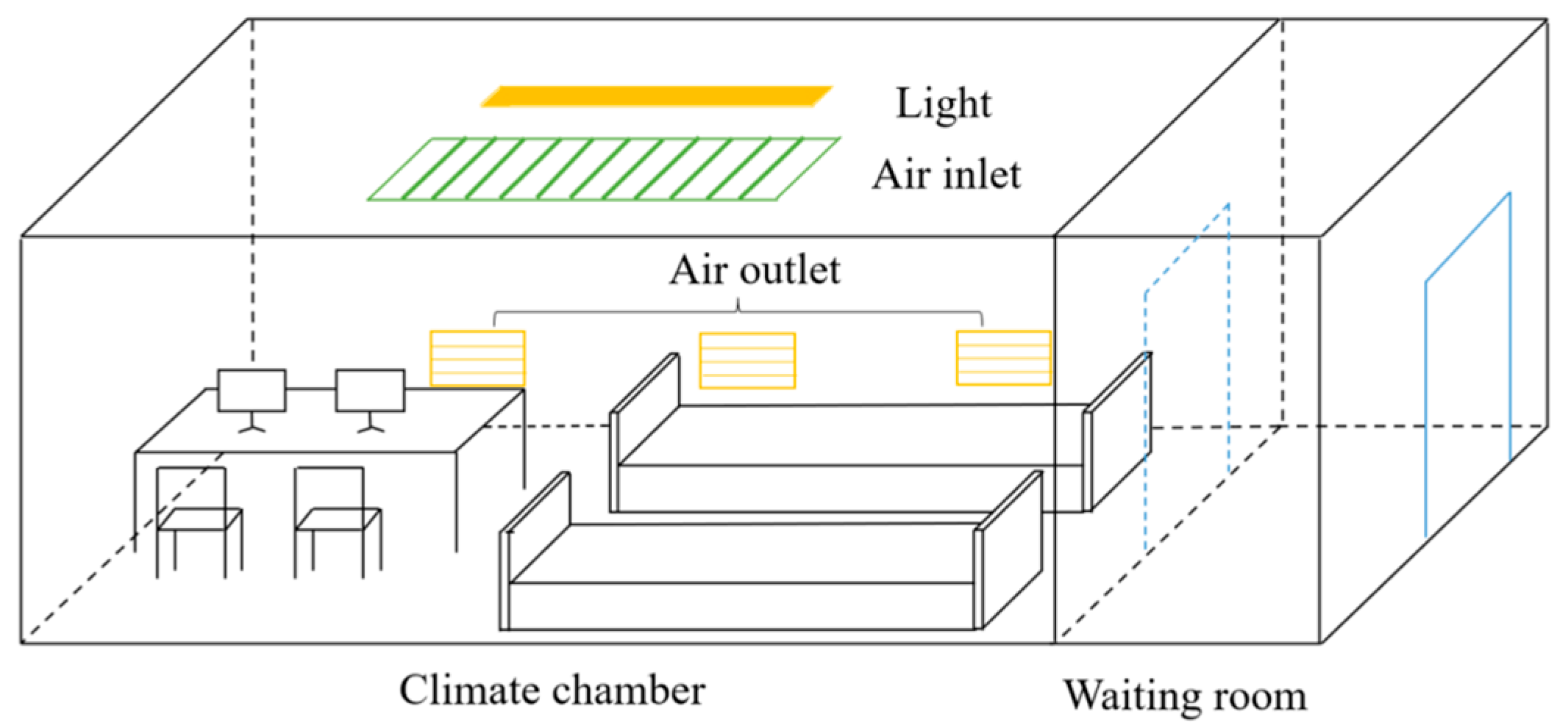
Figure 1. Climatic chamber.
Buildings 15 03021 g001
Figure 2. Experimental procedures.
Buildings 15 03021 g002
Figure 3. Changes in thermal sensation under three sleep restrictions.
Buildings 15 03021 g003
Figure 4. Changes in sleepiness under three sleep-restriction conditions.
Buildings 15 03021 g004
Figure 5. Changes in perceived workload under three sleep-restriction conditions.
Buildings 15 03021 g005
Figure 6. Changes in heart rate during and after these three sleep restrictions.
Buildings 15 03021 g006
Figure 7. Changes in RMSSD during and after these three sleep restrictions.
Buildings 15 03021 g007
Figure 8. Changes in PI of SRT during and after three sleep-restriction conditions.
Buildings 15 03021 g008
Figure 9. Changes in PI of CRT during and after three sleep-restriction conditions.
Buildings 15 03021 g009
Figure 10. Changes in PI of Stroop during and after three sleep-restriction conditions.
Buildings 15 03021 g010
Figure 11. Changes in maximum memory span of the Corsi Block task under and after three sleep-restriction conditions.
Buildings 15 03021 g011
Figure 12. Changes in OPI under and after three sleep-restriction conditions.
Buildings 15 03021 g012
Table 1. The selected measurements and the reasons for selections.
MetricPhysiological/Psychological SignificanceReason(s)
TSVReflects subjective perception of environmental temperatureContinuous monitoring of thermal sensation to simulate the physical environment during real overtime work during the night [48]
SSSQuantifies momentary sleepiness levels (1–7 scale)Validated for sleep-deprivation studies to capture transitions from alertness to near-sleep states [49]
NASA-TLXMulti-dimensional assessment of perceived workload (six weighted dimensions)Evaluates mental, physical, and temporal demands to quantify the perceived workload [50,51,52]
HRVHeart Rate
RMSSD
Indicates the dynamic balance of the autonomic nervous systemLinks sympathetic–parasympathetic modulation to cognitive functions (e.g., working memory, sustained attention) [53]
Table 2. Details of three cognitive tasks.
Task TypeCognitive FunctionsDuration
Deary–Liewald Task [54]Simple Response Time taskSustained Attention3.5 min
Choice Response Time taskSelective Attention
Stroop Task [55]Inhibitory Control, Working memory, Executive function3 min
Corsi Block Task [56]Spatial working memory, Short-term Memory3.5 min
Table 3. Summary of changes in cognitive functions due to sleep restrictions (SR).
Cognitive
Functions
Mild SR
(Before 1:20 a.m.)
Moderate SR
(Before 2:30 a.m.)
Severe SR
(Before 3:40 a.m.)
End of SRMorningEnd of SRMorningEnd of SRMorning
Sustained attention↓*↓*
Selective attention↓*↓*↓*
Working memory, Executive function, Inhibitory control↓*↓*
Short-term memory
Note: → means no changes compared with baselines; ↓ means reduction compared with baselines; ↓* means significant reduction compared with baselines; ↑ means increase compared with baselines.
Disclaimer/Publisher’s Note: The statements, opinions and data contained in all publications are solely those of the individual author(s) and contributor(s) and not of MDPI and/or the editor(s). MDPI and/or the editor(s) disclaim responsibility for any injury to people or property resulting from any ideas, methods, instructions or products referred to in the content.