Article

New Bactericide Orthodonthic Archwire: NiTi with Silver Nanoparticles

by
F. Javier Gil
1,2,
Eduardo Espinar-Escalona
3,
Nuria Clusellas
2,
Javier Fernandez-Bozal
2,
Montserrat Artes-Ribas
2 and
Andreu Puigdollers
2,*
1
Bioengineering Institute of Technology, Universitat Internacional de Catalunya, 08195 Barcelona, Spain
2
Department of Orthodontics, Faculty of Dentistry, Universitat Internacional de Catalunya, 08195 Barcelona, Spain
3
Department of Orthodontics, Faculty of Dentistry, Universidad de Sevilla, 41009 Sevilla, Spain
*
Author to whom correspondence should be addressed.
Metals 2020, 10(6), 702; https://doi.org/10.3390/met10060702
Submission received: 20 April 2020 / Revised: 22 May 2020 / Accepted: 25 May 2020 / Published: 26 May 2020
(This article belongs to the Special Issue Metallic Biomaterials Surface Engineering)

Abstract

:
A potential new bactericide treatment for NiTi orthodontic archwires based in the electrodeposition of silver nanoparticles on the surface was studied. Twenty-five archwires were treated by electrodeposition, obtaining nanoparticles of silver embedded on the archwire surface. These were evaluated in order to investigate the possible changes on the superelastic characteristics (critical temperatures and stresses), the nickel ion release, and the bacteria culture behavior. The chemical composition was analyzed by Energy Dispersive X-Ray Spectroscopy-microanalysis; the singular temperatures of the martensitic transformation were obtained by a flow calorimeter. Induced martensitic transformation stresses were obtained by mechanical testing apparatus. Nickel ion release was analyzed by inductively coupled plasma-mass spectrometry (ICP-MS) equipment using artificial saliva solution at 37 °C. Bacterial tests were studied with the most used oral bacterial strains: Streptococcus sanguinis and Lactobacillus salivarius. NiTi samples were immersed in bacterial suspensions for 2 h at 37 °C. Adhered bacteria were separated and seeded on agar plates: Tood-Hewitt (TH) and Man-Rogosa-Sharpe (MRS) for S. sanguinis and for L.salivarius, respectively. These were then incubated at 37 °C for 1 day and the colonies were analyzed. The results showed that the transformation temperatures and the critical stresses have not statistically significant differences. Likewise, nickel ion release at different immersion times in saliva at 37 °C does not present changes between the original and treated with silver nanoparticles archwires. Bacteria culture results showed that the reduction of the bacteria due to the presence to the nanoparticles of silver is higher than 90%. Consequently, the new treatment with nanoparticles of silver could be a good candidate as bactericidic orthodontic archwire.

1. Introduction

NiTi arch wires, and specially thermoelastic alloys, are used routinely in the first phase of orthodontic treatments because of their flexibility and resistance that can align and level efficiently misaligned teeth.
NiTi superelastic archwires can present two phases: austenite which is the stable phase at high temperature and martensite which is stable at low temperatures. Thermoelastic transformation can be produced by cooling from the austenite or when the austenite is subjected under load transforming to stress induced martensite (SIM). Both transformations are elastic and the material by heat or by unloading recovers the austenite phase and the original shape. In orthodontics, deformed archwire reverts to its original shape correcting the dental places [1]. This stress remains practically constant over a wide range of archwire activation. This property produces an adequate low level of forces which result a good tissular behavior. This unusual property in metallic materials is named ‘superelasticity’ or ‘pseudoelasticty’ due to the small hysteresis between the load and unload cycles [2,3,4,5,6,7,8].
This has a two-fold effect. On one side is important because is more comfortable for the patients. On the other, from the tisular, cellular and molecular point of view low forces allows a more physiologic dental movement [9]. When the loads applied are high, they produce necrosis of the soft tissues such as periodontal ligament, and may cause serious problems as for example root resorption [10].
The arch wires exert the forces to the teeth when placed into the slot of the brackets, that are bonded to the labial or lingual surface of the teeth. Oral hygiene, then, is more complicated for patients. The normal development of dental biofilm is favored due to the adhesion of dental plaque around the brackets [11,12,13,14,15]. Then, the chances for developing an enamel decay or a gingivitis and further development of periodontal disease is a real situation [10,11,12].
The aim of this article is the design of a new bactericide orthodontic NiTi arch wire by means of electrodeposition of silver nanoparticles, without any further loss of mechanical properties, that can help to control the dental plaque in patients bearing brackets.

2. Materials and Methods

2.1. Material

NiTi orthodontic archwires studied presented a chemical composition near equiatomic. This chemical analysis was obtained EDS-microanalysis resulting 55.8 ± 0.2% of Nickel and 44.1 ± 0.3% wt %. A scheme of the orthodontic archwires can be observed in Figure 1. Twenty-five NiTi archwires were analyzed in this study.

2.2. Silver Nanoparticles Electrodeposition

In order to electrodeposit silver onto nickel-titanium archwires is necessary performs a multiple-step process which consists in:
a.
Nickel-titanium samples are cleaned by ultrasonication in ethylic alcohol, double distilled water, and acetone for 10 min each. Samples were dried with argon gas. This process removes accumulated contaminants.
b.
Samples are treated in acid solution (HNO3 60%, NH4HF2, and ultrapure distilled water) for 10 s to clean the contamination.
c.
Samples are rinsed in distilled water eliminating constituent particles not removed by the previous treatment or product from previous treatment solution.
d.
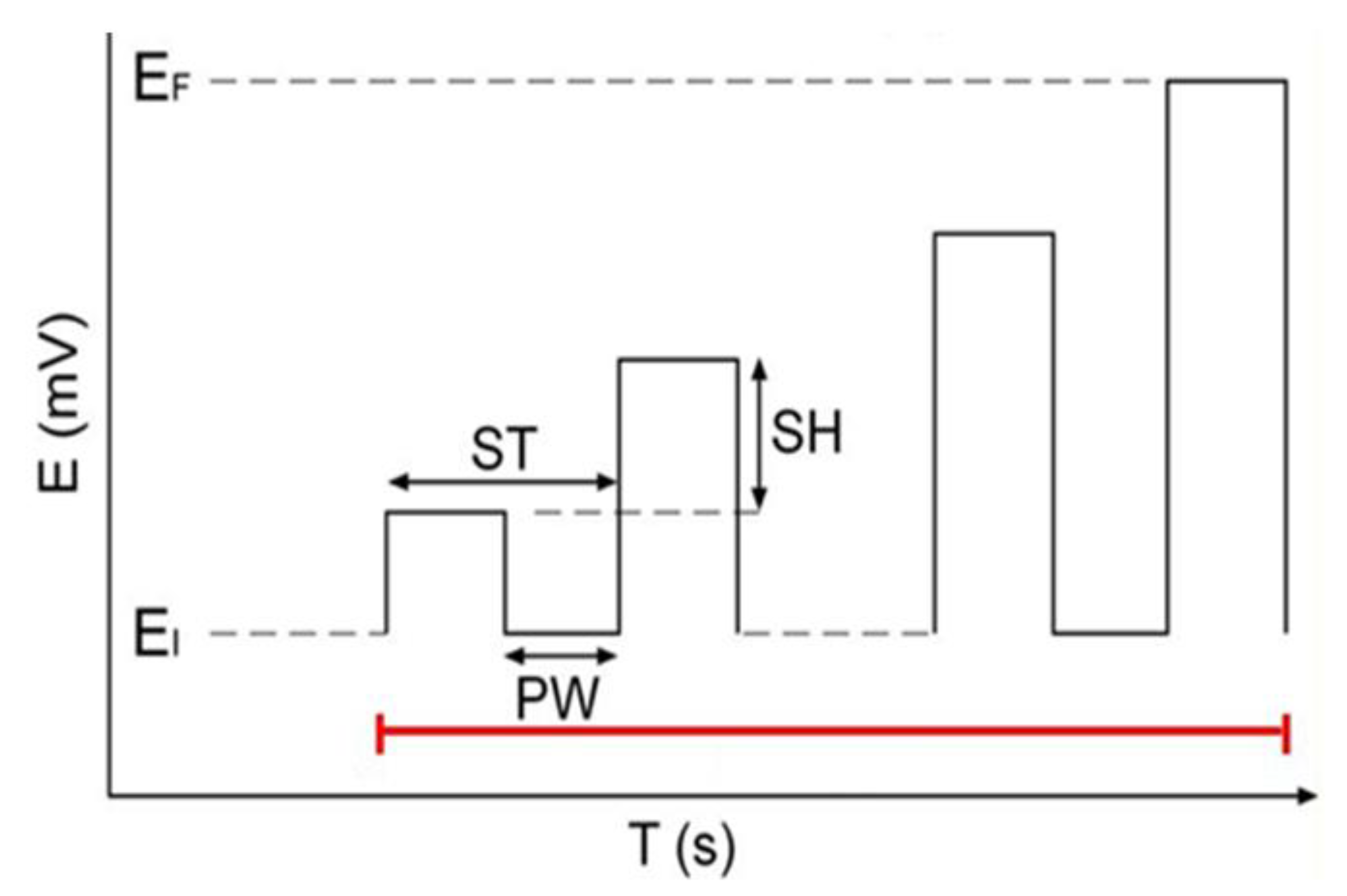
Samples are immersed in a reactor containing an electrolyte with silver nitrate (AgNO3) and a coordinating compound sodium thiosulphate (Na2S2O3) at 25 °C and with magnetic stirring. Electric current is passed through the electrolyte and the titanium is made the anode in the electrolytic cell; the platinum electrode is the cathode. A rectangular pulsed potential is applied to the electrode (EI = 0 V, EF = 5 V, ST = 500 ms, SH = 10 mV, PW = 100 ms). The full-cycle period is around 25 s [12,13]. From Figure 2 is shown a diagram of the anodization process.
The samples anodized were cleaned by ultrasounds in ethylic alcohol, double-distilled water and acetone for 15 min each one.
Silver nanoparticles were observed by means of Focus Ion Beam-Scanning Electron Microscope Zeiss Neon 4 (Oberkochen, Germany) using 25 kV and high resolution mode.

2.3. Calorimetric Tests

Six original samples and six with silver nanoparticles were analyzed to observe the possible influence on the silver on the transformation temperatures. These singular temperatures are: Ms and Mf when the martensitic transformation starts and when the 100% of the transformation from austenite is achieved, respectively. Besides, As and Af when the retransformation to austenite starts form the martensite and when the retransformation has been competed, respectively. These temperatures were detected by means of Pt-100 probe and increment signals (ΔT) by means of a flow calorimeter Melcor S 10, equipped with 30 thermocouples [16,17]. The temperature was measured by means. The start transformation (Ms) and retransformation (As) temperatures are detected when there is an abrupt increment or decrement in the calorimetric signal and final temperatures, (Mf and Af), were determined as when the signal returned to the base line [18].

2.4. Mechanical Tests

The tests were realized with a servo-hydraulic testing machine (MTS-Bionix 858, Minneapolis, MN, USA) with a small load cell in oder to increase the sensitivity. Ten NiTi original archwires and with silver nanoparticles were tested. The critical stresses (stress needed to transform austenitic phase to stress-induced martensitic phase and the stress to retransform from stress induced martensitic phase to austenitic phase when unloading cycle is realized) were obtained, which were tested in saliva at 37 °C (Table 1). The cross-head rate was of 10 mm/min [18,19,20,21].

2.5. Nickel Ion Release

Nickel ion release analysis was realized by immersing the NiTi archwires (originals and with silver nanoparticles) in 10 mL of saliva (Table 1) at pH 6.8 and 37 °C [18,19,20]. The released Ni was measured by means of ICP-MS PerkinElmer Óptima 320 RL (Waltham, MA, USA) at 1 and 5, 24, 48, 120, 360, 700, 1000, 1100, and 1900 h. Five measurements different were analyzed for each archwires.

2.6. Bacteria Cultures

Bacterial tests were realized with two oral bacterial strains. These are commonly used in the experimental tests [22,23,24,25,26]: Streptococcus sanguinis (CECT 480; Spanish Culture Collection, Valencia, Spain) and Lactobacillus salivarius (CCUG 17826; Culture Collection University of Göteborg, Göteborg, Sweden). S. sanguinis was growth and maintained on Todd-Hewitt (TH) broth (Scharlab SL, Sentmenat, Spain) and L. salivarius on MRS broth (Scharlab SL).
Before the tests, the cultures were incubated for 10 h at 37 °C. The bacterial suspension was adjusted to 0.2 ± 0.01 units at 600 nm, giving around 1 × 10⁸ colony forming units (CFU)/mL for each strain.
Samples were introduced in 1 mL of bacterial suspensions (1 × 10⁸ CFU/mL) for 2 h at 37 °C. Once, the medium was separated by aspiration, the samples were washed with Phosphate Buffer Solution (PBS). Adherent bacteria were detached by vortexing the disks for 5 min in 1 mL of PBS. Then, these bacteria were seeded using dilutions on Tood-Hewitt (TH) agar plates for S. sanguinis and Man-Rogosa-Sharpe (MRS) ones for L. salivarius. These plates were incubated at 37 °C for 1 day and the colonies were quantified. Results were normalized with the surface.

2.7. Statistical Analysis

The results were studied in order to determine any statistically significant differences between the original and bactericide archwires using one-way ANOVA tables, Student’s t-tests and Tukey’s multiple comparison tests. These analyses were carried out using MinitabTM software (Minitab 13.0, Minitab Inc., State College, PA, USA). The statistical differences were significant when p < 0.005.

3. Experimental Results and Discussion

Silver nanoparticles on the titanium alloy surface can observe in Figure 3a and at more magnification in Figure 3b. These particles on titanium surface are very good adhered as embedded into the surface and show a plastic deformation. In some cases, nanoparticles clusters can be observed on the surface. These clusters present an average diameter 100–500 nm. On the metallic surface, the nanoparticles are agglomerated producing clusters. However, when nanoparticles are on the bacteria membrane are isolated and can determine that the silver nanoparticles are lower than 50 nm (Figure 3c). Nano size avoids the oxidation of the silver and consequently there are not tattoos in the soft tissue. This fact is due to the reduced surface of the nanoparticle, this is not sufficient to activate the oxidation chemical reaction. The oxidation can be produced by long treatments in the presence of H2O2. In general, this process must be accelerated by high concentrations or electrolytically. Silver is stable in water and physiological medium and needs an oxidizing element to achieve oxidative dissolution. When oxidizing agents such as hydrogen peroxide or oxygen are present, they dissolute silver nanoparticles to release Ag+. The release of Ag+ leads to creation of reactive oxygen species (ROS) inside electrolytic cells, which can further dissolve the nanoparticles [27,28,29]. Figure 3d presents the EDS-HRSEM microanaylisis realized in a silver nanoparticle around 40 nm of size.
Ms and Mf, were determined during the cooling cycle and As and Af during the heating cycle. It can be observed that the singular temperatures do not change with statistical significance differences (p < 0.001) when the cooling/heating cycles are repeated. The singular temperatures are practically constant with the thermal cycles. The incorporation of the silver nanoparticles does not modify (p = 0.0001) the transformation temperatures, as can be observed in Table 2.
The stress necessary to produce the formation of martensitic plates form austenitic archwire (stress-induced martensite), σβΜ and the stress in the unloading process required for the retransformation (formation of austenite from martensite plates) σΜβ at 20 °C and 37 °C were obtained. Silver nanoparticles did not produce significant changes in the transformation temperatures (p = 0.002). This is due to the fact that the silver does not influence the thermoelastic martensitic transformation induced by stress, because the quantity of silver is very small and is not alloyed with NiTi.
The quantities of silver deposited on the NiTi are around 20 ± 8 µg/cm2, these values were obtained by the determination of the difference between the silver in the reactive before the electrodeposition and after the treatment. The difference is that the silver is deposited on the NiTi. This difference is divided by the total surface of the NiTi archwire.
Besides, the electrodeposition is made at room temperature and consequently grain growth or modification of phases by solid state diffusion is avoided. There are not diffusion is solid state. These critical stresses (β↔Stress-Induced Martensite) can be observed in Table 3.
The nickel ions release are shown in Figure 4 for as-received and with silver nanoparticles treatment. The Ni ion release in both samples are very similar with low values ranging from 20 ppb at 2 h to 210 ppb at 2000 h. The silver ion release cannot determine due to the sensibility of the equipment, the equipment only detects when the ion release is higher than 1 ppb. Consequently, the presence of silver nanoparticles in the NiTi archwires maintains the nickel release level as the original archwires.
Different studies pay attention to nickel ion release in physiological medium [30,31,32,33] as an element able to induce allergenic reactions. The analysis in Europe showed the prevalence is from 10% to 15% for females and from 1% to 3% for males. For these people, around 40% to 70% of Ni contact dermatitis develops hand eczema, which can be acute or chronic [30,31,32,33]. From the results of Figure 4 the nickel ion release is very low, 200 ppb after 15 days for original and treated with silver nanoparticles-NiTi archwires and consequently these are values are safe in order to use as orthodontic archwires [34,35,36,37]. The nickel ion release between two types of archwires do not have statistically significant differences at p = 0.001.
The metabolism of bacteria and the formation of the plaque on hard oral surfaces are considered the main reason of dental caries, periodontitis, gingivitis, stomatitis, and peri-implant infections [11]. In vitro tests have been realized with the bacteria more usual in these studies in oral cavity. In general, the orthodontic archwires have an use from 15 days to 4–5 months depends on the movement of the teeth, age, sex, and magnitude of correction. The bactericide action of the nanoparticles of silver has been very efficient and it activity remains during the period of the orthodontic therapy. The bactericide time of the NiTi depends on the density of silver nanoparticles on NiTi and the quantity and nature of the bacteria present. From Figure 5, an efficient bactericide effect presents the silver nanoparticles in two families of bacteria.
The usual situation in patients is that biofilms effectively protect bacteria from antimicrobial agents. Treatment with antimicrobial substances is often unsuccessful unless the deposits are mechanically removed [11]. In the case of the values obtained of the present research, bactericide mechanism of the silver nanoparticles is the contact with the bacteria. When the bacteria contact with silver, this element react with sulphur of the amino-acids and produce AgS [38]. This chemical reaction produces the fracture of the DNA of the bacteria producing the death of the microorganism. This mechanism was studied by Khalandi et al. [38]. This mechanism is only for the bacteria; the membrane of humans cells avoids this reaction. The inhibition of the silver nanoparticles reaction on the human cells can be demonstrated when human-osteoblasts were seeded in a metal with nanoparticles. In Figure 6a shows the osteoblasts after 24 h in contact with nanoparticles and Figure 6b after three days, where it can be observed that the cells have prolifereated and these present a good morphology.
This phenomenon provokes that the bacteria cannot produce the biofilm. It is the attack in the first stage of the bacteria colonization.
Although the mechanism of silver’s antibacterial action is not fully understood, it exhibits a significant antimicrobial efficacy against a wide spectrum of bacterial species. A number of different approaches to the mechanism are reported by Khalandi [38]. Silver nanoparticles have been reported to exhibit enhanced antibacterial activity due to their increased surface-area-to-volume ratio [34,35,36]. Interactions between viral biomolecules and silver nanoparticles suggest that the use of nanosystems may contribute importantly for the enhancement of current prevention of infection and antiviral therapies. Recently, it has been suggested that silver nanoparticles bind with external membrane of lipid enveloped virus to prevent the infection. Nevertheless, the interaction of silver nanoparticles with viruses is a largely unexplored field. These nanoparticles have been studied particularly on HIV where it was demonstrated the mechanism of antiviral action of the nanoparticles as well as the inhibition the transmission of HIV-1 infection in human cervix organ culture [39,40,41,42].
Tian et al. [43] demonstrated, across a spectrum of bacterial pathogens, that the effects of silver nanoparticles on the beneficial bacteria are less clear. These authors compared the antibacterial activity of silver nanoparticles against two beneficial lactobacilli (Lactobacillus delbrueckii subsp. bulgaricus and Lactobacillus casei) and two common opportunistic pathogens (Escherichia coli and Staphylococcus aureus). Their results demonstrate that those lactobacilli are highly susceptible to silver nanoparticles, while the opportunistic pathogens are not. Acidic environment caused by the lactobacilli is associated with the bactericidal effects of silver nanoparticles. The mechanistic study suggests that the acidic growth environment of lactobacilli promotes silver nanoparticles dissolution and hydroxyl radical (•OH) overproduction. Furthermore, increases in silver ions (Ag+) and •OH deplete the glutathione pool inside the cell, which is associated with the increase in cellular reactive oxygen species (ROS). High levels of ROS may further induce DNA damage and lead to cell death. When E. coli and S. aureus are placed in a similar acidic environment, they also become more susceptible to silver nanoparticles [44].
Studies realized with stainless steel and NiTi orthodontic archwires coated with silver nanoparticles by Maskher et al. [45] showed an antiadherent effect against L. acidophilus compared with uncoated wires. Uncoated stainless steel and NiTi wires respectively showed 35.4% and 20.5% increase in weight which was statistically significant (p  <  0.001), whereas surface-modified wires showed only 4.08% and 4.4% increase in weight (statistically insignificant p  >  0.001). The groups containing surface-modified wires showed an important decrease in the survival rate of L. acidophilus expressed as CFU and as log of colony count when compared to groups containing uncoated wires. It was 836.60  ±  48.97 CFU in the case of uncoated stainless steel whereas it was 220.90  ±  30.73 CFU for silver-modified stainless steel, 748.90  ±  35.64 CFU for uncoated NiTi, and 203.20  ±  41.94 CFU for surface-modified NiTi.
Some metals—such as gold, platinum, copper, zinc—present antimicrobial properties. However, silver is known to exert the most effective antibacterial action. The bactericide effect of the silver nanoparticles on NiTi in comparison with other metals used as biomaterials is very similar such as titanium and Ti-6Al-4V alloys [25,26]. Nevertheless, Chen and Schluesener [46] demonstrated that silver nanoparticles are non-toxic and non-mutageneic to human primary organ systems and are considered as a safe and promising antibactericidal agent against highly infectious drug-resistant bacteria such as E. coli, P. aeruginosa, and S. aureus [47,48]. The bactericide effect of silver nanoparticles can be improved with is mixed with antibiotics such as amoxicillin, clindamycin, erythromycin, penicillin, and vancomycin. However, similar to antibiotics, prolonged exposure of bacteria to silver nanoparticles may result in the development of resistant bacterial cells. For instance, E. coli K12 MG1655 strain has developed resistance toward silver nanoparticles; however, the bacterium does not possess any Ag-resistance element [49]. It is therefore necessary in future to carefully examining the development of Ag-resistance in bacteria.
Panpaliya et al. [50] studied strains of Streptococcus mutans (MTCC 497), Streptococcus oralis (MTCC 2696), Lactobacillus acidophilus (MTCC 10307), Lactobacillus fermentum (MTCC 903), and Candida albicans (MTCC 183). We used commercially available silver nanoparticles (experimental group) and chlorhexidine gluconate (positive control). Silver nanoparticles inhibited bacterial growth moderately. The mean minimum inhibitory concentration of silver nanoparticles against S. mutans was 60 ± 22.36 μg/mL, S. oralis—45 ± 11.18 μg/mL, L. acidophilus—15 ± 5.59 μg/mL, L. fermentum—90 ± 22.36 μg/mL, and Candida albicans—2.82 ± 0.68 μg/mL respectively. For chlorhexidine gluconate, mean minimum inhibitoy concentration for S. mutans was 300 ± 111.80 μg/mL, S. oralis—150 ± 55.90 μg/mL, L. acidophilus—450 ± 111.80 μg/mL, L. fermentum—450 ± 111.80 μg/mL and Candida albicans—150 ± 55.90 μg/mL. As can observe the values of silver nanoparticles were significantly lower than chlorhexidine gluconate. Silver nanoparticles exhibited better bacteriostatic and bactericidal effect with concentration less than five-fold as compared to chlorhexidine. Silver nanoparticles when used in appropriate concentration as safe alternative to present chemically derived other antimicrobial agents.
Several studies have consistently demonstrated the great antimicrobial activity and antiadherence properties of silver nanoparticles against a wide variety of microorganisms, including oral bacteria. However, there are not enough works that have determined the bactericidal and the substantivity activities of silver nanoparticles against oral biofilms taken clinically from young and young-adult patients with active caries and periodontal disease. Studies have reported that silver nanoparticles covered human dentin and different alloy medical implants can significantly inhibit the biofilm formation on the surface of the dentin and the implants as well as control the bacterial growth around them [51,52]. Also, the authors determined the antimicrobial and antibiofilm properties of silver nanoparticles (~17 nm) included in orthodontic appliances and standardized microbiocidal assays against S. mutans, Lactobacillus casei (L. casei), Staphylococcus aureus (S. aureus), and Escherichia coli (E. coli) as well as in vitro biofilms using cariogenic bacteria (S. mutans), suggesting the potential use to prevent the dental biofilm, decreasing the incidence of demineralization activities associated with caries during conventional dental treatments [53,54]. A recent in vitro study reported the antimicrobial activity of silver nanoparticles (30–50 nm) and chlorhexidine (main antimicrobial agent in dentistry) against different oral pathogenic bacteria (S. mutans, S. oralis, Lactobacillus acidophilus, Lactobacillus fermentum, and Candida albicans), determining good bactericidal and bacteriostatic properties of AgNPs for all microbial strains with concentrations less than five holds as compared to chlorhexidine supporting its potential and safe use in the dentistry field [50,55].

4. Conclusion

The electrodeposition of silver nanoparticles onto thermoelastic NiTi orthodontic archwires has a bactericidic effect on cultures of oral bacteria. This effect is without changing their calorimetric and mechanical properties, and nickel release levels of the NiTi wires. This in vitro research should be replicated in a clinical setting. Surface modification of NiTi orthodontic archwires with silver nanoparticles can be used to prevent the accumulation of dental plaque and the development of dental caries during orthodontic treatment.

Author Contributions

Conceptualization, F.J.G., A.P., E.E.-E. and M.A.-R.; methodology, F.J.G.,J.F.-B.,N.C.; validation, N.C., A.P., M.A.-R and E.E.-E.; formal analysis, F.J.G.; investigation, F.J.G., M.A.-R., E.E.-E., N.C.; resources, A.P.; data curation, A.P., J.F.-B., N.C., M.A.-R.; writing—original draft preparation, A.P., E.E.-E., F.J.G.; writing—review and editing, F.J.G.; visualization, A.P., E.E.-E.; supervision, J.F.-B., N.C., A.P.; project administration, A.P., N.C.; funding acquisition, A.P. All authors have read and agreed to the published version of the manuscript.

Funding

This research was funded by Spanish Government and and European Union FEDER by the concession of the grant RTI2018-098075-B-C22 and the research group Generalitat de Catalunya 2017SGR708.

Conflicts of Interest

The authors declare no conflict of interest.

References

  1. Miura, F.; Mogi, M.; Ohura, Y.; Hamanaka, H. The super-elastic property of the Japanese NiTi alloy wire for use in orthodontics. Am. J. Orthod. Dentofac. Orthop. 1986, 90, 1–10. [Google Scholar] [CrossRef]
  2. Haarsters, J.; Salis-Solio, G.; Bensmann, T. The use of NiTi as a implant material in orthopedics. In Shape Memory in Engineering Aspects of Shape Memory Alloys; Duering, T.W., Melton, K.N., Stöckel, D., Wayman, C.M., Eds.; Butterworth-Heinemann Ed.: London, UK, 1990. [Google Scholar]
  3. Andreasen, G.F.; Morrow, R.E. Laboratory and clinical analysis of Nitinol wire. Am. J. Orthod. 1978, 73, 142–145. [Google Scholar] [CrossRef]
  4. Sevilla, P.; Martorell, F.; Libenson, C.; Planell, J.A.; Gil, F.J. Laser welding of NiTi orthodontic archwires for selective force application. J. Mater. Sci. Mater. Med. 2008, 19, 525–529. [Google Scholar] [CrossRef] [PubMed]
  5. Krousbroeck, R.; Van der Perre, G.; Aernoudt, E.; Mulier, J.C. Shape memory effect in biomedical devices. In Advances in Biomaterials; Winter, G.D., Gibbons, D.F., Plenk, H., Eds.; John Wiley & Sons: Hoboken, NJ, USA, 1982. [Google Scholar]
  6. Arciniegas, M.; Casals, J.; Manero, J.M.; Peña, J.; Gil, F.J. Study of hardness and wear behaviour of NiTi shape memory alloys. J. Alloys Comp. 2008, 460, 213–219. [Google Scholar] [CrossRef]
  7. Gil, F.J.; Cenizo, M.; Espinar, E.; Rodriguez, A.; Rúperez, E.; Manero, J.M. NiTi superelastic orthodontic wires with variable stress obtained by ageing treatments. Mater. Lett. 2013, 104, 5–7. [Google Scholar] [CrossRef]
  8. Gil, F.J.; Solano, E.; Peña, J.; Engel, E.; Mendoza, A.; PLanell, J.A. Microstructural. Mechanical and citotoxicity evaluation of different NiTi and NiTiCu shape memory alloys. J. Mater. Sci Mater. Med. 2004, 15, 1181–1185. [Google Scholar] [CrossRef]
  9. Khouw, F.E.; Goldhaber, P. Changes in vasculature of the periodontium associated with tooth movement in the rhesus monkey and dog. Arch. Oral. Biol. 1970, 15, 1125–1132. [Google Scholar] [CrossRef]
  10. Nakano, T.; Hotokezaka, H.; Hashimoto, M. Effects of different types of tooth movement and force magnitudes on the amount of tooth movement and root resorption in rats. Angle Orthod 2014, 84, 1079–1085. [Google Scholar] [CrossRef] [Green Version]
  11. Lang, N.P.; Mombelli, A.; Attström, R. Oral Biofilms and Calculus. In Clinical Periodontology and Implant Dentistry, 5th ed.; Lindhe, J., Lang, N.P., Karring, T., Eds.; Wiley-Blackwell Ed.: Hoboken, NJ, USA, 2008. [Google Scholar]
  12. Godoy-Gallardo, M.; Rodríguez-Hernández, A.G.; Delgado, L.M.; Manero, J.M.; Javier Gil, F.; Rodríguez, D. Silver deposition on titanium surface by electrochemical anodizing process reduces bacterial adhesion of Streptococcus sanguinis and Lactobacillus salivarius. Clin. Oral. Implants Res. 2014, 26, 1170–1179. [Google Scholar] [CrossRef] [Green Version]
  13. Godoy-Gallardo, M.; Manzanares-Cespedes, M.C.; Sevilla, P.; Nart, J.; Manzanares, N.; Manero, J.M.; Gil, F.J.; Boyd, S.K.; Rodriguez, D. Evaluation of bone los in antibacterial coated dental implants: An experimental study in dogs. Mater. Sci. Eng. C 2016, 69, 538–545. [Google Scholar] [CrossRef] [Green Version]
  14. Godoy-Gallardo, M.; Guillem-Marti, J.; Sevilla, P.; Manero, J.M.; Gil, F.J.; Rodriguez, D. Anhydride-functional silane immobilized onto titanium surfaces induces osteoblast cell differentiation and reduces bacterial adhesion and biofilm formation. Mater. Sci. Eng. C 2016, 1, 524–532. [Google Scholar] [CrossRef] [PubMed] [Green Version]
  15. Zhao, L.; Chu, P.K.; Zhang, Y.; Wu, Z. Antibacterial coatings on titanium implants. J. Biomed. Mater. Res. B Appl. Biomater. 2009, 91, 470–480. [Google Scholar] [CrossRef] [PubMed]
  16. Suárez, C.; Vilar, T.; Gil, F.J.; Sevilla, P. In vitro evaluation of surface topographic changes and nickel release of lingual orthodontic archwires. J. Mater Sci. Mater Med. 2010, 21, 675–683. [Google Scholar] [CrossRef] [PubMed] [Green Version]
  17. Saburi, T.; Tatsumi, T.; Nenno, S. Effects of heat treatment on mechanical behaviour of TiNi alloys. J. Phys. Colloq. C4 1982, 12, 261–266. [Google Scholar]
  18. Barrabés, M.; Michiardi, A.; Aparicio, C.; Sevilla, P.; Planell, J.A.; Gil, F. Oxidized nickel-titanium foams for bone reconstructions: Chemical and mechanical characterization. J. Mater. Sci. Mater. Med. 2007, 18, 2123–2129. [Google Scholar] [CrossRef]
  19. Preetha, A.; Barnejee, R. Comparison of artificial saliva substitutes. Trends Biomater. Artif. Organs 2005, 18, 178–186. [Google Scholar]
  20. Messer, L.M.; Lucas, L.C. Cytotoxicity of nickel-chromium alloys:bulk alloys compared to multiple ion salts solutions. Dent. Mater. 2000, 16, 207–212. [Google Scholar] [CrossRef]
  21. Park, H.Y.; Shearer, T.R. In vitro release of nickel and chromium from simulated orthodontic appliances. Am. J. Orthod. 1983, 84, 156–159. [Google Scholar] [CrossRef]
  22. Sinha, M.; Kaushik, S.; Kaur, P.; Sharma, S.; Singh, T.P. Antimicrobial lactoferrin peptides: The hidden players in the protective function of a multifunctional protein. Int. J. Pept. 2013, 13, 1–13. [Google Scholar] [CrossRef] [Green Version]
  23. Godoy-Gallardo, M.; Mas-Moruno, C.; Fernández-Calderón, M.C.; Pérez-Giraldo, C.; Manero, J.M.; Albericio, F.; Gil, F.J.; Rodríguez, D. Covalent immobilization of hLf1-11 peptide on a titanium surface reduces bacterial adhesion and biofilm formation. Acta Biomater. 2014, 10, 3522–3534. [Google Scholar] [CrossRef]
  24. Huo, L.; Zhang, K.; Ling, J.; Peng, Z.; Huang, X.; Liu, H.; Gu, L. Antimicrobial and DNA-binding activities of the peptide fragments of human lactoferrin and histatin 5 against Streptococcus mutans. Arch. Oral Biol. 2011, 56, 869–876. [Google Scholar] [CrossRef] [PubMed]
  25. Godoy-Gallardo, M.; Mas-Moruno, C.; Yu, K.; Manero, J.M.; Gil, F.J.; Kizhakkedathu, J.N.; Rodriguez, D. Antibacterial Properties of hLf1–11 Peptide onto Titanium Surfaces: A Comparison Study Between Silanization and Surface Initiated Polymerization. Biomacromolecules 2015, 16, 483–496. [Google Scholar] [CrossRef] [PubMed] [Green Version]
  26. Godoy-Gallardo, M.; Wang, Z.; Shen, Y.; Manero, J.M.; Gil, F.J.; Rodriguez, D.; Haapasalo, M. Antibacterial Coatings on Titanium Surfaces: A Comparison Study Between in Vitro in Single-Species and Multispecies Biofilm. ACS Appl. Mater. Interfaces 2015, 7, 5992–6001. [Google Scholar] [CrossRef] [PubMed] [Green Version]
  27. Valenti, L.E.; Giacomelli, C.E. Stability of silver nanoparticles: Agglomeration and oxidation in biological relevant conditions. J. Nanopart. Res. 2017, 19, 156. [Google Scholar] [CrossRef]
  28. Bastiaan, M.; Hiemstra, T. Surface Structure of Silver Nanoparticles as a Model for Understanding the Oxidative Dissolution of Silver Ions. Langmuir 2015, 31, 13361–13372. [Google Scholar]
  29. Ching-Ming, H.; Yau, S.K.W.; Lok, C.N.; So, M.H.; Che, C.M. Oxidative Dissolution of Silver Nanoparticles by Biologically Relevant Oxidants: A Kinetic and Mechanistic Study. Chem. Asian J. 2010, 2, 2016. [Google Scholar]
  30. Setcos, J.C.; Babaei-Mahamani, A.; Di Silvio, L.; Mijör, M.A.; Wilson, N.H.F. The safety of nickel containing dental alloys. Dent Mater. 2006, 22, 1163–1168. [Google Scholar] [CrossRef]
  31. Wiltshire, W.A.; Noble, J. Allergies to dental materials. Vital Autumn 2007, 27, 39. [Google Scholar]
  32. Huang, H.H. Variation in corrosion resistance of nickel-titanium wires from different manufacturers. Angle Orthod 2005, 75, 661–665. [Google Scholar]
  33. Huang, H.H.; Chiu, Y.H.; Lee, T.H.; Wu, S.C.; Yang, H.W.; Su, K.H. Ion release from Ni-Ti orthodontic wires in artificial saliva with various acidities. Biomaterials 2003, 20, 3585–3592. [Google Scholar] [CrossRef]
  34. Wang, J.; Li, N.; Rao, G.; Han, E.H.; Ke, W. Stress corrosion cracking of Ni-Ti in artificial saliva. Dent Mater. 2007, 23, 133–137. [Google Scholar] [CrossRef] [PubMed]
  35. Michiardi, A.; Aparicio, C.; Planell, J.A.; Gil, F.J. New oxidation treatment of NiTi shape memory alloys to obtain Ni-free surfaces and to improve biocompatibility. J. Biomed. Mater. Res. Part B Appl. Biomater. 2006, 77B, 249–256. [Google Scholar] [CrossRef] [PubMed]
  36. Schultz, J.C.; Connelly, E.; Glesene, L.; Warshaw, E.M. Cutaneous and oral eruption from oral exposure to nickel dental braces. Dermatitis 2004, 15, 154–157. [Google Scholar] [CrossRef] [PubMed]
  37. Dakal, T.C.; Kumar, A.; Majumdar, R.S.; Yadav, V. Mechanistic Basis of Antimicrobial Actions of Silver Nanoparticles. Front. Microbiol. 2016, 7, 1831. [Google Scholar] [CrossRef] [PubMed] [Green Version]
  38. Khalandi, B.; Asadi, N.; Milani, M.; Davaran, S.; Jafari, A.; Abadi, N.; Abasi, E.; Akbarzadeh, A. A Review on Potential Role of Silver Nanoparticles and Possible Mechanisms of their Actions on Bacteria. Drug Res. 2017, 67, 70–76. [Google Scholar] [CrossRef]
  39. Morones, J.R.; Elechiguerra, J.L.; Camacho, A.; Holt, K.; Kouri, J.B.; Ramírez, J.T.; Yacaman, M.J. The Bactericidal Effect of Silver Nanoparticles. Nanotechnology 2005, 16, 2346–2353. [Google Scholar] [CrossRef] [Green Version]
  40. Kaur, A.; Kumar, R. Enhanced bactericidal efficacy of polymer stabilized silver nanoparticles in conjugation with different classes of antibiotics. RSC Adv. 2019. [Google Scholar] [CrossRef] [Green Version]
  41. Lara, H.H.; Garza-Treviño, E.N.; Ixtepan-Turrent, L.; Singh, D.K. Silver nanoparticles are broad-spectrum bactericidal and virucidal compounds. J. Nanobiotechnol. 2011. [Google Scholar] [CrossRef] [Green Version]
  42. Shatan, A.B.; Venclíková, K.; Zasońska, B.A.; Patsula, V.; Pop-Georgievski, O.; Petrovský, E.; Horák, D. Antibacterial Silver-Conjugated Magnetic Nanoparticles: Design, Synthesis and Bactericidal Effect. Pharm. Res. 2019, 36, 147. [Google Scholar] [CrossRef]
  43. Tian, X.; Jiang, X.; Welch, C.; Croley, T.R.; Wong, T.Y.; Chen, C.; Fan, S.; Chong, Y.; Li, R.; Ge, C.; et al. Bactericidal Effects of Silver Nanoparticles on Lactobacilli and the Underlying Mechanism. ACS Appl. Mater. Interfaces 2018, 10, 8443–8450. [Google Scholar] [CrossRef]
  44. Rangasamy, S.; Purushothaman, B.; Song, J.M. The Application of Bactericidal Silver Nanoparticles in Wound Treatment. Rev. Nanomater. Nanotechnol. 2015. [Google Scholar] [CrossRef]
  45. Mhaske, A.R.; Shetty, P.C.; Bhat, N.S.; Ramachandra, C.S.; Laxmikanth, S.M.; Nagarahalli, K.; Tekale, P.D. Antiadherent and antibacterial properties of stainless steel and NiTi orthodontic wires coated with silver against Lactobacillus acidophilus—An in vitro study. Prog. Orthod. 2015, 16, 40. [Google Scholar] [CrossRef] [PubMed] [Green Version]
  46. Chen, X.; Schluesener, H.J. Nanosilver: A nanoproduct in medical application. Toxicol. Appl. Pharmacol. Lett. 2008, 176, 1–12. [Google Scholar] [CrossRef] [PubMed]
  47. Namasivayam, S.K.; Ganesh, S.; Avimanyu, B. Evaluation of anti-bacterial activity of silver nanoparticles synthesized from Candida glabrata and Fusarium oxysporum. Int. J. Med. Res. 2011, 1, 131–136. [Google Scholar]
  48. El-Kheshen, A.A.; El-Rab, S.F.G. Effect of reducing and protecting agents on size of silver nanoparticles and their anti-bacterial activity. Schol. Res. Librar. 2012, 4, 53–65. [Google Scholar]
  49. Graves, J.L., Jr.; Tajkarimi, M.; Cunningham, Q.; Campbell, A.; Nonga, H.; Harrison, S.H. Rapid evolution of silver nanoparticle resistance in Escherichia coli. Front. Genet. 2015, 6, 42. [Google Scholar] [CrossRef] [Green Version]
  50. Panpaliya, N.P.; Prasanna, T.D.; Yogesh, J.K.; Mahesh, V.D.; Shrikant, B.K.; Ayesha, G.S.; Ulka, R.M. In vitro evaluation of antimicrobial property of silver nanoparticles and chlorhexidine against five different oral pathogenic bacteria. S Dent J. 2019, 31, 76–83. [Google Scholar] [CrossRef]
  51. Besinis, A.; Hadi, S.D.; Le, H.R.; Tredwin, C.; Handy, R.D. Antibacterial activity and biofilm inhibition by surface modified titanium alloy medical implants following application of silver, titanium dioxide and hydroxyapatite nanocoatings. Nanotoxicology 2017, 11, 327–338. [Google Scholar] [CrossRef] [Green Version]
  52. Besinis, A.; De Peralta, T.; Handy, R.D. Inhibition of biofilm formation and antibacterial properties of a silver nano-coating on human dentine. Nanotoxicology 2014, 8, 745–754. [Google Scholar] [CrossRef]
  53. Hernández-Gómora, A.; Lara-Carrillo, E.; Robles-Navarro, J. Biosynthesis of silver nanoparticles on orthodontic elastomeric modules: Evaluation of mechanical and antibacterial properties. Molecules 2017, 22, 1407. [Google Scholar] [CrossRef] [Green Version]
  54. Yin, I.X.; Yu, O.Y.; Zhao, I.S. Developing biocompatible silver nanoparticles using epigallocatechin gallate for dental use. Arch. Oral Biol. 2019, 102, 106–112. [Google Scholar] [CrossRef] [PubMed]
  55. Espinosa-Cristóbal, L.F.; Holguín-Meráz, C.; Zaragoza-Contreras, E.A.; Martínez-Martínez, R.E.; Donohue-Cornejo, A.; Loyola-Rodríguez, J.P.; Cuevas-González, J.C.; Reyes-López, S. Antimicrobial and Substantivity Properties of Silver Nanoparticles against Oral Microbiomes Clinically Isolated from Young and Young-Adult Patients. J. Nanomat. 2019, 2019, 14. [Google Scholar] [CrossRef] [Green Version]
Figure 1. Geometry of the orthodontic archwires studied. The measuremts are in millimeters.
Metals 10 00702 g001
Figure 2. Scheme of the anodization procedure applied to the NiTi made in an electrolytic reactor.
Metals 10 00702 g002
Figure 3. (a) Silver nanoparticles on metallic surface, (b) silver nanoparticles at greater magnification. The agglomeration in clusters can be seen in (a,b). (c) Silver nanoparticles on the bacteria. (d) EDS microanalysis of silver nanoparticle.
Metals 10 00702 g003
Figure 4. Nickel ion release for NiTi and NiTi with silver nanoparticles archwires analyzed at different immersion times.
Metals 10 00702 g004
Figure 5. In vitro test (NiTi: nickel-titanium. NiTi + Ag: Nickel-Titanium with silver nanoparticles). Quantification of CFU/mm2 for both NiTi archwires. (a) Streptococcus sanguinis. (b) Lactobacillus salivarius.
Metals 10 00702 g005
Figure 6. Human-osteoblast cells on NiTi with silver nanoparticles (a) after 24 h on metallic surface. (b) After 3 days.
Metals 10 00702 g006
Table 1. Compounds of the artificial saliva.
Chemical ProductComposition (g/dm3)
K2HPO40.22
KCI1.19
KSCN0.29
Na2HPO40.26
NaCl0.69
NaHCO31.49
Urea1.49
Lactic acidup to pH = 6.8
Table 2. Singular temperatures (°C) for the Ni-Ti and NiTi with silver nanoparticles
AlloyMsMfAsAf
NiTi26.2 ± 0.316.3 ± 0.420.4 ± 0.532.4 ± 1.7
NiTi + Ag26.5 ± 2.416.9 ± 1.219.4 ± 1.733.2 ± 4.5
Table 3. Critical stresses for transformation and retransformation at different test temperatures
ArchwireσβSIM (MPa)σSIMβ (MPa)
20 °C37 °C20 °C37 °C
NiTi230 ± 15321 ± 2058 ± 10212 ± 15
NiTi + Ag237 ± 12333 ± 2265 ± 32216 ± 10

