


Liver Enhancement on Computed Tomography Is Suboptimal in Patients with Liver Steatosis


Abstract
1. Introduction
2. Materials and Methods
2.1. Patient Selection
2.2. CT Scanning Protocols
2.3. Contrast Material Injection
2.4. Quantitative Image Analysis
2.5. Qualitative Image Analysis
2.6. Statistical Analysis
3. Results
3.1. Baseline Characteristics
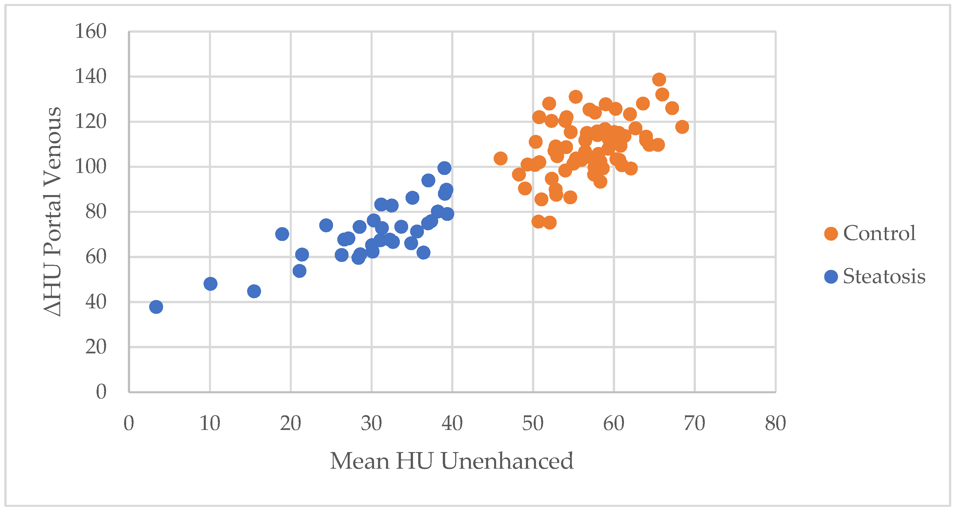
3.2. Quantitative Image Quality
3.3. Qualitative Analysis
3.4. Regression Analysis
3.5. Predicted Dose of Contrast Media
4. Discussion
5. Conclusions
Author Contributions
Funding
Institutional Review Board Statement
Informed Consent Statement
Data Availability Statement
Acknowledgments
Conflicts of Interest
References
- Foley, W.D.; Mallisee, T.A.; Hohenwalter, M.D.; Wilson, C.R.; Quiroz, F.A.; Taylor, A.J. Multiphase hepatic CT with a multirow detector CT scanner. AJR Am. J. Roentgenol. 2000, 175, 679–685. [Google Scholar] [CrossRef] [Scilit]
- Soyer, P.; Poccard, M.; Boudiaf, M.; Abitbol, M.; Hamzi, L.; Panis, Y.; Valleur, P.; Rymer, R. Detection of hypovascular hepatic metastases at triple-phase helical CT: Sensitivity of phases and comparison with surgical and histopathologic findings. Radiology 2004, 231, 413–420. [Google Scholar] [CrossRef] [Scilit] [PubMed]
- Kanematsu, M.; Kondo, H.; Goshima, S.; Kato, H.; Tsuge, U.; Hirose, Y.; Kim, M.-J.; Moriyama, N. Imaging liver metastases: Review and update. Eur. J. Radiol. 2006, 58, 217–228. [Google Scholar] [CrossRef] [Scilit] [PubMed]
- Younossi, Z.M.; Koenig, A.B.; Abdelatif, D.; Fazel, Y.; Henry, L.; Wymer, M. Global epidemiology of nonalcoholic fatty liver disease-Meta-analytic assessment of prevalence, incidence, and outcomes. Hepatology 2016, 64, 73–84. [Google Scholar] [CrossRef] [Scilit]
- Oliver, J.H.; Baron, R.L.; Federle, M.P.; Rockette, H.E. Detecting hepatocellular carcinoma: Value of unenhanced or arterial phase CT imaging or both used in conjunction with conventional portal venous phase contrast-enhanced CT imaging. AJR Am. J. Roentgenol. 1996, 167, 71–77. [Google Scholar] [CrossRef] [Scilit] [PubMed]
- Brink, J.A.; Heiken, J.P.; Forman, H.P.; Sagel, S.S.; Molina, P.L.; Brown, P.C. Hepatic spiral CT: Reduction of dose of intravenous contrast material. Radiology 1995, 197, 83–88. [Google Scholar] [CrossRef] [Scilit]
- Bae, K.T. Intravenous contrast medium administration and scan timing at CT: Considerations and approaches. Radiology 2010, 256, 32–61. [Google Scholar] [CrossRef] [Scilit]
- Koiwahara, G.; Tsuda, T.; Matsuda, M.; Hirata, M.; Tanaka, H.; Hyodo, T.; Kido, T.; Mochizuki, T. Different enhancement of the hepatic parenchyma in dynamic CT for patients with normal liver and chronic liver diseases and with the dose of contrast medium based on body surface area. Jpn. J. Radiol. 2015, 33, 194–200. [Google Scholar] [CrossRef] [Scilit]
- Lawrence, D.A.; Oliva, I.B.; Israel, G.M. Detection of hepatic steatosis on contrast-enhanced CT images: Diagnostic accuracy of identification of areas of presumed focal fatty sparing. AJR Am. J. Roentgenol. 2012, 199, 44–47. [Google Scholar] [CrossRef] [Scilit]
- Kodama, Y.; Ng, C.S.; Wu, T.T.; Ayers, G.D.; Curley, S.A.; Abdalla, E.K.; Vauthey, J.N.; Charnsangavej, C. Comparison of CT methods for determining the fat content of the liver. AJR Am. J. Roentgenol. 2007, 188, 1307–1312. [Google Scholar] [CrossRef] [Scilit]
- de Jong, D.J.; Veldhuis, W.B.; Wessels, F.J.; de Vos, B.; Moeskops, P.; Kok, M. Towards Personalised Contrast Injection: Artificial-Intelligence-Derived Body Composition and Liver Enhancement in Computed Tomography. J. Pers. Med. 2021, 11, 159. [Google Scholar] [CrossRef] [Scilit]
- Heiken, J.P.; Brink, J.A.; McClennan, B.L.; Sagel, S.S.; Crowe, T.M.; Gaines, M.V. Dynamic incremental CT: Effect of volume and concentration of contrast material and patient weight on hepatic enhancement. Radiology 1995, 195, 353–357. [Google Scholar] [CrossRef] [Scilit]
- Francque, S.; Verrijken, A.; Mertens, I.; Hubens, G.; Van Marck, E.; Pelckmans, P.; Van Gaal, L.; Michielsen, P. Noncirrhotic human nonalcoholic fatty liver disease induces portal hypertension in relation to the histological degree of steatosis. Eur. J. Gastroenterol. Hepatol. 2010, 22, 1449–1457. [Google Scholar] [CrossRef] [Scilit] [PubMed]
- Hashimoto, K.; Murakami, T.; Dono, K.; Hori, M.; Kim, T.; Kudo, M.; Marubashi, S.; Miyamoto, A.; Takeda, Y.; Nagano, H.; et al. Assessment of the severity of liver disease and fibrotic change: The usefulness of hepatic CT perfusion imaging. Oncol. Rep. 2006, 16, 677–683. [Google Scholar] [CrossRef] [Scilit] [PubMed]
- Farrell, G.C.; Teoh, N.C.; McCuskey, R.S. Hepatic microcirculation in fatty liver disease. Anat. Rec. 2008, 291, 684–692. [Google Scholar] [CrossRef] [Scilit]
- Ryou, M.; Stylopoulos, N.; Baffy, G. Nonalcoholic fatty liver disease and portal hypertension. Explor. Med. 2020, 1, 149–169. [Google Scholar] [CrossRef] [Scilit] [PubMed]
- Martens, B.; Hendriks, B.M.F.; Eijsvoogel, N.G.; Wildberger, J.E.; Mihl, C. Individually Body Weight-Adapted Contrast Media Application in Computed Tomography Imaging of the Liver at 90 kVp. Invest. Radiol. 2019, 54, 177–182. [Google Scholar] [CrossRef] [Scilit]
- Costa, A.F.; Peet, K.; Abdolell, M. Dosing Iodinated Contrast Media According to Lean Versus Total Body Weight at Abdominal CT: A Stratified Randomized Controlled Trial. Acad. Radiol. 2020, 27, 833–840. [Google Scholar] [CrossRef] [Scilit]
- Zanardo, M.; Doniselli, F.M.; Esseridou, A.; Agrò, M.; Panarisi, N.A.R.; Monti, C.B.; Di Leo, G.; Sardanelli, F. Lean body weight versus total body weight to calculate the iodinated contrast media volume in abdominal CT: A randomised controlled trial. Insights Imaging 2020, 11, 132. [Google Scholar] [CrossRef] [Scilit]
- Nakai, H.; Arizono, S.; Isoda, H.; Togashi, K. Imaging Characteristics of Liver Metastases Overlooked at Contrast-Enhanced CT. AJR Am. J. Roentgenol. 2019, 212, 782–787. [Google Scholar] [CrossRef] [Scilit]
- Torres, D.M.; Harrison, S.A. Nonalcoholic Fatyy Liver Disease: Clinical Features, Disease Modifiers, and Natural History. In Alcoholic and Non-Alcoholic Fatty Liver Disease; Chalasani, N., Szabo, G., Eds.; Springer: Cham, Germany, 2016; pp. 183–194. ISBN 978-3-319-20537-3. [Google Scholar]
- Marin, D.; Nelson, R.C.; Schindera, S.T.; Richard, S.; Youngblood, R.S.; Yoshizumi, T.T.; Samei, E. Low-tube-voltage, high-tube-current multidetector abdominal CT: Improved image quality and decreased radiation dose with adaptive statistical iterative reconstruction algorithm--initial clinical experience. Radiology 2010, 254, 145–153. [Google Scholar] [CrossRef] [Scilit]
- Colorectal Liver Metastases: CT, MR Imaging, and PET for Diagnosis—Meta-Analysis|Radiology. Available online: https://pubs-rsna-org.proxy.library.uu.nl/doi/10.1148/radiol.2371042060 (accessed on 2 September 2021).
- Böttcher, J.; Hansch, A.; Pfeil, A.; Schmidt, P.; Malich, A.; Schneeweiss, A.; Maurer, M.H.; Streitparth, F.; Teichgräber, U.K.; Renz, D.M. Detection and classification of different liver lesions: Comparison of Gd-EOB-DTPA-enhanced MRI versus multiphasic spiral CT in a clinical single centre investigation. Eur. J. Radiol. 2013, 82, 1860–1869. [Google Scholar] [CrossRef] [Scilit]
- Semelka, R.C.; Martin, D.R.; Balci, C.; Lance, T. Focal liver lesions: Comparison of dual-phase CT and multisequence multiplanar MR imaging including dynamic gadolinium enhancement. J. Magn. Reson. Imaging 2001, 13, 397–401. [Google Scholar] [CrossRef] [Scilit] [PubMed]
- Wanless, I.R.; Lentz, J.S. Fatty liver hepatitis (steatohepatitis) and obesity: An autopsy study with analysis of risk factors. Hepatology 1990, 12, 1106–1110. [Google Scholar] [CrossRef] [Scilit] [PubMed]
- Birnbaum, B.A.; Hindman, N.; Lee, J.; Babb, J.S. Multi-detector row CT attenuation measurements: Assessment of intra- and interscanner variability with an anthropomorphic body CT phantom. Radiology 2007, 242, 109–119. [Google Scholar] [CrossRef] [Scilit] [PubMed]
- Zhang, Y.N.; Fowler, K.J.; Hamilton, G.; Cui, J.Y.; Sy, E.Z.; Balanay, M.; Hooker, J.C.; Szeverenyi, N.; Sirlin, C.B. Liver fat imaging-a clinical overview of ultrasound, CT, and MR imaging. Br. J. Radiol. 2018, 91, 20170959. [Google Scholar] [CrossRef] [Scilit]
- Hamer, O.W.; Aguirre, D.A.; Casola, G.; Lavine, J.E.; Woenckhaus, M.; Sirlin, C.B. Fatty liver: Imaging patterns and pitfalls. Radiographics 2006, 26, 1637–1653. [Google Scholar] [CrossRef] [Scilit] [PubMed]

| Characteristic | Control | Liver Steatosis | p-Value |
|---|---|---|---|
| No. participants | 70 | 35 | - |
| Sex male | 53 (75.7%) | 25 (71.4%) | 0.636 a |
| Age (years) | 65.5 (55.8–73.0) | 64.0 (57.0–69.0) | 0.273 b |
| Length (m) | 1.77 (1.70–1.83) | 1.72 (1.68–1.83) | 0.360 b |
| Weight (kg) | 81.0 (73.0–88.0) | 92.0 (84.0–103.0) | <0.001 b |
| BMI | 26.0 (23.8–28.9) | 29.5 (26.2–35.7) | <0.001 b |
| Grams of iodine (g I) | 45.0 (39.0–45.0) | 45.0 (43.5–45.0) | 0.080 b |
| Grams of iodine/TBW (g I/kg) | 0.53 (±0.074) | 0.48 (±0.066) | 0.002 c |
| Flow rate (ml/s) | 4.5 (4.0–4.5) | 4.5 (4.5–4.75) | 0.187 b |
| Iodine delivery rate (g I/s) | 1.35 (1.20–1.35) | 1.35 (1.35–1.43) | 0.187 b |
| Injection duration (s) | 33.3 (33.3–33.3) | 33.3 (31.2–33.3) | 0.154 b |
| Phase | Segment | Control (n = 70) | Liver Steatosis (n = 35) | p-Value |
|---|---|---|---|---|
| Unenhanced | S2 | 58.6 (± 4.9) | 32.2 (± 9.3) | |
| S7 | 55.8 (± 5.5) | 29.1 (± 8.2) | ||
| S8 | 56.9 (± 5.6) | 28.2 (± 9.9) | ||
| Mean | 57.1 (± 5.0) | 29.8 (± 8.4) | <0.001 | |
| Portal | S2 | 109.8 (± 13.2) | 73.5 (± 12.8) | |
| S7 | 107.2 (± 12.9) | 69.2 (± 13.4) | ||
| S8 | 109.8 (± 14.4) | 68.6 (± 15.7) | ||
| Mean | 108.9 (± 13.0) | 70.4 (± 13.3) | <0.001 | |
| ΔHU portal venous a | 51.9 (± 11.5) | 40.6 (± 8.4) | <0.001 |
| Enhancement | Label | Control (n = 70) | Steatosis (n = 35) |
|---|---|---|---|
| ≥50 HU | Adequate | 39 (55.7%) | 7 (20.0%) |
| 40–50 HU | Moderate | 21 (30.0%) | 8 (22.9%) |
| ≤40 HU | Insufficient | 10 (14.3%) | 20 (57.1%) |
| Label | Control (n = 70) | Steatosis (n = 35) |
|---|---|---|
| Good | 46 (65.7%) | 1 (8.6%) |
| Moderate | 24 (34.3%) | 33 (88.5%) |
| Poor | 0 (0.0%) | 1 (2.9%) |
| Variable | Simple R2 |a | p-Value | Multiple Adjusted R2 |b | Significant F Change |
|---|---|---|---|---|
| Grams of Iodine per TBW | 0.213 | <0.001 | 0.205 | <0.001 |
| Liver steatosis | 0.205 | <0.001 | 0.303 | <0.001 |
| Weight | 0.122 | <0.001 | 0.310 | 0.174 |
| BMI | 0.057 | 0.017 | ||
| Length | 0.029 | 0.087 | ||
| Injection duration | 0.013 | 0.257 | ||
| Grams of Iodine | 0.008 | 0.357 | ||
| Flow rate/IDR | <0.001 | 0.978 | ||
| Age | <0.001 | 0.996 |
Publisher’s Note: MDPI stays neutral with regard to jurisdictional claims in published maps and institutional affiliations. |
© 2021 by the authors. Licensee MDPI, Basel, Switzerland. This article is an open access article distributed under the terms and conditions of the Creative Commons Attribution (CC BY) license (https://creativecommons.org/licenses/by/4.0/).
Share and Cite
van Cooten, V.V.; de Jong, D.J.; Wessels, F.J.; de Jong, P.A.; Kok, M. Liver Enhancement on Computed Tomography Is Suboptimal in Patients with Liver Steatosis. J. Pers. Med. 2021, 11, 1255. https://doi.org/10.3390/jpm11121255
van Cooten VV, de Jong DJ, Wessels FJ, de Jong PA, Kok M. Liver Enhancement on Computed Tomography Is Suboptimal in Patients with Liver Steatosis. Journal of Personalized Medicine. 2021; 11(12):1255. https://doi.org/10.3390/jpm11121255
Chicago/Turabian Stylevan Cooten, Véronique V., Daan J. de Jong, Frank J. Wessels, Pim A. de Jong, and Madeleine Kok. 2021. "Liver Enhancement on Computed Tomography Is Suboptimal in Patients with Liver Steatosis" Journal of Personalized Medicine 11, no. 12: 1255. https://doi.org/10.3390/jpm11121255
APA Stylevan Cooten, V. V., de Jong, D. J., Wessels, F. J., de Jong, P. A., & Kok, M. (2021). Liver Enhancement on Computed Tomography Is Suboptimal in Patients with Liver Steatosis. Journal of Personalized Medicine, 11(12), 1255. https://doi.org/10.3390/jpm11121255

