Measurement of Oral Epithelial Thickness by Optical Coherence Tomography

 ,
,  ,
, 
Abstract
1. Introduction
2. Materials and Methods
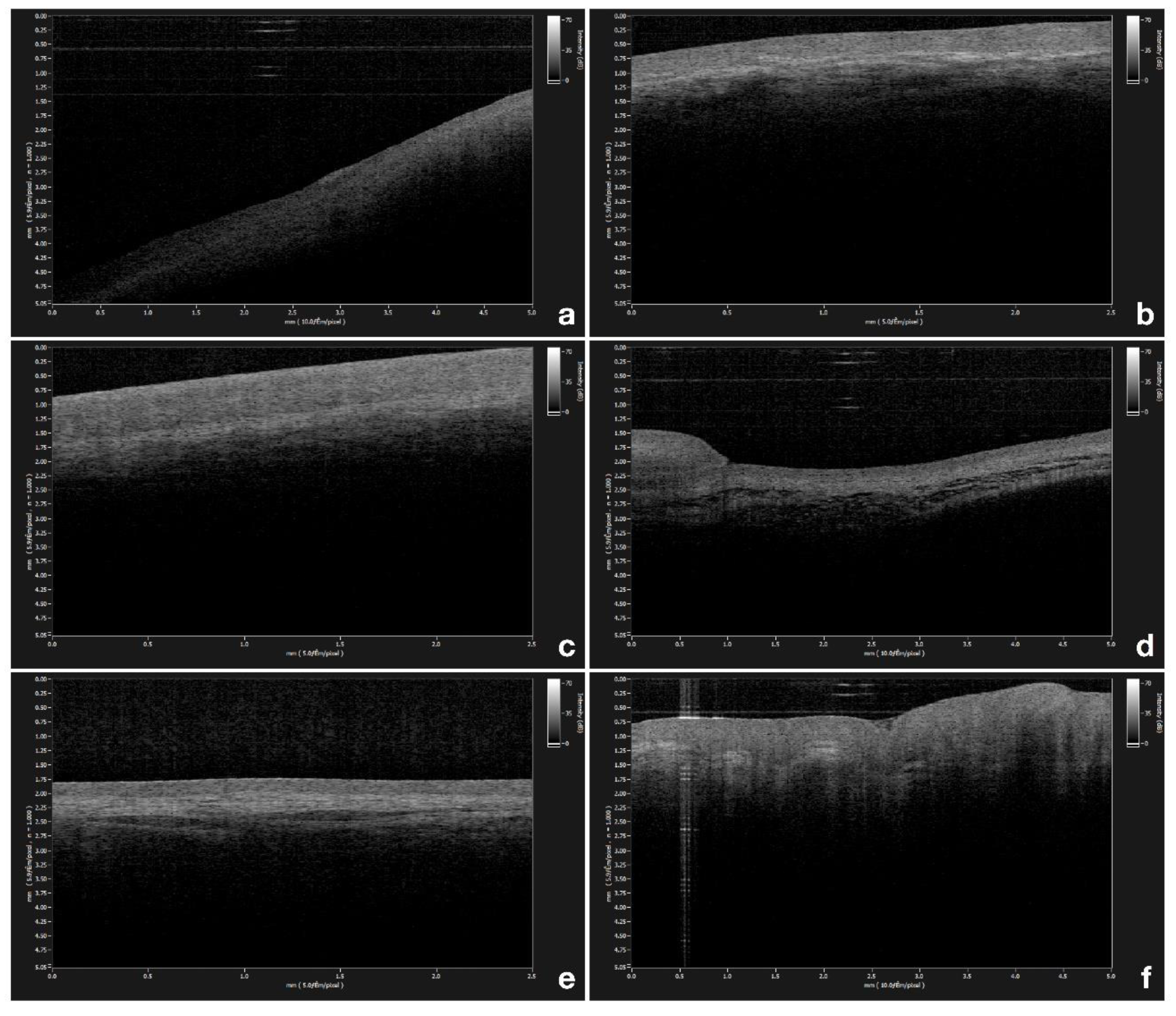
2.1. Epithelial Thickness Mesurement
2.2. OCT System
2.3. Statistical Analysis
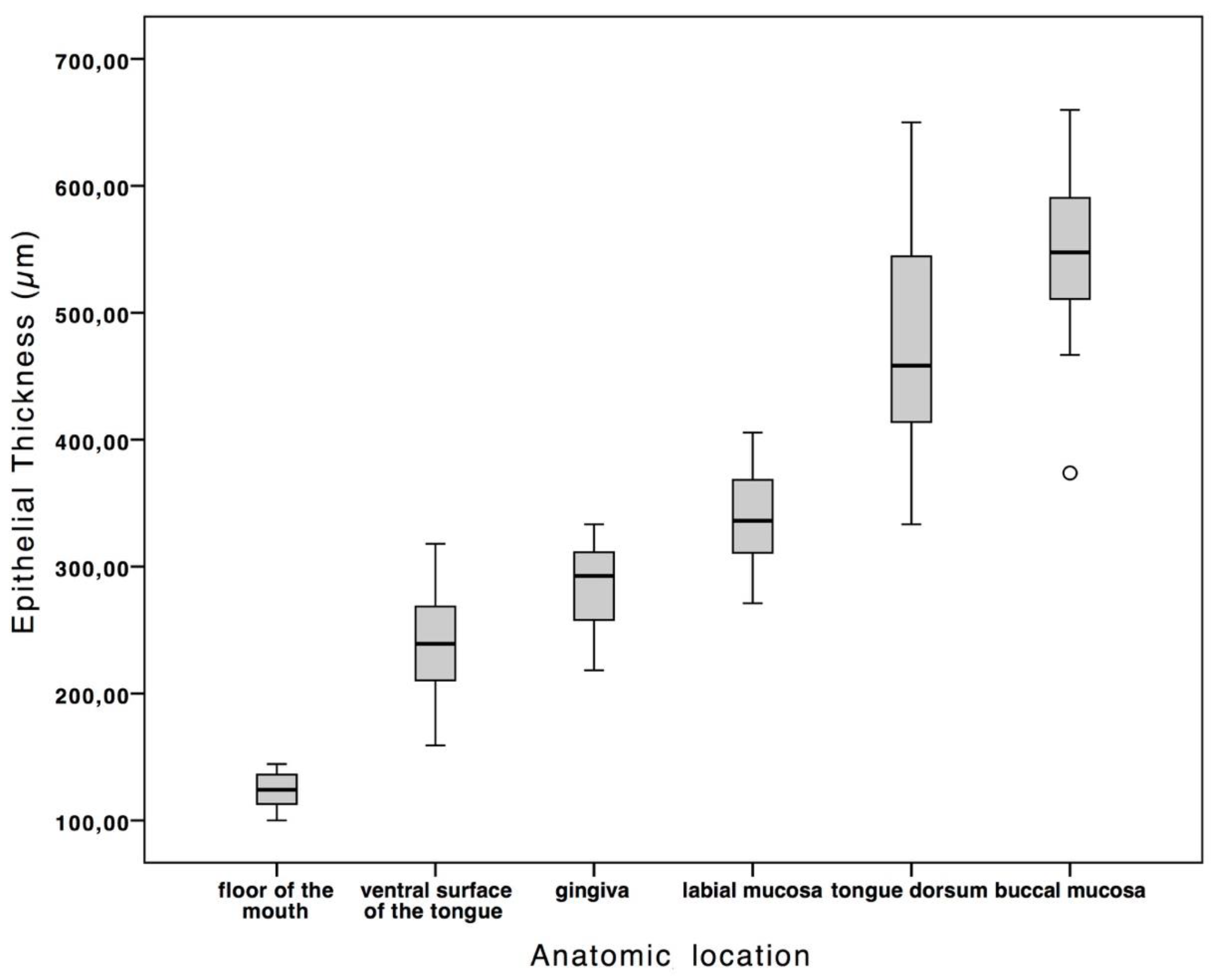
3. Results
4. Discussion
5. Conclusions
Author Contributions
Funding
Conflicts of Interest
References
- Gentile, E.; Maio, C.; Romano, A.; Laino, L.; Lucchese, A. The potential role of in vivo optical coherence tomography for evaluating oral soft tissue: A systematic review. J. Oral Pathol. Med. 2017, 1–13. [Google Scholar] [CrossRef] [PubMed]
- Feldchtein, F.I.; Gelikonov, V.M.; Iksanov, R.R.; Gelikonov, G.V.; Kuranov, R.V.; Sergeev, A.M.; Gladkova, N.D.; Ourutina, M.N.; Reitze, D.H.; Warren, J.A.; et al. In vivo OCT imaging of hard and soft tissue of the oral cavity. Opt. Express 1998, 3, 239–250. [Google Scholar] [CrossRef] [PubMed]
- Steele, T.O.; Meyers, A. Early Detection of Premalignant Lesions and Oral Cancer. Otolaryngol. Clin. N. Am. 2011, 44, 221–229. [Google Scholar] [CrossRef] [PubMed]
- Hsieh, Y.-S.; Ho, Y.-C.; Lee, S.-Y.; Chuang, C.-C.; Tsai, J.; Lin, K.-F.; Sun, C.-W. Dental optical coherence tomography. Sensors (Basel). 2013, 13, 8928–8949. [Google Scholar] [CrossRef] [PubMed]
- Jung, U.; Cho, N.; Kim, S.-H.; Jeong, H.; Kim, J.; Ahn, Y.-C. Simple Spectral Calibration Method and Its Application Using an Index Array for Swept Source Optical Coherence Tomography. J. Opt. Soc. Korea 2011, 15, 386–393. [Google Scholar] [CrossRef]
- Kim, K.H.; Park, B.H.; Maguluri, G.N.; Lee, T.W.; Rogomentich, F.J.; Bancu, M.G.; Bouma, B.E.; de Boer, J.F.; Bernstein, J.J. Two-axis magnetically-driven MEMS scanning catheter for endoscopic high-speed optical coherence tomography. Opt. Express 2007, 15, 18130. [Google Scholar] [CrossRef] [PubMed]
- Huber, R.; Adler, D.C.; Fujimoto, J.G. Buffered Fourier domain mode locking: Unidirectional swept laser sources for optical coherence tomography imaging at 370,000 lines/s. Opt. Lett. 2006, 31, 2975. [Google Scholar] [CrossRef] [PubMed]
- Choi, W.J.; Wang, R.K. In vivo imaging of functional microvasculature within tissue beds of oral and nasal cavities by swept-source optical coherence tomography with a forward/side-viewing probe. Biomed. Opt. Express 2014, 5, 2620. [Google Scholar] [CrossRef]
- Huang, D.; Swanson, E.A.; Lin, C.P.; Schuman, J.S.; Stinson, W.G.; Chang, W.; Hee, M.R.; Flotte, T.; Gregory, K.; Puliafito, C.A. Optical coherence tomography. Science 1991, 254, 1178–1181. [Google Scholar] [CrossRef]
- Alibhai, A.Y.; Or, C.; Witkin, A.J. Swept Source Optical Coherence Tomography: A Review. Curr. Ophthalmol. Rep. 2018, 7–16. [Google Scholar] [CrossRef]
- Wang, F.; Zhang, Q.; Deegan, A.J.; Chang, J.; Wang, R.K. Comparing imaging capabilities of spectral domain and swept source optical coherence tomography angiography in healthy subjects and central serous retinopathy. Eye Vis. 2018, 5, 19. [Google Scholar] [CrossRef] [PubMed]
- Arens, C.; Glanz, H.; Wönckhaus, J.; Hersemeyer, K.; Kraft, M. Histologic assessment of epithelial thickness in early laryngeal cancer or precursor lesions and its impact on endoscopic imaging. Eur. Arch. Otorhinolaryngol. 2007, 264, 645–649. [Google Scholar] [CrossRef] [PubMed]
- Tsai, M.-T.; Lee, H.-C.; Lee, C.-K.; Yu, C.-H.; Chen, H.-M.; Chiang, C.-P.; Chang, C.-C.; Wang, Y.-M.; Yang, C.C. Effective indicators for diagnosis of oral cancer using optical coherence tomography. Opt. Express 2008, 16, 15847–15862. [Google Scholar] [PubMed]
- Prestin, S.; Rothschild, S.I.; Betz, C.S.; Kraft, M. Measurement of epithelial thickness within the oral cavity using optical coherence tomography. Head Neck 2012, 34, 1777–1781. [Google Scholar] [CrossRef] [PubMed]
- Ridgway, J.M.; Armstrong, W.B.; Guo, S.; Mahmood, U.; Su, J.; Jackson, R.P.; Shibuya, T.; Crumley, R.L.; Gu, M.; Chen, Z.; et al. In Vivo Optical Coherence Tomography of the Human Oral Cavity and Oropharynx. Arch. Otolaryngol. Neck Surg. 2006, 132, 1074. [Google Scholar] [CrossRef] [PubMed]
- Wilder-Smith, P.; Lee, K.; Guo, S.; Zhang, J.; Osann, K.; Chen, Z.; Messadi, D. In vivo diagnosis of oral dysplasia and malignancy using optical coherence tomography: Preliminary studies in 50 patients. Lasers Surg. Med. 2009, 41, 353–357. [Google Scholar] [CrossRef] [PubMed]
- Di Stasio, D.; Lauritano, D.; Paparella, R.; Franco, R.; Montella, M.; Serpico, R.; Lucchese, A. Ultrasound imaging of oral fibroma: A case report. J. Biol. Regul. Homeost. Agents 2017, 31, 23–26. [Google Scholar] [PubMed]
- Vogt, M.; Knüttel, A.; Hoffmann, K.; Altmeyer, P.; Ermert, H. Comparison of high frequency ultrasound and optical coherence tomography as modalities for high resolution and non invasive skin imaging. Biomed. Tech. (Berl). 2003, 48, 116–121. [Google Scholar] [CrossRef] [PubMed]
- Warszawik-Hendzel, O.; Olszewska, M.; Maj, M.; Rakowska, A.; Czuwara, J.; Rudnicka, L. Non-invasive diagnostic techniques in the diagnosis of squamous cell carcinoma. J. Dermatol. Case Rep. 2015, 9, 89–97. [Google Scholar] [CrossRef]
- Rueden, C.T.; Schindelin, J.; Hiner, M.C.; DeZonia, B.E.; Walter, A.E.; Arena, E.T.; Eliceiri, K.W. ImageJ2: ImageJ for the next generation of scientific image data. BMC Bioinformatics 2017, 18, 1–26. [Google Scholar] [CrossRef]
- Di Stasio, D.; Lauritano, D.; Romano, A.; Salerno, C.; Minervini, G.; Minervini, G.; Gentile, E.; Serpico, R.; Lucchese, A. In vivo characterization of oral pemphigus vulgaris by optical coherence tomography. J. Biol. Regul. Homeost. Agents 2015, 29, 39–41. [Google Scholar] [PubMed]
- de Boer, J.F.; Leitgeb, R.; Wojtkowski, M. Twenty-five years of optical coherence tomography: The paradigm shift in sensitivity and speed provided by Fourier domain OCT [Invited]. Biomed. Opt. Express 2017, 8, 3248. [Google Scholar] [CrossRef] [PubMed]
- Lucchese, A.; Gentile, E.; Romano, A.; Maio, C.; Laino, L.; Serpico, R. The potential role of in vivo reflectance confocal microscopy for evaluating oral cavity lesions: A systematic review. J. Oral Pathol. Med. 2016, 45, 723–729. [Google Scholar] [CrossRef] [PubMed]
- Grassia, V.; Gentile, E.; Di Stasio, D.; Jamilian, A.; Matarese, G.; D’Apuzzo, F.; Santoro, R.; Perillo, L.; Serpico, R.; Lucchese, A. In vivo confocal microscopy analysis of enamel defects after orthodontic treatment: A preliminary study. Ultrastruct. Pathol. 2016, 40, 317–323. [Google Scholar] [CrossRef] [PubMed]
- De Rosa, A.; Di Stasio, D.; Lauritano, D.; Santoro, R.; Marotta, A.; Itro, A.; Lucchese, A. Non-invasive analysis of bleaching effect of hydrogen peroxide on enamel by reflectance confocal microscopy (RCM): Study of series of cases. Odontology 2019, 107, 285–290. [Google Scholar] [CrossRef] [PubMed]
- Lee, A.M.D.; Cahill, L.; Liu, K.; MacAulay, C.; Poh, C.; Lane, P. Wide-field in vivo oral OCT imaging. Biomed. Opt. Express 2015, 6, 2664. [Google Scholar] [CrossRef] [PubMed]
- Paulsen, F.; Thale, A. Epithelial-connective tissue boundary in the oral part of the human soft palate. J. Anat. 1998, 193, 457–467. [Google Scholar] [CrossRef]
- Klein-Szanto, A.J.; Schroeder, H.E. Architecture and density of the connective tissue papillae of the human oral mucosa. J. Anat. 1977, 123, 93–109. [Google Scholar]
- Kraft, M.; Glanz, H.; von Gerlach, S.; Wisweh, H.; Lubatschowski, H.; Arens, C. Clinical value of optical coherence tomography in laryngology. Head Neck 2008, 30, 1628–1635. [Google Scholar] [CrossRef]


| Anatomic Location | Thickness (µm) | SD | |
|---|---|---|---|
| gingiva | Mean | 285.04 | ± 32.98 |
| Min | 218.30 | ||
| Max | 333.33 | ||
| labial mucosa | Mean | 339.83 | ± 36.44 |
| Min | 271.19 | ||
| Max | 405.56 | ||
| buccal mucosa | Mean | 545.40 | ± 62.45 |
| Min | 373.75 | ||
| Max | 659.79 | ||
| ventral surface of the tongue | Mean | 239.79 | ± 37.30 |
| Min | 159.09 | ||
| Max | 318.00 | ||
| floor of the mouth | Mean | 124.09 | ± 13.53 |
| Min | 100.07 | ||
| Max | 144.44 | ||
| tongue dorsum | Mean | 479.32 | ± 83.56 |
| Min | 333.33 | ||
| Max | 650.02 |
© 2019 by the authors. Licensee MDPI, Basel, Switzerland. This article is an open access article distributed under the terms and conditions of the Creative Commons Attribution (CC BY) license (http://creativecommons.org/licenses/by/4.0/).
Share and Cite
Di Stasio, D.; Lauritano, D.; Iquebal, H.; Romano, A.; Gentile, E.; Lucchese, A. Measurement of Oral Epithelial Thickness by Optical Coherence Tomography. Diagnostics 2019, 9, 90. https://doi.org/10.3390/diagnostics9030090
Di Stasio D, Lauritano D, Iquebal H, Romano A, Gentile E, Lucchese A. Measurement of Oral Epithelial Thickness by Optical Coherence Tomography. Diagnostics. 2019; 9(3):90. https://doi.org/10.3390/diagnostics9030090
Chicago/Turabian StyleDi Stasio, Dario, Dorina Lauritano, Hasan Iquebal, Antonio Romano, Enrica Gentile, and Alberta Lucchese. 2019. "Measurement of Oral Epithelial Thickness by Optical Coherence Tomography" Diagnostics 9, no. 3: 90. https://doi.org/10.3390/diagnostics9030090
APA StyleDi Stasio, D., Lauritano, D., Iquebal, H., Romano, A., Gentile, E., & Lucchese, A. (2019). Measurement of Oral Epithelial Thickness by Optical Coherence Tomography. Diagnostics, 9(3), 90. https://doi.org/10.3390/diagnostics9030090