Experimental Lung Ultrasound Scoring in a Murine Model of Aspiration Pneumonia: Challenges and Diagnostic Perspectives

 , , and
Abstract
1. Introduction
2. Materials and Methods
2.1. Animal Ethics and Experimental Design
2.2. Aspiration Pneumonia Model
2.3. Lung Ultrasound Scoring
3. Ultrasound Acquisition
3.1. Equipment and Setup
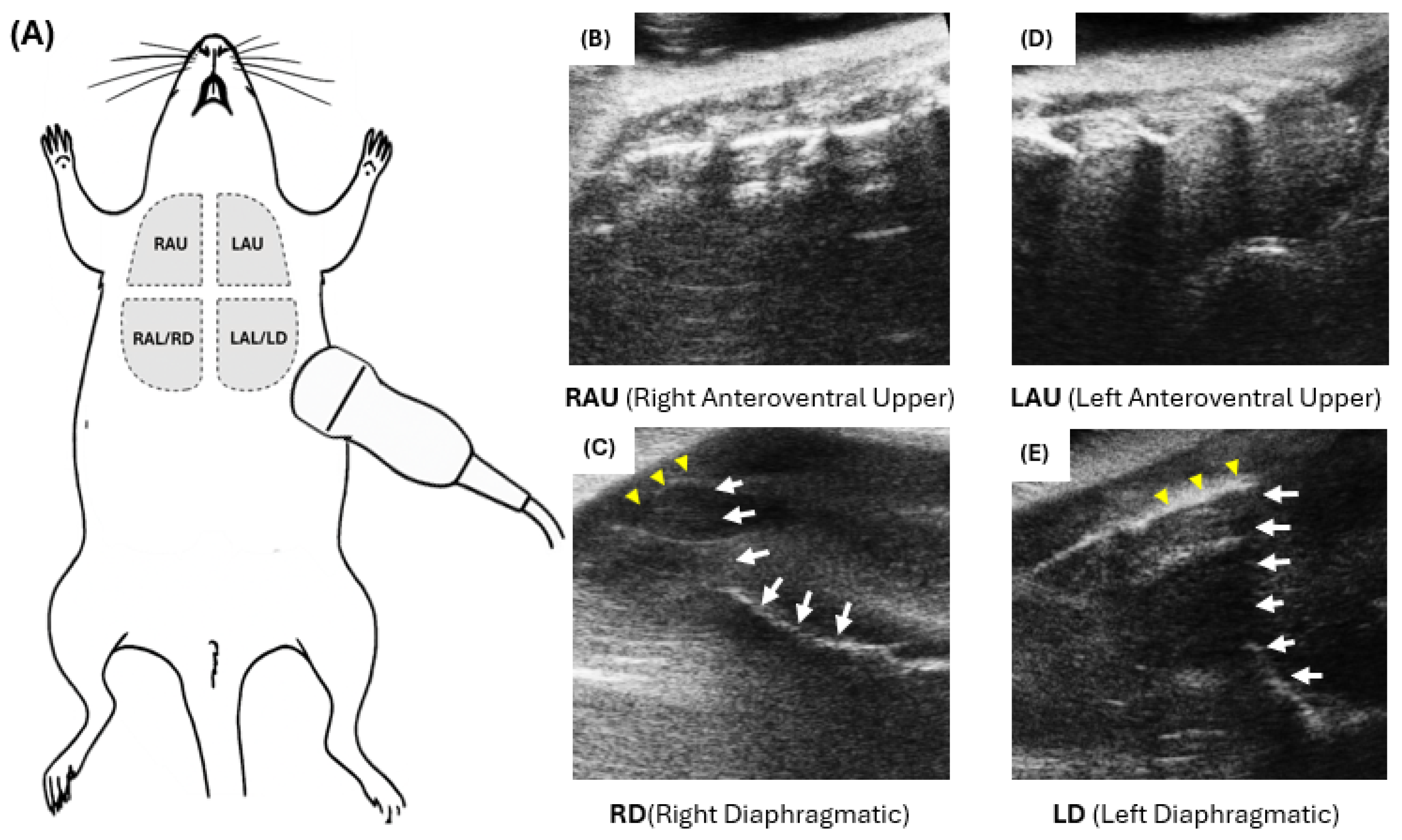
3.2. Scanning Protocol and Zoning
- •
- RAU: Right anteroventral upper
- •
- RAL (RD): Right anteroventral lower/right diaphragmatic
- •
- LAU: Left anteroventral upper
- •
- LAL (LD): Left anteroventral lower/left diaphragmatic
3.2.1. Zone Acquisition and Recording Handling
- (i)
- LU-only views were not used for correlation analyses due to poor reproducibility in pilot assessments;
- (ii)
- Mixed LU + LD recordings were retained only for the diaphragmatic (LD) portion, because the pleuro-diaphragmatic angle and lung base remained consistently identifiable, whereas the LU component was more susceptible to rib shadowing and cardiac-motion artifacts; thus, these mixed recordings were reassigned to LD for analysis;
- (iii)
- Regions demonstrating poor reproducibility were excluded from quantitative correlation analyses to minimize interpretive noise.
3.2.2. Rationale for LU Exclusion
3.3. Regional Lung Ultrasound (LUS) Analysis
4. MLEUS Scoring System
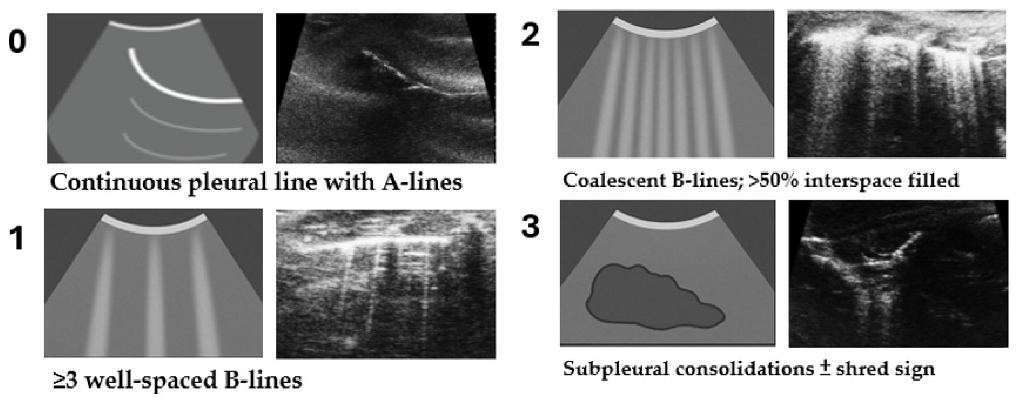
4.1. Criteria and Interpretation
4.2. Inter-Rater Reliability and Blinding
4.3. Validation of MLEUS Score
4.4. Wet-to-Dry Weight Ratio (W/D Ratio)
4.5. Histological Lung Injury Score
4.6. Definition of Injury Severity
4.7. Statistical Analysis
- •
- Group comparisons were performed using the Kruskal–Wallis test followed by Dunn’s multiple comparisons test.
- •
- Pairwise comparisons were conducted using the Mann–Whitney U test.
- •
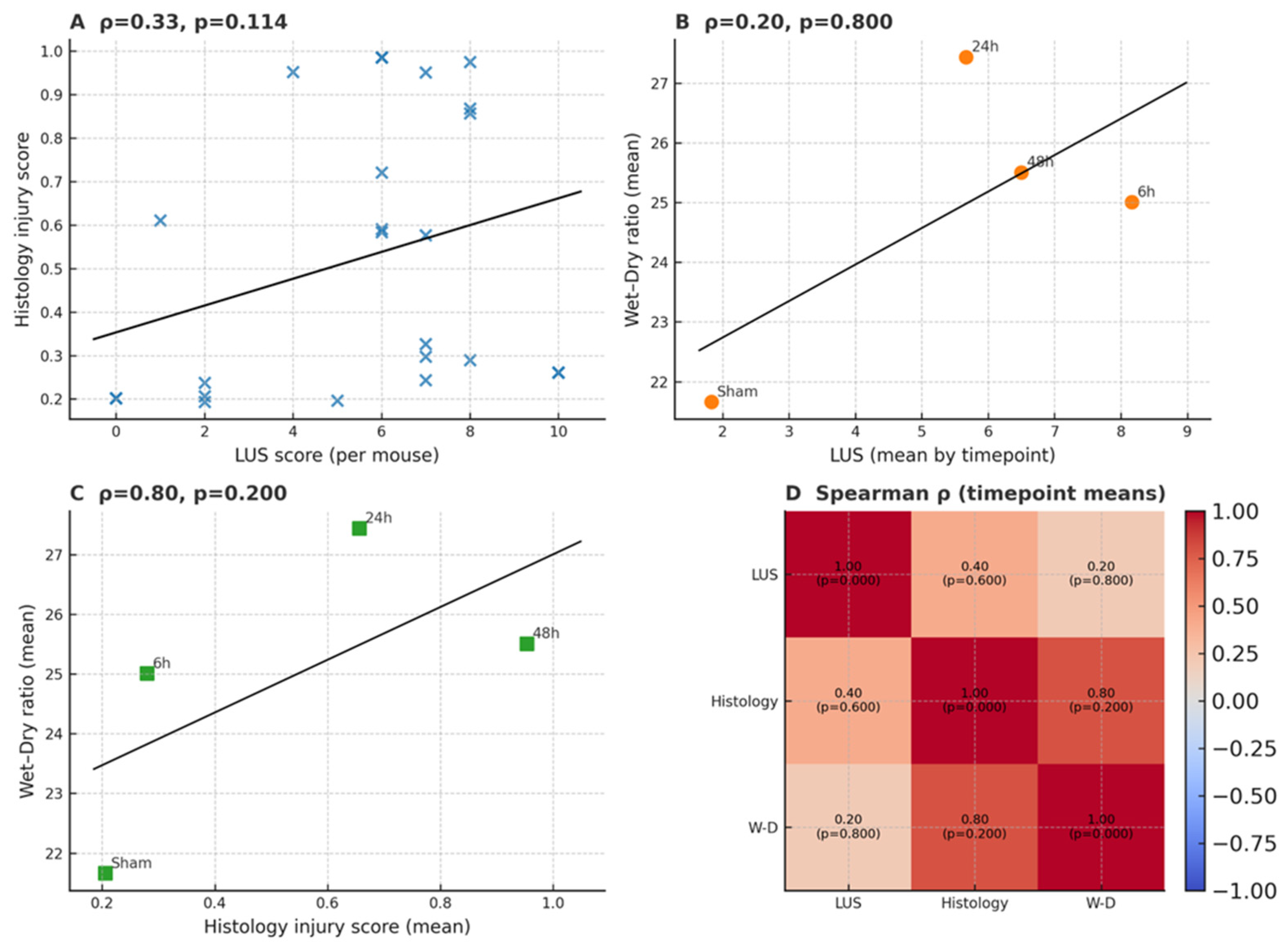
- Spearman’s rank correlation was applied to evaluate associations between MLEUS scores, W/D ratios, and histological scores.
- •
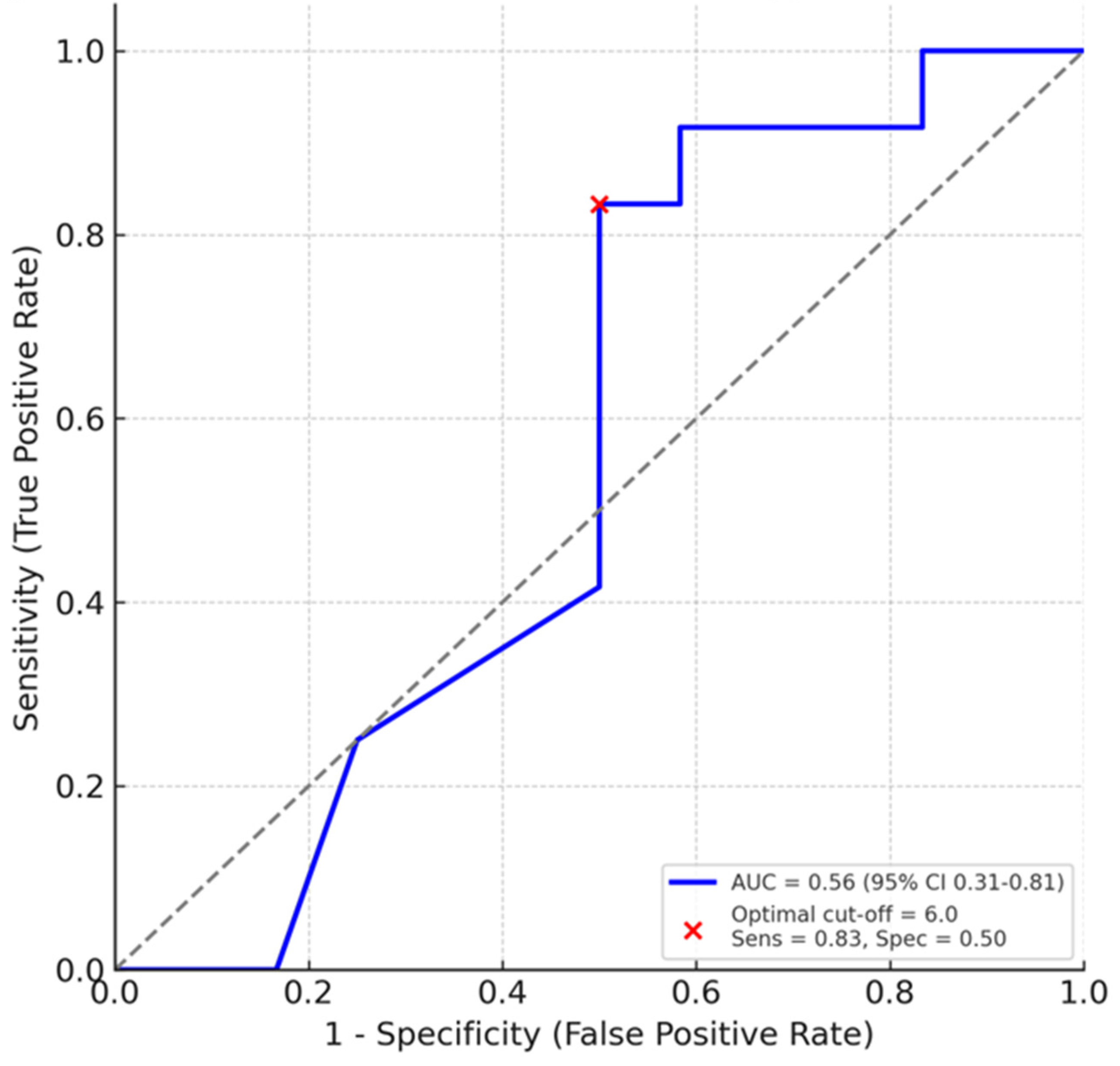
- Receiver Operating Characteristic (ROC) analysis was used in an exploratory fashion to assess the diagnostic performance of regional LUS scores. ROC analysis was performed on the composite LUS score rather than on individual regions because the sample size was insufficient for reliable zone-specific ROC curves. The ROC evaluation was exploratory and intended only to provide a global assessment of LUS discrimination. Area under the curve (AUC) values were interpreted as follows: 0.7–0.8 (acceptable), 0.8–0.9 (excellent), and >0.9 (outstanding). Statistical significance was defined as p < 0.05.
4.8. Minimum Criteria Rationale for Scoring System Design
- ▪
- The final system was designed to satisfy three essential methodological criteria:
- ▪
- Clearly defined, region-based scoring metrics to ensure anatomical consistency.
- ▪
- Inter-rater reliability formally assessed and reported to confirm reproducibility.
- ▪
- Measurable correlations with histopathological and physiological indices of lung injury, providing construct validity.
4.9. Use of Generative Artificial Intelligence (GenAI)
5. Results
5.1. Overview of Study Findings
5.1.1. Early Detection of Pulmonary Injury
5.1.2. Temporal Progression and Regional Susceptibility
5.1.3. Correlation with Established Injury Markers
5.1.4. Diagnostic Performance and Threshold-Based Prediction
5.1.5. Representative Histopathology
6. Discussion
6.1. Region-Specific Reliability of LUS
6.2. Diagnostic Correlation with Established Injury Markers
6.3. Median-Based Thresholds as a Pragmatic Diagnostic Tool
6.4. Temporal Sensitivity and Early Detection
6.5. Reproducibility and Translational Implications
6.6. Limitations and Future Directions
7. Conclusions
Supplementary Materials
Author Contributions
Funding
Institutional Review Board Statement
Informed Consent Statement
Data Availability Statement
Acknowledgments
Conflicts of Interest
Abbreviations
| ALI | Acute Lung Injury |
| AP | Aspiration Pneumonia |
| AUC | Area Under the Curve |
| CI | Confidence Interval |
| H&E | Hematoxylin and Eosin |
| ICC | Intraclass Correlation Coefficient |
| IQR | Interquartile Range |
| LUS | Lung Ultrasound |
| MLEUS | Modified Lung Edema Ultrasound Score |
| MoLUS | Mouse Lung Ultrasound Score |
| RAU | Right Anteroventral Upper |
| RD (RAL) | Right Diaphragmatic (Right Anteroventral Lower) |
| ROC | Receiver Operating Characteristic |
| RU | Right Upper (zone) |
| LD (LAL) | Left Diaphragmatic (Left Anteroventral Lower, including LU+LD) |
| SD | Standard Deviation |
| SEM | Standard Error of the Mean |
| W/D ratio | Wet-to-Dry Weight Ratio |
References
- Marik, P.E. Aspiration pneumonitis and aspiration pneumonia. N. Engl. J. Med. 2001, 344, 665–671. [Google Scholar] [CrossRef]
- Raghavendran, K.; Nemzek, J.; Napolitano, L.M.; Knight, P.R. Aspiration-induced lung injury. Crit. Care Med. 2011, 39, 818–826. [Google Scholar] [CrossRef] [PubMed]
- Kikuchi, R.; Watabe, N.; Konno, T.; Mishina, N.; Sekizawa, K.; Sasaki, H. High incidence of silent aspiration in elderly patients with community-acquired pneumonia. Am. J. Respir. Crit. Care Med. 1994, 150, 251–253. [Google Scholar] [CrossRef]
- Teramoto, S.; Fukuchi, Y.; Sasaki, H.; Sato, K.; Sekizawa, K.; Matsuse, T.; Japanese Study Group on Aspiration Pulmonary Disease. High incidence of aspiration pneumonia in community- and hospital-acquired pneumonia in hospitalized patients: A multicenter, prospective study in Japan. J. Am. Geriatr. Soc. 2008, 56, 577–579. [Google Scholar] [CrossRef] [PubMed]
- Kosutova, P.; Mikolka, P. Aspiration syndromes and associated lung injury: Incidence, pathophysiology and management. Physiol. Res. 2021, 70, S567–S583. [Google Scholar] [CrossRef]
- Mandell, L.A.; Niederman, M.S. Aspiration Pneumonia. N. Engl. J. Med. 2019, 380, 651–663. [Google Scholar] [CrossRef]
- Teramoto, S. The current definition, epidemiology, animal models and a novel therapeutic strategy for aspiration pneumonia. Respir. Investig. 2022, 60, 45–55. [Google Scholar] [CrossRef]
- Son, Y.G.; Shin, J.; Ryu, H.G. Pneumonitis and pneumonia after aspiration. J. Dent. Anesth. Pain Med. 2017, 17, 1–12. [Google Scholar] [CrossRef] [PubMed]
- Thompson, B.T.; Chambers, R.C.; Liu, K.D. Acute Respiratory Distress Syndrome. N. Engl. J. Med. 2017, 377, 562–572. [Google Scholar] [CrossRef]
- Matute-Bello, G.; Frevert, C.W.; Martin, T.R. Animal models of acute lung injury. Am. J. Physiol. Lung Cell. Mol. Physiol. 2008, 295, L379–L399. [Google Scholar] [CrossRef]
- Matute-Bello, G.; Downey, G.; Moore, B.B.; Groshong, S.D.; Matthay, M.A.; Slutsky, A.S.; Kuebler, W.M.; Acute Lung Injury in Animals Study, G. An official American Thoracic Society workshop report: Features and measurements of experimental acute lung injury in animals. Am. J. Respir. Cell Mol. Biol. 2011, 44, 725–738. [Google Scholar] [CrossRef]
- Teramoto, S. Animal Models of Aspiration Pneumonia. In Aspiration Pneumonia: The Current Clinical Giant for Respiratory Physicians; Teramoto, S., Komiya, K., Eds.; Springer: Singapore, 2020; pp. 143–151. [Google Scholar]
- Amigoni, M.; Bellani, G.; Scanziani, M.; Masson, S.; Bertoli, E.; Radaelli, E.; Patroniti, N.; Di Lelio, A.; Pesenti, A.; Latini, R. Lung injury and recovery in a murine model of unilateral acid aspiration: Functional, biochemical, and morphologic characterization. Anesthesiology 2008, 108, 1037–1046. [Google Scholar] [CrossRef]
- Knight, P.R.; Rutter, T.; Tait, A.R.; Coleman, E.; Johnson, K. Pathogenesis of Gastric Particulate Lung Injury: A Comparison and Interaction with Acidic Pneumonitis. Anesth. Analg. 1993, 77, 754–760. [Google Scholar] [CrossRef]
- Alluri, R.; Kutscher, H.L.; Mullan, B.A.; Davidson, B.A.; Knight, P.R. Open Tracheostomy Gastric Acid Aspiration Murine Model of Acute Lung Injury Results in Maximal Acute Nonlethal Lung Injury. J. Vis. Exp. 2017, 120, 54700. [Google Scholar] [CrossRef] [PubMed]
- Meli, A.; Barbeta Vinas, E.; Battaglini, D.; Li Bassi, G.; Yang, H.; Yang, M.; Bobi, J.; Motos, A.; Fernandez-Barat, L.; Chiumello, D.; et al. Lateral position during severe mono-lateral pneumonia: An experimental study. Sci. Rep. 2020, 10, 19372. [Google Scholar] [CrossRef]
- Lichtenstein, D.A.; Meziere, G.A. Relevance of lung ultrasound in the diagnosis of acute respiratory failure: The BLUE protocol. Chest 2008, 134, 117–125. [Google Scholar] [CrossRef]
- Volpicelli, G.; Elbarbary, M.; Blaivas, M.; Lichtenstein, D.A.; Mathis, G.; Kirkpatrick, A.W.; Melniker, L.; Gargani, L.; Noble, V.E.; Via, G.; et al. International evidence-based recommendations for point-of-care lung ultrasound. Intensive Care Med. 2012, 38, 577–591. [Google Scholar] [CrossRef] [PubMed]
- Rodenas Monteagudo, M.A.; Albero Rosello, I.; Del Mazo Carrasco, A.; Carmona Garcia, P.; Zarragoikoetxea Jauregui, I. Update on the use of ultrasound in the diagnosis and monitoring of the critical patient. Rev. Esp. Anestesiol. Reanim. (Engl. Ed.) 2022, 69, 567–577. [Google Scholar] [CrossRef]
- Haji, K.; Haji, D.; Canty, D.J.; Royse, A.G.; Green, C.; Royse, C.F. The impact of heart, lung and diaphragmatic ultrasound on prediction of failed extubation from mechanical ventilation in critically ill patients: A prospective observational pilot study. Crit. Ultrasound J. 2018, 10, 13. [Google Scholar] [CrossRef]
- Fernandes Rodrigues, N.; Giraud, L.; Bolen, G.; Fastres, A.; Clercx, C.; Boysen, S.; Billen, F.; Gommeren, K. Comparison of lung ultrasound, chest radiographs, C-reactive protein, and clinical findings in dogs treated for aspiration pneumonia. J. Vet. Intern. Med. 2022, 36, 743–752. [Google Scholar] [CrossRef] [PubMed]
- Lin, C.H.; Lo, P.Y.; Lam, M.C.; Wu, H.D. Usefulness of Chest Ultrasonography in Predicting Diagnosis in Non-emergency Small Animal Patients With Lung Parenchymal and Pleural Disease. Front. Vet. Sci. 2020, 7, 616882. [Google Scholar] [CrossRef]
- Jambrik, Z.; Gargani, L.; Adamicza, A.; Kaszaki, J.; Varga, A.; Forster, T.; Boros, M.; Picano, E. B-lines quantify the lung water content: A lung ultrasound versus lung gravimetry study in acute lung injury. Ultrasound Med. Biol. 2010, 36, 2004–2010. [Google Scholar] [CrossRef]
- Gargani, L.; Lionetti, V.; Di Cristofano, C.; Bevilacqua, G.; Recchia, F.A.; Picano, E. Early detection of acute lung injury uncoupled to hypoxemia in pigs using ultrasound lung comets. Crit. Care Med. 2007, 35, 2769–2774. [Google Scholar] [CrossRef]
- Vezzosi, T.; Mannucci, T.; Pistoresi, A.; Toma, F.; Tognetti, R.; Zini, E.; Domenech, O.; Auriemma, E.; Citi, S. Assessment of Lung Ultrasound B-Lines in Dogs with Different Stages of Chronic Valvular Heart Disease. J. Vet. Intern. Med. 2017, 31, 700–704. [Google Scholar] [CrossRef]
- Rademacher, N.; Pariaut, R.; Pate, J.; Saelinger, C.; Kearney, M.T.; Gaschen, L. Transthoracic lung ultrasound in normal dogs and dogs with cardiogenic pulmonary edema: A pilot study. Vet. Radiol. Ultrasound 2014, 55, 447–452. [Google Scholar] [CrossRef]
- Brusasco, C.; Santori, G.; Bruzzo, E.; Tro, R.; Robba, C.; Tavazzi, G.; Guarracino, F.; Forfori, F.; Boccacci, P.; Corradi, F. Quantitative lung ultrasonography: A putative new algorithm for automatic detection and quantification of B-lines. Crit. Care 2019, 23, 288. [Google Scholar] [CrossRef] [PubMed]
- Villalba-Orero, M.; Lopez-Olaneta, M.M.; Gonzalez-Lopez, E.; Padron-Barthe, L.; Gomez-Salinero, J.M.; Garcia-Prieto, J.; Wai, T.; Garcia-Pavia, P.; Ibanez, B.; Jimenez-Borreguero, L.J.; et al. Lung ultrasound as a translational approach for non-invasive assessment of heart failure with reduced or preserved ejection fraction in mice. Cardiovasc. Res. 2017, 113, 1113–1123. [Google Scholar] [CrossRef] [PubMed]
- Chuang, C.W.; Vo, H.N.; Huang, Y.H.; Tsai, I.L.; Chang, C.Y.; Huang, C.J. Human placental mesenchymal stem cell-derived exosomes carrying hsa-let-7i-5p mitigate lung injury in a murine model of aspiration pneumonia. Korean J. Anesthesiol. 2025. [Google Scholar] [CrossRef] [PubMed]
- Shrout, P.E.; Fleiss, J.L. Intraclass correlations: Uses in assessing rater reliability. Psychol. Bull. 1979, 86, 420–428. [Google Scholar] [CrossRef]
- Hassanzad, M.; Hajian-Tilaki, K. Methods of determining optimal cut-point of diagnostic biomarkers with application of clinical data in ROC analysis: An update review. BMC Med. Res. Methodol. 2024, 24, 84. [Google Scholar] [CrossRef]
- Soldati, G.; Smargiassi, A.; Inchingolo, R.; Buonsenso, D.; Perrone, T.; Briganti, D.F.; Perlini, S.; Torri, E.; Mariani, A.; Mossolani, E.E.; et al. Is There a Role for Lung Ultrasound During the COVID-19 Pandemic? J. Ultrasound Med. 2020, 39, 1459–1462. [Google Scholar] [CrossRef] [PubMed]
- Grune, J.; Beyhoff, N.; Hegemann, N.; Lauryn, J.H.; Kuebler, W.M. From bedside to bench: Lung ultrasound for the assessment of pulmonary edema in animal models. Cell Tissue Res. 2020, 380, 379–392. [Google Scholar] [CrossRef] [PubMed]
- Tusman, G.; Acosta, C.M.; Costantini, M. Ultrasonography for the assessment of lung recruitment maneuvers. Crit. Ultrasound J. 2016, 8, 8. [Google Scholar] [CrossRef] [PubMed]



| Score | Ultrasound Features | Interpretation |
|---|---|---|
| 0 | Continuous pleural line with A-lines; no B-lines | Normal aeration |
| 1 | ≥3 well-spaced B-lines | Mild interstitial syndrome |
| 2 | Coalescent B-lines; >50% interspace filled | Moderate alveolar-interstitial syndrome |
| 3 | Subpleural consolidations ± shred sign | Severe loss of aeration or alveolar flooding |
Disclaimer/Publisher’s Note: The statements, opinions and data contained in all publications are solely those of the individual author(s) and contributor(s) and not of MDPI and/or the editor(s). MDPI and/or the editor(s) disclaim responsibility for any injury to people or property resulting from any ideas, methods, instructions or products referred to in the content. |
© 2026 by the authors. Licensee MDPI, Basel, Switzerland. This article is an open access article distributed under the terms and conditions of the Creative Commons Attribution (CC BY) license.
Share and Cite
Chuang, C.-W.; Lai, W.-Y.; Chang, K.-W.; Chang, C.-Y.; Yeoh, S.-R.; Huang, C.-J. Experimental Lung Ultrasound Scoring in a Murine Model of Aspiration Pneumonia: Challenges and Diagnostic Perspectives. Diagnostics 2026, 16, 361. https://doi.org/10.3390/diagnostics16020361
Chuang C-W, Lai W-Y, Chang K-W, Chang C-Y, Yeoh S-R, Huang C-J. Experimental Lung Ultrasound Scoring in a Murine Model of Aspiration Pneumonia: Challenges and Diagnostic Perspectives. Diagnostics. 2026; 16(2):361. https://doi.org/10.3390/diagnostics16020361
Chicago/Turabian StyleChuang, Ching-Wei, Wen-Yi Lai, Kuo-Wei Chang, Chao-Yuan Chang, Shang-Ru Yeoh, and Chun-Jen Huang. 2026. "Experimental Lung Ultrasound Scoring in a Murine Model of Aspiration Pneumonia: Challenges and Diagnostic Perspectives" Diagnostics 16, no. 2: 361. https://doi.org/10.3390/diagnostics16020361
APA StyleChuang, C.-W., Lai, W.-Y., Chang, K.-W., Chang, C.-Y., Yeoh, S.-R., & Huang, C.-J. (2026). Experimental Lung Ultrasound Scoring in a Murine Model of Aspiration Pneumonia: Challenges and Diagnostic Perspectives. Diagnostics, 16(2), 361. https://doi.org/10.3390/diagnostics16020361