Article

LungINFseg: Segmenting COVID-19 Infected Regions in Lung CT Images Based on a Receptive-Field-Aware Deep Learning Framework


1
Department of Computer Engineering and Mathematics, Universitat Rovira i Virgili, 43007 Tarragona, Spain
2
Department of Electrical Engineering, Aswan University, 81542 Aswan, Egypt
3
Department of Medicine and Health Sciences, Universitat Rovira i Virgili, 43201 Reus, Spain
*
Author to whom correspondence should be addressed.
Diagnostics 2021, 11(2), 158; https://doi.org/10.3390/diagnostics11020158

Submission received: 10 December 2020 / Revised: 7 January 2021 / Accepted: 14 January 2021 / Published: 22 January 2021
(This article belongs to the Section Machine Learning and Artificial Intelligence in Diagnostics)

Abstract

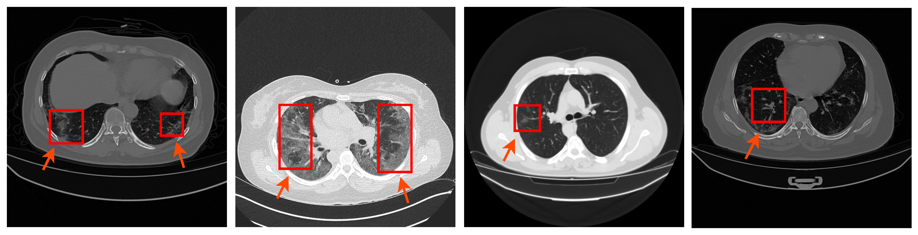
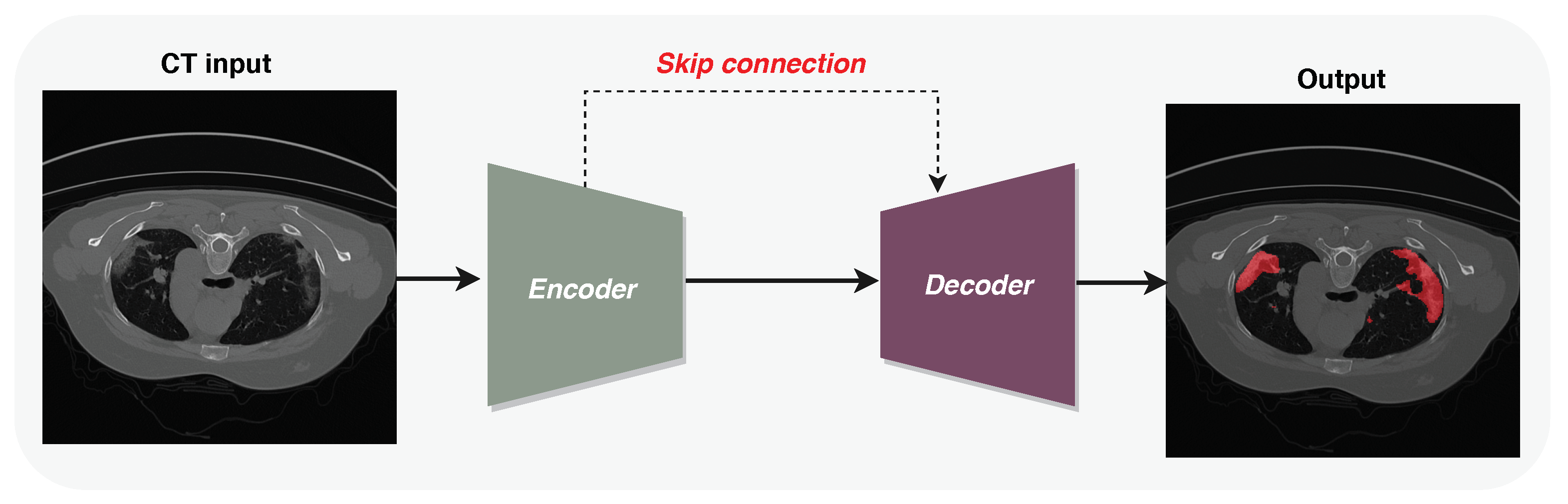
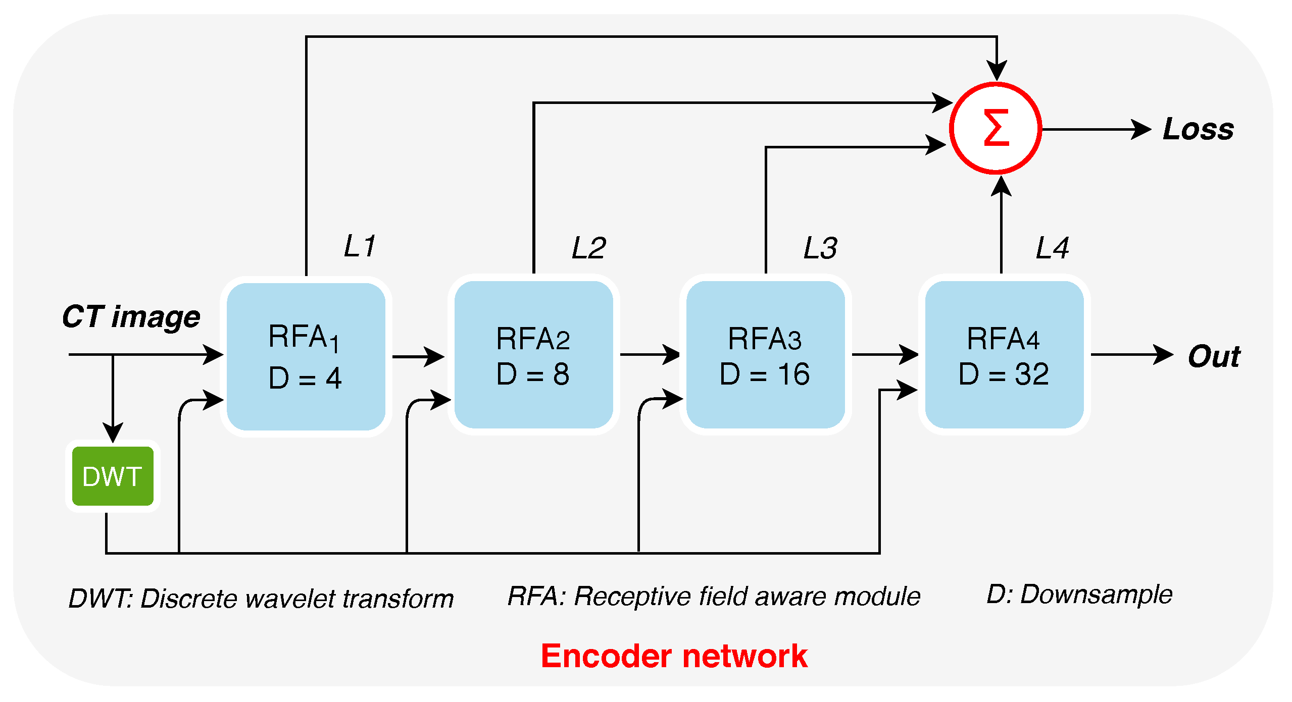
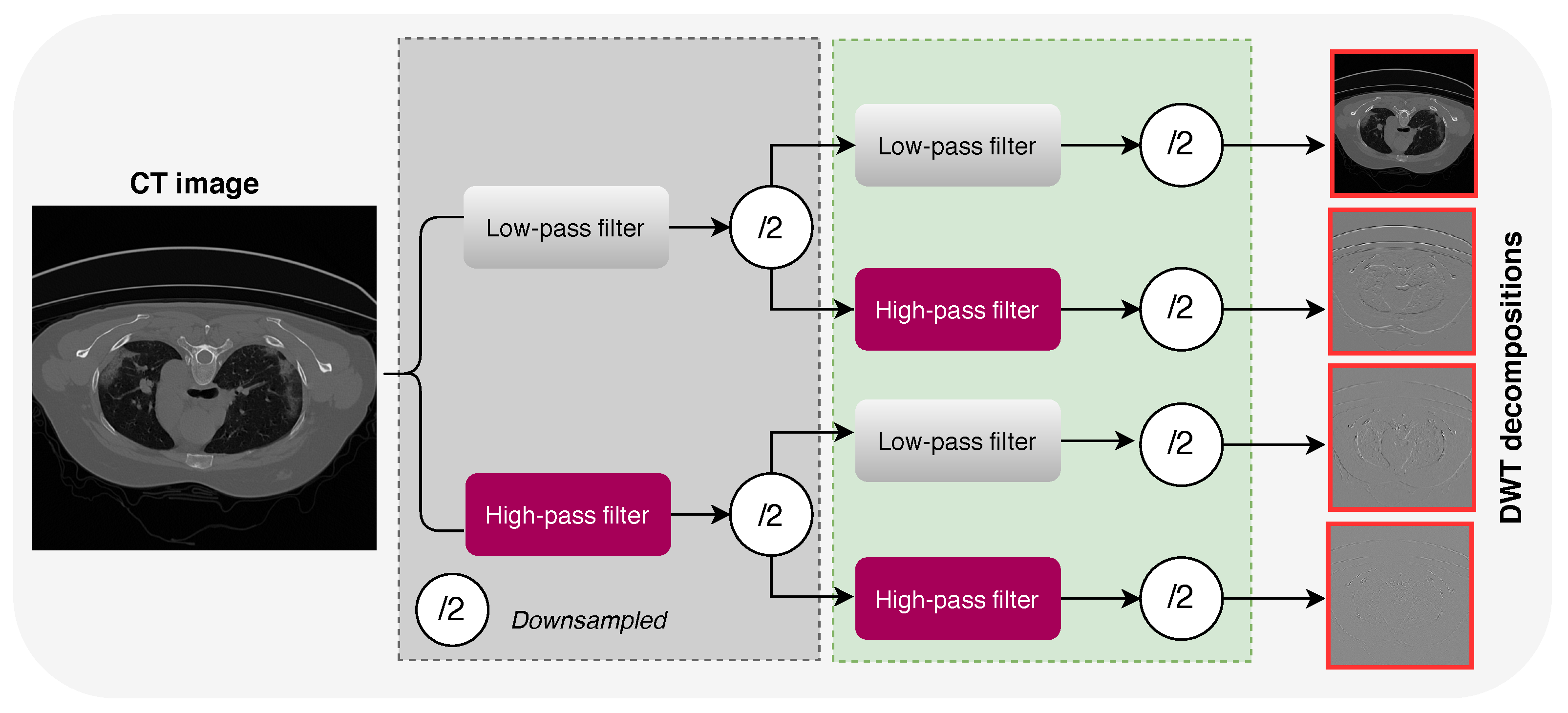
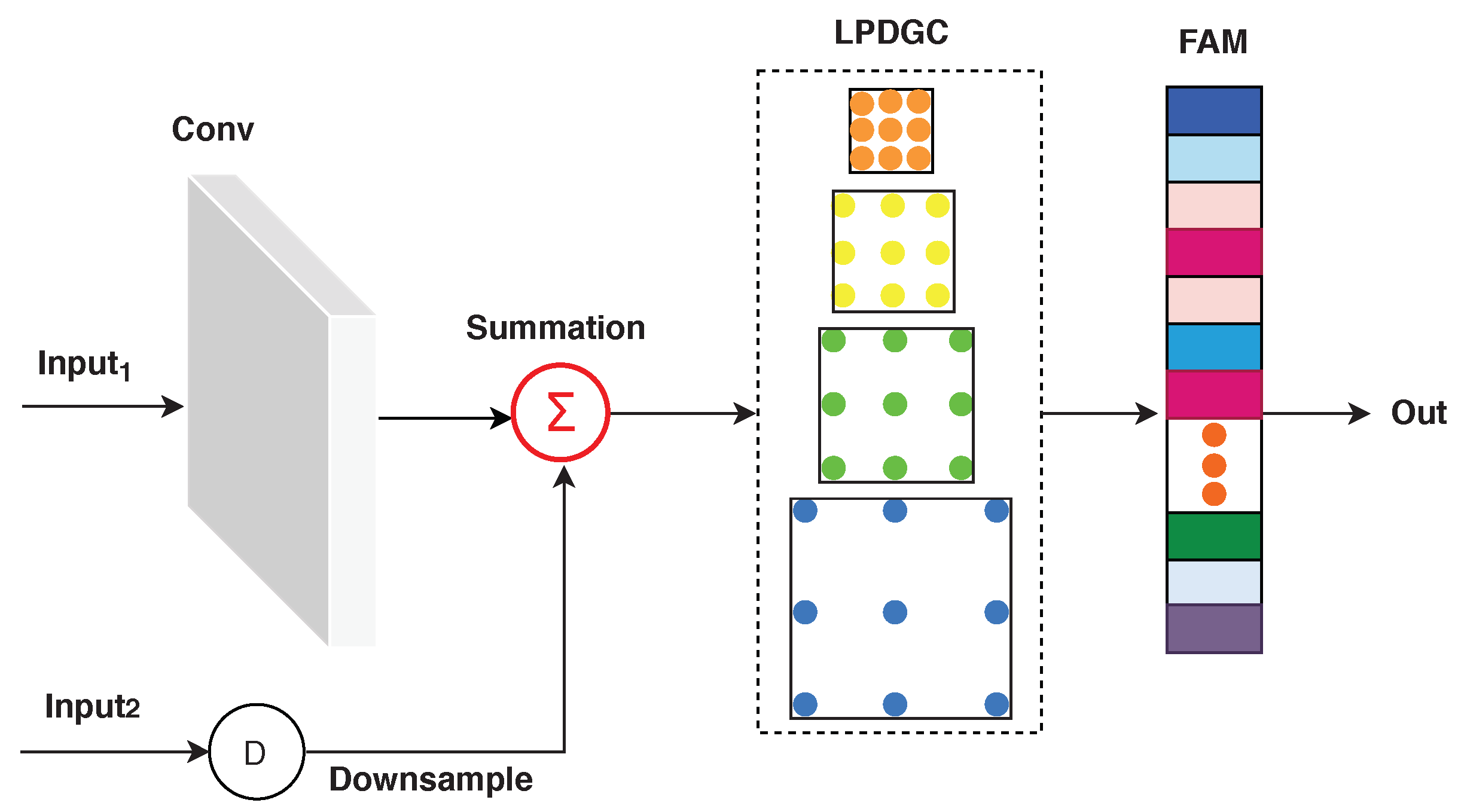
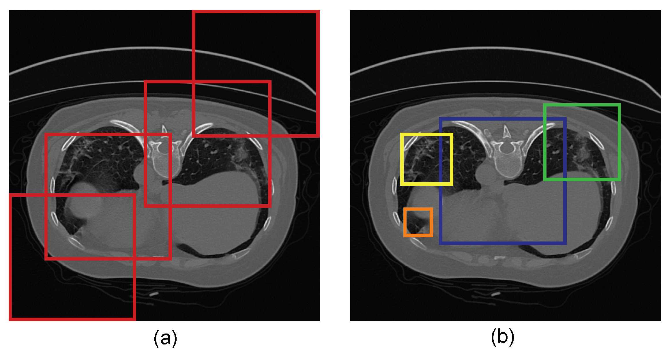
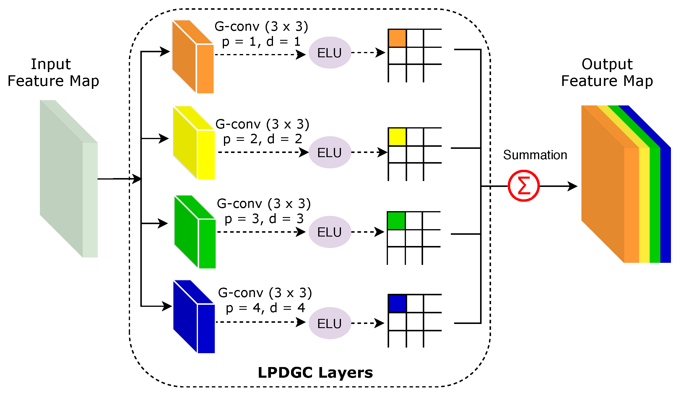
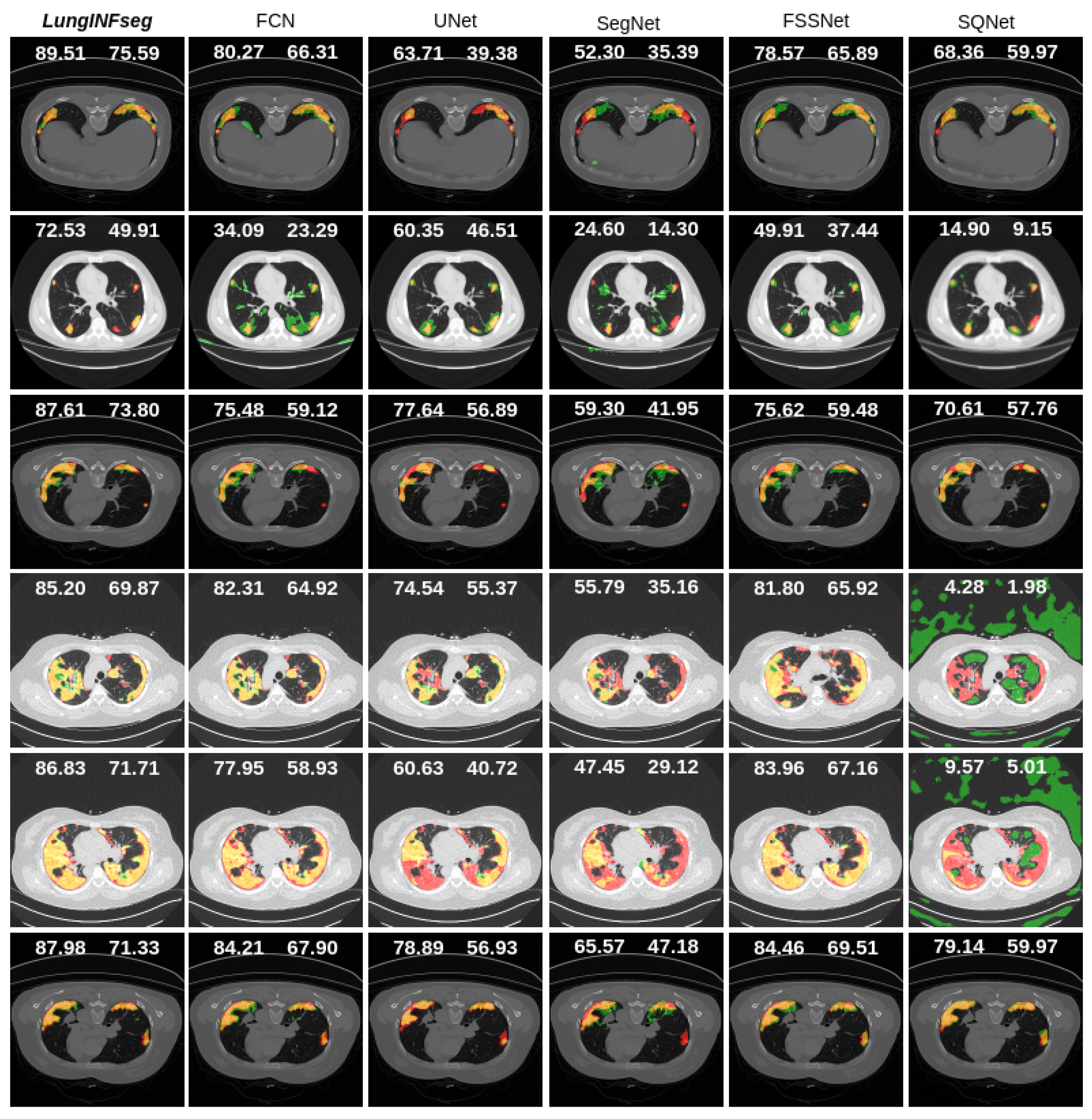
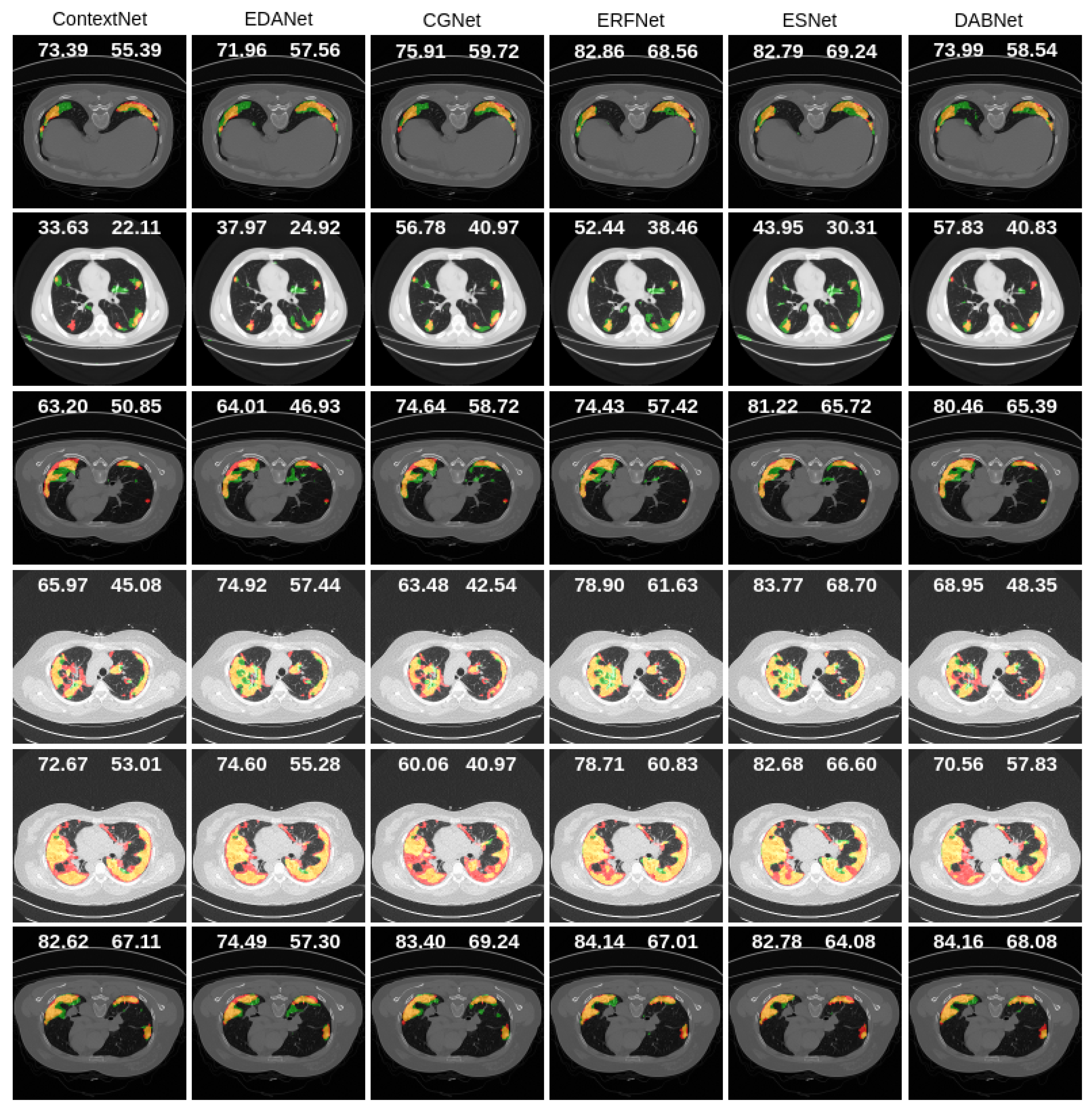
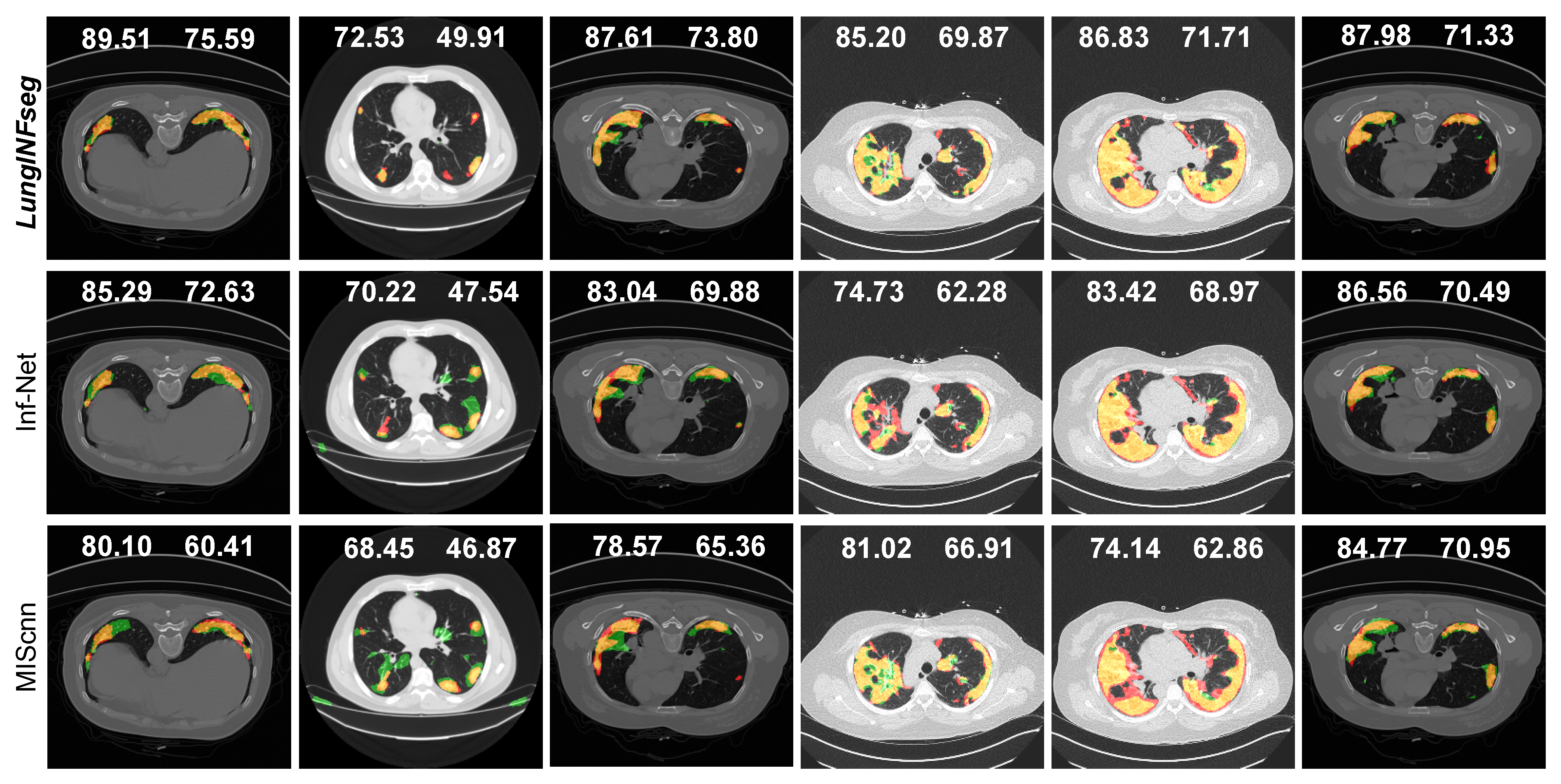
COVID-19 is a fast-growing disease all over the world, but facilities in the hospitals are restricted. Due to unavailability of an appropriate vaccine or medicine, early identification of patients suspected to have COVID-19 plays an important role in limiting the extent of disease. Lung computed tomography (CT) imaging is an alternative to the RT-PCR test for diagnosing COVID-19. Manual segmentation of lung CT images is time consuming and has several challenges, such as the high disparities in texture, size, and location of infections. Patchy ground-glass and consolidations, along with pathological changes, limit the accuracy of the existing deep learning-based CT slices segmentation methods. To cope with these issues, in this paper we propose a fully automated and efficient deep learning-based method, called LungINFseg, to segment the COVID-19 infections in lung CT images. Specifically, we propose the receptive-field-aware (RFA) module that can enlarge the receptive field of the segmentation models and increase the learning ability of the model without information loss. RFA includes convolution layers to extract COVID-19 features, dilated convolution consolidated with learnable parallel-group convolution to enlarge the receptive field, frequency domain features obtained by discrete wavelet transform, which also enlarges the receptive field, and an attention mechanism to promote COVID-19-related features. Large receptive fields could help deep learning models to learn contextual information and COVID-19 infection-related features that yield accurate segmentation results. In our experiments, we used a total of 1800+ annotated CT slices to build and test LungINFseg. We also compared LungINFseg with 13 state-of-the-art deep learning-based segmentation methods to demonstrate its effectiveness. LungINFseg achieved a dice score of 80.34% and an intersection-over-union (IoU) score of 68.77%—higher than the ones of the other 13 segmentation methods. Specifically, the dice and IoU scores of LungINFseg were 10% better than those of the popular biomedical segmentation method U-Net.
Keywords: COVID-19; CT slices; deep learning; image segmentation COVID-19; CT slices; deep learning; image segmentation