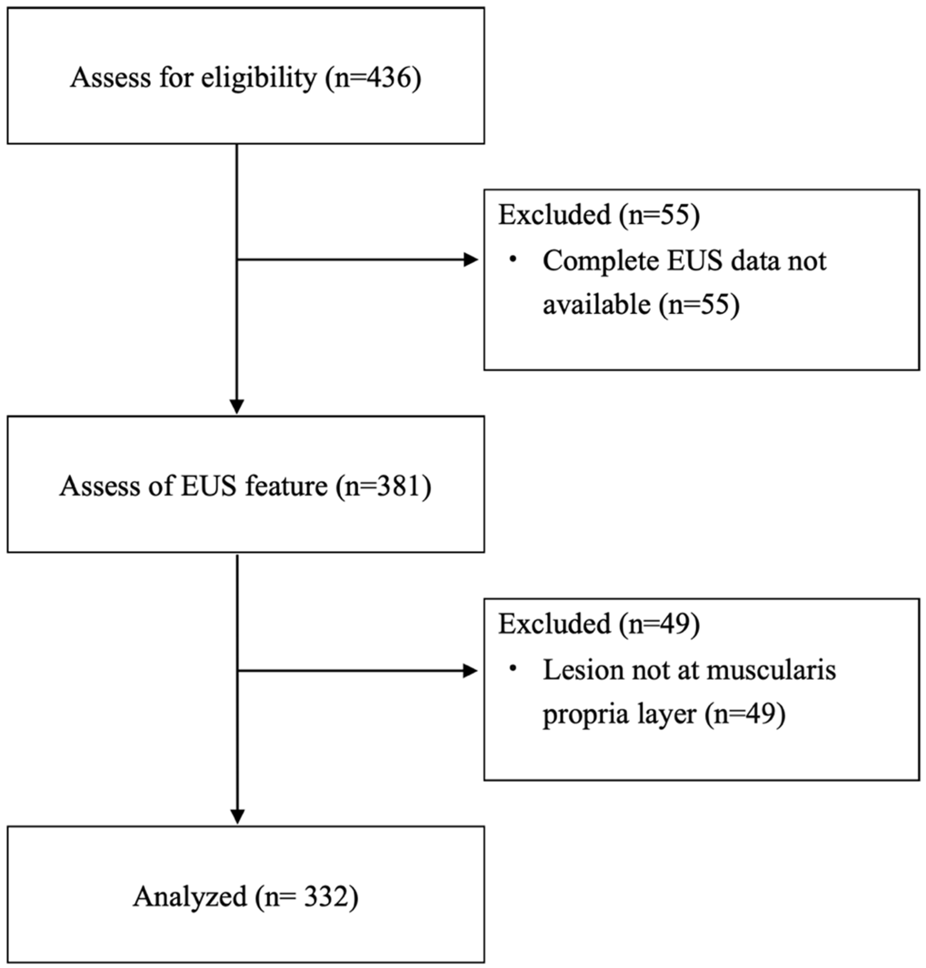
1. Introduction
The incidence of gastric subepithelial lesions (SELs) is on the rise, attributed to the widespread use of gastroscopy [
1]. Approximately 1.9% of patients who undergo gastroscopy are found to have SELs [
2]. A subset of these lesions originates from the muscularis propria (MP) layer, which includes gastrointestinal stromal tumors (GISTs), leiomyomas, and schwannomas. It is imperative to identify SELs with malignant potential, such as GISTs, for timely management. Certain characteristics observed in computed tomography (CT) scans, including location of the lesions, size, presence of necrosis, and enhancement during various phases, may assist in distinguishing GISTs from leiomyomas [
3]. Endoscopic ultrasonography (EUS) serves as a superior modality for characterizing the features of SELs; however, it still demonstrates a limited accuracy of 43% in predicting histological diagnoses [
4,
5]. Notable features of EUS that may aid in differentiating GISTs from leiomyomas include heterogeneity, irregular borders, echogenic foci, cystic (or anechoic) spaces, and the presence of dimpling or ulceration [
4,
6].
Histological assessment is essential for the definitive diagnosis of SELs, necessitating tissue acquisition for effective risk stratification. Proposed methods for obtaining tissue include mucosal incision-assisted biopsy (MIAB), bite-on-bite stacked biopsy, and EUS-guided fine needle aspiration or biopsy [
5]. However, the yield rates of these techniques have proven inadequate, failing to provide the mitotic rate necessary for assessing malignant potential [
7]. Furthermore, the incidence of bleeding complications associated with forceps biopsy methods can reach as high as 8%, and the small size of the lesions complicates EUS-guided tissue acquisition [
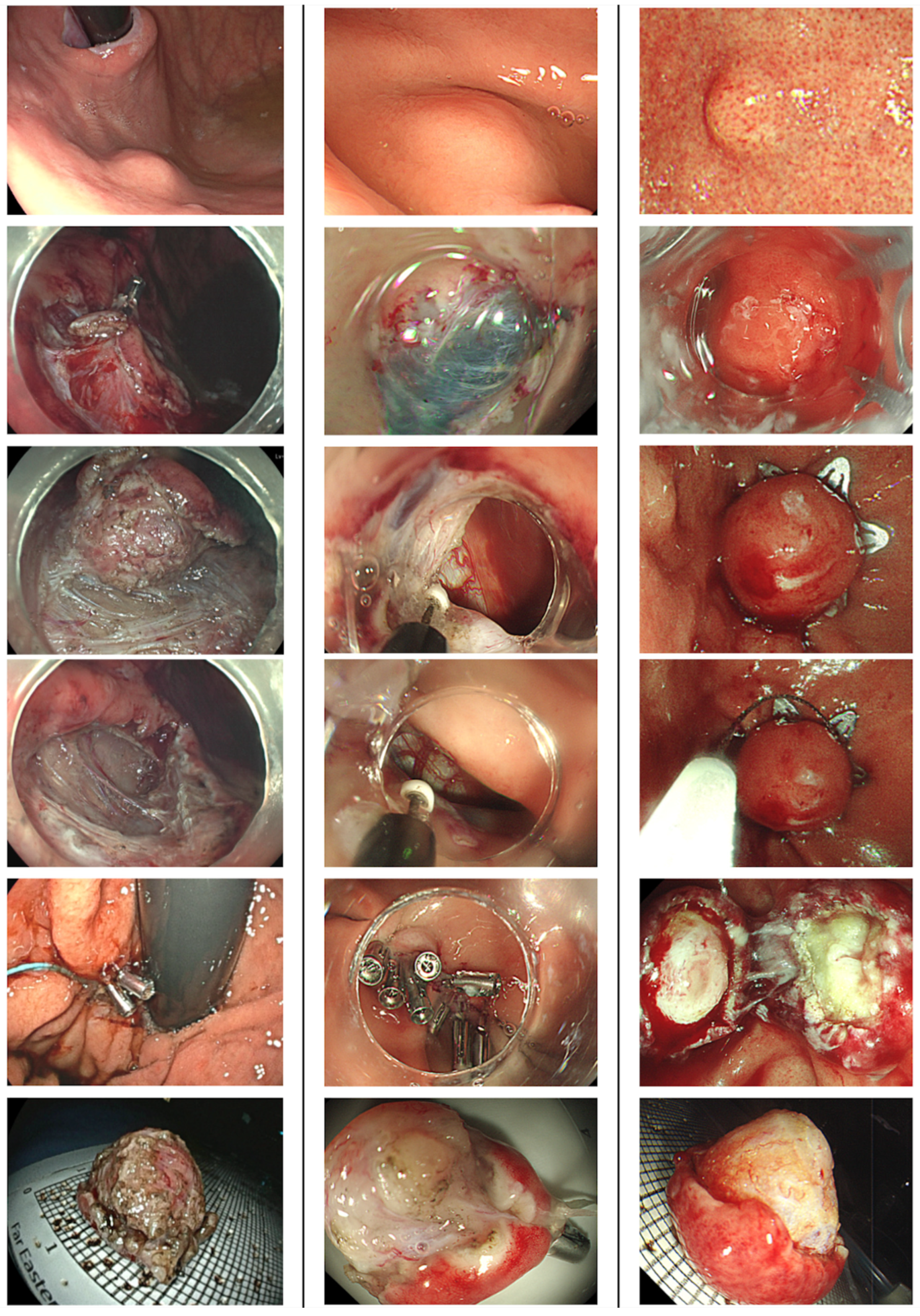
8]. Recent advancements in endoscopic resection (ER) and wound closure techniques, such as endoscopic muscularis dissection (EMD), endoscopic subserosal dissection (ESSD), submucosal tunneling endoscopic resection (STER), and endoscopic full-thickness resection (EFTR), have been developed to excise SELs that extend beyond the submucosal layer, facilitating both definitive histological diagnosis and curative resection [
9,
10,
11]. The clinical guidelines from the American College of Gastroenterology (ACG) and the European Society for Medical Oncology (ESMO) recognize ER as an alternative to surgical intervention for small gastric GISTs [
4,
12]. Additionally, the European Society of Gastrointestinal Endoscopy (ESGE) guidelines recommend considering ER—specifically STER, EFTR, or endoscopic submucosal excavation (ESE)—as a treatment option when there is a clinical indication or when attempts to obtain a diagnosis for the resection of SELs have failed [
13]. The objective of this study was to assess the
Gastric
Stromal
Tumor
Resection
Outcomes (GASTRO Trial) by different ER techniques and identify EUS predictors for diagnosis of GISTs.
4. Discussion
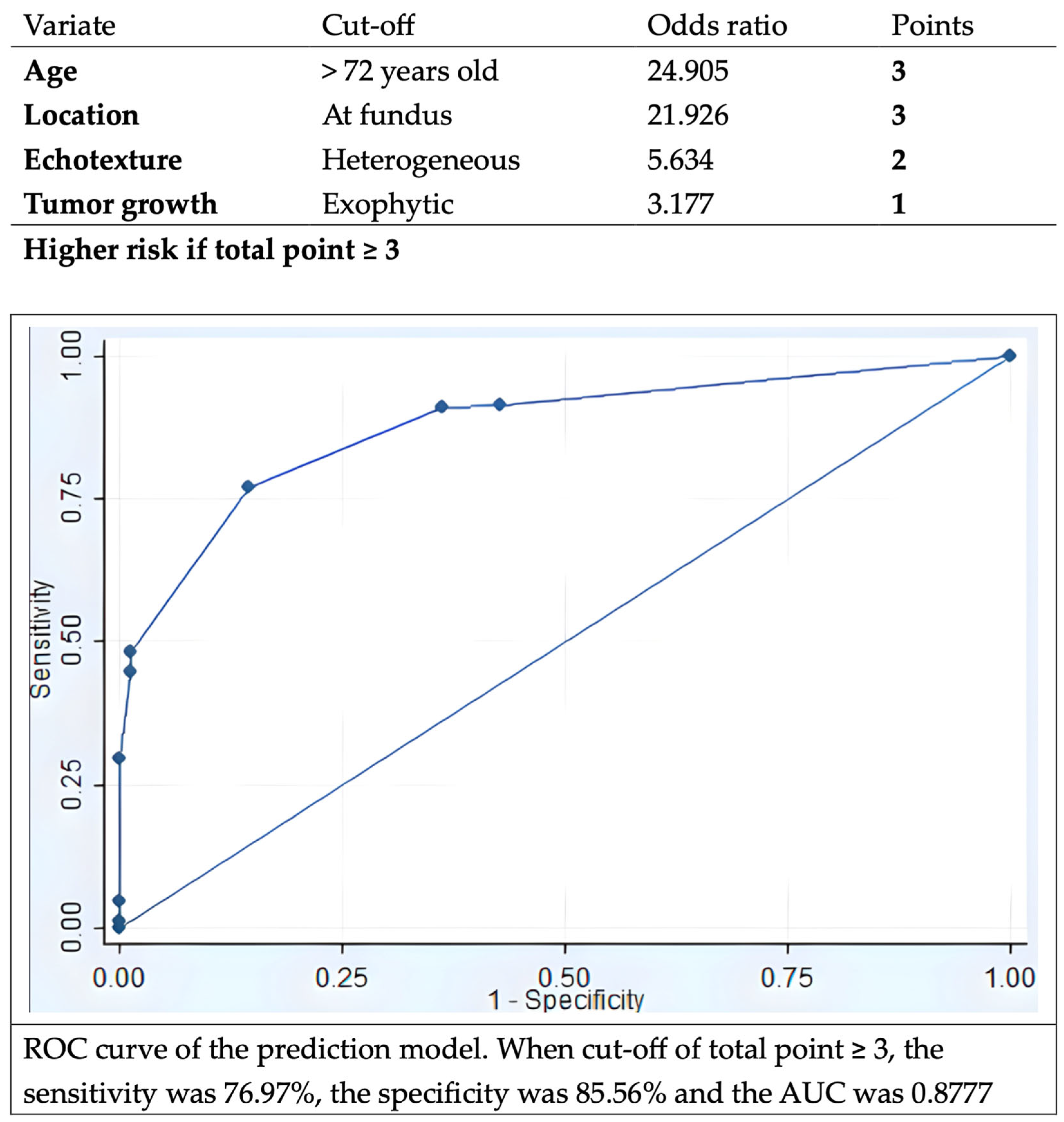
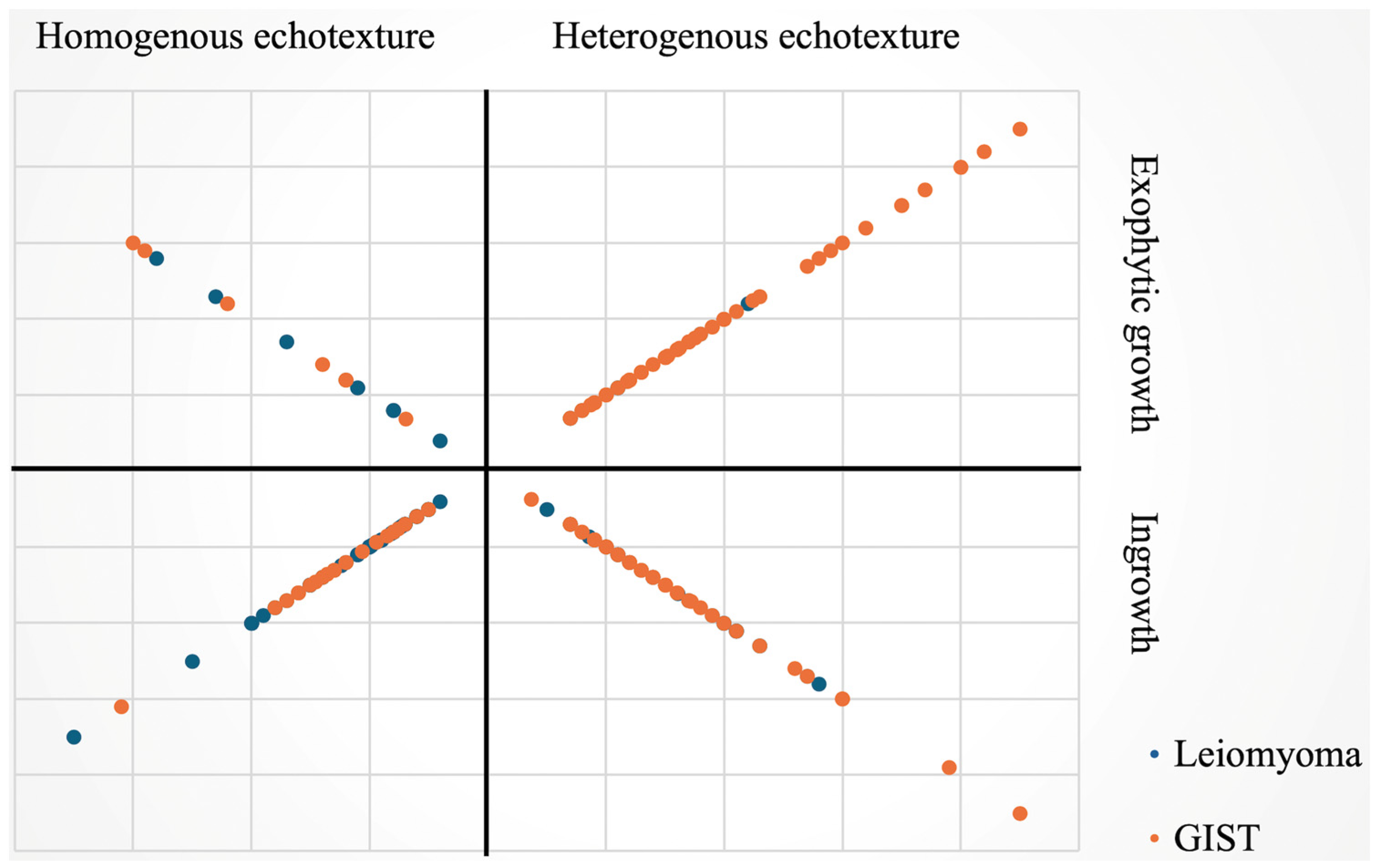
This retrospective study demonstrates ER being a valid and reliable strategy for the management of gastric SELs originating from MP layer, with high technical success and en bloc rates as well as low complication rate. In addition, EUS features, including heterogeneous echotexture and exophytic growth rather than the tumor size, could be used to predict malignant potential of gastric myogenic tumors. A model with factors of age, location at fundus, and EUS features aid in predicting malignant potential of gastric myogenic tumors.
The prevalence of GISTs is estimated to be 10 to 15 per 100,000 in the general population and counts for up to 3% for malignancies in the GI tract [
15]. The incidence of GISTs was reported to be as high as up to 2 per 100,000 in some studies [
16]. The natural course of gastric SELs is still poorly understood; yet, about 8.4% of the lesions were found with enlargement during serial endoscopy follow-up [
2]. Proportions with malignant potential within gastric SELs were reported in some studies, at around 23–34% [
17]. One cohort revealed that 35.1% small (i.e., less than 2 cm) gastric SELs were GISTs [
18]. In updated international guidelines, the malignant potential of GISTs was emphasized [
12,
19]. The World Health Organization (WHO) classified GIST as a malignant tumor, disregarding location, tumor size, and mitotic count [
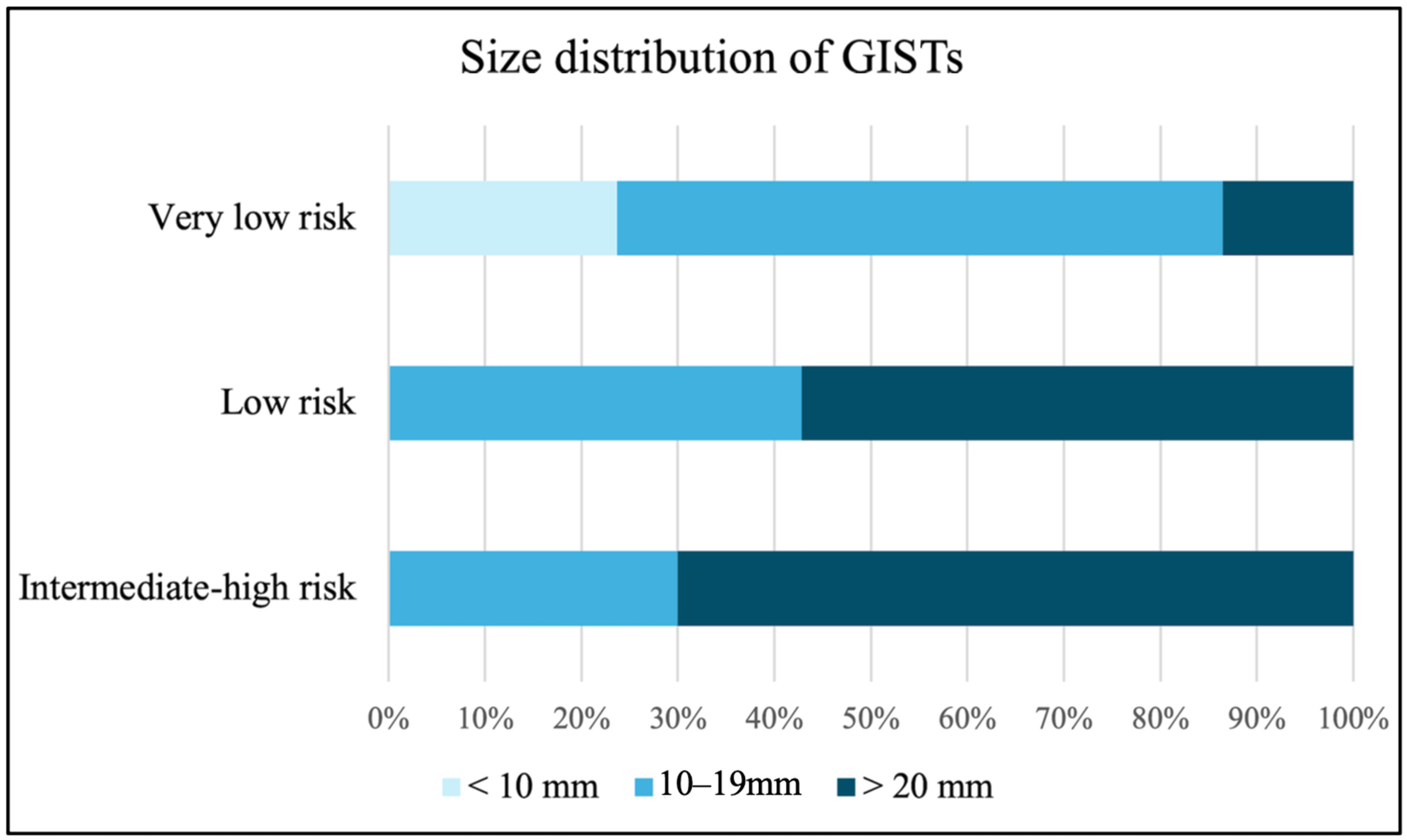
20]. Within our cases, almost half (45.8%) of gastric SELs originating from the MP layer are GISTs, even those of smaller sizes. Resection of all gastric myogenic tumors disregarding tumor size, particularly in those with high-risk EUS features, should be taken into consideration as the appropriate initial management.
Resection is recommended as standard treatment for GISTs larger than 20 mm [
4,
19]. Based on recent evidence, the ESGE guideline suggests that surveillance or resection are both acceptable in small (<20 mm) proven GISTs in the stomach [
13]. Nevertheless, ESMO guidelines suggest complete excision of all GISTs regardless of size, and ER could be considered for small tumors to minimize morbidity [
12]. The American College of Gastroenterology (ACG) and ESGE guidelines also describe ER as alternative therapies in gastric GISTs < 20 mm to avoid long-term surveillance [
4,
13]. More evidence supports ER in managing GISTs, due to shorter procedure times, better post operative recovery without increased of recurrence or complications compared with operation [
21]. Nevertheless, a higher proportion of R1 resection in ER compared to surgery was observed, although this phenomenon was not related to a higher recurrence rate [
22,
23]. In our study, the R0 resection rate is slightly lower than 90%, although there was no recurrence noted during follow-up. We believe that the electrocauterization artifacts contributed to the pathological findings of R1 resection. As a result, there was no recurrence after such an R1 resection rate. Another possible reason for low recurrence was that the predominance of lesions was for very low-risk GISTs in this study, which are characterized by slow growth rate. Complication rate for ER in GISTs was disclosed between 0% and 14.4% [
24,
25]. Pneumoperitoneum was described at about 10.6% in a large-scale study of EFTR [
25]. However, perforation may not be seen as a severe adverse event if successful closure is completed, especially in EFTR with intentional perforating gastric wall. Therefore, the true “complication” is hard to define. The complication rate in our cohort is 6.3% while inadvertent perforation is the most common one. About one-third of patients with perforation needed an additional operation while another two-thirds of patients underwent either intra-procedure needle decompression with successful endoscopic closure and conservative treatment with antibiotics. Two mortalities were recorded during follow-up but none of them were related to the procedure or the tumor.
Advancements in techniques of ER beyond the submucosal layer provide alternative minimally invasive treatment options for GISTs. EMD was found to have a complete resection rate of 96%, with some chance of perforation able to be managed by endoscopic techniques [
9]. ESSD was reported to have a similar complete resection, en bloc rate, and adverse event with EMD [
10,
26]. The efficacy and safety of STER and EFTR for the management of gastric myogenic tumors were also proven [
27]. Either exposed or non-exposed EFTR were found to have technical success rates close to 100% without major adverse events [
11]. One analysis comparing EFTR with STER showed earlier enteric feeding and shorter stay after STER, with a higher en bloc rate in EFTR [
28]. This may be due to the chance of breaching the tumor capsule when dissection in tunnel is accomplished by STER. Total complication rate was 6.5% in the analysis and there was no difference between these two procedures [
25]. Our study demonstrates high success and en bloc rate using variant types of ER procedures. Amount 24 procedures were classified as unintentional EFTR, nineteen of them were GISTs. This may be because of larger size and exophytic growth of GISTs. For the diagnostic value of image study, one recent study found that artificial intelligence-based models using CT features demonstrated good performance for prediction of risk stratification of GISTs [
29]. According to our analysis, the EUS evaluation, heterogeneous echotexture, and exophytic growth under EUS may indicate higher risk of SELs with malignancy potential. EUS features for prediction of GISTs were conducted in multiple studies. A cohort found heterogeneity and marginal halo signs were more observed in GISTs while irregular border as well as cystic change under EUS did not reach a significant difference [
30]. This is similar to our result that heterogeneous echotexture was more important than irregular border. Another study with 138 patients with larger (mean size 34 mm) SELs revealed that non-smooth border, blurring of the layers, presence of blood flow or special inner structure, hypoechoic and heterogeneous echotexture were related to histology of GISTs [
31]. As for correlation of EUS findings with risk stratification, one study suggested EUS features are not reliable for prediction and that only tumor size is related to mitotic count [
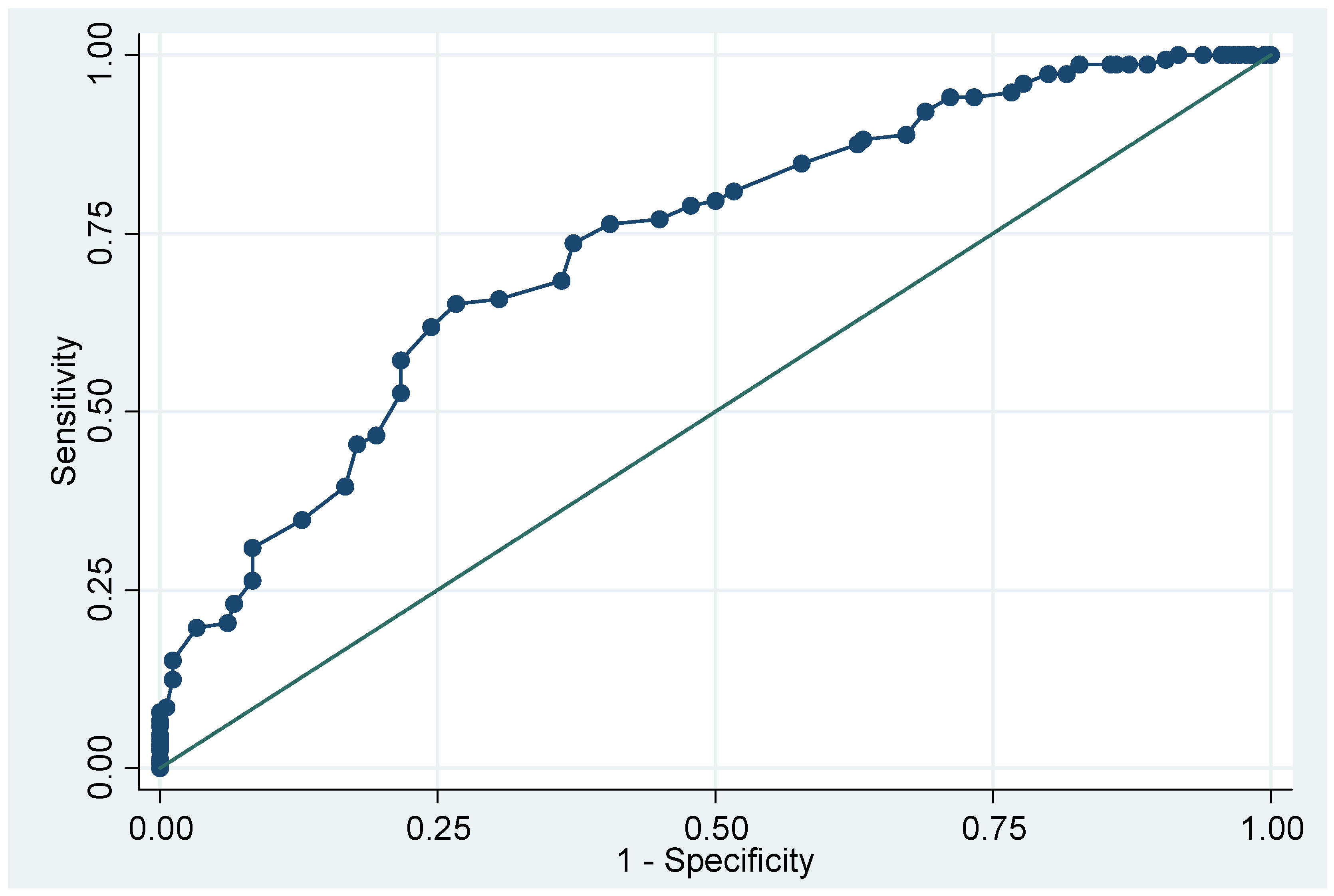
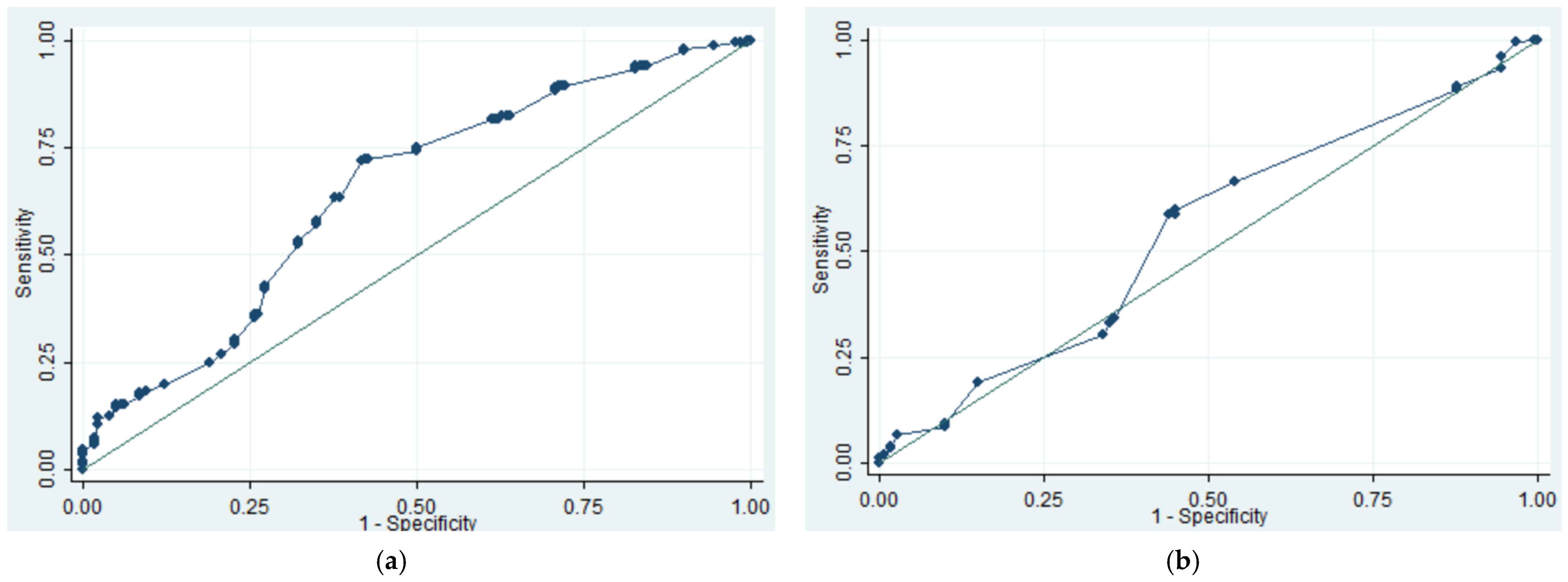
32]. The findings of our study were comparable to previous studies. Using ROC analysis, we found 15 mm for EUS size and 12 mm for endoscopic size were cut point for higher risk being SELs with malignant potential. Nevertheless, low AUC values were found in both groups. One study enrolled small SELs for suspected GIST and found that lesions of more than 9.5 mm had a significantly higher risk for tumor growth [
33]. The size necessary to predict malignant potential of gastric myogenic tumor is still debatable. One study developed a scoring system using CT and EUS features to differentiate GIST and schwannoma from leiomyoma, for which tumor location and uneven echogenicity were also suggested as key factors [
34]. As for prediction of GIST, one retrospective study used multiple variants of demographic data, and EUS features were used to distinguish GIST from leiomyoma, showing a high AUC [
6]. Compared to the prediction model we built, ours might be more simple to apply clinically. We apply only four significant factors (aged over 72, fundus location, heterogeneous echotexture, and exophytic growing), and do not include tumor size. This means that even small lesions could be evaluated by this model. Additionally, we simplified the points of each variant, making it easy to calculate the score. We believe that this prediction model may aid clinicians in deciding whether endoscopic resection should be considered directly or after failed multiple attempts of tissue acquisition, as well as predicting a higher risk of complications and inadvertently perforation, enabling the preparation of suitable closure devices in advance.
There are some limitations of this study. First, this is a multicenter, retrospective study. Therefore, heterogenicity in determination of endoscopic procedures, including resection methods as well as closure techniques may occur. Second, the EUS images are evaluated by endoscopists at each study institute rather than from central reading. Interobserver variations should be considered. Third, the prediction model was only internal validated at present. For clinical practice, endoscopists still need to evaluate the lesion, respectively, upon their characteristics, including the factors that were not mentioned in our prediction model. Furthermore, a future prospective study is therefore warranted to achieve external validation of the model. Lastly, there is wide range of follow-up periods of the patients. Recurrence in those patients with R1 resection of GISTs with lower risk may not be discernible during the study period.




 ,
,