Review

Synchronous Ileal Metastasis from Pancreatic Ductal Adenocarcinoma: Case Report and Narrative Review with Practical Diagnostic and Management Points

by
Tiberiu Stefăniță Țenea Cojan
1,
Valeriu Șurlin
2,
Stelian-Stefaniță Mogoantă
2,
Nicolae-Dragoș Mărgăritescu
2,
Daniel-Cosmin Caragea
3,
Ioana-Alexia Țenea Cojan
4,
Valentina Căluianu
1,
Marius Cristian Marinaș
5,
Gabriel Florin Răzvan Mogoș
1,*,
Liviu Vasile
2 and
Laurențiu Augustus Barbu
1
1
Department of Surgery, Railway Clinical Hospital Craiova, University of Medicine and Pharmacy of Craiova, 2 Petru Rares Street, 200349 Craiova, Romania
2
Department of Surgery, Emergency County Hospital, University of Medicine and Pharmacy of Craiova, 2 Petru Rares Street, 200349 Craiova, Romania
3
Department of Internal Medicine, University of Medicine and Pharmacy of Craiova, 200349 Craiova, Romania
4
Faculty of Medicine, University of Medicine and Pharmacy of Craiova, 200349 Craiova, Romania
5
Department of Obstetrics and Gynecology, Emergency County Hospital, University of Medicine and Pharmacy of Craiova, 2 Petru Rares Street, 200349 Craiova, Romania
*
Author to whom correspondence should be addressed.
Life 2025, 15(11), 1684; https://doi.org/10.3390/life15111684
Submission received: 7 October 2025 / Revised: 23 October 2025 / Accepted: 28 October 2025 / Published: 29 October 2025

Abstract

Background: Pancreatic ductal adenocarcinoma (PDAC) is an aggressive malignancy with poor prognosis, most frequently metastasizing to the liver, peritoneum, and lungs. Intestinal metastases are exceptionally rare and easily misinterpreted as primary small-bowel tumors, typically presenting with acute complications such as obstruction, perforation, or bleeding. Methods: We combined a detailed case description with a narrative literature review. PubMed/MEDLINE and Embase (2000–2025) were searched for case reports and case series describing intestinal metastases from PDAC with histopathological and immunohistochemical confirmation. Case presentation: We report a female patient presenting with acute intestinal obstruction caused by a synchronous ileal metastasis from PDAC. Imaging revealed an ileal stenosing lesion and a pancreatic body mass. An exploratory laparotomy identified a 3 cm transmural ileal tumor with additional serosal nodules. Histopathology confirmed a moderately differentiated adenocarcinoma. Immunohistochemistry supported pancreatic origin (CK7+, CA19-9+, faint CDX2), with mutant-type p53 positivity, ultra-low HER2/Neu expression, and a Ki-67 index of ~50%. The patient underwent segmental enterectomy with terminal ileostomy, followed by systemic therapy. Conclusions: This represents an exceptional and rare clinical finding rather than a presentation from which broad conclusions can be drawn. Histopathological and immunohistochemical analysis supported pancreatic origin and helped avoid misclassification as a primary intestinal neoplasm. It underscores the importance of careful clinicopathological correlation and multidisciplinary evaluation in atypical metastatic scenarios, while illustrating how surgery can provide symptom control and enable systemic therapy. Given its rarity, these observations should be interpreted with caution and regarded as descriptive rather than generalizable.

1. Introduction

Pancreatic ductal adenocarcinoma (PDAC) is a highly aggressive gastrointestinal malignancy, typically diagnosed at an advanced stage, with limited therapeutic options and poor survival outcomes. The 5-year survival rate remains below 10% overall and under 3% in metastatic disease. The liver, peritoneum, and lungs represent the most common metastatic sites, and systemic therapy remains the standard of care according to current ESMO and ASCO guidelines [1,2,3,4,5,6,7].
In contrast, small bowel metastases from PDAC are exceptionally rare, with only isolated reports in the literature [8,9,10,11]. When present, they often manifest through acute complications such as obstruction or bleeding, and synchronous presentations are particularly uncommon. We present a case of synchronous ileal metastasis causing acute small bowel obstruction, underscoring the diagnostic challenges of atypical metastatic patterns and the critical role of timely surgical palliation to enable systemic therapy.

2. Case Presentation

2.1. Patient Information

A female patient, previously diagnosed by imaging with a pancreatic tumor, was admitted to the Department of Surgery, Railway Clinical Hospital Craiova. Her medical history was significant for arterial hypertension and dyslipidemia. She presented with diffuse abdominal pain, absence of intestinal transit for feces and gas, and fecaloid vomiting with onset 2 days prior to admission.

2.2. Clinical Findings

On examination, the abdomen was mobile with respiratory movements, diffusely tender on palpation, distended in volume, and showed diffuse muscular guarding. Abdominal resonance was absent.

2.3. Timeline

A structured timeline of the patient’s clinical course, diagnostic workup, surgical intervention, and postoperative recovery is summarized below (Table 1).

2.4. Diagnostic Assessment

Abdominal radiography: No pneumoperitoneum; hydro-aerial levels in the mesogastrium.
Ultrasonography: Dilated intestinal loops with liquid content and hypermotility; no abdominopelvic free fluid.
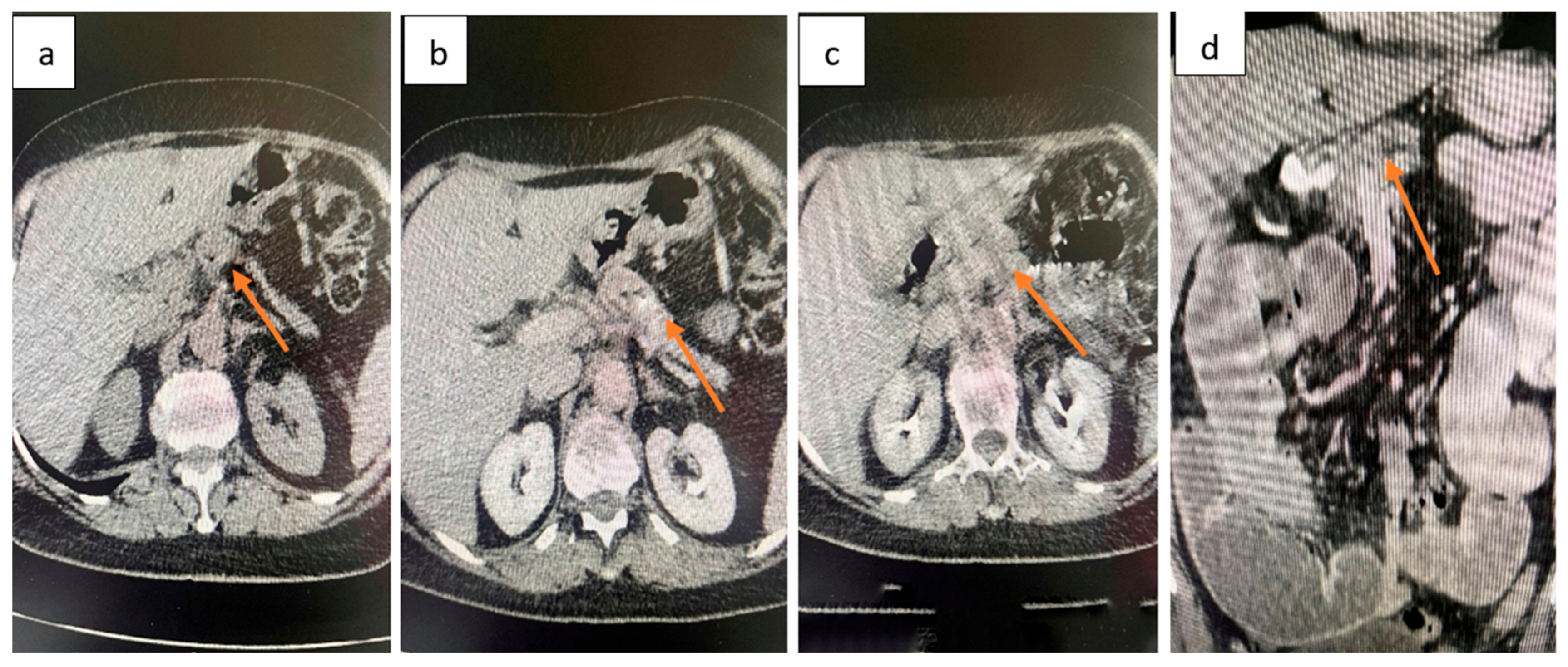
CT abdomen and pelvis: Focal, irregular parietal thickening with stenosing features located in the ileum, producing upstream intestinal distension with fluid content and hydro-aerial levels, without pneumoperitoneum (Figure 1). At the pancreatic body, a spontaneously isodense lesion with discrete iodophilia was identified, measuring 3.2 cm in axial diameter, without ductal dilatation (Figure 2). Given the acute obstructive presentation and the patient’s overall clinical status, EUS-FNB of the pancreatic mass was not performed preoperatively. This decision was agreed upon in a multidisciplinary tumor board, with the plan to reassess the lesion at a later stage for histological confirmation.
Laboratory investigations upon admission demonstrated leukocytosis with neutrophilia, indicating an acute inflammatory response. Coagulation studies were at the upper normal limit, while fibrinogen and ESR were markedly elevated, consistent with systemic inflammation. Renal function was preserved. Tumor markers showed a normal CEA but a mildly elevated CA 19-9, supporting possible pancreatic involvement (Table 2).

2.5. Therapeutic Intervention

Exploratory laparotomy revealed multiple dilated small bowel loops (8–10 cm in diameter, with thin walls). Approximately 50 cm proximal to the ileocecal valve, a 3 cm stenosing tumor extending beyond the serosa was identified. Two additional non-stenosing serosal nodules (1 cm and 1.5 cm, respectively) were also found upstream of the stenosis. In the corporeocaudal region of the pancreas, a firm, fixed mass measuring 5–5.5 cm was palpated, adherent posteriorly (Figure 3). The stomach and duodenum were markedly distended.
A segmental enterectomy was performed with terminal ileostomy in a “double-barrel” configuration.

2.6. Follow-Up and Outcomes

The postoperative course was favorable. The patient tolerated oral intake, had a functional ileostomy, and was discharged on postoperative day 10 in good general condition. She was subsequently referred to the oncology department and initiated systemic oncological treatment.

2.7. Histopathological Findings

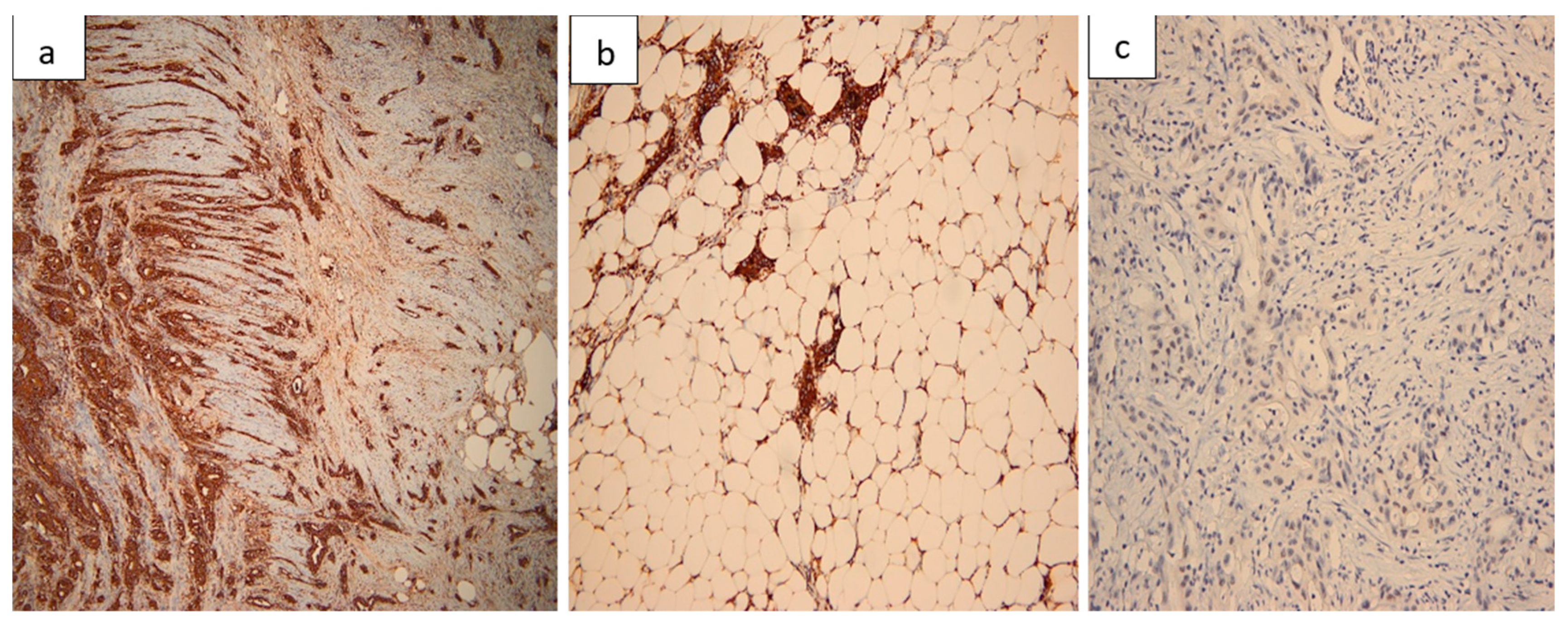
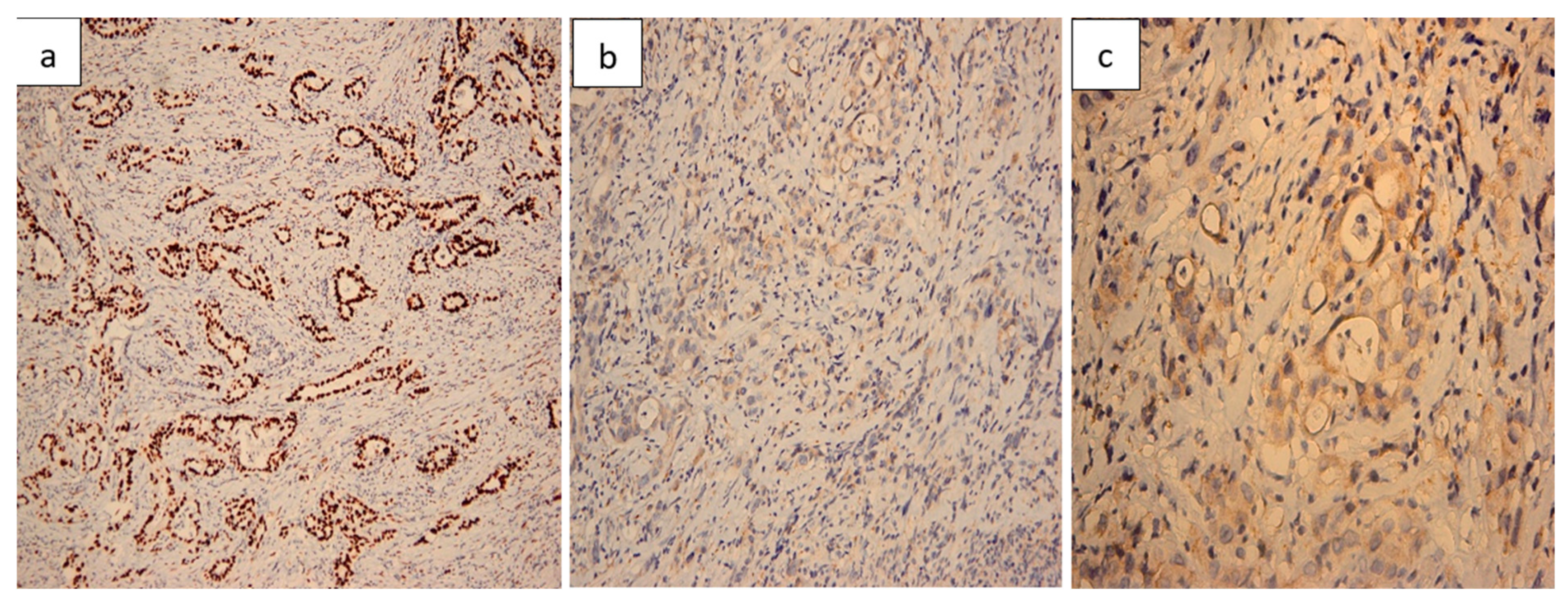
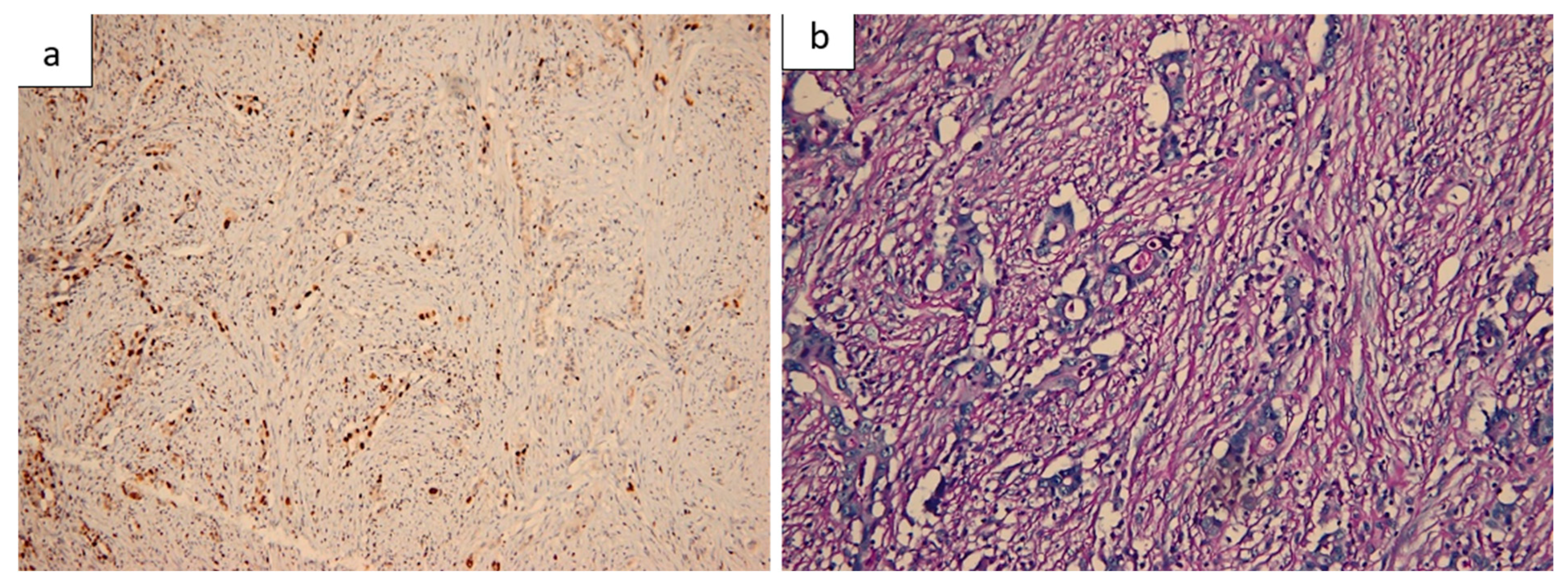
Histopathological evaluation of the resected small bowel segment revealed a moderately differentiated adenocarcinoma, measuring 3 cm, with transmural infiltration and extension beyond the serosa (Figure 4). Immunohistochemical staining demonstrated CK7 (Figure 5) and CA19-9 positivity, with only faint CDX2 expression (Figure 6), consistent with a pancreatic origin. Tumor cells exhibited strong and diffuse mutant-type p53 positivity (Figure 7a), ultra-low HER2/Neu expression (Figure 7b,c), and a Ki-67 proliferation index of approximately 50% (Figure 8a). Scant mucinous secretion in tubular lumina and within some tumor cells was highlighted by PAS staining (Figure 8b). MMR protein expression was preserved, indicating a low probability of high microsatellite instability (MSI-H). Taken together, these findings supported the diagnosis of intestinal metastasis from a moderately differentiated pancreatic adenocarcinoma. We expanded the IHC panel to differentiate from a primary colonic or ileal tumor (CK20, SATB2, MUC1, SMAD4, CK19). The resulting profile (CK7+, CA19-9+, CK20−, SATB2−, MUC1+, SMAD4 loss, very faint CDX2) supports a pancreatic origin; however, in the absence of a pancreatic mass biopsy, the conclusion remains qualified as probable.

3. Materials and Methods

3.1. Literature Search and Selection

A narrative literature review was performed in PubMed/MEDLINE and Embase databases, covering the period 2000–2025. The following search terms were applied: “pancreatic ductal adenocarcinoma” OR “PDAC” AND “intestinal metastasis” OR “small bowel metastasis” OR “colonic metastasis”. In addition, the reference lists of relevant articles were screened manually to identify further eligible studies.
Inclusion criteria were case reports or case series reporting histologically confirmed intestinal metastases from PDAC. Studies were excluded if they described direct contiguous invasion without evidence of metastatic spread, lacked immunohistochemical confirmation, or were published in languages other than English.

3.2. Limitations

The evidence base is restricted to isolated case reports, which are inherently prone to publication bias. Further heterogeneity results from variability in immunohistochemical panels and occasional diagnostic uncertainty in differentiating true metastases from direct invasion. Consequently, quantitative synthesis was not feasible, and the results were analyzed qualitatively.

4. Discussion

More than half of patients with pancreatic ductal adenocarcinoma (PDAC) present with metastatic disease at diagnosis, most frequently involving the liver, peritoneum, and lungs [1,2]. Reports of small-bowel metastases are limited to isolated case descriptions, and no reliable incidence data are available [8,9,10].

4.1. Epidemiology and Mechanisms of Spread

Pancreatic ductal adenocarcinoma (PDAC) is one of the most lethal gastrointestinal malignancies and currently the seventh leading cause of cancer-related death worldwide, projected to rank second by 2030 [1,2]. Despite therapeutic progress, the 5-year survival remains 6–10%, and below 3% in metastatic disease [1]. Over half of patients present with advanced disease, with the liver (60–70%), peritoneum (20–40%), and lungs (10–20%) as the predominant metastatic sites [2,3]. Gastrointestinal involvement is exceptionally rare and reported almost exclusively as isolated case reports, with colonic and small-bowel metastases documented by immunohistochemical confirmation of pancreatic origin [8,9,10].
Three main mechanisms are proposed to explain these rare metastatic patterns. Hematogenous dissemination may account for distant intestinal lesions via tumor thrombi in the portal or systemic circulation, often without peritoneal involvement [1,3,11]. Lymphatic spread through mesenteric or retroperitoneal pathways can lead to skip metastases in the absence of dominant hepatic disease [3,9]. Finally, peritoneal seeding through transcoelomic spread allows exfoliated tumor cells to implant on the bowel surface and invade the wall, particularly in carcinomatosis or ascites, explaining some synchronous obstructive presentations [10,11].

4.2. Correlation with Reported Cases

Published cases illustrate these mechanisms (Figure 9). Metachronous jejunal and colonic metastases occurring after pancreaticoduodenectomy presented with obstruction and fever, requiring emergency resection; the patient died within seven months, consistent with aggressive disease and hematogenous or lymphatic spread [9]. A jejunal metastasis diagnosed five years after curative resection, confirmed by IHC (CK7+, CK20−), was resected with favorable recovery, and the absence of peritoneal disease suggested hematogenous or lymphatic dissemination [8]. A synchronous sigmoid metastasis with pancreatic-type IHC (CK7+, CK20−) was reported as an unusual metastatic pattern, with hematogenous, lymphatic, and transcoelomic routes considered plausible [11].

4.3. Clinical Presentation and Diagnostic Work-Up

The clinical presentation of intestinal metastases from pancreatic ductal adenocarcinoma (PDAC) is nonspecific and often leads to delayed diagnosis. Typical symptoms include abdominal pain, nausea, vomiting, altered bowel habits, and signs of subocclusion or obstruction [9,10]. Occult gastrointestinal bleeding with anemia may be the initial manifestation [8], while hematemesis, melena, and iron-deficiency anemia can mimic other acute gastrointestinal conditions [12]. Obstructive symptoms may resemble mesenteric lymphatic cysts or appendiceal tumors [13,14]. Rarely, patients present with perforation and peritonitis requiring emergency surgery [10], and differential diagnoses should include colonic, rectal, or retroperitoneal pathologies [15]. Because of its rarity, intestinal involvement is often mistaken for a primary gastrointestinal tumor, underscoring the importance of histopathological confirmation.
The diagnosis is challenging and relies on appropriate imaging and histopathology. Contrast-enhanced CT is the first-line investigation to assess obstruction and metastatic spread [1], but sensitivity decreases for small or early lesions. Careful review of multiphasic CT is important in pancreatic tail primaries near the splenic hilum or flexure, where direct invasion may mimic metastasis. PET-CT can identify hypermetabolic bowel lesions when CT or endoscopy are inconclusive, as shown in a reported case of jejunal recurrence [8]. Endoscopy or capsule endoscopy may detect intraluminal lesions but have limited value for extramural disease. A definitive diagnosis requires histopathological and immunohistochemical confirmation of pancreatic origin [1].

4.4. Histopathological and Immunohistochemical Evidence

True small-bowel metastases from pancreatic ductal adenocarcinoma (PDAC) must be differentiated from direct invasion of adjacent loops and from primary intestinal adenocarcinoma. Immunohistochemistry (IHC) plays a key role in this distinction; a CK7+/CK20−/CDX2− profile supports pancreatic origin, while CK20+/CDX2+/SATB2+ suggests a colorectal primary [8,11]. Neuroendocrine tumors (NETs) of the small intestine, another important differential diagnosis, are typically chromogranin A and synaptophysin positive and may present with obstruction, anemia, or abdominal pain [16]. Primary intestinal lymphomas can also mimic these presentations, often debuting with obstruction or upper gastrointestinal symptoms [17].
Histologically, PDAC metastases usually involve the subserosa or submucosa with preservation of the mucosa. Deep biopsies are recommended because superficial samples can mimic a colorectal phenotype overlying a pancreatic metastasis. A practical diagnostic profile includes CK7+/CK20−/CDX2−, often MUC1+, with possible SMAD4 loss, whereas colorectal primaries are typically CK20+/CDX2+/SATB2+ and CK7−. Additional markers such as CK19 and CA19-9 may provide further support. Strong and diffuse p53 positivity, as in our case, is consistent with tumor progression and aids in distinguishing PDAC metastases [18].
Published reports describe identical IHC profiles in pancreatic primaries and intestinal lesions, most commonly diffuse CK7 positivity with CK20 negativity, supporting the diagnosis of metastatic spread rather than synchronous primaries [8,11]. This confirmation often guides therapy, favoring palliative surgery followed by systemic treatment. When feasible, pancreatic mass biopsy with IHC comparison (±KRAS/SMAD4) is recommended to exclude a synchronous luminal primary. If biopsy is not possible, extended IHC on the metastasis, correlated with morphology and imaging, helps reduce misclassification, though the conclusion remains probabilistic. Because aberrant marker expression may occur, final diagnosis should integrate clinical, radiologic, and pathological data, and when necessary, additional molecular testing.

4.5. Molecular and Therapeutic Considerations

Molecular profiling complements histopathology and immunohistochemistry in pancreatic ductal adenocarcinoma (PDAC) by identifying clinically relevant alterations. KRAS mutations are present in approximately 95% of cases, with the G12R variant—around 15%—showing distinct signaling that may confer relative sensitivity to MEK inhibition, though this remains investigational [19,20,21]. A subset of tumors harbor DNA damage repair alterations (BRCA1, BRCA2, PALB2), associated with platinum sensitivity and potential response to PARP inhibitors. Rare MSI-high tumors may benefit from immune checkpoint blockade [22,23]. For this reason, baseline next-generation sequencing is increasingly recommended in advanced PDAC to identify actionable targets, in line with ESMO, NCCN, and ASCO guidelines [6,24,25].
Accurate classification of intestinal lesions—distinguishing metastasis from a primary tumor or direct invasion—is crucial for therapeutic planning. Surgery or endoscopic procedures are typically reserved for complications such as obstruction or bleeding, or for carefully selected oligometastatic cases, while systemic therapy remains the standard of care [26]. Surgical intervention is most often used for acute complications, with reported procedures including segmental bowel resection, bypass, or stoma formation, resulting in short-term symptom relief [8,9,10]. Endoscopic palliation with self-expanding metal stents can decompress obstructive colonic lesions and serve as a bridge to systemic treatment. Chemotherapy regimens include FOLFIRINOX for fit patients and gemcitabine plus nab-paclitaxel for those with lower performance status, often initiated or resumed after palliation [2,27]. Given the aggressive course of PDAC, multidisciplinary decision-making is essential to balance surgical risks, expected benefits, and the feasibility of continuing systemic therapy.

4.6. Practical Management Algorithm for Intestinal Metastasis from PDAC

Step 1. Clinical suspicion
Patient with pancreatic adenocarcinoma presenting with abdominal pain, nausea, vomiting, subocclusion/obstruction, anemia, or GI bleeding [9,10].
Step 2. Diagnostic work-up
CT scan → first-line to assess site of obstruction and metastases [1].
PET-CT → for occult or equivocal lesions [8].
Histopathology + IHC (CK7+, CK19+, CA19-9+, CK20−, CDX2−) → confirm pancreatic origin [11].
Step 3. Emergency/urgent management
Endoscopic palliation (self-expanding metal stents) → in selected colonic obstruction.
Surgical resection (segmental bowel resection ± anastomosis or bypass) → for obstruction, perforation, bleeding [10,28].
Re-resection may be considered in oligometastatic disease with good performance status [8].
Step 4. Post-palliation systemic therapy
Resume or initiate systemic treatment:
FOLFIRINOX (fit patients).
Gemcitabine + nab-paclitaxel (more common in reported cases, tolerable) [2,27].
Step 5. Multidisciplinary follow-up
Regular monitoring with imaging + CA19-9.
Focus on symptom relief, nutritional support, and maintaining chemotherapy eligibility.

4.7. Prognosis and Clinical Lessons

The prognosis of pancreatic ductal adenocarcinoma (PDAC) remains poor, with a 5-year survival of 9–10% across all stages and less than 3% in metastatic disease [1,2]. Median survival in advanced or unresectable stages is short, underscoring the need for individualized multimodal strategies [1]. Outcomes of intestinal metastases are rarely documented; Miyasaka et al. reported death seven months after emergency resection for jejunal and colonic involvement [9], whereas Tseng et al. described favorable recovery and ongoing systemic therapy after re-resection of a metachronous jejunal metastasis nearly five years post-pancreatic surgery [8].
Prognosis is influenced by several factors. Tumor burden is critical—patients with oligometastatic disease may benefit from selective resection, while those with disseminated disease have a significantly worse outcome [8,9]. Performance status, reflected by ECOG score and suitability for systemic therapy (FOLFIRINOX or gemcitabine plus nab-paclitaxel), strongly impacts survival [1,8]. In selected patients, re-resection of intestinal metastases may offer symptom control and enable continuation of treatment [8].
Although long-term survival is uncommon, palliative surgery can provide rapid symptom relief, prevent life-threatening complications, and allow systemic therapy to continue. Miyasaka et al. reported good postoperative recovery with normalization of fever and inflammatory markers and hospital discharge [9]. Similarly, Meng et al. described initiation of gemcitabine plus nab-paclitaxel following IHC confirmation of pancreatic origin in a case of colonic metastasis [11].

4.8. Reported Cases in the Literature

A review of the literature (Table 3) highlights the extreme rarity of intestinal metastases from PDAC, with fewer than 15 well-documented cases over the last decade. Most cases were reported as single case reports, reflecting the anecdotal nature of the available evidence.
Most patients with intestinal metastases from pancreatic ductal adenocarcinoma (PDAC) present with acute complications such as obstruction, abdominal pain, or perforation, while fewer are diagnosed incidentally during colonoscopy or imaging. Rare metachronous cases may occur years after pancreatic resection. The jejunum, ileum, and colon (sigmoid, transverse, rectum) are most often affected, with exceptional cases involving colonization of preexisting colorectal carcinoma, suggesting atypical spread [8,9,10,11].
Management is mainly palliative, typically involving emergency or symptom-driven segmental colectomy or enterectomy, with endoscopic stenting used selectively in colonic obstruction. Systemic chemotherapy (gemcitabine plus nab-paclitaxel or FOLFIRINOX) is often initiated or resumed after palliation. Prognosis remains poor, though re-resection may benefit carefully selected oligometastatic patients. In most cases, surgery provides symptom relief, improved quality of life, and enables continuation of systemic therapy [8,9,10,11].

4.9. Key Messages

Key practice points in diagnosing and managing intestinal metastases from PDAC are summarized in Table 4.
  • Intestinal metastases from PDAC are exceptional events, often presenting with acute surgical emergencies.
  • Surgical resection provides palliative benefit and may enable systemic therapy in selected patients.
  • The role of systemic chemotherapy is crucial, but evidence remains anecdotal.
  • Multidisciplinary evaluation is essential, as no standardized guidelines exist.

4.10. Review Synthesis and Future Directions

Available evidence indicates that intestinal metastases from pancreatic ductal adenocarcinoma (PDAC) are exceptionally rare and usually present with obstruction, bleeding, or acute abdomen. Because clinical and imaging findings are nonspecific, histopathological and immunohistochemical confirmation is crucial. Although mainly palliative, surgery provides symptom relief and preserves eligibility for systemic therapy, the mainstay of treatment. Recognizing this entity helps avoid misdiagnosis and supports timely management.
Current knowledge relies on isolated case reports and small series, with no standardized guidelines. Future research should focus on registries to clarify incidence, risk factors, and outcomes, and on systematic reporting to refine diagnosis. Consensus recommendations are needed to standardize palliation, integrate systemic therapy, and optimize follow-up. This case adds to the limited literature by documenting a rare synchronous intestinal metastasis with acute obstruction and highlighting the value of such reports for future clinical practice.

5. Conclusions

Synchronous ileal metastasis from PDAC represents an exceptional and rare clinical finding, rather than a typical presentation from which general conclusions can be drawn. Histopathological and immunohistochemical analysis supported a pancreatic origin and helped avoid misclassification as a primary intestinal neoplasm. Surgical palliation provided symptom control and allowed systemic therapy to be initiated. As pancreatic mass biopsy was not performed in the acute setting, the diagnosis should be regarded as probable and interpreted cautiously, in conjunction with clinical, imaging, and IHC findings. This highlights the importance of multidisciplinary evaluation and careful, case-by-case decision-making in unusual metastatic scenarios, as well as the value of reporting rare cases to support future evidence-building rather than to establish clinical standards.

Author Contributions

Conceptualization L.A.B., D.-C.C. and N.-D.M.; writing—original draft L.A.B. and T.S.Ț.C.; resources V.C., L.V., S.-S.M., G.F.R.M., T.S.Ț.C. and V.Ș.; supervision M.C.M. and S.-S.M.; investigation M.C.M., V.C., L.V., G.F.R.M. and V.Ș.; writing—review M.C.M., L.A.B., T.S.Ț.C. and G.F.R.M.; validation D.-C.C., L.V., S.-S.M. and N.-D.M.; software D.-C.C.,V.C. and I.-A.Ț.C.; visualization V.C. and G.F.R.M.; data curation I.-A.Ț.C., T.S.Ț.C. and N.-D.M.; project administration L.A.B., D.-C.C. and V.Ș.; formal analysis M.C.M., I.-A.Ț.C. and N.-D.M.; methodology I.-A.Ț.C., L.V., S.-S.M. and V.Ș. All authors have read and agreed to the published version of the manuscript.

Funding

The article processing charges were funded by the University of Medicine and Pharmacy of Craiova, Romania.

Institutional Review Board Statement

The study was conducted in accordance with the Declaration of Helsinki and approved by the Ethics Committee of the Railway Clinical Hospital Craiova (approval no. 8457/24 September 2025).

Informed Consent Statement

Informed consent was obtained from the patient, whereby individual written informed consent was obtained, ensuring full compliance with ethical standards.

Data Availability Statement

The data presented in this study are available on request from the corresponding author. The data are not publicly available due to patient confidentiality.

Conflicts of Interest

The authors declare no conflicts of interest.

References

  1. Pavlidis, E.T.; Galanis, I.N.; Pavlidis, T.E. Updates in the diagnosis and management of ductal adenocarcinoma of the pancreas. World J. Clin. Oncol. 2025, 16, 105601. [Google Scholar] [CrossRef]
  2. Zhang, C.Y.; Liu, S.; Yang, M. Clinical diagnosis and management of pancreatic cancer: Markers, molecular mechanisms, and treatment options. World J. Gastroenterol. 2022, 28, 6827–6845. [Google Scholar] [CrossRef]
  3. Cannistrà, M.; Ruggiero, M.; Zullo, A.; Serafini, S.; Grande, R.; Nardo, B. Metastases of pancreatic adenocarcinoma: A systematic review of literature and a new functional concept. Int. J. Surg. 2015, 21 (Suppl. 1), S15–S21. [Google Scholar] [CrossRef] [PubMed]
  4. Conroy, T.; Pfeiffer, P.; Vilgrain, V.; Lamarca, A.; Seufferlein, T.; O’Reilly, E.M.; Hackert, T.; Golan, T.; Prager, G.; Haustermans, K.; et al. Pancreatic cancer: ESMO Clinical Practice Guideline for diagnosis, treatment and follow-up. Ann. Oncol. 2023, 34, 987–1002. [Google Scholar] [CrossRef]
  5. Springfeld, C.; Bailey, P.; Büchler, M.W.; Neoptolemos, J.P. ESMO 2023 pancreatic cancer guidelines signal stepwise progress. Hepatobiliary Surg. Nutr. 2024, 13, 362–365. [Google Scholar] [CrossRef]
  6. Sohal, D.P.S.; Kennedy, E.B.; Cinar, P.; Conroy, T.; Copur, M.S.; Crane, C.H.; Garrido-Laguna, I.; Lau, M.W.; Johnson, T.; Krishnamurthi, S.; et al. Metastatic Pancreatic Cancer: ASCO Guideline Update. J. Clin. Oncol. 2020, 38, 3217–3230. [Google Scholar] [CrossRef]
  7. Hayat, U.; Croce, P.S.; Saadeh, A.; Desai, K.; Appiah, J.; Khan, S.; Khan, Y.I.; Kumar, K.; Hanif, A. Current and Emerging Treatment Options for Pancreatic Cancer: A Comprehensive Review. J. Clin. Med. 2025, 14, 1129. [Google Scholar] [CrossRef] [PubMed]
  8. Tseng, C.-W.; Tseng, I.-T.; Wu, C.-C. Rare Case of Metachronous Small-Bowel Metastasis from Pancreatic Cancer Presenting as Anemia Approximately 5 Years Postpancreatoduodenectomy. J. Gastroenterol. Dig. Syst. 2024, 8, 1–6. [Google Scholar] [CrossRef]
  9. Miyasaka, M.; Noji, T.; Tanaka, K.; Nakanishi, Y.; Asano, T.; Ebihara, Y.; Kurashima, Y.; Nakamura, T.; Murakami, S.; Tsuchikawa, T.; et al. Oncological emergency surgery for metachronous large and small bowel metastases after pancreaticoduodenectomy for pancreatic cancer: A case report. Surg. Case Rep. 2018, 4, 99. [Google Scholar] [CrossRef]
  10. Fasano, M.; Della Corte, C.M.; Vicidomini, G.; Scotti, V.; Rambaldi, P.F.; Fiorelli, A.; Accardo, M.; De Vita, F.; Santini, M.; Ciardiello, F.; et al. Small bowel metastasis from pancreatic cancer in a long-term survival patient with synchronous advanced malignant pleural mesothelioma: A case report and literature review. Oncol. Lett. 2016, 12, 4505–4509. [Google Scholar] [CrossRef]
  11. Meng, N.; Han, P.; Liu, L.; Liu, J.; Liu, J. Colon Metastasis from Pancreatic Cancer: A Case Report. OncoTargets Ther. 2023, 16, 739–744. [Google Scholar] [CrossRef] [PubMed]
  12. Barbu, L.A.; Vasile, L.; Cercelaru, L.; Șurlin, V.; Mogoantă, S.-S.; Mogoș, G.F.R.; Țenea Cojan, T.S.; Mărgăritescu, N.-D.; Buliman, A. Non-Variceal Upper Gastrointestinal Bleeding: A Retrospective Cohort of 364 Cases, Historical Comparison, and Updated Management Algorithm. Life 2025, 15, 1320. [Google Scholar] [CrossRef]
  13. Vasile, L.; Barbu, L.A.; Mogoş, G.F.R.; Şurlin, V.; Vîlcea, I.D.; Cercelaru, L.; Mogoantă, S.Ş.; Mărgăritescu, N.D.; Nimigean, V.; Nimigean, V.R.; et al. Neuroendocrine tumors of the appendix: A comprehensive review of the literature and case presentation. Rom. J. Morphol. Embryol. 2025, 66, 269–278. [Google Scholar] [CrossRef] [PubMed]
  14. Barbu, L.A.; Cercelaru, L.; Vîlcea, I.-D.; Șurlin, V.; Mogoantă, S.-S.; Țenea Cojan, T.S.; Mărgăritescu, N.-D.; Popescu, M.; Mogoș, G.F.R.; Vasile, L. Coexistence of Acute Appendicitis and Mesenteric Cystic Lymphatic Malformation in an Adult: A Case Report and Narrative Review of Intraoperative Management Strategies. Life 2025, 15, 1390. [Google Scholar] [CrossRef]
  15. Barbu, L.A.; Vasile, L.; Cercelaru, L.; Vîlcea, I.-D.; Șurlin, V.; Mogoantă, S.-S.; Mogoș, G.F.R.; Țenea Cojan, T.S.; Mărgăritescu, N.-D. Fused Ischiorectal Phlegmon with Pre- and Retroperitoneal Extension: Case Report and Narrative Literature Review. J. Clin. Med. 2025, 14, 4959. [Google Scholar] [CrossRef] [PubMed]
  16. Calomino, N.; Fusario, D.; Cencini, E.; Lazzi, S. Two secondary localisation of non-Hodgkin’s lymphomas in the upper gastrointestinal tract. BMJ Case Rep. 2022, 15, e247607. [Google Scholar] [CrossRef]
  17. Waters, A.M.; Der, C.J. KRAS: The Critical Driver and Therapeutic Target for Pancreatic Cancer. Cold Spring Harb. Perspect. Med. 2018, 8, a031435. [Google Scholar] [CrossRef]
  18. Ardalan, B.; Azqueta, J.; Sleeman, D. Cobimetinib Plus Gemcitabine: An Active Combination in KRAS G12R-Mutated Pancreatic Ductal Adenocarcinoma Patients in Previously Treated and Failed Multiple Chemotherapies. J. Pancreat. Cancer 2021, 7, 65–70. [Google Scholar] [CrossRef]
  19. Mehdi, M.; Thalji, S.Z.; Shreenivas, A.; Chakrabarti, S.; Thomas, J.P.; Christians, K.K.; Evans, D.B.; Hall, W.A.; Erickson, B.; Thapa, B.; et al. MEK-Inhibitor (inh) and Hydroxychloroquine (HCQ) in KRAS-Mutated Advanced Pancreatic Ductal Adenocarcinoma (PDAC). J. Clin. Oncol. 2022, 40, e16260. [Google Scholar] [CrossRef]
  20. Golan, T.; Hammel, P.; Reni, M.; Van Cutsem, E.; Macarulla, T.; Hall, M.J.; Park, J.O.; Hochhauser, D.; Arnold, D.; Oh, D.Y.; et al. Maintenance Olaparib for Germline BRCA-Mutated Metastatic Pancreatic Cancer. N. Engl. J. Med. 2019, 381, 317–327. [Google Scholar] [CrossRef]
  21. Ghidini, M.; Lampis, A.; Mirchev, M.B.; Okuducu, A.F.; Ratti, M.; Valeri, N.; Hahne, J.C. Immune-Based Therapies and the Role of Microsatellite Instability in Pancreatic Cancer. Genes 2020, 12, 33. [Google Scholar] [CrossRef]
  22. Mosele, F.; Remon, J.; Mateo, J.; Westphalen, C.B.; Barlesi, F.; Lolkema, M.P.; Normanno, N.; Scarpa, A.; Robson, M.; Meric-Bernstam, F.; et al. Recommendations for the use of next-generation sequencing (NGS) for patients with metastatic cancers: A report from the ESMO Precision Medicine Working Group. Ann. Oncol. 2020, 31, 1491–1505. [Google Scholar] [CrossRef]
  23. Tempero, M.A.; Malafa, M.P.; Al-Hawary, M.; Behrman, S.W.; Benson, A.B.; Cardin, D.B.; Chiorean, E.G.; Chung, V.; Czito, B.; Del Chiaro, M.; et al. Pancreatic Adenocarcinoma, Version 2.2021, NCCN Clinical Practice Guidelines in Oncology. J. Natl. Compr. Cancer Netw. 2021, 19, 439–457. [Google Scholar] [CrossRef]
  24. O’Sullivan, B.; Burton, T.; Van Dalen, R.; Welsh, F.; Pandita, A.; Fischer, J. Beware the pancreatic incidentaloma in colorectal tumours: Pancreatic adenocarcinoma with metastases to the colon and rectum. J. Surg. Case Rep. 2022, 2022, rjab629. [Google Scholar] [CrossRef]
  25. Daamen, L.A.; Molenaar, I.Q.; Groot, V.P. Recent Advances and Future Challenges in Pancreatic Cancer Care: Early Detection, Liquid Biopsies, Precision Medicine and Artificial Intelligence. J. Clin. Med. 2023, 12, 7485. [Google Scholar] [CrossRef] [PubMed]
  26. Bugazia, D.; Al-Najjar, E.; Esmail, A.; Abdelrahim, S.; Abboud, K.; Abdelrahim, A.; Umoru, G.; Rayyan, H.A.; Abudayyeh, A.; Al Moustafa, A.E.; et al. Pancreatic ductal adenocarcinoma: The latest on diagnosis, molecular profiling, and systemic treatments. Front. Oncol. 2024, 14, 1386699. [Google Scholar] [CrossRef] [PubMed]
  27. Nakaya, T.; Oshiro, H.; Saito, T.; Sakuma, Y.; Horie, H.; Sata, N.; Tanaka, A. Metastasis of pancreatic cancer within primary colon cancer by overtaking the stromal microenvironment. Int. J. Clin. Exp. Pathol. 2018, 11, 3141–3146. [Google Scholar] [PubMed]
  28. Park, D.Y.; Krishnamurthi, S.; Chahal, P.; Downs-Kelly, E.; Morris-Stiff, G. Pancreatic metastases to the colon: An unusual cause of colonic obstruction. BMJ Case Rep. 2019, 12, e228578. [Google Scholar] [CrossRef]
  29. Kahl, R.; George, K.; Patel, K.; Stawick, L. Pancreatic Adenocarcinoma with Rare Sigmoid Colon Metastasis. ACG Case Rep. J. 2019, 6, e00132. [Google Scholar] [CrossRef]
  30. Ardalan, B.; Azqueta, J.; England, J.; Hartmann, R. Pancreatic cancer presenting as bowel obstruction and role of next generation sequencing: A case report. Int. J. Surg. Case Rep. 2022, 90, 106654. [Google Scholar] [CrossRef]
  31. Pacheco, F.; Luciano, E.; Hebert, D.; Marar, O. Metastatic pancreatic adenocarcinoma presenting as large bowel obstruction: A case report. Int. J. Surg. Case Rep. 2023, 102, 107801. [Google Scholar] [CrossRef]
  32. Casolino, R.; Biankin, A.V. Treatment of pancreatic cancer in 2022. Camb. Prism. Precis. Med. 2023, 1, e14. [Google Scholar] [CrossRef]
  33. Netto, D.; Frizziero, M.; Foy, V.; McNamara, M.G.; Backen, A.; Hubner, R.A. Systemic Therapy for Metastatic Pancreatic Cancer—Current Landscape and Future Directions. Curr. Oncol. 2024, 31, 5206–5223. [Google Scholar] [CrossRef]
  34. Stoop, T.F.; Javed, A.A.; Oba, A.; Koerkamp, B.G.; Seufferlein, T.; Wilmink, J.W.; Besselink, M.G. Pancreatic cancer. Lancet 2025, 405, 1182–1202. [Google Scholar] [CrossRef] [PubMed]
  35. Wainberg, Z.A.; Melisi, D.; Macarulla, T.; Pazo Cid, R.; Chandana, S.R.; De La Fouchardière, C.; Dean, A.; Kiss, I.; Lee, W.J.; Goetze, T.O.; et al. NALIRIFOX versus nab-paclitaxel and gemcitabine in treatment-naive patients with metastatic pancreatic ductal adenocarcinoma (NAPOLI 3): A randomised, open-label, phase 3 trial. Lancet 2023, 402, 1272–1281. [Google Scholar] [CrossRef]
  36. Miyabayashi, K.; Nakagawa, H.; Koike, K. Molecular and Phenotypic Profiling for Precision Medicine in Pancreatic Cancer: Current Advances and Future Perspectives. Front. Oncol. 2021, 11, 682872. [Google Scholar] [CrossRef] [PubMed]
  37. Ratner, E.S.; Sartorelli, A.C.; Lin, Z.P. Poly (ADP-ribose) polymerase inhibitors: On the horizon of tailored and personalized therapies for epithelial ovarian cancer. Curr. Opin. Oncol. 2012, 24, 564–571. [Google Scholar] [CrossRef]
  38. Wang, H.; Li, Z.W.; Ou, Q.; Wu, X.; Nagasaka, M.; Shao, Y.; Ou, S.I.; Yang, Y. NTRK fusion positive colorectal cancer is a unique subset of CRC with high TMB and microsatellite instability. Cancer Med. 2022, 11, 2541–2549. [Google Scholar] [CrossRef] [PubMed]
  39. Joshi, D.C.; Sharma, A.; Prasad, S.; Singh, K.; Kumar, M.; Sherawat, K.; Tuli, H.S.; Gupta, M. Novel therapeutic agents in clinical trials: Emerging approaches in cancer therapy. Discov. Oncol. 2024, 15, 342. [Google Scholar] [CrossRef]
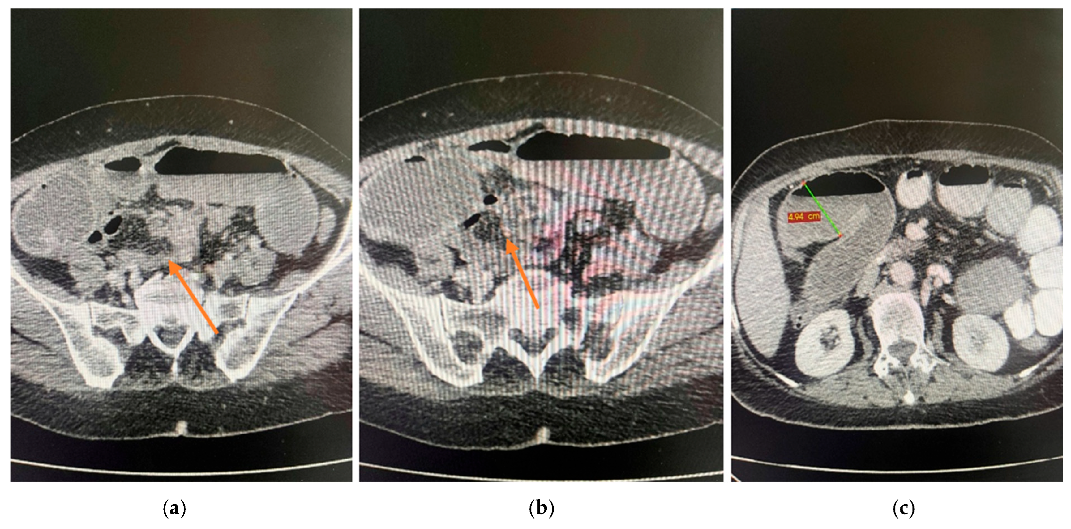
Figure 1. CT examination of the ileum. (a) Focal irregular parietal thickening (orange arrow). (b) Stenosing lesion (orange arrow). (c) Upstream intestinal dilatation (green measurement line).
Life 15 01684 g001
Figure 2. Contrast-enhanced abdominal CT. At the pancreatic body, a spontaneously isodense lesion with discrete iodophilia was identified, measuring 3.2 cm in axial diameter, without ductal dilatation (orange arrow). (a) Non-contrast phase. (b) Arterial phase. (c) Venous phase. (d) Delayed phase.
Life 15 01684 g002
Figure 3. Intraoperative findings. (a) Stenosing ileal tumor (black arrow) with markedly dilated small bowel loops upstream and collapsed loops downstream. (b) Serosal tumor measuring 1.5 cm (black arrow). (c) Serosal tumor of 1 cm (black arrow) associated with intestinal lymphadenopathy (blue arrow).
Life 15 01684 g003
Figure 4. Moderately differentiated pancreatic adenocarcinoma. (a) Tumoral ducts with cuboidal cells, polymorphous nuclei, and frequent mitoses (H&E, 200×). (b) Cribriform structures and solid nests with desmoplastic stroma (H&E, 200×). (c) High-power view showing marked atypia and numerous mitotic figures (H&E, 400×).
Life 15 01684 g004
Figure 5. CK7 immunostaining. (a) Tubular pattern with complex architecture and isolated tumor cells at the invasion front (40×). (b) CK7 positivity highlighting the architectural complexity (100×). (c) Peripancreatic fat invasion on CK7 stain (100×).
Life 15 01684 g005
Figure 6. CA19-9/CDX2 immunoprofile. (a) CA19-9 positivity showing radial invasion in the muscularis propria of the duodenum (40×). (b) Peripancreatic fat invasion highlighted on CA19-9 stain (100×). (c) Very faint nuclear positivity for CDX2 in tumor cells (100×).
Life 15 01684 g006
Figure 7. p53 and HER2/Neu immunoprofile. (a) Strong and diffuse mutant-type p53 positivity in tumor cells, with lighter wild-type staining in stromal cells (100×). (b) HER2/Neu ultra-low expression, with very faint membranous staining (20×). (c) Faint, discontinuous HER2/Neu immunostaining in scattered tumor cells (“hot spot”) (40×).
Life 15 01684 g007
Figure 8. Ki-67 and PAS staining. (a) Ki-67 immunostaining showing a proliferation index of ~50% in tumor cells (100×). (b) Scant mucinous secretion in tubular lumina and within some tumor cells, highlighted by PAS staining (200×).
Life 15 01684 g008
Figure 9. Proposed mechanisms of intestinal metastasis in PDAC. Legend: Three main routes have been proposed—(1) hematogenous dissemination to distant bowel, (2) lymphatic spread via mesenteric/retroperitoneal channels, and (3) peritoneal seeding with secondary bowel wall invasion [1,3,10].
Life 15 01684 g009
Table 1. Clinical Timeline of Patient Presentation and Management.
DayClinical Event
Day 0Onset of diffuse abdominal pain, fecaloid vomiting, and absence of intestinal transit.
Day 2Admission to hospital; clinical and imaging workup performed.
Day 2 (evening)Decision for surgical intervention; exploratory laparotomy performed.
Postoperative Days 1–3Gradual clinical improvement; ileostomy functional; oral intake initiated.
Postoperative Days 4–9Continued recovery without complications.
Postoperative Day 10Discharged home in good general condition.
Table 2. Laboratory tests upon admission.
ParameterResultNormal Range
White blood cell count13.40 × 103/μL4–10 × 103/μL
Neutrophil proportion77.9%40–70%
Hemoglobin13.4 g/dL12–16 g/dL
Platelet count304 × 103/μL150–450 × 103/μL
Creatinine0.7 mg/dL0.6–1.3 mg/dL
Urea28.37 mg/dL15–45 mg/dL
INR1.210.8–1.2
Prothrombin time14.3 s11–15 s
Fibrinogen1115 mg/dL200–400 mg/dL
C-reactive protein<0.5 mg/dL
ESR55 mm/h<20 mm/h
CEA1.23 ng/mL<5 ng/mL
CA 19-953.11 U/mL<37 U/mL
Table 3. Reported cases of intestinal metastases from pancreatic ductal adenocarcinoma (PDAC).
Author/YearPatient (Age/Sex)Interval from Primary PDACSite of MetastasisPresentationManagementOutcome
Fasano [10]Long-term survivor (M)SynchronousSmall bowelAcute abdomenEmergency small bowel resectionFirst described intestinal metastasis in long-term PDAC survivor with mesothelioma
Miyasaka [9]63/M3 months post-pancreaticoduodenectomyJejunum + colonAbdominal pain, fever, diarrheaEmergency resection (jejunum + colon)Death at 7 months due to relapse
Nakaya [27]72/MSynchronousColon (within preexisting carcinoma)Bowel obstructionColectomyRare phenomenon of PDAC metastasis colonizing colon carcinoma
Park [28]73/FSynchronousTransverse colonLarge bowel obstructionSegmental colectomyPalliative survival, <1 year
Kahl [29]91/FSynchronousSigmoid colonLarge bowel obstructionSigmoid colectomyDied shortly after surgery
Meng [11]65/MConcomitantSigmoid colonColon lesion detected on work-upDiagnosis confirmed by IHC; chemotherapyExtremely rare metastatic route
Ardalan [30]66/FSynchronousSigmoid colonAcute obstructionEmergency left hemicolectomy + IHC; FOLFIRINOXPoor tolerance, palliative course
O’Sullivan [24]Cases (2)SynchronousRectum/colonObstruction, GI symptomsSurgical resection/supportiveReported as very rare synchronous cases
Tseng [8]56/F~5 years post-pancreaticoduodenectomyJejunumAnemia (Hb 8–9 g/dL), occult bleedingSegmental jejunal resectionUneventful recovery; continued systemic therapy
Pacheco [31]68/MMetachronous (interval not specified)Sigmoid colonSymptomatic metastasis, bowel obstructionSurgical resectionReported as the third symptomatic colonic metastasis from PDAC
Present case (2025)FConcomitantIleumAcute intestinal obstructionEmergency ileal resection + anastomosisFavorable recovery; referred to oncology.
Table 4. Lessons learned/Practice points in diagnosing and managing intestinal metastases from PDAC.
Practice PointRationale
When technically feasible, target submucosaSuperficial mucosal biopsies may mimic a colorectal phenotype; submucosal sampling can improve diagnostic accuracy, but should not be considered mandatory
Always run CK7/CK20/CDX2/SATB2 ± MUC1/SMAD4Distinguishes PDAC metastasis (CK7+/CDX2−/SATB2−) from primary intestinal adenocarcinoma.
Re-discuss at MDT before major surgeryAvoids inappropriate CRC-style resections and aligns care with systemic PDAC therapy. Avoiding inappropriate surgery in such rare scenarios is not based on a standardized algorithm, but rather on individualized, case-by-case multidisciplinary decision-making, aimed at aligning management with the underlying pancreatic primary.
Note: Adapted from published case reports and small series on intestinal metastases from PDAC [8,9,10,11,32,33,34,35,36,37,38,39].
Disclaimer/Publisher’s Note: The statements, opinions and data contained in all publications are solely those of the individual author(s) and contributor(s) and not of MDPI and/or the editor(s). MDPI and/or the editor(s) disclaim responsibility for any injury to people or property resulting from any ideas, methods, instructions or products referred to in the content.