Communication

Dementia Is Induced via the AGEs/Iba1/iNOS Pathway in Aged KK-Ay/Tajcl Mice

Department of Pharmaceutical Sciences, Suzuka University of Medical Science, Suzuka 513-8670, Japan
*
Author to whom correspondence should be addressed.
Life 2023, 13(7), 1540; https://doi.org/10.3390/life13071540
Submission received: 14 June 2023 / Revised: 7 July 2023 / Accepted: 10 July 2023 / Published: 11 July 2023
(This article belongs to the Special Issue Feature Papers in Animal Science: 2nd Edition)

Abstract

:
The onset and exacerbation of dementia have been observed in elderly patients with type 2 diabetes. However, the underlying mechanism remains unclear. In this study, we investigated the effects of aging on the cognitive function in a mouse model of type 2 diabetes. Pathogen-free KK-Ay/TaJcl mice were used in this study. The cognitive abilities and memory declined in the mice and worsened in the 50-week-olds. The levels of advanced glycation end products (AGEs), receptor for AGE (RAGE), and Iba1 in the hippocampus were increased in the mice compared to those in the control mice. Hippocampal levels of CC-chemokine receptor 7 and inducible nitric oxide synthase, which are from M1-type macrophages that shift from microglia, were higher in KK-Ay/TaJcl mice than in control mice. Tumor necrosis factor (TNF)-α and nitric oxide (NO) levels secreted by M1-type macrophages were similarly elevated in the mice and were even higher at the age of 50 weeks. NO levels were markedly elevated in the 50-week-old mice. In contrast, differentiation of CD163 and arginase-1 did not change in both mouse types. Memory and learning declined with age in diabetic mice, and the AGEs/RAGE/M1-type macrophage/NO and TNF-α pathways played an important role in exacerbating memory and learning in those mice.

1. Introduction

Aging is progressing in modern times, and the number of patients with lifestyle-related diseases is increasing. Lifestyle-related diseases are also involved in dementia and are deeply involved not only in vascular dementia (VD), but also in the development and progression of Alzheimer’s disease (AD). Among the lifestyle-related diseases occurring in middle-aged individuals, hypertension, diabetes, dyslipidemia, and metabolic syndrome increase the risk of developing AD and promote pathological degenerative processes [1].
Notably, diabetes increases the incidence of dementia in the elderly [2]. Elderly patients with diabetes have a higher incidence of VD than chronic vascular lesions, and recent epidemiological studies have reported that the risk of AD complications is also high [3,4,5]. The mechanisms by which diabetes modifies cognitive function can be divided into vascular and metabolic factors and could be reversible and irreversible [6]. AD-type dementia is related to metabolic factors and accumulates in the brain, causing neuropathy. Insulin-degrading enzymes break down amyloids and prevent their accumulation in the brain. When diabetes causes insulin resistance, a large amount of insulin is required (hyperinsulinemia); therefore, a large number of insulin-degrading enzymes are used. Consequently, brain amyloids cannot decompose and accumulate [7]. In addition, choline acetyltransferase activity is decreased in patients with AD, and cholinergic neuropathy is thought to occur, affecting memory and learning abilities [8]. Recent reports have shown that the receptor for advanced glycation end product (RAGE) is abundant in brain microglia and astrocytes of patients with AD, caused by inflammation and apoptosis, resulting from AGEs/RAGE [9,10,11]. However, the details of the onset mechanism of AD in the diabetic elderly are not known.
In this study, we used KK-Ay/TaJcl mice, a type 2 diabetes mouse model, for 50 weeks to investigate the mechanism of AD in elderly patients with diabetes. We then focused on microglia in the hippocampus of the mouse brain and examined the M1 and M2 shifts in macrophages.

2. Materials and Methods

2.1. Animal Experiments

Specific pathogen-free (SPF) 7- or 50-week-old C57BL/6N and KK-Ay/TaJcl male mice were obtained from CLEA Japan Inc. (Tokyo, Japan). The mice were separately kept in cages in specific pathogen-free conditions at 23 ± 1 °C, with a 12 h light:12 h darkness schedule. The mice were housed with unlimited access to drinking water and a pelleted basal diet. On the final day of the study, the body weights were measured. Blood glucose levels were measured from the tail vein of each mouse. We defined diabetic as more than a 300 mg/dL blood glucose level. In order to eliminate factors affecting the experiment, each animal was kept under constant conditions, such as free access to the same food, minimization of stress load, and freedom of movement within the cage. This work was conducted in accordance with the recommendations of the Guide for the Care and Use of Laboratory Animals of Suzuka University of Medical Science (approval number: 34). All dissections were performed under pentobarbital anesthesia. All efforts were made to minimize mouse suffering.

2.2. Open Field Test

The 50 × 50 × 40 cm3 open field area consisted of plastic. The mice’s distance moved (cm) was measured by a video tracking system (Smart2, Bio Research Center, Nagoya, Japan) during 15 min and was used as motor activity. The data obtained were graphed.

2.3. Step-Through Passive Avoidance Test

This test was used to evaluate non-spatial long-term memory following a previously described method [12]. The experimental apparatus (Bio Research Center, Nagoya, Aichi, Japan) consisted of two compartments: a light compartment and a dark compartment separated by a grid door. A stainless-steel shock grid floor was presented in the dark compartment. During the acquisition trial, the mouse was placed in the light compartment. After 60 s, the grid door between the compartments was opened. The step-through latency for animals to enter the dark compartment was measured, and the door was closed. As soon as the animals entered the dark compartment, there was an inescapable foot-shock (0.5 mA for 3 s). The retention test was performed 24 h after the training trial in a similar manner without the electric shock, and the step-through latency to the dark compartment was recorded. The maximal cutoff time for step-through latency was 600 s.

2.4. Measurement of AGEs, TNF-α, RAGE, Iba1, CCR7, iNOS, NO, CD163, and Arginase-1 in the Hippocampus

At the end of the study, the brains were harvested. Following this, the hippocampus was rapidly dissected, excised, and homogenized in lysis buffer (Kurabo, Osaka, Japan), and the lysate was centrifuged at 10,000 rpm. ELISA kits were used to measure the hippocampus concentrations of AGEs, tumor necrosis factor (TNF)-α, RAGE (MyBioSource, San Diego, CA, USA), cluster of differentiation (CD) 163 (Elabscience, Houston, TX, USA), ionized calcium binding adapter molecule 1 (Iba1: South San Francisco, CA, USA), CC-chemokine receptor 7 (CCR7: abbexa, Cambridge, UK), inducible nitric oxide synthase (iNOS), and arginase-1 (CUSABIO, Houston, TX, USA). NO was assayed using assay kits (NO3+NO2 colorimetric assay; Dojindo, Kumamoto, Japan) according to the manufacturer’s instructions. The microplate reader (Molecular Devices, Sunnyvale, CA, USA) was used for the measurement of optical density.

2.5. Statistical Analysis

The results are expressed as the means ± the standard deviation, and the data were analyzed using one-way ANOVA followed by Tukey’s post hoc test or the Steel–Dwass test was applied. The statistical significance level was set at p < 0.05 and 0.01.

3. Results

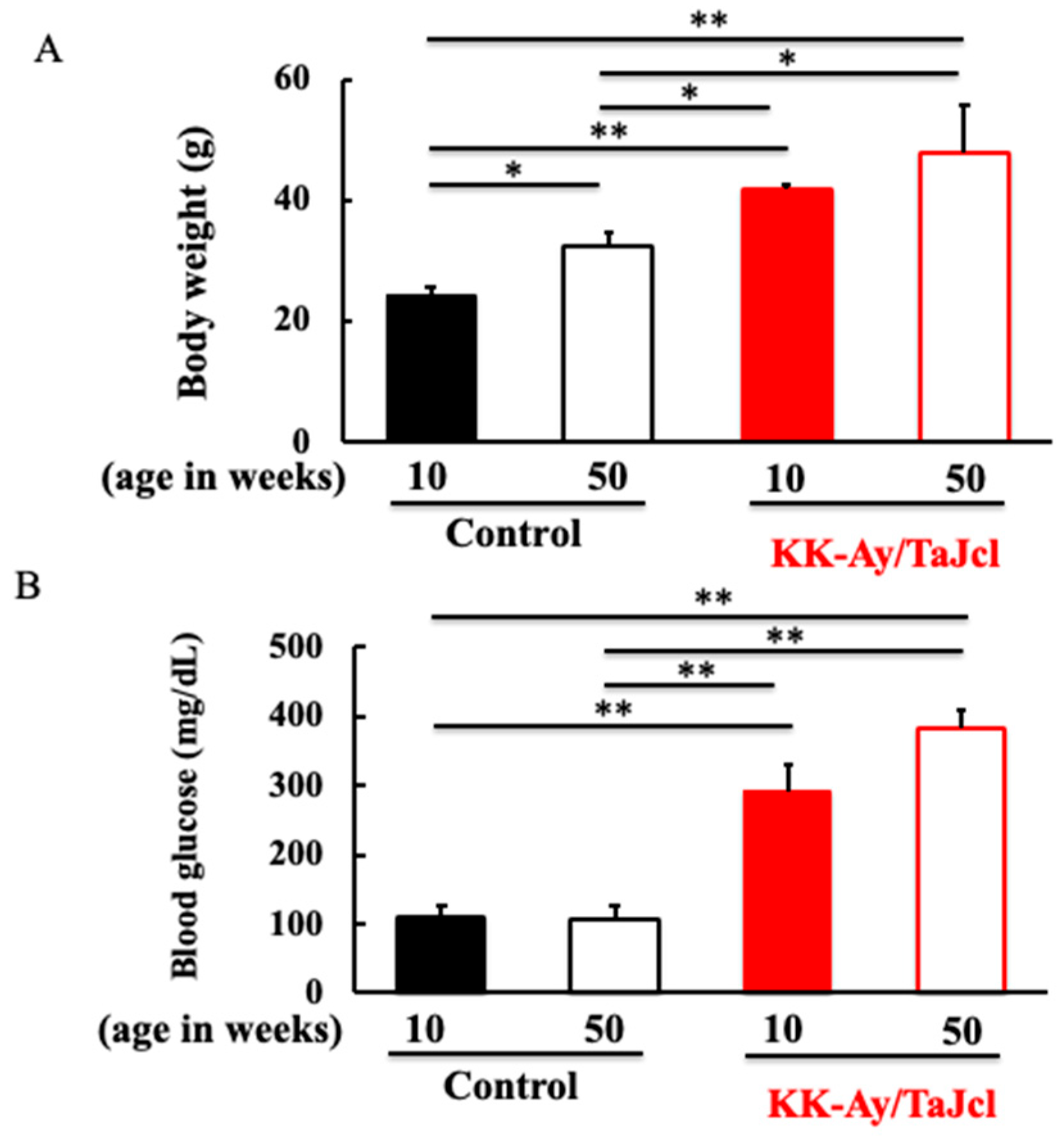
3.1. Effect of Aging on Body Weight and Blood Glucose Level in KK-Ay/Tajcl Mice

The body weight was higher in KK-Ay/Tajcl mice than in the controls. The body weight of the control mice increased with age, but that of the KK-Ay/Tajcl mice did not differ between 10 and 50 weeks of age (Figure 1A). The blood glucose levels were higher in the KK-Ay/Tajcl mice than in the control mice. In the control and KK-Ay/Tajcl mice, the blood glucose levels did not change with age; however, in the KK-Ay/Tajcl mice, the 50-week-old mice showed higher blood glucose levels than the 10-week-old mice (Figure 1B).

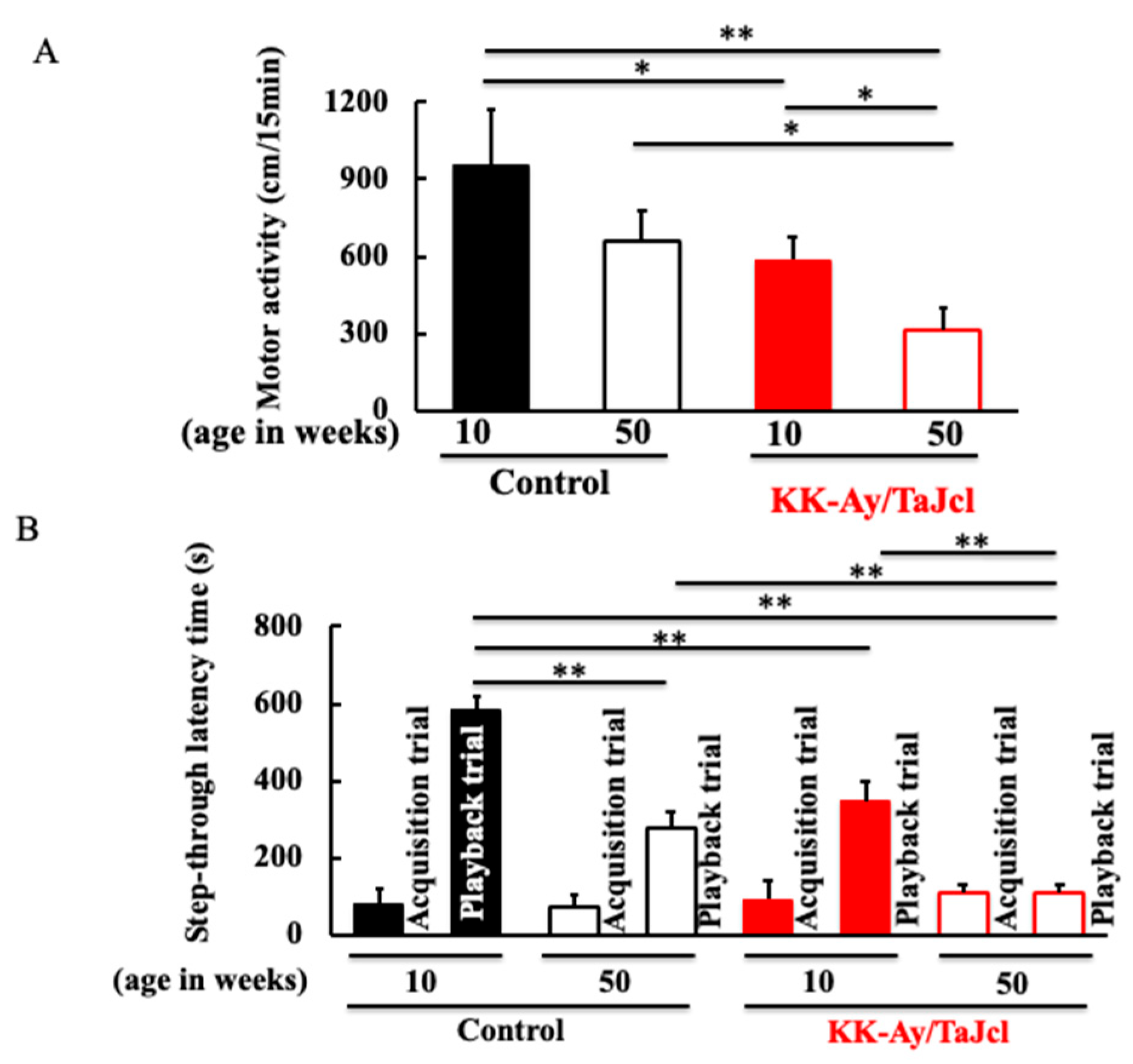
3.2. Behavioral Effects on Aging in KK-Ay/TaJcl Mice

Locomotor activity was lower in the KK-Ay/Tajcl mice than in the controls. In both the control and KK-Ay/Tajcl mice, the 50-week-old mice showed decreased locomotion compared to the 10-week-old mice (Figure 2A). The control and KK-Ay/Tajcl mice had the same results in the first trial (conditioning: acquisition trial). In the second trial (playback trial), high values were obtained from animals with established memory and learning, and lower values indicate lower memory and learning ability. In both the control and KK-Ay/Tajcl mice, there were lower memory and learning abilities in the 50-week-old mice than in the 10-week-old mice. Memory and learning abilities were not observed in the 50-week-old KK-Ay/Tajcl mice (Figure 2B).

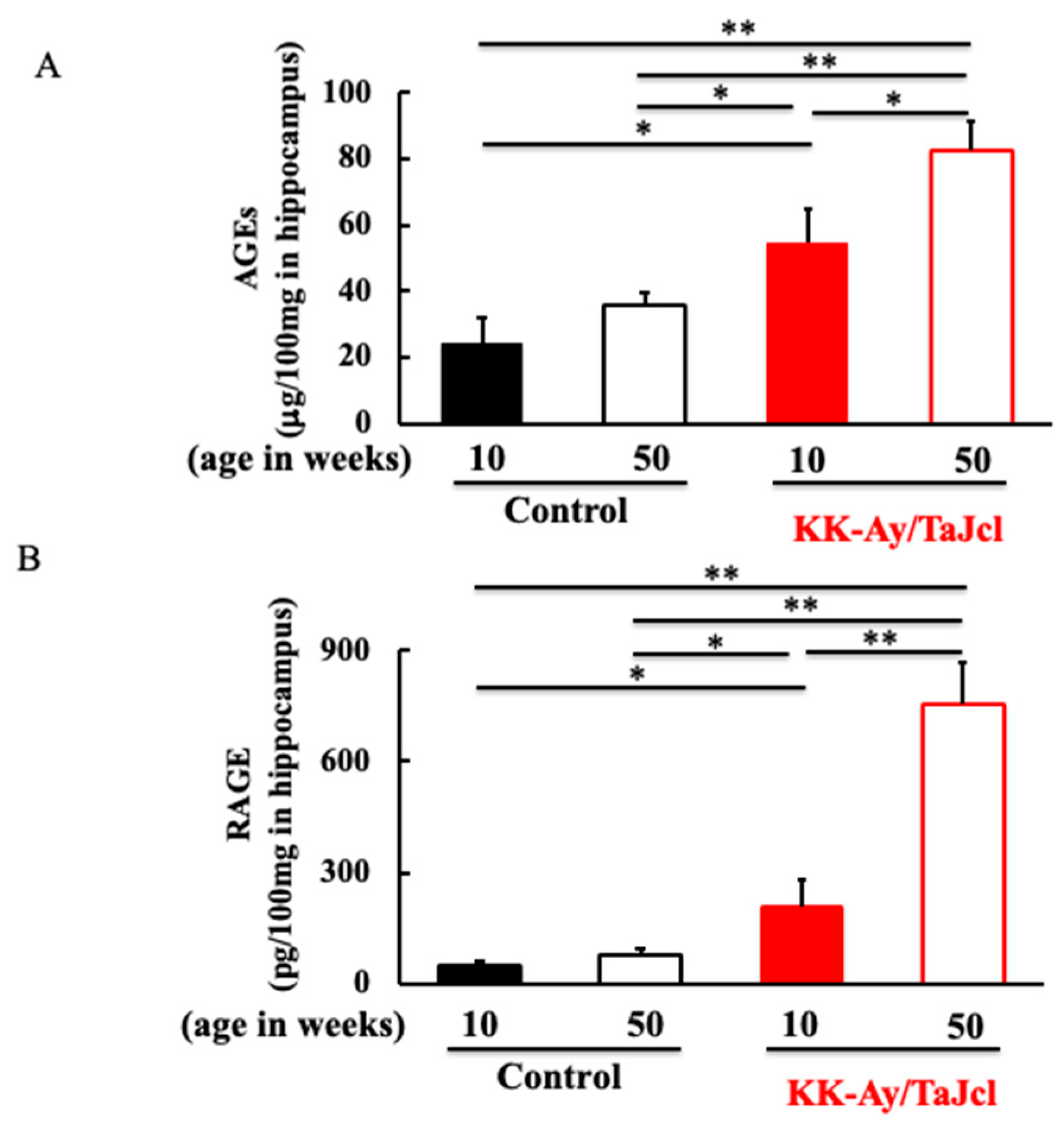
3.3. Effect of Aging on AGEs and RAGE in KK-Ay/TaJcl Mice

AGEs are produced in large quantities in patients with diabetes. We investigated the expression levels of AGEs and their receptor, RAGE, which are involved in memory and learning, in the hippocampus. Both the AGE and RAGE levels were higher in the KK-Ay/Tajcl mice than in the controls (Figure 3A,B). In addition, in the KK-Ay/Tajcl mice, it significantly increased at 50 weeks of age compared to that at 10 weeks of age (Figure 3A,B).

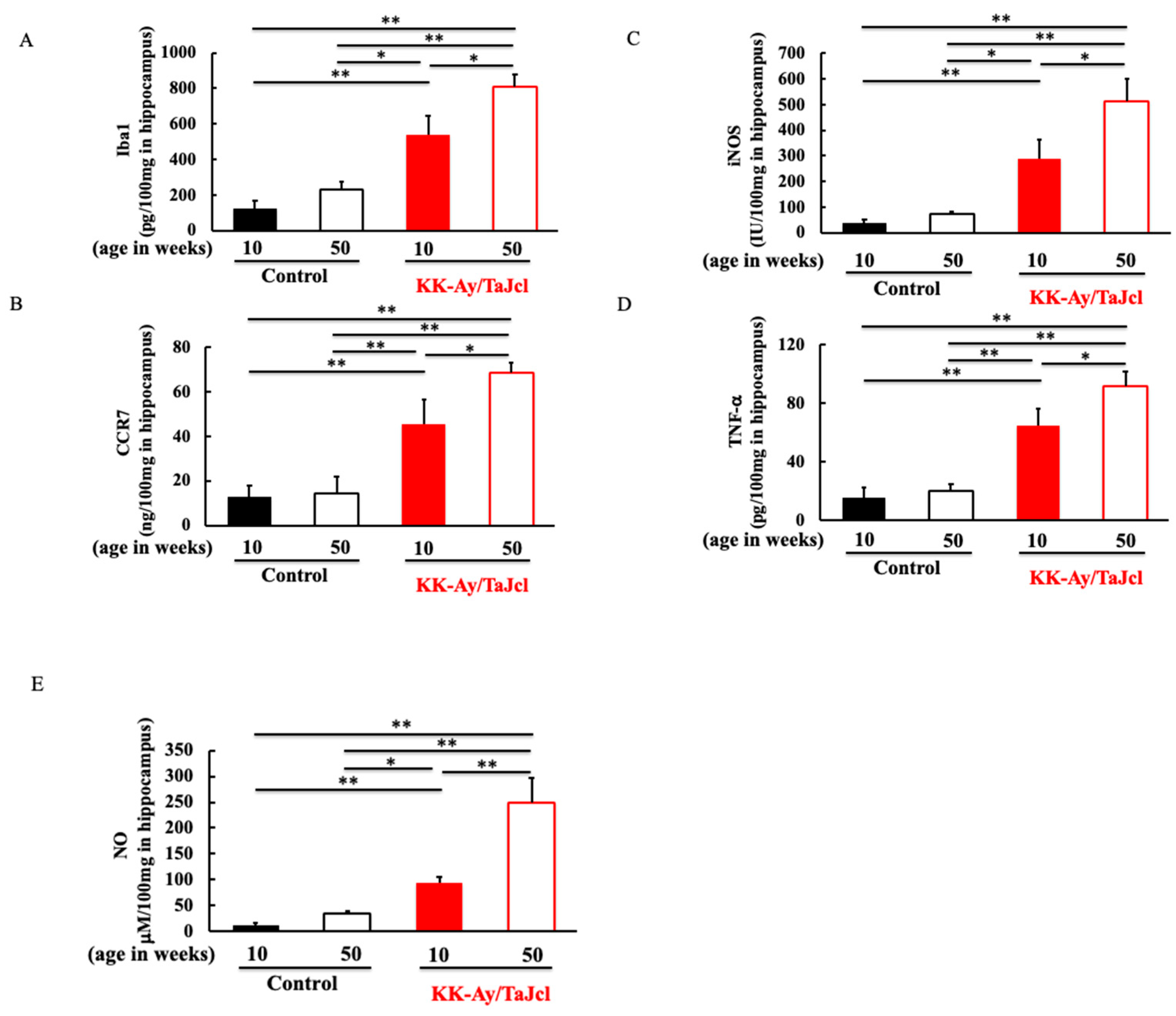
3.4. Effect of Aging on Iba1, CCR7, iNOS, TNF-α, and NO in KK-Ay/TaJcl Mice

Next, we investigated the expression levels of microglia (Iba1) involved in AGEs, RAGE, and the M1/M2 switch in microglia. Firstly, we investigated the M1 macrophages [13]. The expression of Iba1 in the hippocampus was higher in the KK-Ay/Tajcl mice than in the control mice. Moreover, both the control and KK-Ay/Tajcl mice showed higher levels in the 50-week-old mice than in the 10-week-old mice (Figure 4A). CCR7 and iNOS, which are M1 macrophages, were higher in the KK-Ay/Tajcl mice than in the control mice, and their levels in the 50-week-old mice were higher than those in the 10-week-old mice (Figure 4B,C). In particular, the 50-week-old KK-Ay/Tajcl mice exhibited a remarkable increase. Furthermore, the TNF-α and NO secreted by the M1 macrophages were also increased in the KK-Ay/Tajcl mice at 50 weeks of age compared to those at 10 weeks of age (Figure 4D,E). In particular, NO production by iNOS was markedly increased in the 50-week-old KK-Ay/Tajcl mice (Figure 4E).

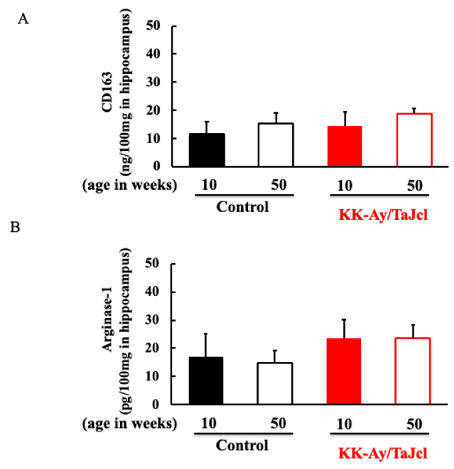
3.5. Effect of Aging on CD163 and Arginase-1 in KK-Ay/Tajcl Mice
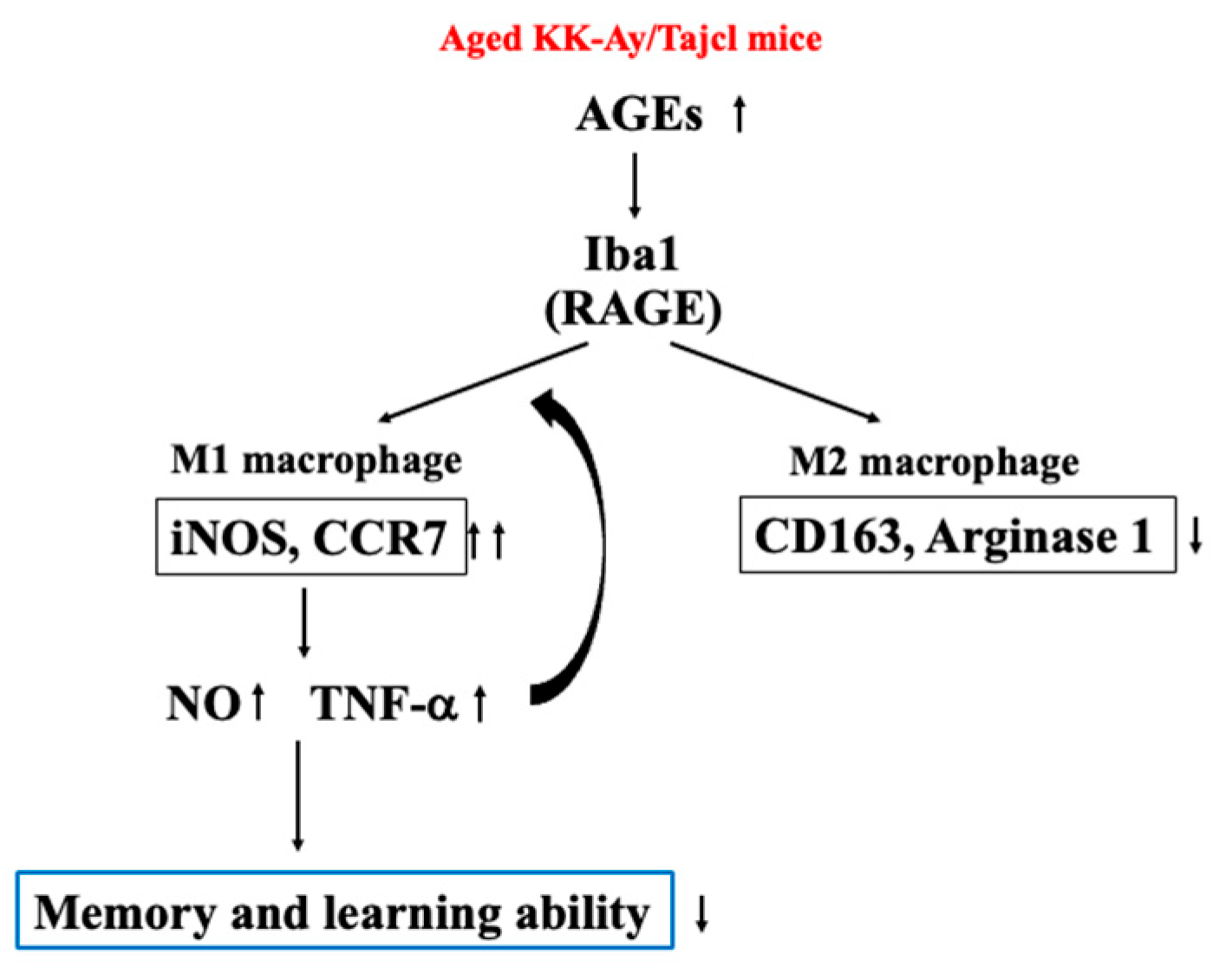
We confirmed the expression of M2 macrophages in the hippocampus, which antagonizes M1 macrophages. Neither CD163 nor arginase-1, the markers of M2 macrophages [13], differed between the groups (Figure 5A,B).

4. Discussion

With aging, many molecules that are recognized as foreign substances are produced [14]. Amyloids are well-known molecules that are found in the brain. In particular, amyloid-β, in which the APP protein has been cleaved, aggregates very easily, damages nerve cells, and activates microglia, which are macrophages in the brain [15]. Numerous senile plaques are found in the brains of patients with AD. Neuritic plaques are composed of amyloid-β, and in the presence of glucose, amyloid-β is AGE and aggregated [16]. Microglia and astrocytes in the brain contain RAGE, a receptor for AGEs, and the number of patients with AD is increasing compared to healthy elderly people [9,17]. In the present study, the expression levels of AGEs and RAGE in the hippocampus were increased in aged diabetic mice (Figure 3). Microglia are a kind of resident macrophage, and when activated, they secrete inflammatory cytokines such as TNF-α and IL-1β, which damage neurons [10,11].
Macrophages are functionally categorized into the M1 type, which acts injuriously, and the M2 type, which acts as an anti-inflammatory agent [18,19]. The M1/M2 classification has also been applied to brain-resident microglia [20]. In this study, hippocampal microglia shifted to the M1 type. IFN-g, LPS, and TNF-g induce the M1-type macrophages [20,21]. In diabetes, the production of TNF-α is increased [22]. We reported previously that aged diabetic mice secreted a large amount of TNF-α from liver and kidney [23]. AGEs, which are upregulated by hyperglycemia, bind to RAGE to increase TNF-α secretion [24]. This increased TNF-α induces M1-type microglia and increases the secretion of inflammatory cytokines, such as CCR7 and iNOS [20]. Furthermore, AGEs bind to RAGE expressed in microglia and increase the secretion of TNF-α from Iba1, thereby accelerating its shift to M1 [18]. However, when AGEs accumulate in the hippocampus and cause neuropathy, cathepsin B and E levels increase in microglia accumulated in the hippocampus prior to neuropathy. Cathepsin B binds to the transcription factor NF-kB and degrades the repressor IkB, which inactivates it and induces the gene conversion of damaging molecules. In addition, cathepsin E induces an increase in cathepsin B. Thus, it has been reported that these two processes transform macrophages into the neurotoxic M1 type [25]. In the present study, an increase in cathepsin E was observed in the hippocampus, suggesting that the inflammatory M1 type occupied the brains of the aged diabetic mice.
Notably, a marked increase in iNOS secretion was observed (Figure 4), which causes overproduction of NO and increases the release of inflammatory cytokines [26]. iNOS-derived NO is not only a major marker of endothelial function, but is also involved in cerebral circulation disorders [27]. Currently, cognitive and memory disorders, such as AD, are also involved in chronic oxygen supply reduction and metabolic disorders due to decreased cerebral circulation [28]. Cerebrovascular endothelial disorders are strongly associated with vascular risk factors (diabetes in this study) and aging [29,30]. These results suggest that a marked increase in iNOS in M1 macrophages impairs vascular endothelial function in aged diabetic mice and induces neuropathy due to the degeneration of the cerebral microvascular endothelium.
Unfortunately, our study has some weaknesses. First, in this test, the value was obtained using the ELISA kit. However, the source of each marker cannot be specified, and immunohistological analysis is required. Second, the use of FACS gives more specific results for the analysis of M1 and M2 macrophages. Thirdly, hippocampal findings can identify the tissue identity better by the immunohistological approach. In this way, it is necessary to investigate the immunohistology in more detail, and we will add further investigations.
Thus, in aged diabetic mice, brain hippocampal AGEs activate microglia and shift to type 1 macrophages through the action of cathepsins and TNF-α. It has been suggested that M1-type macrophages secrete large amounts of inflammatory cytokines and NO, which cause neuropathy (Figure 6).
Many reports have been published on the factors that cause age-related type 2 diabetes, especially dementia and AD [31]. In AD, amyloid-β begins to accumulate in the brain, deposits in the brain as senile plaques, and kills nerve cells. Blood vessel pulsation and lipid metabolism are important for excreting this amyloid-β. However, in elderly people with diabetes mellitus, blood vessels are damaged, making pulsation less likely to occur, making it easier to induce dementia. In addition, the nerve energy of the brain is glucose, and brain nerve cells must always take in glucose, while insulin plays an important role. Glucose is taken up by glial cells, but in elderly diabetic patients, insulin that acts on glial cells is insufficient and glial cells are unable to take up glucose from the blood. Therefore, insulin resistance also influences the progression of dementia [32,33]. In this way, there are many factors between elderly type 2 diabetes and dementia, and it is necessary to examine the relationship with the results of this experiment.

Author Contributions

Conceptualization, K.H. and K.O.; methodology, S.T.; software, S.T.; validation, K.H., M.I. and K.O.; formal analysis, S.T.; investigation, K.H.; resources, K.H.; data curation, K.H. and S.T.; writing—original draft preparation, K.H.; writing—review and editing, K.O.; visualization, M.I.; supervision, S.T.; project administration, K.H. and K.O.; funding acquisition, K.H. and K.O. All authors have read and agreed to the published version of the manuscript.

Funding

This study was supported by the JSPS KAKENHI grant (18K11085) and a grant from Maruho Co., Ltd.

Institutional Review Board Statement

The animal study protocol was approved by the Institutional Review Board of Laboratory Animals of the Suzuka University of Medical Science (approval number: 34, 7 October 2017).

Informed Consent Statement

Not applicable.

Data Availability Statement

Not applicable.

Acknowledgments

We would like to thank Vikas Narang for English language editing.

Conflicts of Interest

The authors declare no conflict of interest.

References

  1. Fratiglioni, L.; Paillard-Borg, S.; Winblad, B. An active and socially integrated lifestyle in late life might protect against dementia. Lancet Neurol. 2004, 3, 343–353. [Google Scholar] [CrossRef] [PubMed]
  2. Kimura, R.; Tomiyasu, H.; Takeuchi, T.; Shimizu, M.; Hayashi, Y.; Okayama, N.; Kamiya, Y.; Joh, T. Prevalence of Alzheimer’s disease with diabetes in the Japanese population. Psychogeriatrics 2008, 8, 73–78. [Google Scholar] [CrossRef]
  3. Ott, A.; Stolk, R.P.; van Harskamp, F.; Pols, H.A.P.; Hofman, A.; Breteler, M.M.B. Diabetes mellitus and the risk of dementia: The Rotterdam Study. Neurology 1999, 53, 1937–1942. [Google Scholar] [CrossRef] [PubMed]
  4. Cukierman, T.; Gerstein, H.C.; Williamson, J.D. Cognitive decline and dementia in diabetes—Systematic overview of prospective observational studies. Diabetologia 2005, 48, 2460–2469. [Google Scholar] [CrossRef] [Green Version]
  5. Cheng, G.; Huang, C.; Deng, H.; Wang, H. Diabetes as a risk factor for dementia and mild cognitive impairment: A meta-analysis of longitudinal studies. Intern. Med. J. 2012, 42, 484–491. [Google Scholar] [CrossRef]
  6. Sato, N.; Morishita, R. Roles of vascular and metabolic components in cognitive dysfunction of Alzheimer disease: Short- and long-term modification by non-genetic risk factors. Front. Aging Neurosci. 2013, 5, 64. [Google Scholar] [CrossRef] [Green Version]
  7. Takeda, S.; Sato, N.; Uchio-Yamada, K.; Sawada, K.; Kunieda, T.; Takeuchi, D.; Kurinami, H.; Shinohara, M.; Rakugi, H.; Morishita, R. Diabetes-accelerated memory dysfunction via cerebrovascular inflammation and Aβ deposition in an Alzheimer mouse model with diabetes. Proc. Natl. Acad. Sci. USA 2010, 107, 7036–7041. [Google Scholar] [CrossRef]
  8. Francis, P.T.; Palmer, A.M.; Snape, M.; Wilcock, G.K. The cholinergic hypothesis of Alzheimer’s disease: A review of progress. J. Neurol. Neurosurg. Psychiatry 1999, 66, 137–147. [Google Scholar] [CrossRef]
  9. Srikanth, V.; Maczurek, A.; Phan, T.; Steele, M.; Westcott, B.; Juskiw, D.; Münch, G. Advanced glycation endproducts and their receptor RAGE in Alzheimer’s disease. Neurobiol. Aging 2011, 32, 763–777. [Google Scholar] [CrossRef]
  10. Dukic-Stefanovic, S.; Gasic-Milenkovic, J.; Deuther-Conrad, W.; Münch, G. Signal transduction pathways in mouse microglia N-11 cells activated by advanced glycation endproducts (AGEs). J. Neurochem. 2003, 87, 44–55. [Google Scholar] [CrossRef] [Green Version]
  11. Takeuchi, M.; Bucala, R.; Suzuki, T.; Ohkubo, T.; Yamazaki, M.; Koike, T.; Kameda, Y.; Makita, Z. Neurotoxicity of Advanced Glycation End-Products for Cultured Cortical Neurons. J. Neuropathol. Exp. Neurol. 2000, 59, 1094–1105. [Google Scholar] [CrossRef] [PubMed] [Green Version]
  12. Lee, S.-S.; Kim, C.-J.; Shin, M.-S.; Lim, B.-V. Treadmill exercise ameliorates memory impairment through ERK-Akt-CREB-BDNF signaling pathway in cerebral ischemia gerbils. J. Exerc. Rehabil. 2020, 16, 49–57. [Google Scholar] [CrossRef] [PubMed] [Green Version]
  13. Hu, X.; Leak, R.K.; Shi, Y.; Suenaga, J.; Gao, Y.; Zheng, P.; Chen, J. Microglial and macrophage polarization—New prospects for brain repair. Nat. Rev. Neurol. 2015, 11, 56–64. [Google Scholar] [CrossRef]
  14. Chen, G.Y.; Nuñez, G. Sterile inflammation: Sensing and reacting to damage. Nat. Rev. Immunol. 2010, 10, 826–837. [Google Scholar] [CrossRef] [PubMed] [Green Version]
  15. Ito, S.; Sawada, M.; Haneda, M.; Fujii, S.; Oh-Hashi, K.; Kiuchi, K.; Takahashi, M.; Isobe, K.-I. Amyloid-β peptides induce cell proliferation and macrophage colony-stimulating factor expression via the PI3-kinase/Akt pathway in cultured Ra2 microglial cells. FEBS Lett. 2005, 579, 1995–2000. [Google Scholar] [CrossRef] [PubMed] [Green Version]
  16. Yagi, M.; Yonei, Y. Glycative stress and antiaging: 12. Glycative stress and dementia. Glycative Stress Res. 2019, 6, 87–91. [Google Scholar]
  17. Takeda, A.; Yasuda, T.; Miyata, T.; Goto, Y.; Wakai, M.; Watanabe, M.; Yasuda, Y.; Horie, K.; Inagaki, T.; Doyu, M.; et al. Advanced glycation end products co-localized with astrocytes and microglial cells in Alzheimer’s disease brain. Acta Neuropathol. 1998, 95, 555–558. [Google Scholar] [CrossRef]
  18. Block, M.L.; Hong, J.-S. Microglia and inflammation-mediated neurodegeneration: Multiple triggers with a common mechanism. Prog. Neurobiol. 2005, 76, 77–98. [Google Scholar] [CrossRef]
  19. Lenz, K.M.; Nugent, B.M.; Haliyur, R.; McCarthy, M.M. Microglia Are Essential to Masculinization of Brain and Behavior. J. Neurosci. 2013, 33, 2761–2772. [Google Scholar] [CrossRef] [Green Version]
  20. Gordon, S. Alternative activation of macrophages. Nat. Rev. Immunol. 2003, 3, 23–35. [Google Scholar] [CrossRef]
  21. Mosser, D.M.; Edwards, J.P. Exploring the full spectrum of macrophage activation. Nat. Rev. Immunol. 2008, 8, 958–969. [Google Scholar] [CrossRef] [PubMed]
  22. Okuno, A.; Tamemoto, H.; Tobe, K.; Ueki, K.; Mori, Y.; Iwamoto, K.; Umesono, K.; Akanuma, Y.; Fujiwara, T.; Horikoshi, H.; et al. Troglitazone increases the number of small adipocytes without the change of white adipose tissue mass in obese Zucker rats. J. Clin. Investig. 1998, 101, 1354–1361. [Google Scholar] [CrossRef]
  23. Hiramoto, K.; Goto, K.; Tanaka, S.; Horikawa, T.; Ooi, K. Skin, Liver, and Kidney Interactions Contribute to Skin Dryness in Aging KK-Ay/Tajcl Mice. Biomedicines 2022, 10, 2648. [Google Scholar] [CrossRef]
  24. Jahan, H.; Choudhary, M.I. Gliclazide alters macrophages polarization state in diabetic atherosclerosis in vitro via blocking AGE-RAGE/TLR4-reactive oxygen species-activated NF-kβ nexus. Eur. J. Pharmacol. 2021, 894, 173874. [Google Scholar] [CrossRef] [PubMed]
  25. Ni, J.; Wu, Z.; Peterts, C.; Yamamoto, K.; Qing, H.; Nakanishi, H. The Critical Role of Proteolytic Relay through Cathepsins B and E in the Phenotypic Change of Microglia/Macrophage. J. Neurosci. 2015, 35, 12488–12501. [Google Scholar] [CrossRef] [PubMed]
  26. Biswas, S.K.; Mantovani, A. Macrophage plasticity and interaction with lymphocyte subsets: Cancer as a paradigm. Nat. Immunol. 2010, 11, 889–896. [Google Scholar] [CrossRef] [PubMed]
  27. Toda, N.; Ayajiki, K.; Okamura, T. Cerebral blood flow regulation by nitric oxide in neurological disorders. Can. J. Physiol. Pharmacol. 2009, 87, 581–594. [Google Scholar] [CrossRef] [PubMed]
  28. Cho, D.-H.; Nakamura, T.; Fang, J.; Cieplak, P.; Godzik, A.; Gu, Z.; Lipton, S.A. S-Nitrosylation of Drp1 Mediates β-Amyloid-Related Mitochondrial Fission and Neuronal Injury. Science 2009, 324, 102–105. [Google Scholar] [CrossRef] [Green Version]
  29. Taddei, S.; Virdis, A.; Mattei, P.; Natali, A.; Ferrannini, E.; Salvetti, A. Effect of Insulin on Acetylcholine-Induced Vasodilation in Normotensive Subjects and Patients With Essential Hypertension. Circulation 1995, 92, 2911–2918. [Google Scholar] [CrossRef]
  30. Higashi, Y.; Sasaki, S.; Nakagawa, K.; Kimura, M.; Noma, K.; Hara, K.; Jitsuiki, D.; Goto, C.; Oshima, T.; Chayama, K.; et al. Tetrahydrobiopterin improves aging-related impairment of endothelium-dependent vasodilation through increase in nitric oxide production. Atherosclerosis 2006, 186, 390–395. [Google Scholar] [CrossRef]
  31. Fukazawa, R.; Hanyu, H.; Sato, T.; Shimizu, S.; Koyama, S.; Kanetaka, H.; Sakurai, H.; Iwamoto, T. Subgroups of Alzheimer’s Disease Associated with Diabetes Mellitus Based on Brain Imaging. Dement. Geriatr. Cogn. Disord. 2013, 35, 280–290. [Google Scholar] [CrossRef] [PubMed]
  32. Shin, H.J.; Kim, K.E.; Jeong, E.A.; An, H.S.; Lee, S.J.; Lee, J.; Roh, G.S. Amyloid b oligomer promotes microglial galection-3 and astrocytic lipocalin-2 levels in the hippocampus of mice fed a high-fat diet. Biochem. Biophy. Res. Commun. 2023, 667, 10–17. [Google Scholar] [CrossRef] [PubMed]
  33. Natunen, T.; Martiskainen, H.; Marttinen, M.; Gabbouj, A.; Koivisto, H.; Kemppainen, S.; Kaipainen, S.; Takalo, M.; Svo-bodova, H.; Leppanen, L.; et al. Diabetic phenotype in mouse and humans reduces the number of microglia atound b-amyloid plaques. Mol. Neurodegener. 2020, 15, 66. [Google Scholar] [CrossRef] [PubMed]
Figure 1. Effect of aging on body weight (A) and blood glucose levels (B) in KK-Ay/TaJcl mice. Values are expressed as the mean ± SD of five animals. * p < 0.05, ** p < 0.01. Control: C57BL/6j mice.
Life 13 01540 g001
Figure 2. Effect of aging on motor activity (A) and memory and learning activity (B) in KK-Ay/TaJcl mice. The step-through passive avoidance test (B) was used to assess memory and learning abilities. Values are expressed as the mean ± the SD of five animals. * p < 0.05, ** p < 0.01. Control: C57BL/6j mice.
Life 13 01540 g002
Figure 3. Effect of aging on levels of AGEs (A) and RAGE (B) in hippocampus of KK-Ay/TaJcl mice. The data show one representative experiment containing five animals. The hippocampus levels of AGEs and RAGE in mice were measured using ELISA. Values are expressed as the mean ± the SD derived from five animals. * p < 0.05, ** p < 0.01. Control: C57BL/6j mice.
Life 13 01540 g003
Figure 4. Effect of aging on the levels of Iba1 (A), CCR7 (B), iNOS (C), TNF-α (D), and NO (E) in the hippocampus of the KK-Ay/TaJcl mice. The data show one representative experiment containing five animals. The hippocampus levels of Iba1, CCR7, iNOS, and TNF-α in the mice were measured using an ELISA kit. The hippocampus level of NO was measured using the assay kit. Values are expressed as the mean ± the SD derived from five animals. * p < 0.05, ** p < 0.01. Control: C57BL/6j mice.
Life 13 01540 g004
Figure 5. Effect of aging on levels of CD163 (A) and arginase-1 (B) in hippocampus of KK-Ay/TaJcl mice. The data show one representative experiment containing five animals. The hippocampus levels of CD163 and arginase-1 in the mice were measured using an ELISA kit. Values are expressed as the mean ± the SD derived from five animals. Control: C57BL/6j mice.
Life 13 01540 g005
Figure 6. Mechanism of the effect of aging on memory and learning abilities in KK-Ay/TaJcl mice.
Life 13 01540 g006
Disclaimer/Publisher’s Note: The statements, opinions and data contained in all publications are solely those of the individual author(s) and contributor(s) and not of MDPI and/or the editor(s). MDPI and/or the editor(s) disclaim responsibility for any injury to people or property resulting from any ideas, methods, instructions or products referred to in the content.

