


Juglans regia Linn.: A Natural Repository of Vital Phytochemical and Pharmacological Compounds

 , ,
, ,  and
and
Abstract
1. Introduction
2. Traditional and Ethnobotanical Uses
3. Pharmacological Applications
3.1. Antibacterial Activity
3.2. Antioxidant Activity
3.3. Analgesic and Anti-Inflammatory Properties
3.4. Antidepressant Activity
3.4.1. Forced Swimming Test
3.4.2. Tail Suspension Test
3.5. Antiviral Activities
Enzyme Inhibition Mechanism
3.6. Antidiabetic Activity
3.7. Anticancer Activity
3.8. Antifungal Activity
3.9. Cardiovascular Activity
3.10. Brain Enhancing Activity
4. Toxicity Activity on Plants and Animals
5. Conclusions
Author Contributions
Funding
Institutional Review Board Statement
Informed Consent Statement
Data Availability Statement
Acknowledgments
Conflicts of Interest
References
- Acarsoy Bilgin, N. Morphological Characterization of Pollen in Some Varieties of Walnut (Juglans regia). Int. J. Fruit Sci. 2022, 22, 471–480. [Google Scholar] [CrossRef]
- Hayes, D.; Angove, M.J.; Tucci, J.; Dennis, C. Walnuts (Juglans regia) chemical composition and research in human health. Crit. Rev. Food Sci. Nutr. 2016, 56, 1231–1241. [Google Scholar] [CrossRef] [PubMed]
- Gupta, A.; Behl, T.; Panichayupakaranan, P. A review of phytochemistry and pharmacology profile of Juglans regia. Obes. Med. 2019, 16, 100142. [Google Scholar] [CrossRef]
- Dawid-Pać, R. Medicinal plants used in treatment of inflammatory skin diseases. Postep. Dermatol. Alergol. 2013, 30, 170–177. [Google Scholar] [CrossRef] [PubMed]
- Bourais, I.; Elmarrkechy, S.; Taha, D.; Mourabit, Y.; Bouyahya, A.; El Yadini, M.; Machich, O.; El Hajjaji, S.; El Boury, H.; Dakka, N.; et al. A Review on Medicinal Uses, Nutritional Value, and Antimicrobial, Antioxidant, Anti-Inflammatory, Antidiabetic, and Anticancer Potential Related to Bioactive Compounds of J. regia. Food Rev. Int. 2022, 1–51. [Google Scholar] [CrossRef]
- Chand, S.P.; Arif, H. Depression. In StatPearls; StatPearls Publishing: Treasure Island, FL, USA, 2022. [Google Scholar]
- Nabavi, S.; Ebrahimzadeh, M.; Nabavi, S.; Mahmoudi, M.; Rad, S. Biological activities of Juglans regia flowers. Rev. Bras. Farmacogn. 2011, 21, 465–470. [Google Scholar] [CrossRef]
- Mahmoudi, M.; Ebrahimzadeh, M.; Ansaroudi, F.; Nabavi, S.F.; Nabavi, S.M. Antidepressant and antioxidant activities of Artemisia absinthium L. at flowering stage. Afr. J. Biotechnol. 2009, 8, 7170–7175. [Google Scholar] [CrossRef]
- Rath, B.P.; Pradhan, D. Antidepressant Activity of Juglans regia L. Fruit Extract. Int. J. Toxicol. Pharmacol. Res. 2009, 1, 24–29. [Google Scholar]
- Mouhajir, F.; Hudson, J.B.; Rejdali, M.; Towers, G.H.N. Multiple Antiviral Activities of Endemic Medicinal Plants Used by Berber Peoples of Morocco. Pharm. Biol. 2001, 39, 364–374. [Google Scholar] [CrossRef]
- Vardhini, S.R. Exploring the antiviral activity of juglone by computational method. J. Recept. Signal Transduct. 2014, 34, 456–457. [Google Scholar] [CrossRef]
- Liu, Z.M.; Wen, R.X.; Ma, H.T.; Yang, Y.S.; Wang, X.L.; Lv, X.H.; Li, Z.L. Investigation on inhibition of HIV III B virus with extractions of Juglans regia. Zhongguo Zhong Yao Za Zhi 2008, 33, 2535–2538. [Google Scholar] [PubMed]
- D’Angeli, F.; Malfa, G.A.; Garozzo, A.; Li Volti, G.; Genovese, C.; Stivala, A.; Nicolosi, D.; Attanasio, F.; Bellia, F.; Ronsisvalle, S.; et al. Antimicrobial, Antioxidant, and Cytotoxic Activities of Juglans regia L. Pellicle Extract. Antibiotics 2021, 10, 159. [Google Scholar] [CrossRef] [PubMed]
- Cui, J.; Jia, J. Discovery of juglone and its derivatives as potent SARS-CoV-2 main proteinase inhibitors. Eur. J. Med. Chem. 2021, 225, 113789. [Google Scholar] [CrossRef] [PubMed]
- Pan, A.; Sun, Q.; Manson, J.E.; Willett, W.C.; Hu, F.B. Walnut Consumption Is Associated with Lower Risk of Type 2 Diabetes in Women. J. Nutr. 2013, 143, 512–518. [Google Scholar] [CrossRef] [PubMed]
- Nasiry, D.; Khalatbary, A.R.; Ahmadvand, H.; Talebpour Amiri, F.B. Effects of Juglans regia L. leaf extract supplementation on testicular functions in diabetic rats. Biotech. Histochem. 2021, 96, 41–47. [Google Scholar] [CrossRef]
- Qanash, H.; Yahya, R.; Bakri, M.M.; Bazaid, A.S.; Qanash, S.; Shater, A.F.; TM, A. Anticancer, antioxidant, antiviral and antimicrobial activities of Kei Apple (Dovyalis caffra) fruit. Sci. Rep. 2022, 12, 5914. [Google Scholar] [CrossRef]
- Wani, A.K.; Akhtar, N.; Sharma, A.; El-Zahaby, S.A. Fighting Carcinogenesis with Plant Metabolites by Weakening Proliferative Signaling and Disabling Replicative Immortality Networks of Rapidly Dividing and Invading Cancerous Cells. Curr. Drug Deliv. 2023, 202, 2. [Google Scholar] [CrossRef]
- Al-Rajhi, A.M.H.; Qanash, H.; Almuhayawi, M.S.; Al Jaouni, S.K.; Bakri, M.M.; Ganash, M.; Salama, H.M.; Selim, S.; Abdelghany, T.M. Molecular Interaction Studies and Phytochemical Characterization of Mentha pulegium L. Constituents with Multiple Biological Utilities as Antioxidant, Antimicrobial, Anticancer and Anti-Hemolytic Agents. Molecules 2022, 27, 4824. [Google Scholar] [CrossRef]
- Sugie, S.; Okamoto, K.; Rahman, K.M.; Tanaka, T.; Kawai, K.; Yamahara, J.; Mori, H. Inhibitory effects of plumbagin and juglone on azoxymethane-induced intestinal carcinogenesis in rats. Cancer Lett. 1998, 127, 177–183. [Google Scholar] [CrossRef]
- Segura-Aguilar, J.; Jönsson, K.; Tidefelt, U.; Paul, C. The cytotoxic effects of 5-OH-1,4-naphthoquinone and 5,8-diOH-1,4-naphthoquinone on doxorubicin-resistant human leukemia cells (HL-60). Leuk. Res. 1992, 16, 631–637. [Google Scholar] [CrossRef]
- Salimi, M.; Ardestaniyan, M.H.; Mostafapour Kandelous, H.; Saeidnia, S.; Gohari, A.R.; Amanzadeh, A.; Sanati, H.; Sepahdar, Z.; Ghorbani, S.; Salimi, M. Anti-proliferative and apoptotic activities of constituents of chloroform extract of Juglans regia leaves. Cell Prolif. 2014, 47, 172–179. [Google Scholar] [CrossRef] [PubMed]
- Zhang, X.B.; Zou, C.L.; Duan, Y.X.; Wu, F.; Li, G. Activity guided isolation and modification of juglone from Juglans regia as potent cytotoxic agent against lung cancer cell lines. BMC Complement. Altern. Med. 2015, 15, 396. [Google Scholar] [CrossRef]
- Shah, T.I.; Sharma, E.; Shah, G.A. Anti-proliferative, Cytotoxicity and Anti-oxidant Activity of uglans regia Extract. Am. J. Cancer Prev. 2015, 3, 45–50. [Google Scholar]
- Bazaid, A.S.; Aldarhami, A.; Patel, M.; Adnan, M.; Hamdi, A.; Snoussi, M.; Qanash, H.; Imam, M.; Monjed, M.K.; Khateb, A.M. The Antimicrobial Effects of Saudi Sumra Honey against Drug Resistant Pathogens: Phytochemical Analysis, Antibiofilm, Anti-Quorum Sensing, and Antioxidant Activities. Pharmaceuticals 2022, 15, 1212. [Google Scholar] [CrossRef]
- Wianowska, S.; Garbaczewska, A.; Cieniecka-Roslonkiewicz, A.L.; Dawidowicz, A.; Jankowska, A. Comparison of antifungal activity of extracts from different Juglans regia cultivars and juglone. Microb. Pathog. 2016, 100, 263–267. [Google Scholar] [CrossRef] [PubMed]
- Choub, V.; Ajuna, H.B.; Won, S.-J.; Moon, J.-H.; Choi, S.-I.; Maung, C.E.H.; Kim, C.-W.; Ahn, Y.S. Antifungal Activity of Bacillus velezensis CE 100 against Anthracnose Disease (Colletotrichum gloeosporioides) and Growth Promotion of Walnut (Juglans regia L.) Trees. Int. J. Mol. Sci. 2021, 22, 10438. [Google Scholar] [CrossRef]
- Upadhyay, V.; Kambhoja, S.; Harshaleena, K. Antifungal activity and preliminary phytochemical analysis of stem bark extracts of Juglans regia linn. IJPBA 2010, 1, 442. [Google Scholar]
- Sytykiewicz, H.; Chrzanowski, G.; Czerniewicz, P.; Leszczyński, B.; Sprawka, I.; Krzyżanowski, R.; Matok, H. Antifungal Activity of Juglans regia (L.) Leaf Extracts Against Candida albicans Isolates. Pol. J. Environ. Stud. 2015, 24, 1339–1348. [Google Scholar] [CrossRef]
- Oliveira, I.; Sousa, A.; Ferreira, I.C.; Bento, A.; Estevinho, L.; Pereira, J.A. Total phenols, antioxidant potential and antimicrobial activity of walnut (Juglans regia L.) green husks. Food Chem. Toxicol. 2008, 46, 2326–2331. [Google Scholar] [CrossRef]
- Hamazaki, T.; Okuyama, H. The Japan Society for Lipid Nutrition recommends to reduce the intake of linoleic acid. A review and critique of the scientific evidence. World Rev. Nutr. Diet 2003, 92, 109–132. [Google Scholar]
- Czernichow, S.; Thomas, D.; Bruckert, E. n-6 Fatty acids and cardiovascular health: A review of the evidence for dietary intake recommendations. Br. J. Nutr. 2010, 104, 788–796. [Google Scholar] [CrossRef] [PubMed]
- Simopoulos, A.P. The importance of the ratio of omega-6/omega-3 essential fatty acids. Biomed. Pharmacother. 2002, 56, 365–379. [Google Scholar] [CrossRef]
- El Haouari, M.; Rosado, J.A. Medicinal Plants with Antiplatelet Activity. Phytother. Res. 2016, 30, 1059–1071. [Google Scholar] [CrossRef] [PubMed]
- Haider, S.; Batool, Z.; Tabassum, S.; Perveen, T.; Saleem, S.; Naqvi, F.; Javed, H.; Haleem, D.J. Effects of walnuts (Juglans regia) on learning and memory functions. Plant Foods Hum. Nutr. 2011, 66, 335–340. [Google Scholar] [CrossRef]
- Shi, D.; Chen, C.; Zhao, S.; Ge, F.; Liu, D.; Song, H. Effects of Walnut Polyphenol on Learning and Memory Functions in Hypercholesterolemia Mice. J. Food Nutr. Res. 2014, 2, 450–456. [Google Scholar] [CrossRef]
- Shabani, M.; Nazeri, M.; Parsania, S.; Razavinasab, M.; Zangiabadi, N.; Esmaeilpour, K.; Abareghi, F. Walnut consumption protects rats against cisplatin-induced neurotoxicity. Neurotoxicology 2012, 33, 1314–1321. [Google Scholar] [CrossRef] [PubMed]
- Muthaiyah, B.; Essa, M.M.; Lee, M.; Chauhan, V.; Kaur, K.; Chauhan, A. Dietary supplementation of walnuts improves memory deficits and learning skills in transgenic mouse model of Alzheimer’s disease. J. Alzheimers Dis. 2014, 42, 1397–1405. [Google Scholar] [CrossRef]
- Ravanbakhsh, A.; Mahdavi, M.; Jalilzade-Amin, G.; Javadi, S.; Maham, M.; Mohammadnejad, D.; Rashidi, M.R. Acute and Subchronic Toxicity Study of the Median Septum of Juglans regia in Wistar Rats. Adv. Pharm. Bull. 2016, 6, 541–549. [Google Scholar] [CrossRef]
- Yang, L.; Ma, S.; Han, Y.; Wang, Y.; Guo, Y.; Weng, Q.; Xu, M. Walnut Polyphenol Extract Attenuates Immunotoxicity Induced by 4-Pentylphenol and 3-methyl-4-nitrophenol in Murine Splenic Lymphocyte. Nutrients 2016, 8, 287. [Google Scholar] [CrossRef]
- Hausen, B.M. Woods Injurious to Human Health; De Gruyter: Berlin, Germany; Boston, MA, USA, 2016. [Google Scholar]
- Bonamonte, D.; Foti, C.; Angelini, G. Hyperpigmentation and contact dermatitis due to Juglans regia. Contact Dermat. 2001, 44, 101–102. [Google Scholar] [CrossRef]
- Utiérrez Ortiz, A.L.; Berti, F.; Navarini, L.; Crisafulli, P.; Colomban, S.; Forzato, C. Aqueous extracts of walnut (Juglans regia L.) leaves: Quantitative analyses of hydroxycinnamic and chlorogenic acids. J. Chromatogr. Sci. 2018, 56, 753–760. [Google Scholar] [CrossRef] [PubMed]
- Calabrò, S.; Alzoubi, K.; Bissinger, R.; Jilani, K.; Faggio, C.; Lang, F. Enhanced eryptosis following juglone exposure. Basic Clin. Pharmacol. Toxicol. 2015, 116, 460–467. [Google Scholar] [CrossRef] [PubMed]
- Belknap, J.K. Black walnut extract: An inflammatory model. Vet. Clin. N. A. Equine Pract. 2010, 26, 95–101. [Google Scholar] [CrossRef]
- Carvalho, M.; Ferreira, P.J.; Mendes, V.S.; Silva, R.; Pereira, J.A.; Jerónimo, C.; Silva, B.M. Human cancer cell antiproliferative and antioxidant activities of Juglans regia L. Food Chem. Toxicol. 2010, 48, 441–447. [Google Scholar] [CrossRef]
- Arslan, H.; Ondul Koc, E.; Ozay, Y.; Canli, O.; Ozdemir, S.; Tollu, G.; Dizge, N. Antimicrobial and antioxidant activity of phenolic extracts from walnut (Juglans regia L.) green husk by using pressure-driven membrane process. J. Food Sci. Technol. 2022, 60, 73–83. [Google Scholar] [CrossRef]
- Almeida, I.; Fernandes, E.; Lima, J.; Costa, P.; Bahia, M. Walnut (Juglans regia) leaf extracts are strong scavengers of pro-oxidant reactive species. Food Chem. 2008, 106, 1014–1020. [Google Scholar] [CrossRef]
- Żurek, N.; Pawłowska, A.; Pycia, K.; Grabek-Lejko, D.; Kapusta, I.T. Phenolic Profile and Antioxidant, Antibacterial, and Antiproliferative Activity of Juglans regia L. Male Flowers. Molecules 2022, 27, 2762. [Google Scholar] [CrossRef]
- Hosseinzadeh, H.; Behravan, E.; Soleimani, M.M. Antinociceptive and Anti-inflammatory Effects of Pistacia vera LeafExtract in Mice. Iran. J. Pharm. Res. 2011, 10, 821–828. [Google Scholar]
- Bhat, A.A.; Tandon, N.; Tandon, R. Pyrrolidine derivatives as antibacterial agents, current status and future prospects: A patent review. Pharm. Patent Analyst. 2022. [Google Scholar] [CrossRef]
- Chiavaccini, L.; Hassel, D.M.; Shoemaker, M.L.; Charles, J.B.; Belknap, J.K.; Ehrhart, E.J. Detection of calprotectin and apoptotic activity within the equine colon from horses with black walnut extract-induced laminitis. Vet. Immunol. Immunopathol. 2011, 144, 366–373. [Google Scholar] [CrossRef]
- Stacey Lockyer, Anne E de la Hunty, Simon Steenson, Ayela Spiro, Sara A Stanner, Walnut consumption and health outcomes with public health relevance—A systematic review of cohort studies and randomized controlled trials published from 2017 to present. Nutr. Rev. 2023, 81, 26–54. [CrossRef]
- Amaral, J.S.; Casal, S.; Pereira, J.A.; Seabra, R.M.; Oliveira, B.P. Determination of sterol and fatty acid compositions, oxidative stability, and nutritional value of six walnut (Juglans regia L.) cultivars grown in Portugal. J. Agric. Food Chem. 2003, 51, 7698–7702. [Google Scholar] [CrossRef] [PubMed]
- Colaric, M.; Veberic, R.; Solar, A.; Hudina, M.; Stampar, F. Phenolic acids, syringaldehyde, and juglone in fruits of different cultivars of Juglans regia L. J. Agric. Food Chem. 2005, 53, 6390–6396. [Google Scholar] [CrossRef] [PubMed]
- Trandafir, I.; Cosmulescu, S.; Botu, M.; Nour, V. Antioxidant activity, and phenolic and mineral contents of the walnut kernel (Juglans regia L.) as a function of the pellicle color. Fruits 2016, 71, 173–184. [Google Scholar] [CrossRef]
- Akbari, V.; Jamei, R.; Heidari, R.; Jahanban Esfahlan, A. Antiradical activity of different parts of Walnut (Juglans regia L.) fruit as a function of genotype. Food Chem. 2012, 135, 2404–2410. [Google Scholar] [CrossRef]
- Owens, N.; Lee, D. The use of micro bubble flotation technology in secondary and tertiary produced water treatment—A technical comparison with other separation technologies. In Proceedings of the the TUV NEL, 5th Produced Water Workshop, Aberdeen, Scotland, 30–31 May 2007. [Google Scholar]
- Altun, T.; Pehlivan, E. Removal of Cr (VI) from aqueous solutions by modified walnut shells. Food Chem. 2012, 132, 693–700. [Google Scholar] [CrossRef]
- Ding, D.; Zhao, Y.; Yang, S.; Shi, W.; Zhang, Z.; Lei, Z.; Yang, Y. Adsorption of cesium from aqueous solution using agricultural residue–Walnut shell: Equilibrium, kinetic and thermodynamic modeling studies. Water Res. 2013, 47, 2563–2571. [Google Scholar] [CrossRef]
- Kazemipour, M.; Ansari, M.; Tajrobehkar, S.; Majdzadeh, M.; Kermani, H.R. Removal of lead, cadmium, zinc, and copper from industrial wastewater by carbon developed from walnut, hazelnut, almond, pistachio shell, and apricot stone. J. Hazard. Mater. 2008, 150, 322–327. [Google Scholar] [CrossRef]
- Köhler, S.J.; Cubillas, P.; Rodríguez-Blanco, J.D.; Bauer, C.; Prieto, M. Removal of cadmium from wastewaters by aragonite shells and the influence of other divalent cations. Environ. Sci. Technol. 2007, 41, 112–118. [Google Scholar] [CrossRef]
- Pehlivan, E.; Altun, T. Biosorption of chromium (VI) ion from aqueous solutions using walnut, hazelnut and almond shell. J. Hazard. Mater. 2008, 155, 378–384. [Google Scholar] [CrossRef]
- Saadat, S.; Karimi-Jashni, A. Optimization of Pb (II) adsorption onto modified walnut shells using factorial design and simplex methodologies. Chem. Eng. J. 2011, 173, 743–749. [Google Scholar] [CrossRef]
- Saifuddin, M.; Kumaran, P. Removal of heavy metal from industrial wastewater using chitosan coated oil palm shell charcoal. Electron. J. Biotechnol. 2005, 8, 43–53. [Google Scholar]
- Saqib, A.N.S.; Waseem, A.; Khan, A.F.; Mahmood, Q.; Khan, A.; Habib, A.; Khan, A.R. Arsenic bioremediation by low cost materials derived from Blue Pine (Pinus wallichiana) and Walnut (Juglans regia L.). Ecol. Eng. 2013, 51, 88–94. [Google Scholar] [CrossRef]
- Shah, J.; Jan, M.R.; Haq, A.; Khan, Y. Removal of Rhodamine B from aqueous solutions and wastewater by walnut shells: Kinetics, equilibrium and thermodynamics studies. Front. Chem. Sci. Eng. 2013, 7, 428–436. [Google Scholar] [CrossRef]
- Soleimani, M.; Kaghazchi, T. Low-Cost Adsorbents from Agricultural By-Products Impregnated with Phosphoric Acid. Adv. Chem. Eng. Res. 2014, 3, 34–41. [Google Scholar]
- Nowicki, P.; Pietrzak, R.; Wachowska, H. Sorption properties of active carbons obtained from walnut shells by chemical and physical activation. Catal. Today 2010, 150, 107–114. [Google Scholar] [CrossRef]
- Rahman, S. Evaluation of filtering efficiency of walnut granules as deep-bed filter media. J. Petrol. Sci. Eng. 1992, 7, 239–246. [Google Scholar] [CrossRef]
- Zare, H.; Najafpour, G.; Rahimnejad, M.; Tardast, A.; Gilani, S. Biofiltration of ethyl acetate by Pseudomonas putida immobilized on walnut shell. Bioresour. Technol. 2012, 123, 419–423. [Google Scholar] [CrossRef]
- Blumenschein, C.D.; Severing, K.; Boyle, E. Walnut shell filtration for oil and solids removal from steel mill recycle systems. AISE Steel Technol. 2001, 78, 33–37. [Google Scholar]
- Srinivasan, A.; Viraraghavan, T. Removal of oil by walnut shell media. Bioresour. Technol. 2008, 99, 8217–8220. [Google Scholar] [CrossRef]
- Srinivasan, A.; Viraraghavan, T. Oil removal from water using biomaterials. Bioresour. Technol. 2010, 101, 6594–6600. [Google Scholar] [CrossRef] [PubMed]
- Wei, Q.; Ma, X.; Zhao, Z.; Zhang, S.; Liu, S. Antioxidant activities and chemical profiles of pyroligneous acids from walnut shell. J. Anal. Appl. Pyrolysis 2010, 88, 149–154. [Google Scholar] [CrossRef]
- Zhai, M.; Shi, G.; Wang, Y.; Mao, G.; Wang, D.; Wang, Z. Chemical compositions and biological activites of pyroligneous acids from walnut shell. Bioresources 2015, 10, 1715–1729. [Google Scholar] [CrossRef]
- Meshkini, A.; Tahmasbi, M. Anti-platelet aggregation activity of walnut hull extract via suppression of ROS generation and caspase activation. J. Acupunct. Meridian Stud. 2017, 10, 193–203. [Google Scholar] [CrossRef]
- Fernández-Agulló, A.; Pereira, E.; Freire, M.S.; Valentão, P.; Andrade, P.B.; González-Álvarez, J.; Pereira, J.A. Influence of solvent on the antioxidant and antimicrobial properties of walnut (Juglans regia L.) green husk extracts. Ind. Crops Prod. 2013, 42, 126–132. [Google Scholar] [CrossRef]
- Van Hellemont, J. Compendium de Phytotherapie; Association Pharmaceutique: Bruxelles, Belgium, 1986. [Google Scholar]
- Bruneton, J. Pharmacognosy, Phytochemistry, Medicinal Plants; Intercept: Hampshire, UK, 1999. [Google Scholar]
- Girzu, M.; Carnat, A.; Privat, A.-M.; Fialip, J.; Carnat, A.-P.; Lamaison, J.-L. Sedative effect of walnut leaf extract and juglone, an isolated constituent. Pharm. Biol. 1998, 36, 280–286. [Google Scholar] [CrossRef]
- Hadis, E.; Mostafa, H.-Z.S.; Saeedeh, N.; Mehdi, M. Study of the effects of walnut leaf on the levels of a number of Blood Biochemical Factors in normal male rats fed with high cholesterol diet. Clin. Biochem. 2011, 44, S331. [Google Scholar] [CrossRef]
- Hosseini, S.; Fallah Huseini, H.; Larijani, B.; Mohammad, K.; Najmizadeh, A.; Nourijelyani, K.; Jamshidi, L. The hypoglycemic effect of Juglans regia leaves aqueous extract in diabetic patients: A first human trial. DARU J. Pharm. Sci. 2014, 22, 19. [Google Scholar] [CrossRef]
- Hosseini, S.; Mehrzadi, S.; Najmizadeh, A.R.; Kazem, M.; Alimoradi, H.; Fallah Huseini, H. Effects of Juglans regia L. leaf extract on hyperglycemia and lipid profiles in type two diabetic patients: A randomized double-blind, placebo-controlled clinical trial. J. Ethnopharmacol. 2014, 152, 451–456. [Google Scholar] [CrossRef]
- Mahmoodi, M.; Eghbali, H.; Hosseini Zijoud, S.M.; Pourrashidi, A.; Mohamadi, A.; Borhani, M.; Hassanshahi, G.; Rezaeian, M. Study of the effects of walnut leaf on some blood biochemical parameters in hypercholesterolemic rats. Biochem. AnalBiochem. 2011, 1, 1–2. [Google Scholar] [CrossRef]
- Pitschmann, A.; Zehl, M.; Atanasov, A.G.; Dirsch, V.M.; Heiss, E.; Glasl, S. Walnut leaf extract inhibits PTP1B and enhances glucose-uptake in vitro. J. Ethnopharmacol. 2014, 152, 599–602. [Google Scholar] [CrossRef] [PubMed]
- Alkhawajah, A.M. Studies on the antimicrobial activity of Juglans regia. Am. J. Chin. Med. 1997, 25, 175–180. [Google Scholar] [CrossRef] [PubMed]
- Qa’dan, F.; Thewaini, A.-J.; Ali, D.A.; Afifi, R.; Elkhawad, A.; Matalka, K.Z. The antimicrobial activities of Psidium guajava and Juglans regia leaf extracts to acne-developing organisms. Am. J. Chin. Med. 2005, 33, 197–204. [Google Scholar] [CrossRef] [PubMed]
- Valdebenito, D.; Farías, D.; Oyanedel, E.; Castro, M.; Lampinen, B.; Tixier, A.; Saa, S. The morphology of a Walnut (Juglans regia L.) shoot is affected by its position in the canopy and correlated to the number and size of its fruits. Sci. Hortic. 2017, 220, 303–309. [Google Scholar] [CrossRef]
- Claudot, A.-C.; Ernst, D.; Sandermann, H.; Drouet, A. Chalcone synthase activity and polyphenolic compounds of shoot tissues from adult and rejuvenated walnut trees. Planta 1997, 203, 275–282. [Google Scholar] [CrossRef]
- Radix, P.; Seigle-Murandi, F.; Charlot, G. Walnut blight: Development of fruit infection in two orchards. Crop. Prot. 1994, 13, 629–631. [Google Scholar] [CrossRef]
- Mahoney, N.; Molyneux, R.J.; Campbell, B.C. Regulation of aflatoxin production by naphthoquinones of walnut (Juglans regia). J. Agric. Food Chem. 2000, 48, 4418–4421. [Google Scholar] [CrossRef]
- Taha, N.A.; Al-wadaan, M.A. Utility and importance of walnut, Juglans regia Linn: A review. Afr. J. Microbiol. Res. 2011, 5, 5796–5805. [Google Scholar]
- Kale, A.; Shah, S.; Gaikwad, S.; Mundhe, K.; Deshpande, N.; Salvekar, J. Elements from Stem Bark of Orchard Tree-Juglans regia. Int. J. Chemtech. Res. 2010, 2, 548–550. [Google Scholar]
- Noumi, E.; Snoussi, M.; Hajlaoui, H.; Valentin, E.; Bakhrouf, A. Antifungal properties of Salvadora persica and Juglans regia L. extracts against oral Candida strains. Eur. J. Clin. Microbiol. Infect. Dis. 2010, 29, 81–88. [Google Scholar] [CrossRef]
- Haque, R.; Bin-Hafeez, B.; Parvez, S.; Pandey, S.; Sayeed, I.; Ali, M.; Raisuddin, S. Aqueous extract of walnut (Juglans regia L.) protects mice against cyclophosphamide-induced biochemical toxicity. Hum. Exp. Toxicol. 2003, 22, 473–480. [Google Scholar] [CrossRef] [PubMed]
- Bhatia, K.; Rahman, S.; Ali, M.; Raisuddin, S. In vitro antioxidant activity of Juglans regia L. bark extract and its protective effect on cyclophosphamide-induced urotoxicity in mice. Redox Rep. 2006, 11, 273–279. [Google Scholar] [CrossRef] [PubMed]
- Nirmla Devi, T.; Apraj, V.; Bhagwat, A.; Mallya, R.; Sawant, L.; Pandita, N. Pharmacognostic and Phytochemical Investigation of Juglans regia Linn. bark. Pharm. J. 2011, 3, 39–43. [Google Scholar]
- Bhat, A.A.; Iqubal, S.; Nitin, T.; Runjhun, T. Structure activity relationship (SAR) and anticancer activity of pyrrolidine derivatives: Recent developments and future prospects (A review). Eur. J. Med. Chem. 2022, 246, 114954. [Google Scholar] [CrossRef] [PubMed]
- Abdelghany, T.M.; Yahya, R.; Bakri, M.M.; Ganash, M.; Amin, B.H.; Qanash, H. Effect of Thevetia peruviana seeds extract for microbial pathogens and cancer control. Int. J. Pharmacol. 2021, 17, 643–655. [Google Scholar] [CrossRef]
- Freeman, P. Tyler’s Herbs of Choice-The Therapeutic Use of Phytomedicinals. J. E. Robbers and V. E. Tyler. New York, NY: Haworth Herbal Press. 2000. ISBN 0-7890- 0160-8. Br. J. Nutr. 2000, 84, 583. [Google Scholar] [CrossRef]
- Fujita, T.; Sezik, E.; Tabata, M.; Yesilada, E.; Honda, G.; Takeda, Y.; Tanaka, T.; Takaishi, Y. Traditional medicine in Turkey VII. Folk medicine in middle and west Black Sea regions. Econ. Bot. 1995, 49, 406–422. [Google Scholar] [CrossRef]
- Kaileh, M.; Vanden Berghe, W.; Boone, E.; Essawi, T.; Haegeman, G. Screening of indigenous Palestinian medicinal plants for potential anti-inflammatory and cytotoxic activity. J. Ethnopharmacol. 2007, 113, 510–516. [Google Scholar] [CrossRef]
- Spaccarotella, K.J.; Kris-Etherton, P.M.; Stone, W.L.; Bagshaw, D.M.; Fishell, V.K.; West, S.G.; Lawrence, F.R.; Hartman, T.J. The effect of walnut intake on factors related to prostate and vascular health in older men. Nutr. J. 2008, 7, 13. [Google Scholar] [CrossRef]
- Dominic, R.; Ramanujam, S.N. Traditional knowledge and ethnobotanical uses of piscicidal plants of Nagaland, north east India. Indian J. Nat. Prod. Resour. 2012, 3, 582–588. [Google Scholar]
- Bazaid, A.S.; Alamri, A.; Almashjary, M.N.; Qanash, H.; Almishaal, A.A.; Amin, J.; Binsaleh, N.K.; Kraiem, J.; Aldarhami, A.; Alafnan, A. Antioxidant, Anticancer, Antibacterial, Antibiofilm Properties and Gas Chromatography and Mass Spectrometry Analysis of Manuka Honey: A Nature’s Bioactive Honey. Appl. Sci. 2022, 12, 9928. [Google Scholar] [CrossRef]
- Sharma, P.; Verma, P.K.; Sood, S.; Pankaj, N.K.; Agarwal, S.; Raina, R. Neuroprotective potential of hydroethanolic hull extract of Juglans regia L. on isoprenaline induced oxidative damage in brain of Wistar rats. Toxicol. Rep. 2021, 8, 223–229. [Google Scholar] [CrossRef] [PubMed]
- Pereira, J.; Oliveira, I.; Sousa, A.; Valentão, P.; Andrade, P.; Ferreira, I.; Ferreres, F.; Bento, A.; Seabra, R.; Estevinho, L. Walnut (Juglans regia L.) leaves: Phenolic compounds, antibacterial activity and antioxidant potential of different cultivars. Food Chem. Toxicol. 2007, 45, 2287–2295. [Google Scholar] [CrossRef] [PubMed]
- Bennacer, A.; Sahir-Halouane, F.; Aitslimane-Aitkaki, S.; Oukali, Z.; Oliveira, I.V.; Rahmouni, N.; Aissaoui, M. Structural characterization of phytochemical content, antibacterial, and antifungal activities of Juglans regia L. leaves cultivated in Algeria. Biocatal. Agric. Biotechnol. 2022, 40, 102304. [Google Scholar] [CrossRef]
- Pereira, J.A.; Oliveira, I.; Sousa, A.; Ferreira, I.C.; Bento, A.; Estevinho, L. Bioactive properties and chemical composition of six walnut (Juglans regia L.) cultivars. Food Chem. Toxicol. 2008, 46, 2103–2111. [Google Scholar] [CrossRef]
- Rather, M.A.; Dar, B.A.; Dar, M.Y.; Wani, B.A.; Shah, W.A.; Bhat, B.A.; Ganai, B.A.; Bhat, K.A.; Anand, R.; Qurishi, M.A. Chemical composition, antioxidant and antibacterial activities of the leaf essential oil of Juglans regia L. and its constituents. Phytomedicine 2012, 19, 1185–1190. [Google Scholar] [CrossRef]
- Fang, Y.Z.; Yang, S.; Wu, G. Free radicals, antioxidants, and nutrition. Nutrition 2002, 18, 872–879. [Google Scholar] [CrossRef]
- Gaunt, M.W.; Turner, S.L.; Rigottier-Gois, L.; Lloyd-Macgilp, S.A.; Young, J.P. Phylogenies of atpD and recA support the small subunit rRNA-based classification of rhizobia. Int. J. Syst. Evol. Microbiol. 2001, 51, 2037–2048. [Google Scholar] [CrossRef]
- Okatan, V.; Bulduk, I.; Kaki, B.; Gundesli, M.A.; Usanmaz, S.; Alas, T.; Hajizadeh, H.S. Identification and quantification of biochemical composition and antioxidant activity of walnut pollens. Pak. J. Bot. 2021, 53, 1–10. [Google Scholar] [CrossRef]
- Wani, A.K.; Hashem, N.M.; Akhtar, N.; Singh, R.; Madkour, M.; Prakash, A. Understanding microbial networks of farm animals through genomics, metagenomics and other meta-omic approaches for livestock wellness and sustainability—A Review. Ann. Anim. Sci. 2022, 22, 839–853. [Google Scholar] [CrossRef]
- Iwamoto, M.; Sato, M.; Kono, M.; Hirooka, Y.; Sakai, K.; Takeshita, A.; Imaizumi, K. Walnuts lower serum cholesterol in Japanese men and women. J. Nutr. 2000, 130, 171–176. [Google Scholar] [CrossRef] [PubMed]


| Compound | Class | Structure | Biological Activity | References |
|---|---|---|---|---|
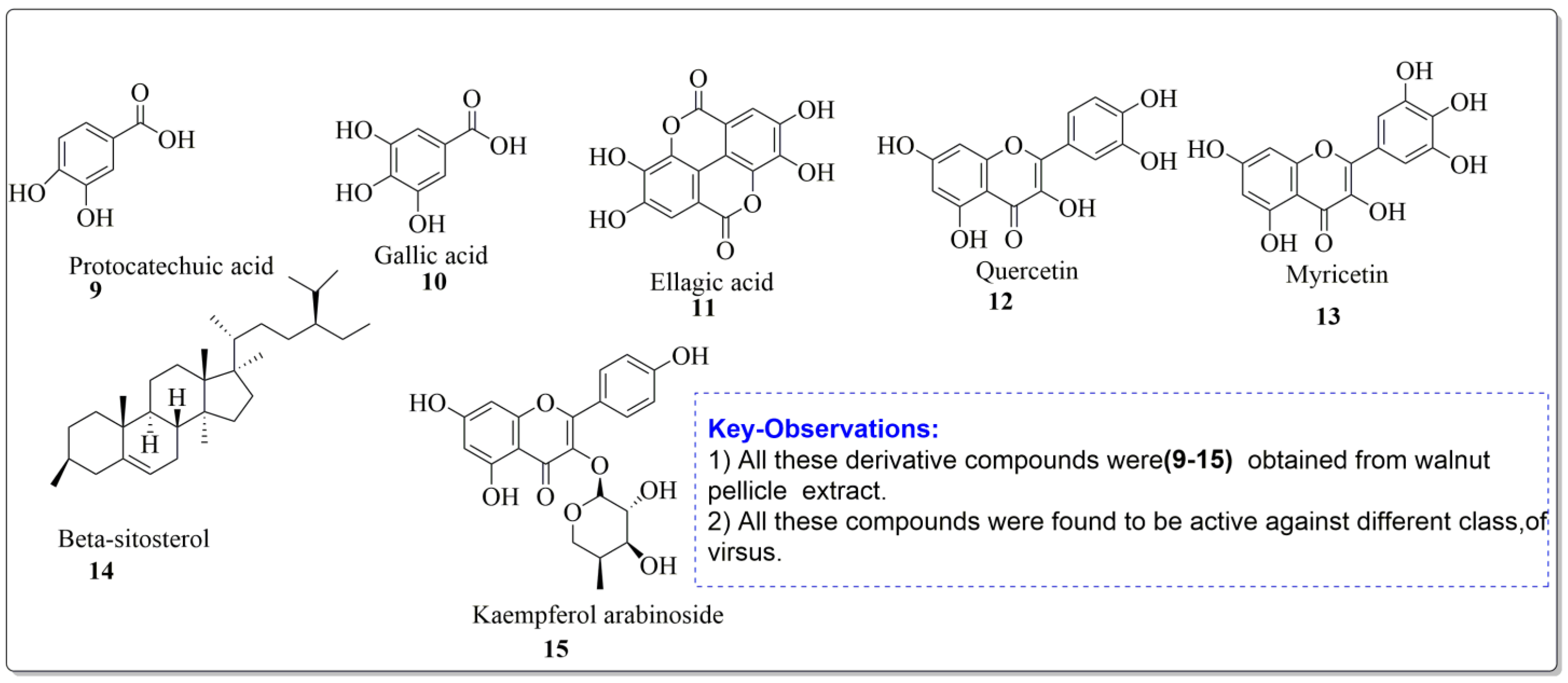
| Gallic Acid | Phenolic |  | Anti-inflammatory | [5,6] |
| Protocatechuic acid | Phenolic |  | Anti-inflammatory and antiapoptotic activities. | [7] |
| Ferulic acid | Phenolic |  | Anticancerous | [8] |
| Sinapate | Phenolic |  | Antioxidant, Antimicrobial | [9] |
| Protocatechuic acid derivative | Phenolic |  | Anti-inflammatory | [10] |
| Ellagic acid | Phenolic |  |
Cytotoxic, Anti-proliferative | [11] |
| p-hydroxybenzoic acid | Phenolic |  | Antimicrobial | [12] |
| p-coumaric acid | Phenolic |  | Antioxidant | [13] |
| Quercetin 3-galactosid | Phenolic |  | Antimicrobial, Antioxidant | [14] |
| Galactose | Phenolic |  | Anticancer, Antidiabetic | [15] |
| Pentose | Phenolic |  | Amino acid synthesis | [16,17] |
| Arabinose | Phenolic |  | Antimicrobial | [18] |
| Xylose | Phenolic |  | Antimicrobial | [19,20] |
| Rhamnose | Phenolic |  | Antioxidant, Antimicrobial | [21] |
| Juglone | Phenolic |  | Antibacterial, Anticancer, Antioxidant | [22] |
| Caffeic acid | Phenolic |  |
Antioxidant, Anti-inflammatory and Anticarcinogenic activity | [23] |
| Vannilic acid | Phenolic |  | Antibacterial, Antioxidant | [24] |
| Quercetin | Flavonoid |  | Analgesic, Antibacterial, Antiviral | [25] |
| Myricetin | Flavonoid |  | Anticancer, Antidiabetic, Antibacterial, Antiviral | [26,27] |
| Kaempferol | Flavonoid |  | Acute and chronic inflammation | [28] |
| Apigenin | Flavone |  | Anti-inflammatory, Antioxidant, Neuroprotective | [29] |
| Luteolin | Flavone |  | Anticancer, Anti-inflammatory | [30] |
| Daidzein | Isoflavonoid |  | Neurobiological activities | [31] |
| Naringenin | Flavanone |  | Antiviral, Antibacterial, Antioxidant | [32] |
| Hesperetin | Flavonone |  | Scavenging activity and Cardioprotective activity | [33,34] |
| Gallocatechin | Flavanol |  | Antioxidant, Antitumor | [35] |
| Sitosterol | Steroid |  | Anticancer, Antidiabetic, Antimicrobial | [36] |
| Stigmast-5-en-3β,7α- diol | Steroid |  | Antimicrobial | [37] |
| Campesterol | Steroid |  | Anti-inflammatory, Anticancer, Antidiabetic | [38] |
| Stigmasterol | Steroid |  | Anticancerous Dyslipidemia, diabetes and metabolic syndrome | [39] |
| Oleanic acid | Tereponoid |  | Dyslipidemia, diabetes, and metabolic syndrome | [40] |
| 3-alpha-Corosolic acid | Tereponoid |  | Antidiabetic, Anti-obesity, Anti-inflammatory | [41] |
| Urosolic acid | Tereponoid |  | Anti-inflammatory, Anticancer, Antidiabetic, Antioxidant | [42] |
| 3-Epikatonic acid | Tereponoid |  | Cytotoxic | [43] |
| Pharmacological Effect | Extract | Model/Experimental-Reference | Dosage-Range | References |
|---|---|---|---|---|
| Antibacterial | Hull extract of ethanol, ethyl-acetate and water | E. coli, B. subtilis, K. aerogenes, S. Aureus | 5 mg/mL | [52,53] |
| Antibacterial | Aqueous leaf extract | Gram-positive and Gram-negative bacteria | 0.1 mg/mL (MIC) | [54,55] |
| Antibacterial | Essential oil and components | Gram-positive and Gram-negative bacteria | Wide range | [56] |
| Antioxidant | Ethyl acetate, butanol, ether, and aq. Extracts of (kernels, husk, and leaves) | NR | 125 µL | [57] |
| Antioxidant | Leaf extract (ethanol and water) | NR | 34.5 and 56.4 µg/mL | [58,59] |
| Analgesic and anti-inflammatory | Aqueous and ethanolic extract of leaf | Wistar rats, human arthritis | 250, 500, 1000, 1500 mg/kg | [60] |
| Antiviral | Methanolic extract | HSC, polio virus and SINV | 2 mg/mL | [61] |
| Antidiabetic | Leaf extract | Human type-ii diabetic patients | Wide range | [62] |
| Anticancer | Juglone | Intestinal carcinogenesis | Wide range | [63] |
| Anticancer | Methanol and aqueous extract of leaf | Mice melanoma (B16F10) and human melanoma (A375) cell lines | Wide range | [64] |
| Anticancer | Juglone and modified juglone | (NCl-H322 and A549) Lung cancers | Wide range | [65] |
| Anticancer | Chloroform leaf extract | Human oral and breast cancer cell lines | Wide range | [66] |
Disclaimer/Publisher’s Note: The statements, opinions and data contained in all publications are solely those of the individual author(s) and contributor(s) and not of MDPI and/or the editor(s). MDPI and/or the editor(s) disclaim responsibility for any injury to people or property resulting from any ideas, methods, instructions or products referred to in the content. |
© 2023 by the authors. Licensee MDPI, Basel, Switzerland. This article is an open access article distributed under the terms and conditions of the Creative Commons Attribution (CC BY) license (https://creativecommons.org/licenses/by/4.0/).
Share and Cite
Bhat, A.A.; Shakeel, A.; Rafiq, S.; Farooq, I.; Malik, A.Q.; Alghuthami, M.E.; Alharthi, S.; Qanash, H.; Alharthy, S.A. Juglans regia Linn.: A Natural Repository of Vital Phytochemical and Pharmacological Compounds. Life 2023, 13, 380. https://doi.org/10.3390/life13020380
Bhat AA, Shakeel A, Rafiq S, Farooq I, Malik AQ, Alghuthami ME, Alharthi S, Qanash H, Alharthy SA. Juglans regia Linn.: A Natural Repository of Vital Phytochemical and Pharmacological Compounds. Life. 2023; 13(2):380. https://doi.org/10.3390/life13020380
Chicago/Turabian StyleBhat, Aeyaz Ahmad, Adnan Shakeel, Sadaf Rafiq, Iqra Farooq, Azad Quyoom Malik, Mohammed E. Alghuthami, Sarah Alharthi, Husam Qanash, and Saif A. Alharthy. 2023. "Juglans regia Linn.: A Natural Repository of Vital Phytochemical and Pharmacological Compounds" Life 13, no. 2: 380. https://doi.org/10.3390/life13020380
APA StyleBhat, A. A., Shakeel, A., Rafiq, S., Farooq, I., Malik, A. Q., Alghuthami, M. E., Alharthi, S., Qanash, H., & Alharthy, S. A. (2023). Juglans regia Linn.: A Natural Repository of Vital Phytochemical and Pharmacological Compounds. Life, 13(2), 380. https://doi.org/10.3390/life13020380