
Article

Non-Invasive Trace Element Fingerprinting of the Lion Man, a Left Mammoth Tusk and Mammoth Ivory Fragments Found at the Hohlenstein-Stadel Cave, Swabian Alb, Germany

1
Laboratoire de Développement Instrumental et de Méthodologies Innovantes Pour Les Biens Culturels (Lab-BC), UAR 3506 Centre National de Recherche Scientifique (CNRS)-Centre de Recherche et de Restauration des Musées de France (C2RMF)-Chimie ParisTech-Université Paris Sciences et Lettres, 75001 Paris, France
2
Institut Photonique D’analyse Non-Destructive Européen des Matériaux Anciens (IPANEMA), UAR 3461 CNRS-Ministère de la Culture-Université de Versailles Saint-Quentin-en-Yvelines -Muséum National d’Histoire Naturelle, 91192 Gif-sur-Yvette, France
3
Centre de Recherche et de Restauration des Musées de France (C2RMF), 75001 Paris, France
4
Formerly Museum Ulm, 89073 Ulm, Germany
*
Author to whom correspondence should be addressed.
Minerals 2026, 16(7), 755; https://doi.org/10.3390/min16070755
Submission received: 31 March 2026 / Revised: 24 June 2026 / Accepted: 7 July 2026 / Published: 19 July 2026

Abstract

The Lion Man sculpture (UNESCO World heritage), a left mammoth tusk and mammoth ivory fragments from the excavations in the Hohlenstein-Stadel cave, Lone valley, Swabian Alb, Germany, were non-invasively analysed by means of external ion beam analysis (IBA) at the microfocus beamline at the particle accelerator AGLAE. The Lion Man was reconstructed from about three hundred mammoth ivory fragments, while the tusk is a separate find, and the fragments are individual pieces that could not be placed in the Lion Man. A characteristic trace element fingerprint, based on zinc, bromine and strontium contents, was established according to previous IBA studies of Aurignacian-era mammoth ivory and allowed for a comparison of the mammoth ivory objects. The specific Hohlenstein-Stadel cave trace element fingerprint could be distinguished from that of other Aurignacian sites in Europe but closely resembles that of the contemporary ivories from the neighbouring Hohle Fels cave. Although this study highlights that the Lion Man sculpture is chemically inhomogeneous due to diagenetic alterations, the left tusk and individual ivory fragments are chemically very similar to the Lion Man. However, further analyses are required to relate them to the same animal. Secondary minerals such as black manganese oxide dendrites and iron-rich aluminosilicates could be identified on the Lion Man and the other ivory objects at the surface. While dendrites are a characteristic diagenetic feature of mammoth ivory, the origin of iron-rich aluminosilicates can be linked either to sediment traces or surface treatment and use wear of the sculpture. Iron-rich zones are identified on particular parts of the Lion Man, namely, at the snout and at the left forearm with decorative signs. The snout is also particularly enriched in carbon, whose origin still needs to be clarified.

1. Introduction

In the Upper Palaeolithic, mammoth ivory was an important raw material to produce tools and jewellery, as well as figurative objects, which are among the oldest preserved works of art of humankind. The earliest use of mammoth ivory was evidenced in Europe for the Aurignacian period, ca. 43.000–33.000 cal BP, and seems to be related to the arrival of the modern humans in Europe. The scientific examination of ivory artefacts can provide us with essential information about the expertise of Stone Age people, their choice and procurement of materials, the techniques they used and their cultural practices.
The World Heritage site ‘Caves and Ice Age Art in the Swabian Jura’ is a globally unique archaeological landscape in the Ach and Lone valleys in the south of Germany, Baden-Württemberg, inscribed on the UNESCO World Heritage List in 2017 [1]. Six caves are united within it, which are known for their extensive and significant finds from the Aurignacian period, many of which consist of mammoth ivory (e.g., [2]). To date, over fifty figurines carved from mammoth ivory or bone have been found there. Most of these art objects depict the fauna of the Ice Age landscape, such as mammoths, horses, bison, etc. (e.g., [3], Vogelherd cave), but also humans, such as the Venus of Hohle Fels, which is known worldwide as the oldest female representation of its kind [4]. In addition, the world’s oldest flutes were found here [5,6]. Other outstanding examples of the representation of hybrid creatures with human and animal characteristics were found in the region, such as the Lion Man statuette in the remote area of the Hohlenstein-Stadel (Ho-St) cave [7,8,9,10].
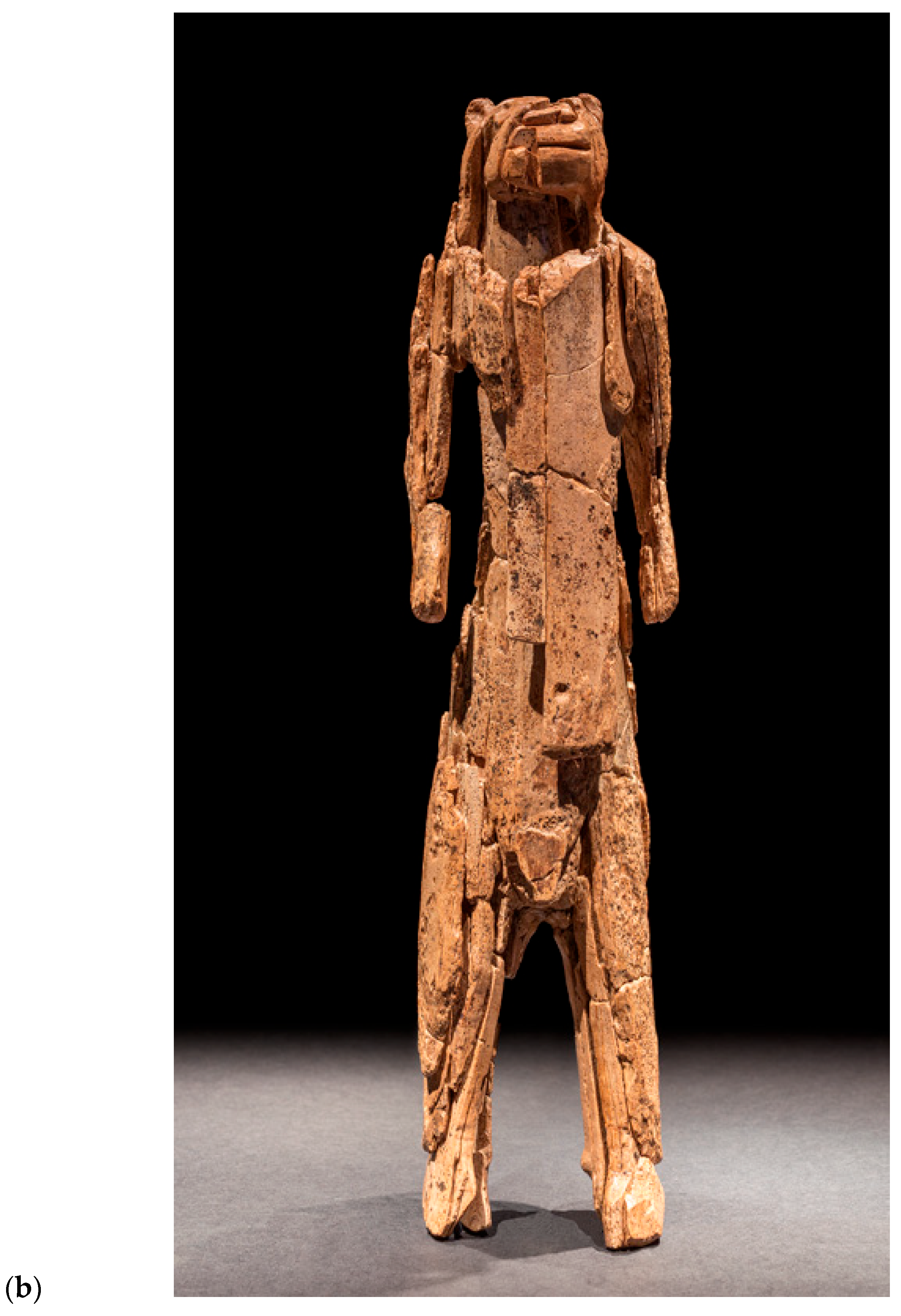
The Hohlenstein-Stadel cave is located in the Lone valley of the Swabian Alb. In 1939, 1960 and 2008–2013, several hundred mammoth ivory fragments were excavated there among other archaeological objects, from a part of which the Lion Man, the largest Ice Age figurine (ca. 31 cm high), could be assembled like a puzzle [11]. In total, the statuette is composed of about three hundred different ivory pieces, which were identified through careful examinations as part of the Lion Man. The therianthropic figurine has animal features—specifically, the head of a cave lion—and the body of a human. The Lion Man figurine was dated by the radiocarbon (14C) method to a range between 41.100 and 39.400 cal BP according to the data obtained for objects from the lower Aurignacian layer (Au) [11,12]. It is thus considered not only the largest but also the oldest known animal/human sculpture in the world [7]. Due to various factors, such as the type of objects, its specific raw material, age, amount of work involved, specific find location, and the presence of a sign composed of seven carved parallel lines on the left arm and use wear, the figurine is of particular significance. It is believed that it could have been a cult object.
In the same cave, a left tusk of a young mammoth and other fragments that could not be assigned so far were found during excavations.
Mammoth ivory artefacts from several European Aurignacian sites were studied previously by our team by means of external micro-proton-induced X-ray and gamma-ray emission (PIXE/PIGE) imaging at the microfocus beamline NewAGLAE of the 2-MV tandem particle accelerator AGLAE, installed at the C2RMF [13]. They showed that ion beam analyses (IBA) such as PIXE and PIGE are powerful and effective methods for examining ancient ivory artefacts non-invasively [14]. Specifically, the studies allowed for the identification and quantification of biogenic trace elements (strontium (Sr), zinc (Zn), and bromine (Br)) in Aurignacian ivories. These elemental patterns show inter-regional differences for different Aurignacian ivories [14]. The marker elements Zn and Sr are also detectable in recent ivory and are incorporated into the ivory during tusk growth. It is known that the Sr content, in particular, varies with the habitat and thus the diet of the individual. Diagenetic changes in the trace element patterns during burial (leaching, enrichment, exchange, etc.) also need to be considered. Therefore, these trace element ratios could be defined as ‘combined biogenic/diagenetic trace element pattern characteristic of each site’, or in short, ‘site-specific markers’, because they not only provide insights into the animal’s living conditions (the origin of the ivory) but also likely reflect specific influences of diagenetic changes that occurred while the material was buried at a particular archaeological site. Our previous studies also evidenced characteristic minor and trace elements (especially iron (Fe), manganese (Mn), and fluor (F)) in the archaeological ivory composition that were enriched in the artefacts through exchange with the environment and are therefore considered ‘diagenesis-related markers’ [15,16,17]. Furthermore, secondary minerals such as Fe-rich aluminosilicates and Mn oxides were also identified on the surface of archaeological and fossil ivories [18,19]. The latter can be formed during diagenetic processes. In the case of Fe-rich aluminosilicate, they can represent either sediment traces from the site or traces of surface treatments such as use wear or ochre traces linked to the production of worked ivory objects.
The aim of the present study was to characterize as precisely as possible the chemical composition and thereby establishing a characteristic fingerprint of the mammoth ivory from the Lion Man, the left tusk and the individual fragments from Hohlenstein-Stadel.
On the one hand, the identified ‘site-specific’ or ‘diagenesis-related markers’ of the Lion Man are discussed based on the data obtained, and on the other hand, possible relationships between the figurine and the other ivory objects are stressed. Furthermore, further insights into possible use wear of the figurine or specific surface treatments of the Lion Man are expected. Specifically, this study aims to answer the following research questions:
(1)
Is it possible to define a site-specific composition of the Aurignacian-period mammoth ivory from the Ho-St cave, and does this composition correspond to that of other contemporaneous ivory finds from the same region, the Swabian Alb?
(2)
Does the Lion Man have a consistent chemical fingerprint, and how does this relate to the chemical composition of the fragments and the unworked tusk? Can conclusions be drawn about whether they belong together based on their chemical composition? Can taphonomic processes be evidenced by IBA on the different objects, and how can the very different states of preservation of the various sculpture fragments be explained?
(3)
Is it possible to carry out a use wear analysis by IBA? In this respect the comparison of the worked sculpture with the unworked tusk is also of importance.
Specific questions from an archaeological perspective are as follows:
(4)
Can we find further evidence that would enable us to relate the fragments to the sculpture, or indications that the unworked tusk is the other tusk of the same animal that also provided the material for the production of the Lion Man figurine?
(5)
Is it possible to detect chemical traces that indicate specific use of the object, possibly of a ritual nature?
Given the inestimable value of the artifacts, the investigations had to be carried out non-invasively and without taking any sample or causing harm to the objects. Moreover, the data of the Ho-St ivories should be comparable to those obtained on mammoth ivory from the other European Aurignacian sites. Therefore, the analyses were carried out using exactly the same IBA setup and analogous measurement conditions as our previous studies of Aurignacian mammoth ivory [14].

2. Materials and Methods

2.1. Lion Man Sculpture and Ivory Objects from Hohlenstein-Stadel Cave

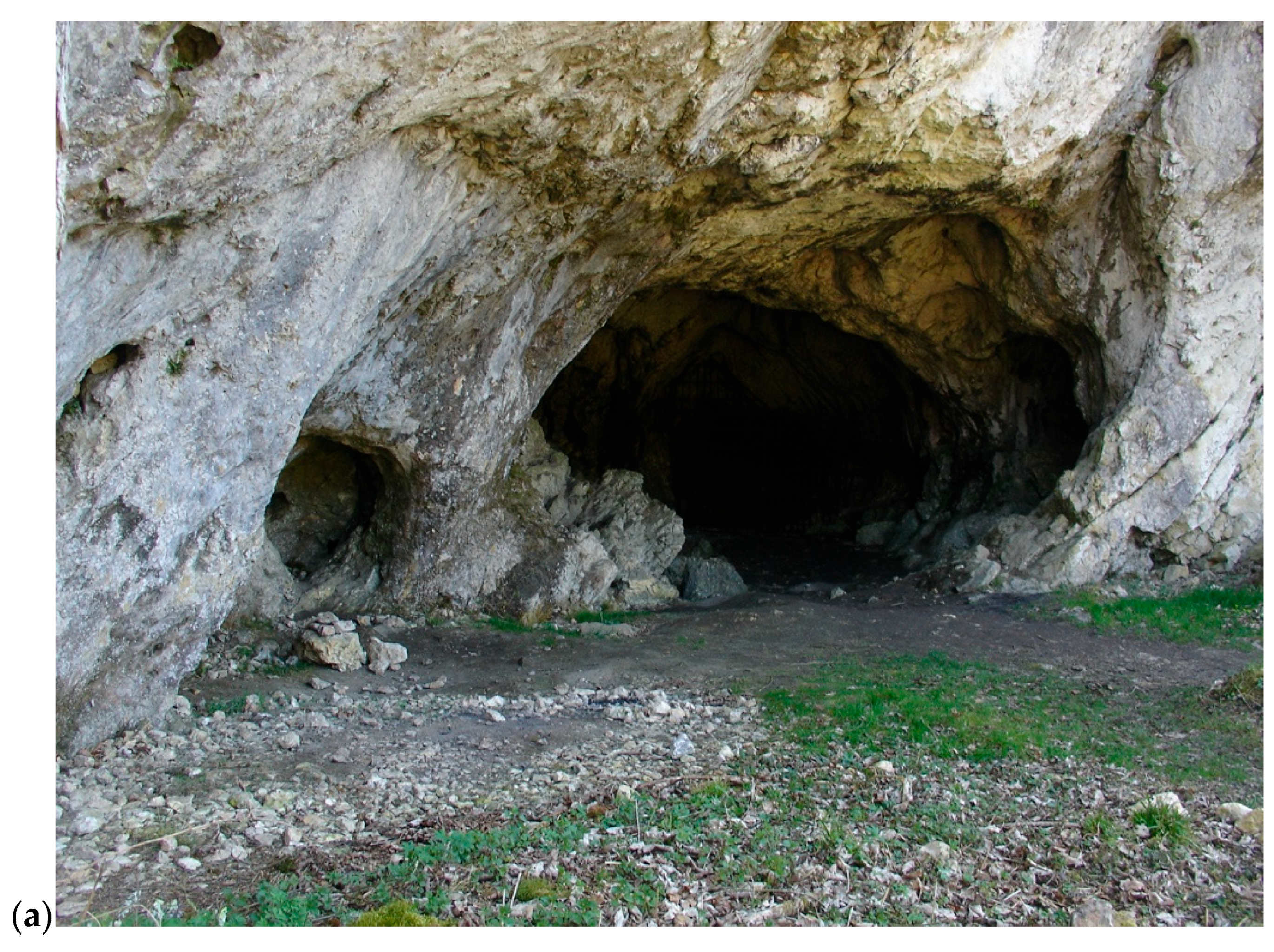
The study objects in this work originate from the Stadel cave on the Hohlenstein rock massif, Lone valley, Swabian Alb, Germany (Figure 1). During archaeological excavations in 1939, shortly before the outbreak of the Second World War, numerous fragments of mammoth ivory were discovered in a small niche at the end of the cave, in an Aurignacian layer, together with some pierced fox teeth and reindeer antlers. It was not until thirty years later that J. Hahn succeeded in piecing the fragments together to form an animal/human figurine, the so-called Lion Man [8,10]. More recent excavations have unearthed further finds, which were added during restoration work. Given its decades-long history of discovery, assemblage and restoration, the Lion Man has, of course, been exposed to many external influences, both during storage and whilst undergoing various restoration processes. After its discovery amongst the finds from the cave by Joachim Hahn, conventional adhesive UHU® was used for the initial provisional assembling. During the first professional restoration in the workshops of the Württemberg State Museum in Stuttgart in 1987/88, the restorer Ute Wolf deliberately continued to use UHU Hard® so as not to introduce any other adhesives into the figurine. Older (incorrect) adhesions were dissolved using acetone and ethyl acetate, and many fragments were hardened with Mowilith 50® [9]. Following the discovery of further fragments during the excavations carried out by the State Office for the Preservation of Historical Monuments between 2008 and 2013, the sculpture was finally restored in the State Office’s workshops in Esslingen. During this work, led by restorer Nicole Ebinger-Rist, no further impregnation or consolidation was carried out; in some areas, however, previous treatments were reversed. New adhesives were applied using aryl resin [20].
Today, the Lion Man figurine is made up of almost three hundred individual parts. Measuring 311 mm in height and 73 mm in width, it is the largest of its time. The figurine is nearly complete, although some fragments of the original surface are lost. Investigations using X-ray radiography (XRR) have shown that the figurine was carved from a right tusk [9].
Various factors contribute to the figurine being accorded special significance:
(1)
The type of hybrid representation between a man and a cave lion;
(2)
The enormous amount of work involved (estimated at >360 h) [21];
(3)
Its deposition location: a small niche deep inside the cave, which may have been a hiding place or a space for ritual activities;
(4)
The presence of a sign sequence of seven parallel carved lines on the left forearm;
(5)
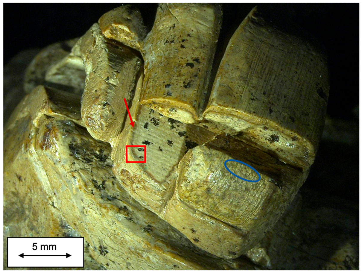
Signs of use wear, like the ‘infiltration zone’ visible at the cross-section and numerous fine grooves in the snout area (Figure 2).
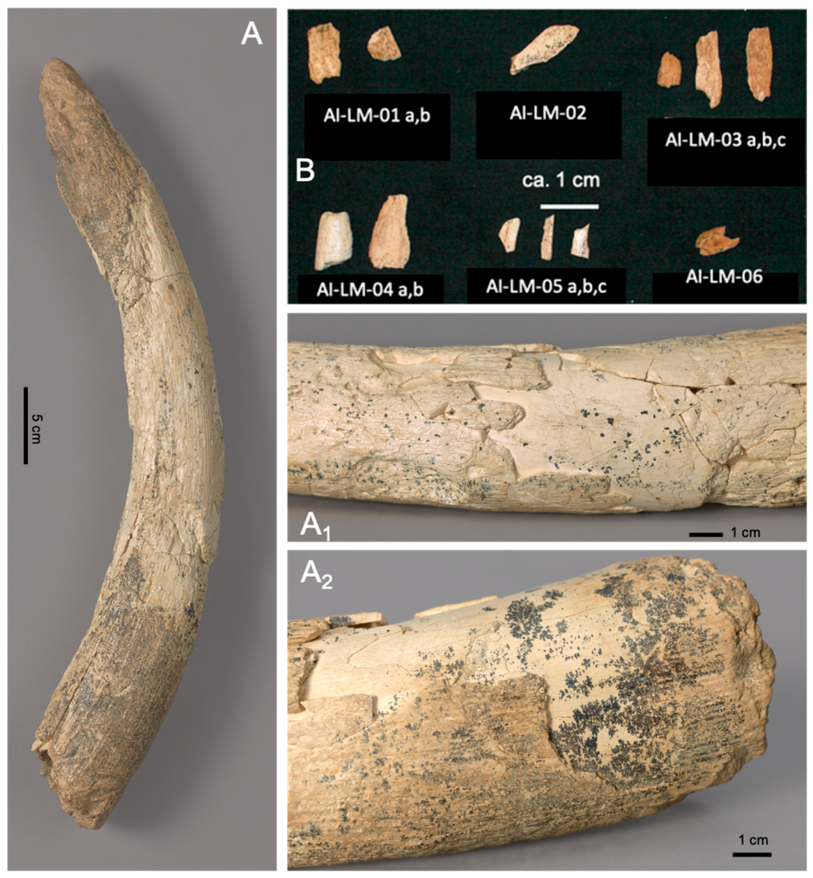
Furthermore, an almost intact left tusk of a young mammoth from the Ho-St cave was included in the investigations. The tusk was recovered in 1960 about 6 m away from the figurine’s location from a deeper layer, which is assigned to the Middle Palaeolithic. The tusk shows typical ivory degradation features, visible to the naked eye: formation of Mn oxide dendrites and flaking of outer layers (Figure 3). It is not known which substances were used during the restoration of the left tusk [9]. Characteristic features such as similar size, opposite curvature and the end of the pulp cavity corresponding roughly to the crotch area of the figurine led to the hypothesis that this tusk might be the unworked counterpart to the one used for the Lion Man. This is, however, contradicted by its stratigraphically deeper position in Middle Palaeolithic layers. One attempted explanation was the rather crude excavation technique used in the early excavations of the 1930s and 1960s, during which a depression from higher levels might not have been recognised; i.e., the unworked tooth may have been buried to preserve its carvability. But this cannot be substantiated in retrospect.
In addition, several mammoth ivory fragments which also originate from excavations in the same or similar archaeological layers in the cave were included in this study.
Certain fragments have been classified as likely belonging to the Lion Man (although they could not be integrated into the figurine) on the basis of the specific find circumstances, the presence of traces of processing or their colour (AI-LM-01, -02 and -03). For the others, they are either not belonging to the figurine (AI-LM-04), coming from the same archaeological layer (AI-LM-05), or their classification remains uncertain (AI-LM-06). It is hoped that this research study will help refine the attribution of the fragments. A total of nine out of twelve individual ivory fragments (all except AI-LM-01a, -03a and -04a) could be investigated in this study (Figure 3).

2.2. Experimental Conditions of Ion Beam Analysis (IBA) of Hohlenstein-Stadel Ivory Objects

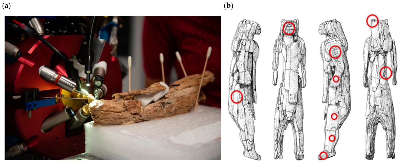
The investigations of the Ho-St ivories were carried out in one run of two days at the external microbeam setup of the 2MV tandem accelerator (Pelletron 6SDH-2) New-AGLAE at Centre de recherche et de restauration des musées de France (C2RMF) in Paris without any surface cleaning prior to the analyses. The setup thus allows for simultaneous micro-proton-induced X-ray emission (PIXE)/gamma-ray emission (PIGE) and Rutherford Backscattering (RBS) analyses enabling the qualitative and quantitative determination of all elements from carbon (C) to uranium (U) (Figure 4a).
A 3 MeV proton beam with a beam size of about 40 µm was used for the measurements in a helium (He) atmosphere. The device operates in imaging mode with high spatial resolution, the analysed area ranges from a few tens of µm2 to 2.5 cm2. The imaging setup is equipped with a designed detection system consisting of three silicon (Si) drift detectors (SDDs) for high-energy X-rays, one SDD for low-energy X-rays, an HPGe detector for the detection of emitted gamma-rays and an external RBS detector (Passivated Implanted Planar Silicon, PIPS). The low-energy X-ray detector is used for the detection of lighter elements from sodium (Na) to Z = 20 (Ca) using a He flux to reduce absorption of X-ray fluorescence and the high-energy detectors for the analysis of heavier elements with Z > 20 using a 50 µm aluminium (Al) filter.
The study object was safely positioned in front of the measuring head of the New-AGLAE beamline on a motorized sample stage (Figure 4a). Two-dimensional scans were carried out by precisely moving the object. A pixel size of 10 µm was selected for the spatial resolution of the scans, which is smaller than the actual beam size. This oversampling allows chemical maps of various areas to be generated with higher resolution.
Twenty-two small scans ranging in size from 500 × 500 µm2 to 1600 × 1500 µm2 were performed on the figurine, depending on accessibility and local conditions (Figure 4b). Five scans were performed on the tusk, as well as at least one scan per fragment, typically of 1000 × 1000 µm2. The measurement time per scan ranged from 10 min to 1 h 40 min for the longest scan. The typical charge for a 10 min scan (500 × 500 µm2 corresponding to a dose of 1,000,000) was of 3.35 µC.
The criteria for selecting measurement locations were guided by three conditions: first, the safe positioning of the object while accessing the chosen zone; second, the representativity of the zone with respect to the whole object, as the analyses should cover as many parts of the figurine as possible; and third, the surface state of the zone (absence of other mineral deposits, unconsolidated or consolidated fragments to study the effect of consolidation, and zones with specific mineral deposits to characterize the deposits as well). Consolidated zones can be recognized by their shiny surface features. The surface of unconsolidated fragments is much lustreless in comparison.
Selected low-energy PIXE spectra are shown in Figures S1–S5 and the corresponding high-energy PIXE spectra in Figures S6–S10. Representative spectra for different measurement spots on consolidated or unconsolidated fragments of the Lion Man (0016, 0020, 0040, and 0041) were compared to a modern elephant ivory reference (0027). The corresponding RBS spectra of different consolidated and unconsolidated parts of the Lion Man are shown in Figures S11–S15 besides that of modern elephant ivory.
Sediments from the cave could not be included in this study. Because of the complex excavation history of the finds, the influence of burial sediment on the ivory fragments can hardly be studied in a systematic way.

2.3. Quantitative IBA Data Evaluation

PIXE analysis is an elemental surface-sensitive method. The information depths vary depending on the element-specific X-ray emission energy and are, in ivory, approx. 14 µm for Si, 50 µm for Ca, 65 µm for Zn, and 68 µm for Sr [13]. The quantitative elemental contents were calculated by PIXE with the pivot method described in [22]. The quantification uses GUPIX® software, which considers fundamental parameters of the ion beam–material interactions. Several standards (DrN, Brill A and B) were analysed additionally to check the quality of the quantification procedure. No calibration was required. The detection limits have been estimated and are in the range of some tens of ppm. They lie in the range of some hundreds of ppm for lighter elements between Na and Ca. For the visualization of the chemical PIXE maps, data are expressed by measured elemental peak area pixel by pixel.
PIGE has a larger information depth followed by RBS. Fluorine (F) and Na contents were calculated using the characteristic gamma-rays at 110 and 197 keV for F and 440 keV for Na of the PIGE spectra. Adequate references including Fossil Bone Standard (FBS) [23] and fluorapatite (FAP) were used for calibration.
RBS spectra were used to evaluate the presence of carbon (C). The spectra were not simulated using traditional simulation software such as SIMNRA. Since with a 3 MeV proton beam, C and oxygen (O) fronts are well separated with kinematic factors of 0.73 and 0.79 (approx. 180 keV in energy), they can be easily separated. For quantification and reporting, elemental concentrations are expressed as oxides. Therefore, O can be considered a constant background in the elastic (non-Rutherford) backscattering spectrometry (EBS) spectra. Consequently, C distribution maps can be obtained using the corresponding region of interest (ROI; channels 317 to 327) of the EBS spectrum. The contribution of heavier elements has been subtracted using an ROI at higher energy (channels 330 to 340) to get the net C distribution (see Figure S16). RBS maps are expressed as a function of subtraction of the areas of the hereabove ROIs defined pixel by pixel.

3. Results

3.1. Major Element Composition of the Ivories

The analytical results of the quantitative elemental analyses of the ivory finds from the Ho-St site are summarized in Table 1. Full data sets are found in Tables S1 and S2. The data show the typical composition of archaeological ivory with about 50 wt.% of calcium oxide (CaO) and about 30 wt.% of phosphorus oxide (P2O5) as main components. The chemical composition of the studied ivory objects is characteristic of archaeological ivories and relatively inhomogeneous, especially for elements such as Al and silicon (Si), indicated by a high standard deviation for the mean compositional values; the reasons for this are discussed in detail in Discussion Section 4.1, Section 4.2, Section 4.3, Section 4.4, Section 4.5, Section 4.6 and Section 4.7.

3.2. Minor and Trace Element Composition of the Ivories

Different minor and trace elements, such as Mn, Fe, Zn, Br and Sr, could be identified in the ivory composition. Variations in chemical composition at the level of minor and trace elements can be used to draw conclusions about the provenance of the artifacts, their diagenesis, or other important issues, such as, in our case, use wear or traces of production (Table 2). The detailed results of all individual measurements are presented in Tables S1 and S2.
The Lion Man statue and the individual fragments have higher average values for Fe2O3, ZnO and SrO contents than the unworked tusk, while the average SiO2 content is similar across all types of finds. However, trace element levels vary quite significantly between the individual measurement areas. The obtained data are discussed and contextualized in detail below, in Discussion Section 4.1, Section 4.2, Section 4.3, Section 4.4, Section 4.5, Section 4.6 and Section 4.7.

4. Discussion

4.1. Combined Biogenic/Diagenetic Trace Element Pattern Characteristic of the Hohlenstein-Stadel Assemblage

The analysed major element oxide contents of 50 wt.% of CaO and 30 wt.% of P2O5 are consistent with carbonated hydroxyapatite, a calcium phosphate phase, the main mineral compound of bone and teeth materials including mammoth ivory. A minor element of elephant and mammoth ivory, which distinguishes it from bone and other teeth, is magnesium (Mg). Modern ivory contains up to 6 wt.% of Mg, but the Mg-containing phase is highly soluble; therefore this element is generally partly or completely washed out in archaeological ivory [24]. Magnesium oxide with values ranging between 0.3 and 0.6 wt.% is depleted in the Ho-St ivories. Zinc, Sr, and to a certain extent also Br are typical ‘biogenic’ trace elements found in archaeological ivories, which our previous studies have shown [15,16] appear to be relatively well preserved in the material. Therefore, these three elements were used in previous work to define so-called ‘site-specific markers’ [14]. Although ‘site-specific markers’ are representative of an individual’s specific living environment, they are also subject to a certain degree of influence from the diagenetic processes at their site of discovery, as the chemical composition of ivory is generally altered during burial through complex biogeochemical transformation processes. These processes depend on the specific conditions of the various types of archaeological sites, such as karst caves, rock shelters, or open-air sites. Therefore, these markers should be more likely considered combined biogenic/diagenetic trace element patterns characteristic of the corresponding site.
It should be noted that archaeological artefacts always have some degree of secondary minerals, such as sediment deposits on their surfaces, and that this can affect measurement results, particularly in surface-sensitive analyses. Sediments from the Ho-St cave could not been studied here, but in a general way, sediments are rich in Si, Al and titanium (Ti). Silicon can be used as indicator of the presence of sediment deposits on mammoth ivory. However, the same type of compound is also characteristic of red colouring matter, which consists of Fe oxide-rich aluminosilicates. Such red colouring matter or Fe oxides are frequently found in prehistoric contexts, including in the cave sites of the Swabian Alb, such as the Vogelherd cave in the Lone valley [25,26]. There are numerous examples of objects from the Palaeolithic era on which Fe oxide-rich deposits have been identified, such as personal ornaments or other statues [26]. It was for instance used as a polishing agent. Therefore, it is not inconceivable that the Lion Man was also worked with ochre or Fe oxides. Red Fe-rich compounds could also have been mixed with water to represent blood during ritual use of the statuette. Therefore, Fe traces at the object’s surface may indicate both sediment deposits or traces related to the object’s production or use. However, the fragmentary nature of the statuette limits the study of potential use wear and traces of manufacture on the original object surface.
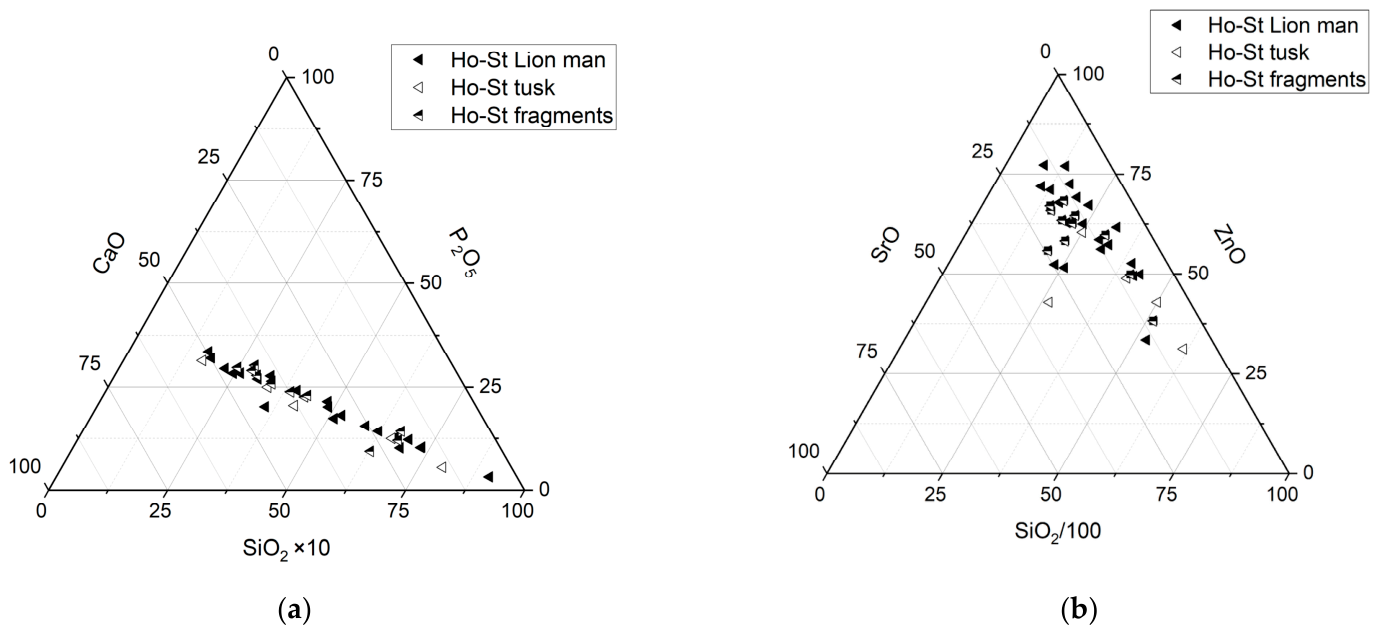
The major elements of Ho-St ivory and the characteristic biogenic trace elements are related to the Si content to evaluate the reliability of the ‘site-specific’ elemental fingerprint. Figure 5a shows an anti-correlation between the Si contents and the measured values for the major elements in ivory (Ca and P). Both the Ca and P values decrease as the Si value increases. The presence of aluminosilicate deposits on the measured ivory surface therefore affects the quantification of the ivory material. Consequently, areas with sediment deposits characterized by high Si content (>10% SiO2) should be avoided, whenever possible, when analysing the ‘site-specific’ chemical fingerprint. Otherwise, the overall composition—particularly P and Ca contents—must always be taken into account in order to assess the impact of surface deposits on the quantitative results. Generally, measurements of lighter elements, for which the depth of information provided by the method used is lower, are more strongly influenced by surface changes. This is why the effect is particularly noticeable for P values.
Nevertheless, the measured concentrations of the trace elements Sr and Zn appear to be relatively independent of Si content, indicating their validity as ‘site-specific markers’. For some of the measurements, Zn content shows a slight negative correlation with Si, suggesting that the presence of aluminosilicate traces could influence the measured Zn content (Figure 5b). In these cases, the overall composition will be considered to assess the reliability of the results. The Br value is close to the detection limit for Ho-St ivory finds; therefore its relationship with SiO2 content cannot be studied.

4.2. Inter-Regional Comparison of Ivories from Aurignacian Sites

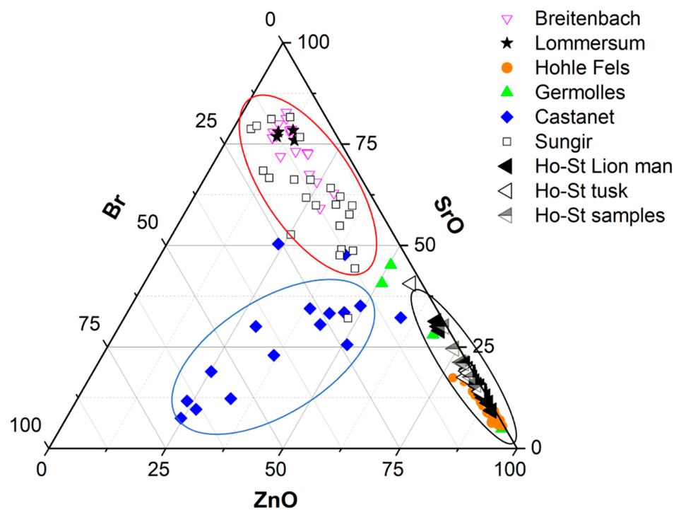
Figure 6 shows the relative Zn, Sr, and Br contents in the ivory from seven different archaeological sites, including the Ho-St cave, where ivory artefacts were found in Aurignacian layers. Three main groups can be distinguished: the northernmost sites with relatively high Sr content (Breitenbach, Lommersum, and Sungir, all open-air sites), the Castanet site in southern France with relatively Br-rich ivory, and the sites in southern Germany (Hohle Fels and Ho-St caves), which are characterized by very low Br content and comparatively low Sr values. The ivory finds from the Germolles site, Burgundy, France, fall with a few exceptions between those from northern and southern Germany and differ also from the ivory finds from Castanet [27]. Figure 6 illustrates that the results for the ivory from Ho-St correspond very well with those from the Hohle Fels cave. This finding is consistent with the regional concept, as the two are cave sites very close to each other, in two neighbouring valleys of the Swabian Alb and thus in the same region.

4.3. Intra-Regional Comparison of Ivories from Lone and Ach Valleys, Swabian Alb

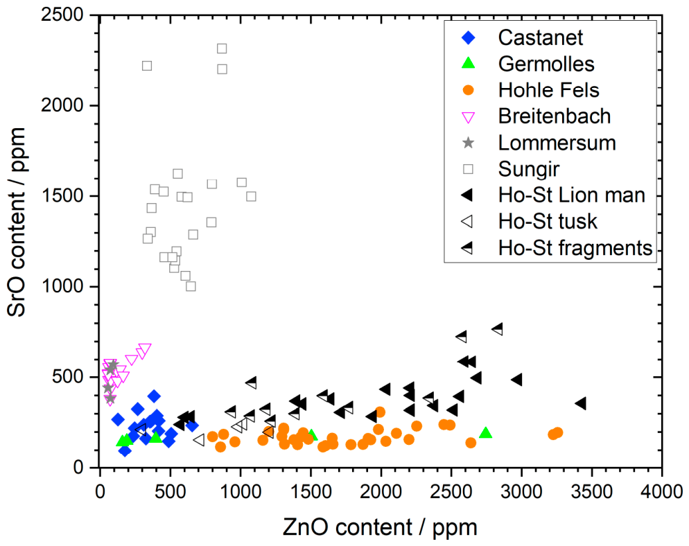
Basing the intra-regional comparison on the defined ‘site-specific markers’ Zn, Sr and Br, Br content can be disregarded in ivories from southern Germany because it is close to the detection limit. Considering Sr and Zn contents, it can be observed that on the one hand, Zn content varies considerably in both caves and, on the other hand, the Sr content in the ivory objects from Ho-St is systematically higher than that in the finds from the Hohle Fels cave (Figure 7). This observation still needs to be explained, but it can be used to distinguish ivories from Ho-St and Hohle Fels.

4.4. Comparison of Trace Element Composition of Hohlenstein-Stadel Ivories

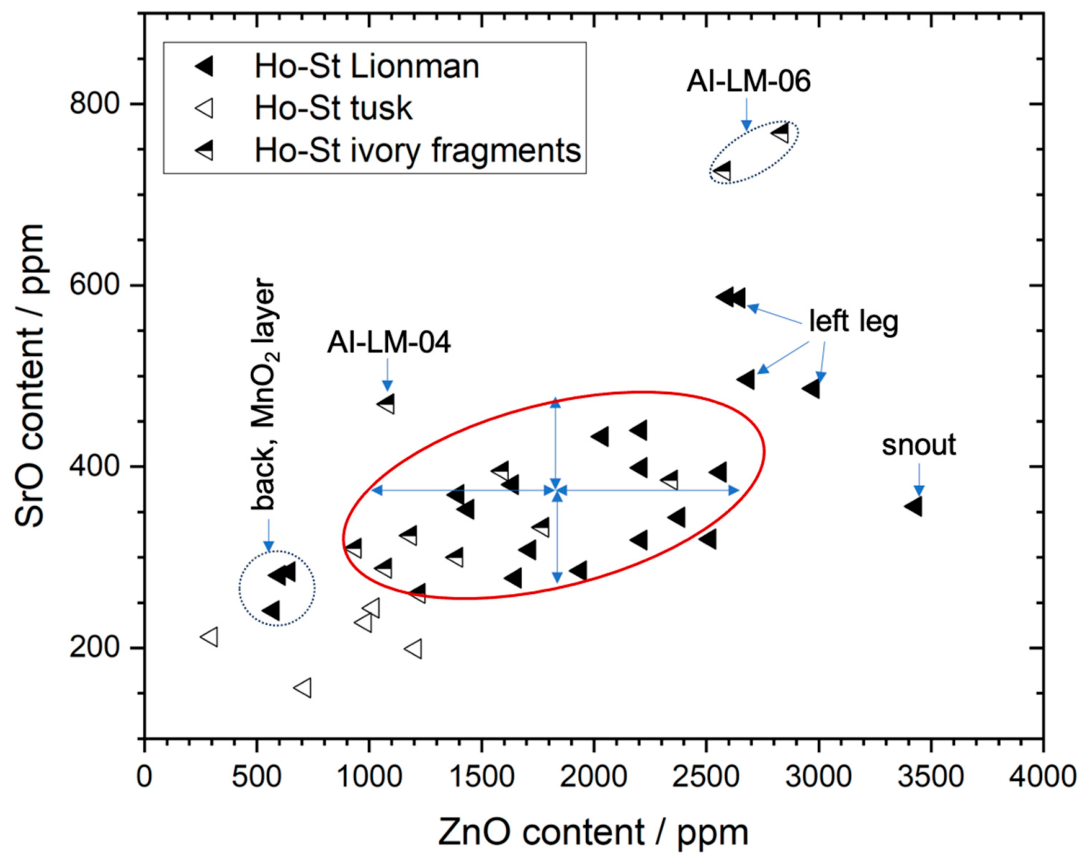
Most of the characteristic trace element data points of the Lion Man lie within the red oval, representing the variations in SrO and ZnO around the mean values obtained for the figurine (Figure 8). There are a few outliers, such as the three measurement points on a fragment of the back, which is thickly covered with a layer of Mn oxide dendrites. It can be assumed that this layer influenced the determined values and led to supposedly lower Zn and Sr contents (Table S4). In contrast, the Sr and Zn values of the left leg and of the snout are increased with respect to the other parts of the Lion Man (Table S2). This discrepancy still needs to be explained.
The SrO and ZnO results for the left mammoth tusk are close to those of the main Lion Man group. With only five measurement points, the statistics are low, but a trend is visible, and a few points can be discussed. As the detailed images of the tusk (Figure 3) show, the outer layer has flaked off in a lamellar pattern in some areas, a typical sign of aging in ivory. Two of the measurements were taken in a small zone of the inner layer and two others on the outer layer. The Sr values on the inner layer are higher in comparison with the other ones. These variations fall within the ‘natural’ Sr range due varying Sr uptake during the growth of the tusk, probably based on migration-related or seasonal variations in the mammoth’s diet like those found for elephants [28,29]. In addition, the measurements taken in different areas of the tusk—near the pulp, more toward the centre, or at the tip of the tusk—differ in their Zn content. This probably has a similar cause, namely, variations in Zn uptake over time or local diagenetic changes. A tusk grows in a helical way, meaning simultaneously in length and width, and the length growth is decisive here [30,31]. The ’natural’ variation in element uptake during the growth of the tusk can, of course, also be applied to the Lion Man figurine, since its carved surface corresponds to the various growth layers.
Regarding the individual ivory fragments, the measured values of the characteristic trace elements show extensive overlap with those of the Lion Man. This applies to fragments AI-LM-01, -02, -03, and -05, so that these fragments could, from a chemical point of view, belong to the Lion Man, or at least to the same archaeological layer, without representing direct proof, as it had indeed been assumed from archaeological considerations for the samples AI-LM-01 to AI-LM-03 and AI-LM-05. Deviating values were obtained for ivory fragments AI-LM-04 and AI-LM-06, whose affiliation with the Lion Man statue is therefore confirmed to be uncertain or has been questioned from an archaeological point of view. Our analyses therefore confirm the archaeological observations [32].

4.5. Diagenetic Processes of Mammoth Ivory at Hohlenstein-Stadel Cave

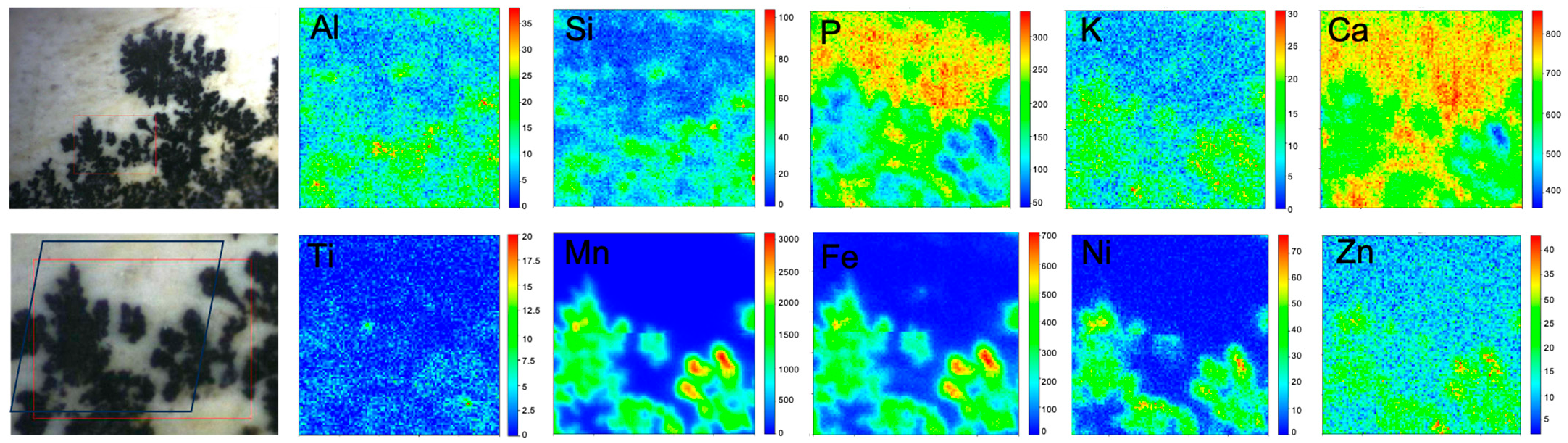
At the macro-scale, the left mammoth ivory tusk and the Lion Man statuette exhibit a concentric, layered structure. As the material ages, water can penetrate between the lamellae in particular and introduce exogenous ions. If the infiltrating water contains Mn and O ions, Mn dendrites begin to form; their shape (branching) and density depend on the Mn/O ratio in the liquid and the size of the crack opening or the interlayer between two lamellae (Figure 3). The development of Mn dendrites between the ivory layers probably fosters, among other processes, the flaking of the lamellae from the outside inwards. The presence of Mn dendrites on substrates in soils is often related to biomineralisation [33,34].
Figure 9 shows PIXE maps obtained on the tusk surface beside a microscopic image of the scanned area. They show the distribution of diagenetic and biogenic elements on the surface of the mammoth tusk obtained by a PIXE scan over an area, where the grown Mn dendrites are clearly visible. These images can be used to draw conclusions about the qualitative composition of the Mn dendrites. Apparently, the Mn dendrites also contain some Fe, nickel (Ni) and small amounts of potassium (K) and Zn. A fragment of the back of the Lion Man figurine is covered with a thick black layer consisting of densely grown manganese dendrites. Analyses of this fragment reveal not only high Mn concentrations (2.2 ± 0.4 wt.% MnO) but also significantly higher Ni levels (290 ± 51 ppm NiO) compared with the other parts of the figurine. Future studies should examine whether the composition of Mn oxide dendrites differs between different locations and whether they can be used as site-specific markers for diagenetic processes.
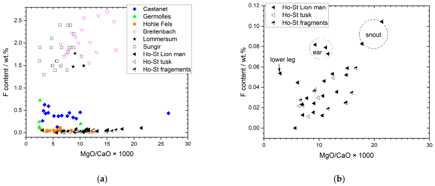
In addition to the PIXE data, the simultaneously recorded PIGE data can provide us information about the state of preservation of the ivory. The F contents in ivory can be determined using PIGE [16]. Fluorine is an element that is typically enriched in ancient ivories through diagenetic processes depending on the burial conditions. This element is integrated into the apatite matrix in the form of fluoride. Karstic environments are known to lead to low F enrichment in bone and ivory. The F content vs. the Mg-to-Ca ratio can be considered an estimate of the state of preservation of the ivory material [23,34,35]. As a general rule, the more the ivory is enriched in F, the poorer its state of preservation is. The opposite is true for the Mg/Ca ratio, expressed as MgO/CaO: the lower it is, the poorer the state of preservation is [24]. In the case of the Ho-St site, the low F content indicates a rather good state of preservation of the ivories, enabling elemental fingerprint studies to establish archaeological considerations (Figure 10a). No specific differences can be discerned in the state of preservation of the Lion Man figurine, the unworked tusk, and the fragments (Figure 10b).
In an inter-regional comparison, the ivory finds from Ho-St exhibit the same characteristics as those from Hohle Fels. Both sites are karst caves that provide favourable conditions for the preservation of archaeological ivory material.

4.6. Iron Oxide Enrichment at the Surface of the Lion Man

Archaeological ivory generally contains ‘diagenesis-related’ trace elements such as Fe and Mn, which are either absorbed from the soil and incorporated into the material or deposited on the surface in the form of secondary minerals through various diagenetic processes [17,36]. In addition, signs of use (e.g., ochre or Fe oxide colouring matter) may be found on the ivory surface [27].
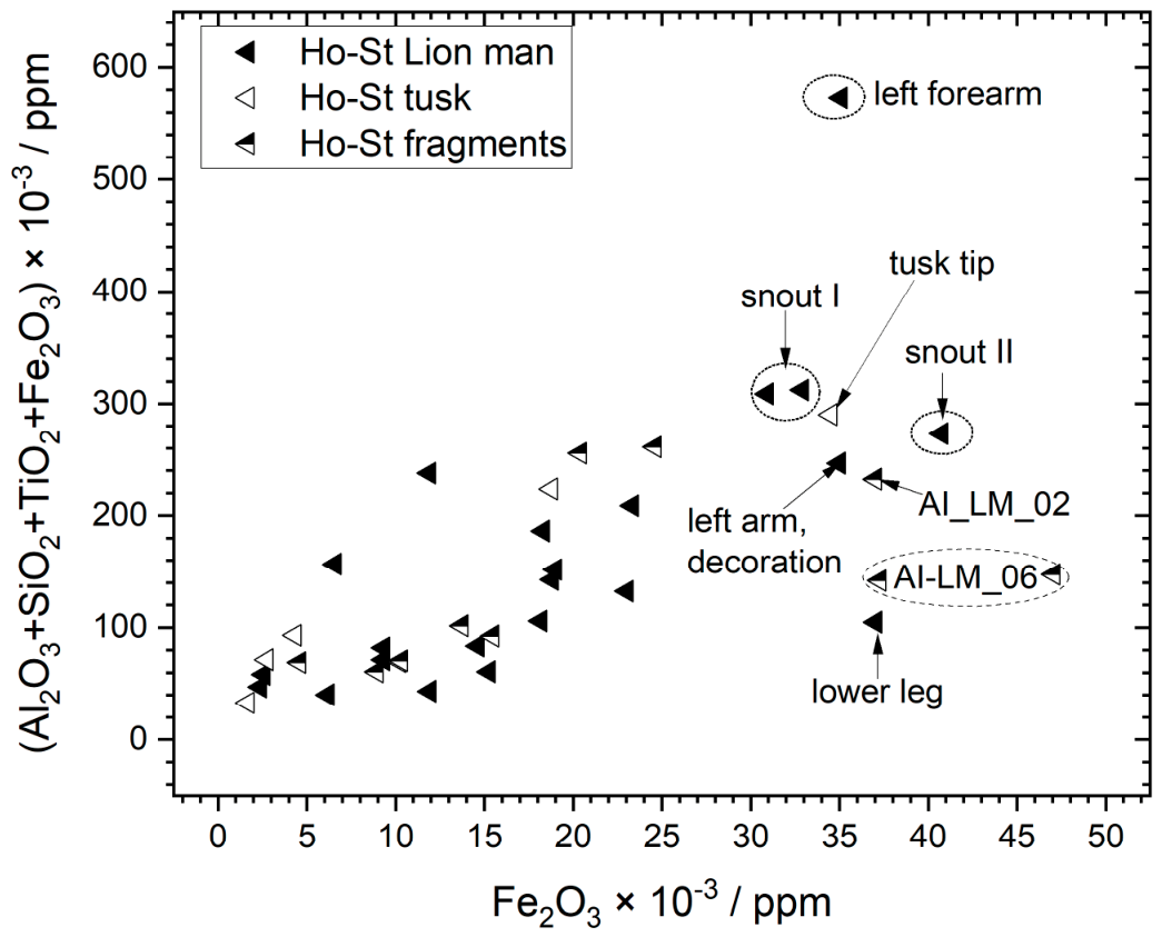
In Figure 11, the sum of the detected earth elements (Al, Si, Ti, and Fe) is plotted against the Fe contents in the different ivory finds from Ho-St. The graph shows that most of the areas measured on the figurine have higher relative Fe content compared with the unworked tusk. The black fragment from the back of the figurine is an exception, as it is poor in Si and Fe (Table S4). The tip of the tusk is also an exception, as it has a completely different composition and cannot be directly compared.
The average ratio of Fe content to the sum of all the detected earth elements (Si, Al, Ti, and Fe) is significantly higher in the Lion Man figurine (0.14 ± 0.08) than in the unworked tusk (0.07 ± 0.03). If measurements with very high Si content are excluded from the calculation, the difference is even greater (0.16 ± 0.09 for the Lion Man versus 0.05 ± 0.005 for the tusk). It can therefore be concluded that the Fe enrichment may not be related to diagenetic changes such as sedimentary deposits. The most Fe-enriched areas of the figurine are the snout, a decoration at the left arm, the left forearm and the left lower leg. This side of the figurine is more complete, exhibiting more likely the original surface of the statuette, than the right-hand side. Interestingly, the left forearm presents a sign sequence in the form of seven parallel carved lines that are interpreted as symbols [10,37], and at the snout, a special surface treatment was suspected. Therefore, this Fe enrichment at the surface of the Lion Man tends to indicate that it might be linked to the production or use of the sculpture. However, further analyses are necessary to clarify this issue.

4.7. Additional Analyses of a Special Part of the Lion Man Figurine: The Snout

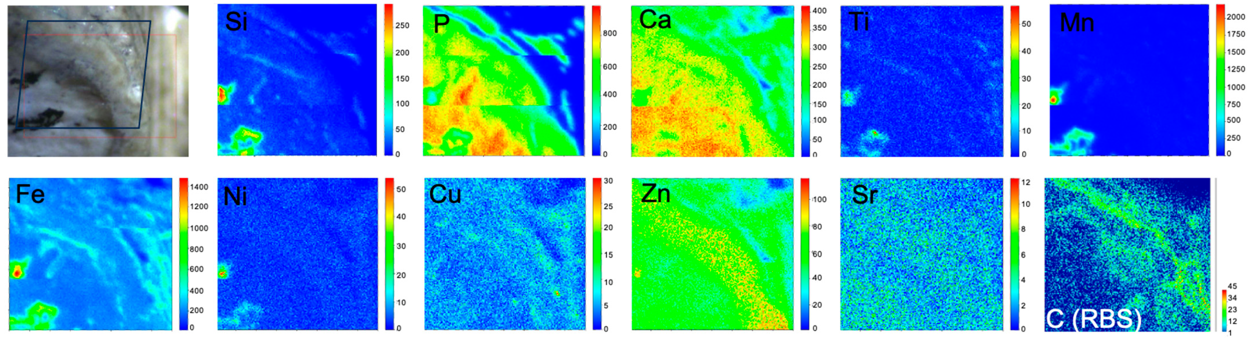
The snout region of the Lion Man statue is of particular interest in relation to possible use wear of the statue. The fragmentary nature of the lion head allows for insights into the inner ivory material because of the presence of a naturally fractured (taphonomical) cross-section of the snout region (Figure 2). This specific area reveals unique internal structures within the sculpture that were not observed anywhere else on the figurine, such as the so-called ’infiltration zone’. This ’infiltration zone’, located approximately 1 mm below the surface of the snout and exhibiting a distinct brownish colouration, was observed on both consolidated and unconsolidated fragments of the snout. This suggests that this ’infiltration zone‘ is likely unrelated to previous conservation treatments performed on the figurine. An archaeological hypothesis was that this zone might have been formed by compounds such as plant or animal substances, e.g., with blood, with which the figurine could have been in contact during its use. Two IBA mappings (2D scans 0039 and 0040) were carried out on the surface of an unconsolidated fragment in the lower jaw area, which was found during the last excavation at the cave. They showed the presence of C in their RBS spectra (Figure S14). To understand the C distribution, a somewhat coarser scan (1.6 × 1.5 mm2) was performed on the naturally formed cross-section in the upper part of the snout, which allows one to obtain a view into the inner zones of the ivory (Figure 12). Interestingly, this inner ivory layer shows the presence of Mn oxide dendrites on the naturally formed cross-section, indicating that the secondary deposit was not recent but must have happened during the long-term burial process, as Mn oxide stains generally form in soil under humid conditions. We assume that the brown stain of the ‘infiltration zone’ was not induced by consolidation.
To verify this assumption, RBS imaging was employed, as it allows for the study of the C distribution, thereby indicating the presence of organic matter or the presence of carbonates [30,38].
Combined elemental imaging reveals enrichment of Zn and C on the naturally formed cross-section in the area of the yellowish-grey layer beneath the surface. If the C enrichment were due to the consolidation, it would have been concentrated at the very surface. It seems that the C enrichment zone is not perfectly aligned with that of Zn but slightly above the Zn-rich layer with respect to the object surface. Zinc is also slightly enriched in the C-rich area, but this enrichment seems more diffuse. At this stage of our research, we cannot fully explain the origin of this enrichment. Fe is found to correlate with the presence of Mn oxide dendrites, as well as with the distribution of Si and Ni on the naturally formed cross-section, and therefore likely linked to diagenetic processes. A very small amount of Cu is also found on the cross-section (Figure 12, Table 3).
In contrast, the quantification of the PIXE-PIGE data of the unconsolidated surface of the snout (measurement spots 0039 and 0040) shows that the F, Fe, and Zn values are slightly elevated compared with the average values for the other parts of the Lion Man. Iron correlates in these areas less strongly with SiO2, so it may also have other sources than the sediment. These results tend to support the hypothesis of a special use wear of the zone of the snout with respect to the other parts of the figurine, without being able to identify its origin.

5. Conclusions

Mammoth ivory objects from the Hohlenstein-Stadel cave including the iconic Lion Man were successfully studied at the microfocus beamline of the particle accelerator AGLAE, installed at the C2RMF in Paris, France. Although the chemical compositions of the Lion Man, the left tusk and several mammoth ivory fragments are relatively inhomogeneous with respect to their minor and trace elements, a combined biogenic/diagenetic trace element fingerprint for the mammoth ivory assemblage from the Hohlenstein-Stadel cave could be determined based on the trace element ratios of Sr, Zn and Br. This fingerprint stands out from that of ivories from other Aurignacian sites in other European regions but, as expected, closely resembles that of the contemporary Aurignacian ivory finds from the Hohle Fels cave, in the other valley of the Swabian Alb. These results underline the validity of this approach to be used to track the circulation of mammoth ivory in Europe in deep time. It is of importance for mammoth ivory objects that are found in archaeological sites where mammoths were rare in Palaeolithic times, like in the Dordogne region in Southern France. Other non-invasive methods such as synchrotron radiation-induced X-ray fluorescence (SR-XRF) analyses could also be interesting to improve detection sensitivity for trace elements. Our previous work suggested possible new marker elements, such as Cr, Ni and Cu, in mammoth ivory [13,17]. While IBA is more sensitive for the analysis of light elements and provides quantification, SR-XRF measurements allow for a better detection sensitivity for heavy trace elements. A decisive development step would be to develop a quantification method for SR-XRF measurements. Finer intra-regional site discrimination should also be possible by means of Sr isotope measurements; these, however, are micro-invasive.
The trace element fingerprint is characteristic of the whole Lion Man sculpture, which is composed of about three hundred individual fragments, although its Zn content is a bit scattered. Thanks to the defined fingerprint, individual ivory fragments and the tusk can be chemically compared to that of the Lion Man. The left tusk and some of the fragments attributed archaeologically to the Lion Man show chemical similarities, even at the trace element level, to the figurine, supporting the hypothesis that they could belong together. However, our analyses do not provide a prove that the tusk and the figurine belonged to the same animal. Further investigations (e.g., using DNA, proteomic analyses or direct dating) are needed to answer this question, but these methods would require sampling.
Interestingly, trace elements like Fe and Zn show systematic concentration anomalies in particular zones of the figurine, especially on the more complete left-hand side, such as the left arm, which presents a decorative sign, and the left leg, in addition to the snout, which might be related to its production or use. Whether these trace element anomalies are related to the production of the figurine (e.g., through polishing), shamanistic rituals (e.g., treatment with red ochre) or other types of use wear on the Lion Man must remain an open question at this point. Obviously, the distinction of these traces from those of sediment deposits or diagenetic processes during burial of the statuette remains a challenging issue.

Supplementary Materials

The following supporting information can be downloaded at: https://www.mdpi.com/article/10.3390/min16070755/s1, Table S1: Results of quantitative PIXE and PIGE analyses of the study objects (AI_LM-XX) from the Hohlenstein-Stadel cave in terms of major and minor elements with Z ≤ 20. Values are given in weight%. Table S2: Results of quantitative PIXE and PIGE analyses of the study objects (AI_LM-XX) from the Hohlenstein-Stadel cave in terms of minor and trace elements with Z > 20. Values are given in ppm. Table S3: Results of combined quantitative PIXE and PIGE analyses without those with very high Si content (>10 wt.% SiO2). In a second step, the results for the back fragment of the Lion Man figurine with a thick MnO2 layer were excluded from the calculation (see Table S2). Mean values and standard deviations are provided in ppm for each element. Table S4: Results of combined quantitative PIXE and PIGE analyses. Composition of a Mn oxide-densely covered fragment of the Lion Man statuette from the Hohlenstein-Stadel cave in terms of major, minor, and trace elements. Mean values and standard deviations are provided in ppm for each element. Table S5: Results of combined quantitative PIXE and PIGE analyses. Mean composition of the Lion Man statuette from the Hohlenstein-Stadel cave in terms of major, minor, and trace elements (without results of regions with high Si and high Mn contents) in comparison with the results of the snout region of the statuette. Mean values and standard deviations are provided in ppm for each element. Snout I = measurements on the unconsolidated fragment; snout II = scan on the ‘natural cross-section’; snout III = mean values of all measurements. Figures S1, S2, S4 and S5: Low-energy PIXE spectra of the Lion Man (measurement points 0016, 0020, 0040, and 0041) and, in Figure S3, of a modern elephant ivory reference (measurement point 0027); Figures S6, S7, S9 and S10: High-energy PIXE spectra of the Lion Man (measurement points 0016, 0020, 0040, and 0041) and, in Figure S8, of a modern elephant ivory reference (measurement point 0027); Figures S11, S12, S14 and S15: RBS spectra of the Lion Man (measurement points 0016, 0020, and 0040) and, in Figure S13, of a modern elephant ivory reference (measurement point 0027); Figure S16: (a) EBS-RBS spectrum of the C-rich zone on the mapped area of the naturally formed cross-section of the snout of the Lion Man (measurement point 0041, 1.6 × 1.5 mm2). (b) Defined regions of interest (ROIs) for carbon (C) and background (B) depicted in light orange and light blue, respectively. (c,d) Corresponding C map (2500 pixels/24000, ROI 308-331). e) Extraction of a zone poor in C of the map on the naturally formed cross-section of the snout of the Lion Man (measurement point 0041) (2508 pixels/24,000). (f) Corresponding RBS spectrum to the C poor zone. (g) Calculated net C distribution of the RBS map (corresponding to net counts C(net) = (C + B) − B).

Author Contributions

Conceptualization, I.R. and K.W.; methodology, I.R. and K.M.; software, L.P.; validation, I.R. and K.M.; formal analysis, K.M. and I.R.; investigation, all authors; resources, I.R.; data curation, Q.L. and K.M.; writing—original draft preparation, I.R. and K.M.; writing—review and editing, I.R., K.M., Q.L. and K.W.; visualization, K.M. and L.P.; supervision, I.R.; project administration, I.R. and K.W.; funding acquisition, I.R. and K.W. All authors have read and agreed to the published version of the manuscript.

Funding

Financial support from the Access to Research Infrastructures activity in the Horizon 2020 Programme of the EU (IPERION HS Grant Agreement No. 871034) is gratefully acknowledged for FIXLab access in the framework of the granted Lion Man project. The FR NewAGLAE (now Lab-BC UAR3506 CNRS) unit is acknowledged for the financial support for secure object transport.

Data Availability Statement

The original contributions presented in this study are included in the article/supplementary material. Further inquiries can be directed to the corresponding author.

Acknowledgments

This article is a revised and expanded version of a paper entitled “Nicht-invasive Ionenstrahlanalyse des Löwenmenschen aus Mammut-Elfenbein aus der Schwäbischen Alb“, which was presented at the Jahrestagung für Archäometrie und Denkmalpflege 2023, Reiss-Engelhorn-Museen (rem), Mannheim, Germany, 28 March to 1 April 2023. We acknowledge the measurement time allowance for ion beam analysis experiments at the New AGLAE facility (ANR—10—EQPX 22) thanks to IPERION HS FIXlab under Grant Agreement No. 871034 (Lion Man project), as well as the members of the New AGLAE team for their support during measurement. We thank the support of Hasenkamp Internationale Transporte, Stuttgart, Germany, for the transport of the objects from Museum Ulm to the Louvre Palace, Paris. We thank Museum Ulm for the provision of the object for the analyses and the reproduction permission of the photo of the Lion Man in this publication. The IRN 0871 Taphonomy European Network (TaphEN) is acknowledged for the support of the project “Non-invasive study of the taphonomical phenomena in Aurignacian ivories by combined RBS/PIXE/PIGE imaging” by I.R. together with Claire Heckel, Vladislav Zhitenev and Nicholas J. Conard. Giulia Gallo is acknowledged for the kind revision of the English language.

Conflicts of Interest

The authors declare no conflicts of interest. The funders had no role in the design of the study; in the collection, analyses, or interpretation of data; in the writing of the manuscript; or in the decision to publish the results.

Abbreviations

The following abbreviations are used in this manuscript:
AGLAEAccélérateur Grand Louvre d’Analyse élémentaire
14Cradiocarbon
C2RMFCentre de recherche et de restauration des musées de France
Ho-StHohlenstein-Stadel cave
HPGeHigh-Purity Germanium
IBAIon beam analysis
PIGEProton-induced gamma-ray emission
PIPSPassivated Implanted Planar Silicon
PIXEProton-induced X-ray emission
RBSRutherford Backscattering
SDDSilicon drift detectors
UNESCOUnited Nations Educational, Scientific and Cultural Organization

References

  1. Conard, N.J.; Meister, C.; Sanz, N.; Wolf, S. The Path to UNESCO World Heritage Caves and Ice Age Art in the Swabian Jura. In Images, Gestures, Voices, Lives. What Can We Learn from Palaeolithic Art? Haidle, M.N., Paar, M., Wolf, S., Conard, N.J., Eds.; ROCEEH Communications: Heidelberg, Germany, 2025; pp. 223–246. [Google Scholar]
  2. Conard, N. Palaeolithic Ivory Sculptures from Southwestern Germany and the Origins of Figurative Art. Nature 2003, 426, 830–832. [Google Scholar] [CrossRef] [Scilit] [PubMed]
  3. Dutkiewicz, E. The Vogelherd Cave and the Discovery of the Earliest Art—History, Critics and New Questions. In Human Origin Sites and the World Heritage Convention in Eurasia; World Heritage Papers 41 Heads 4; The World Heritage Centre: Paris, France, 2015; Volume II, pp. 74–91. [Google Scholar]
  4. Conard, N.J. A Female Figurine from the Basal Aurignacian of Hohle Fels Cave in Southwestern Germany. Nature 2009, 459, 248–252. [Google Scholar] [CrossRef] [Scilit] [PubMed]
  5. Conard, N.J.; Malina, M.; Münzel, S.; Seeberger, F. Eine Mammutelfenbeinflöte aus dem Aurignacien des Geißenklösterle: Neue Belege für eine Musiktradition im frühen Jungpaläolithikum auf der Schwäbischen Alb. Archäol. Korresp. 2004, 34, 447–462. (In German) [Google Scholar]
  6. Conard, N.J.; Malina, M.; Münzel, S. New Flutes Document the Earliest Musical Tradition in South-Western Germany. Nature 2009, 460, 737–740. [Google Scholar] [CrossRef] [Scilit] [PubMed]
  7. Ebinger-Rist, N.; Wolf, S.; Wehrberger, K.; Kind, C.-J. L’homme-lion d’Hohlenstein—Stadel. L’Anthropologie 2018, 122, 415–436. [Google Scholar] [CrossRef] [Scilit]
  8. Hahn, J. Stellung der männlichen Statuette aus dem Hohlenstein-Stadel in der jungpaläolithischen Kunst. Ger. Anz. Röm.-Ger. Komm. Dtsch. Archäol. Inst. 2023, 48, 1–12. [Google Scholar] [CrossRef]
  9. Schmid, E.; Hahn, J.; Wolf, U. Die altsteinzeitliche Elfenbeinstatuette aus der Höhle Stadel im Hohlenstein bei Asselfingen, Alb-Donau-Kreis. Fundber. Baden-Württ. 2017, 14, 33–118. [Google Scholar] [CrossRef]
  10. Hahn, J. Aurignacien, das ältere Jungpaläolithikum in Mittel- und Osteuropa; Fundamenta: Monographien zur Urgeschichte; Böhlau: Köln, Germany; Vienna, Austria, 1977. [Google Scholar]
  11. Kind, C.-J.; Ebinger-Rist, N.; Wolf, S.; Beutelspacher, T.; Wehrberger, K. The Smile of the Lion Man. Recent Excavations in Stadel Cave (Baden-Württemberg, South-Western Germany) and the Restoration of the Famous Upper Palaeolithic Figurine. Quartär—Int. Jahrb. Erforsch. Eiszeitalt. Steinzeit 2021, 61, 129–145. [Google Scholar] [CrossRef] [Scilit] [PubMed]
  12. Beutelspacher, T.; Ebinger-Rist, N.; Kind, C.-J. Neue Funde Aus Der Stadelhöhle Im Hohlenstein Bei Asselfingen. In Archäologische Ausgrabungen in Baden-Württemberg 2010; wbg Theiss: Baden-Württemberg, Germany, 2011; pp. 65–70. [Google Scholar]
  13. Tranchant, L.; Müller, K.; Lemasson, Q.; Pichon, L.; Schöder, S.; Conard, N.J.; Reiche, I. Performance Comparison of External IBA and SR-XRF Imaging for the Study of Ivory. Nucl. Instrum. Methods Phys. Res. Sect. B Beam Interact. Mater. At. 2023, 545, 165146. [Google Scholar] [CrossRef] [Scilit]
  14. Reiche, I.; Heckel, C.; Müller, K.; Jöris, O.; Matthies, T.; Conard, N.J.; Floss, H.; White, R. Combined Non-Invasive PIXE/PIGE Analyses of Mammoth Ivory from Aurignacian Archaeological Sites. Angew. Chem. Int. Ed. 2018, 57, 7428–7432. [Google Scholar] [CrossRef] [Scilit] [PubMed]
  15. Heckel, C.; Müller, K.; White, R.; Floss, H.; Conard, N.J.; Reiche, I. Micro-PIXE/PIGE Analysis of Palaeolithic Mammoth Ivory: Is It Possible to Find Characteristic Chemical Markers of Mammoth Ivory Provenance and Relative Dating in Specimens of Great Age? Palaeogegraphy Palaeoclimatol. Palaeoecol. 2014, 416, 133–141. [Google Scholar] [CrossRef] [Scilit]
  16. Heckel, C.; Müller, K.; White, R.; Wolf, S.; Conard, N.J.; Normand, C.; Floss, H.; Reiche, I. F-Content Variation in Mammoth Ivory from Aurignacian Contexts: Preservation, Alteration, and Implications for Ivory-Procurement Strategies. Quat. Int. 2016, 403, 40–50. [Google Scholar] [CrossRef] [Scilit]
  17. Tranchant, L.; Müller, K.; Lemasson, Q.; Pichon, L.; Schöder, S.; Conard, N.J.; Reiche, I. Improved Discrimination of Biogenic and Diagenetic Elements in Palaeolithic Mammoth Ivory and Bone from Hohle Fels Cave in the Swabian Jura of Southwestern Germany. Quat. Int. 2023, 660, 4–12. [Google Scholar] [CrossRef] [Scilit]
  18. Reiche, I.; Vignaud, C.; Menu, M. Heat Induced Transformation of Fossil Mastodon Ivory into Turquoise ‘Odontolite’. Structural and elemental characterisation. Solid State Sci. 2000, 2, 625–636. [Google Scholar] [CrossRef] [Scilit]
  19. Chadefaux, C.; Vignaud, C.; Chalmin, E.; Robles-Camacho, J.; Aarroyo-Cabrales, J.; Johnson, E.; Reiche, I. Color Origin and Heat Evidence of Paleontological Bones: Case Study of Blue and Gray Bones from San Josecito Cave, Mexico. Am. Mineral. 2009, 94, 27–33. [Google Scholar] [CrossRef] [Scilit]
  20. Ebinger-Rist, N.; Wolf, S. A Giant Puzzle. Restoration of the Statuette 2012/13. In The Return of the Lion Man. History-Myth-Magic; Jan Thorbecke Verlag der Schwabenverlag: Ostfildern, Germany, 2013; pp. 52–62. [Google Scholar]
  21. Hein, W.; Wehrberger, K. Löwenmensch 2.0 Nachbildung der Elfenbeinstatuette aus der Hohlenstein-Stadel-Höhle mit authentischen Werkzeugen. In Experimentelle Archäologie in Europa: Bilanz 2010; Isensee, Florian, GmbH: Oldenburg, Germany, 2010; pp. 47–53. [Google Scholar]
  22. Pichon, L.; Calligaro, T.; Lemasson, Q.; Moignard, B.; Pacheco, C. Programs for Visualization, Handling and Quantification of PIXE Maps at the AGLAE Facility. Nucl. Instrum. Methods Phys. Res. Sect. B Beam Interact. Mater. At. 2015, 363, 48–54. [Google Scholar] [CrossRef] [Scilit]
  23. Chavagnac, V.; Milton, J.A.; Green, D.R.H.; Breuer, J.; Bruguier, O.; Jacob, D.E.; Jong, T.; Kamenov, G.D.; Le Huray, J.; Liu, Y.; et al. Towards the Development of a Fossil Bone Geochemical Standard: An Inter-Laboratory Study. Anal. Chim. Acta 2007, 599, 177–190. [Google Scholar] [CrossRef] [Scilit] [PubMed]
  24. Müller, K.; Reiche, I. Differentiation of Archaeological Ivory and Bone Materials by Micro-PIXE/PIGE with Emphasis on Two Upper Palaeolithic Key Sites: Abri Pataud and Isturitz, France. J. Archaeol. Sci. 2011, 38, 3234–3243. [Google Scholar] [CrossRef] [Scilit]
  25. Velliky, E.C.; Porr, M.; Conard, N.J. Ochre and Pigment Use at Hohle Fels Cave: Results of the First Systematic Review of Ochre and Ochre-Related Artefacts from the Upper Palaeolithic in Germany. PLoS ONE 2018, 13, e0209874. [Google Scholar] [CrossRef] [Scilit] [PubMed]
  26. Velliky, E.C.; Schmidt, P.; Bellot-Gurlet, L.; Wolf, S.; Conard, N.J. Early Anthropogenic Use of Hematite on Aurignacian Ivory Personal Ornaments from Hohle Fels and Vogelherd Caves, Germany. J. Hum. Evol. 2021, 150, 102900. [Google Scholar] [CrossRef] [Scilit] [PubMed]
  27. Lebon, M.; Pichon, L.; Beck, L. Enhanced Identification of Trace Element Fingerprint of Prehistoric Pigments by PIXE Mapping. Nucl. Instrum. Methods Phys. Res. Sect. B Beam Interact. Mater. At. 2018, 417, 91–95. [Google Scholar] [CrossRef] [Scilit]
  28. Codron, J.; Codron, D.; Sponheimer, M.; Kirkman, K.; Duffy, K.J.; Raubenheimer, E.J.; Melice, J.L.; Grant, R.; Clauss, M.; Lee-Thorp, J.A. Stable Isotope Series from Elephant Ivory Reveal Lifetime Histories of a True Dietary Generalist. Proc. Biol. Sci. R. Soc. 2012, 279, 2433–2441. [Google Scholar] [CrossRef] [Scilit] [PubMed]
  29. Prozesky, V.M.; Raubenheimer, E.J.; Heerden, W.F.P.V.; Grotepass, W.P.; Przybylowicz, W.J.; Pineda, C.A.; Swart, R. Trace Element Concentration and Distribution in Ivory. Nucl. Instrum. Methods Phys. Res. Sect. B Beam Interact. Mater. At. 1995, 104, 638–644. [Google Scholar] [CrossRef] [Scilit]
  30. Albéric, M.; Dean, M.N.; Gourrier, A.; Wagermaier, W.; Dunlop, J.W.C.; Staude, A.; Fratzl, P.; Reiche, I. Relation between the Macroscopic Pattern of Elephant Ivory and Its Three-Dimensional Micro-Tubular Network. PLoS ONE 2017, 12, e0166671. [Google Scholar] [CrossRef] [Scilit] [PubMed]
  31. Albéric, M.; Gourrier, A.; Wagermaier, W.; Fratzl, P.; Reiche, I. The Three-Dimensional Arrangement of the Mineralized Collagen Fibers in Elephant Ivory and Its Relation to Mechanical and Optical Properties. Acta Biomater. 2018, 72, 342–351. [Google Scholar] [CrossRef] [Scilit] [PubMed]
  32. Reiche, I.; Müller, K.; Lemasson, Q.; Pichon, L.; Wehrberger, K. Nicht-invasive Ionenstrahlanalyse des Löwenmenschen aus Mammut-Elfenbein aus der Schwäbischen Alb. In Proceedings of the German Archaeometry Conference, Mannheim: METALLA; Klein, S., Berthold, C., Berger, D., Eds.; Bergbaumuseum: Bochum, Germany, 2023; Special Volume 12, pp. 33–35. [Google Scholar]
  33. Potter, R.M.; Rossman, G.R. Mineralogy of Manganese Dendrites and Coatings. Am. Mineral. 1979, 64, 1219–1226. [Google Scholar]
  34. Skinner, H.G.W.; Fitzpatrick, R.W. Biomineralization Processes of Iron and Manganese—Modern and Ancient Environments; Catena-Verlag: Cremlingen-Destedt, Germany, 1992. [Google Scholar]
  35. Reiche, I.; Müller, K. Marqueur d’identification à micro-échelle de l’ivoire de mammouth dans les objets préhistoriques. L’Anthropologie 2018, 122, 316–326. [Google Scholar] [CrossRef] [Scilit]
  36. Albéric, M.; Müller, K.; Pichon, L.; Lemasson, Q.; Moignard, B.; Pacheco, C.; Fontan, E.; Reiche, I. Non-Invasive Quantitative Micro-PIXE–RBS/EBS/EBS Imaging Reveals the Lost Polychromy and Gilding of the Neo-Assyrian Ivories from the Louvre Collection. Talanta 2015, 137, 100–108. [Google Scholar] [CrossRef] [Scilit] [PubMed]
  37. Bentz, C.; Dutkiewicz, E. Humans 40,000 y Ago Developed a System of Conventional Signs. Proc. Natl. Acad. Sci. USA 2026, 123, e2520385123. [Google Scholar] [CrossRef] [Scilit] [PubMed]
  38. Beck, L.; Cuif, J.-P.; Pichon, L.; Vaubaillon, S.; Dambricourt Malassé, A.; Abel, R.L. Checking Collagen Preservation in Archaeological Bone by Non-Destructive Studies (Micro-CT and IBA). Nucl. Instrum. Methods Phys. Res. Sect. B Beam Interact. Mater. At. 2012, 273, 203–207. [Google Scholar] [CrossRef] [Scilit]
Figure 1. (a) Entrance of the Hohlenstein-Stadel cave (2020) © Kurt Wehrberger and (b) photo of the Lion Man (H, 311 mm, max.; L, 73 mm, max.; D, 59 mm, max.), Hohlenstein-Stadel cave, Asselfingen, Alb-Donau-Kreis, Baden-Württemberg, Germany © Museum Ulm, photo: Oleg Kuchar, Ulm.
Minerals 16 00755 g001aMinerals 16 00755 g001b
Figure 2. Close-up view of the Lion Man’s snout. The red arrow marks the so-called “infiltration zone.” The blue oval indicates the area on an unconsolidated fragment from recent excavations. This latter fragment also exhibits the infiltration zone. The red rectangle marks the scanned area on the naturally broken cross-section of the snout. © Photo: Ewa Dutkiewicz.
Minerals 16 00755 g002
Figure 3. Photo of the tusk ((A,A1,A2) © C2RMF, photo: Anne Maigret) and the studied mammoth ivory fragments ((B), © Museum Ulm, photo: Kurt Wehrberger, 2021).
Minerals 16 00755 g003
Figure 4. (a) Photo of the Lion Man figurine placed in front of the measuring head of the microfocus beam line NewAGLAE of the AGLAE accelerator. © C2RMF, photo: Anne Maigret. (b) Drawings of the Lion Man statue with marked areas of analysis (red circles) modified from a drawing by Christina v. Elm © Museum Ulm [7].
Minerals 16 00755 g004
Figure 5. Ternary diagrams of the studied samples from the Hohlenstein-Stadel (Ho-St) cave: (a) relative Ca and P contents in the form of oxides vs. that of Si and (b) relative Zn and Sr contents in the form of oxides vs. that of Si.
Minerals 16 00755 g005
Figure 6. Ternary diagram showing the relative contents of the biogenic trace elements Zn and Sr as oxides and Br for seven different sites, including all studied objects from the Hohlenstein-Stadel (Ho-St) cave. The differently coloured ovals mark the three main groups that can be distinguished: red for the northernmost sites, blue for Castanet site in southern France and dark grey for the sites in southern Germany.
Minerals 16 00755 g006
Figure 7. Zoom-in section of the binary diagram showing the relative contents of the ‘biogenic’ trace element oxides of Sr and Zn for seven different sites, including all studied objects from the Hohlenstein-Stadel (Ho-St) cave.
Minerals 16 00755 g007
Figure 8. Comparison of SrO and ZnO contents obtained by PIXE analysis for the studied samples from the Hohlenstein-Stadel (Ho-St) cave. The blue double-headed arrows mark the mean values of the Sr and Zn measurements for the Lion Man, as well as the respective standard deviations. The red oval frames the measured values that fell within this range.
Minerals 16 00755 g008
Figure 9. Left: Microscope images of the analysed area at the inner layer of the mammoth tusk (the dark blue parallelogram corresponds to the scanned area) and the corresponding elemental distribution maps for Al, Si, P, K, Ca, Ti, Mn, Fe Kβ, Ni and Zn. The boxes mark the area analysed on the sample: red = area indicated by the measurement software, and blue = measured area (1000 × 1000 µm2).
Minerals 16 00755 g009
Figure 10. Plot of F content versus MgO-to-CaO ratio (a) for all ivory Aurignacian ivory assemblages studied and (b) for the studied samples from Hohlenstein-Stadel (Ho-St). The dashed lines outline measurement points derived from specific areas on the figurine.
Minerals 16 00755 g010
Figure 11. Plot of the sum of the detected earth elements (Al, Si, Ti, and Fe) against Fe content for all studied samples from the Hohlenstein-Stadel (Ho-St) cave. Values are expressed as element oxides. The dashed lines and ovals mark measurement points originating from specific areas of the statue or specific ivory fragments.
Minerals 16 00755 g011
Figure 12. Top left: Microscope image of the analysed area at the inner layer of the mammoth tusk (the dark blue parallelogram corresponds to the scanned area of 1.6 × 1.5 mm2). Elemental distribution PIXE maps of Si, P, and Ca using the low-energy X-ray detector, whereas those of Ti, Mn, Fe, Ni, Cu, Zn and Sr were measured using the high-energy X-ray detectors. Lower right: The distribution of net C intensity obtained by RBS imaging (see Figure S16). The boxes mark the area analysed on the sample: red = area indicated by the measurement software, and blue = measured area.
Minerals 16 00755 g012
Table 1. Results of quantitative PIXE and PIGE analyses. Chemical composition of the study objects from the Hohlenstein-Stadel cave in terms of major and minor trace elements with Z ≤ 20, expressed as element oxides. Mean values and standard deviations are provided for each element. Values are given in weight%.
ElementF
PIGE
Na2O PIGEMgOAl2O3SiO2P2O5SO3ClK2OCaO
Lion Man0.0450.520.544.649.6430.50.710.260.6349.3
±0.027±0.21±0.17±3.39±8.32±5.8±0.28±0.13±0.46±7.8
Tusk0.0300.560.443.659.0827.14.800.400.7449.1
±0.014±0.25±0.05±2.12±7.35±8.3±8.40±0.28±0.40±9.0
Fragments0.0260.280.534.297.0532.40.520.150.3351.1
±0.015±0.05±0.16±2.55±4.40±6.0±0.24±0.09±0.40±4.8
Table 2. Results of quantitative PIXE analyses. Chemical composition of the study objects from the Hohlenstein-Stadel cave in terms of minor and trace elements with Z > 20, expressed as element oxides. Mean values and standard deviations are provided for each element. Values are given in weight% (wt.%) for minor elements and in ppm for trace elements.
ElementTiO2MnOFe2O3NiOCuOZnOBrSrOY2O3ZrO2SnO2BaOPbO
Unitwt.%ppm
Lion Man0.320.611.921841671969183683410928317032
±0.29±0.74±1.17±352±116±758±6±99±26±202±300±229±27
Tusk0.252.351.24252111844142081557238523
±0.18±2.41±1.42±316±75±352±4±34±19±78±22±154±3
Fragments0.230.732.0910811616371841420956910214
±0.24±0.95±1.39±116±76±663±6±175±14±240±83±147±18
Table 3. Results of quantitative PIXE and PIGE (for F) analyses. Mean composition of the Lion Man statuette from the Hohlenstein-Stadel (Ho-St) cave showing representative elements (without results for high-Si and high-Mn locations) compared with the results for the snout region of the figurine. Snout I = measurements on the unconsolidated fragment; snout II = scan on the ‘natural cross-section’. Mean values and standard deviations are provided. Major and minor elements are expressed in wt.% and trace elements in ppm. n.a. = not applicable.
SiO2P2O5CaOMnOFe2O3ZnOBrSrOF
wt.%ppm
Lion Man mean4.9133.7653.530.321.660.2219408456
±2.27±3.40±3.18±0.12±0.85±0.05±6±84±235
Snout I17. 8125.0540.170.353.190.2925350935
±0.13±0.004±0.79±0.02±0.14±0.07±2±8±154
Snout II1.9037.8255.660.321.190.1824352445
n.a.n.a.n.a.n.a.n.a.n.a.n.a.n.a.n.a.
Disclaimer/Publisher’s Note: The statements, opinions and data contained in all publications are solely those of the individual author(s) and contributor(s) and not of MDPI and/or the editor(s). MDPI and/or the editor(s) disclaim responsibility for any injury to people or property resulting from any ideas, methods, instructions or products referred to in the content.