Article

Influence of Geological Origin on the Physicochemical Characteristics of Sepiolites

by
Leticia Lescano
1,
Silvina A. Marfil
1,
Luciana A. Castillo
2,3,* and
Silvia E. Barbosa
2,3
1
Departamento de Geología, Universidad Nacional del Sur. CGAMA (CIC-UNS), Bahía Blanca 8000, Argentina
2
Planta Piloto de Ingeniería Química, PLAPIQUI (UNS-CONICET), Bahía Blanca 8000, Argentina
3
Departamento de Ingeniería Química, Universidad Nacional del Sur, Bahía Blanca 8000, Argentina
*
Author to whom correspondence should be addressed.
Minerals 2025, 15(9), 950; https://doi.org/10.3390/min15090950
Submission received: 11 July 2025 / Revised: 28 August 2025 / Accepted: 2 September 2025 / Published: 5 September 2025

Abstract

In this study the influence of the geological formation environment on the physicochemical properties of two natural sepiolites, as collected, was investigated. The samples analyzed were a lacustrine-derived sample from Tolsa, Spain (ST), and a hydrothermal-derived sample from La Adela, Argentine (SA). Comprehensive characterization was carried out using chemical analysis (XRF), X-ray diffraction (XRD), thermogravimetric analysis (TGA), Fourier-transform infrared spectroscopy (FTIR), scanning electron microscopy (SEM), and evaluations of hydrophobicity/hydrophilicity behavior. The results indicate that the ST sample exhibits a higher SiO2/MgO ratio and contains amorphous silica impurities, while the SA sample shows a composition more closely aligned with the theoretical stoichiometry of sepiolite. Furthermore, the SA sample demonstrates greater crystallinity compared to the ST sample. Morphological analysis revealed that ST consists of compact, aggregated fibrous structures, while SA is composed of disaggregated, needle-like fibers with high aspect ratios and nanometric diameters. Both samples display predominantly hydrophilic behavior; however, only the SA sample exhibits suspended particles at the interface, suggesting a slightly higher hydrophobic character than ST sample. These findings highlight the significant impact of the geological formation environment on the structural and surface characteristics of sepiolite, which, in turn, influence its performance in applications involving dispersion, adsorption, and interfacial interactions.

Graphical Abstract

1. Introduction

Sepiolite is a natural occurring mineral, belonging to the palygorskite/sepiolite polysomatic series, which can originate in a wide variety of geological environments. It is a magnesium phyllosilicate with the chemical formula Mg4Si6O15(OH)2(H2O)2·4H2O. In the sepiolite crystal lattice, the octahedral (O) sheet is divided into ribbons that extend along the z-axis, positioned between two continuous, undulating tetrahedral (T) layers. This arrangement arises from a periodic reversal in the orientation of the apical oxygen atoms. These TOT ribbons are alternately connected in a chessboard-like pattern, creating parallel nanochannels with an effective maximum width of approximately 10.6 Å, which run along the c-axis and are typically occupied by weakly held zeolitic water molecules [1]. At the ribbon edges, tightly bound structural water (H2O) completes the coordination sphere of the octahedral magnesium atoms. These channels and the external surface of the fibers can accommodate small molecules or exchangeable cations, making sepiolite not only industrially valuable but also geochemically informative. Sepiolite typically exhibits a needle-like morphology, often forming porous aggregates and fibrous sheets. Due to its unique structural and physicochemical properties, sepiolite plays a significant role in various geological, environmental, and industrial contexts [1,2,3].
Besides its physical and chemical characteristics, sepiolite has increasingly been recognized as a valuable indicator of paleoenvironmental conditions. Its formation is highly sensitive to specific geological and climatic settings. Typically, sepiolite forms in lacustrine to palustrine environments, under semi-arid to arid climatic regimes, frequently associated with high evaporation rates [4,5,6,7,8,9,10]. Moreover, sepiolite may also form through hydrothermal alteration of rocks and minerals, particularly in volcanic or metamorphic settings. Its formation requirements include a fluid with moderately to highly alkaline pH, sufficient concentrations of magnesium and silica, and minimal aluminum [11]. Giustetto et al. [1] analyzed a sepiolite from Sassello (Ligurian Apennines, Italy) which precipitated from hydrothermal fluids saturated in Mg and silica by the interaction with the host serpentinites. The authors studied its crystal-chemistry and structure, revealing, among other findings, that the fibers are extremely long and flexible, with a length/width aspect ratio significantly greater than 3. Despite this, there are relatively few studies exploring how geological origin impacts sepiolite’s physicochemical behavior. The environmental context, whether lacustrine or hydrothermal, can influence parameters such as crystallinity, surface area, trace element distribution, and adsorption capacity. A comprehensive understanding of these contrasts is essential not only for reconstructing the mineral’s geological history but also for optimizing its industrial utility.
The objective of this study is to examine how the formation environment of sepiolite influences its physicochemical characteristics. For this purpose, two distinct samples were selected for the analysis: one from a lacustrine setting in the Tajo Basin (Madrid, Spain), and another of hydrothermal origin from the Colorado Basin (province of Río Negro, Argentina). Comprehensive characterization of their physicochemical properties was performed, using X-ray diffraction (XRD) for crystal structure, X-ray fluorescence (XRF) for chemical analysis, Fourier-transform infrared spectroscopy (FTIR) to identify functional groups, and thermogravimetric analysis (TGA) for evaluating their thermal behavior. Morphological features were assessed through scanning electron microscopy (SEM). Additionally, the hydrophobic/hydrophilic behavior of sepiolite samples was qualitatively assessed.

2. Geological Setting

2.1. Tolsa Quarry
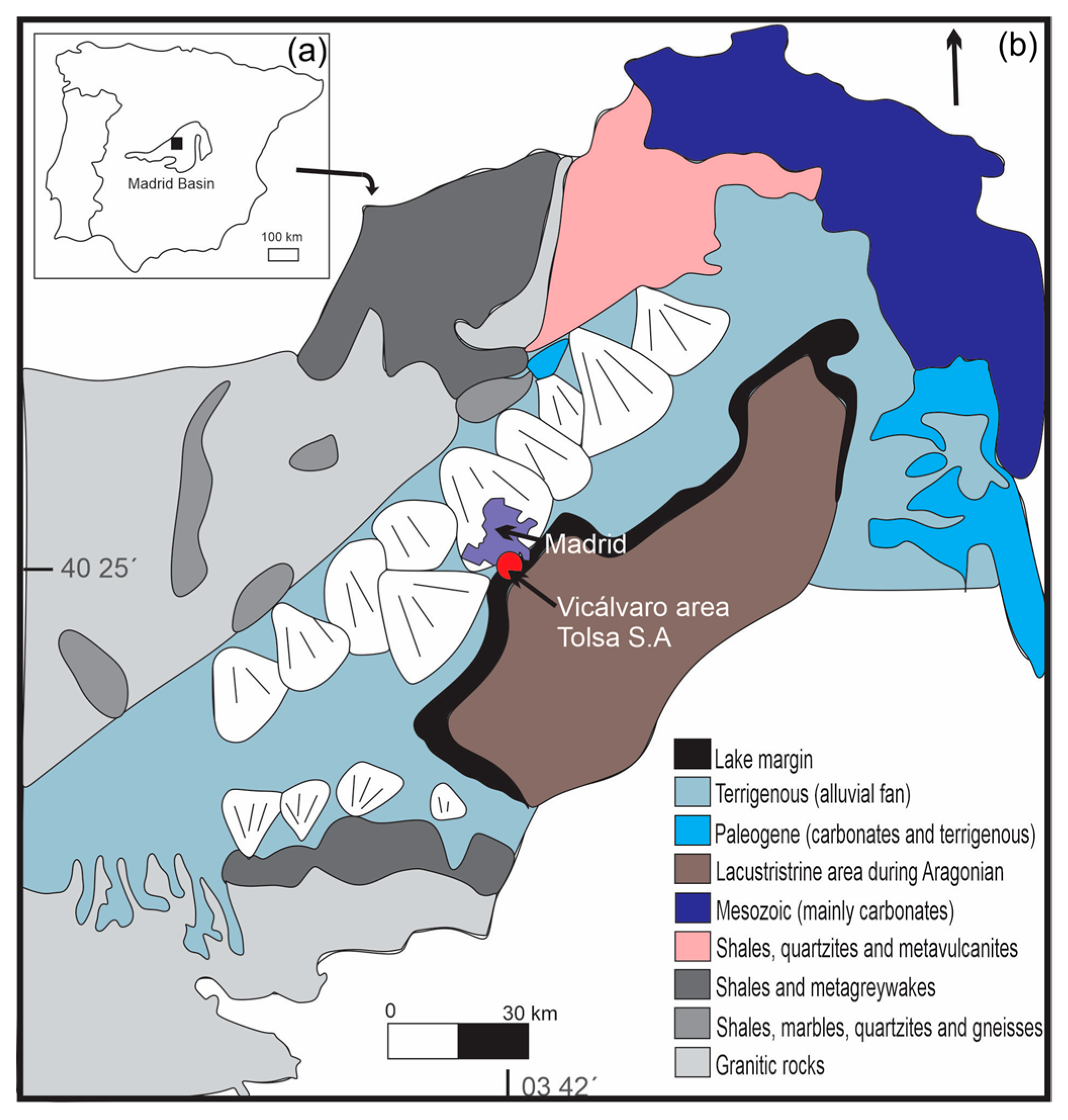
The Madrid Basin (Spain) is mainly composed of granitic, metamorphic and sedimentary rocks. During the early and middle Miocene, it was an endorheic sedimentary basin, characterized by central lacustrine and palustrine systems, fringed by alluvial fans and fluvial distributary facies [12]. Figure 1a shows the location of the basin in the center of the Iberian Peninsula, covering an area of more than 10,000 km2.
The basin’s stratigraphy is typically divided into three units: lower, intermediate, and upper [13]. The intermediate unit, dated to the Middle Aragonian–Upper Vallesian, is approximately 60 m thick and is composed of two distinct subunits with different lithologies and depositional environments: the lower, or detrital subunit, primarily consisting of detrital gypsum, and an upper, dolomitic subunit. The facies assemblage within this unit reflects a moderately saline, very shallow lacustrine system with fluctuating water level. Sepiolite deposits are located in marginal lacustrine zones within this unit and at the top of the underlying detrital unit [14]. The mineral occurs in two main stratigraphic levels within these detrital units, interbedded with fine arkosic sands and silts derived from alluvial fans. These deposits may also contain palygorskite and traces of quartz or amorphous silica. Although this sepiolite is acicular and generally poorly crystallized, its fibrous habit is distinctive. Fiber length is typically less than 2 mm, rarely exceeding 5 mm. Fibers are arranged parallel to bedding within fine-grained sediments. Under optical microscopy in thin sections, sepiolite appears colorless to slightly whitish, fibrous, and acicular, with low birefringence, straight extinction, and positive elongation, comparable to descriptions in other lacustrine-evaporitic deposits [14]. From a genetic perspective, the mineralization is interpreted as resulting from chemical precipitation under evaporitic and alkaline conditions within shallow lacustrine settings. The progressive concentration of Mg- and Si-rich waters, coupled with fluctuations in pH and salinity, favored the formation of fibrous Mg-silicates such as sepiolite and palygorskite [11]. These processes were enhanced by the endorheic character of the basin, where limited outflow promoted silica supersaturation and episodic precipitation. The studied deposit occurrence reflects a sedimentary-evaporitic origin, controlled by lacustrine geochemistry and periodic desiccation events. Since 1963, this sepiolite has been extracted in the southeastern area of Madrid municipality, specifically in Vicálvaro. Initially, mining was carried out underground, but since 1970 it has been conducted by open-pit methods [15]. Figure 1b shows the location of the mine.
Figure 1. (a) Location, and (b) geological map of the Madrid Basin (modified from [16]).
Minerals 15 00950 g001

2.2. La Adela Quarry

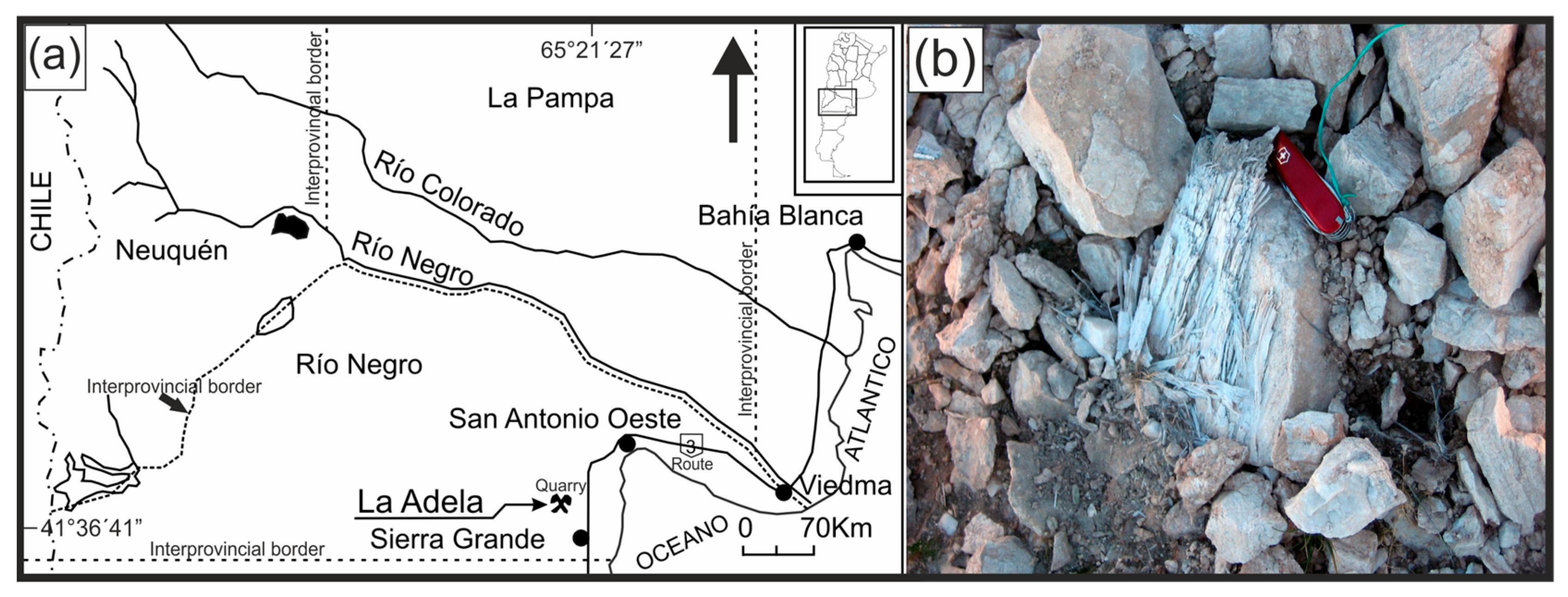
The La Adela quarry is located within the Santa Euriciana Ranch, approximately 75 km southwest of the town of San Antonio Oeste, in the province of Río Negro, Argentina (Figure 2a) [17]. The area features around eight small elevations composed mainly of dolomite beds with strike directions ranging from E–W to N 80° W, and dips varying between 45° and 65° to the north. The quarry (currently inactive) was developed on one of these outcrops. It measures approximately 600 m in length and between 70 and 90 m in width, with an estimated thickness of 50 m; the base does not outcrop. It is hosted in pre-Silurian micaceous schists belonging to the Valcheta Group [18]. The country rock consists of a massive, compact dolomite, bluish-white to gray in color, slightly reddish due to the presence of iron oxides. It exhibits a granoblastic texture with medium to coarse grain size, with dolomite crystals reaching up to 0.5 cm [19]. These authors also identified tremolite-actinolite, phlogopite, talc, montmorillonite, and iron oxides. The exploited material is dolomite, containing low amounts of silica and clay minerals as impurities. It was mainly used in the metallurgical processing of iron ore from a nearby Sierra Grande mine. The quarry shows intense jointing, with three predominant directions, variably mineralized. The eastern sector of the quarry is highly dislocated and contains the highest concentration of alteration minerals, the most significant being sepiolite (Figure 2b).
Sepiolite occurs within joints, where it fills the fractures without replacing the host rock, exhibiting exceptional crystal development. Fibers exceed 10 cm in length and occur in bundles up to 1 cm thick. The mineral is white, soft, flexible, and has a waxy luster. It is arranged parallel to the fractures it fills, forming high-purity monomineralic veins, with minor dolomite impurities. Under optical microscopy in thin section, sepiolite appears colorless, fibrous, and acicular, with refractive indices n = 1.512 and n = 1.5222, birefringence of 0.01, straight extinction, and positive elongation. The mineralization was developed along pre-existing fractures and bedding planes in the dolomite, which acted as both structural and lithological controls. The process occurred under hydrothermal conditions, beginning with the precipitation of talc and phlogopite under acidic conditions at ~300 °C. As the pH approached neutrality, illite formed, followed by the crystallization of sepiolite under alkaline conditions, resulting from the alteration of dolomite in the presence of alkali elements. This alteration process is related to Miocene–Pliocene volcanism, to which similar mineralizations in the northern Somuncurá Plateau have been attributed [20,21].
Figure 2. (a) Location map of the La Adela quarry within the province of Río Negro (Argentina) (modified from [17]). (b) Photograph showing bundles of highly crystalline sepiolite fibers growing within dolomitic host rock.
Minerals 15 00950 g002

3. Materials and Methods

Two natural sepiolite samples were analyzed in this study, both previously milled to a uniform fineness of 400 mesh (37 μm). A representative portion of each sepiolite was then selected for analysis following a quartering procedure performed on the ground bulk material. This method ensured the homogeneity and representativeness of the sample used for subsequent physicochemical and mineralogical characterizations. The samples are referred to as ST (from the Tolsa quarry, Madrid, Spain, supplied by Tolsa S.A.) and SA (from La Adela quarry, province of Río Negro, Argentina). These samples were analyzed according to chemical, mineralogical, and physical characterization.
Chemical composition was determined by XRF using a PANalytical PW 4400/40 Axios with CrKα radiation (Malvern Panalytical, Almelo, The Netherlands). Mineralogical analysis was conducted by XRD, using a Rigaku D Max III-C X-ray diffractometer (Rigaku, Tokyo, Japan) operating at 35 kV and 15 mA, with CuKα radiation (wavelength of 0.154060 nm), and a graphite monochromator in the secondary diffracted beam was used to eliminate CuKβ radiation. Diffractograms were recorder over a 2θ ranges of 3–60° 2θ, in 0.04° 2θ increments with a counting time of 1 s per increment. Samples were mounted on dimpled glasses and pressed with a flat glass plate to obtain a uniform surface. Phase identification was performed using JADE software version 8 (Materials Data Inc., Livermore, CA, USA), with intensities expressed in counts per second (CPS). Raw, unsmoothed diffractograms were used for all measurements. FTIR analysis was carried out using a Nicolet 520 FT-IR spectrophotometer (Thermo Fisher Scientific, Waltham, MA, USA) equipped with a DTGS detector. A 2 mg sample was mixed with KBr at 1:49 ratio (sepiolite/KBr) and pressed under 120 MPa until a disk was obtained. Spectra were recorder from 4000 cm−1 to 400 cm−1, using 32 scans at resolution of 4 cm−1 under a nitrogen atmosphere. Thermal behavior was evaluated by thermogravimetric analysis (TGA) and derivative thermogravimetry (DTG) using a thermogravimetric analyzer (TGA 5500, Discovery Series, TA Instruments, New Castle, DE, USA). In total, 25 mg of sample was placed in a ceramic crucible and heated at 10 °C/min up to 800 °C under a nitrogen atmosphere (25 mL/min). Morphological characterization was performed using a SEM (LEO EVO 40 XVP, Carl Zeiss AG, Cambridge, UK) equipped with an energy-dispersive X-ray spectroscopy (EDS) detector (Oxford X-Max 50, Oxford, Oxford, UK), operating at an accelerating voltage of 15 kV and under a vacuum of 2.1 × 10⁻⁵ Torr. The optimum working distance for sepiolite morphology observation was 7 mm. Samples were prepared by gently dispersing the particles with the aid of a dry air stream onto a double-sided aluminum adhesive tape mounted on bronze stubs. Then, they were coated with a 30 Å gold layer argon plasma PELCO 91000 coater (Ted Pella, Redding, CA, USA), in order to ensure conductivity. Particle length and width were determined by analyzing hundreds of particles by using AnalySIS2.1 software (Soft Imaging System GmbH, Münster, Germany) on high-resolution SEM micrographs. The hydrophobic/hydrophilic behavior of the samples was qualitatively evaluated by dispersing 1 g of each sample in a biphasic system composed of octane and distilled water. The settling behavior was visually assessed after 1 h, and photographic documentation was recorded. To complement this test, UV-Vis spectroscopy was performed to verify the presence of particles at the interface. The experiment was replicated in a quartz cuvette with identical proportions of components. A specific window was created in the cuvette to allow UV–Vis light to pass through the interfacial region. Spectra were acquired using a Shimadzu UV-160 spectrophotometer (Shimadzu Corporation, Kyoto, Japan), in the range of 200 nm–600 nm, with a spectral resolution of 1 nm.

4. Results and Discussion

The influence of the formation environment on the physicochemical characteristics of the sepiolite samples investigated in this study was assessed through integral comprehensive chemical, mineralogical, and physical characterization.
Table 1 shows the chemical composition of the two samples (ST and SA), along with the theoretical values derived from the ideal formula for sepiolite (S) Mg4Si6O15(H2O)2•4H2O [22]. The SiO2/MgO ratio is 2.65 for ST and 2.21 for SA, the latter being very close to the theoretical value of 2.23. The high ratio observed in the ST sample is attributed to the higher silica content, which is consistent with a lower loss on ignition (LOI), likely due to the presence amorphous silica as an impurity. The calcium oxide content in both samples is associated with the presence of calcite (CaCO3) or dolomite (CaMg(CO3)2) as accessory minerals. According to [23], the Mg/(Mg + Al) atomic ratio must exceed 0.6 for a mineral to be classified as sepiolite. The calculated values for this ratio are 0.96 for ST and 0.93 for SA, confirming their classification as sepiolite.
Figure 3 shows the XRD patterns of the analyzed samples. The principal reflections corresponding to sepiolite were identified in both diffractograms according to the ICDD 26-1226 reference card [24]. Although the overall diffractograms are comparable, several differences exist between the ST and SA samples, attributable not only to the presence of impurities but also to variations in their degree of crystallinity. In the ST sample, the elevation of the background, between 20° and 30° 2θ, suggests the presence of amorphous material, likely corresponding to amorphous silica, as previously reported in the literature [14]. This interpretation is consistent with the elevated SiO2 content and the lower LOI observed in the chemical analysis. In contrast, the SA sample exhibits reflections associated with dolomite impurities, the most intense being located at 0.289 nm. The crystallinity of each sepiolite sample can be inferred from the presence, shape, and intensity of specific reflections as well as the full width at half maximum (FWHM) parameter, measured in degrees 2θ, which reflects the mineral crystallinity [25]. The diffraction peaks in the ST sample are broader and less defined, whereas those in the SA sample are sharper and more intense, indicating a higher degree of crystallinity and a more ordered structural arrangement. These differences are particularly noticeable in the reflections corresponding to the d-spacings of 0.374 nm, 0.334 nm, 0.318 nm, 0.261 nm, and 0.256 nm, although additional variations are also observed. Quantitatively, FWHM values for the SA sample range from 0.095° 2θ to 0.331° 2θ, while those for the ST sample range from 0.422° 2θ to 1.065° 2θ. The broader peaks and higher FWHM values in the ST sample confirm its relatively lower crystallinity.
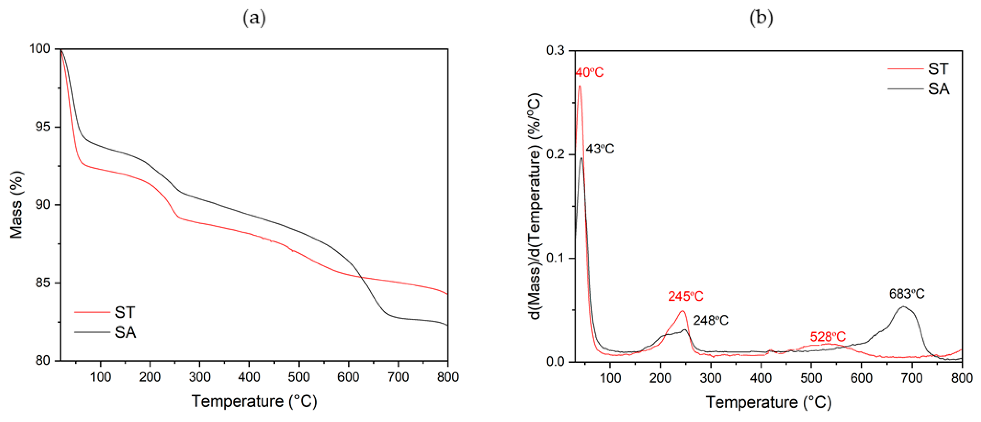
Figure 4 presents the thermogravimetric curves of the two sepiolite samples. Both exhibit broadly similar slopes, suggesting a comparable thermal behavior. At ~100 °C, the hydration water is released, with mass loss values of 7.5% and 5.0% for the ST and SA samples, respectively. The total mass loss values are consistent with those obtained from the chemical composition analysis. The lower total mass loss observed in the ST sample is likely due to the presence of impurities identified (amorphous silica). The mass loss observed at approximately 700 °C corresponds to two overlapping thermal decomposition processes. On one hand, water molecules coordinated to Mg2+ cations in octahedral sites within the sepiolite structure are released at this temperature [26,27]. On the other hand, this event coincides with the decomposition of dolomite, which occurs within the same temperature range [28,29]. The DTG curves (Figure 4b) provide further insight into the thermal events underlying the overall mass loss. Both samples exhibit an initial peak around 40–45 °C, associated with the release of physically adsorbed water. The slightly higher intensity in the ST sample could indicate a higher affinity for surface water. A second thermal event is observed at 245–248 °C, corresponding to the loss of loosely bound water within the sepiolite channels. The similarity in temperature between both samples indicates comparable structural features, although the peak intensity in ST is higher, suggesting a greater amount of intracanalicular water. At higher temperatures, a distinct peak at 528 °C is evident in the ST sample, indicating the dehydroxylation of structural hydroxyl groups. In SA, this event is greatly attenuated or absent, which could indicate a lower proportion of structural hydroxyl groups and/or a higher crystallinity. Only the SA sample presents a clear signal at 683 °C, which may be attributed to the decomposition of dolomite impurities, as indicated by the thermogravimetrical analysis [28,29].
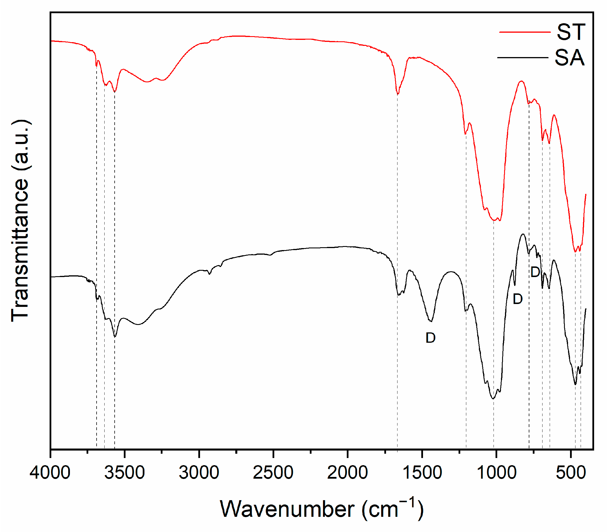
Figure 5 presents the FTIR spectra of both sepiolite samples. Most absorption bands in both SA and ST, identified with stripped lines, correspond to characteristic sepiolite vibration. The band at 3689 cm−1 is assigned to the stretching vibrations of MgOH. The band observed at 3566 cm−1 corresponds to the OH stretching vibrations of bound water, while the band at 3621 cm−1 is associated with zeolitic water. The OH bending vibration corresponding to the bound water can be observed at 1657 cm−1. The SiOSi bands at 1209 cm−1 and 1016 cm−1 are due to SiO vibrations. A minor band at 785 cm−1 is assigned to the OH bending vibration of MgFeOH. The bands observed at 690 cm−1 and 646 cm−1 correspond to the bending vibration of MgOH. The band at 470 cm−1 is attributable to the SiOSi bending vibration and 442 cm−1 arising from the SiOMg of the octahedral–tetrahedral linkage [30,31,32,33,34,35,36,37]. In the SA sample, additional bands at 1437 cm−1, 877 cm−1 and 728 cm−1 were detected, indicating the presence of carbonate impurities [38,39,40], consistent with TGA and XRD results.
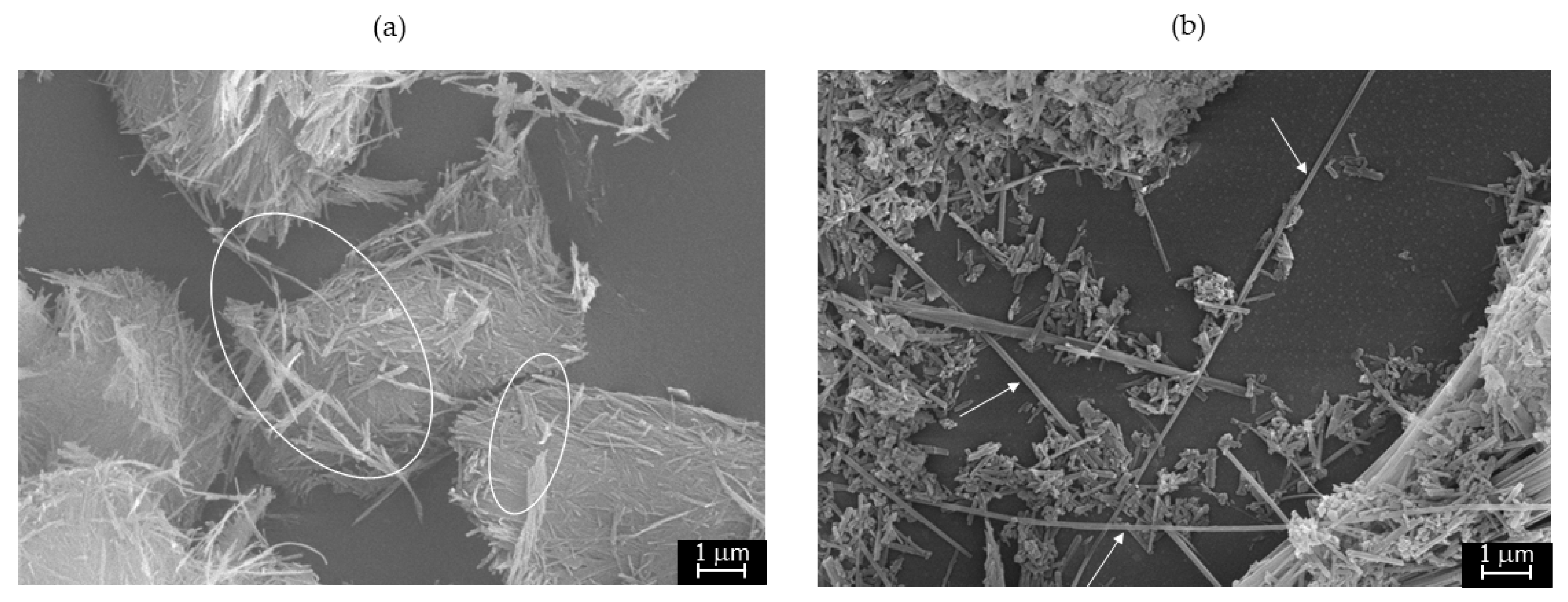
The fibrous morphology of the ST and SA milled samples is revealed in the SEM micrographs shown in Figure 6. The sample ST exhibits densely aggregated fibrous structures, forming entangled mats and compact bundles with a homogeneous appearance (Figure 6a). This aggregation is likely promoted by hydrogen bonding between surface SiOH groups and a small amount of adsorbed water, which facilitates network formation [41]. The ST fibers appear oriented in planar arrangements, consistent with their sedimentary depositional origin. Despite the fact that the fibers are mostly densely and compactly aggregated, it was possible to determine the length and width of loose fibers among these fiber aggregates (e.g., circles in Figure 6a). The length and width values are 1.51 ± 0.47 μm and 0.12 ± 0.04 μm, respectively, resulting in a length-to-width ratio of 13 ± 5. However, a more representative size of ST sample is the average length of the aggregated fibers (4.5 μm). These aggregates were not included in the individual fiber statistical analysis. From a textural perspective, no discernible porosity is evident in the ST, at the used observation conditions, as the fibers form a compact and planar structure. This reduced porosity may be attributed to the presence of amorphous silica impurities, identified in previous analyses, which could fill voids between fibers or promote a denser packing, thereby modifying the material’s overall texture and fiber arrangement. In contrast, the SA sample (Figure 6b) displays elongated needle-like and rod-shaped particles, along with short disaggregated fibers dispersed throughout the field of view. Individual long fibers, exceeding 5 microns in length with notable flexibility, are clearly visible (see arrows in Figure 6b). Statistical analysis of measurable loose fibers revealed a bimodal particle distribution, associated with two distinct size populations. One of them consists of short fibers (~70%) with length and width values of 1.16 ± 0.39 μm, and 0.10 ± 0.02 μm, respectively, resulting in a length-to-width ratio of 11.22. Meanwhile, ~30% is composed of long and flexible fibers, with a length of 7.08 ± 2.83 μm and a width of 0.18 ± 0.07 μm, yielding a length-to-width ratio of 44. Taking into account the relative abundance of each population, the weighted mean fiber length for the SA sample is 2.94 µm. The nanometric thicknesses and high aspect ratio of these needle-like crystals are characteristic of the anisotropic crystal growth of the SA sample. Importantly, the overall mean fiber length for SA, weighted by the relative abundance of the two size populations is ~3 µm, which remains on the same order of magnitude as the average length of the aggregated fibers in ST (4.5 µm).
Although both samples exhibit a similar degree of fineness, the fibers display different dispersion/aggregation behavior, likely associated with their origin. These morphological features have been explored in various studies using different defibrillation methods, without affecting sepiolite length and flexibility [42]. Moreover, the potential to obtain tridimensional (3D) networks and bidimensional (2D) layered structures from hydrogels containing high-aspect-ratio sepiolite nanofibers has been demonstrated [43]. In addition, Giustetto et al. also analyzed hydrothermal-origin sepiolite, similar to SA, finding extremely long (up to several centimeters) and flexible fibers, composed of bundles of thinner fibrils [1]. These previous works revealed the influence of geological formation environment on sepiolite, highlighting that sepiolite crystallized from hydrothermal solution presents large size of the fibers and high degree of crystallinity.
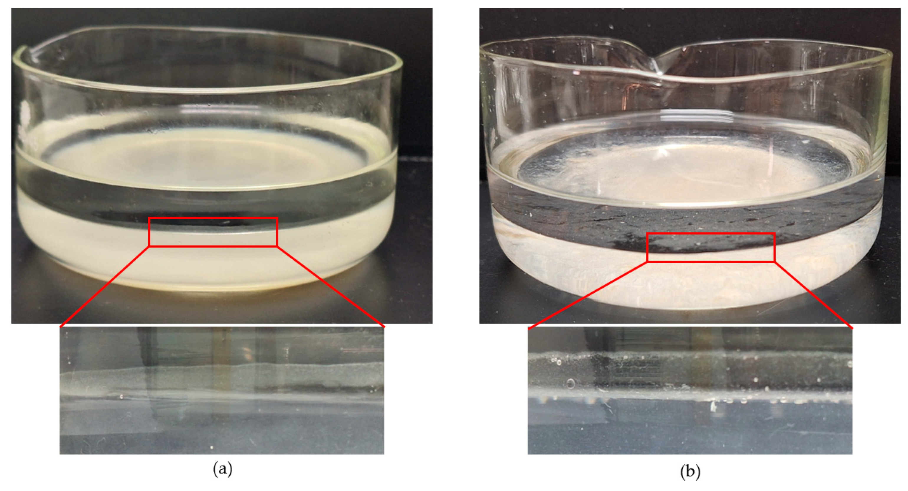
To further investigate potential differences in surface properties, a simple hydrophobic/hydrophilic test was conducted on both sepiolite samples, ground under identical conditions. Figure 7 shows photographs of octane–water systems one hour after the addition of the samples. Upon introduction, particles predominantly settled, indicating a primarily hydrophilic nature. This is consistent with the expected behavior of sepiolite, which is inherently hydrophilic due to its composition as a hydrated magnesium silicate and its high surface area, rich in silanol groups (SiOH). Nevertheless, distinct interfacial behaviors were observed since the system containing ST samples exhibits an interface sharp and clear, whereas for SA, a cloudy interfacial region appeared. Under higher magnification, small air bubbles and fine particles were observed suspended at the interface for the system containing the SA sample.
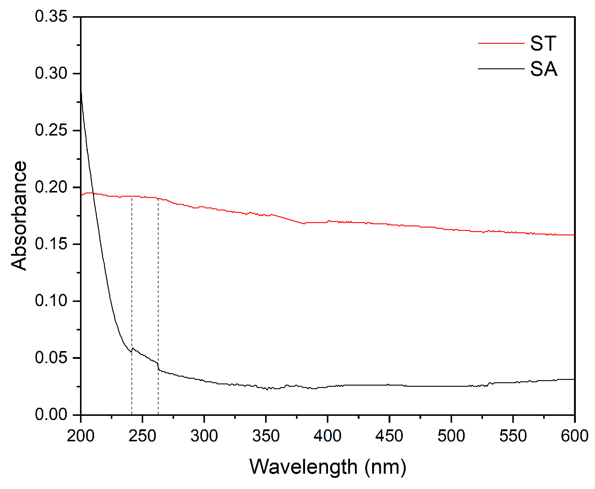
To confirm the presence and nature of particles at the interface, the hydrophobic/hydrophilic test was replicated directly in a UV-Vis cuvette, limiting UV-Vis exposure to the interfacial region. Figure 8 displays the resulting UV-Vis spectra obtained for each sample. The ST sample shows no significant absorption consistent with visual observations of a clear interface. In contrast, the SA sample exhibits a characteristic absorption band between approximately 242 nm and 262 nm [44], delimited with stripped lines; confirming the presence of sepiolite at the interface. This interfacial behavior is likely related to morphological and textural differences: the SA sample comprises smaller, disaggregated particles with a higher specific surface area compared to ST. As a fibrous mineral, sepiolite has greater lateral surface area than basal surface area. Furthermore, the chemical composition of these surfaces depends on particle size and exposed surface orientation [45], with the lateral surface being more hydrophilic than the basal surface. Consequently, the hydrophilic nature of the particles varies according to their size, with very small SA particles exhibiting lower hydrophilicity than ST particles.

5. Industrial and Environmental Applications

Sepiolite from Tolsa has been commercially distributed worldwide since 1957. Its applications are diverse, ranging from functional additives (biocidal, fungicidal, algicidal, and virucidal agents, synergistic agents for flame retardants, photocatalytic decontamination materials, and self-cleaning additives) to industrial solutions including paints and coatings, bitumen and asphalt formulations, drilling muds, construction material additives, foundry aids, and paper industry inputs. Additionally, it is used in environmental applications such as absorbents, bleaching earths, filtration, purification, and clarification. In the life sciences sector, this sepiolite is also employed in animal feed and agricultural additives, while in the pet care sector, it is widely used in cat litter and other hygiene products [46].
Regarding the SA sample, characterized by its disaggregated fibers, higher aspect ratio, and greater crystallinity than ST, it exhibits properties that could be advantageous for adsorption and interfacial processes. Its enhanced surface accessibility and higher specific surface area make it particularly suitable for wastewater treatment applications, such as the removal of heavy metals or organic contaminants. It is important to mention that the high-aspect sepiolite fibers of the SA sample allow obtaining 3D networks and 2D layered nanostructures [43]. These sepiolite-based nanoarchitectures show promising potential for the development of inorganic membranes with pore size and pore size distribution appropriated for ultrafiltration process. Moreover, the incipient hydrophobicity observed in the interfacial tests suggests possible applications in emulsion stabilization, or as a carrier for active agents in catalytic or controlled-release systems. The SA sample’s well-defined structure and surface reactivity also support its potential use as a catalyst support or matrix for functional nanocomposites. These application pathways highlight the importance of geological origin in tailoring sepiolite for specific technological uses, expanding its utility beyond traditional sectors.

6. Conclusions

The geological origin of the sepiolite samples, lacustrine-derived (ST) and hydrothermal-derived (SA), has an impact on their physicochemical properties. Comparative analysis reveals differences in chemical composition, crystallinity, thermal behavior, morphology, and hydrophobic/hydrophilic behavior. The ST sample exhibited elevated SiO2 content, likely due to amorphous silica impurities, consistent with its lower crystallinity, and higher structural defects. In contrast, the SA sample presented a chemical composition aligned with ideal sepiolite stoichiometry, accompanied by sharper XRD reflections and lower FWHM values, indicating a greater structural order than ST. SEM observations further underscore these contrasts. ST displays dense, aggregated fiber bundles forming planar textures, associated with the sedimentary depositional origin. SA, by contrast, reveals disaggregated needle-like fibers with high aspect ratios and nanometric thickness, characteristics indicative of anisotropic hydrothermal crystal growth. Hydrophilicity/hydrophobicity tests for both samples confirm their predominantly hydrophilic nature; however, SA exhibits additional interfacial behavior since fine particles suspended at the octane–water boundary, suggesting relatively less hydrophilicity or incipient hydrophobicity. This is attributable to the high specific surface area and surface orientation effects in the SA sample, which enhance interfacial activity. Thus, the sepiolite formation environment critically defines the mineral’s structural, morphological, and surface characteristics, directly influencing its interaction with fluids, adsorption capacities, and dispersion behavior, essential for both geological interpretation and industrial utility. These characteristics confer a wide range of applications on sepiolite. ST is mainly used as an adsorbent, especially for wastewater treatment, whereas SA enables the formation of 3D networks that could be applied in the fabrication of inorganic membranes.

Author Contributions

Conceptualization, L.A.C. and S.E.B.; methodology, L.A.C., L.L.; software, L.A.C., L.L.; validation, L.A.C., L.L.; formal analysis, L.A.C., L.L.; investigation, L.A.C., L.L., S.A.M. and S.E.B.; resources, S.A.M., S.E.B.; data curation, L.A.C., L.L.; writing—original draft preparation, L.A.C., L.L.; writing—review and editing, L.A.C., L.L., S.A.M. and S.E.B.; visualization, L.A.C., L.L.; supervision, S.E.B.; project administration S.A.M., S.E.B. All authors have read and agreed to the published version of the manuscript.

Funding

This research received no external funding.

Acknowledgments

The authors thank the Consejo Nacional de Investigaciones Científicas y Técnicas (CONICET), the Centro de Geología Aplicada, Agua y Medio Ambiente (CGAMA, CIC-UNS) and the Universidad Nacional del Sur for their support.

Conflicts of Interest

The authors declare no conflict of interest.

References

  1. Giustetto, R.; Macaluso, L.; Berlier, G.; Ganjkhanlou, Y.; Barale, L. Characterisation and possible hazard of an atypical asbestiform sepiolite associated with aliphatic hydrocarbons from Sassello, Ligurian Apennines, Italy. Mineral. Mag. 2019, 83, 209–222. [Google Scholar] [CrossRef]
  2. Jones, B.F.; Galán, E. Sepiolite and palygorskite. Rev. Mineral. Geochem. 1988, 19, 631–674. [Google Scholar]
  3. Ruiz, A.I.; Ruiz-García, C.; Ruiz-Hitzky, E. From old to new inorganic materials for advanced applications: The paradigmatic example of the sepiolite clay mineral. Appl. Clay Sci. 2023, 235, 106874. [Google Scholar] [CrossRef]
  4. Stoessell, R.K.; Hay, R.L. The geochemical origin of sepiolite and kerolite at Amboseli, Kenya. Contrib. Mineral. Petrol. 1978, 65, 255–267. [Google Scholar] [CrossRef]
  5. Herranz, J.E.; Pozo, M. Sepiolite and other authigenic Mg-clay minerals formation in different Palustrine environments (Madrid Basin, Spain). Minerals 2022, 12, 987. [Google Scholar] [CrossRef]
  6. Tateo, F.; Sabbadini, R.; Morandi, N. Palygorskite and sepiolite occurrence in Pliocene lake deposits along the River Nile: Evidence of an arid climate. J. Afr. Earth Sci. 2000, 31, 633–645. [Google Scholar] [CrossRef]
  7. Mokatse, T.; Prud’Homme, C.; Vainer, S.; Adatte, T.; Shemang, E.; Verrecchia, E.P. Sepiolite as a multifactorial indicator of paleoenvironments in the Chobe Enclave (northern Botswana). Sediment. Geol. 2023, 454, 106459. [Google Scholar] [CrossRef]
  8. Kadir, S.; Erkoyun, H.; Eren, M.; Huggett, J.; Önalgil, N. Mineralogy, geochemistry, and genesis of sepiolite and palygorskite in Neogene lacustrine sediments, Eskişehir Province, west central Anatolia, Turkey. Clays Clay Miner. 2016, 64, 145–166. [Google Scholar] [CrossRef]
  9. Calvo, J.P.; Pozo, M. Geology of magnesian clays in sedimentary and non-sedimentary environments. In Magnesian Clays: Characterization, Origin and Applications; Pozo, M., Galán, E., Eds.; Digilabs: Bari, Italy, 2015; pp. 123–174. [Google Scholar]
  10. Draidia, S.; Ouahabi, M.E.; Daoudi, L.; Havenith, H.B.; Fagel, N. Occurrences and genesis of palygorskite/sepiolite and associated minerals in the Barzaman formation, United Arab Emirates. Clay Miner. 2016, 51, 763–779. [Google Scholar] [CrossRef]
  11. Baldermann, A.; Mavromatis, V.; Frick, P.M.; Dietzel, M. Effect of aqueous Si/Mg ratio and pH on the nucleation and growth of sepiolite at 25 °C. Geochim. Cosmochim. Acta 2018, 227, 211–226. [Google Scholar] [CrossRef]
  12. Cañaveras, J.C.; Calvo, J.P.; Ordóñez, S.; Muñoz-Cervera, M.C.; Sánchez-Moral, S. Tectono-Sedimentary Evolution of the Madrid Basin (Spain) during the Late Miocene: Data from Paleokarst Profiles in Diagenetically-Complex Continental Carbonates. Geosciences 2020, 10, 433. [Google Scholar] [CrossRef]
  13. Reijmer, J.J.; Blok, C.N.; El-Husseiny, A.; Kleipool, L.M.; Hogendorp, Y.C.; Alonso-Zarza, A.M. Petrophysics and sediment variability in a mixed alluvial to lacustrine carbonate system (Miocene, Madrid Basin, Central Spain). Depos. Rec. 2022, 8, 317–339. [Google Scholar] [CrossRef]
  14. Herranz, J.E.; Pozo, M. Authigenic Mg-clay minerals formation in lake margin deposits (the Cerro de los Batallones, Madrid Basin, Spain). Minerals 2018, 8, 418. [Google Scholar] [CrossRef]
  15. Pozo, M.; Galán, E.; González, J.L. Sepiolite and palygorskite from the Madrid Basin, Spain: Mineralogy and genetic relations. Appl. Clay Sci. 2014, 95, 31–45. [Google Scholar]
  16. Ordóñez, S.; Calvo, J.P.; García del Cura, M.A.; Alonso Zarza, A.M.; Hoyos, M. Sedimentology of sodium sulphate deposits and special clays from the Tertiary Madrid Basin (Spain). In Lacustrine Facies Analysis; Anadón, P., Cabrera, L., Kelts, K., Eds.; Blackwell Scientific Publications: Oxford, UK, 1991; pp. 39–55. [Google Scholar]
  17. Cortelezzi, C.R.; Marfil, S.A.; Maiza, P.J. A sepiolite of large crystalline growth from “La Adela” mine province of Río Negro, Argentina. N. Jb. Miner. Mh. 1994, 4, 157–166. [Google Scholar] [CrossRef]
  18. Caminos, R.; Llambías, E.J. El basamento cristalino. In Proceedings of the Geología y Recursos Naturales de la Provincia de Río Negro, IX Congreso Geológico Argentino, Bariloche, Argentina, 5–9 November 1984; Ramos, V., Ed.; pp. 37–63. [Google Scholar]
  19. Maiza, P.; Marfil, S.A. Diaclasas mineralizadas con sepiolita de cantera La Adela, provincia de Río Negro, Argentina. In Proceedings of the XII Congreso Geológico Argentino y II Congreso de Exploración de Hidrocarburos, Mendoza, Argentina, 10–15 October 1993; pp. 82–86. [Google Scholar]
  20. Dominguez, E.A.; Maiza, P.J. Yacimientos no metalíferos y rocas de aplicación. In Proceedings of the Geología y Recursos Naturales de la Provincia de Río Negro, IX Congreso Geológico Argentino, Bariloche, Argentina, 5–9 November 1984; Ramos, V., Ed.; pp. 611–628. [Google Scholar]
  21. Cordenons, P.D.; Remesal, M.B.; Salani, F.M.; Cerredo, M.E. Temporal and spatial evolution of the Somún Curá magmatic province, northern extra-andean Patagonia, Argentina. J. S. Am. Earth Sci. 2020, 104, 102881. [Google Scholar] [CrossRef]
  22. International Mineralogical Association (IMA). 2025. Available online: https://cnmnc.units.it/files/editor/IMA_Master_List_(2025-07).pdf (accessed on 15 August 2025).
  23. Velde, B. Introduction to Clay Minerals: Chemistry, Origins, Uses and Environmental Significance; Chapman and Hall Ltd.: London, UK, 1992; Volume 198, 198p. [Google Scholar]
  24. JCPDS, International Centre for Diffraction Data (ICDD). Mineral Powder Diffraction File Databook; ICDD: Swarthmore, PA, USA, 1993; pp. 614–615. [Google Scholar]
  25. Pozo, M.; Calvo, J.P.; Pozo, E.; Moreno, A. Genetic constraints on crystallinity, thermal behavior and surface area of sepiolite from the Cerro de los Batallones deposits (Madrid Basin, Spain). Appl. Clay Sci. 2014, 91–92, 30–45. [Google Scholar] [CrossRef]
  26. Dikmen, S. Zeta potential study of natural- and acid-activated sepiolites in electrolyte solutions. Can. J. Chem. Eng. 2012, 90, 421–427. [Google Scholar] [CrossRef]
  27. Locatelli, D.; Pavlovic, N.; Barbera, V.; Giannini, L.; Galimberti, M. Sepiolite as reinforcing filler for rubber composites: From the chemical compatibilization to the commercial exploitation. Kautsch. Gummi Kunststoffe 2020, 73, 34–41. [Google Scholar]
  28. Zhou, F.; Ye, G.; Gao, Y.; Wang, H.; Zhou, S.; Liu, Y.; Yan, C. Cadmium adsorption by thermal-activated sepiolite: Application to in-situ remediation of artificially contaminated soil. J. Hazard. Mater. 2022, 423A, 127104. [Google Scholar] [CrossRef]
  29. Shahraki, B.; Mehrabi, B.; Dabiri, R. Thermal behavior of Zefreh dolomite mine (Central Iran). J. Min. Metall. 2009, 45B, 35–44. [Google Scholar] [CrossRef]
  30. Suárez, M.; García-Rivas, J.; García-Romero, E.; Jara, N. Mineralogical characterisation and surface properties of sepiolite from Polatli (Turkey). Appl. Clay Sci. 2016, 131, 124–130. [Google Scholar] [CrossRef]
  31. Largo, F.; Haounati, R.; Ouachtak, H.; Hafid, N.; Jada, A.; Addi, A.A. Design of organically modified sepiolite and its use as adsorbent for hazardous Malachite Green dye removal from water. Water Air Soil Pollut. 2023, 234, 183. [Google Scholar] [CrossRef]
  32. Wei, S.; Wang, L.; Wu, Y.; Liu, H. Study on removal of copper ions from aqueous phase by modified sepiolite flocs method. Environ. Sci. Pollut. Res. 2022, 29, 73492–73503. [Google Scholar] [CrossRef]
  33. Jiang, X.; Wang, S.; Ge, L.; Lin, F.; Lu, Q.; Wang, T.; Lu, B. Development of organic–inorganic hybrid beads from sepiolite and cellulose for effective adsorption of malachite green. RSC Adv. 2017, 7, 38965–38972. [Google Scholar] [CrossRef]
  34. Zhou, X.; Li, H.; Liu, Y.; Hao, J.; Liu, H.; Lu, X. Improvement of stability of insecticidal proteins from Bacillus thuringiensis against UV-irradiation by adsorption on sepiolite. Adsorpt. Sci. Technol. 2018, 36, 1233–1245. [Google Scholar] [CrossRef]
  35. Yang, L.; Deng, Y.; Gong, D.; Luo, H.; Zhou, X.; Jiang, F. Effects of low molecular weight organic acids on adsorption of quinclorac by sepiolite. Environ. Sci. Pollut. Res. 2021, 28, 9582–9597. [Google Scholar] [CrossRef]
  36. Alves, L.; Ferraz, E.; Santarén, J.; Rasteiro, M.G.; Gamelas, J.A. Improving colloidal stability of sepiolite suspensions: Effect of the mechanical disperser and chemical dispersant. Minerals 2020, 10, 779. [Google Scholar] [CrossRef]
  37. Zhang, G.; Liu, L.; Shiko, E.; Cheng, Y.; Zhang, R.; Zeng, Z.; Zhao, T.; Zhou, Y.; Chen, H.; Liu, Y.; et al. Low-price MnO2 loaded sepiolite for Cd2+ capture. Adsorption 2019, 25, 1271–1283. [Google Scholar] [CrossRef]
  38. Caicedo-Pineda, G.A.; Prada-Fonseca, M.C.; Casas-Botero, A.E.; Martínez-Tejada, H.V. Effect of the tryptone concentration on the calcium carbonate biomineralization mediated by Bacillus cereus. Dyna 2018, 85, 69–75. [Google Scholar] [CrossRef]
  39. Nguyen, M.B.; Le, G.H.; Pham, T.T.; Pham, G.T.; Quan, T.T.; Nguyen, T.D.; Vu, T.A. Novel Nano-Fe2O3-Co3O4 Modified Dolomite and Its use as highly efficient catalyst in the ozonation of ammonium solution. J. Nanomater. 2020, 2020, 4593054. [Google Scholar] [CrossRef]
  40. Sánchez-Sánchez, A.; Cerdán, M.; Jordá, J.D.; Amat, B.; Cortina, J. Characterization of soil mineralogy by FTIR: Application to the analysis of mineralogical changes in soils affected by vegetation patches. Plant Soil 2019, 439, 447–458. [Google Scholar] [CrossRef]
  41. Ruiz-Hitzky, E.; Ruiz-García, C.; Fernandes, F.M.; Lo Dico, G.; Lisuzzo, L.; Prevot, V.; Darder, M.; Aranda, P. Sepiolite-hydrogels: Synthesis by ultrasound irradiation and their use for the preparation of functional clay-based nanoarchitectured materials. Front. Chem. 2021, 9, 733105. [Google Scholar] [CrossRef] [PubMed]
  42. Lescano, L.; Castillo, L.; Marfil, S.; Barbosa, S.; Maiza, P. Separation and purification of sepiolite fibers. Contribution to special processing for industrial use. Appl. Clay Sci. Intern. J. Appl. Technol. Clays Clay Miner. 2014, 93, 378–382. [Google Scholar]
  43. Castillo, L.; Lescano, L.; Sirvent, L.; Barbosa, S.; Marfil, S.; Maiza, P. Separación y purificación de fibras de sepiolita: Contribución al procesamiento de arcillas especiales para uso industrial. Geoacta 2011, 36, 113–127. [Google Scholar]
  44. Fajdek-Bieda, A.; Wróblewska, A.; Miądlicki, P.; Szymańska, A.; Dzięcioł, M.; Booth, A.M.; Michalkiewicz, B. Influence of technological parameters on the isomerization of geraniol using sepiolite. Catal. Lett. 2020, 150, 901–911. [Google Scholar] [CrossRef]
  45. Tian, G.; Han, G.; Wang, F.; Liang, J. Sepiolite nanomaterials: Structure, properties and functional applications. In Nanomaterials from Clay Minerals; Wang, A., Wang, W., Eds.; Elsevier: Amsterdam, The Netherlands, 2019; pp. 135–201. [Google Scholar]
  46. Tolsa. Available online: https://www.tolsa.com/es/ (accessed on 15 August 2025).
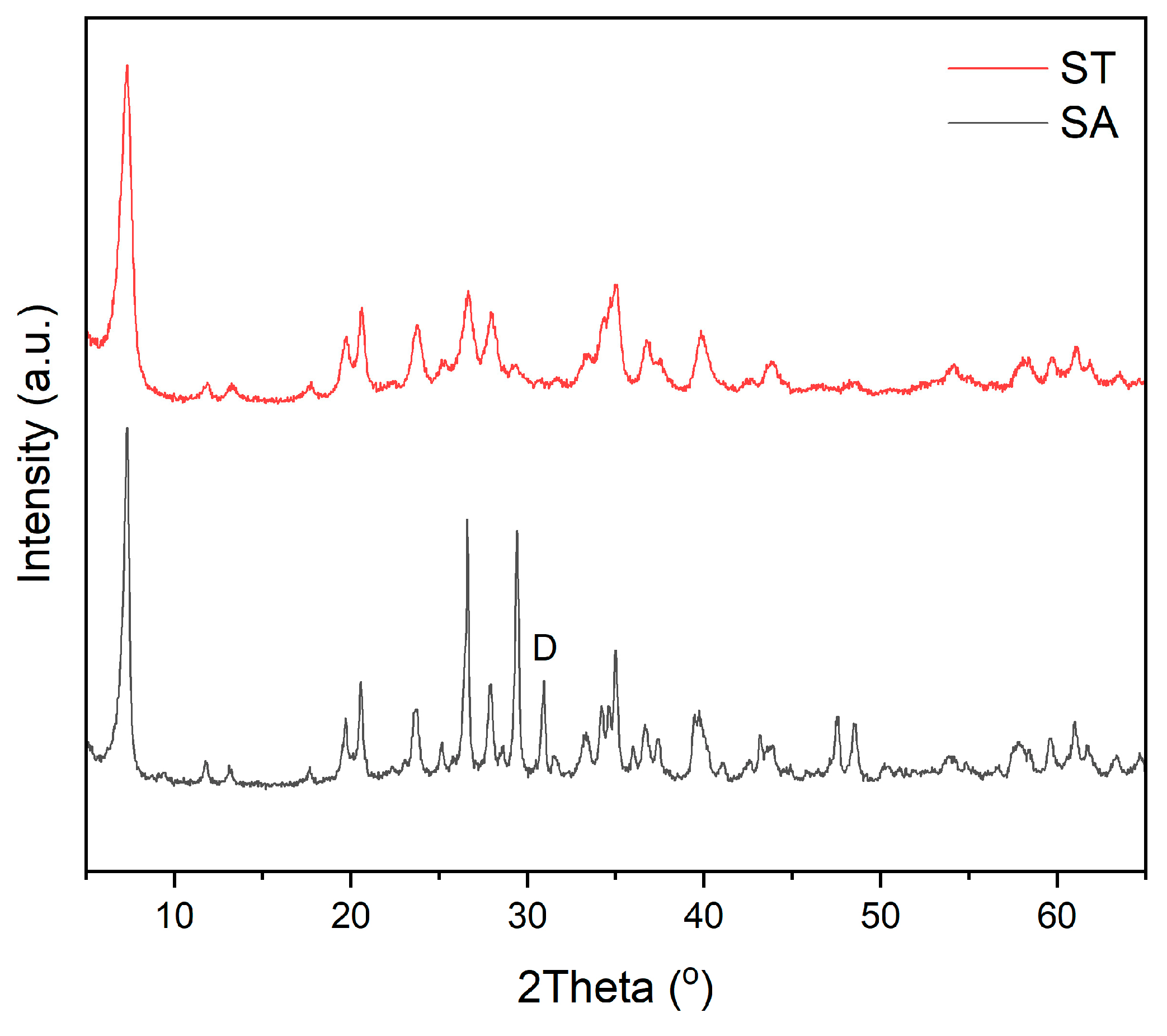
Figure 3. X-ray diffractograms of ST and SA samples. D: dolomite.
Minerals 15 00950 g003
Figure 4. (a) Mass loss curve, and (b) first derivative of the mass loss curve of ST and SA samples from TGA.
Minerals 15 00950 g004
Figure 5. FTIR spectra of ST and SA samples. Ref: D: dolomite.
Minerals 15 00950 g005
Figure 6. SEM micrographs (20,000×) of (a) ST sample, showing densely aggregated fibrous structures with a few loose fibers (enclosed in circles), and (b) SA sample displaying short disaggregated fibers and individual long fibers (indicated by arrows).
Minerals 15 00950 g006
Figure 7. Photographs of hydrophilicity/hydrophobicity test for: (a) ST and (b) SA.
Minerals 15 00950 g007
Figure 8. UV-vis spectra at the water–octane interfaces of the systems containing ST and SA particles.
Minerals 15 00950 g008
Table 1. Chemical composition of analyzed samples from Tolsa (ST) and La Adela (SA) compared with ideal sepiolite (S).
SamplesSiO2 (%)Al2O3 (%)Fe2O3 (%)MgO (%)CaO (%)Na2O (%)K2O (%)LOI (%)
ST63.101.080.2723.800.490.090.2110.96
SA53.151.831.3124.080.900.100.0818.54
S56.26--25.27---18.57
Disclaimer/Publisher’s Note: The statements, opinions and data contained in all publications are solely those of the individual author(s) and contributor(s) and not of MDPI and/or the editor(s). MDPI and/or the editor(s) disclaim responsibility for any injury to people or property resulting from any ideas, methods, instructions or products referred to in the content.