Article

Controls on the Formation and Stability of Siderite (FeCO3) and Chukanovite (Fe2(CO3)(OH)2) in Reducing Environment

Department of Earth System Sciences, Yonsei University, Seoul 03722, Korea
*
Author to whom correspondence should be addressed.
Minerals 2020, 10(2), 156; https://doi.org/10.3390/min10020156
Submission received: 30 December 2019 / Revised: 30 January 2020 / Accepted: 7 February 2020 / Published: 11 February 2020
(This article belongs to the Special Issue Clays and Micro-Organisms: From Nature to Industry)

Abstract

:
The formation of ferrous carbonate mineral is a significant geochemical reaction linked to iron and carbon cycling in the sedimentary environment. However, knowledge of the controlling factors and conditions for the mineral formation is limited. Two types of ferrous carbonate mineral, siderite (FeCO3) and chukanovite (Fe2(CO3)(OH)2) were synthesized under a FeCl2–NaHCO3 system with various concentration ranges (10–100 mmolal) and ratios (Fe:Dissolved inorganic carbon (DIC) = 1:1, X:50, and 50:X) to verify the concentration limit and control species for the formation of those minerals. The mineralogy of filtered precipitates at the reaction time of 1 week and 1 month were identified by X-ray diffraction (XRD), and scanning/transmission electron microscopic (S/TEM) analyses were applied for direct identification of the synthesized siderite and chukanovite at various conditions. A semi-quantitative calculation to estimate siderite proportion (siderite/[siderite + chukanovite]) in the precipitates was carried out using peak intensity ratios of siderite (d104 [2θ = 32.02°]) and chukanovite (d211 [2θ = 33.98°]) from XRD profiles. The framboids or trigonal-rhombohedron crystals and flaky rosette-shaped minerals were identified in SEM analysis. In addition, the chemical compositions of Fe and C of framboid (Fe:C = 1:1.01) and flaky mineral (1:2.04) were identified as siderite and chukanovite, respectively. The formation of siderite was predominated over chukanovite in 50 mmolal (both Fe and DIC) or higher conditions (siderite proportion = 49–100%). The estimated siderite proportion increased (27–100%) as DIC concentration increased (15–100 mmolal) in conditions of varying ratios of iron and DIC (50:X), indicating that DIC is a decisive factor in siderite formation. The increase in the reaction time promotes the siderite proportion increase, so that chukanovite may be dissolved and re-precipitated as siderite for the long-term reaction, except in the enriched DIC condition (Fe:DIC = 15:50). This study demonstrates that various conditions, not limited to the concentrations or reaction time, may affect the geochemical pathways of carbonate mineral formations.

1. Introduction

Siderite (FeCO3) is a common ferrous mineral in oxygen-free environmental conditions including lakes, rivers, and marine sediments [1,2,3], and even in extraterrestrial materials such as meteorites [4,5,6]. Ferrous iron is particularly unstable in aqueous solutions because it reacts readily with (dissolved) oxygen, oxidizing to ferric iron and precipitating as iron oxides and/or oxyhydroxides, including goethite, hematite, magnetite, maghemite, and ferrihydrite [7,8,9]. Therefore, siderite could be used as a good tracer to reveal paleo-environmental redox conditions [10,11]. In addition, carbon dioxide and hydrocarbons are formed when siderite reacts with water to form magnetite, which could be used as a tracer for tracking life activities in extraterrestrial conditions [12,13,14,15]. The Fe-reducing microorganisms use organic carbon as an electron donor and carbon source for their metabolic processes, so they produce carbonate and/or carbon dioxide and, as a consequence, enrich the Fe(II) in supernatant or pore water [16,17] that could enhance the formation of secondary phase mineral. Although the formation of siderite has been identified in lab experiments associated with microbial Fe(III) reduction of Fe-oxide (akaganeite: [18]; magnetite: [19]; ferrihydrite: [20]) and Fe-rich clay minerals (nontronite: [21]), no detailed conditions or controlling factors on biotic siderite formation have been studied.
Iron carbonate minerals are very useful as tracers for the geochemical reactions by utilizing stable isotope analysis. Therefore, synthetic methods of siderite have mostly been reported in previous studies that discussed carbon and oxygen isotope fractionation in siderite [22,23,24,25]. The method used in previous studies is “active degassing experiments” which was modified after Kim and O’Neil [26]. In this method, siderite is precipitated upon pH increase by bubbling N2 or Ar-gas into the Fe-titrated bicarbonate solution [23,24,25]. In addition to siderite, a noncrystalline phase under low temperature (4 and 9 °C [24]) or green rust under low concentrations of iron and DIC (1 mM and 3 mM, respectively, [25]) were identified during the siderite synthesis. However, the relationships between those phases and siderite are not clearly understood. Even when using other synthetic methods, such as controlled-chemistry and free-drift methods using ferrous perchlorate (Fe(ClO4)2) and bicarbonate solution for siderite formation [22], phases A and B were identified in the middle stage of the siderite formation. However, the mineralogy of phases A and B has not been identified nor has it been proven to be related to siderite. On the other hand, the synthesis of siderite through thermal treatment was reported [27]. The optimal condition was 1.5 h at 140 °C with iron sulfate, ammonium carbonate, and ascorbic acid as starting materials. In this case, iron was oxidized if ascorbic acid was not added, indicating that anaerobic conditions should be kept or reducing agents should exist during the reaction. Nevertheless, optimum conditions (concentration, ratios between iron and DIC, etc.) for siderite formation at ambient temperatures and pressure conditions have not yet been clearly understood.
Recently, chukanovite (Fe2(CO3)(OH)2), an iron hydroxide-carbonate mineral, was identified as a new mineral phase in the weathered iron meteorite, Dronino [28]. Chukanovite was observed in the cavity of the meteorite with goethite, hematite, akaganite, and iron hydroxides. Iron (Fe) hydroxide-carbonate (FHC) was also recently reported on the surface corrosion of steel [29,30] that was identified as chukanovite [31]. Similar to siderite, chukanovite or FHC could be formed associated with the metabolic process of Fe-reducing microorganisms [32]. As Shewanella putrefaciens reduced ferrihydrite and akaganeite, ferrous-enriched magnetite (Fe2+/Fetot = 0.5–0.6) was formed within a day; however, it transformed to FHC with time. In the next 3 years of incubation, the Fe2+/Fetot ratio of magnetite decreased as stoichiometric form (0.33) and the FHC became the dominant mineral phase in the system. As such, ferrous iron supplied by microbial metabolism forms the various types of iron carbonate minerals, including siderite and chukanovite. However, there is no clear understanding of the relationship between those minerals. Nonetheless, the solubility model under a carbonate-chloride system was reported, chukanovite was formed in a low-carbonate concentration condition, and siderite was formed in carbonate-enriched condition [33].
Therefore, the present study aims to find ideal conditions, concentration range of iron and dissolved inorganic carbon (DIC), concentration ratio (Fe:DIC), and reaction time for siderite formation. Moreover, the proportion of siderite (siderite/[siderite+chukanovite], in percent) was estimated in each experimental condition to clarify concentration limit and control species for siderite formation. To identify the mineralogy of the precipitates, X-ray diffractometer (XRD) and scanning/transmission electron microscope (S/TEM) analyses were applied to the filtered dried powder after 1 week or 1 month of reaction for each experimental setting.

2. Materials and Methods

2.1. Experimental Setup

The entire experiment was performed in an anaerobic chamber to prevent oxidation of ferrous ion. The distilled water for the experiment and washing step was boiled for 1 h, then nitrogen gas was purged for 3 h to remove the dissolved oxygen prior to use. The mass of iron chloride (FeCl2·4H2O, KANTO CHEMICAL, Tokyo, Japan) and sodium bicarbonate (NaHCO3, Sigma-Aldrich, ACS grade, St. Louis, MO, USA) powder was measured then transferred to the anaerobic chamber to make a solution with degassed distilled water.
A total of 4 sets of experiments were performed to elucidate the mechanism of iron carbonate mineral formation (Table 1). The first and second sets were designed to identify the concentration limit of the siderite formation. The difference between the first and the second set was the method of adding the iron to the bicarbonate solution. The total volume was the same, 500 mL, but the first set was made by adding the solid phase of iron chloride to the 500 mL of sodium bicarbonate solution while the second set was conducted by mixing 250 mL of an iron chloride solution and sodium bicarbonate solution. Within each set, the concentrations of iron and DIC were same (1:1), and the range was from 10 to 100 mmolal. The third and fourth sets were performed to identify which ions, iron or bicarbonate, determine the formation of siderite. To do this, the third and fourth sets were made with different concentration ratios of iron and DIC (X:50 and 50:X, respectively). For the third set, concentration of DIC was fixed (50 mmolal in final concentration), and the concentration of iron chloride solution was varied (15, 30, 70, and 100 mmolal in final concentration). In contrast, the iron concentration was fixed in the fourth set (50 mmolal in final concentration), and the concentration of DIC was varied (15, 30, 70, and 100 mmolal in final concentration). During the synthesis, the temperature in the anaerobic chamber was maintained at 30 °C. The pH level of the solution was measured right after mixing the iron and bicarbonate solution and before filtering to stop the reaction using a portable pH meter (AB150, Fischer Scientific, Atlanta, GA, USA). The experiments were stopped at 1 week and 1 month and the solid phase was collected by filtering using an aspirator in the anaerobic chamber. For rapid drying to prevent oxidation, methanol was used in the final step of filtering; then, the dried powder was stored in sealed glass vial in the anaerobic chamber for further analysis.

2.2. Analysis and Calculation

In order to minimize oxidation of synthesized minerals by atmospheric oxygen, the entire sample preparation steps for the analysis were performed in the anaerobic chamber. The XRD profiles were obtained using a MiniFlex(II) (Rigaku, Tokyo, Japan) automated diffractometer, utilizing Cu-Kα radiation at Yonsei University. Dried precipitates were gently crushed and ground using a mortar to make a fine powder and then loaded on the Al-plate to make a random-oriented XRD specimen. Then, the surface was flattened by slashing it using slide glass rather than pressing to minimize the preferred-orientation effect. The XRD profiles were recorded for 2–70° with 0.02° steps at a scan speed of 1.5°/min. The first experimental set samples were reanalyzed after the first analysis to verify if any oxidation by atmospheric oxygen occurred during the XRD. No deterministic differences due to oxidation during the analysis were observed, therefore, subsequent samples were not reanalyzed.
The correlation between the proportion of siderite in the precipitates (siderite/[siderite+chukanovite]) and peak intensity ratio of the major peaks of siderite and chukanovite (d104 [2θ = 32.02°] and d211 [2θ = 33.98°], respectively) was obtained for the semi-quantitative calculation of the estimated siderite proportion. To obtain the function of the correlation, XRD profiles of various proportions of mixture of siderite [34] and chukanovite [28] were simulated using CrystalDiffract. Then the correlation equation between the peak intensity and siderite proportion was conducted as an exponential function:
Peak   Intensity   ratio   ( I 2 θ = 32.02 / I 2 θ = 33.98 ) = 0.1089 e 5.798 × s i d e r i t e   p r o p o r t i o n
The semi-quantitatively-estimated siderite proportion was then calculated by applying the peak ratio from each XRD profile to the correlation curve equation.
A JEOL JSM-5610LV (JEOL, Tokyo, Japan), equipped with OXFORD INCAx-act Model 51-ADD0021 (Oxford Instruments, Abingdon, UK) at the Department of Earth System Sciences, Yonsei University, was used to observe particle size and morphology. The dried powder was loaded on adhesive carbon tape, then coated with a palladium coater for 60 s prior to SEM observation.
For the TEM analysis, the dried powder of the 50:50 condition in the Experiment 1 sample was dispersed in degassed distilled water and sonicated for ~3 min. Then, a droplet of the dispersed particle-distilled water solution was dropped onto a 200-mesh copper grid. Bright-field TEM images, selected-area electron diffraction (SAED) patterns, and EDS analysis were obtained using a JEOL JEM-ARM200F (JEOL, Tokyo, Japan) equipped with EDS (INCA Energy TEM for JEM-ARM200F, Oxford Instruments, Abingdon, UK), operated at 200 keV at Yonsei University.

3. Results

3.1. Concentration Limit of Siderite Formation

The XRD profiles for the 1-week reaction samples from the first and second experiments showed two types of minerals: siderite (red dashed line with diamonds) and chukanovite (blue dashed line with circles), in accordance with the concentration conditions (Figure 1 and Figure 2). For the lowest-concentration condition of 10 mmolal, only chukanovite was observed, and for the following conditions of 15 to 50 mmolal, both siderite and chukanovite were observed (Figure 1A). For the high-concentration conditions of 70 and 100 mmolal, a dominant siderite phase was observed, even though it had a short reaction period (1 week, Figure 1A). After 1 month, only siderite was observed in the concentration range of 50 mmolal and higher (Figure 1B).
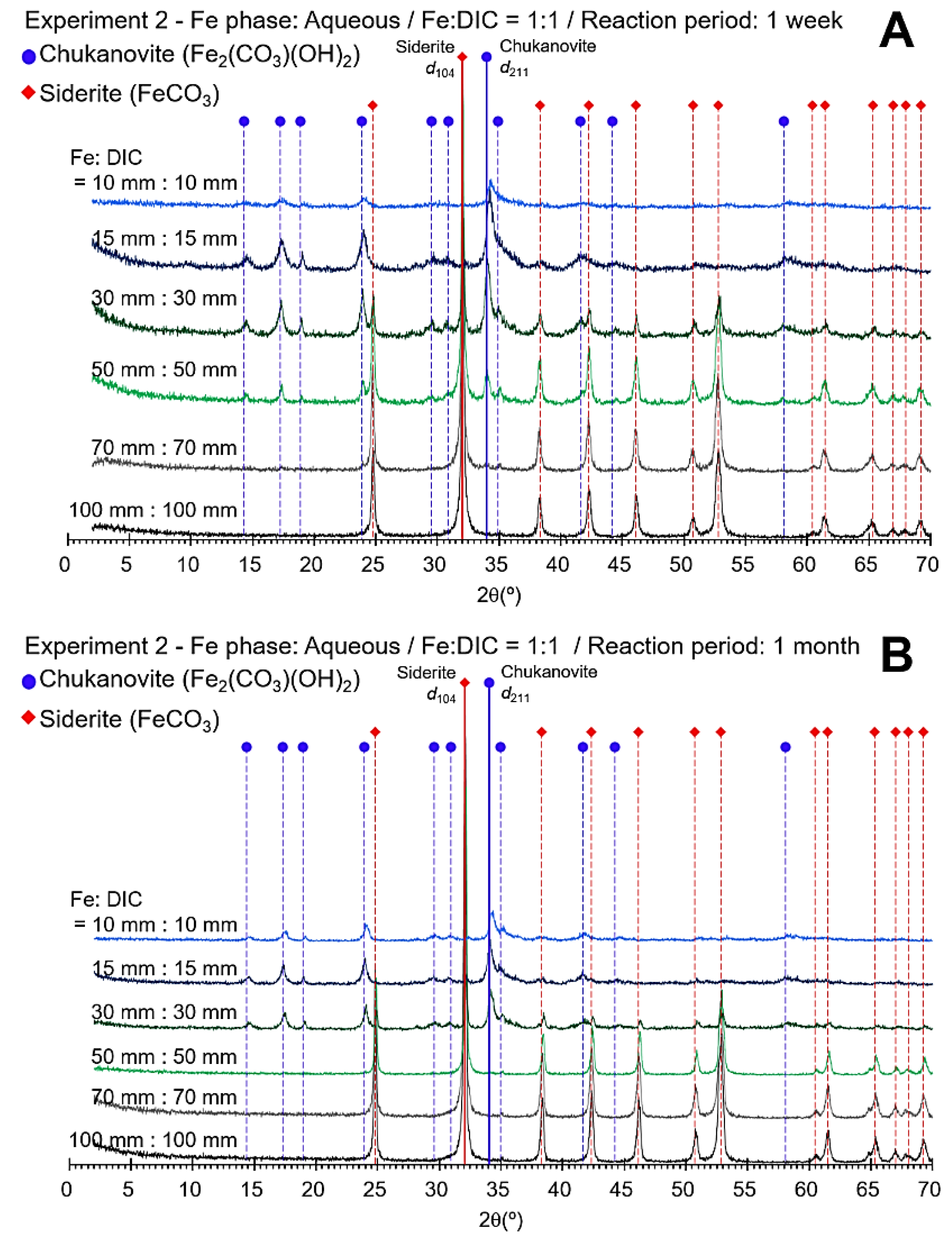
Compared to Experiment 1, in the second experiment set with aqueous iron, only chukanovite was observed under a low-concentration condition, but at a concentration of 50 mmolal, dominant siderite was observed even in the 1-week reaction samples (Figure 2A). After 1 month, only siderite was observed in the concentration range of 50 mmolal and was higher, like Experiment 1 (Figure 2B).
For each XRD profile, the proportion of siderite was estimated using the correlation curve to check the change in the content of siderite numerically (Table 2). In the case of Experiment 1, the proportion of siderite was calculated from 2.8% to 100% depending on the concentration of iron and DIC in the system at 1 week of reaction. The estimated siderite proportion increased as reaction time increased (i.e., 2.8 to 7.0 for 10:10 in Experiment 1). Similarly, the siderite proportion increased relative to the concentration increase in Experiment 2.

3.2. Control Species for Siderite Formation

In order to verify which ions determine the formation of siderite and chukanovite, various concentration ratios of iron and DIC were tested, and changes in the mineral composition were confirmed through XRD analysis (Figure 3 and Figure 4). In the third experiment, the concentration of DIC was fixed at 50 mmolal. It was found that the siderite and chukanovite were mixed even in the lowest iron concentration (15 mmolal of iron) with a short reaction time (1 week). There was a dominant siderite phase with a small amount of chukanovite in the 70 and 100 mmolal conditions (Figure 3A). After 1 month of reaction, insignificant peaks of chukanovite were identified in XRD profiles. In contrast, weak-intensity peaks of siderite with a dominant chukanovite phase were observed in the lowest iron concentration condition (Figure 3B).
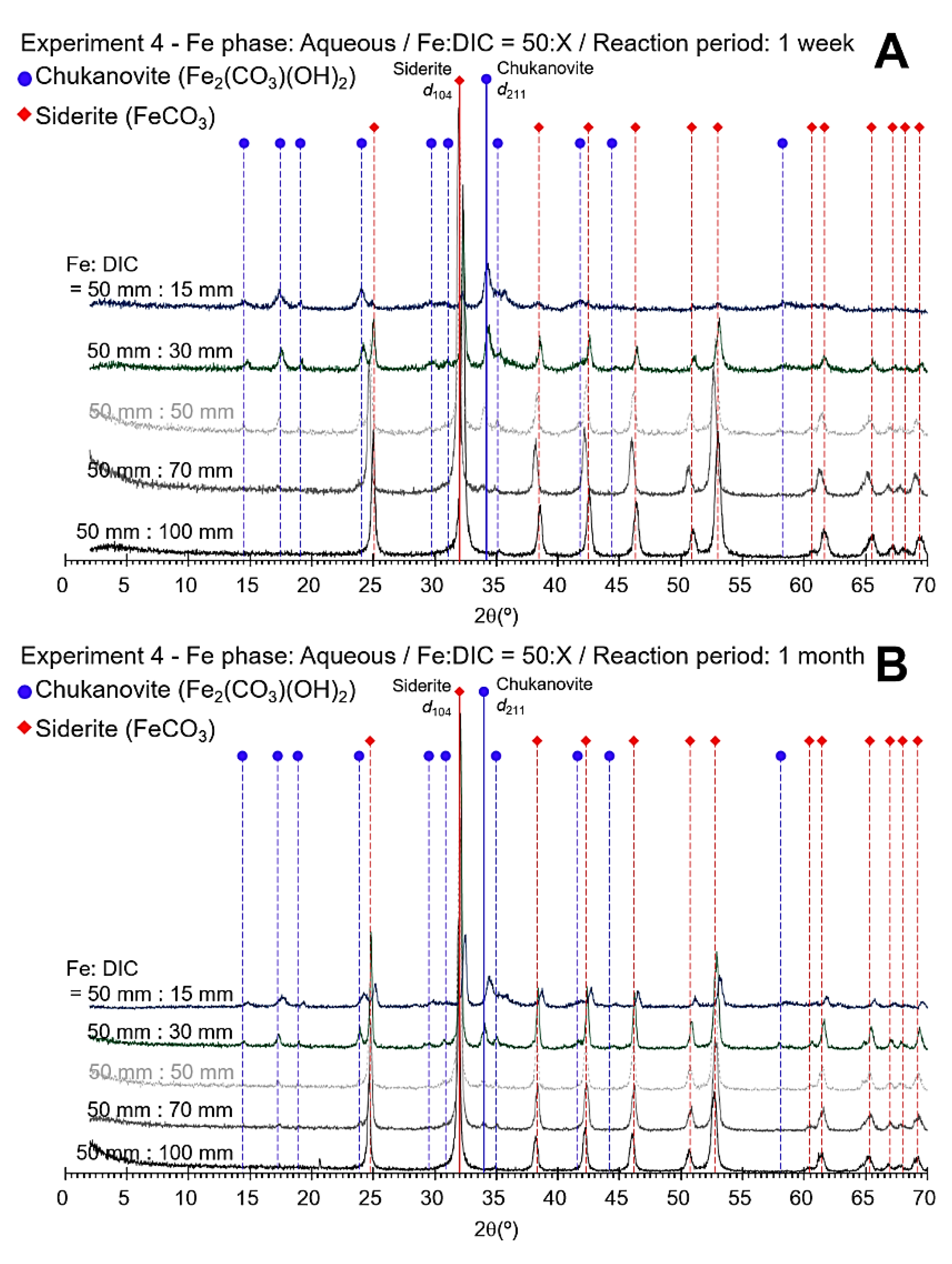
The XRD profiles of the samples in Experiment 4 showed a mixture of siderite and chukanovite under low-concentration conditions (50:15 and 50:30) as in Experiment 3, and for the high DIC concentration conditions, the precipitates were mostly siderite (Figure 4A). Similar to the 1 week of reaction samples, siderite and chukanovite were observed together under a low-concentration condition, but only siderite was observed at a high concentration in the XRD profiles of 1 month of reaction samples (Figure 4B).
In the 1-week reaction of Experiment 3, the estimated siderite proportion increased from 49.2% to 91.4% as the iron concentration increased. In the case of the 1 month of reaction, in conditions where the iron concentration was higher than 30 mmolal, the precipitates were made up of mostly siderite (92.5–100%, Table 3). The siderite proportion decreased in the lowest iron concentration (49.2% to 25.6%). In Experiment 4, the proportion of siderite increased according to carbonate concentration and increase in reaction period.
Measured pH values of the solution at the beginning (initial pH) and end (final pH) of Experiment 3 and 4 are summarized in Table 4. The initial pH values varied from 6.67 to 7.21 depending on the DIC concentration, then decreased to 5.30 to 6.31 after the reaction.

3.3. Electron Microscopic Observation of Synthesized Siderite and Chukanovite

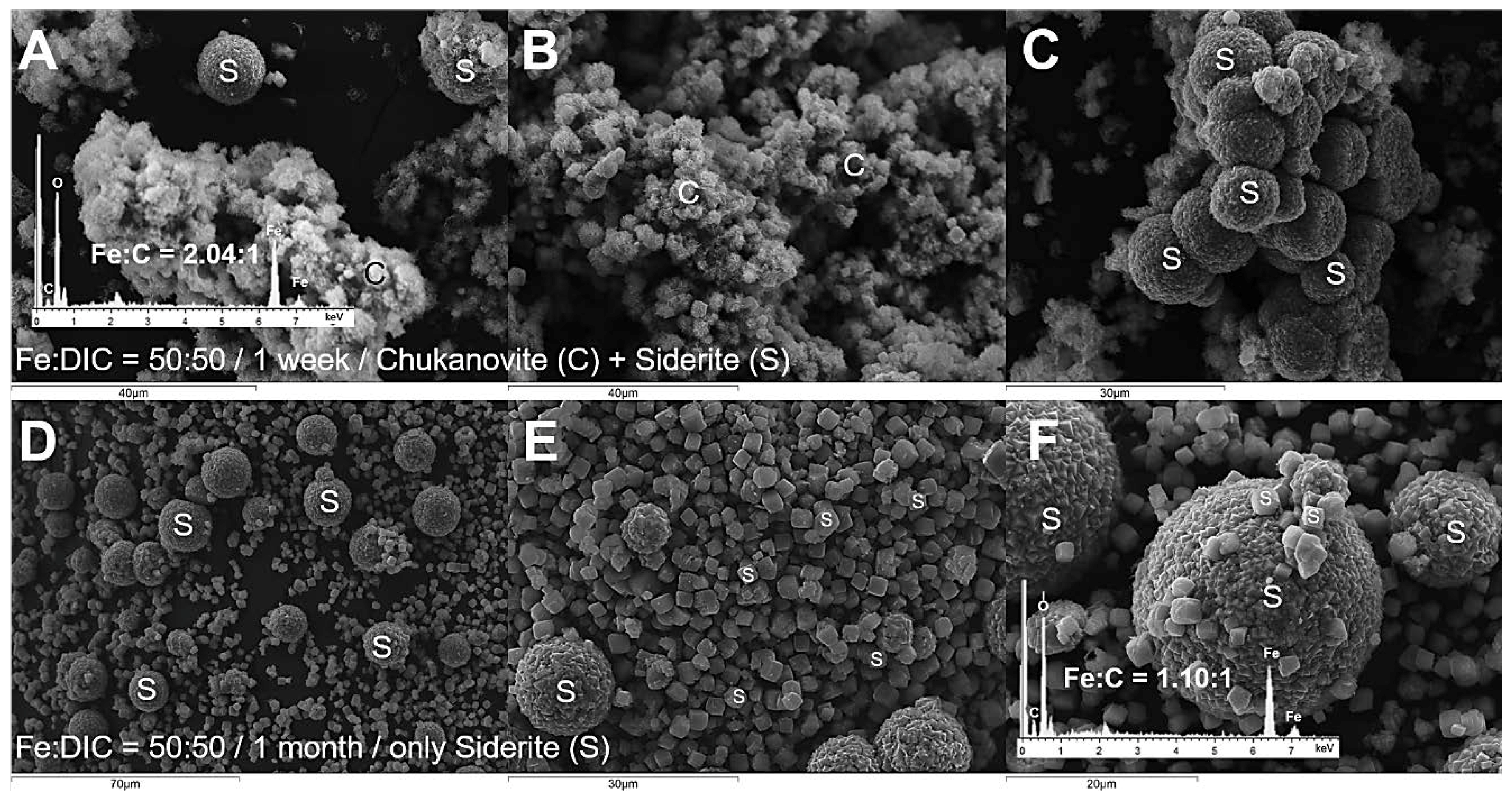
The SEM micrographs of both the 1-week and 1-month reactions for the 50:50 condition (Fe:DIC) in the first experimental set are displayed in Figure 5. For the 1-week reaction, both siderite and chukanovite were observed (Figure 5A–C) whereas only siderite was observed in the 1-month reaction (Figure 5D–F). Chukanovite showed a cluster of ~5 µm crystals with flaky rosette shapes (Figure 5A,B) and siderite framboids (Figure 5A,C). In the 1-month reaction sample, two types of siderite were observed (Figure 5D–F): framboids (over 10 micro-sized) and trigonal-rhombohederon crystals (2–4 µm). Framboids showed the angle of edges with 120° (Figure 5F) indicating that trigonal-rhombohedron crystals of siderite assembled to form the framboids. Both types were reported as synthesized siderite in previous studies [22,23,25]. The chemical composition of siderite and chukanovite was confirmed with EDS analysis (inset spectrum, Figure 5). The Fe:C ratio was 2.04:1 in the chukanovite cluster whereas siderite showed 1.10:1, consistent with their chemical formula.
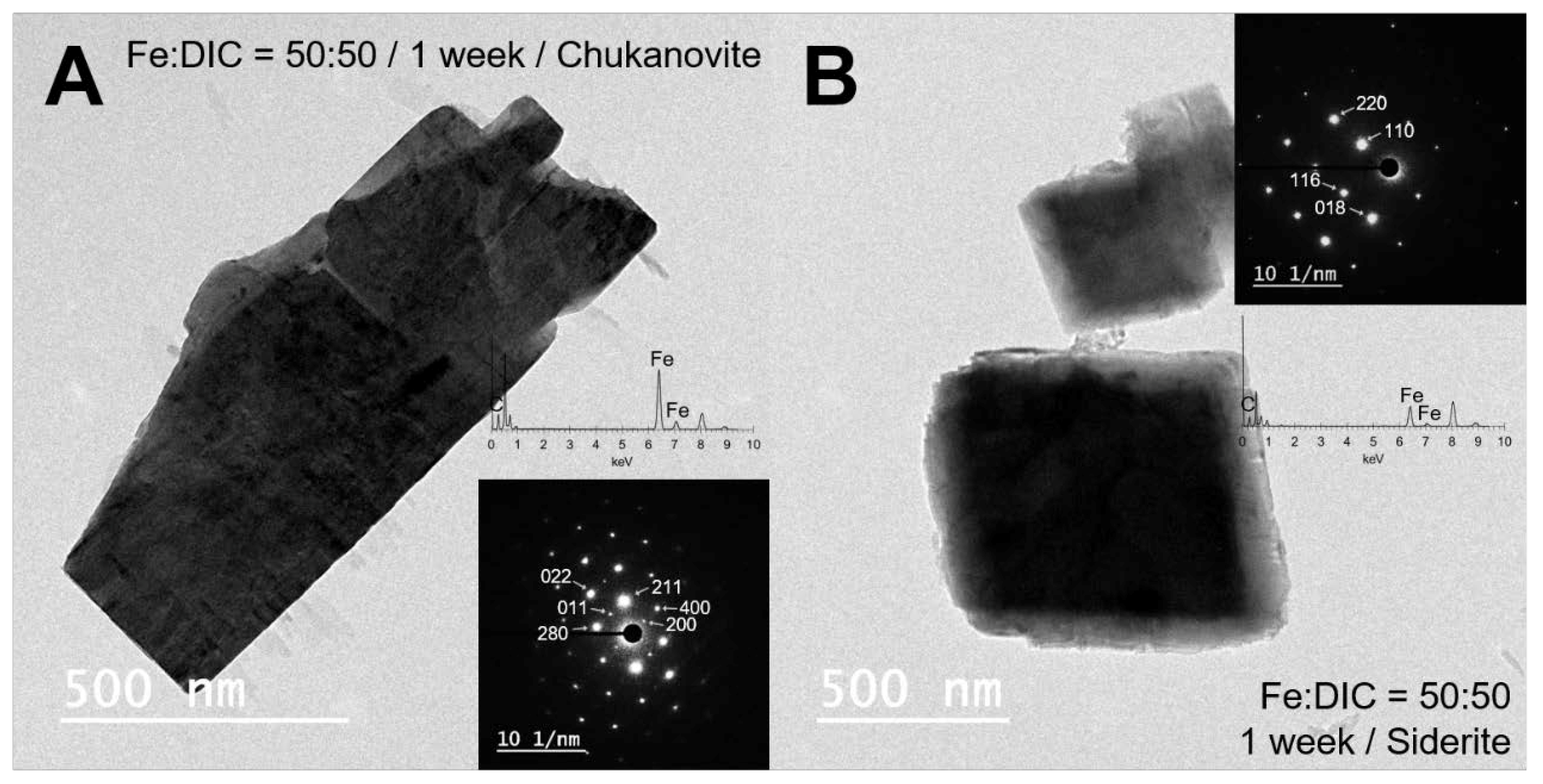
Both of the two minerals, chukanovite (Figure 6A) and siderite (Figure 6B) were identified in the bright-field images of TEM with corresponding SAED patterns (insets of Figure 6). A lath-shaped mineral with chemical composition Fe and C was identified as chukanovite with corresponding distinct reflections [(200), (011), (211), (280), (400), and (022)] in the SAED pattern (Figure 6A). Siderite was easily distinguished with chukanovite in the EM analysis because of its trigonal crystal structure (Figure 6B) and fewer reflections [(110), (018), (116), and (220)] in the SAED pattern (inset, Figure 6B).

4. Discussion

4.1. Concentration Limit of Siderite Formation

In conditions where the iron and DIC concentrations were higher than 50 mmolal, regardless of the phase of iron added, mostly siderite was identified in the 1-month reaction samples (Table 2). Particularly, conditions with low Fe and DIC concentration (10:10 and 15:15 in both Experiment 1 and 2) showed low estimated siderite proportion (~19.8%, Table 2) that suggests siderite is formed in the higher Fe and DIC concentration compared to chukanovite. Chukanovite is in a more stable phase under a lower DIC concentration, whereas siderite is more stable in high DIC conditions, according to the stability diagram of ferrous minerals (siderite, chukanovite, ferrous iron hydroxide, and hibbingite) [33] that supports a low estimated proportion of siderite in low DIC conditions. The low estimated proportion of siderite in the 1-week reaction sample of 10 and 15 mmolal of DIC (~3%, Table 2) compared to 20% in 30 mmolal condition, suggests that the concentration limit of the stable conditions for chukanovite is between 15 and 30 mmolal of DIC under 30 °C of FeCl2-NaHCO3 system.
Nonetheless, even in the DIC concentration range of 50 mmolal and 70 mmolal where chukanovite is less stable, chukanovite was formed in 1 week of reaction (51.4% and 18.8%, respectively; calculated from Table 2 [100-estimated siderite proportion, %]). However, after the reaction occurred for up to 1 month, there was only siderite in both conditions (Table 2). These results may be a consequence of differences in the initial reaction rate (kinetics) of the formation and stability of each mineral. There is not enough data to study kinetics in the present study because we did not track a detailed time-scale, however, it can be inferred that the reaction rate of chukanovite was higher than that of siderite because chukanovite was formed in even less stable conditions in the 1-week reaction. These results suggest that chukanovite could be formed with siderite even if the DIC concentration is high in a short-term reaction.
On the other hand, the estimated siderite proportion had increased in the 1-month reaction in all of the experimental conditions, which suggested that stability played a more significant role than reaction rate after 1 week. Dissolution of chukanovite may be dominant in long-term reactions because chukanovite has a large surface area with smaller particle size (~2 µm particle in cluster) compared to the siderite framboids (~10 µm, Figure 5A–C). Moreover, once carbonate minerals were formed in the system, the pH level would be decreased because the carbonate ions were consumed, which could enhance the dissolution of chukanovite by proton [33]. The concentration of ferrous iron and bicarbonate in the solution would increase due to the dissolution of chukanovite [33]. From our experiments, the pH of the solution after dissolution of chukanovite in all the conditions of Experiment 3 and 4, except Fe:DIC = 15:50 condition, however, was lower (~5.8, Table 4) than the initial solution (6.6–7.2, Table 4), which reduces the concentration of the ferrous monohydroxide ion Fe(OH)+ in solution. To form chukanovite, the Fe(OH)+ should be formed before reacting with the carbonate [31,35], therefore, the formation of chukanovite may be further diminished compared to the formation of siderite if Fe(OH)+ is insufficient in the system. Thus, after the 1-week reaction, the siderite proportion increased because the chukanovite was dissolved and its re-formation was hindered, whereas siderite was able to form continuously.

4.2. Control Species for Siderite Formation

The only difference between Experiment 1 and 2 was how the iron and bicarbonate solution was mixed, either by input of FeCl2 powder or pouring of the FeCl2 solution. However, the estimated siderite proportions of 1-week reacted samples were different under the same concentration of Fe and DIC in Experiment 1 and 2. The color of the FeCl2 solution that was used in Experiment 2 was clear yellow, which then turned to muddy yellow after mixing with bicarbonate solution, whereas the mixed solution in Experiment 1 was turquoise. This may be due to the geochemical conditions of the matrix when the iron was ionized. The pH of the FeCl2 solution is low because of the chloride ion, so ferrous ion is stable under reducing condition. However, when iron is dissolved into a bicarbonate solution, the pH was buffered by carbonate, which makes it easier to form ferrous monohydroxide ions (Fe(OH)+), the intermediate phase of chukanovite formation. Therefore, chukanovite formation could be enhanced in Experiment 1 rather than Experiment 2.
Comparing the three experimental conditions with different combinations of iron and DIC concentrations (i.e., 50:50, 50:100, and 100:50 (Fe:DIC in mmolal) from Experiment 2, 3, and 4, respectively), only siderite was observed in the 1-week reaction sample from the 50:100 condition (Figure 4A and Table 3), while chukanovite was observed (26% for 50:50 and 9% for 100:50, calculated from Table 3 [100-estimated siderite proportion, %]) in the other two conditions. Moreover, the estimated siderite proportion increased inconsistently in Experiment 3 when the iron concentration increased from 30 to 50 mmolal (70.6% to 73.2%) and 70 to 100 mmolal (90.7% to 91.4%), whereas it increased gradually according to the DIC concentration increases in Experiment 4 (26.6–100%, Table 3). These results suggest that the concentration of DIC is a critical factor for determining the stability of chukanovite and siderite. The fact that the boundary of chukanovite and siderite in the stability diagram is determined only by the activity of bicarbonate [33] also supports the idea that DIC is a major factor controlling the formation of ferrous carbonate mineral. In particular, it is consistent with a previous study suggesting that anion activity is the most important chemical control in nature or lab-scale experiments to form ferrous compounds [36]. Therefore, when the concentration of DIC is equal to or higher than the ferrous iron concentration in solution, siderite is the dominant precipitate. When iron is more concentrated than DIC, however, the excess iron in solution could form Fe(OH)+ and consequently form chukanovite in an early stage of the reaction. Nevertheless, chukanovite is unstable above 50 mmolal of DIC, so it could be dissolved, and only siderite remains after 1 month.
It is noteworthy that the estimated siderite proportion was decreased in the 1-month reaction compared to 1 week (49.2% to 25.6%, Table 3) in only one condition, 15 mmolal of iron and 50 mmolal of DIC mixed condition, in Experiment 3. This result may have been due to the pH of the solution. The Eh-pH diagram of potential Earth surface aqueous solution at 25 °C [37] showed that chukanovite is stable in basic conditions, whereas siderite is stable in slightly acidic to neutral conditions. The final pH level for the 1-month reaction of 15:50 conditions was higher (6.31) than other conditions (5.3–5.8, Table 4). The concentration of DIC was higher (50 mmolal) than the concentration limit of the stable conditions for chukanovite suggested in the present study (15–30 mmolal), so chukanovite and siderite may have formed at similar levels in the initial 1 week of the reaction (estimated siderite proportion = 49.2%, Table 3), even though the pH level was high (6.39, Table 4). However, in the 1-month reaction, the pH level did not decrease compared to other conditions that may have majorly controlled the stability of chukanovite and siderite, consequently decreasing the estimated siderite proportion. Thus, concentration of DIC is a major factor in short-term reactions, whereas both the pH and concentration of DIC affect the stability of chukanovite in long-term reactions.

5. Conclusions

For the formation of pure siderite, a synthetic experiment in a FeCl2-NaHCO3 system was tested. However, chukanovite was precipitated with siderite formation under low-concentration conditions (10–15 mmolal of Fe and DIC) and a short reaction time (1 week). This suggests that chukanovite should be considered as a transient phase for the synthetic formation of siderite. This is a significant observation that could apply to the understanding of experimental variables that control the pure siderite formation by semi-quantification of siderite and chukanovite formation in each experimental setup.

Author Contributions

T.-h.K. contributed to the experimental settings, data production, and manuscript writing. J.K. supervised the entire process. All authors have read and agreed to the published version of the manuscript.

Funding

This research was funded by National Research Foundation of Korea (NRF), grant number NRF-2018R1A2B6002036 to J.K. and NRF-2019R1A6A3A01092955 to T.K.

Acknowledgments

The authors are grateful to Sang-Tae Kim (McMaster University, Canada) for advising on the experimental setting.

Conflicts of Interest

The authors declare no conflict of interest.

References

  1. Anthony, T.; Cline, H. Surface rippling induced by surface-tension gradients during laser surface melting and alloying. J. Appl. Phys. 1977, 48, 3888–3894. [Google Scholar] [CrossRef]
  2. Fernández-Remolar, D.C.; Knoll, A.H. Fossilization potential of iron-bearing minerals in acidic environments of Rio Tinto, Spain: Implications for Mars exploration. Icarus 2008, 194, 72–85. [Google Scholar] [CrossRef]
  3. Hałas, S.; Chlebowski, R. Unique siderite occurrence in Baltic Sea: A clue to siderite-water oxygen isotope fractionation at low temperatures. Geol. Q. 2004, 48, 317–322. [Google Scholar]
  4. Romanek, C.S.; Grady, M.M.; Wright, I.; Mittlefehldt, D.; Socki, R.; Pillinger, C.; Gibson, E.K., Jr. Record of fluid–rock interactions on Mars from the meteorite ALH84001. Nature 1994, 372, 655. [Google Scholar] [CrossRef] [PubMed]
  5. Valley, J.W.; Eiler, J.M.; Graham, C.M.; Gibson, E.K.; Romanek, C.S.; Stolper, E.M. Low-temperature carbonate concretions in the Martian meteorite ALH84001: Evidence from stable isotopes and mineralogy. Science 1997, 275, 1633–1638. [Google Scholar] [CrossRef]
  6. Treiman, A.H.; Romanek, C.S. Bulk and stable isotopic compositions of carbonate minerals in Martian meteorite Allan Hills 84001: No proof of high formation temperature. Meteorit. Planet. Sci. 1998, 33, 737–742. [Google Scholar] [CrossRef]
  7. Seguin, M. Instability of FeCO3 in air. Am. J. Sci. 1966, 264, 562–568. [Google Scholar] [CrossRef]
  8. McMillan, S.; Schwertmann, U. Morphological and genetic relations between siderite, calcite and goethite in a Low Moor Peat from southern Germany. Eur. J. Soil Sci. 1998, 49, 283–293. [Google Scholar] [CrossRef]
  9. Duckworth, O.W.; Martin, S.T. Role of molecular oxygen in the dissolution of siderite and rhodochrosite. Geochim. Cosmochim. Acta 2004, 68, 607–621. [Google Scholar] [CrossRef]
  10. Poulton, S.W.; Canfield, D.E. Ferruginous conditions: A dominant feature of the ocean through Earth’s history. Elements 2011, 7, 107–112. [Google Scholar] [CrossRef] [Green Version]
  11. Rodrigues, A.G.; De Ros, L.F.; Neumann, R.; Borghi, L. Paleoenvironmental implications of early diagenetic siderites of the Paraíba do Sul Deltaic Complex, eastern Brazil. Sediment. Geol. 2015, 323, 15–30. [Google Scholar] [CrossRef]
  12. McKay, D.S.; Gibson, E.K.; Thomas-Keprta, K.L.; Vali, H.; Romanek, C.S.; Clemett, S.J.; Chillier, X.D.; Maechling, C.R.; Zare, R.N. Search for past life on Mars: Possible relic biogenic activity in Martian meteorite ALH84001. Science 1996, 273, 924–930. [Google Scholar] [CrossRef] [PubMed] [Green Version]
  13. Clemett, D.; Cockett, M.; Marsden, C.; Fone, K. Antisense oligonucleotide-induced reduction in 5-hydroxytryptamine7 receptors in the rat hypothalamus without alteration in exploratory behaviour or neuroendocrine function. J. Neurochem. 1998, 71, 1271–1279. [Google Scholar] [CrossRef] [PubMed]
  14. Friedmann, E.I.; Wierzchos, J.; Ascaso, C.; Winklhofer, M. Chains of magnetite crystals in the meteorite ALH84001: Evidence of biological origin. Proc. Natl. Acad. Sci. 2001, 98, 2176–2181. [Google Scholar] [CrossRef] [PubMed] [Green Version]
  15. Gibson, E.K., Jr.; McKay, D.S.; Thomas-Keprta, K.L.; Wentworth, S.; Westall, F.; Steele, A.; Romanek, C.S.; Bell, M.; Toporski, J. Life on Mars: Evaluation of the evidence within Martian meteorites ALH84001, Nakhla, and Shergotty. Precambrian Res. 2001, 106, 15–34. [Google Scholar] [CrossRef]
  16. Dong, H.; Kukkadapu, R.K.; Fredrickson, J.K.; Zachara, J.M.; Kennedy, D.W.; Kostandarithes, H.M. Microbial reduction of structural Fe (III) in illite and goethite. Environ. Sci. Technol. 2003, 37, 1268–1276. [Google Scholar] [CrossRef] [Green Version]
  17. Koo, T.-H.; Lee, G.; Kim, J.-W. Biogeochemical dissolution of nontronite by Shewanella oneidensis MR-1: Evidence of biotic illite formation. Appl. Clay Sci. 2016, 134, 13–18. [Google Scholar] [CrossRef]
  18. Roh, Y.; Zhang, C.-L.; Vali, H.; Lauf, R.; Zhou, J.; Phelps, T. Biogeochemical and environmental factors in Fe biomineralization: Magnetite and siderite formation. Clays Clay Miner. 2003, 51, 83–95. [Google Scholar] [CrossRef]
  19. Kim, J.; Dong, H. Application of electron energy-loss spectroscopy (EELS) and energy-filtered transmission electron microscopy (EFTEM) to the study of mineral transformation associated with microbial Fe-reduction of magnetite. Clays Clay Miner. 2011, 59, 176–188. [Google Scholar] [CrossRef]
  20. Johnson, C.M.; Roden, E.E.; Welch, S.A.; Beard, B.L. Experimental constraints on Fe isotope fractionation during magnetite and Fe carbonate formation coupled to dissimilatory hydrous ferric oxide reduction. Geochim. Cosmochim. Acta 2005, 69, 963–993. [Google Scholar] [CrossRef]
  21. Kim, J.; Dong, H.; Seabaugh, J.; Newell, S.W.; Eberl, D.D. Role of microbes in the smectite-to-illite reaction. Science 2004, 303, 830–832. [Google Scholar] [CrossRef] [PubMed] [Green Version]
  22. Jimenez-Lopez, C.; Romanek, C.S. Precipitation kinetics and carbon isotope partitioning of inorganic siderite at 25 °C and 1 atm. Geochim. Cosmochim. Acta 2004, 68, 557–571. [Google Scholar] [CrossRef]
  23. Van Dijk, J.; Fernandez, A.; Müller, I.A.; Lever, M.; Bernasconi, S.M. Oxygen isotope fractionation in the siderite-water system between 8.5 and 62 °C. Geochim. Cosmochim. Acta 2018, 220, 535–551. [Google Scholar] [CrossRef]
  24. Fernandez, A.; Tang, J.; Rosenheim, B.E. Siderite ‘clumped’isotope thermometry: A new paleoclimate proxy for humid continental environments. Geochim. Cosmochim. Acta 2014, 126, 411–421. [Google Scholar] [CrossRef]
  25. Wiesli, R.A.; Beard, B.L.; Johnson, C.M. Experimental determination of Fe isotope fractionation between aqueous Fe (II), siderite and “green rust” in abiotic systems. Chem. Geol. 2004, 211, 343–362. [Google Scholar] [CrossRef]
  26. Kim, S.-T.; O’Neil, J.R. Equilibrium and nonequilibrium oxygen isotope effects in synthetic carbonates. Geochim. Cosmochim. Acta 1997, 61, 3461–3475. [Google Scholar] [CrossRef]
  27. Nassar, M.Y.; Ahmed, I.S.; Mohamed, T.Y.; Khatab, M. A controlled, template-free, and hydrothermal synthesis route to sphere-like α-Fe2O3 nanostructures for textile dye removal. Rsc Adv. 2016, 6, 20001–20013. [Google Scholar] [CrossRef]
  28. Pekov, I.V.; Perchiazzi, N.; Merlino, S.; Kalachev, V.N.; Merlini, M.; Zadov, A.E. Chukanovite, Fe(CO3)(OH)2, a new mineral from the weathered iron meteorite Dronino. Eur. J. Mineral. 2007, 19, 891–898. [Google Scholar] [CrossRef]
  29. Erdös, E.; Altorfer, H. Ein dem Malachit ähnliches basisches Eisenkarbonat als Korrosionsprodukt von Stahl. Mater. Corros. 1976, 27, 304–312. [Google Scholar] [CrossRef]
  30. Azoulay, I.; Rémazeilles, C.; Refait, P. Determination of standard Gibbs free energy of formation of chukanovite and Pourbaix diagrams of iron in carbonated media. Corros. Sci. 2012, 58, 229–236. [Google Scholar] [CrossRef]
  31. Pandarinathan, V.; Lepková, K.; Van Bronswijk, W. Chukanovite (Fe2(OH)2CO3) identified as a corrosion product at sand-deposited carbon steel in CO2-saturated brine. Corros. Sci. 2014, 85, 26–32. [Google Scholar] [CrossRef]
  32. Kukkadapu, R.K.; Zachara, J.M.; Fredrickson, J.K.; Kennedy, D.W. Biotransformation of two-line silica-ferrihydrite by a dissimilatory Fe (III)-reducing bacterium: Formation of carbonate green rust in the presence of phosphate. Geochim. Cosmochim. Acta 2004, 68, 2799–2814. [Google Scholar] [CrossRef] [Green Version]
  33. Kim, S.; Marrs, C.; Nemer, M.; Je-Hun Jang, J. Solubility Model for Ferrous Iron Hydroxide, Hibbingite, Siderite, and Chukanovite in High Saline Solutions of Sodium Chloride, Sodium Sulfate, and Sodium Carbonate. ACS Earth Space Chem. 2017, 1, 647–663. [Google Scholar] [CrossRef]
  34. Graf, D.L. Crystallographic tables for the rhombohedral carbonates. Am. Mineral. J. Earth 1961, 46, 1283–1316. [Google Scholar]
  35. De Marco, R.; Jiang, Z.-T.; John, D.; Sercombe, M.; Kinsella, B. An in situ electrochemical impedance spectroscopy/synchrotron radiation grazing incidence X-ray diffraction study of the influence of acetate on the carbon dioxide corrosion of mild steel. Electrochim. Acta 2007, 52, 3746–3750. [Google Scholar] [CrossRef]
  36. Curtis, C.; Spears, D. The formation of sedimentary iron minerals. Econ. Geol. 1968, 63, 257–270. [Google Scholar] [CrossRef]
  37. Lee, S.; Xu, H. The crystal structure and Gibbs free energy of formation of chukanovite as an oxidation product of carbon steel in human liver. Chem. Geol. 2018, 488, 180–188. [Google Scholar] [CrossRef]
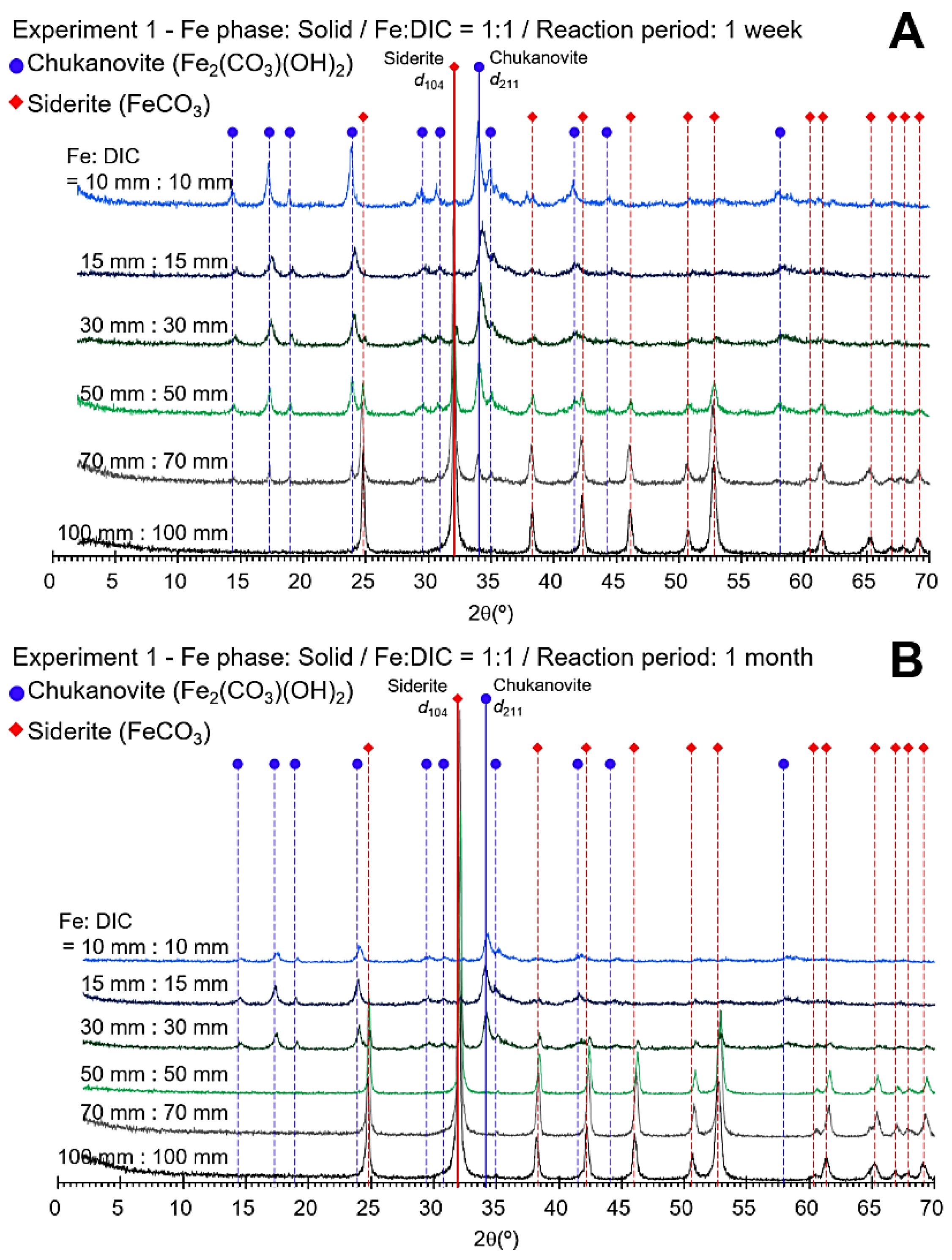
Figure 1. The XRD profiles for Experiment 1 (solid phase Fe:DIC = 1:1 mixing) at 1 week (A) and 1 month of reaction (B). The red dashed lines with the diamonds indicate peaks of siderite and the blue dashed lines with the circles indicate peaks of chukanovite. The markers with solid lines are the major peaks of siderite and chukanovite (d104 [2θ = 32.02°] and d211 [2θ = 33.98°], respectively).
Minerals 10 00156 g001
Figure 2. The XRD profiles for Experiment 2 (aqueous phase of Fe:DIC = 1:1 mixing) at 1 week (A) and 1 month of reaction (B). The red dashed lines with the diamonds indicate peaks of siderite and the blue dashed lines with the circles indicate peaks of chukanovite. The markers with solid lines are the major peaks of siderite and chukanovite (d104 [2θ = 32.02°] and d211 [2θ = 33.98°], respectively).
Minerals 10 00156 g002
Figure 3. The XRD profiles for Experiment 3 (aqueous phase of Fe:DIC = X:50 mixing) at 1 week (A) and 1 month of reaction (B). The red dashed lines with the diamonds indicate peaks of siderite and the blue dashed lines with the circles indicate peaks of chukanovite. The markers with solid lines are the major peaks of siderite and chukanovite (d104 [2θ = 32.02°] and d211 [2θ = 33.98°], respectively). The gray colored 50 mmolal:50 mmolal profiles that were obtained in Experiment 2 were added for reference.
Minerals 10 00156 g003
Figure 4. The XRD profiles for Experiment 4 (aqueous phase of Fe:DIC = 50:X mixing) at 1 week (A) and 1 month of reaction (B). The red dashed lines with the diamonds indicate peaks of siderite and the blue dashed lines with the circles indicate peaks of chukanovite. The markers with solid lines are the major peaks of siderite and chukanovite (d104 [2θ = 32.02°] and d211 [2θ = 33.98°], respectively). The gray colored 50 mmolal:50 mmolal profiles that were obtained in Experiment 2 were added for reference.
Minerals 10 00156 g004
Figure 5. The SEM micrographs of the chukanovite (C) and siderite (S) of 50 mmolal:50 mmolal condition of Experiment 1 (solid phase Fe) at 1 week (AC) and 1 month of aging (DF). In the 1 week of aging sample, a mixture of chukanovite cluster (labeled C in A and B) and siderite framboids (labeled S in A–C) were identified, whereas only siderite framboids and trigonal-rhombohedron crystals (both labeled S in D–F) were identified in 1 month of aging sample (D–F).
Minerals 10 00156 g005
Figure 6. Representative bright-field TEM micrographs with selected-area electron diffraction (SAED) patterns and EDS spectrum displaying chukanovite [(200): d200 = 0.468nm, (011): d011 = 0.316 nm, (211): d211 = 0.266 nm, (280): d280 = 0.263 nm, (400): d400 = 0.236 nm, and (022): d022 = 0.158 nm] (A) and siderite [(110): d110 = 0.236 nm, (018): d018 = 0.174 nm, (116): d116 = 0.173 nm, and (220): d220 = 0.118 nm] (B) of 1 week of reaction sample of Experiment 1, 50 mmolal condition.
Minerals 10 00156 g006
Table 1. Experimental conditions for iron carbonate mineral formation.
ExperimentFeCl2·4H2ONaHCO3Reaction Time
PhaseConcentration
(mmolal)
Concentration
(mmolal)
Experiment 1
(Fe:DIC = 1:1)
Solid (powder)10, 15, 30, 50, 70, 10010, 15, 30, 50, 70, 1001 week
1 month
Experiment 2
(Fe:DIC = 1:1)
Aqueous10, 15, 30, 50, 70, 10010, 15, 30, 50, 70, 1001 week
1 month
Experiment 3
(Fe:DIC = X:50)
Aqueous15, 30, 70, 100501 week
1 month
Experiment 4
(Fe:DIC = 50:X)
Aqueous5015, 30, 70, 1001 week
1 month
Table 2. Semi-quantitative estimation for siderite proportion in Experiments 1 and 2.
ExperimentConcentration
Fe:DIC
(mmolal:mmolal)
1 Week1 Month
Intensity RatioSiderite Proportion (%)Intensity RatioSiderite Proportion (%)
Experiment 110:100.1282.80.1647.0
15:150.1303.00.22112.2
30:300.35120.21.36443.6
50:501.82148.655.750100
70:7012.10481.267.229100
100:10043.125100388.696100
Experiment 210:100.23913.50.1849.1
15:150.24413.90.34419.8
30:301.38343.89.74777.5
50:507.59073.223.25692.5
70:708.48175.137.667100
100:10024.41593.335.702100
Table 3. Semi-quantitative estimation for siderite proportion of Experiments 3 and 4.
ExperimentConcentration
Fe:DIC
(mmolal:mmolal)
1 Week1 Month
Intensity RatioSiderite ProportionIntensity RatioSiderite Proportion
Experiment 315:501.88749.20.48125.6
30:506.54170.630.49397.2
50:50 *7.59073.223.25692.5
70:5020.93190.757.000100
100:5021.76691.454.086100
Experiment 450:150.51026.62.21151.9
50:303.15258.011.95281.0
50:50 *7.59073.223.25692.5
50:7025.26293.929.04796.3
50:10037.89710048.484100
* Results from Experiment 2.
Table 4. pH values of the solution at the initial and final of reactions.
ExperimentConcentration
Fe:DIC
(mmolal:mmolal)
1 Week1 Month
Initial pHFinal pHInitial pHFinal pH
Experiment 315:507.196.397.216.31
30:506.995.386.995.87
50:50 *6.935.556.935.88
70:506.785.636.785.38
100:506.675.506.685.30
Experiment 450:156.786.276.605.87
50:306.815.956.865.73
50:50 *6.935.556.935.88
50:706.795.806.776.08
50:1006.775.886.765.62
* Results from Experiment 2.