Article

Wearable Activity Monitors to Quantify Gait During Stroke Rehabilitation: Data from a Pilot Randomised Controlled Trial Examining Auditory Rhythmical Cueing

1
Department of Sport, Exercise and Rehabilitation, Faculty of Health and Life Science, Northumbria University, Newcastle upon Tyne NE7 7YT, UK
2
Stroke Research Group, Faculty of Medical Sciences, Population Health Sciences Institute, Newcastle University, Newcastle upon Tyne NE2 4HH, UK
3
The Newcastle upon Tyne Hospitals NHS Foundation Trust, Newcastle upon Tyne NE4 5PL, UK
4
Institute of Translational and Clinical Research, Faculty of Medical Sciences, Newcastle University, Newcastle upon Tyne NE1 7RU, UK
5
National Institute for Health and Care Research (NIHR), Newcastle Biomedical Research Centre (BRC), Newcastle University, Newcastle upon Tyne NE4 5PL, UK
6
Stroke Northumbria, Northumbria Healthcare NHS Foundation Trust, Rake Lane, North Shields, Tyne and Wear NE29 8NH, UK
*
Authors to whom correspondence should be addressed.
Symmetry 2025, 17(10), 1640; https://doi.org/10.3390/sym17101640
Submission received: 14 July 2025 / Revised: 7 August 2025 / Accepted: 5 September 2025 / Published: 3 October 2025

Abstract

Hemiparesis is a disabling consequence of stroke, causing abnormal gait patterns with biomechanical asymmetries. Gait mechanics for stroke survivors appear resistant to conventional rehabilitation. Auditory rhythmical cueing (ARC) represents an emerging intervention option. To determine effective gait interventions, objective measures of gait collected from real-world environments may be required in addition to standard clinical outcomes to aid understanding of gait mechanics. This study reports on the ability of wearable activity monitors to quantify an ARC intervention for fifty-nine stroke survivors randomised into an ARC gait and balance training programme or an equivalent training programme without ARC. Gait assessments were undertaken at baseline and at 6 weeks for 4-metre walks and continuously for 7 days following each home assessment using a wearable activity monitor. The success rates of data collection using the wearable activity monitors ranged from 64 to 95%. Forty-eight Digital Mobility Outcomes representing a broad range of gait mechanics were calculated. Visualisation of all DMOs using radar plots indicated changes from baseline in both groups, with individual data indicating large variability in response to the intervention and control programme. Including wearable activity monitors to evaluate gait interventions for stroke survivors provides additional value to traditional methods and aids understanding of individual responses; as such, they should be used for future intervention-based research.

1. Introduction

Hemiparesis is a common disabling consequence of stroke that leads to abnormal gait patterns marked by biomechanical asymmetries that contribute to decreased gait velocity, increased susceptibility to falls, hospitalisations, and reduced independence and quality of life [1,2]. One of the main rehabilitation goals for stroke survivors is to regain the ability to ambulate independently in real-world settings [3,4]. Despite current rehabilitation efforts, gait problems are experienced by ≈80% of stroke survivors [5,6], and 50–70% of individuals are classified as household or limited community ambulators based on their walking function [1,7,8]. Given the disabling impacts of a stroke, more effective interventions to improve gait mechanics and, consequently, increase ambulation within the context of the participants’ own environments are required [1,9].
Stroke survivors would ideally benefit from practicing their gait rehabilitation outside the limited face-to-face therapy time available to them in real-world settings [8,10]. A potential low-cost method of enhancing the efficacy of gait rehabilitation post-stroke that can be practiced unsupervised is auditory rhythmical cueing (ARC). ARC is where a metronome beat (or music) is delivered during exercise training to normalise and train stepping. If the metronome is equally matched for each step, in theory, it should improve symmetry when walking [11]. The efficacy of ARC has been well established in Parkinson’s disease over the last 20 years [12], and there is evidence for its efficacy when applied in a clinic/laboratory setting for stroke survivors [13]. ARC is emerging as a feasible and acceptable method of targeting gait within and around the home after stroke, but ARC’s efficacy within real-world settings post-stroke has yet to be established [8,14,15].
To establish the efficacy of ARC in home and community settings, sensitive and specific measurements are required. During stroke rehabilitation, gait is conventionally assessed by measuring walking distance and speed using tests such as the 10 m walk test or the 6 min walk test (6MWT) [16]. Tests like these do not capture real-world walking behaviours or cardinal gait features of stroke survivors such as asymmetry and variability [2,17]. Although correlated to gait speed, these metrics are important as they are associated with several negative consequences, such as inefficiency, challenges to balance control, risk of musculoskeletal injury to the non-paretic lower limb, and loss of bone mass density in the paretic lower limb [18]. The lack of specific measures that capture gait asymmetry might explain why there is a current lack of evidence and effective interventions targeting gait asymmetry [8,19,20]. The same argument can be made for improvements in free-living walking performance (i.e., defined as what a person does in their own environment [17]), as this also lacks an objective and specific method to be quantified.
Wearable activity monitors are valid and reliable at capturing whole-body movement symmetry [21,22] and gait from real-world environments for stroke survivors [9,23]. A large variety of Digital Mobility Outcomes (DMOs) (defined as a digitally measured mobility parameter used to assess an individual’s health status, particularly in the context of movement and walking) can be calculated from wearable activity monitors [24]. Wearable activity monitors appear to be an ideal tool to objectively quantify the impact of interventions designed to improve specific gait mechanics for stroke survivors from real-world environments due to the increased specificity of measuring mechanical changes relative to conventional gait tests [2,6,9,25]. However, to our knowledge, they have yet to be tested for this purpose. Understanding effective gait interventions for stroke survivors is limited [26]. Interventions are still being developed by researchers and tested on relatively small single-site samples (N < 100), with variable methodological rigour undertaken in research laboratories with selected patient samples [6,27]. If wearable activity monitors can quantify specific changes in gait mechanics, it would help overcome these limitations while providing a step towards integrating wearable activity monitors to quantify the impact of future gait intervention-based research [10,25]. We have previously reported findings of a pilot RCT exploring the use of auditory rhythmical cueing to improve gait in community-dwelling stroke survivors [15]. The aim of this current paper is to report the data from wearable activity monitors collected within this pilot RCT and to visualise how, relative to a control group, the gait mechanics of stroke survivors may change following 6 weeks of ARC training. To achieve this aim, the objectives of this study are as follows:
  • To quantify the success rate of integrating wearable activity monitors to collect data within a pilot RCT of ARC training post-stroke.
  • Use 48 DMOs to visualise how, relative to a control group, the gait mechanics of stroke survivors are impacted following a 6-week ARC intervention,
  • Provide DMO reference data recorded with the use of wearable activity monitors for both a control group and following 6 weeks of an ARC intervention.
  • Discuss our findings with the purpose of informing future intervention studies aiming to integrate wearable activity monitors and quantify intervention impact for stroke survivors.

2. Materials and Methods

Study design
The focus of this study is to report data from wearable activity monitors collected as part of a pilot RCT exploring the use of auditory rhythmical cueing to improve gait in community-dwelling stroke survivors. This study’s methods and its details have been reported previously [14,15]. In brief, the design was a parallel-group observer blind multicentre pilot RCT set in the Northeast of England. The focus of this current study is to report the wearable activity monitor data collected as part of the pilot RCT.
Intervention
The ARC gait and balance programme was designed through a literature review and stakeholder consultation [14]. It consisted of three 30 min sessions per week over six weeks (eighteen sessions in total), with one session per week supervised by a trained researcher. Rhythmic cueing was delivered using either a metronome device (Metro Tuner MT-100, Musedo) or a smartphone application (“ZyMi” for Android or “Pro Metronome” for iOS) based on the participant’s preference. A single auditory cue initiated the frequency of each step, with frequency adjusted to exercise demands. Ten gait and balance exercises were implemented and progressively adapted by increasing cueing frequency, repetitions, duration, or task complexity (e.g., additional turns). From weeks four to six, supervised sessions incorporated outdoor walking.
Control
The control group completed the gait and balance training programme without ARC. The duration, content, supervision, and materials were identical to those of the intervention group, with the sole exception being rhythmic cueing.
Wearable activity monitor data collection:
Four-metre walk test
A four-metre walk test was performed in the participant’s home whilst wearing an AX3 wearable activity monitor (see below for details) at the fifth lumbar vertebrae (L5). The walk was undertaken in the most appropriate space possible to record straight-line walking under the observation of a member of the research team. The walk was repeated five times so that an average for each visit could be calculated using the AX3 wearable activity monitor. These 5 × 4 m walks were performed both at baseline and after 6 weeks for both the control and the intervention group.
Seven-day assessment
In addition to the four-metre walk test, all participants wore the AX3 wearable activity monitor at L5 in the community for seven days both before and after the 6-week intervention. The AX3 was worn continuously for 7 days except for skin cleaning or during bathing/swimming (the wearable activity monitor was water-resistant but not waterproof). Participants, or a family member who could assist, were instructed on how to remove and reattach the wearable activity monitor. Following the data collection, the wearable activity monitor was returned by participants in a pre-paid envelope and data was downloaded to be processed using custom algorithms within MATLAB (version 9.4.0, R2018a) to calculate the subsequently described gait variables.
Wearable activity monitor details
The AX3 (Axivity, York, UK) is a tri-axial accelerometer-based wearable activity monitor (costs ≈ £100; 23.0 × 32.5 × 7.6 mm; weight 11 g). It features 512 MB memory, a 14-day battery life, and records acceleration at 100 Hz (16-bit resolution, ±8 g). Data were stored locally and downloaded after each walking trial. The device was secured at the fifth lumbar vertebra (L5) using double-sided tape and Hypafix (BSN Medical Ltd., Hull, UK) (Figure 1).
Digital Mobility Outcome Calculation
The following Digital Mobility Outcomes were calculated from the four-metre walk test and the seven-day assessment.
Micro DMOs: Fourteen temporal and spatial gait parameters, as defined by [29], were derived from the AX3. Mean values were calculated for the step time, stance time (the duration of foot contact per stride), swing time (the duration for which the foot was off the ground per stride), step length, and step velocity. Step variability was quantified using the standard deviation across all steps, and asymmetry was defined as the absolute difference between consecutive left and right steps. The algorithms and data segmentation methods applied to the AX3, along with their rationale, have been described previously [23,30]. Briefly, vertical acceleration was analysed using continuous wavelet transformation to identify initial and final contact within the gait cycle, while the inverted pendulum model was applied to derive spatial parameters.
Macro DMOs: A validated free-living algorithm [31] was employed to quantify macro-level gait characteristics, including volume, pattern, and variability. Volume was defined as the total daily step count. Pattern was characterised by the number of daily walking bouts (a minimum of three steps per bout), the mean bout duration (s), and α, representing the distribution of ambulatory bouts, where lower α values indicate a higher proportion of longer bouts [32,33]. Variability was assessed as the within-subject variation in bout length, calculated using a maximum likelihood approach to account for non-normal data [34]. Higher variability reflects a more diverse walking pattern.
Signal-derived DMOs:
Previously, we showed that DMOs measured from accelerometer signals placed at L5 during walking are both valid and reliable measures of gait asymmetry post-stroke and more favourable than spatiotemporal-based DMOs reliant on detecting an accurate measure of initial contact [35]. However, to accurately calculate gait asymmetry, it is vital that straight-line walking is maintained. Using an accelerometer alone, it is not possible to solely segment and analyse straight-line walking from continuous real-world data because, if a participant is gradually turning while walking, this could be interpreted as asymmetrical gait. Consequently, the asymmetry variables were only calculated from the confirmed four-metre walk recorded in the participant’s homes under the observation of a member of the research team. The DMOs calculated are described in detail elsewhere [36]; however, in brief, the measures represent signal asymmetry and, therefore, gait asymmetry using different signal processing techniques. Firstly, the Harmonic Ratio quantifies the step-to-step symmetry within a stride by calculating the ratio of the odd and even harmonics of a signal following fast Fourier transformation [37]. The other measure involves autocorrelation, which combines the estimated as well as the normalised unbiased autocovariance for a lag of one step/stride time. This feature reflects the similarity between subsequent steps of the acceleration pattern over a step/stride. Values of this feature close to 1.0 (maximum possible value) reflect repeatable patterns between subsequent steps. This can then be used to measure asymmetry directly by creating a ratio between step and stride regularity designed to quantify the level of symmetry between them and indicative of symmetry during straight walking [38]. Lastly, the gait symmetry index was calculated based on the concept of the summation of the biased autocorrelation from all three components of movement and the subsequent calculation of step and stride timing asymmetry, where a value of one represents perfectly symmetrical gait [39].
Data analysis
Objective one: The success rate of data collection using a wearable activity monitor was calculated using percentages based on how much successful data was obtained relative to how much data was collected from participants. For the 7-day assessment data, successfully collected data was defined as at least 24 hours’ worth of data collected.
Objective two: To aid understanding of the impact of ARC training on individuals relative to the control group, the analysis only included successfully collected data at baseline and after the 6 weeks in a paired fashion. To visualise the intervention effect for both groups, a z score was calculated by the equation, z = (x − μ)/σ, where “x” is the 6-week data mean, “μ” is the baseline mean score, and “σ” is the baseline population standard deviation. This was performed for both groups separately and relative to their baseline values, so that it was possible to see the relative impact for each intervention separately for both groups, but also between the groups. The calculation of the z score, due to the normalisation of the baseline values for each DMO, allowed radar plots to be created whereby all variables could be visualised for interpretation on the same figure irrespective of the DMO’s base unit, therefore, providing a clinically useful means to understand both individual and group changes in gait mechanics.
Objective three: The mean and standard deviation of all DMOs were calculated for the paired individuals in both groups at baseline and post-trial. Due to the use of the recommended pilot study sample size, there was a high number of DMOs relative to the population size and no primary outcome to dictate a power calculation; the results are descriptive only, and no inferential statistics were performed.

3. Results

3.1. Objective 1: Success Rates of Collecting Wearable Activity Monitor Gait Data

At baseline, out of the 59 participants enrolled in the pilot trial, wearable activity monitor data were successfully collected from 51 (86%) participants for the 7-day analysis and 56 (95%) participants from the four-metre walk assessments. Successful data collection was reduced after 6 weeks of the intervention, where data was obtained from 38 (64%) and 46 (77%) participants for the 7-day and four-metre assessments, respectively. As data was not uniformly available for the same participant and condition, it meant 34 full sets of 7-day assessment data were available for individual participants (e.g., pre- and post-intervention), and 43 full participant data sets were available from the four-metre analysis. The unsuccessful data was a product of participants dropping out, data not being recorded, the monitors being lost, and follow-ups not being possible due to the impact of the COVID-19 pandemic (three participants per group).

3.2. Objective 2: Visualisation of the Impact of Both Interventions on All DMOs

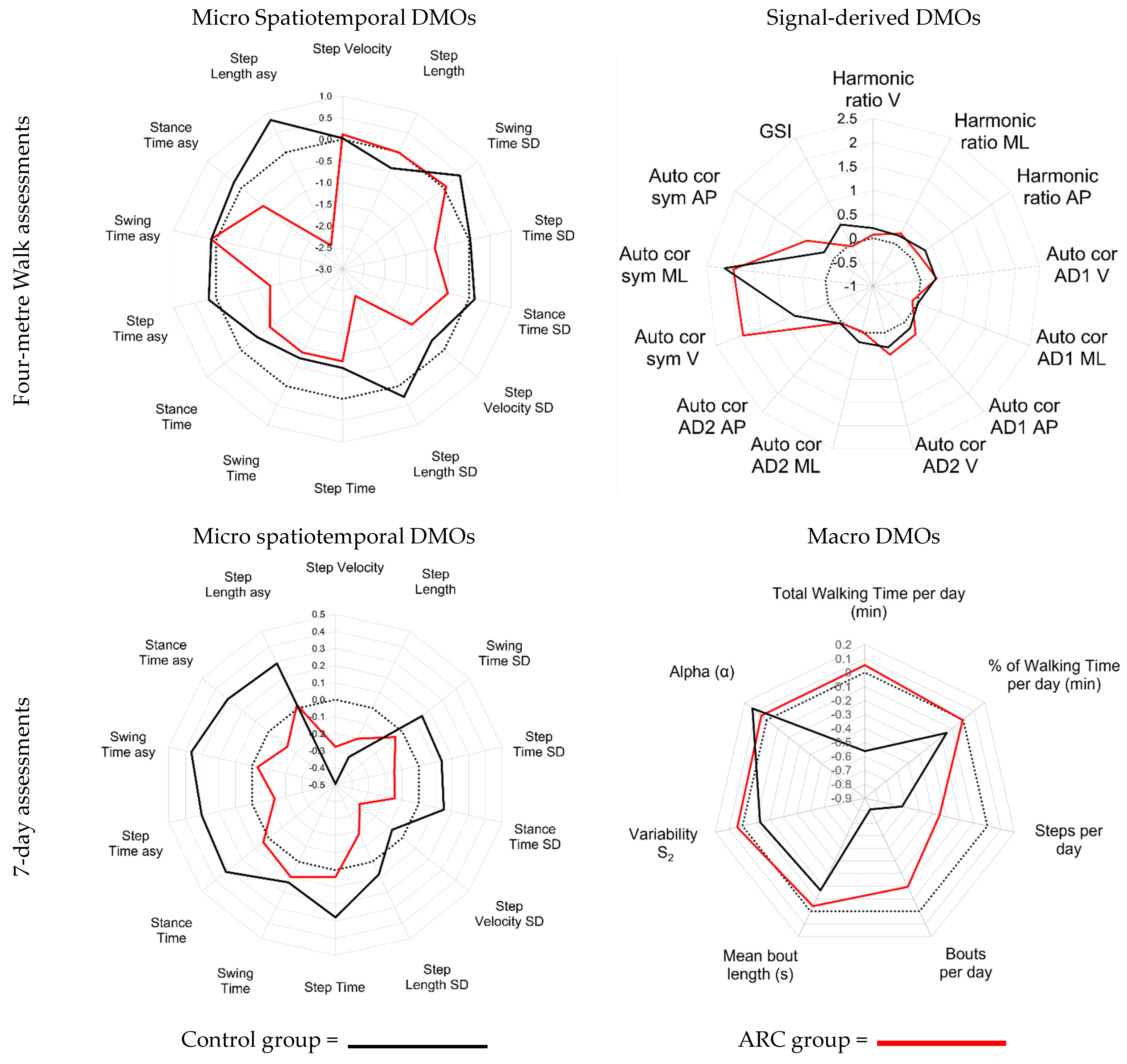
Figure 2 shows the change seen for all DMOs for all tested conditions using the z score derived relative to the baseline values for the continuous and at-home assessments. For the at-home data, the radar plots show that for the spatiotemporal variables, a similar trend of intervention effect was found for both groups. Visual inspection shows that, except for step velocity, step length, and swing time asymmetry, all variables were reduced for the ARC group. For the control group, the majority of the DMOs stayed the same, with the exception of step length asymmetry, which increased the most, while stance time, swing time, and step time all decreased the most relative to the other DMOs. The biggest differences between the ARC and the control group were found for step length asymmetry and SD, where the ARC group recorded a reduction, while the opposite was recorded for the control group. For the signal-derived variables, the changes that occurred were most evident for the autocorrelation symmetry DMOs, where an increase was observed for both groups. In the vertical direction, the increase was more evident for the ARC group. For the control group, the gait symmetry index increased, whereas it did not increase for the ARC group. The other DMOs displayed a similar trend for both groups.
For the continuous data collected from 7 days of analysis, the micro spatiotemporal variables showed that, except for step velocity and step velocity SD, the control group achieved an increase in all variables. The ARC group mainly achieved reduced values or values similar to their baseline. Step length asymmetry, stance time, swing time and step time were the exceptions, as a slight increase was observed. The macro variables showed that mean bout length, bouts per day, and steps per day reduced for both groups. The control group was reduced for all DMOs except for alpha, which increased. The ARC group recorded little impact of the intervention but did appear to reduce the number of bouts per day and steps per day, although this reduction was less than that of the control group.
The wearable activity monitors derived DMOs displayed using radar plots, showing that it is possible to see objective changes that occur because of an intervention designed to improve gait mechanics for stroke survivors. Between the conditions, the higher z values achieved for select variables from the data collected at the participants’ homes indicate that the short walks under the observation of the research team highlight greater changes occurring because of the 6 weeks of gait training with or without the ARC feedback.
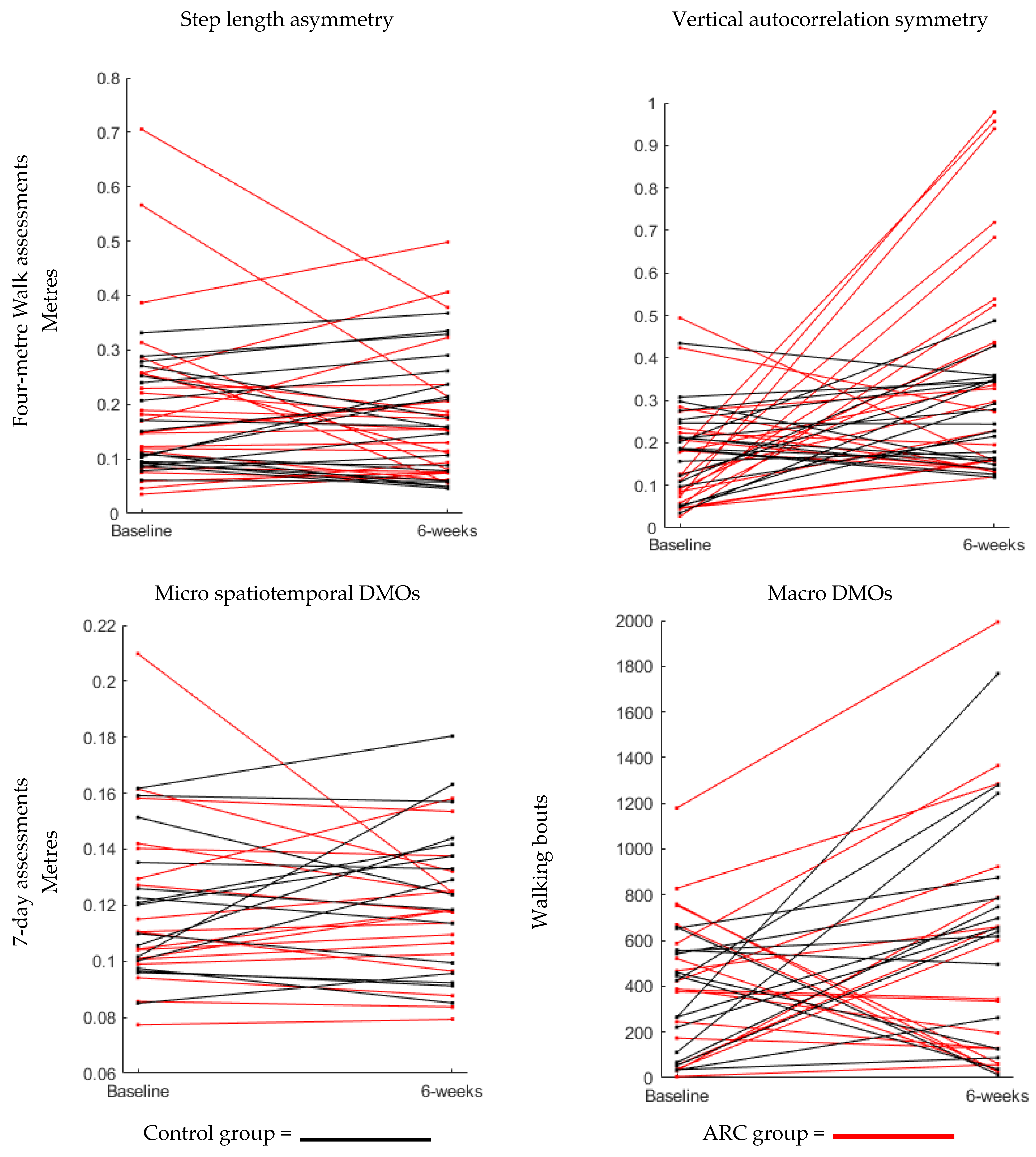
Further visualisation of the variables with the greatest differences achieved in the change scores between the ARC and control group for each test condition is displayed in Figure 3. Here, we see how each individual responded to step length asymmetry and the vertical autocorrelation symmetry recorded during the four-metre assessments, as well as for swing time asymmetry and the number of bouts per day for the 7-day assessment data following the 6 weeks of interventions.

3.3. Objective 3: Reference Values Obtained for All DMOs Analysed

Table 1 indicates the mean and standard deviation for all DMOs calculated for both the ARC and control group when recorded at baseline and after the 6 weeks of interventions. The four-metre walking DMOs were calculated from 23 ARC participants and 20 controls. For the 7-day assessments, values were calculated from 18 and 16 participants from the ARC and control group, respectively. The purpose of providing all reference values is for future researchers to have a value for these DMOs, for the creation of sample size calculations, and to help decide which DMOs might be the most valuable for different interventions with the goal of improving gait mechanics for stroke survivors.

4. Discussion

The aim of this current paper was to explore the use of wearable activity monitors within a pilot RCT to objectively quantify gait performance following 6 weeks of ARC training. We were able to quantify a range of DMOs in stroke survivors in the home and community, both before and after intervention, to enable the exploration of specific gait mechanics. To our knowledge, this study is the first to trial ARC compared to a control group where the intervention was both administered and objectively quantified at the participants’ homes. The wearable activity monitors did appear to add value to a pilot trial where previously, when compared to a control group who slightly increased their gait speed from 0.51 to 0.60 m/s, the ARC group’s speed did not appear to change because of the 6 weeks of ARC intervention (0.53 to 0.54 m/s) [15]. The success rates of data collection using the wearable activity monitors ranged from 64 to 95%. When compared to the previous study using the same data set, these values are lower as gait speed data was collected using traditional methods for 59 (100%) and 47 (80%) participants at baseline and after 6 weeks, respectively. From the available data, our study indicates that wearable activity monitors can be used to quantify group and individual differences in a range of DMOs in stroke survivors in the home and community in response to an auditory rhythmical cueing intervention. The collection of this range of DMOs provides insight into how interventions influence specific gait mechanics beyond standard clinical measures and in real-world settings, rather than a laboratory. Furthermore, the results observed in this study support past views, which stated that, due to home-based interventions being available to all, they are advantageous over traditional methods of gait rehabilitation, especially for individuals where transport is problematic [8]. Although home-based therapy poses a challenge as clinicians cannot observe any exercise adherence/quality, these problems are likely outweighed by facilitating more opportunities for increasing quantity and duration of treatment [1,8]. Future research, such as a full RCT, using a home-based gait rehabilitation intervention measured using wearable activity monitors, appears to be warranted to determine the impact of novel gait interventions for stroke survivors.
To achieve our second and third objectives of attempting to visualise any mechanical changes in gait and to provide reference values for DMOs using wearable activity monitors located on the lower back, a broad range of variables were quantified. Although useful for our objectives, this pilot RCT and the broad range of DMOs calculated did not facilitate a clear and clinically meaningful understanding of whether the ARC intervention impacted gait in a more favourable way compared to the control group, as this was a pilot RCT and did not include measuring efficacy. Future research with large cohorts establishing clinically meaningful differences and variables most relevant to specific interventions is required [2,9]. To achieve this goal, despite using valid and reliable measures already tested for stroke survivors, the large heterogeneity observed between the participants for different variables, as seen in Figure 3, indicates that certain participants responded well compared to others who did not, highlighting the difficulty in researching which interventions are effective for stroke survivors. Future research into the impact of specific gait interventions, to avoid the large heterogeneity seen in certain DMOs, could benefit from stricter inclusion criteria or from larger sample sizes, allowing for a stratification/adaptive trial design [40]. In addition, further validation of DMOs at different stages of rehabilitation/severity, both in the home and in free-living environments, is required to accurately capture gait improvements even when gait mechanics may change [25,41,42]. Only then, and after larger cohort studies have been conducted, can wearable activity monitors contribute towards a precision medicine approach where DMOs can allow the classification of pheno(sub)types at a participant-specific level and identify those who do and do not respond to an evidence-based intervention [27].
Alternatively, if acknowledging that overcoming the heterogeneity in stroke survivors’ gait will continue to be a barrier against the best practice for gait rehabilitation, an individual/participant-centred approach might be best for future research [27]. The results from the current study appear to confirm that “one size does not fit all” for stroke survivors’ gait rehabilitation, and instead, individual participants might benefit from a more personalised rehabilitation target [2,9]. For example, if a participant wants to focus on a symmetrical gait pattern and move with a gait speed that does not facilitate the use of an algorithm to accurately detect initial contact, a therapist may solely focus on signal-derived gait measures prior to focusing on gait speed and the other spatiotemporal measures of asymmetry. For this purpose, z scores and radar plots might be a useful approach, but instead of using the group average and standard deviation, they could be used to track the progression of the same participant using multiple gait bouts and only with the variables relevant to the chosen rehabilitation targets [2,9,25].
Although, to our knowledge, this current pilot RCT is the first to use wearable activity monitors to quantify the impact of an ARC intervention within the home/real-world environment, the data from the wearable activity monitors did not feed into the therapy. Due to the advancement in “real-time” analysis of wearable technology and the portability of devices to visualise such analysis, it is now possible for participants to monitor their own gait mechanics in real time, where complex gait variables are adapted to determine if they are improving or worsening [43,44]. If this technology can be fully adapted for individual stroke survivors, where therapists can select the relevant variables based on each participant’s baseline profile, it is feasible that ARC could be integrated into participants’ rehabilitation together with the ability to track their progress towards personalised goals [10]. This would also benefit clinicians who could remotely track progress for each individual and have an objective measure of adherence to the therapy. Collectively, the above approaches between the need for bigger studies or a more person-tailored approach indicate two separate means to understand the same collective goal; future research is needed to consolidate the efforts of researchers if we are to progress our understanding of the most effective way to rehabilitate gait for stroke survivors [20].
The current study has limitations. One limitation is that, although we used a novel approach with the aforementioned benefits, assessing the participants’ gait over four metres in their own homes did not guarantee an adequate distance to create valid measures for all DMOs recorded. Research is required to determine the minimal walking bout lengths for valid DMOs to be recorded using wearable activity monitors [1]. Alternatively, the advancements in battery technology for inertial measurement units (IMUs) and the addition of gyroscopes would allow us to confirm straight-line walking in the free-living environment when tested continuously for a week or longer. If proven to be valid, future work may be able to adopt free-living data only and determine the walking bouts that are adequately long for each DMO and which are confirmed not to involve turning; this may eliminate the need for at-home assessments. Although the L5 placement of the wearable activity monitor is favourable for the signal-derived measures of symmetry, past studies that calculated a greater amount of DMOs using inertial measurement devices located on more segments of the body found that the DMOs they calculated were more reliable from monitors located on the participant’s feet, especially for those with the most severe gait impairments [2]. Therefore, for select participants where events could not accurately be detected from the L5, this might partly explain the large heterogeneity of the intervention impact seen in the current results and imply that for certain individuals, monitors located on both feet would be better suited. Equally, it could be that certain DMOs are potentially better suited to specific gait impairments. Another notable limitation was the broad inclusion criterion in the current study, which did not control for patient characteristics known to impact gait, such as age, balance, cognitive function, and fear of falling [9]. The data loss from the wearable sensors was another limitation, as it did not allow for analysis from all participants. Future RCTs should be informed by this data loss when estimating sample size, while qualitative studies exploring the reason for wearable data loss are warranted. Lastly, a limitation of this study is shared with the field in that there remains uncertainty on the correct duration/intensity of ARC and other gait-training interventions to enable neuroplasticity [6,8]. More longitudinal studies investigating the required intervention intensity to induce clinically meaningful change for gait in a broad range of stroke survivors with varying gait impairments are required. The current study did attempt a washout period with follow-up assessments at 10 weeks post-baseline; however, due to the impact of COVID-19 at the end of this study, the remaining participants would not have allowed for adequate numbers to run the paired analysis used in this current study.

5. Conclusions

The achievement of the four study objectives indicates that it is possible to successfully integrate wearable activity monitors within a pilot RCT aiming to improve the gait mechanics of stroke survivors, and through visual inspection of a broad range of DMOs, it is possible to assess the impact of gait training for both the ARC and the control group. This is a useful initial step to support the idea that wearable activity monitors are the ideal tool to calculate objective and specific DMOs required to support evidence-based-intervention research aiming to improve specific gait mechanics for stroke survivors. This is because wearable activity monitors are objective, valid and reliable tools capable of measuring specific gait mechanics targeted by the intervention; their use can identify the desired changes targeted by the intervention, where conventional clinical measures might lack specificity. Although the results are limited as this is a pilot study and, therefore, not able to show efficacy, the visible changes seen in certain DMOs and individuals in both groups warrant further research into how wearable activity monitors can best support evidence-based interventions and aid in explaining the complexity observed when rehabilitating gait mechanics for stroke survivors.

Author Contributions

Conceptualization, S.A.M., L.S., P.M., S.D.D., L.R., H.H., P.B., and A.L.; methodology, S.D.D., S.A.M., L.S., H.H., P.B., P.M., A.L., and R.F.; software, S.D.D., C.B., L.R., R.F., L.S., and S.A.M.; validation, S.A.M., C.B., S.D.D., and L.R.; formal analysis, C.B., S.D.D., R.F., L.S., S.A.M., L.R., and P.M.; investigation, H.H., P.B., and P.M.; resources, S.D.D., H.H., P.B., L.R., L.S., and S.A.M.; data curation, P.B., C.B., S.D.D., L.S., and S.A.M.; writing—original draft preparation, C.B.; writing—review and editing, C.B., S.D.D., S.A.M., L.S., H.H., and P.B.; visualisation, C.B., S.D.D., S.A.M., and L.S.; supervision, S.D.D., L.S., and L.R.; project administration, S.A.M., P.M., H.H., P.B., S.D.D., R.F., and L.S.; funding acquisition, S.A.M., L.R., L.S., and A.L. All authors have read and agreed to the published version of the manuscript.

Funding

This study was funded by The Stroke Association, reference TSA 2016/06. SAM was supported by Health Education England and the National Institute for Health Research (HEE/NIHR ICA Programme Clinical Lectureship, Dr Sarah Anne Moore, ICA-CL-2015-01-012). SDD and LR were also supported by the IDEA-FAST project, which has received funding from the Innovative Medicines Initiative 2 Joint Undertaking (JU) under grant agreement No. 853981. This JU receives support from the European Union’s Horizon 2020 research and innovation program and the European Federation of Pharmaceutical Industries and Associations (EFPIA). SDD and LR were also supported by the National Institute for Health Research (NIHR) Newcastle Biomedical Research Centre (BRC) based at The Newcastle upon Tyne Hospital NHS Foundation Trust, Newcastle University and the Cumbria, Northumberland and Tyne and Wear (CNTW) NHS Foundation Trust. SDD was also supported by the NIHR/Wellcome Trust Clinical Research Facility (CRF) infrastructure at Newcastle upon Tyne Hospitals NHS Foundation Trust. SDD was supported by the UK Research and Innovation (UKRI) Engineering and Physical Sciences Research Council (EPSRC) (Grant Ref: EP/W031590/1, Grant Ref: EP/X031012/1 and Grant Ref: EP/X036146/1). All opinions are those of the authors and not the funders. The content in this publication reflects the authors’ view, and neither IMI nor the European Union, EFPIA, NHS, NIHR, nor any associated partners are responsible for any use that may be made of the information contained herein.

Institutional Review Board Statement

The study was conducted in accordance with the Declaration of Helsinki and approved by the Institutional Ethics Committee of London-City and East Research Ethics Committee (ref 18/LO/0115 on 12 January 2018).

Informed Consent Statement

Informed consent was obtained from all subjects involved in this study.

Data Availability Statement

Please contact SAM to request data availability.

Acknowledgments

We would like to thank the following for their contributions: 1. Study participants. 2. Staff from the following NHS Trusts who were involved in recruiting participants to the research project: County Durham and Darlington NHS Foundation Trust, Gateshead Health NHS Foundation Trust, Northumbria Healthcare NHS Foundation Trust, Newcastle upon Tyne Hospitals NHS Foundation Trust. 3. Staff at Newcastle University who contributed to the project: Anne Harrison.

Conflicts of Interest

Authors Phillip Brown, Heather Hunter were employed by the Newcastle upon Tyne Hospitals NHS Foundation Trust, and Lynn Rochester had an affiliation with this trust but was employed by Newcastle university and author Sarah A. Moore was employed by the Northumbria Healthcare NHS Foundation Trust. The remaining authors declare that the research was conducted in the absence of any commercial or financial relationships that could be construed as a potential conflict of interest. The authors declare that this study received funding from the Stroke Association reference TSA 2016/06. The funder was not involved in the study design, collection, analysis, interpretation of data, the writing of this article or the decision to submit it for publication.

References

  1. Huizenga, D.; Rashford, L.; Darcy, B.; Lundin, E.; Medas, R.; Shultz, S.T.; DuBose, E.; Reed, K.B. Wearable Gait Device for Stroke Gait Rehabilitation at Home. Top. Stroke Rehabil. 2021, 28, 443–455. [Google Scholar] [CrossRef]
  2. Felius, R.A.W.; Geerars, M.; Bruijn, S.M.; van Dieën, J.H.; Wouda, N.C.; Punt, M. Reliability of IMU-Based Gait Assessment in Clinical Stroke Rehabilitation. Sensors 2022, 22, 908. [Google Scholar] [CrossRef] [PubMed]
  3. Pollock, A.; St George, B.; Fenton, M.; Firkins, L. Top Ten Research Priorities Relating to Life after Stroke. Lancet Neurol. 2012, 11, 209. [Google Scholar] [CrossRef]
  4. Stretton, C.M.; Mudge, S.; Kayes, N.M.; McPherson, K.M. What Does Real-World Walking Mean to People with Stroke? An Interpretive Descriptive Study. Disabil. Rehabil. 2022, 44, 315–322. [Google Scholar] [CrossRef] [PubMed]
  5. Algurén, B.; Lundgren-Nilsson, Å.; Sunnerhagen, K.S. Functioning of Stroke Survivors—A Validation of the ICF Core Set for Stroke in Sweden. Disabil. Rehabil. 2010, 32, 551–559. [Google Scholar] [CrossRef] [PubMed]
  6. Krishnan, V.; Khoo, I.; Marayong, P.; DeMars, K.; Cormack, J. Gait Training in Chronic Stroke Using Walk-Even Feedback Device: A Pilot Study. Neurosci. J. 2016, 2016, 6808319. [Google Scholar] [CrossRef]
  7. Fulk, G.D.; He, Y.; Boyne, P.; Dunning, K. Predicting Home and Community Walking Activity Poststroke. Stroke 2017, 48, 406–411. [Google Scholar] [CrossRef]
  8. Wright, R.L.; Briony Brownless, S.; Pratt, D.; Sackley, C.M.; Wing, A.M. Stepping to the Beat: Feasibility and Potential Efficacy of a Home-Based Auditory-Cued Step Training Program in Chronic Stroke. Front. Neurol. 2017, 8, 412. [Google Scholar] [CrossRef]
  9. Felius, R.A.W.; Wouda, N.C.; Geerars, M.; Bruijn, S.M.; van Dieën, J.H.; Punt, M. Beyond Gait Speed: Exploring the Added Value of Inertial Measurement Unit-Based Measurements of Gait in the Estimation of the Walking Ability in Daily Life. BMC Neurol. 2024, 24, 129. [Google Scholar] [CrossRef]
  10. Spencer, J.; Wolf, S.L.; Kesar, T.M. Biofeedback for Post-Stroke Gait Retraining: A Review of Current Evidence and Future Research Directions in the Context of Emerging Technologies. Front. Neurol. 2021, 12, 637199. [Google Scholar] [CrossRef]
  11. Schaefer, R.S. Auditory Rhythmic Cueing in Movement Rehabilitation: Findings and Possible Mechanisms. Philos. Trans. R. Soc. B Biol. Sci. 2014, 369, 20130402. [Google Scholar] [CrossRef] [PubMed]
  12. Murgia, M.; Pili, R.; Corona, F.; Sors, F.; Agostini, T.A.; Bernardis, P.; Casula, C.; Cossu, G.; Guicciardi, M.; Pau, M. The Use of Footstep Sounds as Rhythmic Auditory Stimulation for Gait Rehabilitation in Parkinson’s Disease: A Randomized Controlled Trial. Front. Neurol. 2018, 9, 348. [Google Scholar] [CrossRef]
  13. Nascimento, L.R.; de Oliveira, C.Q.; Ada, L.; Michaelsen, S.M.; Teixeira-Salmela, L.F. Walking Training with Cueing of Cadence Improves Walking Speed and Stride Length after Stroke More than Walking Training Alone: A Systematic Review. J. Physiother. 2015, 61, 10–15. [Google Scholar] [CrossRef]
  14. McCue, P.; Shaw, L.; Del Din, S.; Hunter, H.; Lord, S.; Price, C.I.M.; Rodgers, H.; Rochester, L.; Moore, S.A. Acceptability and Deliverability of an Auditory Rhythmical Cueing (ARC) Training Programme for Use at Home and Outdoors to Improve Gait and Physical Activity Post-Stroke. Arch. Physiother. 2022, 1, 1. [Google Scholar] [CrossRef]
  15. Shaw, L.; McCue, P.; Brown, P.; Buckley, C.; Del Din, S.; Francis, R.; Hunter, H.; Lambert, A.; Lord, S.; Price, C.I.M.; et al. Auditory Rhythmical Cueing to Improve Gait in Community-Dwelling Stroke Survivors (ACTIVATE): A Pilot Randomised Controlled Trial. Pilot. Feasibility Stud. 2022, 8, 239. [Google Scholar] [CrossRef]
  16. Seo, M.; Shin, M.J.; Park, T.S.; Park, J.H. Clinometric Gait Analysis Using Smart Insoles in Patients with Hemiplegia after Stroke: Pilot Study. JMIR Mhealth Uhealth 2020, 8, e22208. [Google Scholar] [CrossRef]
  17. Moore, S.A.; Boyne, P.; Fulk, G.; Verheyden, G.; Fini, N.A. Walk the Talk: Current Evidence for Walking Recovery after Stroke, Future Pathways and a Mission for Research and Clinical Practice. Stroke 2022, 53, 3494–3505. [Google Scholar] [CrossRef] [PubMed]
  18. Patterson, K.K.; Gage, W.H.; Brooks, D.; Black, S.E.; McIlroy, W.E. Evaluation of Gait Symmetry after Stroke: A Comparison of Current Methods and Recommendations for Standardization. Gait Posture 2010, 31, 241–246. [Google Scholar] [CrossRef] [PubMed]
  19. Patterson, K.K.; Mansfield, A.; Biasin, L.; Brunton, K.; Inness, E.L.; McIlroy, W.E. Longitudinal Changes in Poststroke Spatiotemporal Gait Asymmetry over Inpatient Rehabilitation. Neurorehabil Neural Repair. 2015, 29, 153–162. [Google Scholar] [CrossRef]
  20. Fini, N.A.; Simpson, D.; Moore, S.A.; Mahendran, N.; Eng, J.J.; Borschmann, K.; Moulaee Conradsson, D.; Chastin, S.; Churilov, L.; English, C. How Should We Measure Physical Activity After Stroke? An International Consensus. Int. J. Stroke 2023, 18, 1132–1142. [Google Scholar] [CrossRef]
  21. Smith, B.A.; Lang, C.E. Sensor Measures of Symmetry Quantify Upper Limb Movement in the Natural Environment across the Lifespan. Arch. Phys. Med. Rehabil. 2019, 100, 1176. [Google Scholar] [CrossRef]
  22. Peters, D.M.; O’Brien, E.S.; Kamrud, K.E.; Roberts, S.M.; Rooney, T.A.; Thibodeau, K.P.; Balakrishnan, S.; Gell, N.; Mohapatra, S. Utilization of Wearable Technology to Assess Gait and Mobility Post-Stroke: A Systematic Review. J. Neuroeng. Rehabil. 2021, 18, 67. [Google Scholar] [CrossRef] [PubMed]
  23. Moore, S.A.; Hickey, A.; Lord, S.; Del Din, S.; Godfrey, A.; Rochester, L. Comprehensive Measurement of Stroke Gait Characteristics with a Single Accelerometer in the Laboratory and Community: A Feasibility, Validity and Reliability Study. J. Neuroeng. Rehabil. 2017, 14, 130. [Google Scholar] [CrossRef]
  24. Polhemus, A.; Ortiz, L.D.; Brittain, G.; Chynkiamis, N.; Salis, F.; Gaßner, H.; Gross, M.; Kirk, C.; Rossanigo, R.; Taraldsen, K.; et al. Walking on Common Ground: A Cross-Disciplinary Scoping Review on the Clinical Utility of Digital Mobility Outcomes. NPJ Digit. Med. 2021, 4, 149. [Google Scholar] [CrossRef] [PubMed]
  25. Lanotte, F.; Okita, S.; O’Brien, M.K.; Jayaraman, A. Enhanced Gait Tracking Measures for Individuals with Stroke Using Leg-Worn Inertial Sensors. J. Neuroeng. Rehabil. 2024, 21, 219. [Google Scholar] [CrossRef] [PubMed]
  26. Murphy, M.A.; Munoz-Novoa, M.; Heremans, C.; Branscheidt, M.; Cabanas-Valdés, R.; Engelter, S.T.; Kruuse, C.; Kwakkel, G.; Lakičević, S.; Lampropoulou, S.; et al. European Stroke Organisation (ESO) Guideline on Motor Rehabilitation. Eur. Stroke J. 2025, 23969873251338142. [Google Scholar] [CrossRef]
  27. Van Criekinge, T.; Heremans, C.; Burridge, J.; Deutsch, J.E.; Hammerbeck, U.; Hollands, K.; Karthikbabu, S.; Mehrholz, J.; Moore, J.L.; Salbach, N.M.; et al. Standardized Measurement of Balance and Mobility Post-Stroke: Consensus-Based Core Recommendations from the Third Stroke Recovery and Rehabilitation Roundtable. Int. J. Stroke 2024, 19, 158–168. [Google Scholar] [CrossRef]
  28. Del Din, S.; Lewis, E.G.; Gray, W.K.; Collin, H.; Kissima, J.; Rochester, L.; Dotchin, C.; Urasa, S.; Walker, R. Monitoring Walking Activity with Wearable Technology in Rural-Dwelling Older Adults in Tanzania: A Feasibility Study Nested within a Frailty Prevalence Study. Exp. Aging Res. 2020, 46, 367–381. [Google Scholar] [CrossRef]
  29. Lord, S.; Galna, B.; Verghese, J.; Coleman, S.; Burn, D.; Rochester, L. Independent Domains of Gait in Older Adults and Associated Motor and Nonmotor Attributes: Validation of a Factor Analysis Approach. J. Gerontol.—Ser. A Biomed. Sci. Med. Sci. 2013, 68, 820–827. [Google Scholar] [CrossRef]
  30. Del Din, S.; Godfrey, A.; Rochester, L. Validation of an Accelerometer to Quantify a Comprehensive Battery of Gait Characteristics in Healthy Older Adults and Parkinson’s Disease: Toward Clinical and at Home Use. IEEE J. Biomed. Health Inf. 2016, 20, 838–847. [Google Scholar] [CrossRef]
  31. Hickey, A.; Del Din, S.; Rochester, L.; Godfrey, A. Detecting Free-Living Steps and Walking Bouts: Validating an Algorithm for Macro Gait Analysis. Physiol. Meas. 2017, 38, N1–N15. [Google Scholar] [CrossRef] [PubMed]
  32. Lord, S.; Chastin, S.F.M.; McInnes, L.; Little, L.; Briggs, P.; Rochester, L. Exploring Patterns of Daily Physical and Sedentary Behaviour in Community-Dwelling Older Adults. Age Ageing 2011, 40, 205–210. [Google Scholar] [CrossRef]
  33. Chastin, S.F.M.; Baker, K.; Jones, D.; Burn, D.; Granat, M.H.; Rochester, L. The Pattern of Habitual Sedentary Behavior Is Different in Advanced Parkinson’s Disease. Mov. Disord. 2010, 25, 2114–2120. [Google Scholar] [CrossRef] [PubMed]
  34. Rochester, L.; Chastin, S.F.M.; Lord, S.; Baker, K.; Burn, D.J. Understanding the Impact of Deep Brain Stimulation on Ambulatory Activity in Advanced Parkinson’s Disease. J. Neurol. 2012, 259, 1081–1086. [Google Scholar] [CrossRef] [PubMed]
  35. Buckley, C.; Micó-Amigo, M.E.; Dunne-Willows, M.; Godfrey, A.; Hickey, A.; Lord, S.; Rochester, L.; Del Din, S.; Moore, S.A. Gait Asymmetry Post-Stroke: Determining Valid and Reliable Methods Using a Single Accelerometer Located on the Trunk. Sensors 2019, 20, 37. [Google Scholar] [CrossRef]
  36. Rehman, R.Z.U.R.; Buckley, C.; Micó-Amigo, M.E.; Kirk, C.; Dunne-Willows, M.; Mazzà, C.; Shi, J.Q.; Alcock, L.; Rochester, L.; Del Din, S. Accelerometry-Based Digital Gait Characteristics for Classification of Parkinson’s Disease: What Counts? IEEE Open J. Eng. Med. Biol. 2020, 1, 65–73. [Google Scholar] [CrossRef]
  37. Bellanca, J.L.; Lowry, K.A.; VanSwearingen, J.M.; Brach, J.S.; Redfern, M.S. Harmonic Ratios: A Quantification of Step to Step Symmetry. J. Biomech. 2013, 46, 828–831. [Google Scholar] [CrossRef]
  38. Moe-Nilssen, R.; Helbostad, J.L. Estimation of Gait Cycle Characteristics by Trunk Accelerometry. J. Biomech. 2004, 37, 121–126. [Google Scholar] [CrossRef]
  39. Zhang, W.; Smuck, M.; Legault, C.; Ith, M.A.; Muaremi, A.; Aminian, K. Gait Symmetry Assessment with a Low Back 3D Accelerometer in Post-Stroke Patients. Sensors 2018, 18, 3322. [Google Scholar] [CrossRef]
  40. Stockley, R.C.; Graham, I.S. The Importance of Embracing Complexity in Rehabilitation. J. Eval. Clin. Pr. 2023, 29, 657–661. [Google Scholar] [CrossRef]
  41. Hulleck, A.A.; Menoth Mohan, D.; Abdallah, N.; El Rich, M.; Khalaf, K. Present and Future of Gait Assessment in Clinical Practice: Towards the Application of Novel Trends and Technologies. Front. Med. Technol. 2022, 4, 901331. [Google Scholar] [CrossRef]
  42. Trojaniello, D.; Ravaschio, A.; Hausdorff, J.M.; Cereatti, A. Comparative Assessment of Different Methods for the Estimation of Gait Temporal Parameters Using a Single Inertial Sensor: Application to Elderly, Post-Stroke, Parkinson’s Disease and Huntington’s Disease Subjects. Gait Posture 2015, 42, 310–316. [Google Scholar] [CrossRef] [PubMed]
  43. Wu, J.; Kuruvithadam, K.; Schaer, A.; Stoneham, R.; Chatzipirpiridis, G.; Easthope, C.A.; Barry, G.; Martin, J.; Pané, S.; Nelson, B.J.; et al. An Intelligent In-Shoe System for Gait Monitoring and Analysis with Optimized Sampling and Real-Time Visualization Capabilities. Sensors 2021, 21, 2869. [Google Scholar] [CrossRef] [PubMed]
  44. Hsu, W.-C.; Sugiarto, T.; Liao, Y.-Y.; Lin, Y.-J.; Yang, F.-C.; Hueng, D.-Y.; Sun, C.-T.; Chou, K.-N.; Liao, Y.-Y.; Lin, Y.-J.; et al. Can Trunk Acceleration Differentiate Stroke Patient Gait Patterns Using Time- and Frequency-Domain Features? Appl. Sci. 2021, 11, 1541. [Google Scholar] [CrossRef]
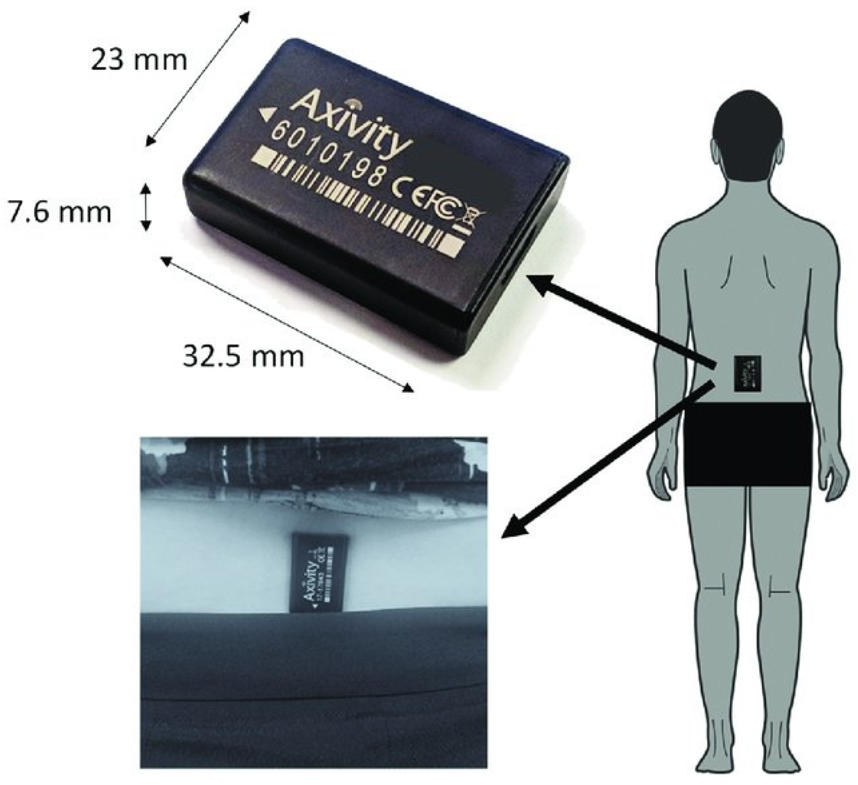
Figure 1. Experimental set-up: the Axivity AX3 device, the site of attachment, and the orientation of the device on the lower back. Image taken from [28].
Symmetry 17 01640 g001
Figure 2. Radar plots illustrating each variable from variables calculated during the four-metre walk assessments (46 participants) and from data collected continuously for 7 days (38 participants). The dashed central line represents the baseline data for both groups. Deviation from zero along the X axis radiating from the centre of the plot represents how many standard deviations differ (based upon the baseline means and standard deviations) between the ARC and the control relative to their original assessments. a. AP = anterior−posterior positions. ML = medio−lateral positions. V = vertical positions. b. SD = standard deviation. Asy = asymmetry. c. AD1 = step regularity. AD2 stride regularity. Auto cor sym = autocorrelation-derived symmetry. GSI = gait symmetry index.
Symmetry 17 01640 g002
Figure 3. A spaghetti diagram illustrating the changes recorded by each individual’s step length variability and the vertical RMS variables recorded in the participants’ homes, as well as step velocity and total walk time for the 7-day data.
Symmetry 17 01640 g003
Table 1. The means and standard deviations for all DMOs calculated for both the ARC and control groups.
ARCControl
VariableBaseline6 WeekBaseline6 Week
Mean SDMean SDMean SDMean SD
Four-metre assessmentsMicroStep Time0.71 ± 0.140.69 ± 0.120.68 ± 0.090.66 ± 0.11
Stance Time0.85 ± 0.140.83 ± 0.100.81 ± 0.090.80 ± 0.10
Swing Time0.54 ± 0.090.52 ± 0.140.53 ± 0.080.51 ± 0.09
Step Length0.57 ± 0.090.56 ± 0.110.58 ± 0.100.55 ± 0.09
Step Velocity0.84 ± 0.110.85 ± 0.160.87 ± 0.140.87 ± 0.18
Step Time SD0.17 ± 0.130.15 ± 0.120.14 ± 0.120.14 ± 0.11
Stance Time SD0.16 ± 0.110.14 ± 0.110.14 ± 0.110.14 ± 0.10
Swing Time SD0.10 ± 0.070.10 ± 0.060.10 ± 0.050.11 ± 0.07
Step Length SD0.13 ± 0.060.11 ± 0.060.11 ± 0.050.12 ± 0.07
Step Velocity SD0.20 ± 0.080.17 ± 0.090.18 ± 0.070.16 ± 0.07
Step Time asy0.15 ± 0.180.12 ± 0.110.11 ± 0.090.11 ± 0.10
Stance Time asy0.14 ± 0.150.12 ± 0.100.12 ± 0.090.12 ± 0.08
Swing Time asy0.10 ± 0.130.11 ± 0.090.11 ± 0.090.11 ± 0.08
Step Length asy0.22 ± 0.160.18 ± 0.120.16 ± 0.090.17 ± 0.11
Signal derived variablesHarmonic ratio V1.59 ± 0.671.64 ± 0.631.50 ± 0.571.62 ± 0.52
Harmonic ratio ML1.63 ± 0.361.72 ± 0.451.59 ± 0.381.66 ± 0.51
Harmonic ratio AP1.38 ± 0.571.47 ± 0.631.20 ± 0.511.37 ± 0.50
Auto cor AD1 V 0.38 ± 0.270.47 ± 0.270.34 ± 0.180.40 ± 0.19
Auto cor AD1 ML 0.46 ± 0.170.43 ± 0.170.46 ± 0.150.46 ± 0.16
Auto cor AD1 AP 0.38 ± 0.230.46 ± 0.220.36 ± 0.300.41 ± 0.23
Auto cor AD2 V 0.47 ± 0.240.59 ± 0.280.48 ± 0.210.54 ± 0.18
Auto cor AD2 ML 0.48 ± 0.210.48 ± 0.220.50 ± 0.240.55 ± 0.24
Auto cor AD2 AP 0.54 ± 0.210.55 ± 0.190.58 ± 0.310.59 ± 0.16
Auto cor sym V 0.16 ± 0.120.39 ± 0.290.19 ± 0.100.27 ± 0.11
Auto cor sym ML 0.12 ± 0.090.29 ± 0.210.12 ± 0.110.34 ± 0.27
Auto cor sym AP 0.28 ± 0.170.39 ± 0.240.35 ± 0.200.40 ± 0.20
Gait symmetry index0.45 ± 0.190.44 ± 0.180.43 ± 0.130.49 ± 0.20
7-day assessmentsMicroStep Time0.62 ± 0.030.62 ± 0.030.62 ± 0.020.62 ± 0.04
Stance Time0.77 ± 0.030.77 ± 0.030.77 ± 0.030.77 ± 0.04
Swing Time0.47 ± 0.030.47 ± 0.030.47 ± 0.030.47 ± 0.04
Step Length0.55 ± 0.040.54 ± 0.040.58 ± 0.060.56 ± 0.07
Step Velocity0.96 ± 0.080.94 ± 0.101.01 ± 0.130.95 ± 0.15
Step Time SD0.20 ± 0.030.19 ± 0.030.19 ± 0.030.19 ± 0.03
Stance Time SD0.21 ± 0.030.21 ± 0.030.20 ± 0.030.21 ± 0.03
Swing Time SD0.17 ± 0.020.16 ± 0.020.16 ± 0.020.16 ± 0.02
Step Length SD0.15 ± 0.010.15 ± 0.010.15 ± 0.010.15 ± 0.01
Step Velocity SD0.37 ± 0.040.36 ± 0.040.36 ± 0.060.35 ± 0.04
Step Time asy0.12 ± 0.030.12 ± 0.020.11 ± 0.020.12 ± 0.03
Stance Time asy0.12 ± 0.030.12 ± 0.020.12 ± 0.020.13 ± 0.03
Swing Time asy0.11 ± 0.030.11 ± 0.020.11 ± 0.020.12 ± 0.03
Step Length asy0.09 ± 0.020.09 ± 0.020.09 ± 0.010.09 ± 0.02
MacroMean bout length (s)14.02 ± 3.2813.88 ± 4.3115.96 ± 3.0515.45 ± 2.89
Variability s20.79 ± 0.100.79 ± 0.110.83 ± 0.070.82 ± 0.09
Alpha1.67 ± 0.081.67 ± 0.081.63 ± 0.041.63 ± 0.04
Total walk time per day (min)116.85 ± 66.02120.38 ± 71.47132.96 ± 56.57101.10 ± 64.02
Steps per day8154 ± 52746305 ± 65709066 ± 38216669 ± 3786
Bouts per day486.19 ± 248.57438.10 ± 321.98506.69 ± 212.47334.48 ± 228.90
% of Walking Time per day8.11 ± 4.588.10 ± 4.889.23 ± 3.938.64 ± 3.52
AP = anterior–posterior position. ML = medio-lateral position. V = vertical position. SD = standard deviation. asy = asymmetry.
Disclaimer/Publisher’s Note: The statements, opinions and data contained in all publications are solely those of the individual author(s) and contributor(s) and not of MDPI and/or the editor(s). MDPI and/or the editor(s) disclaim responsibility for any injury to people or property resulting from any ideas, methods, instructions or products referred to in the content.