Article

Brain Lateralization for Language, Vocabulary Development and Handedness at 18 Months

by
Delphine Potdevin
1,†,
Parvaneh Adibpour
2,3,†,
Clémentine Garric
1,
Eszter Somogyi
1,
Ghislaine Dehaene-Lambertz
4,
Pia Rämä
1,
Jessica Dubois
2,3 and
Jacqueline Fagard
1,*
1
Integrative Neuroscience and Cognition Center, CNRS, Université Paris Cité, F-75006 Paris, France
2
Inserm, NeuroDiderot, Université Paris Cité, F-75019 Paris, France
3
CEA, NeuroSpin, Université Paris-Saclay, UNIACT, F-91191 Gif-sur-Yvette, France
4
Cognitive Neuroimaging Unit, CNRS ERL 9003, INSERM U992, CEA, Université Paris-Saclay, NeuroSpin, F-91191 Gif-sur-Yvette, France
*
Author to whom correspondence should be addressed.
Co-first author, these authors contributed equally to this work.
Symmetry 2023, 15(5), 989; https://doi.org/10.3390/sym15050989
Submission received: 24 February 2023 / Revised: 17 April 2023 / Accepted: 21 April 2023 / Published: 27 April 2023
(This article belongs to the Special Issue Early Laterality in Behaviour and Brain)

Abstract

:
Is hemisphere lateralization for speech processing linked to handedness? To answer this question, we compared hemisphere lateralization for speech processing and handedness in 18-month-old infants, the age at which infants start to produce words and reach a stable pattern of handedness. To assess hemisphere lateralization for speech perception, we coupled event-related potential (ERP) recordings with a syllable-discrimination paradigm and measured response differences to a change in phoneme or voice (different speaker) in the left and right clusters of electrodes. To assess handedness, we gave a 15-item grasping test to infants. We also evaluated infants’ range of vocabulary to assess whether it was associated with direction and degree of handedness and language brain asymmetries. Brain signals in response to a change in phoneme and voice were left- and right-lateralized, respectively, indicating functional brain lateralization for speech processing in infants. Handedness and brain asymmetry for speech processing were not related. In addition, there were no interactions between the range of vocabulary and asymmetry in brain responses, even for a phoneme change. Together, a high degree of right-handedness and greater vocabulary range were associated with an increase in ERP amplitudes in voice condition, irrespective of hemisphere side, suggesting that they influence discrimination during voice processing.

1. Introduction

It is often assumed that handedness and language lateralization are related. This is based on several kinds of observations, and it supports theoretical hypotheses about the origin of language. First, there have been observations of pathologies, mainly aphasia, in which language was impaired differently in right- and left-handers (e.g., [1], and see [2] for a meta-analysis). This led to the notion that even though in most people the left-hemisphere controls important functions of language, the right hemisphere is more likely to control language in non-right-handers than in right-handers, which is referred to as atypical lateralization.
Second, many studies using behavioral techniques (dichotic listening or visual half-field tachistiscopic presentation of linguistic stimuli), or, more recently, brain imaging techniques (fMRI, MEG or functional Transcranial Doppler Ultrasonography (fTCD)), investigated language asymmetries depending on handedness in healthy individuals. Most studies concord in finding (1) that about 95% of right-handers have left-hemisphere dominance for language, (2) that most non-right-handers have also left-hemisphere dominance for language, but (3) that the proportion of non-right-handers having left-hemisphere dominance for language is about 10–20% below that of right-handers, and (4) that there is an equivalent increase in the incidence of right-hemisphere language lateralization or no language lateralization in non-right-handers compared to right-handers (e.g., [3,4,5,6], and see [2] for a meta-analysis). However, there seems to be either no relationship within right-handers between the degree of right-handedness and language lateralization (fMRI 3), or only when non-right-handers were also included (fTCD) [4], and even so, the association found was modest and only about 8% of the variance in total hemisphere laterality index was accounted for (by non-right-handers, we mean the minority of left-handers and the extremely small minority of ambidextrous people [7]).
Third, theoretical hypotheses have been developed for the origin of language. One of them, the gradual theory, states that complex forms of manual skills (tool use) and language share computational principles leading to grammar (skill grammar or language grammar). The fact that tool use and perception or production of language overlap with the posterior part of the Broca area, as identified with fMRI, supports arguments for the share and gradual theory of the origin of language, which suggests that tool-use skills are related to the origin of language [8,9].
All observations cited above might lead to the idea that language and manual skill—leading to handedness—share a common genetic basis. However, one can object to the conclusion of an association between handedness and asymmetries in language-related areas, as most studies used small samples, except for Carey & Johnstone’s [2] meta-analysis. One study using a large sample (1554 participants) found significantly more left-handed than right-handed individuals with atypical language lateralization on the categorical level, but no association between dichotic listening lateralization quotients and handedness quotient when using continuous measures [10]. Another objection is that, despite many studies, there are no data to support a genetic basis for handedness in healthy individuals ([11,12,13], and see [14] for a review).
In addition, the fact that (1) most people are right-handed and (2) most people have a left-hemisphere specialization for speech processing does not necessarily mean that the two phenotypes are linked. Even if both traits were driven by independent factors, statistically, the great majority of right-handers would also have a left-hemisphere specialization for speech processing. Therefore, in order to understand the true relationship between the two asymmetries, it is necessary to track their early development. In this study, we compared hand preference and brain lateralization for speech perception in 18-month-old infants to investigate whether there is a link between the two asymmetries when infants start to produce words and simple sentences.
The earliest signs of behavioral asymmetry seem to be for hand preference. Reaching and grasping are often considered the first manual skills of the infant. However, infants and even fetuses use skillfully, and perhaps intentionally, their hand to touch their body, in particular their face and mouth, before being able to grasp objects. It seems that, from the earliest observed occurrences of mouth touching and thumb sucking, most fetuses use their right hand more than their left hand [15,16,17] (see, however, [18,19] for different results). This manual asymmetry might be reinforced by the tendency of the fetus, increasingly so during pregnancy, to turn its head toward the right [20], which is a tendency also observed at birth and related to hand movement asymmetry at that age [21] and to later hand preference for reaching [22]. As soon as infants start grasping objects, around 5–6 months of age, they do so more with their right hand than with their left [23,24,25,26,27]. However, at that age, manual asymmetry is systematically observed only at the group level, whereas at the individual level, hand preference fluctuates for a few months, at least for the majority of infants [28,29]. There is a tendency to increasingly use the right hand for grasping during the next months [27,30]. At 18 months of age, the majority of infants reach a rather stable pattern of handedness (see [31] for a review).
Although less precociously observed than manual preference, lateralization for speech perception also appears very early, toward the end of pregnancy. For instance, a functional Near-Infrared Spectroscopy (fNIRS) study on 30-week, post-menstrual age preterm neonates showed that several perisylvian regions produce different responses between the left and right hemispheres in response to speech syllables, and notably, that the left frontal region only discriminated a change in phonemes, whereas the right symmetrical region discriminated both a change in phonemes and a change in voices [32]. Other brain imaging [33,34,35,36] and behavioral [37,38] studies indicate that functional asymmetries for speech perception exist at full-term birth and during the first months of life. For instance, functional Magnetic Resonance Imaging (fMRI) studies have shown larger activations in the left relative to right temporal areas when perceiving linguistic compared to non-linguistic stimuli [39,40] or compared to music [41]. Similarly, fNIRS studies have shown that contrary to prosodic contrasts, phonemic contrasts activate the left hemisphere more than the right hemisphere [36] (see also the left hemisphere involvement in grammar repetition [42]). The left-hemisphere specificity for the native-language contrast, as opposed to non-native, emerges later, usually after six months of age [43,44] (see [45] for a review).
The later development of speech production, as compared with speech perception and comprehension, does not allow for direct evaluation of its lateralization before the second year of life. However, lateralization for declarative pointing, considered a proxy for linguistic skills, has been studied behaviorally. For instance, it seems that the hand used for pointing at 14 months is often the right hand for all infants even when the target is positioned slightly to the left [46]. In addition, an EEG coherence study showed that declarative pointing is associated with the maturation of the left hemisphere [47]. As soon as infants are able to name objects, speech production seems to be lateralized to the left hemisphere, as shown with the fTCD brain imaging method more recently used in infants [48].
A link between the emergence of hand preference and vocabulary development has been found in some infant studies. For instance, typically developing infants who were right-handed at 6 months and stayed right-handed all along the 6–24-month longitudinal study showed a larger vocabulary at 24 months than infants with a more fluctuating handedness pattern [49]. On the other hand, no studies, to our knowledge, directly tackled the question regarding the relationship between language lateralization and hand preference in the early phase of speech production in infants. One longitudinal study followed the development of hand preference and language lateralization from 3 to 10 years of age and showed a link between the left cerebral lateralization in speech production and between-hand performance differences in skill: a smaller between-hand difference in the pegboard task was related to a larger left-hemisphere activation [50]. In that study, lateralization for speech production was measured using fTCD during an animation-description task, and between-hand performance differences were evaluated using an electronic version of the pegboard task. In another study, declarative pointing was used as a proxy for language development in 1–2-year-old infants, and pointing was more biased toward the right hand than grasping, but the two asymmetries seemed to be relatively independent 30. Thus, the question of whether hand preference and language lateralization are linked when speech production emerges is still open.
In the study presented here, we compared the cerebral bases of language lateralization with hand preference for grasping objects in infants aged 18 months. To assess hemispheric lateralization for speech perception, we recorded the event-related potentials (ERP) during a syllable-discrimination paradigm. Hand preference was evaluated using a 15-item reaching task. We chose the age of 18 months since at that age, handedness seems to reach a stable individual pattern, as reported before. In addition, there is a vocabulary burst around 18 months [51,52] (however, see [53] for a different point of view), allowing us to compare infants with different levels of language. Vocabulary knowledge was assessed using a questionnaire filled out by the parents.
Thus, the main question in this study was whether the development of brain asymmetry for speech perception and asymmetry in hand use were related. If infants showing hemispheric asymmetry in ERPs during syllable discrimination also show a high handedness index on the grasping tasks, then this association will support the assumption that common factors drive the development of both asymmetries. On the contrary, if there is no link between the development of hemispheric asymmetry in ERPs and the degree of right-handedness, then this will support the assumption that independent factors drive the development of both asymmetries.
A secondary question was to what extent the vocabulary range is associated with the direction and degree of both asymmetries. Since vocabulary has sometimes been related to handedness in infants, it is interesting to ascertain whether vocabulary is also related to brain lateralization for language. If such a relationship is found, then early vocabulary could be used as a proxy to represent brain lateralization for language.

2. Material and Methods

2.1. Participants

A total of 59 18-month-old infants with typical development (born full-term without a history of neurological impairment) participated in the study, including 36 infants from a larger longitudinal study described elsewhere [54]. The remaining 23 infants were recruited from a list of local families who had expressed interest in taking part in studies on infant development. The data from 9 infants were insufficient due to fussiness or excessive artifacts and, therefore, were not included in the analyses. Thus, as detailed below, 50 infants were included in the study (27 girls and 23 boys). Infants were seen at eighteen months ±7 days as the date of the visit sometimes had to be adjusted to fit the family’s schedule. French was the dominant language in all families. Thirty infants were raised in a monolingual family and twenty infants in a bi- or multilingual family. We checked that infants from monolingual and multilingual families were not significantly different in terms of handedness (p = 0.88) and vocabulary development (p = 0.92). Since that was the case, this factor was not considered in subsequent analyses.

2.2. Ethics and Informed Consent

This study was conducted in accordance with the 1975 Declaration of Helsinki and its later amendments and was approved by the local ethics committee of the Paris Cite University (CER U-Paris Cité). Parents provided informed consent before testing.

2.3. Procedure

Infants came with their parents to the BabyLab of the laboratory. Upon arrival, during a warm-up period, the infant was familiarized with the site and the experimenters while playing in the room. The infant and one of the parents were then taken to the EEG room in the BabyLab. After a short pause following the EEG recording, the infant and parent were taken to another room in the BabyLab where the infant was evaluated for handedness. In addition, each family completed a language questionnaire for the infant (see details below).

2.3.1. Evaluation of Brain Lateralization for Language

Brain activity was recorded (band-pass of 0.1–100 Hz and sampling rate of 250 Hz) using an EEG Geodesic Sensor Net cap composed of 128 electrodes (EGI, Eugene, OR, USA) with an online reference located on the vertex. Each infant sat on its parent’s lap in front of a screen. To help the infant relax, the movie “Poccoyo” was presented on the screen while the EEG cap was fitted. The experimenter started the experiment using a computer placed outside the recording booth. A camera installed above the screen permitted the experimenter to follow the infant’s behavior during the experiment. If the infant grew impatient, peevish or started crying, a short movie was presented on the screen in order to keep him or her focused on the task.
To assess the hemispheric lateralization for speech perception, we used a paradigm of syllable discrimination that was used in earlier studies (e.g., [55]). Three different syllables were produced, two from the French language /pa/ et /ta/ and one from the Tamoul language /Ta/. These syllables were on each side of two phonetic contrasts, based on the place of articulation. One contrast, /pa/ vs. /ta/, is discriminated by French speakers only (native phonetic contrast), and the second /ta/ vs. /Ta/ by Tamil but not French speakers (non-native phonetic contrast). The syllables were equalized in duration (200 ms) and loudness.
Each trial was composed of three syllables. The first two syllables were identical, whereas the third one was either identical (standard trial) or different (deviant trials). The deviant syllable was either on the other side of the native contrast (e.g., ta1/ta1/pa1) or on the other side of the non-native contrast (e.g., ta1/ta1/Ta1) relative to the two previous syllables. A third case of deviance was a change in voice (e.g., ta1/ta1/ta2). The change in voice was introduced by a change in speaker with a comparable tone and intensity. Although 24 combinations were possible (4 conditions (standard, deviant native, deviant non-native, deviant voice) × 3 syllables (/pa/, /ta/, /Ta/) × 2 voices (1 and 2)), we used only 20 combinations to shorten the experiment while keeping enough trials in each combination (we then excluded the largest phonetic change from /pa/ to /Ta/ and the reverse).
In each block, trials were presented in random order. In each trial, the three syllables were presented with a 1 s interval, and the interval between the onset of the 3rd syllable in a given trial and the onset of the 1st syllable in the following trial was 2 s. This resulted in a trial duration of 4 s. The entire experiment consisted of 5 blocks (20 trials each), each repeated 3 times, adding up to a total of 300 trials. The total duration of the stimuli presentation was thus 20 min.
Auditory stimuli were played with two loudspeakers located on each side of the computer screen. In addition, a face (the speaker’s face) that seemed to pronounce the syllables synchronously with the auditory stimulus, was presented in the center of the screen. We used this setup in order to keep the infant more focused on the task and to encourage subliminal language production elicited by imitation. The face was presented on a 19-inch computer screen set in front of the infant at eye level. The same face was shown for the duration of the trial. To avoid a mismatch between the articulatory movements seen and the sound heard, the mouth part of the face was hidden with a white strip. Five different women’s faces were used, with one single face per block.

2.3.2. Handedness Evaluation

For the handedness evaluation, the infant sat at a table on the parent’s lap. Before each trial, we ensured that both of the infant’s hands were available. The hand used by the infant to grasp the object was recorded. The test consisted of 15 trials, i.e., 15 objects presented within reaching distance of the infant at a midline position. Objects for grasping were small baby toys that could be grasped unimanually [56]. The number of 15 presentations was chosen following a comparative study showing that beyond this number, additional presentations do not significantly change the degree and direction of handedness [28].

2.3.3. Language Evaluation

Vocabulary size was evaluated using the short version of the French translation of the MacArthur–Bates Communicative Development for Words and Sentences (CDI) [57,58] dedicated to 18-month-olds. The parents were asked to fill out the inventory at the beginning of the session or, when not possible, at home during the week following the study. They had to mark off each word their infant understands and/or produces. The variable considered was the number of words in the child’s vocabulary, which includes the number of words understood only + the number of words understood and produced.

2.4. Data Analyses

2.4.1. EEG Processing

EEG recordings were band-pass filtered between 0.5 and 20 Hz using a zero-phase lag filter, and then were further processed using the MATLAB toolboxes: EEGLAB [59] and Brainstorm Tadel, 2011. Recordings were then segmented into 2200 ms epochs, [−200, 2000] ms according to the onset of the 3rd syllable within each trial. Channels and trials contaminated by eye and movement artifacts were automatically detected on a trial-by-trial basis based on amplitude variations inside an epoch. For each channel, an epoch was rejected when the fast-average amplitude exceeded 100 microvolts or when the difference between the fast- and slow-running averages exceeded 150 microvolts. Channels were rejected for the entire recording if they were marked as bad in more than 50% of trials, and trials were rejected if more than 50% of channels were marked as bad. Using these criteria, 50 out of the 59 infants tested had enough correct data (i.e., at least 10 good trials for each of the experimental conditions, resulting in a mean number of trials for all conditions = 133; SD = 63, 40 to 296) and were kept for further analyses. Recordings were then re-referenced by subtracting the average activity in all channels to each channel, to obtain average-reference recordings, and baseline-corrected by subtracting the average amplitude of the [−200 0] ms time window before the onset of the 3rd syllable.
ERPs were computed for each infant by averaging all artifact-free trials in each of the conditions. The grand average, collapsed over all the conditions, was first visually inspected during the entire trial using Brainstorm representations of the 2D topographies. Evoked responses to each syllable were clearly identifiable with a positive pole extending over frontal areas and a negative posterior pole. To study the left and right hemispheric differences for these activations, we considered two symmetrical channel clusters based on previous EEG work using the same recording net and a very similar auditory paradigm [60], which is consistent with the infant literature on syllable perception [61,62,63]. Each hemispheric cluster consisted of 27 electrodes covering the fronto-temporal regions and extending over T1-F7 on the left side and T2-F8 on the right side. Voltage was averaged over each cluster of electrodes in each infant.
Second, we identified the time windows indicating language and voice processing as those showing a significant difference between the standard condition and voice or phoneme change conditions. To that end, we ran a nonparametric cluster-mass permutation test [64] over each time series (the two clusters of electrodes) and for each change condition (phoneme vs. standard and voice vs. standard). In this procedure, the original ERPs were first compared at each time point using a two-tailed t-test. The t-values of neighboring time points showing a trend in significant effects (p < 0.10) were summed together to obtain a statistical weight for that temporal cluster. We set the alpha threshold here at 0.1 in order to account for the high inter-individual variability in the timing of the responses in infants and to identify the larger possible time window. Second, to obtain the null distribution for these weights in our data, we created 1000 new ERPs for the conditions of interest for each subject, which were obtained with random permutations of the condition labels in the original data. Then, we performed the same procedure for the original ERPs (i.e., a t-test comparison was completed for each time point at the group level, followed by summing the t-values in neighboring time points with p < 0.1). This allowed us to obtain the strongest statistical cluster in each of the 1000 t-test comparisons. The significance of the original cluster was established using the fraction of cases in which the clusters obtained with the permutation procedure obtained higher weights than the original data. For example, in the case of phoneme change vs. standard, the trials corresponding to these conditions were randomly re-attributed to one of the two conditions in each subject (i.e., their labels could be swapped). ERPs were calculated by averaging trials within each condition (i.e., considering their new condition labels) for each subject and were compared using a t-test at the group level. This procedure was repeated 1000 times, each time with a new random permutation of trial labels for each subject. This nonparametric cluster-mass permutation test was performed for each comparison of interest (phoneme change vs. standard and voice change vs. standard) and for the two time series, which identified one time window of interest in the voice comparison and two in the phoneme comparison (see Section 3).
Finally, we obtained 12 EEG measures for each infant by averaging the voltage difference [phoneme change-standard] and [voice change-standard] in each of the 2 hemispheric clusters (right or left) and time windows isolated with the permutation procedure described above (3 levels). As we did not detect significant differences between the ERP and a change in phoneme for the native vs. non-native language (/pa/ -> /ta/, vs. /ta/ -> /Ta/), we merged the two phonetic contrasts into a single phoneme change condition.

2.4.2. Handedness

Following the handedness test, the handedness index (HI) was calculated as follows: [number (nb) of right-hand grasps − nb of left-hand grasps]/[nb of right-hand grasps + nb of left-hand grasps + nb of bimanual grasps]. Based on the HI, for some analyses, infants were categorized as right-handed (HI ≥ 0.3), left-handed (HI ≤ −0.3) or non-lateralized (−0.3 < HI < 0.3) (see [7] for a justification of the 0.3 limit).

2.4.3. Language Development

The raw number of words understood and produced was used as a continuous variable.

2.5. Statistical Analyses

An analysis of covariance (ANCOVA) was performed on the voltage difference with condition (change in phoneme or voice) and hemisphere (right or left cluster) as the within-subject factors and with handedness (HI) and vocabulary (number of words) as the between-subject covariates. We considered each time window separately as we suspected that each captured independent linguistic processing stages, some of which may be more lateralized than others. When the effects were significant, ƞ2 was calculated for effect size, and post hoc analyses were performed using t-tests and regression analyses.

3. Results

3.1. Handedness

Data on handedness at 18 months were obtained for 48 infants (26 girls, 22 boys; for 1 girl and 1 boy, the handedness test could not be performed, see Table 1). The mean HI was 0.39 (from −0.72 to 1, SD = 0.50), i.e., as a group, the infants tended to use their right hand more than their left hand. The majority of the infants were right-handed (n = 31, 64.6%, 16 girls and 15 boys), whereas 16.6% were left-handed (n = 8, 4 girls and 4 boys) and 18.8% were not lateralized (n = 9, 6 girls and 3 boys). When the handedness category was used, left-handed and non-lateralized infants were pooled in the same category as non-right-handers (n = 17, 35.4%, 10 girls and 7 boys).

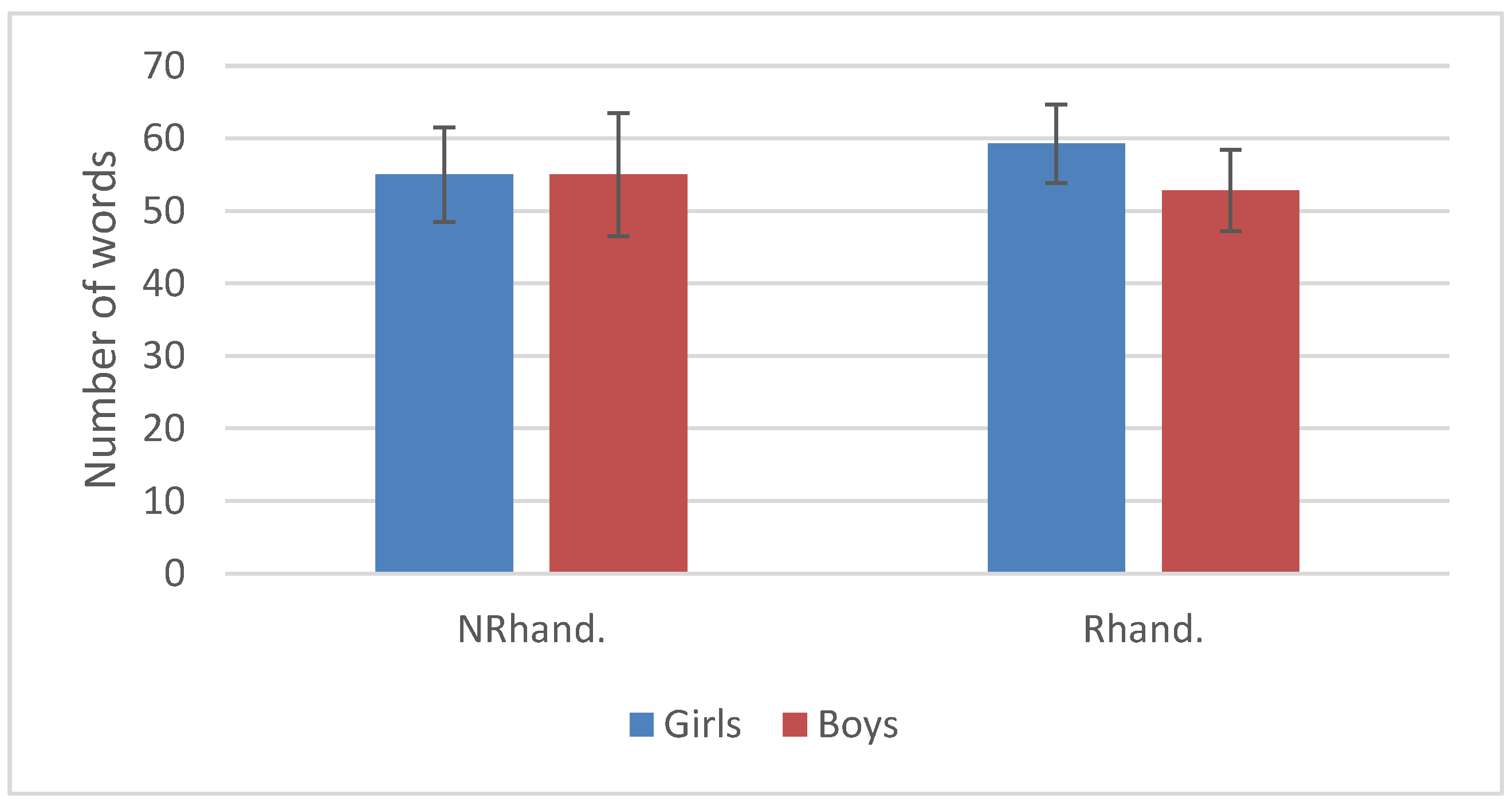
3.2. Language Development

The questionnaire was filled out by the parents of 48 infants (25 girls and 23 boys; the parents of 2 girls did not fill out the questionnaire). The mean number of words understood (understood only + understood and produced) was 55.7 (from 28 to 75, SD = 12.8). An ANCOVA with the number of words as the dependent variable as a function of the category of handedness and sex (independent factors) showed that there was no significant effect. There was no significant difference between the number of words in right-handers’ vocabulary (mean = 56.4, SD = 12.3) and the number of words in non-right-handers’ vocabulary (mean = 55, SD = 13.7) (p = 0.791), as well as no significant sex differences (p = 0.332) and no significant sex × category of handedness interaction (p = 0.413) (Figure 1). Not surprisingly, the correlation between HI and the number of words was very low (r = 0.04) and not significant (p = 0.717). Finally, the model R2 was very low (0.0178), showing that HI explained only 1.78% of the variance in the number of words.

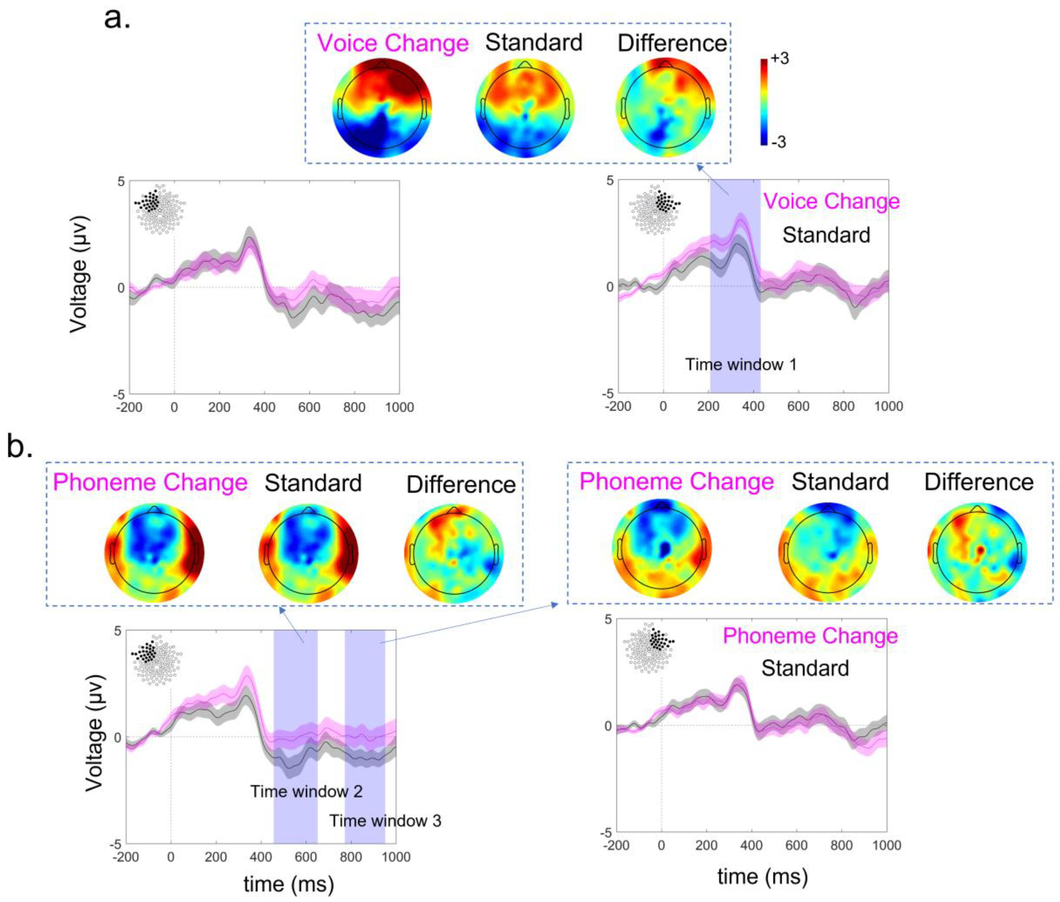
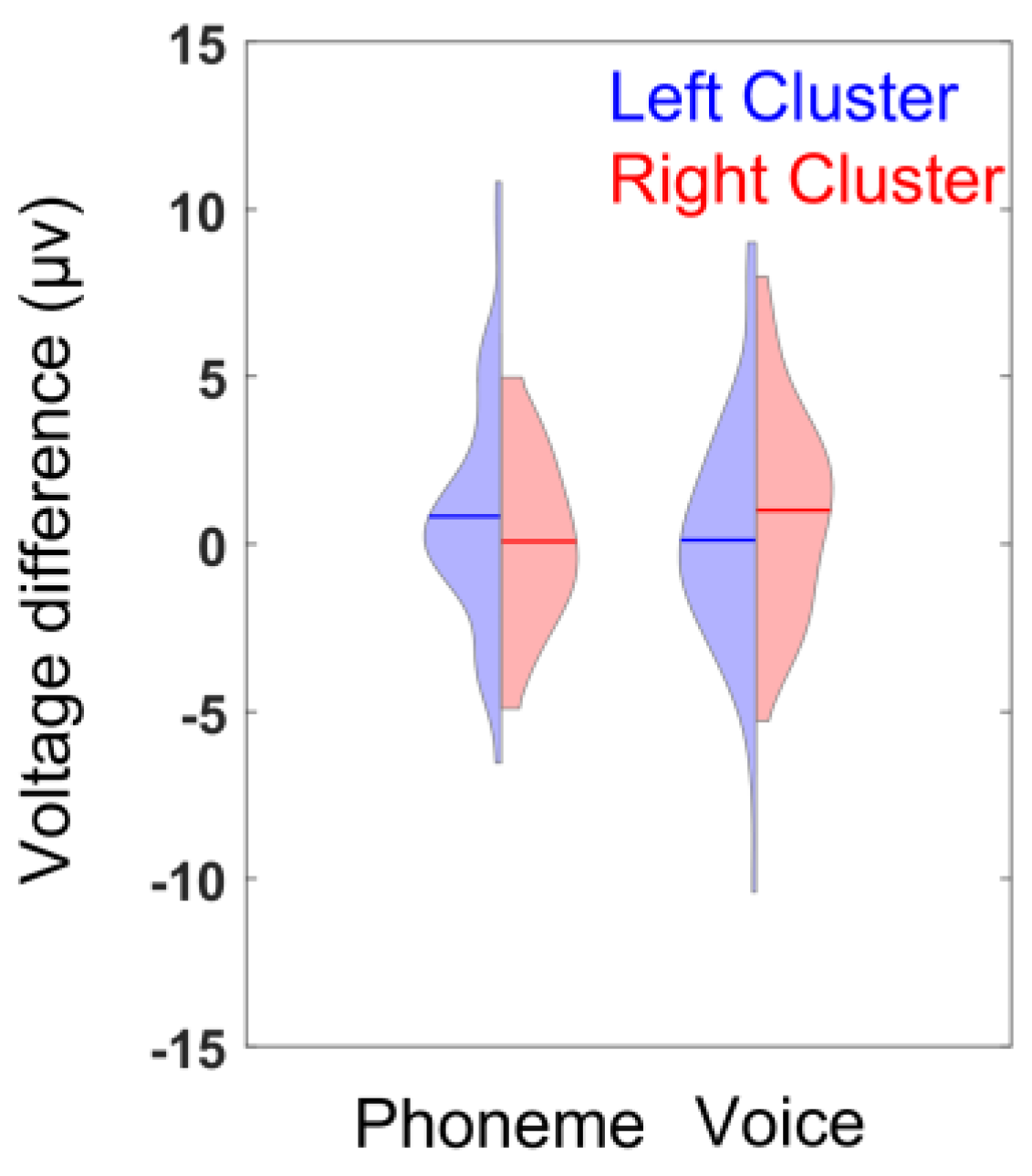
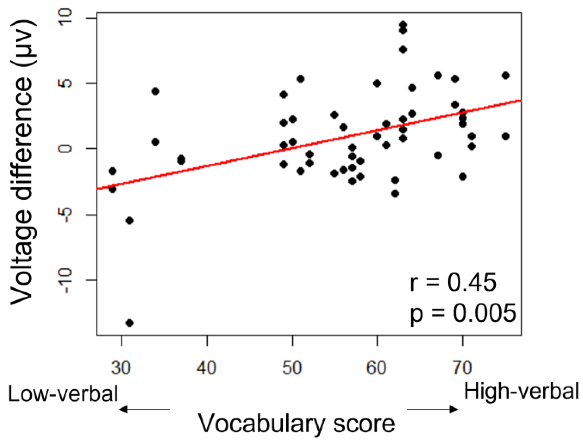
3.3. ERP Lateralization

The nonparametric cluster-mass permutation test revealed three time windows of interest: T1 = [212–436 ms], T2 = [460–656 ms] and T3 = [776–956 ms]. The ERPs over the right (R) cluster differed with the change in voice (pR-T1 = 0.039) during T1 and tended to differ over the left (L) cluster with the change in phoneme during T2 and T3 (pL-T2 = 0.089 and pL-T3 = 0.101 respectively). Although the statistical significance for T2 and T3 was marginal, we kept this partitioning on the three time windows because of their similar length and because they covered the 200 ms−1 s post-stimulus period (Figure 2).
ANCOVAs were performed over the three identified time windows independently.
Within T1 (212–436 ms), the only significant effect was a two-way interaction for condition × cluster (F(1,42) = 6.5, p = 0.015). No other main effect or interaction was significant (Table 2). The post hoc analyses performed with paired t-tests revealed significant voltage differences between the left vs. right cluster in each condition: phonetic change: p = 0.048, due to a larger difference in the left than right cluster; voice change: p = 0.019, due to a larger difference in the right than left cluster. As for the within cluster analyses, the voltage difference tended to be larger in the condition of phoneme than voice change in the left cluster, p = 0.060, and was significantly larger in the condition of voice than phoneme change in the right one, p = 0.015 (Figure 3).
Within T2 (460–656 ms), the two-way interaction for handedness × vocabulary (F(1,42) = 5.3, p = 0.026), as well as the three-way interaction for handedness × vocabulary × condition (F(1,42) = 4.5, p = 0.039) were significant. No other main effect or interaction was significant (Table 3). For post hoc analyses, we considered HI categories and performed regression analyses that revealed a correlation (r = 0.45, p = 0.005) between voice discrimination responses (higher voltage differences for voice change − standard) and vocabulary scores only in right-handers (Figure 4).
Within T3 (776–956 ms), a main effect of vocabulary (F(1,42) = 4.7, p = 0.035) indicated a correlation (r = 0.16, p = 0.029) between general discrimination responses (higher voltage differences for all deviants − standards) and vocabulary scores (Figure 5). In addition to this main effect, the two-way interaction for handedness × cluster was significant (F(1,42) = 4.5, p = 0.041) (Table 4). The post hoc analysis revealed a correlation between the general discrimination responses (higher voltage differences for all deviants − standards) over the right but not the left hemisphere and the handedness index (r = 0.24, p = 0.020) (Figure 6).

4. Discussion

The main goal of our study was to evaluate whether the cerebral bases of language lateralization are related to hand preference in infants at 18 months. To assess hemispheric lateralization for speech perception, we recorded ERPs during a syllable-discrimination paradigm and measured brain response lateralization to a change in phoneme or voice. The nonparametric cluster-mass permutation test highlighted three time windows post-stimulus where the response to a change in phoneme or voice was identified. The response to a change in voice was right-lateralized, whereas the response to a change in phoneme was left-lateralized, significantly in the earliest time window. Thus, according to our results, at 18 months of age, ERP asymmetries for phoneme and voice processing are clear with lateralization to the left and right hemispheres, respectively.
Lateralized ERP responses in 18-month-olds are consistent with previous reports on differential lateralized processing of speech vs. non-speech stimuli in younger infants. Newborns and preterm neonates already demonstrate left-lateralized responses to speech stimuli, which were observed using NIRS [32,42,65], particularly for native speech stimuli [66], and using optical topography [67] as well as other techniques (see [68] for a review; however, see [69] for different results). Contrary to speech stimuli, non-speech auditory stimulation elicits right-lateralized responses in newborns using auditory-evoked potentials [70] or NIRS [71]. In older infants, non-linguistic aspects of speech stimuli, such as prosody, also evoke right-dominant activations using fNIRS [72] (however, for slightly different results, see [73]). Together, these studies highlight a pre-specialization in the left and right hemispheres for phonological and pitch (i.e., voice) processing, which seems to remain stable throughout the second year of life, as we observed here with ERPs (e.g., [3,4,5,6], and see [2] for a meta-analysis).
Although the links remain to be established, this early functional lateralization might be indirectly related to structural asymmetries that appear early in development. In particular, the Sylvian fissure and perisylvian regions show strong asymmetries in morphology from birth [74]. The superior temporal sulcus folds earlier in the right hemisphere than in the left hemisphere as observed in fetuses, preterm and full-term neonates and infants [75,76,77,78], and this asymmetry in-depth persists throughout development [79]. The maturation of the perisylvian regions also progresses differently between the two cerebral hemispheres [80,81]. Finally, the connections between these regions are also asymmetrical early on, particularly the arcuate fasciculus that is more developed in the left hemisphere than in the right hemisphere in both infants and adults [82,83]. These early asymmetries are accompanied by differences in processing times for speech stimuli between the two hemispheres, as observed using ERPs in typical infants but not in infants with agenesis of the corpus callosum in whom hemispheric lateralization may be impacted [60,84]. The combination of these early anatomo-functional asymmetries within the perisylvian network could underlie the emergence of functional lateralization for the processing of speech and non-speech stimuli, as reported here.
Since the main goal of our study was to compare this early development of language asymmetry with that of handedness, we included a test of handedness. We observed that, as a group, the infants tended to use their right hand more than their left hand during the 15-item grasping task. The majority of the infants were right-handed (64.6%), whereas 16.6% were left-handed and 18.8% were not lateralized. This is consistent with many studies showing that there is a majority of right-handed grasping at the group level as soon as grasping develops [23,24,25,26,27], and that, after a decrease in individual fluctuations [28,29], the majority of infants reach a rather stable handedness pattern by 18 months of age [27,30].
The main question here was whether brain asymmetry for speech perception and asymmetry in hand use for grasping were related. We hypothesized that if infants with hemispheric asymmetry in ERPs during syllable discrimination also show a high handedness index in grasping tasks, then this association will support the assumption that common factors drive the development of both asymmetries. On the contrary, if there is no link between the development of hemispheric asymmetry in ERPs and handedness, this will support the assumption that independent factors drive the development of both asymmetries. Although we found a significant interaction between cluster and condition in the first time window, handedness did not interact with cluster or condition, neither in the first time window nor in the other two time windows. Thus, we observed no link between the development of hemispheric asymmetry in ERPs and the degree of right-handedness at 18 months. These results argue in favor of independent ontogenetic influences for driving the development of both asymmetries 12. In addition, this is in line with adult studies showing no link or almost no link between the degree of right-handedness and hemispheric asymmetry for language [3,4].
We found an effect of handedness on the level of voltage difference during the third time window, but only in the right hemisphere and for all conditions. Even though both asymmetries do not seem to be related at 18 months with our experimental set-up, the higher the HI, the higher the discriminative response between standard and deviant stimuli in the right hemisphere.
If these results hold true in future replication studies with different methodologies, one might conclude that early manual asymmetries and language lateralization are determined by independent factors. Interestingly, a recent neuroimaging study highlighted that the morphology of the central sulcus, delineating the sensory and motor regions, shows strong asymmetries early on in preterm infants and that its shape characteristics at term-equivalent age tend to predict later handedness at 5 years of age [74], in a similar way as in adults [85].
A secondary question was to what extent vocabulary range is associated with the direction and degree of both asymmetries. At the behavioral level, i.e., concerning manual asymmetry, we did not find the same relationship between the range of vocabulary (number of words) and category of handedness as found in Nelson’s study [49] or in other studies comparing handedness for pointing and vocabulary [46,52]. This might be due to methodological differences. For instance, in Nelson’s study, the infants were followed from 6 to 24 months, whereas in our study handedness was tested at 18 months, which might have blurred the differences between early-lateralized and late-lateralized infants.
Concerning language asymmetry, we did not find a specific effect of vocabulary range on phoneme discrimination in the left hemisphere. However, we found a significant main effect of vocabulary in the third time window. This means that in both conditions and in both hemispheres, the greater the vocabulary range, the higher the voltage difference between standard and deviant. This late time window (>750 ms) might reflect orientation to novelty following any change detection. To our knowledge, no studies have investigated the correlation between ERP asymmetry for speech perception and the range of vocabulary in infants and toddlers so far, but a few studies have explored related questions. First, a group of researchers described significant differences in lateralized patterns of event-related EEG oscillations in response to rapid successive auditory stimuli in 6-month-old infants with a familial risk for language and learning impairment compared to 6-month-old infants without a familial risk for such an impairment [86]. In addition, differences in ERPs are predictive of expressive vocabulary at 20 months of age [87]. This suggests that neural mechanisms that support rapid auditory processing in infancy interplay with the development of vocabulary. In another study, kindergarten children at risk for dyslexia were tested using a speech–syllable mismatch negativity paradigm (MMN) and received two behavioral assessments important for language skills: phonological awareness (PA) and rapid automatized naming (RAN). The results showed that late MMN amplitude was significantly greater in children with typical PA ability than in children with low PA ability and that laterality in the early and late MMN was significantly different in children with low versus typical RAN ability. This suggests that, in kindergarten children, the amplitude of the MMN may relate to phonological representations and that asymmetry in the MMN may relate to the ability to manipulate them [88]. In addition, relationships were observed between the early organization of functional [89] and structural connectivity [90] within the left hemisphere and later language skills.
In addition, one needs to consider that a link between vocabulary range and brain lateralization might exist for other measures of brain asymmetry (e.g., structural characteristics) that were not tested in our paradigm, as previously suggested [91,92,93]. The work of Aeby and collaborators [91], for instance, suggested a relationship between structural measures of asymmetry in neonates and language development at two years of age.
Of note, we observed a significant interaction between handedness, vocabulary range and condition in the second time window. The higher the HI and the larger the vocabulary range, the higher the voltage difference between the standard and deviant voice. Thus, for voice only, and for both hemispheres, handedness and vocabulary influenced voice discrimination. Therefore, the only effect found for handedness and vocabulary together was not on phoneme discrimination but on voice discrimination, and it was not a lateralized effect. These results are nevertheless interesting to compare with the results of a previous EEG study, which showed that right-handers with greater vocabulary size had a more pronounced N400 effect over the right hemisphere than non-lateralized infants at 18 months but not at 24 months [94]. It would be interesting to continue the present study with other age groups.
In conclusion, from our results, it appears that brain asymmetries in speech and non-speech processing—the left hemisphere for phoneme change and the right hemisphere for voice change—are related neither to handedness nor to range of vocabulary; however, handedness and range of vocabulary together appear to influence the level of brain responses for voice change detection. To explain this absence of a relationship between handedness and cerebral lateralization for speech, one could quote Provins [95] in saying that “although the development of a cerebral lateralization for speech and handedness is dependent on both genetic and environmental factors, the specific role of inborn and postnatal influences is very different”. It might be that a more challenging task for testing manual handedness (such as tool use) could have evidenced a greater relationship between handedness and cerebral lateralization for speech, as found in adult (e.g., [8]) studies. Another limitation of our study might be that handedness was tested on a production task, whereas the language lateralization condition was a comprehension-oriented situation.

Author Contributions

Conceptualization, J.F.; methodology, J.F., J.D., G.D.-L. and P.R.; software, P.A., P.R. and D.P.; validation, J.D., G.D.-L. and P.R.; formal analysis, J.F., P.A. and J.D.; investigation, D.P., E.S. and C.G.; resources, J.F. and J.D.; data curation, P.A., J.F, J.D. and P.R.; writing—original draft preparation, J.F. and D.P.; writing—review and editing, J.F., J.D., P.A., E.S. and G.D.-L.; visualization, P.A., J.D. and J.F.; supervision, J.F.; project administration, J.F.; funding acquisition, J.F. and J.D. All authors have read and agreed to the published version of the manuscript.

Funding

This research was funded by ANR Agence Nationale de Recherche, grant number ANR-13-BSH2–0007-01. The researchers were supported by the IdEx Université de Paris (ANR-18-IDEX-0001), Fondation Médisite (FdF-2018-00092867), Fondation de France (FdF-2012-00033702), ANR Agence Nationale de Recherche (ANR-20-CE17-0014-03).

Institutional Review Board Statement

The research was approved by the Institutional Review Board of “CER U-Paris Cité” (Identifier: 2013-41) on 10 December 2013.

Informed Consent Statement

Informed consent was obtained from all subjects involved in the study.

Data Availability Statement

The data presented in this study are available on request from the corresponding author. The data are not publicly available due to privacy.

Conflicts of Interest

The authors declare no conflict of interest.

References

  1. Bryden, M.; Hécaen, H.; DeAgostini, M. Patterns of cerebral organization. Brain Lang. 1983, 20, 249–262. [Google Scholar] [CrossRef] [PubMed]
  2. Carey, D.P.; Johnstone, L.T. Quantifying cerebral asymmetries for language in dextrals and adextrals with random-effects meta analysis. Front. Psychol. 2014, 5, 1128. [Google Scholar] [CrossRef] [PubMed]
  3. Springer, J.A.; Binder, J.R.; Hammeke, T.A.; Swanson, S.J.; Frost, J.A.; Bellgowan, P.S.; Brewer, C.C.; Perry, H.M.; Morris, G.L.; Mueller, W.M. Language dominance in neurologically normal and epilepsy subjects: A functional MRI study. Brain A J. Neurol. 1999, 122, 2033–2046. [Google Scholar] [CrossRef] [PubMed]
  4. Knecht, S.; Dräger, B.; Deppe, M.; Bobe, L.; Lohmann, H.; Flöel, A.; Ringelstein, E.-B.; Henningsen, H. Handedness and hemispheric language dominance in healthy humans. Brain 2000, 123, 2512–2518. [Google Scholar] [CrossRef] [PubMed]
  5. Pujol, J.; Deus, J.; Losilla, J.M.; Capdevila, A. Cerebral lateralization of language in normal left-handed people studied by functional MRI. Neurology 1999, 52, 1038. [Google Scholar] [CrossRef]
  6. Szaflarski, J.P.; Binder, J.R.; Possing, E.T.; McKiernan, K.A.; Ward, B.D.; Hammeke, T.A. Language lateralization in left-handed and ambidextrous people: FMRI data. Neurology 2002, 59, 238–244. [Google Scholar] [CrossRef]
  7. Fagard, J.; Chapelain, A.; Bonnet, P. How should “ambidexterity” be estimated? Laterality 2015, 20, 543–570. [Google Scholar] [CrossRef]
  8. Higuchi, S.; Chaminade, T.; Imamizu, H.; Kawato, M. Shared neural correlates for language and tool use in Broca’s area. Neuroreport 2009, 20, 1376–1381. [Google Scholar] [CrossRef]
  9. Greenfield, P.M.; Nelson, K.; Saltzman, E. The development of rulebound strategies for manipulating seriated cups: A parallel between action and grammar. Cogn. Psychol. 1972, 3, 291–310. [Google Scholar] [CrossRef]
  10. Packheiser, J.; Schmitz, J.; Arning, L.; Beste, C.; Gunturkun, O.; Ocklenburg, S. A large-scale estimate on the relationship between language and motor lateralization. Sci. Rep. 2020, 10, 13027. [Google Scholar] [CrossRef]
  11. Francks, C.; Maegawa, S.; Laurén, J.; Abrahams, B.S.; Velayos-Baeza, A.; Medland, S.; Colella, S.; Groszer, M.; McAuley, E.Z.; Caffrey, T.M.; et al. LRRTM1 on chromosome 2p12 is a maternally suppressed gene that is associated paternally with handedness and schizophrenia. Mol. Psychiatry 2007, 12, 1129–1139. [Google Scholar] [CrossRef] [PubMed]
  12. Ocklenburg, S.; Beste, C.; Güntürkün, O. Handedness: A neurogenetic shift of perspective. Neurosci. Biobehav. Rev. 2013, 37, 2788–2793. [Google Scholar] [CrossRef] [PubMed]
  13. Armour, J.A.L.; Davison, A.; McManus, I. Genome-wide association study of handedness excludes simple genetic models. Heredity 2014, 112, 221–225. [Google Scholar] [CrossRef] [PubMed]
  14. McManus, I.C.; Davison, A.; Armour, J.A.L. Multilocus genetic models of handedness closely resemble single-locus models in explaining family data and are compatible with genome-wide association studies. Ann. N. Y. Acad. Sci. 2013, 1288, 48–58. [Google Scholar] [CrossRef] [PubMed]
  15. Hepper, P.G.; Shahidullah, S.; White, R. Handedness in the human fetus. Neuropsychologia 1991, 29, 1107–1111. [Google Scholar] [CrossRef] [PubMed]
  16. McCartney, G.; Hepper, P. Development of lateralized behaviour in the human fetus from 12 to 27 weeks’ gestation. Dev. Med. Child Neurol. 1999, 41, 83–86. [Google Scholar] [CrossRef]
  17. Kurjak, A.; Vecek, N.; Hafner, T.; Bozek, T.; Funduk-Kurjak, B.; Ujevic, B. Prenatal diagnosis: What does four-dimensional ultrasound add? J. Perinat. Med. 2002, 30, 57–62. [Google Scholar] [CrossRef]
  18. de Vries, J.; Wimmers, R.; Ververs, I.; Hopkins, B.; Savelsbergh, G.; van Geijn, H. Fetal handedness and head position preference: A developmental study. Dev. Psychobiol. 2001, 39, 171–178. [Google Scholar] [CrossRef]
  19. Myowa-Yamakoshi, M.; Takeshita, H. Do Human Fetuses Anticipate Self-Oriented Actions? A Study by Four-Dimensional (4D) Ultrasonography. Infancy 2006, 10, 289–301. [Google Scholar] [CrossRef]
  20. Ververs, I.A.; de Vries, J.I.; van Geijn, H.P.; Hopkins, B. Prenatal head position from 12-38 weeks. weeks. I. Developmental aspects. Early Hum. Dev. 1994, 39, 83–91. [Google Scholar] [CrossRef]
  21. Van der Meer, A.L. Keeping the arm in the limelight: Advanced visual control of arm movements in neonates. Eur. J. Paediatr. Neurol. 1997, 4, 103–108. [Google Scholar] [CrossRef] [PubMed]
  22. Michel, G.F.; Harkins, D.A. Postural and lateral asymmetries in the ontogeny of handedness during infancy. Dev. Psychobiol. 1986, 19, 247–258. [Google Scholar] [CrossRef] [PubMed]
  23. Corbetta, D.; Bojczyk, K.E. Infants Return to Two-Handed Reaching When They Are Learning to Walk. J. Mot. Behav. 2002, 34, 83–95. [Google Scholar] [CrossRef] [PubMed]
  24. Michel, G.F.; Tyler, A.N.; Ferre, C.; Sheu, C.-F. The manifestation of infant hand-use preferences when reaching for objects during the seven- to thirteen-month age period. Dev. Psychobiol. 2006, 48, 436–443. [Google Scholar] [CrossRef]
  25. Nelson, E.L.; Campbell, J.M.; Michel, G.F. Unimanual to bimanual: Tracking the development of handedness from 6 to 24 months. Infant Behav. Dev. 2013, 36, 181–188. [Google Scholar] [CrossRef]
  26. Fagard, J. The nature and nurture of human infant hand preference. Ann. N. Y. Acad. Sci. 2013, 1288, 114–123. [Google Scholar] [CrossRef]
  27. Ferre, C.L.; Babik, I.; Michel, G.F. Development of infant prehension handedness: A longitudinal analysis during the 6- to 14-month age period. Infant Behav. Dev. 2010, 33, 492–502. [Google Scholar] [CrossRef]
  28. Fagard, J.; Margules, S.; Lopez, C.; Granjon, L.; Huet, V. How should we test infant handedness? Laterality 2016, 22, 294–312. [Google Scholar] [CrossRef]
  29. Michel, G.F.; Babik, I.; Sheu, C.-F.; Campbell, J.M. Latent classes in the developmental trajectories of infant handedness. Dev. Psychol. 2014, 50, 349–359. [Google Scholar] [CrossRef]
  30. Jacquet, A.-Y.; Esseily, R.; Rider, D.; Fagard, J. Handedness for grasping objects and declarative pointing: A longitudinal study. Dev. Psychobiol. 2012, 54, 36–46. [Google Scholar] [CrossRef]
  31. Nelson, E.L.; Gonzalez, S.L. Measuring infant handedness reliably from reaching: A systematic review. Laterality 2020, 25, 430–454. [Google Scholar] [CrossRef] [PubMed]
  32. Mahmoudzadeh, M.; Dehaene-Lambertz, G.; Fournier, M.; Kongolo, G.; Goudjil, S.; Dubois, J.; Grebe, R.; Wallois, F. Syllabic discrimination in premature human infants prior to complete formation of cortical layers. Proc. Natl. Acad. Sci. USA. 2013, 110, 4846–4851. [Google Scholar] [CrossRef] [PubMed]
  33. Dehaene-Lambertz, G.; Spelke, E. The Infancy of the Human Brain. Neuron 2015, 88, 93–109. [Google Scholar] [CrossRef]
  34. Gervain, J.; Macagno, F.; Cogoi, S.; Pena, M.; Mehler, J. The neonate brain detects speech structure. Proc. Natl. Acad. Sci. USA. 2008, 105, 14222–14227. [Google Scholar] [CrossRef] [PubMed]
  35. Becker, A.B.; Schild, U.; Friedrich, C.K. ERP correlates of word onset priming in infants and young children. Dev. Cogn. Neurosci. 2014, 9, 44–55. [Google Scholar] [CrossRef] [PubMed]
  36. Arimitsu, T.; Uchida-Ota, M.; Yagihashi, T.; Kojima, S.; Watanabe, S.; Hokuto, I.; Ikeda, K.; Takahashi, T.; Minagawa-Kawai, Y. Functional Hemispheric Specialization in Processing Phonemic and Prosodic Auditory Changes in Neonates. Front. Psychol. 2011, 2, 202. [Google Scholar] [CrossRef]
  37. Bertoncini, J.; Morais, J.; Bijeljac-Babic, R.; McAdams, S.; Peretz, I.; Mehler, J. Dichotic perception and laterality in neonates. Brain Lang. 1989, 37, 591–605. [Google Scholar] [CrossRef]
  38. DeCasper, A.J.; Prescott, P. Lateralized processes constrain auditory reinforcement in human newborns. Hear. Res. 2009, 255, 135–141. [Google Scholar] [CrossRef]
  39. Shultz, S.; Vouloumanos, A.; Bennett, R.H.; Pelphrey, K. Neural specialization for speech in the first months of life. Dev. Sci. 2014, 17, 766–774. [Google Scholar] [CrossRef]
  40. Perani, D.; Saccuman, M.C.; Scifo, P.; Anwander, A.; Spada, D.; Baldoli, C.; Poloniato, A.; Lohmann, G.; Friederici, A.D. Neural language networks at birth. Proc. Natl. Acad. Sci. USA 2011, 108, 16056–16061. [Google Scholar] [CrossRef]
  41. Dehaene-Lambertz, G.; Montavont, A.; Jobert, A.; Allirol, L.; Dubois, J.; Hertz-Pannier, L.; Dehaene, S. Language or music, mother or Mozart? Structural and environmental influences on infants’ language networks. Brain Lang. 2009, 114, 53–65. [Google Scholar] [CrossRef] [PubMed]
  42. Bouchon, C.; Nazzi, T.; Gervain, J. Hemispheric Asymmetries in Repetition Enhancement and Suppression Effects in the Newborn Brain. PLoS ONE 2015, 10, e0140160. [Google Scholar] [CrossRef] [PubMed]
  43. Cristia, A.; Minagawa-Kawai, Y.; Egorova, N.; Gervain, J.; Filippin, L.; Cabrol, D.; Dupoux, E. Neural correlates of infant accent discrimination: An fNIRS study. Dev. Sci. 2014, 17, 628–635. [Google Scholar] [CrossRef] [PubMed]
  44. Minagawa, Y.; Naoi, N.; Nishijima, N.; Kojima, S.; Dupoux, E. Developmental changes in cerebral responses to native and non-native vowels: A NIRS Study. In Proceedings of the ICPhS XVI Symposium, Saarbrucken, Germany, 6–10 August 2007. ID 1487. [Google Scholar]
  45. Bisiacchi, P.; Cainelli, E. Structural and functional brain asymmetries in the early phases of life: A scoping review. Anat. Embryol. 2022, 227, 479–496. [Google Scholar] [CrossRef]
  46. Esseily, R.; Jacquet, A.-Y.; Fagard, J. Handedness for grasping objects and pointing and the development of language in 14-month-old infants. Laterality 2011, 16, 565–585. [Google Scholar] [CrossRef] [PubMed]
  47. Kühn-Popp, N.; Kristen, S.; Paulus, M.; Meinhardt, J.; Sodian, B. Left hemisphere EEG coherence in infancy predicts infant declarative pointing and preschool epistemic language. Soc. Neurosci. 2016, 11, 49–59. [Google Scholar] [CrossRef]
  48. Kohler, M.; Keage, H.; Spooner, R.; Flitton, A.; Hofmann, J.; Churches, O.; Elliott, S.; Badcock, N. Variability in lateralised blood flow response to language is associated with language development in children aged 1–5 years. Brain Lang. 2015, 145-146, 34–41. [Google Scholar] [CrossRef] [PubMed]
  49. Nelson, E.L.; Campbell, J.M.; Michel, G.F. Early handedness in infancy predicts language ability in toddlers. Dev. Psychol. 2014, 50, 809–814. [Google Scholar] [CrossRef]
  50. Hodgson, J.C.; Hirst, R.; Hudson, J.M. Hemispheric speech lateralisation in the developing brain is related to motor praxis ability. Dev. Cogn. Neurosci. 2016, 22, 9–17. [Google Scholar] [CrossRef]
  51. Goldfield, B.A.; Reznick, J.S. Early lexical acquisition: Rate, content, and the vocabulary spurt. J. Child Lang. 1990, 17, 171–183. [Google Scholar] [CrossRef]
  52. Cochet, H.; Jover, M.; Vauclair, J. Hand preference for pointing gestures and bimanual manipulation around the vocabulary spurt period. J. Exp. Child Psychol. 2011, 110, 393–407. [Google Scholar] [CrossRef] [PubMed]
  53. Ganger, J.; Brent, M.R. Reexamining the Vocabulary Spurt. Dev. Psychol. 2004, 40, 621–632. [Google Scholar] [CrossRef] [PubMed]
  54. Somogyi, E.; Salomon, L.; Fagard, J. Hand movements in communicative and non-communicative situations in very young infants: A preliminary study. J. Mot. Learn. Dev. 2021, 9, 132–152. [Google Scholar] [CrossRef]
  55. Dehaene-Lambertz, G.; Dehaene, S. Speed and cerebral correlates of syllable discrimination in infants. Nature 1994, 370, 292–295. [Google Scholar] [CrossRef] [PubMed]
  56. Fagard, J.; Corbetta, D.; Somogyi, E.; Safar, A.; Bernard, C. Right-handed one day, right-handed the next day? Laterality 2020, 1–14. [Google Scholar] [CrossRef]
  57. Fenson, L.; Dale, P.S.; Reznick, J.S.; Thal, D.; Bates, E.; Hartung, J.P.; Pethick, S.; Reilly, J.S. The MacArthur Development Inventories: User’s Guide and Technical Manual; Singular Publishing Group: San Diego, CA, USA, 1993. [Google Scholar]
  58. Bovet, F.; Danjou, G.; Langue, J.; Moretto, M.; Tockert, E.; Kern, S. Un nouvel outil d’évaluation du développement communicatif du nourrisson. Médecine Enfance 2005, 25, 67–74. [Google Scholar]
  59. Delorme, A.; Makeig, S. EEGLAB: An Open Source Toolbox for Analysis of Single-Trial EEG Dynamics Including Independent Component Analysis. J. Neurosci. Methods 2004, 134, 9–21. [Google Scholar] [CrossRef]
  60. Adibpour, P.; Lebenberg, J.; Kabdebon, C.; Dehaene-Lambertz, G.; Dubois, J. Anatomo-functional correlates of auditory development in infancy. Dev. Cogn. Neurosci. 2020, 42, 100752. [Google Scholar] [CrossRef]
  61. Bristow, D.; Dehaene-Lambertz, G.; Mattout, J.; Soares, C.; Gliga, T.; Baillet, S.; Mangin, J.-F. Hearing Faces: How the Infant Brain Matches the Face It Sees with the Speech It Hears. J. Cogn. Neurosci. 2009, 21, 905–921. [Google Scholar] [CrossRef]
  62. Dehaene-Lambertz, G. Cerebral Specialization for Speech and Non-Speech Stimuli in Infants. J. Cogn. Neurosci. 2000, 12, 449–460. [Google Scholar] [CrossRef]
  63. Wunderlich, J.L.; Cone-Wesson, B.K.; Shepherd, R. Maturation of the cortical auditory evoked potential in infants and young children. Hear. Res. 2006, 212, 185–202. [Google Scholar] [CrossRef] [PubMed]
  64. Maris, E.; Oostenveld, R. Nonparametric statistical testing of EEG- and MEG-data. J. Neurosci. Methods 2007, 164, 177–190. [Google Scholar] [CrossRef] [PubMed]
  65. Kotilahti, K.; Nissilä, I.; Näsi, T.; Lipiäinen, L.; Noponen, T.; Meriläinen, P.; Huotilainen, M.; Fellman, V. Hemodynamic responses to speech and music in newborn infants. Hum. Brain Mapp. 2009, 31, 595–603. [Google Scholar] [CrossRef] [PubMed]
  66. Vannasing, P.; Florea, O.; González-Frankenberger, B.; Tremblay, J.; Paquette, N.; Safi, D.; Wallois, F.; Lepore, F.; Béland, R.; Lassonde, M.; et al. Distinct hemispheric specializations for native and non-native languages in one-day-old newborns identified by fNIRS. Neuropsychologia 2016, 84, 63–69. [Google Scholar] [CrossRef]
  67. Pena, M.; Maki, A.; Kovacic, D.; Dehaene-Lambertz, G.; Koizumi, H.; Bouquet, F.; Mehler, J. Sounds and silence: An optical topography study of language recognition at birth. Proc. Natl. Acad. Sci. USA 2003, 100, 11702–11705. [Google Scholar] [CrossRef]
  68. Bisiacchi, P.; Marzi, C.; Nicoletti, R.; Carena, G.; Mucignat, C.; Tomaiuolo, F. Left-right asymmetry of callosal transfer in normal human subjects. Behav. Brain Res. 1994, 64, 173–178. [Google Scholar] [CrossRef]
  69. Zhang, F.; Gervain, J.; Roeyers, H. Developmental changes in the brain response to speech during the first year of life: A near-infrared spectroscopy study of dutch-learning infants. Infant Behav. Dev. 2022, 67, 101724. [Google Scholar] [CrossRef]
  70. Molfese, D.L.; Freeman, R.B.; Palermo, D.S. The ontogeny of brain lateralization for speech and nonspeech stimuli. Brain Lang. 1975, 2, 356–368. [Google Scholar] [CrossRef]
  71. Telkemeyer, S.; Rossi, S.; Koch, S.P.; Nierhaus, T.; Steinbrink, J.; Poeppel, D.; Obrig, H.; Wartenburger, I. Sensitivity of newborn auditory cortex to the temporal structure of sounds. J. Neurosci. 2009, 29, 14726–14733. [Google Scholar] [CrossRef]
  72. Arimitsu, T.; Minagawa, Y.; Yagihashi, T.; Uchida, M.O.; Matsuzaki, A.; Ikeda, K.; Takahashi, T. The cerebral hemodynamic response to phonetic changes of speech in preterm and term infants: The impact of postmenstrual age. NeuroImage Clin. 2018, 19, 599–606. [Google Scholar] [CrossRef]
  73. Minagawa-Kawai, Y.; Cristià, A.; Vendelin, I.; Cabrol, D.; Dupoux, E. Assessing Signal-Driven Mechanisms in Neonates: Brain Responses to Temporally and Spectrally Different Sounds. Front. Psychol. 2011, 2, 135. [Google Scholar] [CrossRef] [PubMed]
  74. De Vareilles, H.; Rivière, D.; Pascucci, M.; Sun, Z.-Y.; Fischer, C.; Leroy, F.; Tataranno, M.-L.; Benders, M.J.; Dubois, J.; Mangin, J.-F. Exploring the emergence of morphological asymmetries around the brain’s Sylvian fissure: A longitudinal study of shape variability in preterm infants. Cereb. Cortex 2023, bhac533. [Google Scholar] [CrossRef] [PubMed]
  75. Dubois, J.; Benders, M.; Cachia, A.; Lazeyras, F.; Leuchter, R.H.-V.; Sizonenko, S.V.; Borradori-Tolsa, C.; Mangin, J.F.; Huppi, P.S. Mapping the Early Cortical Folding Process in the Preterm Newborn Brain. Cereb. Cortex 2008, 18, 1444–1454. [Google Scholar] [CrossRef]
  76. Habas, P.A.; Scott, J.A.; Roosta, A.; Rajagopalan, V.; Kim, K.; Rousseau, F.; Barkovich, A.J.; Glenn, O.A.; Studholme, C. Early Folding Patterns and Asymmetries of the Normal Human Brain Detected from in Utero MRI. Cereb. Cortex 2012, 22, 13–25. [Google Scholar] [CrossRef] [PubMed]
  77. Hill, J.; Dierker, D.; Neil, J.; Inder, T.; Knutsen, A.; Harwell, J.; Coalson, T.; Van Essen, D. A Surface-Based Analysis of Hemispheric Asymmetries and Folding of Cerebral Cortex in Term-Born Human Infants. J. Neurosci. 2010, 30, 2268–2276. [Google Scholar] [CrossRef]
  78. Glasel, H.; Leroy, F.; Dubois, J.; Hertz-Pannier, L.; Mangin, J.; Dehaene-Lambertz, G. A robust cerebral asymmetry in the infant brain: The rightward superior temporal sulcus. Neuroimage 2011, 58, 716–723. [Google Scholar] [CrossRef]
  79. Leroy, F.; Cai, Q.; Bogart, S.L.; Dubois, J.; Coulon, O.; Monzalvo, K.; Fischer, C.; Glasel, H.; Van der Haegen, L.; Benezit, A.; et al. New human-specific brain landmark: The depth asymmetry of superior temporal sulcus. Proc. Natl. Acad. Sci. USA. 2015, 112, 1208–1213. [Google Scholar] [CrossRef]
  80. Leroy, F.; Glasel, H.; Dubois, J.; Hertz-Pannier, L.; Thirion, B.; Mangin, J.F.; Dehaene-Lambertz, G. Early maturation of the linguistic dorsal pathway in human infants. J. Neurosci. 2011, 31, 1500–1506. [Google Scholar] [CrossRef]
  81. Rolland, C.; Lebenberg, J.; Leroy, F.; Moulton, E.; Adibpour, P.; Riviere, D.; Poupon, C.; Hertz-Pannier, L.; Mangin, J.-F.; Dehaene-Lambertz, G.; et al. Exploring Microstructure Asymmetries in the Infant Brain Cortex: A Methodological Framework Combining Structural and Diffusion Mri. In Proceedings of the 019 IEEE 16th International Symposium on Biomedical Imaging (ISBI 2019), Venice, Italy, 8–11 April 2019; pp. 426–429. [Google Scholar]
  82. Dubois, J.; Hertz-Pannier, L.; Cachia, A.; Mangin, J.F.; Le Bihan, D.; Dehaene-Lambertz, G. Structural Asymmetries in the Infant Language and Sensori-Motor Networks. Cereb. Cortex 2009, 19, 414–423. [Google Scholar] [CrossRef]
  83. Dubois, J.; Poupon, C.; Thirion, B.; Simonnet, H.; Kulikova, S.; Leroy, F.; Hertz-Pannier, L.; Dehaene-Lambertz, G. Exploring the Early Organization and Maturation of Linguistic Pathways in the Human Infant Brain. Cereb. Cortex 2016, 26, 2283–2298. [Google Scholar] [CrossRef]
  84. Adibpour, P.; Dubois, J.; Moutard, M.-L.; Dehaene-Lambertz, G. Early asymmetric inter-hemispheric transfer in the auditory network: Insights from infants with corpus callosum agenesis. Anat. Embryol. 2018, 223, 2893–2905. [Google Scholar] [CrossRef] [PubMed]
  85. Sun, Z.Y.; Klöppel, S.; Rivière, D.; Perrot, M.; Frackowiak, R.; Siebner, H.; Mangin, J.-F. The effect of handedness on the shape of the central sulcus. Neuroimage 2012, 60, 332–339. [Google Scholar] [CrossRef] [PubMed]
  86. Cantiani, C.; Riva, V.; Piazza, C.; Bettoni, R.; Molteni, M.; Choudhury, N.; Marino, C.; Benasich, A.A. Auditory discrimination predicts linguistic outcome in Italian infants with and without familial risk for language learning impairment. Dev. Cogn. Neurosci. 2016, 20, 23–34. [Google Scholar] [CrossRef] [PubMed]
  87. Cantiani, C.; Ortiz-Mantilla, S.; Riva, V.; Piazza, C.; Bettoni, R.; Musacchia, G.; Molteni, M.; Marino, C.; Benasich, A.A. Reduced left-lateralized pattern of event-related EEG oscillations in infants at familial risk for language and learning impairment. NeuroImage Clin. 2019, 22, 101778. [Google Scholar] [CrossRef] [PubMed]
  88. Norton, E.S.; Beach, S.D.; Eddy, M.D.; McWeeny, S.; Ozernov-Palchik, O.; Gaab, N.; Gabrieli, J.D.E. ERP Mismatch Negativity Amplitude and Asymmetry Reflect Phonological and Rapid Automatized Naming Skills in English-Speaking Kindergartners. Front. Hum. Neurosci. 2021, 15, 624617. [Google Scholar] [CrossRef] [PubMed]
  89. Yu, X.; Ferradal, S.L.; Sliva, D.D.; Dunstan, J.; Carruthers, C.; Sanfilippo, J.; Zuk, J.; Zöllei, L.; Boyd, E.; Gagoski, B.; et al. Functional Connectivity in Infancy and Toddlerhood Predicts Long-Term Language and Preliteracy Outcomes. Cereb. Cortex 2021, bhab230. [Google Scholar] [CrossRef]
  90. Zuk, J.; Yu, X.; Sanfilippo, J.; Figuccio, M.J.; Dunstan, J.; Carruthers, C.; Sideridis, G.; Turesky, T.K.; Gagoski, B.; Grant, P.E.; et al. White matter in infancy is prospectively associated with language outcomes in kindergarten. Dev. Cogn. Neurosci. 2021, 50, 100973. [Google Scholar] [CrossRef]
  91. Aeby, A.; De Tiège, X.; Creuzil, M.; David, P.; Balériaux, D.; Van Overmeire, B.; Metens, T.; Van Bogaert, P. Language development at 2 years is correlated to brain microstructure in the left superior temporal gyrus at term equivalent age: A diffusion tensor imaging study. Neuroimage 2013, 78, 145–151. [Google Scholar] [CrossRef]
  92. Groen, M.A.; Whitehouse, A.J.O.; Badcock, N.A.; Bishop, D.V.M. Does cerebral lateralization develop? A study using functional transcranial Doppler ultrasound assessing lateralization for language production and visuospatial memory. Brain Behav. 2012, 2, 256–269. [Google Scholar] [CrossRef] [PubMed]
  93. O’Muircheartaigh, J.; Dean, D.C., 3rd; Dirks, H.; Waskiewicz, N.; Lehman, K.; Jerskey, B.A.; Deoni, S.C. Interactions between white matter asymmetry and language during neurodevelopment. J. Neurosci. 2013, 33, 16170–16177. [Google Scholar] [CrossRef]
  94. Fagard, J.; Sirri, L.; Rã¤Mã¤, P. Effect of handedness on the occurrence of semantic N400 priming effect in 18- and 24-month-old children. Front. Psychol. 2014, 5, 355. [Google Scholar] [CrossRef] [PubMed]
  95. Provins, K.A. Handedness and speech: A critical reappraisal of the role of genetic and environmental factors in the cerebral lateralization of function. Psychol. Rev. 1997, 104, 554–571. [Google Scholar] [CrossRef] [PubMed]
Figure 1. The number of words understood and produced as a function of the category of handedness and sex (NRhand. = Non-right-handers; Rhand. = Right-handers).
Symmetry 15 00989 g001
Figure 2. ERP time course for the deviant (in magenta) and standard conditions (in black) over the left and right cluster of fronto-temporal electrodes. For the contrast voice change (in magenta) vs. standard (in black) (a), the statistically significant time window is highlighted as time window 1. The topographies on top of the ERP time courses represent the average activity within this time window for each of the conditions and also their difference (voice change − standard). For the contrast phoneme change (in magenta) vs. standard (black) (b), the statistically significant time window is highlighted as time windows 2 and 3. The topographies on top of the ERP time courses represent the average activity within each of these time windows for each of the conditions and also their difference (phoneme change—standard). Red and blue colors in the topographies indicate higher and lower voltage values, respectively.
Symmetry 15 00989 g002
Figure 3. Voltage differences (deviant − standard) during time window 1 for the phoneme and voice changes in the left (in blue) and right (in red) clusters.
Symmetry 15 00989 g003
Figure 4. Correlation between the voltage differences (voice change − standard) and vocabulary score in right-handers.
Symmetry 15 00989 g004
Figure 5. Correlation between the voltage differences (all deviants − standards) and vocabulary score.
Symmetry 15 00989 g005
Figure 6. Correlation between the voltage differences (all deviants − standards) over the right hemisphere and handedness index. (NRhand. = Non-right-handers; Rhand. = Right-handers).
Symmetry 15 00989 g006
Table 1. The number of infants tested for handedness and vocabulary range and when the two variables were included.
Mean HISDSEN
Handedness (HI)0.390.380.0548
Handedness (+Voc)0.390.380.0646
Voc (Number of words)55.6912.841.8548
Voc (+Handedness)55.8312.751.8846
Table 2. Results of the ANCOVA on ERP differences within T1 (212–436 ms) as a function of cluster, condition, handedness (HI), and vocabulary (number of words). Significant statistics (p < 0.05) are highlighted with asterisks, and ƞ2 represents effect size.
ANCOVA for Time Window T1Statisticsƞ2
handednessF(1,42) = 0.8, p = 0.3730.18
vocabularyF(1,42) = 0.1, p = 0.7660.02
clusterF(1,42) = 0.3, p = 0.5580.10
conditionF(1,42) = 0.0, p = 0.9250.00
handedness × vocabularyF(1,42) = 3.7, p = 0.0610.81
handedness × clusterF(1,42) = 1.9, p = 0.1770.53
handedness × conditionF(1,42) = 0.2, p = 0.6950.07
vocabulary × clusterF(1,42) = 1.2, p = 0.2890.32
vocabulary × conditionF(1,42) = 1.5, p = 0.2310.66
cluster × conditionF(1,42) = 6.5, p = 0.015 *0.96
handedness × vocabulary × clusterF(1,42) = 0.2, p = 0.6710.05
handedness × vocabulary × conditionF(1,42) = 0.6, p = 0.4480.26
handedness × cluster × conditionF(1,42) = 0.2, p = 0.6600.03
vocabulary × cluster × conditionF(1,42) = 0.0, p = 0.9890.00
handedness × vocabulary × cluster × conditionF(1,42) = 0.1, p = 0.8010.01
Table 3. Results of the ANCOVA on ERP differences within T2 (460–656 ms) as a function of cluster, condition, handedness (HI), and vocabulary (number of words). Significant statistics (p < 0.05) are highlighted with asterisks, and ƞ2 represents effect size.
ANCOVA for Time Window T2Statisticsƞ2
handednessF(1,42) = 0.4, p = 0.5150.06
vocabularyF(1,42) = 1.2, p = 0.2760.18
clusterF(1,42) = 1.2, p = 0.2700.30
conditionF(1,42) = 0.1, p = 0.7440.02
handedness × vocabularyF(1,42) = 5.3, p = 0.026 *0.76
handedness × clusterF(1,42) = 2.8, p = 0.1040.66
handedness × conditionF(1,42) = 0.1, p = 0.7690.02
vocabulary × clusterF(1,42) = 0.0, p = 0.9540.00
vocabulary × conditionF(1,42) = 0.6, p = 0.4570.11
cluster × conditionF(1,42) = 0.1, p = 0.7840.05
handedness × vocabulary × clusterF(1,42) = 0.2, p = 0.6850.04
handedness × vocabulary × conditionF(1,42) = 4.5, p = 0.040 *0.86
handedness × cluster × conditionF(1,42) = 0.0, p = 0.8620.02
vocabulary × cluster × conditionF(1,42) = 0.5, p = 0.4860.32
handedness × vocabulary × cluster × conditionF(1,42) = 0.9, p = 0.3400.61
Table 4. Results of the ANCOVA on ERPs differences within T3 (776–956 ms) as a function of cluster, condition, handedness (HI), and vocabulary (number of words). Significant statistics (p < 0.05) are highlighted with asterisks, and ƞ2 represents effect size.
ANCOVA for Time Window T3Statisticsƞ2
handednessF(1,42) = 0.3, p = 0.6030.05
vocabularyF(1,42) = 4.7, p = 0.035 *0.81
clusterF(1,42) = 1.0, p = 0.3150.13
conditionF(1,42) = 0.1, p = 0.8110.01
handedness × vocabularyF(1,42) = 0.8, p = 0.3670.14
handedness × clusterF(1,42) = 4.5, p = 0.041 *0.56
handedness × conditionF(1,42) = 0.9, p = 0.3360.24
vocabulary × clusterF(1,42) = 0.6, p = 0.4410.08
vocabulary × conditionF(1,42) = 0.2, p = 0.6560.05
cluster × conditionF(1,42) = 0.5, p = 0.5050.12
handedness × vocabulary × clusterF(1,42) = 1.9, p = 0.1770.24
handedness × vocabulary × conditionF(1,42) = 2.7, p = 0.1050.70
handedness × cluster × conditionF(1,42) = 1.0, p = 0.3140.27
vocabulary × cluster × conditionF(1,42) = 1.9, p = 0.1760.49
handedness × vocabulary × cluster × conditionF(1,42) = 0.5, p = 0.4920.13
Disclaimer/Publisher’s Note: The statements, opinions and data contained in all publications are solely those of the individual author(s) and contributor(s) and not of MDPI and/or the editor(s). MDPI and/or the editor(s) disclaim responsibility for any injury to people or property resulting from any ideas, methods, instructions or products referred to in the content.

