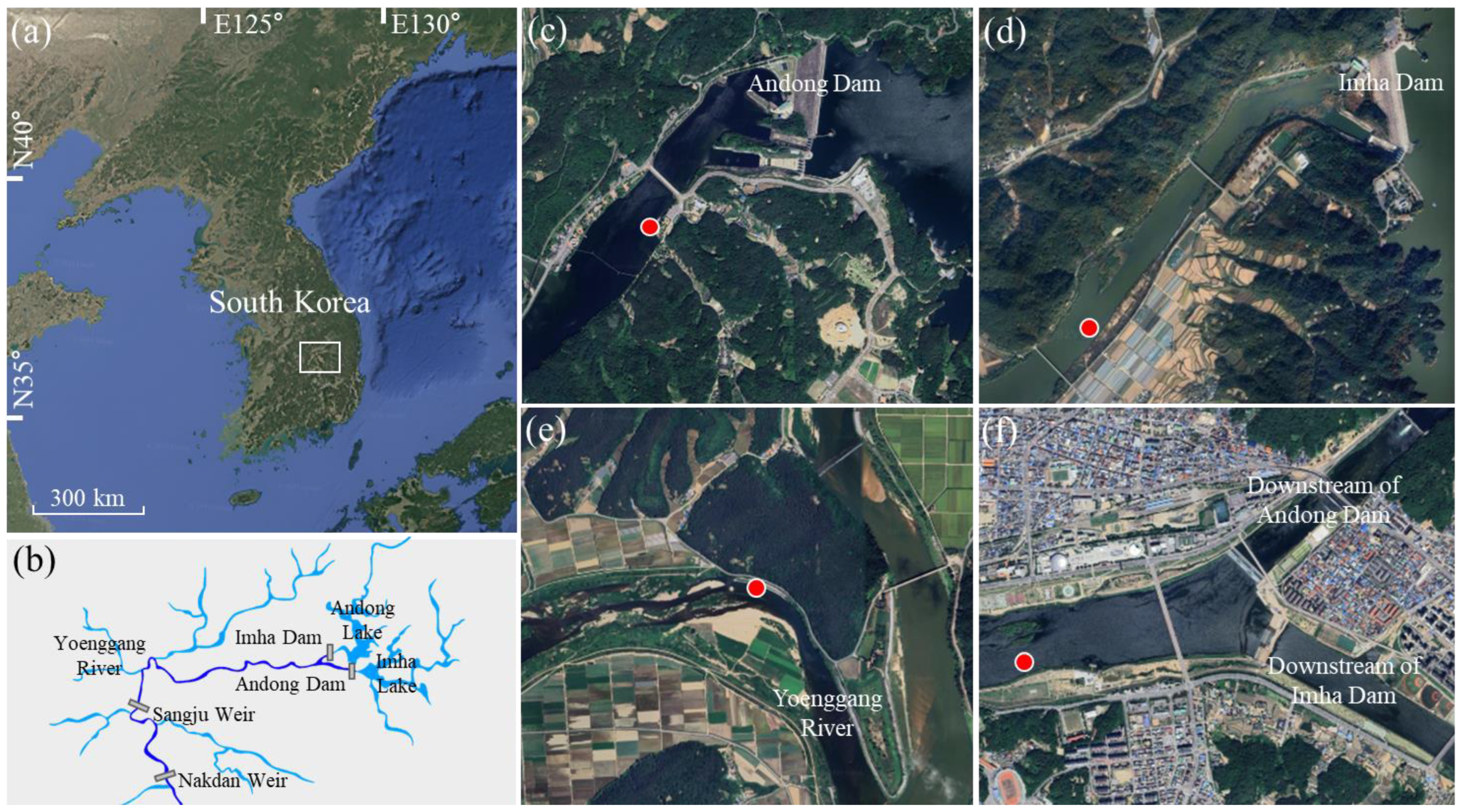
1. Introduction
Diatoms (Bacillariophyta) are among the most diverse and ecologically important groups of microalgae in freshwater ecosystems worldwide. Their siliceous cell walls (frustules), species-specific morphology, and high sensitivity to environmental change make them ideal bioindicators in ecological and water quality assessments [
1,
2]. Diatoms play a fundamental role in primary productivity, nutrient cycling, and shaping microbial communities in aquatic environments. In recent decades, advances in microscopy and molecular biology have significantly improved our understanding of diatom taxonomy and diversity, particularly in regions with historically limited floristic inventories.
The development of scanning electron microscopy (SEM) in the 1980s and 1990s revolutionized diatom taxonomy by enabling high-resolution observation of ultrastructural features, such as areola morphology, raphe architecture, apical pore fields, and striae structure—traits often indistinct under light microscopy (LM) [
3]. This technological advancement, combined with molecular phylogenetics, has led to a paradigm shift in diatom classification frameworks, challenging traditional systems and contributing to more accurate species delimitation [
4]. Round et al. (1990) proposed a modern morphology-based system that integrated fossil and extant characteristics, dividing diatoms into three classes: Coscinodiscophyceae, Fragilariophyceae, and Bacillariophyceae [
1]. Subsequent molecular and cytological evidence further refined higher-level diatom classification and clarified major evolutionary lineages [
4]. Later, molecular phylogenetic evidence led to further refinement of higher-level diatom classification, including recognition of an additional major lineage (e.g., Mediophyceae) [
4]. This revised framework has since been widely adopted in contemporary taxonomic systems, including AlgaeBase and other international phycological references [
5].
Although diatom studies in Korea began in the early 20th century, systematic taxonomic efforts were first established in the 1960s through early national surveys of freshwater algae. Over subsequent decades, diatom classification in Korea followed several international taxonomic systems, including those proposed by Engler, Hustedt, and Bourrelly. In the late 20th century, Simonsen’s phylogenetically oriented system (1979) became influential and was widely adopted in national checklists of marine, freshwater, and fossil diatoms [
6]. Based on advances in molecular phylogenetics and ultrastructural studies, contemporary diatom classification systems have been progressively refined and synthesized into standardized frameworks. These developments are comprehensively summarized in modern taxonomic syntheses, such as that provided by Cox (2015), which integrates morphological, cytological, and molecular evidence to present a coherent higher-level classification of diatoms. Such frameworks underpin current taxonomic practice and are broadly consistent with those adopted in major international references and databases [
7].
Despite the Nakdonggang River being the longest river in South Korea and a vital ecological corridor, it has not been extensively surveyed using SEM-based diatom taxonomy. The river originates in the Taebaek Mountains and stretches over 500 km, passing through varied ecological zones and supporting diverse aquatic biota. Previous studies in the Nakdonggang River and its tributaries have primarily focused on LM-based ecological assessments, which limit taxonomic resolution and may overlook cryptic or morphologically similar taxa [
8].
This study aims to address this gap by documenting ten freshwater diatom taxa newly recorded from both the mainstem and tributary sites of the Nakdonggang River Basin, South Korea. None of these taxa are currently listed in the National List of Species of Korea and are proposed as new records for official inclusion [
9]. Species identification was conducted using high-resolution SEM, enabling detailed morphological characterization, including valve outline, striae arrangement, areola structure, apical pore fields, and spines. In addition to taxonomic observation, basic environmental parameters such as water temperature, pH, electrical conductivity, dissolved oxygen, and turbidity were measured at each sampling site.
By providing precise morphological documentation and associated ecological data, this study contributes to expanding the national inventory of freshwater diatoms. It also highlights the utility of SEM in resolving species boundaries and the potential for novel discoveries even in well-sampled but taxonomically under-characterized river systems. Continued integration of high-resolution imaging with ecological context will be essential for future diatom biodiversity assessments and conservation planning in East Asia.
3. Results
10 freshwater diatom taxa were newly recorded from the Nakdonggang River Basin, South Korea. All taxa represent first confirmed records for the country. Species identification and documentation were based on scanning electron microscopy (SEM), which provided detailed observations of valve morphology, including outline, striae arrangement, areola structure, spines, and apical pore fields. For each taxon, diagnostic features are described, and remarks are included to distinguish them from morphologically similar species based on current taxonomic literature. Ecological data from the sampling sites—including salinity measurements, water temperature, pH, conductivity, dissolved oxygen, record turbidity—were used to characterize environmental conditions and potential habitat preferences (
Table 2). This study contributes to the national diatom inventory and provides foundational data for further ecological, taxonomic, and biogeographical research in East Asian freshwater systems.
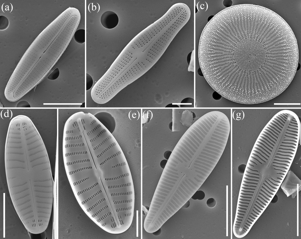
3.1. Achnanthidium druartii Rimet & Couté 2010 (Figure 2a)
Original description: Rimet, F., Couté, A., Piuz, A., Berthon, V. & Druart, J.C. (2010).
Achnanthidium druartii sp. nov. (Achnanthales, Bacillariophyta): A new species invading European rivers. Vie et Milieu 60: 185–195, 6 figs, 3 tables [
11].
SEM Description: The valves (N = 5) are lanceolate with slightly subrostrate apices, lacking any capitate expansion. Valve dimensions range from 14.5 to 15.0 µm in length and 3.9 to 4.1 µm in width. Striae are weakly radiate across the entire valve surface and number around 31 in 10 µm, with no significant difference between the central and apical regions. Areolae are arranged in transapical rows and occur at a density of 3–4 per one µm. On the raphe valve, the raphe is straight with externally drop-shaped proximal ends that are spaced approximately 0.8 µm apart. The axial area is wider at the center and tapers gradually toward the apices. Areolae rows are interrupted near the apices. While internal areolar structures could not be confirmed in this observation, the external foramina appear round to slightly elongate. The overall morphology of the Korean samples conforms well to the original concept of A. druartii. However, in contrast to the type material, the specimens examined here exhibit both a higher stria and areola density and a more even striae distribution, with little difference between the central and apical regions.
Ecology and distribution:
A. druartii was observed from a sample collected downstream of the Andong Dam (Nakdonggang River Basin, South Korea). The habitat is a regulated riverine environment characterized by low salinity (0.1 ppt), cool water temperature (9.5 °C), and slightly alkaline conditions (pH 8.2). Electrical conductivity was relatively low (187 μS/cm), and dissolved oxygen was high, with values of 106% saturation and 12.1 mg/L. The turbidity was moderate at 3.6 NTU. These environmental parameters suggest that
A. druartii prefers well-oxygenated, oligotrophic to mesotrophic freshwater habitats with low mineral content. This observation represents the first confirmed record of
A. druartii in Korea. Originally described from the Roubion River, a calcareous tributary of the Rhône Basin in France, the species was associated with cold, alkaline, and low-conductivity waters rich in dissolved oxygen [
11]. Since its original description, it has also been reported from similarly oligotrophic freshwater systems in parts of Europe and North America [
12]. In these regions,
A. druartii has been found in epilithic habitats within well-oxygenated, low-nutrient streams and canals, often under alkaline conditions [
11,
12]. Kelly et al. [
13] have noted its occurrence in high-status river sites in the United Kingdom, indicating its potential as an indicator species for clean, minimally impacted waters. The ecological characteristics of the Korean habitat are consistent with those previously reported, reinforcing the species’ preference for stable, unpolluted freshwater systems. This finding expands the known geographic distribution of
A. druartii to East Asia.
Remarks: The Korean sample of
Achnanthidium druartii closely matches the type description provided by Rimet & Couté (in Rimet et al. 2010) [
11] in terms of valve outline, size range, and raphe morphology, including the distinctly drop-shaped external proximal raphe ends spaced approximately 0.8 µm apart. However, the observed sample exhibits slightly higher stria density (31 in 10 µm) and a greater number of areolae per unit length (3–4 per µm) than originally reported (15–22 striae in 10 µm at the center and 2–3 areolae per µm) [
11]. These differences likely reflect intraspecific variation or environmental influences, as commonly noted in other
Achnanthidium druartii [
12,
14]. Within the genus
Achnanthidium, two major species groups are recognized: the
minutissimum group and the
pyrenaicum group.
A. druartii clearly belongs to the
pyrenaicum group, and can be distinguished from morphologically similar members of this group (e.g.,
A. pyrenaicum and related taxa) by its broader central area, higher stria and areola density, and the characteristic drop-shaped proximal raphe ends [
12,
15]. Compared with
Achnanthidium pyrenaicum, which typically shows a narrower central area and lower stria density,
A. druartii exhibits a more robust valve outline, a broader central area, and consistently higher stria and areola counts. Other members of the
pyrenaicum group generally display less pronounced drop-shaped proximal raphe ends, whereas this feature is well developed and consistently expressed in
A. druartii. In addition,
A. druartii is readily separable from taxa belonging to the
A. minutissimum group, including
A. microcephalum and
A. lusitanicum, by several SEM-based characters. The latter taxa typically exhibit more distinctly capitate or narrowly protracted apices, lower and more variable stria densities, and less prominently expanded proximal raphe ends. In contrast, the Korean specimens of
A. druartii show weakly subrostrate, non-capitate apices, uniformly high stria density across the valve, and clearly drop-shaped proximal raphe ends, supporting their separation from members of the
minutissimum complex. Although no LM images were available for the original description [
11], the SEM characteristics strongly support its taxonomic placement within the
pyrenaicum group.
3.2. Achnanthidium peetersianum Wetzel, Juttner & Ector 2019 (Figure 2b)
Original description: Wetzel, C.E., Jüttner, I., Gurung, S. & Ector, L. (2019). Analysis of the type material of
Achnanthes minutissima var.
macrocepahala (Bacillariophyta) and description of two new small capitate
Achnanthidium species from Europe and the Himalaya. Plant Ecology and Evolution 152(2): 340–350, 5 fig [
16].
SEM Description: The valves (N = 6) are lanceolate with slightly subcapitate apices, measuring approximately 13.2–14.0 µm in length and 3.0–3.5 µm in width. The raphe is straight and filiform, with distinctly drop-shaped proximal ends that are clearly separated. The axial area is narrow and linear. Striae are weakly radiate across the valve face, becoming more densely arranged toward the apices. A total of 33 striae are observed in 10 µm, with 34–36 striae in 10 µm at the central part of the valve, and no significant increase in density toward the apices. This contrasts with the original description, in which striae become slightly more radiate and denser near the apices, reaching up to 40 in 10 µm. Each stria is composed of 8–12 areolae per 10 µm, with areolae arranged in transapical rows. The external foramina of the areolae are round to slightly elongate. Areola rows are interrupted near the apices. The overall morphology of the Korean sample is consistent with the diagnostic features of A. peetersianum, though slight variation in areola density is observed.
Ecology and distribution:
A. peetersianum was recorded from a regulated lotic environment downstream of the Andong Dam, located in the Nakdonggang River system, South Korea. At the time of sampling, the water temperature was 9.5 °C, with slightly alkaline pH (8.2) and low salinity (0.1 ppt). Electrical conductivity measured 187 μS/cm, and dissolved oxygen levels were high (12.1 mg/L; 106% saturation). These parameters characterize a well-oxygenated, low-mineral, circumneutral to alkaline freshwater system. This is the first report of
A. peetersianum from Korea. Globally,
A. peetersianum was originally described from calcareous headwater streams in France, where it was found in oligotrophic to mesotrophic conditions, typically with high oxygen concentrations and moderate alkalinity [
16]. Subsequent records have reported the species in similarly low-nutrient, hard-water rivers and mountain streams in regions such as Belgium, Luxembourg, and Austria [
14,
16]. It is usually associated with epilithic habitats in unpolluted, stable-flow environments, often with low conductivity and slightly alkaline pH. The presence of
A. peetersianum in the Nakdonggang River expands its known geographical distribution to East Asia and reinforces its ecological preference for cold, well-oxygenated, and relatively nutrient-poor freshwater environments.
Remarks: The Korean sample of
A. peetersianum conforms well to the morphological concept proposed by Wetzel et al. (2019), with valve shape and striae distribution patterns closely matching those illustrated in the original description [
16]. Particularly in valve outline, raphe morphology, and the striae and areolae patterns. The valve outline, raphe shape, and striae patterns are consistent with the type description, although the Korean sample exhibits a slightly narrower valve and a higher areola density (40–60 in 10 µm).
A. peetersianum can be easily confused with several members of the
Achnanthidium genus, particularly
A. minutissimum,
A. pyrenaicum, and
A. druartii. Compared to
A. minutissimum, which has a narrower central area, lower stria density (usually 20–30 in 10 µm), and less distinctly shaped proximal raphe ends [
12,
15],
A. peetersianum displays a more robust valve structure and more pronounced raphe morphology. It differs from
A. pyrenaicum by its higher stria and areola density and more narrowly lanceolate valve outline [
15,
17], and from
A. druartii by its generally straighter striae and slightly broader axial area. Additionally, the shape and spacing of the proximal raphe ends in
A. peetersianum are more distinctly tear-drop shaped and widely spaced, providing a reliable diagnostic character [
11,
16]. These comparisons support the identification of the Korean sample as
A. peetersianum and reinforce the utility of SEM imaging in distinguishing among morphologically similar taxa within the genus.
3.3. Discocyclus hubeianus (Chen & Zhu) Rioual 2025 (Figure 2c)
Basionym: J.Y.Chen & H.Z.Zhu 1985
Homotypic synonym: Cyclotella hubeiana J.Y.Chen & H.Z.Zhu 1985
Heterotypic synonym: Discostella guiyangensis Y.D.Xiang, Q.Liu & S.L.Xie 2024
Original description: Rioual, P., Schultz, K., Joh, G. & Liao, M. (2025).
Cyclotella or
Discostella? an evaluation of the morphological and molecular evidence regarding the generic placement of a centric diatom from eastern Asia and the creation of
Discocyclus gen. nov. Taxonomy 5(17): 1–12, 4 figures, 1 table [
18].
SEM Description: Valves (N = 5) are circular and discoid, with a flat to slightly domed central area and distinctly radiate striae extending from the center to the margin. The valve diameter ranges from 8.7 µm (Andong Dam downstream) to 18.8 µm (Imha Dam downstream). In smaller valves (8.7 µm), striae are uniseriate, numbering 28–32 in 10 µm, and composed of regularly spaced rounded areolae. In larger valves (18.8 µm), striae appear partially biseriate toward the margin and number approximately 9 in 10 µm; counted as uniseriate, the density increases to 32–34 in 10 µm. The central area is ornamented with numerous pustules and irregular areolae, often scattered without a distinct fultoportula. Toward the margin, areolae become smaller, more densely packed, and sometimes arranged in double rows. Areola density in the marginal region ranges from 35 to 38 in 10 µm. Marginal fultoportulae are visible as small interruptions in the striae and number approximately 8–10 per valve. These morphological features match the species concept of
D. hubeianus, including the intraspecific variability previously reported [
18].
Ecology and distribution: Discocyclus hubeianus was recorded for the first time in Korea from two freshwater sites in the upper Nakdonggang River basin—specifically, the downstream reaches of the Andong and Imha Dams. The Andong Dam site exhibited a water temperature of 9.5 °C, pH 8.2, conductivity 187 µS/cm, dissolved oxygen 12.1 mg/L (106%), and salinity of 0.1 ppt. In contrast, the Imha Dam site showed slightly warmer and more mineral-rich conditions, with a water temperature of 14.9 °C, pH 8.6, conductivity 225 µS/cm, dissolved oxygen 11.5 mg/L (114%), and salinity of 0.1 ppt. Both environments were moderately alkaline, well-oxygenated, and exhibited stable physicochemical profiles favorable to benthic diatom growth. These ecological conditions are comparable to those of the species’ type locality in Hubei Province, China, where it was originally described from low-disturbance freshwater habitats with similar water chemistry. The present observations not only confirm the species’ broader ecological tolerance but also extend its known distribution range into northeastern Asia.
Remarks: The Korean samples are morphologically consistent with
Discocyclus hubeianus as originally described [
18], including key features such as a discoid valve outline, radiate striae, scattered central granules, and marginal fultoportulae. Notably, the two observed samples—one from Andong Dam downstream and one from Imha Dam downstream—demonstrate significant intraspecific variation, particularly in valve size and striae arrangement. The smaller individual exhibits tightly spaced uniseriate striae and a compact central area, while the larger valve has a broader central zone, more widely spaced striae, and marginal biseriate areolae. This variation reflects developmental or environmental influences and was also noted within the type population in China [
18]. According to the original description, a diagnostic feature separating
Discocyclus from related genera (
Pantocsekiella,
Lindavia) is the position of the rimoportula: in
Discocyclus, it is located among the ring of marginal fultoportulae, above an alveola, whereas in
Pantocsekiella/
Lindavia it lies outside the ring, generally below the alveola or on the valve face. However, in the present Korean material this character could not be clearly verified in SEM observations. Despite this limitation, other valve features, together with the distinction from
D. zhushanensis and
D. sinensis—which show more regularly organized areolae, larger concentric central areas, or a higher number of marginal fultoportulae—support the identification of the Korean specimens as
D. hubeianus.
3.4. Geissleria cascadensis (Sovereign) Stancheva & Spaulding 2019 (Figure 2d,e)
Basionym: Navicula cascadensis Sovereign 1960
Homotypic synonym: Navicula cascadensis Sovereign 1960
Original description: Sovereign, H.E. (1958). The diatoms of Crater Lake, Oregon Transactions of the American Microscopical Society 77(2): 96–134 [
19].
SEM Description: Valves (N = 4) are elliptic with broadly rounded to slightly cuneate apices. The valve length measures approximately 9.8–13.3 µm, and the width is 4.6–5.0 µm. The raphe is straight and filiform, with slightly expanded proximal ends externally and internally deflected distal ends. The axial area is narrow and linear, slightly widening toward the center. The central area is small, rounded to elliptic, and lacks a distinct fascia. Although a stigma is generally reported for species of
Geissleria, the presence of this feature could not be confirmed in the Korean specimens under SEM. Striae are uniseriate and radiate, more widely spaced near the center and becoming denser toward the apices. Striae number 16–18 in 10 µm, and are composed of round to slightly elongate areolae, approximately 7 per 1 µm, clearly visible under SEM. The areolae are internally occluded by hymenes and appear rimmed on the internal valve surface. A particularly distinct feature observed in the Korean sample is the presence of annulae at both apices, usually consisting of 2–4 concentric striae arranged in circular or semi-circular rings. These annular structures are clearly visible in SEM and correspond well with those described in previous literature [
17,
20]. The consistent occurrence of apical annulae in the Korean material not only affirms the generic placement of this species within
Geissleria but also provides additional support for its taxonomic identification as
G. cascadensis.
Ecology and distribution:
Geissleria cascadensis was recorded from two freshwater sites in Korea: the downstream reach of the Andong Dam and the confluence zone where the outflows of the Andong and Imha Dams merge. These sites are located in the upper Nakdonggang River basin. At the time of sampling, water quality conditions were indicative of moderately alkaline, well-oxygenated freshwater systems, with pH values ranging from 8.2 to 8.6, conductivity between 187 and 226 µS/cm, and dissolved oxygen levels from 11.5 to 12.1 mg/L. Water temperatures were cool, ranging from 9.5 to 14.9 °C. By contrast, the type locality in the Cascade Mountains of Oregon is characterized by low conductivity (24–38 µS/cm) and slightly acidic waters (pH 6.7–6.9), where the species was first described from oligotrophic lakes and streams [
17]. In addition,
G. cascadensis has been observed in several mountain streams in western Oregon, USA, further supporting its distribution across western North America. The occurrence of this taxon in alkaline, higher-conductivity habitats in Korea indicates a broader ecological tolerance than previously recognized. This record represents the first confirmed occurrence of
G. cascadensis in South Korea, expanding its known biogeographic range and highlighting a broader ecological tolerance of the species across geographically distant freshwater systems.
Remarks: The Korean samples of
Geissleria cascadensis display morphological features that conform closely to the original description of the species from the Cascade Range in North America [
17]. The elliptic valve outline with broadly rounded apices, uniseriate radiate striae, and rounded to slightly elongate areolae are all consistent with diagnostic features previously reported [
17,
20]. Both external and internal SEM views reveal a filiform raphe, narrow axial area, and absence of a fascia or expanded central area—characteristics typical of the genus
Geissleria. A particularly important feature confirming this identification is the presence of annulae at both apices, consisting of two to four pairs of striae arranged in a circular pattern. These annulae, clearly visible in SEM, have been proposed as a reliable generic marker for
Geissleria and support its placement in the genus [
20]. This character is seldom emphasized in older literature but has been consistently reported in recent taxonomic treatments.
Geissleria cascadensis can be distinguished from morphologically similar species such as
G. decussis, which typically has a more linear-lanceolate valve outline, fewer striae, and a broader central area with more prominent fascia [
21]. It also differs from
G. paludosa, which shows more lanceolate valves and denser striae that are more closely spaced near the apices [
17]. The annulae, along with the elliptic valve outline and striae arrangement, provide a reliable basis for separating
G. cascadensis from other small
Geissleria taxa.
3.5. Gomphonella perolivaceoides (Levkov) Tuji 2020 (Figure 2f,g)
Basionym: Gomphonema perolivaceoides Levkov 2007
Homotypic synonym: Gomphonema perolivaceoides Levkov 2007
Original description: Levkov, Z., Krstic, S., Metzeltin, D. & Nakov, T. (2007). Diatoms of Lakes Prespa and Ohrid, about 500 taxa from ancient lake system. Iconographia Diatomologica 16: [i–iv], 1–611, 220 plates [
22].
SEM Description: Valves (N = 3) are clavate, with broadly rounded headpoles and gradually tapering footpoles. The observed sample from the Yeonggang River tributary measured 26–28 µm in length and 8–9 µm in width. The axial area is narrow and linear, slightly widened at the center; no fascia is present. The raphe is straight and filiform, with slightly expanded proximal ends and terminal fissures curving onto the mantle. Striae are biseriate, radiate throughout, and composed of closely spaced rounded areolae. The striae number approximately 30–32 in 10 µm, with each stria containing 11 areolae per 2 µm. The areolae are rounded, internally occluded by hymenes, and regularly spaced within each stria. Internally, the valve shows the absence of a septum, and the internal proximal raphe endings curve in opposite directions. No stigmata were observed. These features, particularly the biseriate striae, narrow clavate outline, and absence of apical pore fields or septum, correspond to the concept of
Gomphonella perolivaceoides, as proposed by Tuji (2020) in the revision and recombination of
Gomphonema perolivaceoides Levkov into the genus
Gomphonella [
23,
24].
Ecology and distribution: The sample was collected from the Yeonggang River tributary, a moderately alkaline freshwater habitat with good water clarity. At the time of sampling, the site exhibited pH 8.7, conductivity 321 µS/cm, dissolved oxygen 15.9 mg/L (167%), and water temperature 16.6 °C. These conditions are characteristic of well-oxygenated, low-turbidity environments favored by benthic diatoms in the Gomphonella complex. Although G. perolivaceoides has been previously reported from Europe and parts of Asia, the present finding represents the first confirmed record of the species in South Korea, expanding its known geographic range and contributing to the recognition of Gomphonella diversity in East Asia.
Remarks: The Korean sample of
Gomphonella perolivaceoides closely resembles the original concept of the species described by Levkov (2007) [
24] as
Gomphonema perolivaceoides, and later transferred to
Gomphonella by Tuji (2020) [
23] based on phylogenetic and morphological evidence [
25]. The key diagnostic features—narrow clavate shape, high stria and areolae density, biseriate arrangement, and absence of septa—are all consistent with the revised circumscription. This species differs from
G. olivaceoides in having significantly denser striae (30–32 vs. 16–18 in 10 µm) and more elongated valves.
G. calcifuga can appear similar in outline but has more widely spaced striae and typically larger valves [
26]. The absence of apical pore fields and stigmata also helps distinguish
G. perolivaceoides from stigmata-bearing species such as
G. tetrastigmata or
G. quadripunctata [
23]. Given the precise morphological match and absence of taxonomically significant deviations, the Korean population can be confidently assigned to
G. perolivaceoides.
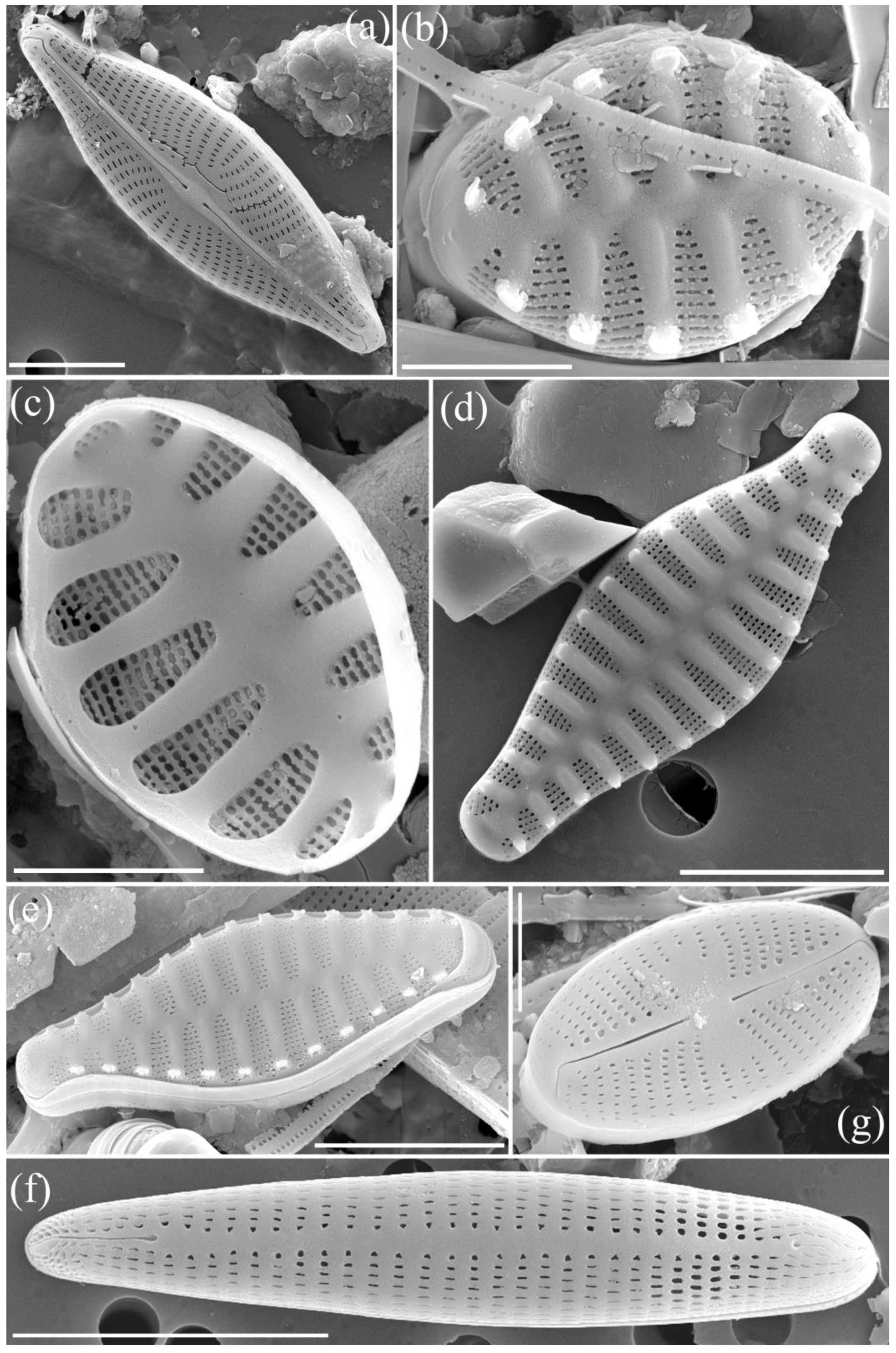
3.6. Navicula venetiformis Van de Vijver & Beyens 2002 (Figure 3a)
Original description: Van de Vijver, B., Frenot, Y. & Beyens, L. (2002). Freshwater diatoms from Île de la Possession (Crozet Archipelago, sub-Antarctica). Bibliotheca Diatomologica 46: 1–412, incl. 132 pls., 1 fig., 4 maps [
27].
SEM Description: Valves (N = 3) are elliptic-lanceolate with narrowly rounded apices. The observed sample from the downstream region of the Imha Dam measured approximately 20.2–21.2 µm in length and 6.0–6.3 µm in width. The axial area is narrow and linear, slightly expanded near the center, without a distinct central fascia. The valve face is slightly convex and transitions smoothly to the mantle. The raphe is filiform and straight, with slightly expanded external proximal ends. Distal fissures are curved toward the secondary side. Striae are uniseriate, radiate across the entire valve, and become slightly more convergent near the apices. Each stria contains 4–5 areolae, which are elongated and rounded in shape. Striae density is approximately 8–10 in 5 µm, and areolae number 4–5 per 1 µm. Areolae are occluded by hymenes. No apical pore fields were visible due to partial obscuration in the SEM image. These morphological features are consistent with previous descriptions of
N. venetiformis [
28].
Ecology and distribution: The sample was collected from the downstream section of the Imha Dam, part of the Nakdonggang River system. The sampling site was a moderately alkaline and oxygen-rich freshwater environment, with the following water quality parameters: pH 8.6, conductivity 226 µS/cm, dissolved oxygen 11.5 mg/L, and temperature 14.9 °C.
Navicula venetiformis was originally described from oligosaprobic, lentic Antarctic lakes where it inhabits cold, low-nutrient freshwater ecosystems [
28]. Its presence in a temperate Korean reservoir suggests that the species may have a broader ecological tolerance than previously reported. This finding extends the known distribution of
N. venetiformis to East Asia.
Remarks: The Korean sample agrees well with the circumscription of
Navicula venetiformis as described by Van de Vijver et al. (2011) from the Maritime Antarctic region [
1]. The overall valve shape, striae density, and areolae arrangement correspond closely with the type population. This taxon may be confused with:
Navicula veneta: which has broader valves and a more expanded central area.;
Navicula cryptocephala: typically has narrower valves and finer striae (14–18 in 10 µm).;
Navicula rhynchocephala: shows more strongly protracted apices and higher striae density. Compared to these species,
N. venetiformis is characterized by its moderate size, coarse radiate striae, and elliptic-lanceolate outline. These features were clearly observed in the Korean sample. This observation constitutes the first confirmed record of
Navicula venetiformis in South Korea.
3.7. Punctastriata glubokoensis Williams, Chudaev & Gololobova 2009 (Figure 3b,c)
Original description: Williams, D.M., Chudaev, D.A. & Gololobova, M.A. (2009).
Punctastriata glubokoensis spec. nov., a new species of ‘fragilarioid’ diatom from Lake Glubokoe, Russia. Diatom Research 24(2): 479–485 [
29].
SEM Description: Valves (N = 5) are broadly elliptical and slightly heteropolar, with rounded apices. The samples collected from the confluence of the downstream reaches of the Andong and Imha dams measured 5.26–5.46 µm in length and 3.84–3.92 µm in breadth. Striae are radiate and multiseriate, beginning as a single row of areolae near the valve center, and gradually increasing to five rows toward the valve margin. Areolae are elongate and densely packed, numbering approximately 7–8 per 1 µm. On the internal valve surface, striae converge laterally to form a series of funnel-shaped internal chambers, each formed by the merging of several striae. These chambers are positioned along both sides of the valve and number approximately 5–7 per side. Interstriae are thickened and raised into robust costae, upon which spines are aligned, at a density of about three spines per 2 µm. Spines are conical and oriented along the costae. An apical pore field is present only at the footpole, composed of several isolated pores. A shallow apical depression is also visible at the headpole. No rimoportulae or mantle plaques were observed. This description is based entirely on Korean material and aligns with key features reported in the original type description, particularly the unique internal chamber morphology [
29].
Ecology and distribution: The samples of
Punctastriata glubokoensis were collected from two distinct epilithic sites in the upper Nakdonggang River basin: the downstream reach of the Imha Dam and the confluence area where the outflows of the Imha and Andong dams meet. Both sites represent clear, lotic freshwater environments with stable rocky substrates. At the time of collection, physicochemical measurements indicated a moderately alkaline, well-oxygenated, and low-conductivity environment. The environmental parameters across both sites ranged from pH 8.17 to 8.57, conductivity from 187.1 to 225.7 µS/cm, dissolved oxygen concentrations from 11.54 to 12.11 mg/L, salinity from 0.11 to 0.12 ppt, and water temperature from 11.5 to 14.9 °C.
Punctastriata glubokoensis was originally described from Lake Glubokoe in the Russian Far East, a remote oligotrophic lake characterized by cold, clear water and rocky littoral habitats [
29]. Its detection at two separate but hydrologically connected locations in South Korea suggests that the species is capable of persisting in regulated freshwater systems with similar environmental characteristics. This study presents the first confirmed record of
P. glubokoensis in South Korea, contributing to a broader understanding of its ecological range and biogeographical distribution.
Remarks: The Korean samples of
Punctastriata glubokoensis are morphologically consistent with the original description by Williams et al. (2009), particularly in their broadly elliptical, slightly heteropolar valve shape, multiseriate radiate striae, and the presence of small spines aligned along the costae [
29]. A key diagnostic feature observed in the Korean material is the internal arrangement of striae forming a series of funnel-shaped chambers near the valve margin, each chamber being formed by the merging of several striae. This structure, visible in internal SEM view, has also been described as a characteristic feature of the species in its type material. The dimensions of the Korean samples (5.26–5.46 µm in length, 3.84–3.92 µm in breadth) fall well within the size range reported in the original description. The density of areolae (7–8 per 1 µm) and the arrangement of striae (increasing from a single row to up to five rows) match the structural complexity described for
P. glubokoensis. The presence of a single apical pore field at the footpole and a shallow apical depression at the headpole also supports this identification. This species may be confused with other members of the genus
Punctastriata, such as
P. mimetica and
P. discoidea. However,
P. glubokoensis can be distinguished from
P. mimetica by its more robust valve shape and coarser striae, and from
P. discoidea by the simpler, flattened spines that lack the complex lateral projections seen in the latter. The diagnostic combination of a multiseriate stria pattern with internal funnel-shaped chambers, consistent valve dimensions, and the specific configuration of apical structures supports the identification of the observed samples as
Punctastriata glubokoensis.
3.8. Punctastriata mimetica Morales 2005 (Figure 3d,e)
Original description: Morales, E.A. (2005). Observations of the morphology of some known and new fragilarioid diatoms (Bacillariophyceae) from rivers in the USA. Phycological Research 53(2): 113–133 [
30].
SEM Description: Valves (N = 3) are cruciform-rhomboid in outline, with distinctly rostrate to cuneate apices and slightly heteropolar ends. The observed samples measured 13.03–13.67 µm in length and 4.36–4.78 µm in breadth. Striae are radiate and multiseriate, composed of up to four rows of round areolae toward the apices, which converge into 1–2 areolae near the axial area. Striae extend continuously from the valve face onto the mantle and terminate sharply at the axial region. Areolae are round to slightly elliptical and densely arranged, with a density of approximately 7 areolae per 1 µm. The costae separating the striae are raised and broader than the striae, contributing to a distinctly undulate valve surface in SEM view. Spines are spatulate and evenly spaced along the striae, except near the apices, and occur at a frequency of approximately 6 spines per 5 µm. Some spines exhibit laminar ligulae projecting toward the mantle. At both apices, small ocellulimbus-type apical pore fields with round poroids are present and more developed at the foot pole. These pore fields are located at the junction between the valve face and the mantle. Rimoportulae are absent. The valvocopula is wider than the other girdle bands and features well-developed fimbriae; girdle bands are open and lack perforations.
Ecology and distribution: Korean samples
Punctastriata mimetica were collected from epilithic habitats at the confluence zone of the Andong and Imha Dams, located in the upper Nakdonggang River basin, South Korea. This site represents a regulated but hydrologically dynamic lotic environment with clear, oligotrophic to mesotrophic water. At the time of collection, physicochemical parameters were as follows: pH 7.7–8.0, conductivity 174.2–187.3 µS/cm, dissolved oxygen 9.9–10.6 mg/L, salinity < 0.12 ppt, and temperature 11.2–14.9 °C. Nutrient levels were low to moderate, with total nitrogen ranging from 1.203 to 1.379 mg/L and total phosphorus between 0.014 and 0.022 mg/L. This observation represents the first confirmed record of
Punctastriata mimetica in South Korea, suggesting that the species may be more widely distributed across temperate lotic systems than previously recognized. Its occurrence under moderately alkaline, well-oxygenated, and nutrient-poor conditions is consistent with habitat preferences reported in previous studies [
30].
Remarks: The Korean samples of
Punctastriata mimetica exhibit key morphological traits consistent with the original description, including cruciform-rhomboid valves, raised costae, and spatulate spines aligned along the striae [
30]. The striae architecture is notable for its multiseriate nature, with up to four rows of areolae at the apices converging to 1–2 areolae near the axial area [
30]. Spines were well-developed, regularly arranged, and in some cases exhibited ligulae extending toward the mantle [
30]. These features distinguish
P. mimetica from similar taxa such as
P. glubokoensis, which lacks continuous mantle-striae and exhibits internal funnel-shaped chambers instead [
29]. The combination of continuous valve-face striae, prominent costae, and fimbriae-bearing valvocopulae supports the identification of the Korean material as
P. mimetica.
3.9. Rhoicosphenia californica Thomas & Kociolek 2015 (Figure 3f)
Original description: Thomas, E.W. & Kociolek, J.P. (2015). Taxonomy of three new
Rhoicosphenia (Bacillariophyta) species from California, USA. Phytotaxa 204(1): 1–21, 110 fig., 3 tables [
31].
SEM Description: Valves (N = 4) are dorsiventral and semi-lanceolate, with a strongly arched dorsal margin and a nearly straight ventral margin. The apices are broadly rounded and heteropolar in outline. The raphe is positioned along the ventral margin, slightly arched, and filiform in external view. The proximal raphe ends are slightly expanded and gently bent to the same side; the distal ends curve in the same direction as well. Internal raphe endings and helictoglossae were not visible in the current material. The axial area is narrow and linear, widening slightly toward a small, rounded central area. Striae are uniseriate and radiate throughout, with more widely spaced striae at the center and denser arrangement near the apices. Areolae are round to slightly elliptical, and number approximately 4 per µm. Striae count ranges from 13 to 15 in 10 µm. The valve length measured between 27.11 and 27.40 µm, and the valve width was approximately 4.91 µm based on SEM observations. Structures such as pseudosepta, apical pore fields, and girdle elements were not visible in the material examined.
Ecology and distribution: Korean samples of
Rhoicosphenia californica were collected from epilithic substrates in the downstream section of Andong Dam, located in the upper Nakdonggang River basin, South Korea. At the time of collection, the water temperature was 14.5 °C, with pH measured at 7.55 and electrical conductivity at 78.9 µS/cm. Dissolved oxygen was 11.44 mg/L, total nitrogen 2.771 mg/L, and total phosphorus 0.031 mg/L. These physicochemical characteristics reflect an oligotrophic to mesotrophic environment with well-oxygenated, slightly alkaline freshwater. This finding represents the first confirmed record of
R. californica in South Korea. The species is widely distributed in temperate regions of the Northern Hemisphere, often inhabiting rivers, lakes, and regulated freshwater systems with moderate nutrient availability and rocky or artificial substrates [
32]. It has also been recorded from a mountain stream in Trinity County, northern California [
33], further supporting its North American distribution.
Remarks: The Korean samples of
R. californica show a strong match with descriptions in the literature in terms of valve outline, striae pattern, and raphe morphology observed in external view [
32]. The characteristic dorsiventral shape, broadly rounded apices, and ventral raphe with slightly expanded proximal ends curved in the same direction are all consistent with features previously reported for this species. The observed striae density (13–15 in 10 µm) and areolae count (approximately 4 per µm) also align with published values. However, due to the unavailability of internal views in the current SEM material, certain diagnostic features such as helictoglossae, pseudosepta, and the reduced raphe valve could not be confirmed. Nevertheless, the external characters—including the asymmetric valve structure, ventral raphe system, and consistent striae arrangement—are sufficient to distinguish
R. californica from other
Rhoicosphenia taxa. Notably,
R. abbreviata tends to have a shorter cell length and lower stria density, while
R. curvata is generally more slender with a more narrowly rounded apex. The combination of broader outline and external raphe architecture in the Korean material supports its assignment to
R. californica.
3.10. Sellaphora nigri (De Notaris) Wetzel & Ector 2015 (Figure 3g)
Basionym: Navicula nigri De Notaris 1872
Homotypic synonym: Navicula nigri De Notaris 1872
Original description: De Notaris, G. & Baglietto, F. (1872). Erbario Crittogamico Italiano. Exsiccata. Ser. II, Fasc.XII. pp. nos 601–650. Genova [
34].
SEM Description: Valves (N = 5) are elliptic with broadly rounded apices, appearing symmetrical along the apical axis. The valve measured 6.20 µm in length and 3.42 µm in width in the examined sample. Striae are uniseriate and radiate throughout the valve face. Near the center, a few shortened striae interrupt the regular pattern, forming a weakly defined and slightly asymmetrical central area. Each stria is composed of a single row of round to slightly angular areolae, which number approximately 8 in 1 µm. Striae are spaced at about 4 per 1 µm. Areolae near the raphe are slightly larger than those toward the valve margin. The raphe is straight and filiform, with externally slightly expanded proximal ends. Apical pore fields and internal structures were not observed in the present material.
Ecology and distribution: Sellaphora nigri was collected from the confluence area of the Andong and Imha Dams, located in the upper Nakdonggang River basin, South Korea. The physicochemical conditions at the time of sampling were as follows: water temperature 11.0 °C, pH 7.56, electrical conductivity 201.1 μS/cm, dissolved oxygen 11.72 mg/L (106.2%), and turbidity 1.4 NTU. These values reflect well-oxygenated, moderately oligotrophic freshwater conditions under low-temperature and low-turbidity environments. This finding constitutes the first documented record of S. nigri in South Korea. Previously, this species has been reported from nutrient-enriched waters in Europe and North America, often occurring in organically influenced or slightly eutrophic environments. Its occurrence in this Korean site suggests a degree of ecological tolerance and possible responsiveness to environmental gradients.
Remarks: The Korean sample of
S. nigri closely resembles the type material described by Wetzel et al. (2015) [
35] in terms of valve outline, striae arrangement, and areola morphology. The observed valve is elliptic with broadly rounded apices and radiate uniseriate striae, consistent with the diagnostic features of the species. However, the Korean sample showed slightly larger areolae and a slightly wider valve breadth (3.42 µm) than most documented populations. A notable diagnostic feature of the Korean sample is the presence of approximately 4 striae and 8 areolae in 1 µm, which aligns well with but slightly exceeds the density ranges mentioned in previous studies [
35]. The central area is asymmetrically bow-tie shaped, bordered by shorter striae, and the proximal raphe ends are tear-drop shaped externally—both being diagnostic features of
S. nigri. This population can be clearly distinguished from morphologically similar taxa such as
Sellaphora seminulum or
Sellaphora pupula by the combination of its elliptic outline, radiate striae, and the regular pattern of areolae bordering the axial area [
35]. Moreover, unlike
S. pupula,
S. nigri lacks significant valve undulation or a lanceolate central area. Additional distinction is also possible from
S. atomoides, which is generally smaller with denser striae,
S. saugerresii, which exhibits differences in central area structure and striae arrangement, and
S. crassulexigua, which is smaller and shows a different areola pattern.
4. Discussion
This study documents ten freshwater diatom taxa newly recorded from the Nakdonggang River Basin, representing a significant addition to the known diatom flora of Korea. The use of high-resolution scanning electron microscopy (SEM) enabled accurate characterization of critical ultrastructural features such as areola morphology, striae density, apical pore fields, and spine architecture, which are essential for resolving taxonomic uncertainties, especially among morphologically cryptic species [
1,
4,
36].
Several of the identified taxa, such as
Punctastriata mimetica and
Geissleria cascadensis, have rarely been reported globally, and their ecological ranges remain underexplored [
29,
30]. For instance,
P. mimetica was previously described from Lake Tahoe, USA, and only sporadically documented in subsequent studies; its occurrence in Korean inland waters suggests a broader ecological amplitude than previously understood [
29]. Likewise,
G. cascadensis, initially reported from montane regions of North America, was found here under relatively eutrophic conditions, suggesting potential ecological plasticity or regional adaptation [
17]. In our material, however, this taxon occurred at very low relative abundance, raising the possibility that it may have originated from more oligotrophic upstream habitats. Additional surveys in headwater sites are needed to determine whether the species is established locally or represents downstream transport. Notably,
G. cascadensis has also been observed in mountain streams of western Oregon [
30,
33], supporting its wider North American distribution. Together, these records indicate that the species may possess greater ecological tolerance and a broader biogeographic range than previously recognized.
The discovery of taxa from genera with taxonomically challenging histories, such as
Staurosirella,
Sellaphora, and
Gomphonella, underscores the limitations of light microscopy in distinguishing fine-scale features used in modern classification systems [
1,
36]. For example, the reliable separation of
Gomphonella olivaceoides and
G. perolivaceoides was only possible through SEM observation of differences in raphe end curvature and areola distribution—characters not consistently visible in LM preparations [
23,
25].
All ten taxa reported in this study are absent from the National List of Species of Korea, and their inclusion will contribute to a more comprehensive biodiversity inventory [
9]. These findings reinforce the growing recognition that even well-surveyed freshwater systems such as the Nakdonggang River Basin may harbor previously undocumented microalgal taxa, especially when high-resolution taxonomic tools are applied. In terms of ecological patterns, the taxa identified were found across diverse microhabitats—ranging from rocky epilithic substrates to open water columns—reflecting their varied ecological niches and adaptive strategies. The species were associated with a range of physicochemical conditions, including conductivity (94.1–174.3 µS/cm), pH (7.4–8.4), and dissolved oxygen (7.7–10.3 mg/L), highlighting that newly recorded species are not confined to pristine or oligotrophic waters [
13,
27].
This study also has implications for biomonitoring frameworks. Some taxa, such as
Navicula venetiformis and
Achnanthidium druartii, have been reported in previous literature as sensitive or moderately tolerant indicator species in European bioassessment indices [
13,
37]. Their detection in Korea opens opportunities for developing localized diatom-based indices that reflect regional taxonomic composition and environmental gradients. Future research should focus on expanding SEM-based diatom surveys across other river basins in Korea, integrating molecular approaches to confirm species boundaries, and evaluating the ecological functions of newly recorded taxa within their respective communities. Furthermore, comparative studies across East Asian river systems may shed light on the dispersal mechanisms and evolutionary histories of rare or disjunct diatom lineages.