Review

Hormonal and Molecular Regulation of Phallus Differentiation in a Marsupial Tammar Wallaby

1
Department of Molecular Genetics and Microbiology, University of Florida, Gainesville, FL 32603, USA
2
School of BioSciences, The University of Melbourne, Parkville, VIC 3010, Australia
*
Authors to whom correspondence should be addressed.
Genes 2020, 11(1), 106; https://doi.org/10.3390/genes11010106
Submission received: 9 December 2019 / Revised: 24 December 2019 / Accepted: 14 January 2020 / Published: 16 January 2020
(This article belongs to the Special Issue Marsupial Genetics and Genomics)

Abstract

:
Congenital anomalies in phalluses caused by endocrine disruptors have gained a great deal of attention due to its annual increasing rate in males. However, the endocrine-driven molecular regulatory mechanism of abnormal phallus development is complex and remains largely unknown. Here, we review the direct effect of androgen and oestrogen on molecular regulation in phalluses using the marsupial tammar wallaby, whose phallus differentiation occurs after birth. We summarize and discuss the molecular mechanisms underlying phallus differentiation mediated by sonic hedgehog (SHH) at day 50 pp and phallus elongation mediated by insulin-like growth factor 1 (IGF1) and insulin-like growth factor binding protein 3 (IGFBP3), as well as multiple phallus-regulating genes expressed after day 50 pp. We also identify hormone-responsive long non-coding RNAs (lncRNAs) that are co-expressed with their neighboring coding genes. We show that the activation of SHH and IGF1, mediated by balanced androgen receptor (AR) and estrogen receptor 1 (ESR1) signalling, initiates a complex regulatory network in males to constrain the timing of phallus differentiation and to activate the downstream genes that maintain urethral closure and phallus elongation at later stages.

1. Introduction

The marsupial tammar wallaby has been used as a molecular research model to study sex determination and sexual differentiation for decades. It is a unique model to investigate sex-related molecular regulation due to its extended period of postnatal sexual differentiation. In the tammar, testicular differentiation occurs from two days after birth, while ovarian differentiation does not begin until day eight postpartum (pp) (reviewed in [1]). Although the genital tubercle is detected about two days before birth [2], sexually dimorphic phallus differentiation does not begin until day 50 pp [3] (Figure 1). After day 50 pp, the anogenital distance in males becomes longer than that in females [2,3,4,5]. The male phallus elongates faster and the urethra begins to fuse along the ventral midline, while the female urethra remains unfused [2,3,4,5]. By day 150 pp, the urethral meatus has reached the glan penis in males, whereas in females, the urethra remains open [3,5].
The development of male tammar phallus is androgen dependent [5,6], like that of eutherian mammals. In males, there is an increase of testicular testosterone from birth to day 40 pp [7]. However, testicular testosterone concentration falls sharply after day 40 pp, but is unmeasurable in ovaries, and plasma levels do not differ between the sexes up to day 50 [3,5,7,8]. There is a critical androgen imprinting window (window of androgen sensitivity or androgen programming window) between days 25 to 30 pp, first described in the tammar in 2004 and then identified in rats and humans [5,9,10,11,12]. Altering androgen concentrations by castration of male young or treatment of female young with the potent androgen androstanediol during this programming window contributes to abnormal phallus development, including hypospadias or phallus sex reversal [5,6]. Interestingly, although androgen controls both urethral closure and phallus elongation, the molecular regulation behind these two phases can differ. When males are castrated at day 25 pp, their phalluses are feminized and the urethra remains unfused [5]. When males are castrated at day 40 pp or at day 80 pp, their phalluses become shorter but the treatment has no effect on urethral closure [5]. These results indicate that urethral closure is regulated by the androgen priming, whereas phallus elongation requires constant or increasing levels of androgen. Although several phallus regulating genes in the tammar have been reported, there has been less attention on the signalling pathways during phallus development.
In this paper, we review the molecular regulation of androgen priming on sonic hedgehog (SHH), insulin-like growth factor 1 (IGF1) and long non-coding RNA (lncRNAs), during phallus differentiation in the tammar. First, we present the molecular mechanism that initiates the phallus differentiation at day 50 pp and, then, the regulatory mechanism of phallus elongation at day 90 pp. We also identify hormonal responsive lncRNAs during phallus development in the tammar and describe their relationship to their neighboring coding genes.

2. A Unique Androgen-Sensitive Regulation Network of Sonic Hedgehog (SHH)

In the tammar, SHH expression remains low in males when testicular testosterone is high, but increases after the content of testosterone (ng/mg protein) in the testes drops [7,9]. Similarly, in phallus transcriptome data (Figure 2), SHH expression increases after removing the testes, but decreases in female phalluses when given androgen [13]. The negative association between SHH expression and androgen is also seen in a lymph node carcinoma of the prostate (LNCaP) cell line [14]. Through steroid treatment and RNA-sequencing (RNA-seq) data analysis in the tammar, a number of genes are shown to have a similar expression pattern to that of SHH. SHH, Wnt family member 5A (WNT5A), and MAF BZIP transcription factor B (MAFB) are all downregulated in female phalluses by androgen treatment at day 50 pp, but are upregulated after castration in males [13,15], while fibroblast growth factor 10 (FGF10) is upregulated by androgen treatment, but downregulated after castration in males [15].

2.1. SHH and WNTs

Like SHH, WNT5A is also downregulated by androgen treatment in females, but increases in males after castration in the tammar [13]. The interaction between androgen and activation of SHH and WNT5A can be critical to maintain masculinization of tammar phalluses, as seen in mice [17,18]. In SHH knockout mice, there is a decrease in proliferation and an increase in apoptosis [19], as well as decreases in Wnt gene expression and WNT/β-catenin signalling activity [18]. WNT inhibitory factor 1 (Wif1) negatively regulates WNT/β-catenin signalling to balance cell apoptosis in mice [20,21,22]. In the tammar, WNT Inhibitory Factor 1(WIF1) is higher in male phalluses and is downregulated by oestrogen treatment, which is opposite to that of SHH [13]. This opposite expression pattern suggests that maintaining phallus development requires a balanced SHH signalling and WIF/WNT signalling in the tammar.

2.2. SHH and MAF BZIP Transcription Factor B (MAFB)

In the tammar, both SHH and MAFB are higher in normal female phalluses and are increased in phalluses after castration in males at day 50 pp [15]. This is in contrast to our expectation due to the predominant role of Mafb in male phalluses in mice [23,24,25]. It is likely that MAFB acts as a downstream target of SHH/WNT signalling in the tammar, as the gene is under the regulation of β-catenin, a transcription factor of the WNT pathway in mice [24]. More interestingly, unlike SHH, which is only transiently increased in male phalluses around day 50 pp [9], MAFB increases in normal male phalluses by day 90 pp [15]. This data suggests that MAFB could have a dual role at the early stage of phallus differentiation and at the later stage of phallus elongation.

2.3. SHH and Fibroblast Growth Factor 10 (FGF10)

In the tammar, FGF10 expression is upregulated by androgen [15], unlike SHH, WNT5A, and MAFB that are downregulated [13,15]. In mice, high levels of SHH inhibits FGF10 transcription in the endoderm during lung morphogenesis [26]. In the tammar, a transient high level of SHH in male phalluses at day 50 pp (mentioned above) may suppress FGF10 expression. When SHH decreases after day 50 pp [9], FGF10 increases [15]. Therefore, it is possible that SHH signalling suppresses FGF10 expression at day 50 pp before phallus elongation. However, after day 50 pp, while SHH expression goes down, FGF10 increases, presumably to maintain phallus elongation at later stages, as seen in mice [27,28,29].

2.4. The SHH Switch

Sonic Hedgehog is negatively regulated by androgen in the tammar, which is unusual as compared with eutherian mammals. SHH levels transiently increase when testicular testosterone drops at around day 40 pp [9]. After day 50 pp, there is no significant difference in plasma testosterone, plasma dihydrotestosterone, and adrenal testosterone between males and females up until day 150 pp [7,8]. However, there are increased levels of the potent androgen androstanediol [5,30] which appears to be critical to maintain phallus elongation and urethral closure after day 50 pp in the tammar.
Sexually dimorphic structures differentiate post-natally in marsupials and over a long time period. Prostate differentiation in the tammar begins at day 25 pp in males [31], while the phallus does not become sexually dimorphic until day 50 to 60 pp. This is in marked contrast to humans, mice and rats in which phallus differentiation begins synchronously with prostatic, ductal, and testicular androgen production. During pregnancy in humans, the prostate and penis differentiate at about 10 weeks [32,33,34,35,36], at 16.5 to 17.5 days in mice [37,38,39,40,41], and at 17 to 19 days of gestation in rats [42,43]. The unique SHH increase might be a regulatory mechanism to constrain the onset of phallus dimorphism up to day 50 to 60 pp in the tammar and switch it on in the males at this time.
SHH and IGF signalling have a synergistic relationship to induce proliferation in multiple tissues in mice [44,45,46]. In addition, SHH-induced proliferation is inhibited by the anti-IGFR1 blocking antibody, cixutumumab (IMC-A12) [44]. IGF2 binds to the IGFR1 [47], and since hepatic IGF2 in the tammar is highest in males from day 50 to 70 pp [16], it may have a similar relationship with SHH signalling at days 50 to 60 pp in tammar phalluses to regulate SHH-induced proliferation.

3. Insulin-Like Growth Factor 1 (IGF1) in Phallus Growth and Urethral Closure

Laron syndrome (OMIM ID #262500), also known as growth hormone (GH) insensitivity syndrome, affects phallus growth and leads to micro-penis [48]. Without GH, IGF1 is not secreted at sufficiently high levels, so IGF1 treatment in human patients can reverse the micro-penis seen in Laron syndrome [49,50]. The lifespan of IGF1 and its pathway activity is affected by the insulin-like growth factor binding proteins (IGFBPs) [51,52,53]. However, the interplay between IGF1 and IGFBPs in phallus development has not been thoroughly investigated in eutherian mammals. Here, we review the role of the IGF network by using RNA-seq analysis and co-expression analysis in phalluses with a tammar as a model.

3.1. IGF1 and Insulin-Like Growth Factor Binding Protein 6 (IGFBP6)

Both IGF1 and IGFBP6 are upregulated by androgen and oestrogen treatment in tammar phalluses [15] (Figure 3). Such androgenic and oestrogenic dependency of IGF1 is also seen in eutherian mammals. For instance, testosterone increases IGF1 in bovine muscle satellite cells [54], rat uterine tissue [55], and human prostate cancer cell lines [56,57]. Oestrogen also increases IGF1 expression in the primate cerebral cortex [58] and in the mouse uterus [59]. Similarly, IGFBP6 decreases in rat epididymides after blocking dihydrotestosterone (DHT) synthesis [60] and is upregulated in prostate cancer cells after treatment with diethylstilbestrol (DES), a synthetic oestrogen [61]. However, the detailed mechanisms remain unknown.

3.2. IGF1 and Insulin-Like Growth Factor Binding Protein 3 (IGFBP3)

In contrast to IGF1, IGFBP3 expression is higher in female phalluses than male phalluses at day 50 pp, day 90 pp, and at day 150 pp, and is downregulated in female phalluses after androgen treatment [15] (Figure 3). A similar response is also found in eutherian mammals in which IGFBP3 is downregulated in prostate cancer cells after treatment of androgen [57,62,63] or synthetic androgen [63]. The opposing expression of IGF1 and IGFBP3 suggests that IGFBP3 may be the agent that inhibits female phallus development by negative regulation of cell proliferation, as seen in many other studies [57,64,65,66,67,68]. Thus, IGF1 may be responsible for maintaining normal male phallus growth at later stages.

3.3. IGF1 and Activator Protein 1 (AP-1)

The transcription of IGF1 is regulated by the DNA binding of Activator Protein 1 (AP-1) complex [69]. Interestingly, both IGF1 and AP-1 genes are higher in males and increase in female phalluses after androgen treatment in the tammar [15] (Figure 3). Similar androgen sensitivity is also seen in other studies. For example, Fos proto-oncogene, AP-1 transcription factor subunit (c-Fos), and activating transcription factor 3 (ATF3) are induced by androgen in the rat hippocampus [70] and in human prostate cancer cells [71], respectively. Since androgen treatment induces phallus elongation [5] and urethral closure [13] in the tammar, it is likely that the AP-1 genes, under the regulation of androgen control cell proliferation in phalluses, as it does in other cells (reviewed in [72]).

3.4. IGF1 and Urethral Closure

Several hypotheses are proposed to explain the mechanism of urethral closure. One of the hypotheses is that the proliferation of cells in urorectal septum (URS) contributes to the urethral closure [73,74,75]. Interestingly, IGF1 is localized in the mesenchyme of the URS only in male phalluses at day 90 pp, but is absent in that of female phalluses [15] (Figure 4). Proliferating cell nuclear antigen (PCNA), a marker for cell proliferation, shows similar localization [15] (Figure 4). The importance of IGF1 at this time is further supported by an earlier study in the tammar that shows that hepatic expression of IGF2 is significantly higher in males than females at day 70 pp, about the time that male and female phalluses become sexually dimorphic [16]. While IGF2 decreases from day 70 and is no longer sexually dimorphic by day 100, hepatic and plasma levels of IGF1 significantly increase in both sexes from day 90 pp to day 250 of pouch life [16]. These data suggest for the first time that urethral closure may involve IGF1-mediated cell proliferation specifically in male URS.

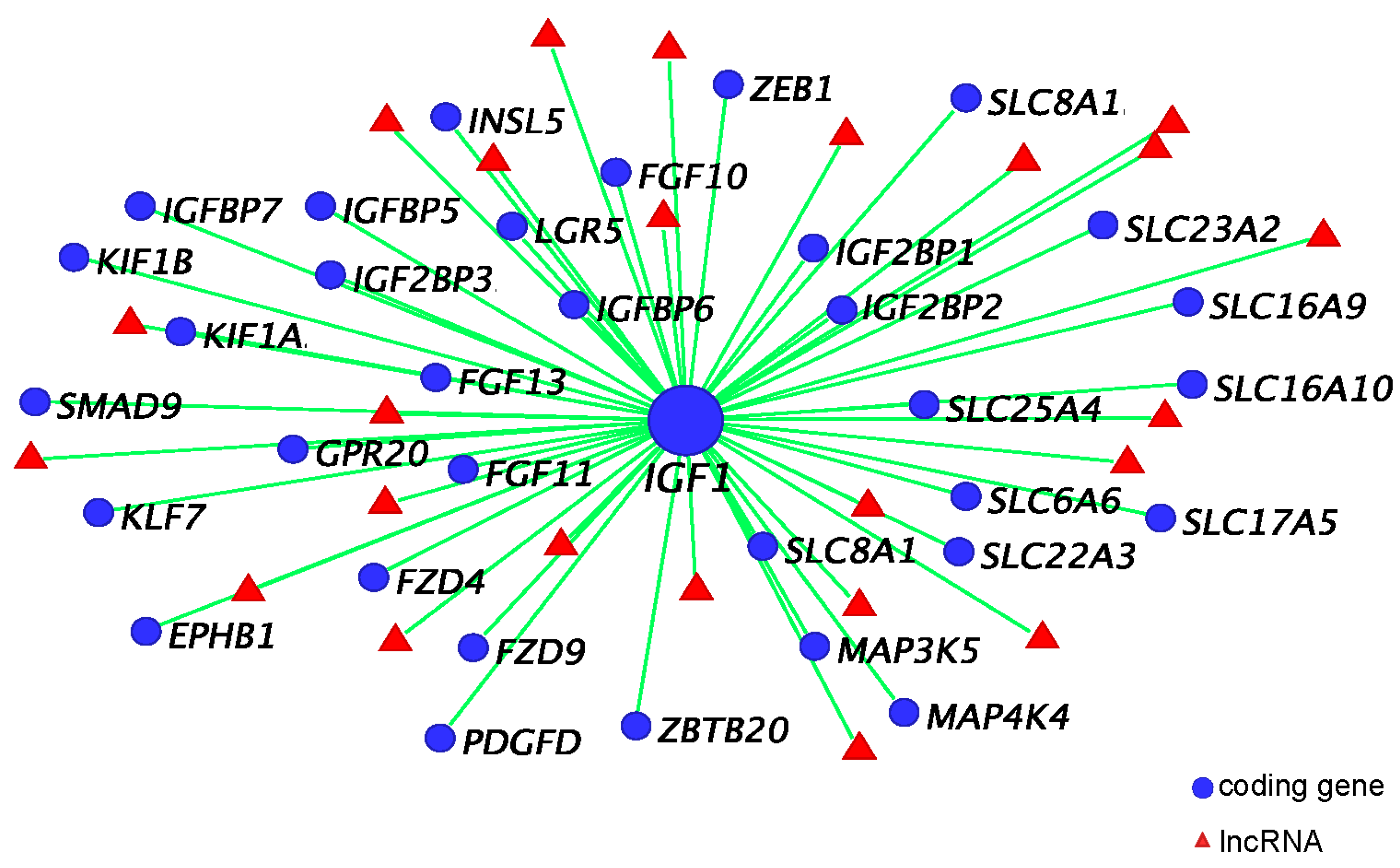
3.5. IGF1 Dependent Phallus Growth

A study conducted by Leihy et al., 2004 demonstrated for the first time an androgen sensitive phase during phallus elongation between days 20 and 40 pp in the tammar [5]. Removing testes in males before day 120 pp reduced phallus length while applying androgen treatment in females before day 120 pp enhances phallus elongation, but has no effect on urethral closure [5]. However, as mentioned before, there is no significant difference in plasma testosterone between male and female at least up to day 50 pp [7,8]. Thus, there must be another regulatory network that is activated by the earlier androgen window of sensitivity to maintain the phallus elongation after day 50 pp. SHH appears to be the key switch that initiates the expression of potential regulatory genes. These may include IGF1, IGFBP3, FGF10, fibroblast growth factor receptor 2 (FGFR2IIIb), Eph-related receptor tyrosine kinase ligand 5 (EFNB2), MAFB, and distal-less homeobox 5 (DLX5). The balance between IGF1 and IGFBP3 could be important in regulating phallus elongation and urethral closure. FGF10, FGFR2IIIb, EFNB2, MAFB, and DLX5 may also involve phallus elongation, since they are significantly higher in male phalluses at day 90 pp [15] and have a conserved localization in urethral epithelium, as seen in mice [28,37,38,76,77,78]. In addition, these genes appear to be important to maintain cell proliferation and survival [79,80,81,82,83,84,85] during male phallus development in mice [23,29,86,87].

4. Co-Expression Network and Hormonally Responsive Long Non-Coding RNAs

Our RNA-seq dataset consists of five different treatment groups with 5 replicates for each group, which makes it hard to interpret with differential expression (DE) analysis. We used weighted genome co-expression network analysis (WGCNA) to find co-expressed genes. It is also a good way to identify lncRNAs as most of them have extremely low sequence conservation, making them difficult to identify cross species with alignment. In our previous paper, we set up a pipeline by combining WGCNA, DE analysis, and the location of lncRNAs and identified the following three coding gene-neighboring lncRNAs: lnc-RSPO4, lnc-BMP5, and lnc-ZBTB16 [88].

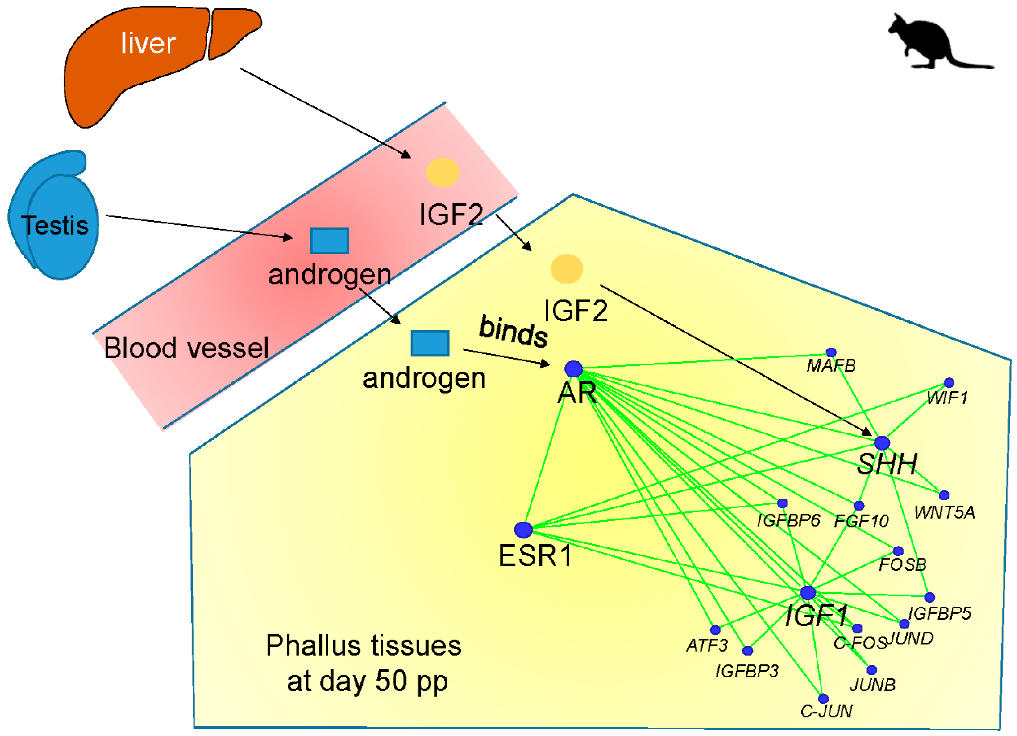
4.1. IGF1, Androgen Receptor (AR), and ESR1 Co-Expression Network

IGF1 is considered as a hub gene in its co-expression network due to its high correlation with a large number of protein-coding genes and lncRNAs. Within the IGF1 co-expression network, both IGFBP5, an IGF signalling regulator (reviewed in [89]) that inhibits SHH-induced proliferation in cerebellar granule cells in mice [44], and FGF10, a phallus regulating gene in mice and the tammar [15,28,29,77,78], have a high correlation (R ≥ 0.8) with IGF1 [88] (Figure 5). IGF1 is also co-expressed with multiple genes that may have a role in regulating reproductive development (Figure 5). For instance, it is co-expressed with other IGF family members, including insulin like growth factor 2 binding protein (IGF2BP) 1–3, insulin like 5 (INSL5), and IGFBP7. Apart from FGF10, IGF1 is also associated with FGF11, FGF13, and tyrosine-protein kinase receptor EPH-2 (EPHB1). Two receptors, frizzled class receptor 4 (FZD4) and FZD9, in the WNT signalling pathway show high association with IGF1. Interestingly, IGF1 is co-expressed with zinc finger and BTB domain containing 20 (ZBTB20), whose mutation causes micro-penis [90]. We also find mutations of several kinesin family members (KIF), such as KIF1A, KIF1B and KIF7, that are associated with IGF1 and can also induce an abnormal phallus phenotype in human [91,92]. These data further confirm the importance of IGF1 in regulating phallus development in the tammar.
In mice, oestrogen signalling clearly has a regulatory role in phallus development [93,94], as we have found in the tammar [13,15,88]. In the tammar co-expression network, about 50% of estrogen receptor 1 (ESR1) co-expressed coding genes and lncRNAs are also associated with AR [88], suggesting an interaction between androgen receptor (AR) signalling and ESR1 signalling during tammar phallus development [88]. However, those lncRNAs could have other genetic targets because none of them were located within 100 kb upstream or downstream of IGF1, AR, and ESR1.

4.2. lnc-RSPO4, lnc-BMP5, and lnc-ZBTB16

We identified three novel lncRNAs using our pipeline. Lnc-RSPO4 is co-expressed with roof plate-specific spondin-4 (RSPO4) coding gene. Both RSPO4 and lnc-RSPO4 are downregulated in tammar female phalluses after androgen treatment [88], in a similar expression pattern to that of SHH and WNT5A [13]. Interestingly, RSPO4 is a ligand of leucine-rich repeat containing G protein-coupled receptor (LGR) 4–6 receptors that potentiate WNT signalling [95,96,97,98]. Thus, RSPO4 and lnc-RSPO4 could also be involved in the molecular regulation mediated by SHH and WNT5A signalling during tammar phallus development.
Both lnc-BMP5 and lnc-ZBTB16 are downregulated in tammar male phalluses after oestrogen treatment [88]. They are also co-expressed with bone morphogenetic protein (BMP5) and zinc finger and BTB domain containing 16 (ZBTB16), respectively, in our co-expression network [88]. Interestingly, Bmp5 is downregulated by flutamide, an androgen signalling inhibitor during phallus development in mice [93]. Mutation of ZBTB16 induces micro-penis [99,100], which is similar to the phenotype observed after oestrogen treatment in the tammar [13]. These data show that there is a complex regulatory system of lncRNAs during phallus development mediated by hormonal signalling.

5. Conclusions and Future Directions

Tammar phallus development is under the regulation of a complex molecular network mediated by endocrine hormones. This review describes two endocrine-mediated networks, the SHH network and the IGF1 network, which may act as molecular switches to constrain and decide male phallus development (Figure 6). The RNA-seq analysis identifies two sets of genes, including WNT5A, MAFB, RSPO4, lnc-RSPO4, FGF10, WIF1 and AP-1, FGF10, IGFBP3, IGFBP6, IGFBP5, EFNB2, that interact with SHH and IGF1, respectively, in the tammar phalluses at day 50 pp. Interestingly, due to the negative association between androgen and SHH transcription, an SHH switch could be a unique regulatory mechanism in the tammar to constrain the timing of phallus differentiation.
The molecular regulatory network that maintains phallus growth after day 50 pp consists of another set of genes, including IGF1, IGFBP3, FGF10, FGFR2IIIb, EFNB2, MAFB, and DLX5. The increased level of those genes may be initiated and enhanced by activation of two endocrine-mediated SHH and IGF1 switches in males, resulting in a phallus with complete urethral closure and elongated shaft. In addition, it is likely that urethral closure is mediated by the activation of IGF1 signalling in the male urorectal septum.
Co-expression analysis to identify novel hormone-responsive lncRNAs, such as lnc-BMP5, lnc-RSPO4, and lnc-ZBTB16, in the tammar phalluses reveals complex regulatory networks of IGF1, AR, and ESR1 that associate with multiple hormone-responsive coding genes and lncRNAs during tammar phallus development. The data also indicate a potential interplay between AR and ESR1 signalling.
Taken together, the activation of the SHH switch and IGF1 switch, mediated by the balance between AR and ESR1 signalling, initiate a complex regulatory network in males to constrain the timing of phallus differentiation and to activate the downstream genes that maintain urethral closure and phallus elongation at later stage.

Author Contributions

Writing—original draft preparation, Y.C. and M.B.R.; writing—review and editing, Y.C. and M.B.R. All authors have read and agreed to the published version of the manuscript.

Funding

This study was supported by grants from the National Health and Medical Research Council of Australia.

Acknowledgments

We thank members of the Wallaby research group (Walgroup) for assistance when handling animals. We also thank members of RIKEN and NIG for the technical support on NGS.

Conflicts of Interest

The authors declare no conflict of interest. The funders had no role in the design of the study; in the collection, analyses, or interpretation of data; in the writing of the manuscript, or in the decision to publish the results.

References

  1. Renfree, M.B.; Pask, A.J.; Shaw, G. Sexual development of a model marsupial male. Aust. J. Zool. 2006, 54, 151–158. [Google Scholar] [CrossRef]
  2. Renfree, M.B.; Short, R.V.; Shaw, G. Sexual differentiation of the urogenital system of the fetal and neonatal tammar wallaby, Macropus eugenii. Anat. Embryol. 1996, 194, 111–134. [Google Scholar] [CrossRef] [PubMed]
  3. Leihy, M.W.; Shaw, G.; Wilson, J.D.; Renfree, M.B. Development of the penile urethra in the tammar wallaby. Sex. Dev. 2011, 5, 241–249. [Google Scholar] [CrossRef]
  4. Butler, C.M.; Shaw, G.; Renfree, M.B. Development of the penis and clitoris in the tammar wallaby, Macropus eugenii. Anat. Embryol. 1999, 199, 451–457. [Google Scholar] [CrossRef]
  5. Leihy, M.W.; Shaw, G.; Wilson, J.D.; Renfree, M.B. Penile development is initiated in the tammar wallaby pouch young during the period when 5α-androstane-3α, 17β-diol is secreted by the testes. Endocrinology 2004, 145, 3346–3352. [Google Scholar] [CrossRef]
  6. Renfree, M.B.; Chew, K.Y.; Shaw, G. Inducing sex reversal of the urogenital system of marsupials. Differentiation 2014, 87, 23–31. [Google Scholar] [CrossRef]
  7. Renfree, M.B.; Wilson, J.D.; Short, R.V.; Shaw, G.; George, F.W. Steroid hormone content of the gonads of the tammar wallaby during sexual differentiation. Biol. Reprod. 1992, 47, 644–647. [Google Scholar] [CrossRef] [Green Version]
  8. Wilson, J.D.; George, F.W.; Shaw, G.; Renfree, M.B. Virilization of the male pouch young of the tammar wallaby does not appear to be mediated by plasma testosterone or dihydrotestosterone. Biol. Reprod. 1999, 61, 471–475. [Google Scholar] [CrossRef] [PubMed] [Green Version]
  9. Chew, K.Y.; Pask, A.J.; Hickford, D.; Shaw, G.; Renfree, M.B. A dual role for SHH during phallus development in a marsupial. Sex. Dev. 2014, 8, 166–177. [Google Scholar] [CrossRef] [PubMed]
  10. Welsh, M.; Saunders, P.T.; Fisken, M.; Scott, H.M.; Hutchison, G.R.; Smith, L.B.; Sharpe, R.M. Identification in rats of a programming window for reproductive tract masculinization, disruption of which leads to hypospadias and cryptorchidism. J. Clin. Investig. 2008, 118, 1479–1490. [Google Scholar] [CrossRef] [Green Version]
  11. Welsh, M.; Suzuki, H.; Yamada, G. The masculinization programming window. Endocr. Dev. 2014, 27, 17–27. [Google Scholar] [PubMed]
  12. Welsh, M.; MacLeod, D.J.; Walker, M.; Smith, L.B.; Sharpe, R.M. Critical androgen-sensitive periods of rat penis and clitoris development. Int. J. Androl. 2010, 33, e144–e152. [Google Scholar] [CrossRef] [PubMed]
  13. Chen, Y.; Yu, H.; Pask, A.J.; Fujiyama, A.; Suzuki, Y.; Sugano, S.; Shaw, G.; Renfree, M.B. Hormone-responsive genes in the SHH and WNT/β-catenin signaling pathways influence urethral closure and phallus growth. Biol. Reprod. 2018, 99, 806–816. [Google Scholar] [CrossRef] [PubMed]
  14. Sirab, N.; Terry, S.; Giton, F.; Caradec, J.; Chimingqi, M.; Moutereau, S.; Vacherot, F.; Taille, A.D.L.; Kouyoumdjian, J.C.; Loric, S. Androgens regulate hedgehog signalling and proliferation in androgen-dependent prostate cells. Int. J. Cancer 2012, 131, 1297–1306. [Google Scholar] [CrossRef] [PubMed]
  15. Chen, Y.; Yu, H.; Pask, A.J.; Fujiyama, A.; Suzuki, Y.; Sugano, S.; Shaw, G.; Renfree, M.B. Effects of androgen and oestrogen on the IGF pathways controlling phallus growth. Reproduction 2019, 157, 1–12. [Google Scholar] [CrossRef] [PubMed] [Green Version]
  16. Menzies, B.R.; Shaw, G.; Fletcher, T.P.; Pask, A.J.; Renfree, M.B. Maturation of the growth axis in marsupials occurs gradually during post-natal life and over an equivalent developmental stage relative to eutherian species. Mol. Cell. Endocrinol. 2012, 349, 189–194. [Google Scholar] [CrossRef]
  17. Miyagawa, S.; Satoh, Y.; Haraguchi, R.; Suzuki, K.; Iguchi, T.; Taketo, M.M.; Nakagata, N.; Matsumoto, T.; Takeyama, K.-I.; Kato, S. Genetic interactions of the androgen and Wnt/β-catenin pathways for the masculinization of external genitalia. Mol. Endocrinol. 2009, 23, 871–880. [Google Scholar] [CrossRef] [Green Version]
  18. Miyagawa, S.; Moon, A.; Haraguchi, R.; Inoue, C.; Harada, M.; Nakahara, C.; Suzuki, K.; Matsumaru, D.; Kaneko, T.; Matsuo, I. Dosage-dependent hedgehog signals integrated with Wnt/β-catenin signaling regulate external genitalia formation as an appendicular program. Development 2009, 136, 3969–3978. [Google Scholar] [CrossRef] [Green Version]
  19. Seifert, A.W.; Zheng, Z.; Ormerod, B.K.; Cohn, M.J. Sonic hedgehog controls growth of external genitalia by regulating cell cycle kinetics. Nat. Commun. 2010, 1, 23. [Google Scholar] [CrossRef] [Green Version]
  20. Kawano, Y.; Kypta, R. Secreted antagonists of the Wnt signalling pathway. J. Cell Sci. 2003, 116, 2627–2634. [Google Scholar] [CrossRef] [Green Version]
  21. Hsieh, J.-C.; Kodjabachian, L.; Rebbert, M.L.; Rattner, A.; Smallwood, P.M.; Samos, C.H.; Nusse, R.; Dawid, I.B.; Nathans, J. A new secreted protein that binds to Wnt proteins and inhibits their activites. Nature 1999, 398, 431–436. [Google Scholar] [CrossRef] [PubMed]
  22. Ng, R.C.; Matsumaru, D.; Ho, A.S.; Garcia-Barceló, M.; Yuan, Z.; Smith, D.; Kodjabachian, L.; Tam, P.K.; Yamada, G.; Lui, V.C. Dysregulation of Wnt inhibitory factor 1 (Wif1) expression resulted in aberrant wnt-β-catenin signaling and cell death of the cloaca endoderm, and anorectal malformations. Cell Death Differ. 2014, 21, 978–989. [Google Scholar] [CrossRef] [PubMed] [Green Version]
  23. Suzuki, K.; Numata, T.; Suzuki, H.; Raga, D.D.; Ipulan, L.A.; Yokoyama, C.; Matsushita, S.; Hamada, M.; Nakagata, N.; Nishinakamura, R.; et al. Sexually dimorphic expression of Mafb regulates masculinization of the embryonic urethral formation. Proc. Natl. Acad. Sci. USA 2014, 111, 16407–16412. [Google Scholar] [CrossRef] [PubMed] [Green Version]
  24. Matsushita, S.; Suzuki, K.; Ogino, Y.; Hino, S.; Sato, T.; Suyama, M.; Matsumoto, T.; Omori, A.; Inoue, S.; Yamada, G. Androgen regulates Mafb expression through its 3’ UTR during mouse urethral masculinization. Endocrinology 2015, 157, 844–857. [Google Scholar] [CrossRef] [Green Version]
  25. Matsushita, S.; Suzuki, K.; Murashima, A.; Kajioka, D.; Acebedo, A.R.; Miyagawa, S.; Haraguchi, R.; Ogino, Y.; Yamada, G. Regulation of masculinization: Androgen signalling for external genitalia development. Nat. Rev. Urol. 2018, 15, 358. [Google Scholar] [CrossRef]
  26. Bellusci, S.; Grindley, J.; Emoto, H.; Itoh, N.; Hogan, B. Fibroblast growth factor 10 (FGF10) and branching morphogenesis in the embryonic mouse lung. Development 1997, 124, 4867–4878. [Google Scholar]
  27. Haraguchi, R.; Suzuki, K.; Murakami, R.; Sakai, M.; Kamikawa, M.; Kengaku, M.; Sekine, K.; Kawano, H.; Kato, S.; Ueno, N. Molecular analysis of external genitalia formation: The role of fibroblast growth factor (FGF) genes during genital tubercle formation. Development 2000, 127, 2471–2479. [Google Scholar]
  28. Petiot, A.; Perriton, C.L.; Dickson, C.; Cohn, M.J. Development of the mammalian urethra is controlled by Fgfr2-IIIb. Development 2005, 132, 2441–2450. [Google Scholar] [CrossRef] [Green Version]
  29. Satoh, Y.; Haraguchi, R.; Wright, T.J.; Mansour, S.L.; Partanen, J.; Hajihosseini, M.K.; Eswarakumar, V.P.; Lonai, P.; Yamada, G. Regulation of external genitalia development by concerted actions of FGF ligands and FGF receptors. Anat. Embryol. 2004, 208, 479–486. [Google Scholar] [CrossRef]
  30. Shaw, G.; Renfree, M.B.; Leihy, M.W.; Shackleton, C.H.; Roitman, E.; Wilson, J.D. Prostate formation in a marsupial is mediated by the testicular androgen 5α-androstane-3α, 17β-diol. Proc. Natl. Acad. Sci. USA 2000, 97, 12256–12259. [Google Scholar] [CrossRef] [Green Version]
  31. Shaw, G.; Renfree, M.B.; Short, R.V. Experimental manipulation of sexual differentiation in wallaby pouch young treated with exogenous steroids. Development 1988, 104, 689–701. [Google Scholar] [PubMed]
  32. Kurzrock, E.A.; Baskin, L.S.; Cunha, G.R. Ontogeny of the male urethra: Theory of endodermal differentiation. Differentiation 1999, 64, 115–122. [Google Scholar] [CrossRef] [PubMed]
  33. Baskin, L.; Lee, Y.; Cunha, G. Neuroanatomical ontogeny of the human fetal penis. Br. J. Urol. 1997, 79, 628–640. [Google Scholar] [CrossRef] [Green Version]
  34. Baskin, L.; Shen, J.; Sinclair, A.; Cao, M.; Liu, X.; Liu, G.; Isaacson, D.; Overland, M.; Li, Y.; Cunha, G.R. Development of the human penis and clitoris. Differentiation 2018, 103, 74–85. [Google Scholar] [CrossRef] [PubMed] [Green Version]
  35. Lowsley, O.S. The development of the human prostate gland with reference to the development of other structures at the neck of the urinary bladder. Am. J. Anat. 1912, 13, 299–349. [Google Scholar] [CrossRef]
  36. Kellokumpu-Lehtinen, P.; Santti, R.; Pelliniemi, L. Correlation of early cytodifferentiation of the human fetal prostate and leydig cells. Anat. Rec. 1980, 196, 263–273. [Google Scholar] [CrossRef]
  37. Perriton, C.L.; Powles, N.; Chiang, C.; Maconochie, M.K.; Cohn, M.J. Sonic hedgehog signaling from the urethral epithelium controls external genital development. Dev. Biol. 2002, 247, 26–46. [Google Scholar] [CrossRef] [Green Version]
  38. Haraguchi, R.; Mo, R.; Hui, C.; Motoyama, J.; Makino, S.; Shiroishi, T.; Gaffield, W.; Yamada, G. Unique functions of sonic hedgehog signaling during external genitalia development. Development 2001, 128, 4241–4250. [Google Scholar]
  39. Liu, G.; Liu, X.; Shen, J.; Sinclair, A.; Baskin, L.; Cunha, G.R. Contrasting mechanisms of penile urethral formation in mouse and human. Differentiation 2018, 101, 46–64. [Google Scholar] [CrossRef]
  40. Cunha, G.R.; Donjacour, A.A.; Cooke, P.S.; Mee, H.; Bigsby, R.M.; Higgins, S.J.; Sugimura, Y. The endocrinology and developmental biology of the prostate. Endocr. Rev. 1987, 8, 338–362. [Google Scholar] [CrossRef]
  41. Timms, B.G.; Mohs, T.J.; Didio, L.J. Ductal budding and branching patterns in the developing prostate. J. Urol. 1994, 151, 1427–1432. [Google Scholar] [CrossRef]
  42. Price, D. Normal development of the prostate and seminal vesicles of the rat with a study of experimental postnatal modifications. Am. J. Anat. 1936, 60, 79–127. [Google Scholar] [CrossRef]
  43. Inomata, T.; Eguchi, Y.; Nakamura, T. Development of the external genitalia in rat fetuses. Exp. Anim. 1985, 34, 439–444. [Google Scholar] [CrossRef] [Green Version]
  44. Fernandez, C.; Tatard, V.M.; Bertrand, N.; Dahmane, N. Differential modulation of sonic-hedgehog-induced cerebellar granule cell precursor proliferation by the IGF signaling network. Dev. Neurosci. 2010, 32, 59–70. [Google Scholar] [CrossRef]
  45. Rao, G.; Pedone, C.A.; del Valle, L.; Reiss, K.; Holland, E.C.; Fults, D.W. Sonic hedgehog and insulin-like growth factor signaling synergize to induce medulloblastoma formation from nestin-expressing neural progenitors in mice. Oncogene 2004, 23, 6156. [Google Scholar] [CrossRef] [PubMed] [Green Version]
  46. Pirskanen, A.; Kiefer, J.C.; Hauschka, S.D. IGFs, insulin, Shh, bFGF, and TFG-β1 interact synergistically to promote somite myogenesis in vitro. Dev. Biol. 2000, 224, 189–203. [Google Scholar] [CrossRef] [PubMed] [Green Version]
  47. Andersen, M.; Nørgaard-Pedersen, D.; Brandt, J.; Pettersson, I.; Slaaby, R. IGF1 and IGF2 specificities to the two insulin receptor isoforms are determined by insulin receptor amino acid 718. PLoS ONE 2017, 12, e0178885. [Google Scholar] [CrossRef] [PubMed] [Green Version]
  48. Laron, Z.; Pertzelan, A.; Mannheimer, S. Genetic pituitary dwarfism with high serum concentation of growth hormone—A new inborn error of metabolism? Isr. J. Med. Sci. 1966, 2, 152–155. [Google Scholar]
  49. Laron, Z.; Klinger, B. Effect of insulin-like growth factor-I treatment on serum androgens and testicular and penile size in males with Laron syndrome (primary growth hormone resistance). Eur. J. Endocrinol. 1998, 138, 176–180. [Google Scholar] [CrossRef] [Green Version]
  50. Levy, J.; Husmann, D. Micropenis secondary to growth hormone deficiency: Does treatment with growth hormone alone result in adequate penile growth? J. Urol. 1996, 156, 214–216. [Google Scholar] [CrossRef]
  51. Stewart, C.E.; Bates, P.C.; Calder, T.A.; Woodall, S.M.; Pell, J.M. Potentiation of insulin-like growth factor-I (IGF-I) activity by an antibody: Supportive evidence for enhancement of IGF-I bioavailability in vivo by IGF binding proteins. Endocrinology 1993, 133, 1462–1465. [Google Scholar] [CrossRef] [PubMed]
  52. Kalus, W.; Zweckstetter, M.; Renner, C.; Sanchez, Y.; Georgescu, J.; Grol, M.; Demuth, D.; Schumacher, R.; Dony, C.; Lang, K.; et al. Structure of the IGF-binding domain of the insulin-like growth factor-binding protein-5 (IGFBP-5): Implications for IGF and IGF-I receptor interactions. EMBO J. 1998, 17, 6558–6572. [Google Scholar] [CrossRef] [PubMed] [Green Version]
  53. Clemmons, D.R. Role of IGF binding proteins in regulating metabolism. Trends Endocrinol. Metab. 2016, 27, 375–391. [Google Scholar] [CrossRef] [PubMed]
  54. Kamanga-Sollo, E.; Pampusch, M.; Xi, G.; White, M.; Hathaway, M.; Dayton, W. IGF-I mRNA levels in bovine satellite cell cultures: Effects of fusion and anabolic steroid treatment. J. Cell. Physiol. 2004, 201, 181–189. [Google Scholar] [CrossRef]
  55. Sahlin, L.; Norstedt, G.; Eriksson, H. Androgen regulation of the insulin-like growth factor-I and the estrogen receptor in rat uterus and liver. J. Steroid Biochem. Mol. Biol. 1994, 51, 57–66. [Google Scholar] [CrossRef]
  56. Arnold, J.T.; Le, H.; McFann, K.K.; Blackman, M.R. Comparative effects of DHEA vs. testosterone, dihydrotestosterone, and estradiol on proliferation and gene expression in human LNCaP prostate cancer cells. Am. J. Physiol. Endocrinol. Metab. 2005, 288, E573–E584. [Google Scholar] [CrossRef] [Green Version]
  57. Le, H.; Arnold, J.T.; McFann, K.K.; Blackman, M.R. DHT and testosterone, but not DHEA or E2, differentially modulate IGF-I, IGFBP-2, and IGFBP-3 in human prostatic stromal cells. Am. J. Physiol.-Endocrinol. Metab. 2006, 290, E952–E960. [Google Scholar] [CrossRef] [Green Version]
  58. Cheng, C.M.; Cohen, M.; Wang, J.; Bondy, C.A. Estrogen augments glucose transporter and IGF1 expression in primate cerebral cortex. FASEB J. 2001, 15, 907–915. [Google Scholar] [CrossRef] [Green Version]
  59. Hewitt, S.C.; Li, Y.; Li, L.; Korach, K.S. Estrogen-mediated regulation of IGF1 transcription and uterine growth involves direct binding of estrogen receptor α to estrogen-responsive elements. J. Biol. Chem. 2010, 285, 2676–2685. [Google Scholar] [CrossRef] [Green Version]
  60. Henderson, N.A.; Cooke, G.M.; Robaire, B. Region-specific expression of androgen and growth factor pathway genes in the rat epididymis and the effects of dual 5α-reductase inhibition. J. Endocrinol. 2006, 190, 779–791. [Google Scholar] [CrossRef]
  61. Koike, H.; Ito, K.; Takezawa, Y.; Oyama, T.; Yamanaka, H.; Suzuki, K. Insulin-like growth factor binding protein-6 inhibits prostate cancer cell proliferation: Implication for anticancer effect of diethylstilbestrol in hormone refractory prostate cancer. Br. J. Cancer 2005, 92, 1538–1544. [Google Scholar] [CrossRef] [PubMed] [Green Version]
  62. Kojima, S.; Mulholland, D.J.; Ettinger, S.; Fazli, L.; Nelson, C.C.; Gleave, M.E. Differential regulation of IGFBP-3 by the androgen receptor in the lineage-related androgen-dependent LNCaP and androgen-independent C4-2 prostate cancer models. Prostate 2006, 66, 971–986. [Google Scholar] [CrossRef] [PubMed]
  63. Peng, L.; Wang, J.; Malloy, P.J.; Feldman, D. The role of insulin-like growth factor binding protein-3 in the growth inhibitory actions of androgens in LNCaP human prostate cancer cells. Int. J. Cancer 2008, 122, 558–566. [Google Scholar] [CrossRef]
  64. Grimberg, A.; Cohen, P. Role of insulin-like growth factors and their binding proteins in growth control and carcinogenesis. J. Cell. Physiol. 2000, 183, 1–9. [Google Scholar] [CrossRef] [Green Version]
  65. Pollak, M. Insulin-like growth factor physiology and cancer risk. Eur. J. Cancer 2000, 36, 1224–1228. [Google Scholar] [CrossRef]
  66. Duan, C.; Xu, Q. Roles of insulin-like growth factor (IGF) binding proteins in regulating IGF actions. Gen. Comp. Endocrinol. 2005, 142, 44–52. [Google Scholar] [CrossRef]
  67. Schmid, C.H.; Rutishauser, J.; Schläpfer, I.; Froesch, E.R.; Zapf, J. Intact but not truncated insulin-like growth factor binding protein-3 (IGFBP-3) blocks IGF I-induced stimulation of osteoblasts: Control of IGF signalling to bone cells by IGFBP-3-specific proteolysis? Biochem. Biophys. Res. Commun. 1991, 179, 579–585. [Google Scholar] [CrossRef]
  68. Valentinis, B.; Bhala, A.; DeAngelis, T.; Baserga, R.; Cohen, P. The human insulin-like growth factor (IGF) binding protein-3 inhibits the growth of fibroblasts with a targeted disruption of the Igf-1 receptor gene. Mol. Endocrinol. 1995, 9, 361–367. [Google Scholar]
  69. Umayahara, Y.; Kawamori, R.; Watada, H.; Imano, E.; Iwama, N.; Morishima, T.; Yamasaki, Y.; Kajimoto, Y.; Kamada, T. Estrogen regulation of the insulin-like growth factor 1 gene transcription involves an AP-1 enhancer. J. Biol. Chem. 1994, 269, 16433–16442. [Google Scholar]
  70. Kerr, J.; Beck, S.; Handa, R. Androgens selectively modulate C-Fos messenger RNA induction in the rat hippocampus following novelty. Neuroscience 1996, 74, 757–766. [Google Scholar] [CrossRef]
  71. Pelzer, A.E.; Bektic, J.; Haag, P.; Berger, A.P.; Pycha, A.; Schäfer, G.; Rogatsch, H.; Horninger, W.; Bartsch, G.; Klocker, H. The expression of transcription factor activating transcription factor 3 in the human prostate and its regulation by androgen in prostate cancer. J. Urol. 2006, 175, 1517–1522. [Google Scholar] [CrossRef]
  72. Angel, P.; Karin, M. The role of Jun, Fos and the AP-1 complex in cell-proliferation and transformation. Biochim. Biophys. Acta Rev. Cancer 1991, 1072, 129–157. [Google Scholar] [CrossRef]
  73. Seifert, A.W.; Harfe, B.D.; Cohn, M.J. Cell lineage analysis demonstrates an endodermal origin of the distal urethra and perineum. Dev. Biol. 2008, 318, 143–152. [Google Scholar] [CrossRef] [PubMed] [Green Version]
  74. Seifert, A.W.; Bouldin, C.M.; Choi, K.S.; Harfe, B.D.; Cohn, M.J. Multiphasic and tissue-specific roles of sonic hedgehog in cloacal septation and external genitalia development. Development 2009, 136, 3949–3957. [Google Scholar] [CrossRef] [Green Version]
  75. Hyuga, T.; Suzuki, K.; Acebedo, A.R.; Hashimoto, D.; Kajimoto, M.; Miyagawa, S.; Enmi, J.-I.; Yoshioka, Y.; Yamada, G. Regulatory roles of epithelial-mesenchymal interaction (EMI) during early and androgen dependent external genitalia development. Differentiation 2019, 110, 29–35. [Google Scholar] [CrossRef] [PubMed]
  76. Dravis, C.; Yokoyama, N.; Chumley, M.J.; Cowan, C.A.; Silvany, R.E.; Shay, J.; Baker, L.A.; Henkemeyer, M. Bidirectional signaling mediated by ephrin-B2 and EphB2 controls urorectal development. Dev. Biol. 2004, 271, 272–290. [Google Scholar] [CrossRef] [PubMed] [Green Version]
  77. Gredler, M.L.; Seifert, A.W.; Cohn, M.J. Tissue-specific roles of Fgfr2 in development of the external genitalia. Development 2015, 142, 2203–2212. [Google Scholar] [CrossRef] [Green Version]
  78. Harada, M.; Omori, A.; Nakahara, C.; Nakagata, N.; Akita, K.; Yamada, G. Tissue-specific roles of FGF signaling in external genitalia development. Dev. Dyn. 2015, 244, 759–773. [Google Scholar] [CrossRef] [Green Version]
  79. Bhushan, A.; Itoh, N.; Kato, S.; Thiery, J.P.; Czernichow, P.; Bellusci, S.; Scharfmann, R. Fgf10 is essential for maintaining the proliferative capacity of epithelial progenitor cells during early pancreatic organogenesis. Development 2001, 128, 5109–5117. [Google Scholar]
  80. Hart, A.; Papadopoulou, S.; Edlund, H. Fgf10 maintains notch activation, stimulates proliferation, and blocks differentiation of pancreatic epithelial cells. Dev. Dyn. 2003, 228, 185–193. [Google Scholar] [CrossRef]
  81. Steinberg, Z.; Myers, C.; Heim, V.M.; Lathrop, C.A.; Rebustini, I.T.; Stewart, J.S.; Larsen, M.; Hoffman, M.P. Fgfr2b signaling regulates ex vivo submandibular gland epithelial cell proliferation and branching morphogenesis. Development 2005, 132, 1223–1234. [Google Scholar] [CrossRef] [PubMed] [Green Version]
  82. Weiler, S.; Rohrbach, V.; Pulvirenti, T.; Adams, R.; Ziemiecki, A.; Andres, A.C. Mammary epithelial-specific knockout of the ephrin-B2 gene leads to precocious epithelial cell death at lactation. Dev. Growth Differ. 2009, 51, 809–819. [Google Scholar] [CrossRef] [PubMed]
  83. Bendall, A.J.; Hu, G.; Levi, G.; Abate-Shen, C. Dlx5 regulates chondrocyte differentiation at multiple stages. Int. J. Dev. Biol. 2003, 47, 335–344. [Google Scholar] [PubMed]
  84. Yu, H.; Jiang, H.; Xu, D.; Jin, J.; Zhao, Z.; Ma, Y.; Liang, J. Transcription factor MafB promotes hepatocellular carcinoma cell proliferation through up-regulation of cyclin D1. Cell. Physiol. Biochem. 2016, 39, 700–708. [Google Scholar] [CrossRef]
  85. Shaulian, E.; Karin, M. Ap-1 in cell proliferation and survival. Oncogene 2001, 20, 2390–2400. [Google Scholar] [CrossRef] [Green Version]
  86. Egea, J.; Klein, R. Bidirectional Eph–ephrin signaling during axon guidance. Trends Cell Biol. 2007, 17, 230–238. [Google Scholar] [CrossRef]
  87. Suzuki, K.; Haraguchi, R.; Ogata, T.; Barbieri, O.; Alegria, O.; Vieux-Rochas, M.; Nakagata, N.; Ito, M.; Mills, A.A.; Kurita, T. Abnormal urethra formation in mouse models of split-hand/split-foot malformation type 1 and type 4. Eur. J. Hum. Genet. 2008, 16, 36–44. [Google Scholar] [CrossRef]
  88. Chen, Y.; Kuroki, Y.; Shaw, G.; Pask, A.J.; Yu, H.; Toyoda, A.; Fujiyama, A.; Renfree, M.B. Androgen and oestrogen affect the expression of long non-coding rnas during phallus development in a marsupial. Non-Coding RNA 2019, 5, 3. [Google Scholar] [CrossRef] [Green Version]
  89. Lindsey, R.C.; Rundle, C.H.; Mohan, S. Role of IGF-I and EFN-EPH signaling in skeletal metabolism. J. Mol. Endocrinol. 2018, 61, T87–T102. [Google Scholar] [CrossRef] [Green Version]
  90. Lovrecic, L.; Rudolf, G.; Veble, A.; Peterlin, B. A new case of rare proximal 3q13 interstitial deletion. Open Med. 2011, 6, 625–630. [Google Scholar] [CrossRef]
  91. Walsh, D.M.; Shalev, S.A.; Simpson, M.A.; Morgan, N.V.; Gelman-Kohan, Z.; Chemke, J.; Trembath, R.C.; Maher, E.R. Acrocallosal syndrome: Identification of a novel KIF7 mutation and evidence for oligogenic inheritance. Eur. J. Med. Genet. 2013, 56, 39–42. [Google Scholar] [CrossRef] [PubMed]
  92. Chen, C.-P.; Lin, C.-J.; Chang, T.-Y.; Chern, S.-R.; Wu, P.-S.; Chen, Y.-T.; Su, J.-W.; Lee, C.-C.; Chen, L.-F.; Wang, W. Prenatal diagnosis of ring chromosome 2 with lissencephaly and 2p25.3 and 2q37.3 microdeletions detected using array comparative genomic hybridization. Gene 2013, 519, 164–168. [Google Scholar] [CrossRef] [PubMed]
  93. Zheng, Z.; Armfield, B.A.; Cohn, M.J. Timing of androgen receptor disruption and estrogen exposure underlies a spectrum of congenital penile anomalies. Proc. Natl. Acad. Sci. USA 2015, 112, E7194–E7203. [Google Scholar] [CrossRef] [PubMed] [Green Version]
  94. Govers, L.C.; Phillips, T.R.; Mattiske, D.M.; Rashoo, N.; Black, J.R.; Sinclair, A.; Baskin, L.S.; Risbridger, G.P.; Pask, A.J. A critical role for estrogen signaling in penis development. FASEB J. 2019, 33, fj-201802586RR. [Google Scholar] [CrossRef] [Green Version]
  95. Carmon, K.S.; Gong, X.; Lin, Q.; Thomas, A.; Liu, Q. R-spondins function as ligands of the orphan receptors LGR4 and LGR5 to regulate Wnt/β-catenin signaling. Proc. Natl. Acad. Sci. USA 2011, 108, 11452–11457. [Google Scholar] [CrossRef] [Green Version]
  96. Carmon, K.S.; Gong, X.; Yi, J.; Thomas, A.; Liu, Q. RSPO–LGR4 functions via IQGAP1 to potentiate Wnt signaling. Proc. Natl. Acad. Sci. USA 2014, 111, E1221–E1229. [Google Scholar] [CrossRef] [Green Version]
  97. Chen, P.-H.; Chen, X.; Lin, Z.; Fang, D.; He, X. The structural basis of R-spondin recognition by LGR5 and RNF43. Genes Dev. 2013, 27, 1345–1350. [Google Scholar] [CrossRef] [Green Version]
  98. Kim, K.-A.; Zhao, J.; Andarmani, S.; Kakitani, M.; Oshima, T.; Binnerts, M.E.; Abo, A.; Tomizuka, K.; Funk, W.D. R-Spondin proteins: A novel link to β-catenin activation. Cell Cycle 2006, 5, 23–26. [Google Scholar] [CrossRef]
  99. Wieczorek, D.; Köster, B.; Gillessen-Kaesbach, G. Absence of thumbs, A/hypoplasia of radius, hypoplasia of ulnae, retarded bone age, short stature, microcephaly, hypoplastic genitalia, and mental retardation. Am. J. Med. Genet. Part A 2002, 108, 209–213. [Google Scholar] [CrossRef]
  100. Fischer, S.; Kohlhase, J.; Böhm, D.; Schweiger, B.; Hoffmann, D.; Heitmann, M.; Horsthemke, B.; Wieczorek, D. Biallelic loss of function of the promyelocytic leukaemia zinc finger (PLZF) gene causes severe skeletal defects and genital hypoplasia. J. Med. Genet. 2008, 45, 731–737. [Google Scholar] [CrossRef]
Figure 1. The timeline of prostate differentiation, phallus development, the androgen imprinting window, the androgen sensitive phase, and the insulin-like growth factor 1 (IGF1) dependent phase in the tammar wallaby.
Genes 11 00106 g001
Figure 2. Gene expression of SHH, WNT5A, MAFB, FGF10, and WIF1 in phalluses at day 50 pp. SHH, WNT5A, and MAFB expression is higher in female phalluses and increases in male phalluses after castration. SHH is upregulated by oestrogen treatment in males but downregulated in females after adiol treatment. WNT5A is downregulated in females after adiol treatment. Both FGF10 and WIF1 are higher in male phalluses at day 50 pp. FGF10 is upregulated by adiol treatment and WIF1 is downregulated by oestrogen treatment. A: adiol, E: oestrogen, *: p-value < 0.05, **: p-value < 0.005, SEM: Standard error of the mean (error bar), Red: control female phalluses, purple: adiol treated female phalluses, green: oestrogen treated male phalluses, blue: control male phalluses, and orange: male phalluses with testes removed (male symbol with a cross). Figure redrawn from [13,16].
Genes 11 00106 g002
Figure 3. Gene expression of IGF1, IGFBP3, IGFBP6, and Activator Protein 1 (AP-1) genes in phalluses at day 50 pp. Both IGF1 and IGFBP6 are upregulated by adiol and oestrogen treatment. IGF1 is higher in normal male phalluses at day 50 pp and is downregulated in males after castration. IGFBP3 is higher in female phalluses at day 50 pp and is downregulated in females after adiol treatment. All six AP-1 genes (C-FOS, FOSB, ATF3, c-JUN, JUNB, and JUND) are higher in male phalluses and upregulated by adiol treatment. C-FOS is downregulated by oestrogen treatment. A: adiol, E: oestrogen, FPKM: Fragments per kilobase million; *: p-value < 0.05, **: p-value < 0.005, ***: p-value < 0.001. Figure redrawn from [15].
Genes 11 00106 g003
Figure 4. IGF1 and proliferating cell nuclear antigen (PCNA) distribution in phalluses at day 90 pp. In both male and female phalluses, IGF1 (top) and PCNA (bottom) are expressed in epithelial cells and in the corpora cavernosa. However, IGF1 and nuclear PCNA are found only in the URS of male phalluses (see insets). CC: corpus cavernosum, D: diverticulum, E: epithelium, U: urethra, UE: urethral epithelium, UG: urethra groove, URS: urorectal septum, red staining: IGF1 (top) and PCNA (bottom), and blue staining: DAPI (4′,6-diamidino-2-fenilindol). Scale bar, 1.0 mm. Figures from [15].
Genes 11 00106 g004
Figure 5. IGF1, androgen receptor (AR), and ESR1 co-expression network. IGF1 co-expressed coding genes (selected based on correlation and reference review) and predicted co-regulatory long non-coding RNAs (R > 0.9). Figure redrawn from [88].
Genes 11 00106 g005
Figure 6. Summary of IGF2-SHH and androgen-IGF1 signalling networks. The activation of IGF2-SHH and androgen-IGF1 signalling networks initiate urethral closure in males, whereas in females, non-activation of those two signalling networks results in an unfused urethra.
Genes 11 00106 g006

