Review

MPN: The Molecular Drivers of Disease Initiation, Progression and Transformation and their Effect on Treatment

1
Cancer Program, QIMR Berghofer Medical Research Institute, Brisbane, QLD 4006, Australia
2
Faculty of Medicine, The University of Queensland, Brisbane, QLD 4072, Australia
3
Cancer Care Services, Royal Brisbane and Women’s Hospital, Brisbane, QLD 4029, Australia
*
Author to whom correspondence should be addressed.
Cells 2020, 9(8), 1901; https://doi.org/10.3390/cells9081901
Submission received: 6 July 2020 / Revised: 7 August 2020 / Accepted: 11 August 2020 / Published: 14 August 2020
(This article belongs to the Special Issue Pathophysiology and Molecular Targets in Myeloid Neoplasia)

Abstract

:
Myeloproliferative neoplasms (MPNs) constitute a group of disorders identified by an overproduction of cells derived from myeloid lineage. The majority of MPNs have an identifiable driver mutation responsible for cytokine-independent proliferative signalling. The acquisition of coexisting mutations in chromatin modifiers, spliceosome complex components, DNA methylation modifiers, tumour suppressors and transcriptional regulators have been identified as major pathways for disease progression and leukemic transformation. They also confer different sensitivities to therapeutic options. This review will explore the molecular basis of MPN pathogenesis and specifically examine the impact of coexisting mutations on disease biology and therapeutic options.

1. Introduction

Myeloproliferative Neoplasms (MPN) are clonal haematological disorders that lead to overproduction of cells derived from the myeloid lineage. Specific driver mutations within stem cells and myeloid progenitors provide cytokine-independent or -hypersensitive proliferative signals leading to the overproduction of myeloid cells. Here, we categorise the genetic changes that lead to these disorders and that contribute to progression of disease.
The 2016 WHO classification of MPN includes BCR-ABL1-positive chronic myeloid leukaemia (CML), polycythemia vera (PV), essential thrombocythemia (ET), primary myelofibrosis (PMF), chronic neutrophilic leukaemia, chronic eosinophilic leukaemia and MPN-unclassifiable. For the purpose of this review, we will focus on the more common MPNs of PV, ET and PMF as they share similar driver mutations. CML is a biologically distinct entity and will not be considered in this review.
PV is characterized by elevated red cell production including increased haemoglobin concentration, increased haematocrit and/or increased red cell mass. The vast majority carry a Janus Kinase 2 JAK2V617F mutation with a rare subset having an JAK2 exon 12 mutation.
ET is characterized by sustained thrombocytosis and increased megakaryocyte numbers. The most common driver mutation is JAK2V617F in 50–60% of cases, with Calreticulin (CALR) mutations in 30% and MPL (Myeloproliferative Leukemia) mutations at <5% of cases.
PMF is characterized by proliferation of abnormal megakaryocytes and granulocytes with deposition of fibrotic tissue in the bone marrow and extramedullary haematopoiesis. The driver mutations most commonly identified are JAK2V617F in 50–60%, with CALR in 30% and MPL in 8% of cases [1].
MPNs can have a variable disease courses, with PV and ET having a more indolent course; however, the progression can be variable with the overall survival for PV ranging from 13.5 to 24 years and ET ranging from 11 to 22.6 years. Progression to myelofibrosis (MF) over a period of 20 years occurs in 26% of PV and 19.9% of ET. The incidence of leukemic transformation over a period of 20 years is 7.9–17% for PV and 8.1% for ET [2,3].
The purpose of this review is to examine the prognostic scoring systems utilized in MPNs. We will explore the driver mutations that are diagnostic hallmarks of MPN, their pathogenesis behind disease and their phenotypic variation accounting for the same mutations in different disease phenotypes. We will then review coexisting mutations that corroborate driver mutations leading to progression. Finally, we explore the therapeutic options in MPN and how they impact the pathogenesis of MPN.

2. Prognostic Scoring Systems
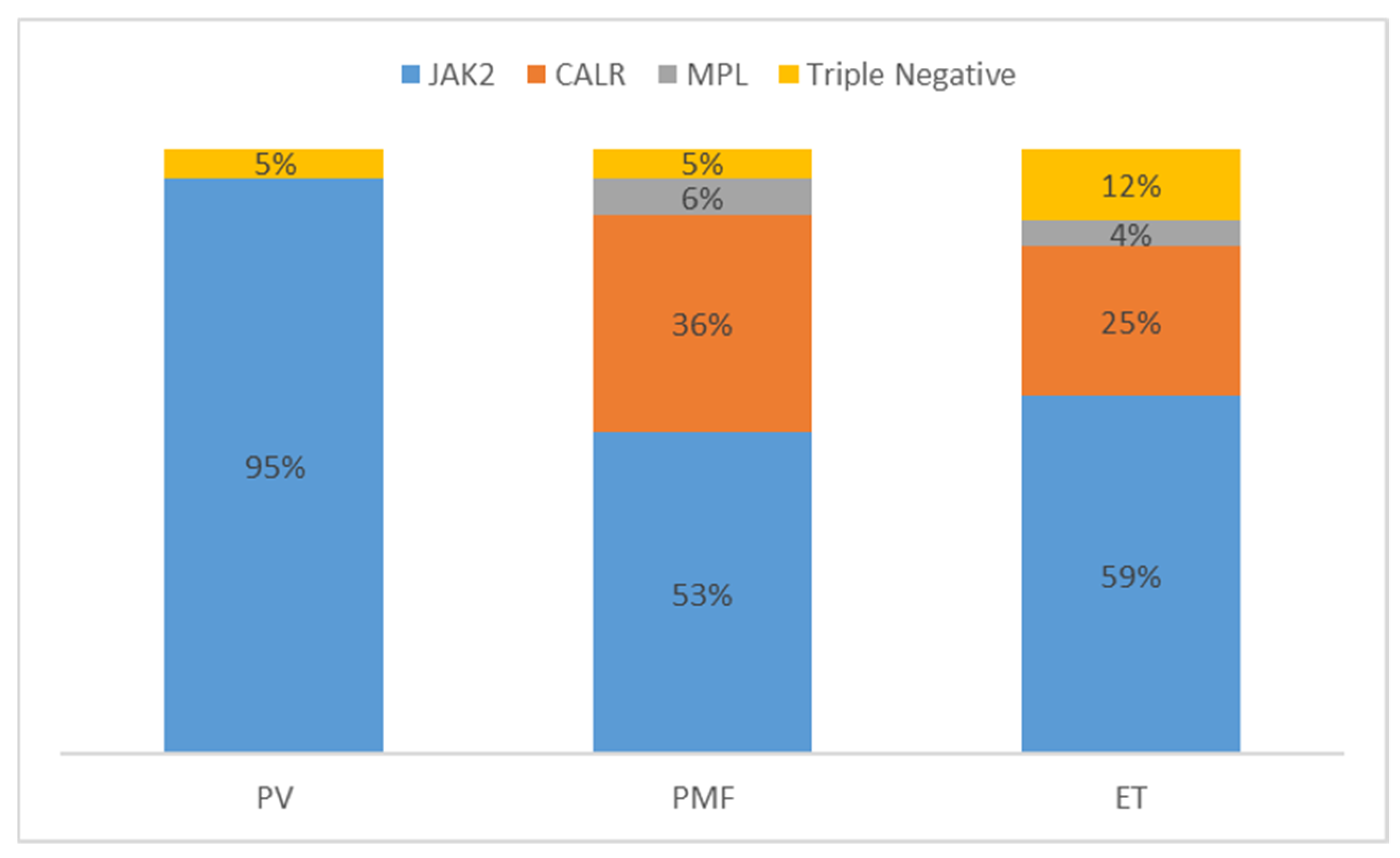
Several prognostic scoring systems have been implemented to predict outcome in MPN with specific focus on each disease phenotype. PV and ET scoring systems focus on thrombotic risk and overall survival, whereas MF scoring systems focus on the risk of leukemic transformation and survival.
For PV, a two-tiered stratification system using age and previous thrombosis to group patients into high or low risk for thrombotic complications guides risk adapted therapy. Overall survival scores initially incorporated both age ≥ 60 years and previous thrombosis. An International Working Group (IWG) included leukocytosis (≥15 × 109/L) to group patients into low, intermediate or high risk [4]. More recent data showing the prognostic impact of JAK2V617F allele burden [5]; cardiovascular risk [6]; and high risk mutations of ASXL1, SRSF2 and IDH2 [7] are not yet incorporated into a prognostic scoring system routinely used in PV [8]. The recently proposed Mutation enhanced International Prognostic Scoring System (MIPSS-PV) adds adverse mutations (specifically SRSF2), further enhancing prognostication (Table 1) [9].
The International Prognostic Score for ET (IPSET) is subdivided into factors that influence thrombosis risk and survival. Risk factors for thrombosis include age ≥ 60 years, previous thrombosis and JAK2V617F mutation, whereas survival incorporates age ≥ 60 years, leukocytosis (≥11 × 109/L) and previous thrombosis [10]. Recent data shows high-risk mutations in ET of SH2B3, SF3B1, U2AF1, TP53, IDH2 and EZH2 and show an effect independent of the current scoring systems [7]. Thus, the MIPSS-ET has been recently developed to include these updated criteria (Table 1) [9].
Several prognostic scoring systems have been developed for MF. Initially, the International Prognostic Scoring System (IPSS) was developed in 2009, followed by the dynamic IPSS (DIPSS), with the advantage of the DIPSS being able to be performed at any time point and with the IPSS being only validated at diagnosis [11]. The advent of increased molecular prognostic markers has led to the development of scoring systems that incorporate these such as the MIPSS70 (mutation enhanced IPSS) or rely solely on cytogenetic and molecular markers, i.e., GIPSS (genetically inspired prognostic score system). The most recent iteration of the MIPSS70+ version 2.0 is detailed in Table 2 together with the DIPSS (Table 3) that is used in the majority of clinical trials in MF [12,13].
Due to differences in disease biology and phenotype between PMF and post PV/ET MF [14], a separate scoring system is utilized, the MYSEC-PM, which includes age at diagnosis of secondary MF, Hb < 110g/L, platelets < 150 × 109/L, unmutated CALR, circulating blasts of 3% and constitutional symptoms [15]. The role of secondary molecular lesions in post PV/ET MF is still under evaluation.
Movement towards a personalized classification and prognosis system has further been enhanced by the work of Grinfeld et al., identifying distinct genetic subgroups and correlating them with disease phenotype and patient outcome. The personalized calculator at https://www.sanger.ac.uk/science/tools/progmod/progmod/ incorporates the results of this work to predict a patient’s progress at dynamic timepoints presenting survival metrics and information on disease biology (chronic phase, myelofibrosis and acute myeloid leukaemia (AML)) [16].

3. Driver Mutations

Mutations in JAK2, CALR and MPL are mutually exclusive [17]. Each driver mutation has unique disease kinetics that lead to clone expansion and acquisition of coexisting mutations. The frequency of each driver mutation is displayed in Figure 1.

4. JAK2 (Janus Kinase 2)

The Janus Kinase 2 gene is located on chromosome 9 at locus p24.1 [18]. The gene consists of a C-terminal JH1 (Jak Homology) domain that is highly conserved region containing activation loop, primary phosphorylation sites and ATP-binding site. The JH2 pseudokinase domain is structurally similar to JH1 but lacks any catalytic activity. The SH2 (Src Homology 2)-like domain and adjacent FERM (4.1/ezrin/radixin/moesin) domain for the N-terminal facilitates JAK2 interaction with cell surface receptors [19]. JAK2 has two main interactions with activation loop of JH1 undergoing phosphorylation and release of the JH2-mediated inhibition of kinase activity to facilitate JAK2-mediated signalling [20].
Somatic mutation of JAK2 with a G to T base change at nucleotide 1849 in exon 14 of the pseudokinase domain JH2 of the gene results in an amino acid substitution of valine to phenylalanine at codon 617 (JAK2V617F) [21,22,23,24]. Mutation within the pseudokinase domain prevents its normal auto inhibitory function and leads to constitutive kinase activation [25]. Constitutive activation leads to cytokine hypersensitivity and cytokine receptor activation independent of ligand binding [26]. JAK2 phosphorylation leads to downstream activation of signalling through STAT, MAPK, PI3K and Akt pathways [25] with STAT signalling in particular showing increased activation of proliferation genes and pro-survival genes such as Bcl-XL [27].
Whilst JAK2 mutation is associated with myeloproliferative neoplasms, it has been identified in asymptomatic clonal haematopoiesis (CH), a, expansion of clonal haematopoietic cells but without diagnostic criteria for a haematologic disease present [28]. The progression of clonal haematopoiesis through to the development of disease is principally determined by JAK2V617F mutant allele burden [29]. Knock in mouse models using a small mutant JAK2V617F population showed progressive expansion of the JAK2V617F clone with expansion of multipotent progenitors (MPP) and megakaryocyte progenitors (MkP) driving the development of disease phenotype [30]. The allele burden has also been implicated in determining disease phenotype with lower allele burden seen in essential thrombocythemia compared to polycythemia vera [31]. The mutation often occurs initially as heterozygous with homozygous mutations associated with progression with loss of 9p during mitotic recombination leading to copy neutral loss of heterozygosity [32]. A rise in allele burden has been correlated with progression to myelofibrosis in mouse models [33] and is consistent with observations of higher allele burden in MF patients compared to PV and ET [31]. Homozygous mutations show erythropoietin (EPO) and thrombopoietin (TPO)-independent growth in cell line experiments, whilst heterozygous mutations show only TPO independence [34] consistent with a higher frequency of homozygous mutations seen in PV. The frequency of homozygous mutations varies by 25–30% in PV patients and 2–4% in ET; homozygous mutations are associated with a greater risk of progression to myelofibrosis [35]. Homozygous subclones in PV are distinct from ET as the homozygous clone often expands early in the disease to become the dominant clone, whereas in ET, homozygous clones are rarely the dominant clone [36]. The heterozygous clones in ET and PV exhibit differential interferon (IFN) signalling and STAT1 phosphorylation. Increased STAT1 activity in CD34 progenitors produce an ET-like phenotype, whereas downregulation leads to a PV-like phenotype [37]. STAT1 knockout mice showed increased BFU-E and reduced colony forming megakaryocyte precursors favouring a PV phenotype. In contrast, STAT1 activation favours higher levels of IFNgamma constraining erythroid differentiation and increasing megakaryocyte proliferation [38]. The balance between STAT1, STAT3 and STAT5 accounts for some of the phenotypic differences in JAK2V617F positive MPN [39]. In JAK2V617 positive mice, Stat3 deletion in haematopoietic cells enhances thrombocytosis, accelerates fibrosis and decreased overall survival [40]. STAT5 is critical to the pathogenesis of JAK2V617F-positive PV with Stat5 conditional knockout mice showing normalization of counts and abrogation of erythropoietin independence in erythroid colonies [41].
Myelofibrosis is distinct from PV and ET as it represents progression to a pro-inflammatory and profibrotic microenvironment beyond expansion of the MPN clone and increasing allele burden. STAT3 is critical to this process with production from both the MPN clone and microenvironment promoting MF with only pan-haematopoietic deletion of STAT3 abrogating the phenotype [42]. Cytokines levels are higher in myelofibrosis compared to PV and ET, with higher IL-2, sIL-2Rα, IL-6 and tumour necrosis factor α (TNFα) production. IL-2 in particular was also higher in ET, where a greater degree of fibrosis was present [43]. Decreased GATA1 expression leads to impaired haematopoiesis [44], and aberrant STAT3, PI3K, Akt and NF-κB signalling leads to megakaryocyte secretion of fibrotic cytokines including IL-1β, fibronectin, vascular endothelial growth factor (VEGF), platelet derived growth factor (PDGF) and tumour growth factor (TGFβ) [45,46].
The vast majority of JAK2 mutant MPNs is due to the JAK2V617F mutation; however. in rare cases of erythrocytosis mutations in JAK2, exon 12 have been identified affecting residues between K537 and E543. Mutations in exon 12 show high levels of phosphorylated JAK2 and STAT5 compared to wildtype and higher than JAK2V617F in the case of K539L substitution. The mutation confers cytokine-independent growth but, unlike JAK2V617F, is only present as heterozygous mutations. The mutation affects common myeloid progenitors but has significant erythroid skewing and is only present in peripheral blood granulocytes at low levels and hence can be easily missed when only peripheral blood is sequenced [47].

5. CALR (Calreticulin)

Mutations in the Calreticulin genes are found in 25% of ET and 36% of PMF [48,49]. Located on chromosome 19, at the p13.13 locus, the gene contains 9 exons and encodes for a multifunctional protein product with an N-terminal lectin domain, proline-rich domain and acidic carboxy (C-) terminal domain that terminates as a KDEL amino acid sequence [17]. Through its c-terminal domain, CALR acts as a major calcium binding and storage protein in the lumen of the endoplasmic reticulum and in the nucleus. The lectin chaperone component also assists folding of glycoprotein substrates to prevent protein aggregation [50]. CALR expression on the cell surface imprints a cell for phagocytosis by macrophages particularly through cleaving the KDEL sequence and binding to LRP1 for surface presentation [51]. The KDEL sequence acts as the key endoplasmic retention signal [48,49].
Mutations of CALR occur as either an insertion and/or deletion in exon 9 of the gene. This leads to a frameshift change, resulting in the loss of the normal KDEL sequence and thus a positively charged amino acid sequence at the C-terminus lacking an endoplasmic retention signal [48,49]. The positive charge of the C-terminus is required for CALR to bind to thrombopoietin receptor (MPL) and to induce a proliferative phenotype. Mutations that result in uncharged glycine residues or complete truncation of exon 9 removing the KDEL sequence and the entire C-terminus leads to total loss of transforming ability [52]. The CALR-MPL interaction is critical to transcriptional activation of STAT5 and can only occur at the cell surface as retention of CALR in the endoplasmic reticulum abrogates STAT5 activity [53,54]. The mutant CALR is able to form a homomultimer at the cell surface with MPL. This homomultimeric structure is required to activate MPL in the absence of a ligand as wildtype CALR lacks the ability to undergo these multimeric changes [55]. Despite the role of STAT5 in JAK2-driven MPN promoting a PV phenotype, CALR mutation is unable to induce polycythemia or ligand independence when co-expressed with the EPO receptor [52,56].
CALR mutations are divided into two types; the first are type 1 (52-bp insertion). and type 1-like mutations lead to complete loss of all negatively charged amino acids. The second is type 2 (5-bp insertion), and type 2-like mutations eliminate approximately half of the negatively charged amino acid sequences [57,58]. The type of mutation confers some phenotypic differences, with type 1 mutations seen more commonly in primary myelofibrosis and is associated with a lower DIPSS score, suggesting a more favourable prognosis in comparison to other forms of PMF. In contrast, type 2 mutations more commonly lead to ET and have higher platelet counts than type 1 CALR ET. However, in primary myelofibrosis, type 2 mutations confer a similar phenotype to JAK2V617F-positive MF and have more pronounced splenomegaly, circulating blasts and cytopenia than the type 1 CALR counterpart [57]. CALR is not associated with a polycythemia phenotype [49].
Unlike JAK2, CALR is a rare mutation in asymptomatic clonal haematopoiesis at 0.16% but comparatively higher variant allele frequency (VAF) at 7.5% (vs 2.1% in JAK2). The average platelet count was also higher in CALR CH compared to normal controls. In contrast to JAK2, the proportion of CALR CH is relatively small compared to the proportion of CALR-mutated MPN [29]. CALR mutation is associated with a rapid development of clonal dominance in comparison to JAK2V617F, with high allele burdens in granulocytes of patients in essential thrombocythemia [31]. High variant allele burden is common in CALR MPN with clonal dominance apparent throughout stem cell, progenitor and granulocyte compartments in both early MPN (essential thrombocythemia) and late stage myelofibrosis. This is in contrast to JAK2 where the variant allele burden in the stem cell compartment is relatively small in early stage MPN (ET or PV) and much higher in primary myelofibrosis and late stage disease (post PV/post ET MF) [59].
Recently, Nam et al. showed that the effect of CALR increases with myeloid differentiation, with higher VAF seen in HSPCs compared to progenitors. Within ET, MkPs show increased cell cycle gene expression and upregulated unfolded protein response (UPR) genes such as IRE1 compared to haematopoietic stem cells (HSCs). In comparison, MF shows no difference in gene enrichment between MkPs and HSCs. Additionally, mutant MkPs show upregulated TGFβ with the degree of upregulation correlating with the degree of fibrosis seen in MF and ET [60]. Mutant CALR is found in 9–20% of leukemic transformation from MPN [61,62,63] and 25% of post-ET MF consistent with the prevalence of CALR-mutated ET [64].

6. MPL (Myeloproliferative Leukaemia)

Mutations in MPL, located on chromosome 1 p34, account for 1–3% of ET and 5% of MF [65]. The gene contains 12 exons with 2 cytokine receptor domains, transmembrane domain and a cytoplasmic domain. It is named after the murine retrovirus, myeloproliferative leukaemia virus (MPLV) [18]. The MPL gene encodes for thrombopoietin receptor protein (TPOR). The normal ligand of TPOR is thrombopoietin with key receptors on HSC, MPP, common myeloid progenitors (CMP), megakaryocyte-erythroid progenitors (MEP) and MkP in additional to receptors on megakaryocytes and platelets [66]. TPO binds to MPL/TPOR and induces receptor homodimerisation that subsequently leads to STAT5 phosphorylation and MAPK signalling [67]. MPL and TPO are critical to HSC self-renewal and expansion in addition to the proliferative effects TPO is able to stimulate DNA repair via the MPL receptor. In mouse models using γ-irradiation, de Laval et al. were able to demonstrate the role of TPO in increasing DNA-PK-dependent non-homologous end-joining (NHEJ) efficiency ensuring chromosomal integrity [68]. Whilst exogenous TPO agonists and endogenous TPO can promote DNA repair [69], the double strand break (DSB) repair function is dependent on an intact MPL receptor at both alleles with Mpl-deficient and haploinsufficient mice showing decreased NHEJ activity [68]. The above findings have been replicated by Barbieri et al. and further demonstrated the ability of MPL activation to promote an interferon-like antiviral gene response in HSCs [70].
There are several mutations of MPL identified, but the two main types occur within exon 10. These are mutations of tryptophan W515 located on the boundary of the transmembrane and cytosolic domains of MPL and lead to a leucine substitution W515L or a lysine substitution W515K [71]. Other mutations are much less common and include W515R, W515A and W515G [72]. Mutations are usually heterozygous, but homozygosity is seen with high mutations burdens and disease progression through copy neutral loss of heterozygosity of chromosome 1p. Variant allele burdens of greater than 50% are commonly associated with fibrosis both PMF and post ET-MF [73]. MPL mutations are mutually exclusive from JAK2 mutations [65] and JAK2V617F shows antagonistic effects by downregulating MPL through enhanced ubiquitination of MPL and increased proteasome degradation [74]. MPLW515L mutations have also shown an upregulation of NHEJ pathways consistent with the effects of MPL receptor activation on DSB repair highlighting the ability of the MPN HSC clone to persist despite DNA damage [75].
MPL is critical at multiple stages of megakaryocyte development evidenced by knockout mice showing marked megakaryocyte deficiency and severe thrombocytosis. Haematopoietic progenitors were reduced and lack megakaryocytic potential [76,77].
MPL expression also acts as a regulator of TPO levels with mature platelets providing a negative feedback mechanism by removing TPO bound to MPL receptor (TPOR) [78]. Through altered MPL expression in late megakaryocytes and platelets, TPO is not cleared as readily leading to increased levels driving proliferation of early megakaryocytes. This leads to subsequent thrombocytosis as defective platelets are not able to provide the normal negative feedback loop [79].

7. Triple Negative MPN

Previously, a histological diagnosis of an MPN lacking a driver mutation was termed triple negative disease. Advances in sequencing technology have been able to identify novel mutations in JAK2 and MPL reclassifying previously “triple negative” disease. Whole exome sequencing has not been able to identify a causative recurrent mutation within this group, and triple negative MPN likely represents a heterogenous group of clonal haematopoietic proliferation [80,81]. For this reason, triple negative MPN will not be explored further in this review.

8. Coexisting Mutations

The prognostic scoring systems for primary myelofibrosis have evolved from phenotype driven (IPSS/DIPSS) to now include information about additional gene mutations (DIPSS-plus and MIPSS, identifying high risk molecular lesions identified as mutations in ASXL1, SRSF2, U2AF1, EZH2, IDH1 and IDH2) [82]. Whilst there has been some discrepancy in broad applicability across all MPNs [14], these highlight the prognostic significance of additional mutational burden. Mutations have been grouped according to the main functional category, being chromatin modifiers, spliceosome complex components, DNA methylation modifiers, tumour suppressors and transcriptional regulators (Figure 2). These mutations are described as coexisting mutations as their presence within a clone can precede the acquisition of the driver mutation or can occur subsequent to the driver mutation. We present a figure that shows the effect of additional mutation acquisition on disease progression, and represented in Figure 3, we propose that specific coexisting mutations drive progression to myelofibrosis and that other coexisting mutations favour leukemic transformation. Frequencies of each mutation in PV, ET, PMF, secondary MF (sMF) and leukemic transformation (LT) are included in Table 4.

9. Chromatin Modifiers

9.1. ASXL1 (Additional Sex Comb Like-1)

ASXL1 mutations have significant impacts on prognosis and have been identified in 47% of MPN in the leukemic phase [61]. Mutations usually occur at the 5′ end of exon 12, mostly as frameshift mutations or less commonly nonsense mutations. Mutation leads to a truncated c-terminal of the resulting ASXL1 protein; this truncated form leads to a gain of function in some cellular pathways and loss of function in others [83]. Mutations are usually present as heterozygous in myeloid malignancy [84].
Normal ASXL1 has both silencing and enhancing effects on homeotic gene expression. It enhances methylation of histone marks through the polycomb repressor complex 2 (PRC2) that leads to repression and silencing of chromatin remodelling genes such as HOXA [85]. Through complexing with BRCA1-associated protein 1 (BAP1), ASXL1 forms the polycomb group repressive deubiquitinase complex, which is involved in stem cell pluripotency [86].
ASXL1 mutation loss of function effects are mediated via a loss of PRC2-mediated histone H3 lysine 27 (H3K27) trimethylation, leading to loss of repression of the HOXA cluster of genes and loss of heterochromatin maintenance [85], whilst gain of function occurs through enhancing the activity of the ASXL1-BAP deubiquitinase complex, resulting in loss of histone H2K119 ubiquitination [87]. Together with loss of H3K27 trimethylation, there is upregulation of genes governing myeloid differentiation with significant myeloid skewing of LT-HSC [88]. This myeloid skewing can partially be attributed to binding of mutant ASXL1 to BRD4, resulting in the phosphorylation of RNA polymerase II, acetylation of H3K27 and H3K122 leading to upregulation of myeloid genes, and an increased sensitivity to BET bromodomain inhibitors [83,89]. Furthermore, mutant ASXL1 truncating protein expression results in more open chromatin in HSC (c-Kit+ cells) and dysregulated expression of genes critical for HSC self-renewal and differentiation [83].
ASXL1 mutations are not limited to a specific molecular driver in MPN, with studies by Lasho et al. and McNamara et al. showing that, in accelerated and blast phase MPN, ASXL1 mutations are present in JAK2, CALR, MPL and triple negative disease [61,62]. ASXL1 mutations are more commonly enriched in leukemic transformation (LT) occurring from PMF, at a frequency of 66.7% compared with 13.6% of LT developing from PV, ET or sMF [63].

9.2. EZH2 (Enhancer of Zeste 2)

EZH2 encodes a histone methyltransferase that constitutes the catalytic component of PRC2 functions to initiate epigenetic silencing of genes involved in cell fate decision. EZH2 activity is particularly enriched with a trimethylation mark at nucleosomal histone H3K27 [90].
The majority of EZH2 mutations in myeloid disease, particularly MPN, are missense loss of function and lead to premature chain termination or direct abrogation of histone methyltransferase domain [91]. This is a direct contrast to lymphoid malignancy, where EZH2 mutations are highly expressed and lead to diffuse large B-cell lymphoma transformation from germinal centre B-cells [92,93]. Ernst at al. identified that deletion of 7q or loss of chromosome 7 leads to loss of the EZH2 tumour suppressor function in myeloid malignancy. The presence of monoalleleic mutations or uniparental disomy leading to homozygous mutations are responsible for oncogenic effects [90].
EZH2 expression is preserved in HSCs through MEPs but decreased in committed megakaryocytes. EZH2 loss in function produces megakaryocyte lineage features and high platelet counts in myeloid malignancy [94]. Commitment of HSCs or MPPs to specific lineages requires the removal of the H3K27me3 mark at the loci of master transcription factor regulators of lineage [95,96].
In combination with JAK2V617F mutations, mouse models with a loss of Ezh2 create an epigenetic switch in PRC target genes including Hmga2, S100a8 and S100a9 that increase colony forming units (CFU-Mk) committed to megakaryopoiesis [12,97,98].
EZH2 mutations have been identified in 6–13% of myelofibrosis and are associated with poor survival [99]. However, its presence is less common in ET at 3% and absent in PV [7]. The presence of EZH2 mutations is indicative of more progressive disease as highlighted above in myelofibrosis and by Lasho and McNamara in leukemic transformation with 15% and 7% identified in each cohort respectively [61,62]. EZH2 mutation is also not specific to any particular driver mutation being identified in JAK2, CALR, MPL and triple negative MPN [16,61]. Whilst EZH2 mutation were not identified in PV amongst the data sets reviewed, post-PV MF has demonstrated EZH2 mutations signifying a phenotypic switch in PV inducing megakaryocyte proliferation and fibrosis consistent with Ezh2 loss of function mouse models [64,97,98].

10. Spliceosome Complex Components

10.1. SRSF2 (Splicing Factor, Serine/Arginine-Rich, 2)

SRSF2 is critical for spliceosome assembly; it contains the ribonucleoprotein (RNP) type RNA binding motif and carboxyl-terminal serine/arginine-rich (SR) domain and binds to the exonic splice enhancer (ESE) with target pre-mRNA species [100].
Mutations in SRSF2 involve a missense at proline 95 (P95H), with altered function leading to preferential recognition of CCNG ESE motifs compared to the wildtype product that recognizes both CCNG and GGNG ESE motifs equally. This alters the balance of splicing of numerous pre-mRNAs, leading to the production of miss-spliced products and nonsense-mediated decay [101,102]. SRSF2 P95H expression in mouse models leads to myeloid bias, impaired B cell development and maturation, and decreased haemopoietic stem cell numbers reminiscent of human myelodysplastic syndrome (MDS) in vivo [103]. MPN with SRSF2 mutations have a high variant allele frequency, suggesting an impact on clonal dominance [104]. SRSF2 mutations are enriched in blast phase MPN (15–22%) and appear to be more common in combination with JAK2 and MPL driver mutations compared to CALR [61,62]. There appears to be a particular predilection for SRSF2 mutations in leukemic transformation after PMF at 50% compared to 6.8% seen in PV, ET and secondary MF-related AML [63].

10.2. U2AF1 (U2 Small Nuclear RNA Auxiliary Factor 1)

U2AF1 forms part of the spliceosome complex. The gene product is involved in pre-RNA splicing and recognizes pyrimidine-rich tracts with a conserved terminal AG present at 3′ splice sites [105]. Somatic mutations occur as heterozygous missense changes in serine 34 with an amino acid substitution to phenylalanine (S34F) or tyrosine (S34Y) [106]. Normally, serine 34 is highly conserved within the zinc finger domain critical for RNA binding. Mutant U2AF1 leads to a splicing defect resulting in increased splicing, exon skipping and subsequent gain of function phenotypes [106].
Mutations in U2AF1 represent less than 2% of chronic phase MPN [16], but their presence is associated with progression identified in 5–9% in leukemic transformation of MPN [61,62].

10.3. SF3B1 (Splicing Factor 3B, Subunit 1)

SF3B1 forms part of the spliceosome complex and has a prominent interaction with polycomb group proteins [107]. SF3B1 mutations occur as missense mutations with the most common being K700E and H662Q amino acid substitutions [108]. Mutations of SF3B1 block erythroid maturation [109], and whilst the mutation does not confer any additional prognostic significance in myelofibrosis [110], it commonly causes ring sideroblast changes to erythroblasts [111]. Consistent with the findings in MDS where SF3B1 mutations generally confer a more benign phenotype, in MPN, it also carries a lower risk of leukemic transformation [108,111].
In combination with CALR mutations, SF3B1 appears to increase the proliferative advantage of MkPs [60]. The subsequent aberrant splicing by SF3B1 leads to increased neoantigen formation. CALR mutants show an ability to bind common major histocompatibility (MHC) class I proteins, and the co-mutations with SF3B1 showed the highest CALR neoantigen presentation on MHC I variants compared to other MPN mutations, suggesting a potential target for future immunotherapy [112].

11. DNA Methylation Modifiers

11.1. DNMT3A (DNA Methyltransferase 3A)

DNMT3A is part of the DNMT family of enzymes responsible for control of DNA methylation at CpG dinucleotides [113]. DNMT3A mutations are the most common mutation identified in clonal haematopoiesis [114,115].
The most common mutation of DNMT3A in myeloid malignancies is a missense mutation leading to substitution of arginine for histidine at position 882 (R882H). This compromises the enzymatic function, CpG specificity and flanking sequence preference of DNMT3A [116]. The degree of subsequent hypomethylation is relative to the mean CpG frequency per base of particular genes. Erythroid transcription factors such as Klf1 and Tal1 having higher CpG frequency per base show an increased erythroid transcriptional priming in Dnmt3a mutant cells in mice [117].
DNMT3A mutations are present in 5–10% of MPN and is present in early stage disease [67]. However, it is not represented in prognostic scoring systems [82]. Loss of DNMT3A function using CRISPR models in JAK2V617F positive models of MPN induces a myelofibrosis phenotype with reduced erythropoiesis, progressive depletion of HSCs and accumulation of MPPs. The transcription signature also shows aberrant self-renewal and inflammatory signalling pathways compared to JAK2V617F positive with wildtype DNMT3A [118].

11.2. TET2 (Ten-Eleven Translocation 2)

TET2 plays a key role in DNA methylation through oxidation of 5-methylcytosine (5mC) into 5-hydroxymethylcytosine (5hmC). This acts a stable epigenetic mark and is active in demethylation [113]. TET2 contains 12 exons with mutations commonly occurring as nonsense or missense mutations leading to its loss of function [119]. Deletion or rearrangement of chromosome 4q24 is an additional mechanism for loss of TET2 function identified by Delhommeau et al. and further identified the mutation in early stage differentiation and its synergistic effects with JAK2V617F mutation [120].
Loss of normal TET2 function decreases 5hmC production, leading to DNA hypermethylation. This has a particular effect on HSCs increasing expression of self-renewal gene signatures, sensitizing haematopoietic cells to cooperating mutations and skewing towards myelomonocytic differentiation, [121] and sensitises haematopoietic cells to cooperation mutations. TET2 loss of function and subsequent hypermethylation patterns favour myelomonocytic lineage in HSC priming. Through increased expression of Hlf, Sox4, and Meis1 there is increased self-renewal and increased susceptibility to hypermethylation of Myc and Myb due to their higher CpG-rich binding motifs [117]. TET2 mutation can precede JAK2 mutations in MPN HSCs and lead to biclonal disease or can occur secondary to JAK2V617F mutation and provide a competitive advantage driving clonal dominance [122]. TET2 mutation order also influences disease biology in MPN, where TET2 arising prior to JAK2V617F favoured an ET phenotype and TET2 as a secondary mutation following JAK2V617F favoured a PV phenotype [123].

11.3. IDH1 and IDH2 (Isocitrate Dehydrogenase 1 and 2)

The isocitrate dehydrogenase enzyme genes are regulators of metabolic pathways that have indirect effects on epigenetic marks. IDH1 is a dimeric cytosolic NADP-dependent isocitrate dehydrogenase that catalyses decarboxylation of isocitrate into alpha ketoglutarate (α-KG) [124]. IDH2 is the mitochondrial version of the enzyme, produces NADPH and plays a key role in mitochondrial redox balance-mitigated oxidative damage [125]. IDH mutants increase production of 2-hydroxyglutarate (2-HG), which prevents histone demethylation. Preventing the expression of inducible lineage-specific differentiation genes leads to a block in differentiation that is characteristic of IDH mutant malignancies [126]. Mutations in IDH1 occur as a missense mutation at codon 132, with the most common mutations being R132H and R132C, leading to an arginine substitution for histidine or a cysteine respectively [127]. Alterations to IDH2 occur as missense mutations most commonly at codon 140 with R140Q the most commonly causing an amino acid substitution of glutamine replacing arginine. Mutations of codon 172 have also been described in transformed MPN with R172G [127,128].
The frequency of IDH 1 and 2 mutations is low in chronic-phase MPN with numerous large cohorts identifying a frequency of between 0.5–4%, with the mutation seen more commonly in PMF than PV or ET [7,16,127,128,129]. There was no impact on overall survival for chronic phase MPN, but in contrast, the presence of IDH mutants in leukemic transformation conferred a worse overall survival than non-IDH mutated leukaemia [127]. The prevalence of IDH mutations is higher in leukemic transformation at 12.5–26% across several cohorts [61,62,63,127,128], suggestive of a strong risk for leukemic transformation with the acquisition of an IDH mutation.
McKenney et al. demonstrated the synergistic effect of JAK2V617F and IDH mutations, showing higher serum 2-HG levels for both IDH1R132H and IDH2R140Q models in combination with JAK2V617F compared to JAK2 or IDH mutant alone. The combination of mutations showed impaired differentiation and increased immature progenitors compared to more late stage differentiated progenitors. Treatment with JAK2 inhibition was not able to significantly reverse the phenotype, but through combined inhibition of JAK2 and IDH2, the double mutant cells reverted to gene clustering patterns closer to WT LSK [130]. The presence of IDH2 mutations has also been shown to enhance aberrant splicing of mutant SRSF2, leading to genomic instability and risk of leukemic transformation, highlighting yet another crosstalk between mutations [131].

12. Tumour Suppressors

TP53 (Tumour Protein p53)

The tumour suppressor gene TP53 is located on chromosome 17, encoding a DNA binding protein that responds to DNA damage by inducing transcriptional programs that result in cell cycle arrest or cell death via apoptotic pathways [132]. The presence of TP53 mutations at low allelic frequency in chronic MPN with subsequent loss of heterozygosity is strongly associated with leukemic transformation [133]. This was confirmed by recent studies showing that TP53 is more significantly enriched in leukemic transformation, found in 16–17% of cases compared to approximately 2% of chronic phase MPNs [16,61,62,134].
There are numerous upstream regulators of TP53; however, key in MPN are MDM2 and MDM4, which inhibit TP53 transcriptional function by facilitating nuclear export and by inducing ubiquitin-mediated degradation. MDM2 overexpression has been reported in MPN, and MDM4 high copy numbers are found in leukemic transformation and are the consequence of amplification of chromosome 1q [135,136].
Trp53 loss leads to leukemic transformation following a short PV phase in Jak2V617F mouse models. Primitive leukaemia cell, MEP and CD71+ erythroid progenitors all retained the ability to cause leukaemia on serial transplantation including CD71+ cells, giving rise to erythroleukaemia. The progenitor population showed enhanced proliferative potential and resistance to apoptosis. Genomic instability was also increased with higher numbers of mice showing an abnormal karyotype compared to Trp53 intact Jak2 mutant mice [137,138]. TP53 mutation appears to be a more common pathway to leukemic transformation in PV and ET compared to AML derived from PMF and secondary MF with 51.6% showing TP53 mutations compared to 16% [63].

13. Transcriptional Regulators

RUNX1 (Runt-Related Transcription Factor 1)

Located on chromosome 21q22.12, the RUNX1 gene plays a critical role in haematopoiesis. All RUNX products contain a Runt homology domain (RHD) which is responsible for DNA binding and interaction with its common heterodimeric partner, CBFbeta. Through this DNA interaction RUNX1 controls the expression of target genes involved in haematopoietic differentiation, cell cycle regulation, ribosome biogenesis, and p53 and TGFβ pathways [139]. RUNX1 mutations occurring in MPN are commonly heterozygous and occur as either missense, nonsense or frameshift mutations. The acquisition of RUNX1 mutation is associated with leukemic transformation, with only 2.2% identified in chronic phase compared to 27.9% in transformed disease. RUNX1-mutated MPN has an adverse prognosis with median survival of 2.8 years compared to not reached (NR) for RUNX1 wildtype [140]. The frequency of somatic RUNX1 mutations is also higher in MPN-related AML compared to de novo AML with 17% vs. 5% in the cohorts examined by Lasho et al. [61].

14. Treatment of MPN—Existing and Emerging

14.1. Chemotherapy

Chronic long-term dosing with oral chemotherapy agents remain a mainstay of MPN therapy. The most widely used chemotherapeutic is the antimetabolite hydroxyurea (hydroxycarbamide). Hydroxyurea reduces deoxyribonucleotide production via inhibition of the enzyme ribonucleotide reductase through scavenging tyrosyl free radicals [141]. Used as a cytoreductive agent, hydroxyurea has been shown to decrease thrombosis rate in both PV [142] and ET [143]. In addition, reduction in splenomegaly and symptoms of MF have been observed although not sustained long term [144]. The limitations in the use of hydroxyurea has concerns regarding risk of leukemic transformation [145] despite some studies showing that the risk is not significantly higher than the background risk for MPN [146,147]. Often used as a comparator arm for other therapies, hydroxyurea can rarely show haematological and molecular responses [148]. Other oral agents include busulfan and pipobroman. Busulfan, an alkylating agent, has shown 80% haematological and 30% molecular response in hydroxyurea-resistant PV patients [149] but has conflicting data about whether it significantly increases risk of leukemic transformation [145,150]; hence, it is often used second line for shorter durations. Whilst European Leukaemia NET (ELN) has a definition for resistance to hydroxyurea, this does not incorporate molecular factors. Recently, p53-dependent mechanisms were identified as a resistance mechanism to hydroxyurea in colon cancer cells, highlighting the importance of DNA damage pathways in hydroxyurea sensitivity [151].

14.2. JAK Inhibitors

Inhibition of Janus kinase enzymes has emerged as the main target in drug development in MPN. JAK inhibition to date has shown clinical benefit independent of JAK2 mutations [152,153] through potent inhibition of JAK1 and JAK2. JAK1-dependent cellular pathways are also activated in myelofibrosis and the downregulation of TNFα, IL-6, and MIP-1β through JAK1 and JAK2 blockade and STAT3 primarily through JAK2 blockade leads to the clinical benefits of decreased symptoms and splenomegaly [154]. JAK inhibitors exert their effect by either type 1 or type 2 inhibition of JAK2. Type 1 inhibition targets the active ATP binding pocket in a competitive manner, stabilizes JAK2 and increases activation loop phosphorylation [155]. Tvogerov et al. recently showed that type 1 JAK inhibition leads to no detectable STAT3 or STAT5 phosphorylation and minimal STAT1 phosphorylation. However, type 1 JAK inhibition prevented dephosphorylation and ubiquitination of JAK2, leading to progressive accumulation of an activation loop phosphorylated pool. The phosphorylated pool is rapidly depleted on JAK inhibitor cessation, accounting for the rapid recrudescence of the clinical syndrome known as JAK inhibitor withdrawal syndrome [156].
Type 2 JAK2 inhibition occurs when the inhibitor binds to the inactive state at a hydrophobic pocket adjacent to the ATP binding site uncovered by conformational change of the activation loop leading to stabilization of inactive JAK2 [155,157,158]. There is an absence of phosphorylation accumulation and hence no washout seen with type 2 inhibitor withdrawal in vitro [156]. JAK2 inhibitors currently in clinical practice and clinical trials include type 1 inhibitors ruxolitinib, fedratinib, momelotinib and pacritinib [158,159]. Type 2 JAK inhibitors BBT594 and CHZ868 has been limited to in vitro and PDX models with no published clinical trial data to date [160,161]. Chronic exposure to JAK2 inhibitors stabilizes activated JAK2 and increased JAK2 mRNA expression facilitates formation of a heterodimer of activated JAK2, JAK1 or TYK2 leading to JAK/STAT signalling escape. Removal of the JAK inhibitor may lead to resensitization by decreasing the drive to heterodimerisation [157].

14.3. Ruxolitinib

Ruxolitinib emerged as the first JAK targeted therapy for myelofibrosis following the results of the COMFORT-I and COMFORT-II phase 3 clinical trials. COMFORT-I showed a ≥50% improvement in symptom score and 41.9% of patients showed a ≥35% spleen volume reduction (SVR) at 24 weeks, whilst COMFORT-II showed that 28% of patients with a ≥35% (SVR) at 48 weeks had a mean reduction in spleen volume of 56% [152,153]. Subsequent analysis showed long-term survival benefit for ruxolitinib with a median overall survival of 5.3 vs. 2.4 years for best available therapy (BAT) after censoring for crossover [162].
The use of ruxolitinib has expanded to PV and ET, particularly to control disease in patient resistant or intolerant of hydroxyurea. In PV, ruxolitinib has proven to be superior to BAT for haematocrit (Hct) control, spleen size reduction and complete haematological response (CHR) [163,164,165,166]. RESPONSE-1 showed Hct control at 60% for ruxolitinib compared to 20% in the BAT arm, an ≥35% SVR of 38% vs. 1% and a CHR of 24% vs. 9% [163]. The results in ET have not shown the same differences between ruxolitinib and BAT with CHR rates of 46.6% vs. 44.2% in MAJIC-ET and no significant difference in molecular responses at 12 months [167]. However, the reduction in allele burden for JAK2V617F was apparent in a separate study with ongoing therapy showing a median decrease of 2.8% at 24 weeks improving to a 60% reduction by week 312 [168].
Ruxolitinib does not reliably eradicate clones harbouring JAK2V617F or CALR mutations in myelofibrosis, with only 12% of patients having a >50% reduction in VAF of JAK2 in the COMFORT-I cohort [169]. Similarly, a significant change in VAF was not seen for CALR in the COMFORT-II cohort [170]. This is in contrast to findings in polycythemia vera with a slight reduction in mean JAK2 allele burden reduction in RESPONSE for the ruxolitinib treatment arm, strongly correlating with spleen reduction of >35% [171]. Type 1 JAK2 inhibitors such as ruxolitinib or fedratinib are unable to eradicate the disease-initiating JAK2V617F mutant LT-HSCs [172,173]. This is due a preferential selection for more committed MPP and MEPs with an inability to significantly block STAT1 phosphorylation in LT-HSCs. Interestingly ex vivo treatment with ruxolitinib was able to completely block STAT1, suggesting an inadequate intracellular drug concentration in LT-HSCs [172].
Clinical data reveal that response to JAK2 inhibition is impaired with concurrent ASXL1 and EZH2 mutations (hazards ratio of 2.94) and that 3 or more high molecular risk mutations have a shorter time to treatment failure [174]. Resistance to JAK inhibitors has been a complication of long-term therapy with loss of spleen response and disease progression with 50% of patients discontinuing ruxolitinib at 3 years due to side effects or loss or response [175].

14.4. Fedratinib

The 2nd JAK inhibitor approved by the U.S Food and Drug Administration (FDA) for myelofibrosis [176], fedratinib, is a selective JAK2 inhibitor with no significant JAK1, JAK3 or TYK3 inhibition [177]. The degree of immunosuppression appears less due to its selectivity for JAK2 with no effect observed on the expression of inflammatory cytokines IL-1α, MCP-1, E-selectin and P-selectin or immunomodulators sIL-17f and HLA-DR [159]. Despite phase 2 and 3 studies opening in 2011–2012, fedratinib underwent a clinical hold from 2013–2017 due to concerns regarding Wernicke encephalopathy for which it now has a black box warning [177]. The principal studies showing efficacy were JAKARTA-I and JAKARTA-II. JAKARTA-I examined fedratinib in frontline therapy for myelofibrosis showing a 36% response in ≥35% SVR and 36% response for a ≥50% improvement in total symptom score compared to placebo [178]. JAKARTA-II was a single arm phase 2 study of fedratinib in patients intolerant or refractory to ruxolitinib; the results showed a 55% response for ≥35% SVR and 26% for improvement of ≥50% in total symptom score [179]. Whilst there is no mutational data reported regarding response to fedratinib, further analysis of JAKARTA-I and II and subsequent studies FREEDOM-I and II may further identify these [180].

14.5. Momelotinib

Momelotinib, a second generation JAK2 inhibitor, has shown efficacy in myelofibrosis in SIMPLIFY-I, a phase 3 non-inferiority study against ruxolitinib. At 24 weeks, 26.5% in the momelotinib arm compared to 29% in the ruxolitinib arm had achieved ≥35% SVR. Whilst showing the benefit of greater transfusion independence (66.5% vs. 49.3% at 24 weeks), momelotinib failed to achieve non-inferiority for symptom control (≥50% reduction in symptom score was 28.4% for momelotinib vs. 42.2% for ruxolitinib) [181]. Following this, SIMPLIFY-II was unable to demonstrate superiority over BAT, 89% of which was patients on ruxolitinib [182]. The difference in efficacy between momelotinib and ruxolitinib might may be due to difference in anti-inflammatory effects particularly a lack of IL-1α suppression from momelotinib [159]. This lower level of suppression over pro-inflammatory signals may account for the lower symptom score reductions seen in SIMPLIFY-I. SIMPLIFY-I and SIMPLIFY-II were both able to demonstrate higher levels of transfusion independence than the comparator arm, and this is due to the ability of momelotinib to bind to bone morphogenic protein receptor kinase, activating A receptor type 1 (ACVR1). This receptor can increase hepcidin expression from the liver, and its inhibition improves iron utilization for erythropoiesis [183]. Long-term follow up of momelotinib therapy in myelofibrosis has shown a median overall survival of 3.6 years and 1.5 years in the high-risk group. Mutations in ASXL1 and SRSF2 were among the predictors of poor survival post-momelotinib therapy [184].

14.6. Pacritinib

Pacritinib is a selective JAK2 inhibitor that also exhibits activity against FLT3. Despite negligible JAK1 activity pacritinib maintains IL-1β suppression through inhibition of IL-1 receptor-associated kinase 1 [185]. The lack of significant JAK1 activity and suppression of IL-1β abrogates some of the myelosuppressive effects of other JAK inhibitors. The PERSIST-1 trial using pacritinib compared to BAT (excluding prior and current JAK inhibitors use) in myelofibrosis showed an increased in platelet count and a greater percentage of transfusion-independent patients at 24 weeks compared to BAT. Pacritinib also showed that 19% of patients achieved ≥35% SVR and that 36% achieved a ≥50% reduction in total symptom score. Reduction in allele burden was also measured at 24 weeks with pacritinib achieving a 15.8% reduction compared to 7.9% in BAT [186]. PERSIST-2 compared pacritinib to BAT but allowed prior ruxolitinib exposure and ruxolitinib treatment in the BAT arm. Pacritinib showed efficacy in 18% of patients achieving ≥35% SVR compared to 3% of BAT and 25% achieving ≥50% reduction in total symptom score compared to 14% (although not statistically significant); 48% of patients had received prior ruxolitinib, and 45% within the BAT arm received ruxolitinib. The improvement in transfusion and thrombocytopenia was also observed with pacritinib in PERSIST-2 [187]. Despite these results, pacritinib is not yet approved by the FDA or the European Medicines Agency (EMA) due to toxicity concerns. Trials are currently ongoing determination of the extent of toxicity and exploring dose modifications due to concerns from early reports of intracranial haemorrhage and cardiac failure identified in the PERSIST studies [188].

14.7. Pegylated Interferon Alpha (IFNα)

The use of interferon in MPNs extends back to 1988 with IFNα used as a treatment for ET [189]. The clinical effects seen occur via its effectors, interferon stimulated genes (ISG). The expression of ISG subsets link to interferon regulatory factor (IRF), NF-κB and other transcriptional regulators. Endogenous interferons are defined as type 1, type 2 and type 3, with type 2 IFNs particularly IFNγ involved in the inflammatory response inducing JAK1 and JAK2 phosphorylation to activate STAT1. Type 1 IFN (IFNα and IFNβ) uses JAK1 phosphorylation and Tyk2 to activate STAT1 and STAT2 and the induction of ISGs [190,191]. In mouse models, IFNα activates dormant HSC by increased phosphorylation of STAT1 and PKD/Akt with a subsequent upregulation of IFNalpha, ISGs and Sca1. Acute exposure promotes cell proliferation from a quiescent state, whereas chronic exposure impairs HSC function and leads to progressive depletion of cycling HSCs through provoking DNA damage [192,193]. HSCs can utilize interferon regulatory factor-2 (IRF2) to protect quiescent stem cells from type 1 IFN-dependent exhaustion. IRF2 is able suppress IFNα signalling to preserve self-renewal and differentiation capacity of HSCs [194].
Treatment with IFNα stimulates both JAK2V617F-positive and WT quiescent stem cells into cycle but leads to a preferential depletion of JAK2V617F LT-HSCs in mice [195] and inability to develop MPN with serial transplantation with JAK2V617F mutant cells subsequent to IFNα treatment [196]. We have previously demonstrated enhanced cell cycle activity at baseline and the increased sensitivity to IFNα-mediated cycle activation of JAK2 mutant cells. The mechanism for this increased sensitivity is not limited to increased cell cycling with other mechanism possibly implicated [195]. Recently, this has been further clarified with JAK2V617F LT-HSC showing increased STAT1 phosphorylation, reactive oxygen species (ROS) production and DNA damage in response to chronic IFNα treatment. The effect is more marked in JAK2 mutant LT-HSCs compared to wildtype [172].
Interferon has shown favourable response rates in PV, with 94.6% of patients achieving CR at 12 months; 89.6% of patients had a reduction in JAK2V617F VAF with 24% achieving a molecular CR with an undetectable JAK2 clone [197]. In a similar study, 70% of PV and 76% of ET patients achieved CRs with 54% and 38% showing a JAK2 molecular response and 14% and 6% of PV and ET respectively showing undetectable JAK2V617F clone [198]. The molecular response with IFNα in PV improves over time with 66% achieving molecular response in JAK2 at 36 months of roPEG-IFNα-2b compared to the decreasing molecular response seen in the control group of best available therapy [148].
Interferon is also active in myelofibrosis with a recent FIM observational study showing that 58% of patients reduced their JAK2 allele burden by more than 50%. The median overall survival for the cohort was 7.4 years, with a strong correlation between overall survival and longer duration of IFNα therapy [199]. In comparison to pooled data on overall survival from COMFORT-I and COMFORT-II, the FIM study showed overall survival for high risk and INT-II DIPSS risk as 4.6 and 6.9 years, respectively, whereas the pooled data showed an overall survival of 4.2 years for high risk and estimated OS of 8.5 years [162,199].
Concomitant TET2 or DNMT3A mutations are associated with molecular resistance to interferon [200]. The FIM study also showed inferior response to IFNα in patients with high molecular risk (HMR) mutations (defined as ASXL1, SRSF2, EZH2 and IDH1/2) [199]. Response to IFNα is not only restricted to JAK2 mutant MPN, with CALR mutated ET also showing a favourable response to IFNα. IFNα leads to a haematological response in all patients and a median decrease in allele burden from 41% to 26%, with 42% of patients showing a greater than 50% reduction in CALR allele burden. The comparator arm of hydroxyurea or aspirin only did not show any significant reduction in allele burden. A suboptimal response was seen in 6 patients and was attributed to the presence of additional mutations of TET2, ASXL1, IDH2 or TP53 [201].
Clinical trials testing the combination of IFNα and ruxolitinib provide an interesting conundrum. On one hand, interferon does increase phosphorylated STAT1 to induce LT-HSCs out of quiescence. Ruxolitinib may be predicted to block STAT1 activation through inhibition of JAK1. We have recently shown that this potential antagonism is avoided in vivo because of the relative sparing of LT-HSC from ruxolitinib-mediated JAK/STAT inhibition [172]. Initial combination phase 2 studies showed a complete haematological response was achieved in 44% of PV patients and 58% of low to intermediate-1 risk MF at 12 months of treatment, with both groups showing a significant reduction in JAK2V617F allele burden with a median decrease in VAF from 47% to 23.5% for PV at 12 months and 45% to 18% for MF at 12 months [202].

14.8. Allogeneic Bone Marrow Transplantation

The only definitive curative option for MPN treatment is allogeneic bone marrow transplantation which is used in both PMF and secondary MF. Transplant selection involves multiple disease-related, recipient-related, donor-related and institutional factors that extend beyond the scope of this review. However, in the setting of myelofibrosis, allogeneic transplant is limited to patients less than 70 years of age and with an estimated survival less than 5 years [203]; this incorporates DIPSS classification of intermediate-2 and high risk based on comparisons between transplant and no transplant survival in the ruxolitinib era. Transplant in intermediate-1 disease could be considered if ASXL1 mutation or >2% blasts are present [204]. The emphasis of molecular risk in MIPSS+ 2.0 and GIPSS and the secondary MF-specific MYSEC-PM are acknowledged as enhanced tools to guide transplant decision in this patient group [203].
A recently published analysis of 1055 patients transplanted for MF showed an overall survival of 74% at 10 years with a disease-free survival at 2 years of 64%. This was compromised by excess mortality of 33% in patients ≥65 years and 14% in patients <45 years, identifying male gender, age, secondary myelofibrosis and lack of any graft versus host disease (GVHD) as risks for relapse and excess of mortality [205]. Comparison of haematopoietic cell transplant (HCT) vs. no HCT in MF was recently evaluated, showing a long-term benefit for allograft but a significant risk of transplant related mortality (TRM) within the first year. Overall survival at 1 year favoured non-HCT with a hazard ratio (HR) of 0.26 for Int-1 and 0.39 for Int-2 and high risk. However, after this initial 1-year period, HCT showed superior outcomes with an HR of 2.64 for Int-1 and 2.55 for Int-2 and high risk [206]. Interestingly, most prognostic mutations such as ASXL1, TP53, SRSF2, IDH1/2 and EZH2 do not alter overall survival or relapse free survival following allogeneic transplant, with only U2AF1 or DNMT3A having adverse effects on overall survival [207].
For further guidance regarding allogeneic transplant in myelofibrosis, the reviews by Devlin and Gupta [208] and the EBMT Handbook [203] are recommended.
Figure 4 provides an overview of treatment modalities and proposed mechanism of these treatments in MPN.

15. Future Directions

15.1. Checkpoint Inhibitors and Cellular Therapies

JAK2 and programmed death ligand 1 (PD-L1) genes are co-located on chromosome 9p24, and recently, Prestipino et al. demonstrated that PD-L1 expression is increased in T cells, megakaryocytes and monocytes of mice with JAK2V617F mutations compared to wildtype controls. They went on to show the ability of STAT3 and STAT5 to bind to the Cd274 promoter encoding PD-L1 and specifically that STAT3 is the main effector mechanism to upregulate Cd274 expression. PD-L1 expression could be subsequently reduced by JAK or STAT3 inhibition. Anti-PD1 antibody therapy was able to successfully reduce the JAK2V617F allele burden in transplanted mice but showed that this was strongly dependent on an intact donor T cell response [209]. Further efficacy of PD-L1-based therapy in human trials remains to be seen, but this work shows early proof of concept.
Additionally, T cell responses against mutant JAK2 have been identified. Through identification of HLA-A2-restricted epitopes in the JAK2V617F mutation, Holmström et al. were able to induce a specific CD8+ cytotoxic T-lymphocyte culture using a JAK201 peptide loaded onto autologous dendritic cells [210]. A similar study with CALR mutant clones has generated HLA-DR-restricted specific CD4+ T-lymphocytes able to target mutated CALR [211], further highlighting a growing potential for cellular based therapies to develop for MPN.

15.2. Ongoing Clinical Trials

Currently, there are several phase 1–3 clinical trials ongoing in MPN that extend beyond the scope of this review, but they include histone deacetylase (HDAC) inhibitors, modulators of cellular apoptosis such as navitoclax and BH3-mimetics, monoclonal antibodies such as tagraxofusp, hypomethylating agents, and combination therapies such as ruxolitinib and lenalidomide [12,212,213].

16. Summary and Conclusions

Myeloproliferative neoplasms have benefitted from significant research into basic biology providing insight into the pathogenesis of these diseases. Whilst there are treatments that control the disease, these is still an absence of a “game changer” that can abrogate the adverse risk features or eradicate the disease in the majority of patients. Leukemic transformation continues to remain a feared complication of MPN, and whilst we can identify the risks for transformation, we still lack any way of stopping it outside of an allograft that is limited to a specific group of patients. Further research into novel combinations, agents that can alter the natural history of disease, decreasing the risk of MF progression or leukemic transformation and overcoming adverse genetic risk features are desperately needed to improve the survival of patients with MPN.

Author Contributions

J.G. and S.W.L. conceptualized the review, J.G. wrote the first manuscript and developed the graphical figures, J.G., J.S., M.B. and S.W.L. contributed to review and editing. All authors reviewed and agreed on the published manuscript. All authors have read and agreed to the published version of the manuscript.

Funding

No external funding was received for this work.

Acknowledgments

S.W.L. has participated in advisory boards for Novartis (ruxolitinib) and Celgene (fedratinib) and has received research funding from Celgene (for an unrelated project). J.G., J.S. and M.B. have no relevant funding acknowledgements.

Conflicts of Interest

The authors declare no conflict of interest.

References

  1. Arber, D.A.; Orazi, A.; Hasserjian, R.; Thiele, J.; Borowitz, M.J.; Le Beau, M.M.; Bloomfield, C.D.; Cazzola, M.; Vardiman, J.W. The 2016 revision to the World Health Organization classification of myeloid neoplasms and acute leukemia. Blood 2016, 127, 2391–2405. [Google Scholar] [CrossRef]
  2. Heppner, J.; Nguyen, L.T.; Guo, M.; Naugler, C.; Rashid-Kolvear, F. Incidence of myeloproliferative neoplasms in Calgary, Alberta, Canada. BMC Res. Notes 2019, 12, 286. [Google Scholar] [CrossRef]
  3. Cerquozzi, S.; Tefferi, A. Blast transformation and fibrotic progression in polycythemia vera and essential thrombocythemia: A literature review of incidence and risk factors. Blood Cancer J. 2015, 5, e366. [Google Scholar] [CrossRef] [Green Version]
  4. Tefferi, A.; Rumi, E.; Finazzi, G.; Gisslinger, H.; Vannucchi, A.M.; Rodeghiero, F.; Randi, M.L.; Vaidya, R.; Cazzola, M.; Rambaldi, A.; et al. Survival and prognosis among 1545 patients with contemporary polycythemia vera: An international study. Leukemia 2013, 27, 1874–1881. [Google Scholar] [CrossRef] [Green Version]
  5. Vannucchi, A.M.; Antonioli, E.; Guglielmelli, P.; Longo, G.; Pancrazzi, A.; Ponziani, V.; Bogani, C.; Ferrini, P.R.; Rambaldi, A.; Guerini, V.; et al. Prospective identification of high-risk polycythemia vera patients based on JAK2V617F allele burden. Leukemia 2007, 21, 1952–1959. [Google Scholar] [CrossRef] [Green Version]
  6. Barbui, T.; Vannucchi, A.M.; Carobbio, A.; Rumi, E.; Finazzi, G.; Gisslinger, H.; Ruggeri, M.; Randi, M.L.; Cazzola, M.; Rambaldi, A.; et al. The effect of arterial hypertension on thrombosis in low-risk polycythemia vera. Am. J. Hematol. 2016, 92, E5–E6. [Google Scholar] [CrossRef] [Green Version]
  7. Tefferi, A.; Lasho, T.L.; Guglielmelli, P.; Finke, C.M.; Rotunno, G.; Elala, Y.; Pacilli, A.; Hanson, C.A.; Pancrazzi, A.; Ketterling, R.P.; et al. Targeted deep sequencing in polycythemia vera and essential thrombocythemia. Blood Adv. 2016, 1, 21–30. [Google Scholar] [CrossRef] [Green Version]
  8. Vannucchi, A.M.; Guglielmelli, P.; Tefferi, A. Polycythemia vera and essential thrombocythemia. Curr. Opin. Hematol. 2018, 25, 112–119. [Google Scholar] [CrossRef]
  9. Tefferi, A.; Guglielmelli, P.; Lasho, T.L.; Coltro, G.; Finke, C.M.; Loscocco, G.G.; Sordi, B.; Szuber, N.; Rotunno, G.; Pacilli, A.; et al. Mutation-enhanced international prognostic systems for essential thrombocythaemia and polycythaemia vera. Br. J. Haematol. 2020, 189, 291–302. [Google Scholar] [CrossRef]
  10. Barbui, T.; Finazzi, G.; Carobbio, A.; Thiele, J.; Passamonti, F.; Rumi, E.; Ruggeri, M.; Rodeghiero, F.; Randi, M.L.; Bertozzi, I.; et al. Development and validation of an International Prognostic Score of thrombosis in World Health Organization–essential thrombocythemia (IPSET-thrombosis). Blood 2012, 120, 5128–5133. [Google Scholar] [CrossRef] [Green Version]
  11. Passamonti, F.; Cervantes, F.; Vannucchi, A.M.; Morra, E.; Rumi, E.; Pereira, A.; Guglielmelli, P.; Pungolino, E.; Caramella, M.; Maffioli, M.; et al. A dynamic prognostic model to predict survival in primary myelofibrosis: A study by the IWG-MRT (International Working Group for Myeloproliferative Neoplasms Research and Treatment). Blood 2010, 115, 1703–1708. [Google Scholar] [CrossRef]
  12. Gangat, N.; Tefferi, A. Myelofibrosis biology and contemporary management. Br. J. Haematol. 2020. [Google Scholar] [CrossRef]
  13. Ciboddo, M.; Mullally, A. JAK2 (and other genes) be nimble with MPN diagnosis, prognosis, and therapy. Hematol. 2018, 2018, 110–117. [Google Scholar] [CrossRef] [Green Version]
  14. Gowin, K.; Coakley, M.; Kosiorek, H.; Mesa, R.A. Discrepancies of applying primary myelofibrosis prognostic scores for patients with post polycythemia vera/essential thrombocytosis myelofibrosis. Haematol. 2016, 101, e405–e406. [Google Scholar] [CrossRef] [Green Version]
  15. Passamonti, F.; Giorgino, T.; Mora, B.; Guglielmelli, P.; Rumi, E.; Maffioli, M.; Rambaldi, A.; Caramella, M.; Komrokji, R.; Gotlib, J.; et al. A clinical-molecular prognostic model to predict survival in patients with post polycythemia vera and post essential thrombocythemia myelofibrosis. Leukemia 2017, 31, 2726–2731. [Google Scholar] [CrossRef]
  16. Grinfeld, J.; Nangalia, J.; Baxter, E.J.; Wedge, D.C.; Angelopoulos, N.; Cantrill, R.; Godfrey, A.L.; Papaemmanuil, E.; Gundem, G.; MacLean, C.; et al. Classification and Personalized Prognosis in Myeloproliferative Neoplasms. N. Engl. J. Med. 2018, 379, 1416–1430. [Google Scholar] [CrossRef]
  17. Prins, D.; Arias, C.G.; Klampfl, T.; Grinfeld, J.; Green, A.R. Mutant Calreticulin in the Myeloproliferative Neoplasms. HemaSphere 2020, 4, e333. [Google Scholar] [CrossRef]
  18. Online Mendelian Iinheritance in Man: An Online Catalog of Human Genes and Genetic Disorders [Internet]. Johns Hopkins University. 2020. Available online: https://omim.org/ (accessed on 27 May 2020).
  19. Gnanasambandan, K.; Sayeski, P.P. A structure-function perspective of Jak2 mutations and implications for alternate drug design strategies: The road not taken. Curr. Med. Chem. 2011, 18, 4659–4673. [Google Scholar] [CrossRef] [Green Version]
  20. Saharinen, P.; Takaluoma, K.; Silvennoinen, O. Regulation of the Jak2 tyrosine kinase by its pseudokinase domain. Mol. Cell. Biol. 2000, 20, 3387–3395. [Google Scholar] [CrossRef]
  21. Levine, R.L.; Wadleigh, M.; Cools, J.; Ebert, B.L.; Wernig, G.; Huntly, B.J.; Boggon, T.J.; Wlodarska, I.; Clark, J.J.; Moore, S.; et al. Activating mutation in the tyrosine kinase JAK2 in polycythemia vera, essential thrombocythemia, and myeloid metaplasia with myelofibrosis. Cancer Cell 2005, 7, 387–397. [Google Scholar] [CrossRef] [Green Version]
  22. Baxter, E.J.; Scott, L.M.; Campbell, P.J.; East, C.; Fourouclas, N.; Swanton, S.; Vassiliou, G.S.; Bench, A.J.; Boyd, E.M.; Curtin, N.; et al. Acquired mutation of the tyrosine kinase JAK2 in human myeloproliferative disorders. Lancet Lond. Engl. J. 2005, 365, 1054–1061. [Google Scholar] [CrossRef]
  23. James, C.; Ugo, V.; Le Couédic, J.-P.; Staerk, J.; Delhommeau, F.; Lacout, C.; Garçon, L.; Raslova, H.; Berger, R.; Bennaceur-Griscelli, A.; et al. A unique clonal JAK2 mutation leading to constitutive signalling causes polycythaemia vera. Nature 2005, 434, 1144–1148. [Google Scholar] [CrossRef]
  24. Kralovics, R.; Passamonti, F.; Buser, A.; Teo, S.-S.; Tiedt, R.; Passweg, J.; Tichelli, A.; Cazzola, M.; Skoda, R.C. A Gain-of-Function Mutation ofJAK2in Myeloproliferative Disorders. N. Engl. J. Med. 2005, 352, 1779–1790. [Google Scholar] [CrossRef] [Green Version]
  25. Levine, R.L.; Pardanani, A.; Tefferi, A.; Gilliland, D.G. Role of JAK2 in the pathogenesis and therapy of myeloproliferative disorders. Nat. Rev. Cancer 2007, 7, 673–683. [Google Scholar] [CrossRef]
  26. Lu, X.; Levine, R.; Tong, W.; Wernig, G.; Pikman, Y.; Zarnegar, S.; Gilliland, D.G.; Lodish, H.F. Expression of a homodimeric type I cytokine receptor is required for JAK2V617F-mediated transformation. Proc. Natl. Acad. Sci. USA 2005, 102, 18962–18967. [Google Scholar] [CrossRef] [Green Version]
  27. Garçon, L.; Rivat, C.; James, C.; Lacout, C.; Camara-Clayette, V.; Ugo, V.; Lécluse, Y.; Bennaceur-Griscelli, A.; Vainchenker, W. Constitutive activation of STAT5 and Bcl-xL overexpression can induce endogenous erythroid colony formation in human primary cells. Blood 2006, 108, 1551–1554. [Google Scholar] [CrossRef] [Green Version]
  28. Steensma, D.P.; Bejar, R.; Jaiswal, S.; Lindsley, R.C.; Sekeres, M.A.; Hasserjian, R.P.; Ebert, B.L. Clonal hematopoiesis of indeterminate potential and its distinction from myelodysplastic syndromes. Blood 2015, 126, 9–16. [Google Scholar] [CrossRef] [Green Version]
  29. Cordua, S.; Kjær, L.; Skov, V.; Pallisgaard, N.; Hasselbalch, H.C.; Ellervik, C. Prevalence and phenotypes of JAK2 V617F and calreticulin mutations in a Danish general population. Blood 2019, 134, 469–479. [Google Scholar] [CrossRef]
  30. Mansier, O.; Kilani, B.; Guitart, A.V.; Guy, A.; Gourdou-Latyszenok, V.; Marty, C.; Parrens, M.; Plo, I.; Vainchencker, W.; James, C. Description of a knock-in mouse model of JAK2V617F MPN emerging from a minority of mutated hematopoietic stem cells. Blood 2019, 134, 2383–2387. [Google Scholar] [CrossRef]
  31. Rumi, E.; Pietra, D.; Ferretti, V.V.; Klampfl, T.; Harutyunyan, A.S.; Milosevic, J.D.; Them, N.C.C.; Berg, T.; Elena, C.; Casetti, I.C.; et al. JAK2 or CALR mutation status defines subtypes of essential thrombocythemia with substantially different clinical course and outcomes. Blood 2014, 123, 1544–1551. [Google Scholar] [CrossRef]
  32. Kralovics, R. Acquired uniparental disomy of chromosome 9p is a frequent stem cell defect in polycythemia vera. Exp. Hematol. 2002, 30, 229–236. [Google Scholar] [CrossRef]
  33. Xing, S.-W.; Wanting, T.H.; Zhao, W.; Ma, J.; Wang, S.; Xu, X.; Li, Q.; Fu, X.; Xu, M.; Zhao, Z.J. Transgenic expression of JAK2V617F causes myeloproliferative disorders in mice. Blood 2008, 111, 5109–5117. [Google Scholar] [CrossRef] [Green Version]
  34. Saliba, J.; Hamidi, S.; Lenglet, G.; Langlois, T.; Yin, J.; Cabagnols, X.; Secardin, L.; Legrand, C.; Galy, A.; Opolon, P.; et al. Heterozygous and Homozygous JAK2V617F States Modeled by Induced Pluripotent Stem Cells from Myeloproliferative Neoplasm Patients. PLoS ONE 2013, 8, e74257. [Google Scholar] [CrossRef] [Green Version]
  35. Vannucchi, A.M.; Antonioli, E.; Guglielmelli, P.; Rambaldi, A.; Barosi, G.; Marchioli, R.; Marfisi, R.M.; Finazzi, G.; Guerini, V.; Fabris, F.; et al. Clinical profile of homozygous JAK2 617V>F mutation in patients with polycythemia vera or essential thrombocythemia. Blood 2007, 110, 840–846. [Google Scholar] [CrossRef]
  36. Godfrey, A.L.; Chen, E.; Pagano, F.; Ortmann, C.A.; Silber, Y.; Bellosillo, B.; Guglielmelli, P.; Harrison, C.N.; Reilly, J.T.; Stegelmann, F.; et al. JAK2V617F homozygosity arises commonly and recurrently in PV and ET, but PV is characterized by expansion of a dominant homozygous subclone. Blood 2012, 120, 2704–2707. [Google Scholar] [CrossRef]
  37. Chen, E.; Beer, P.A.; Godfrey, A.L.; Ortmann, C.A.; Li, J.; Costa-Pereira, A.P.; Ingle, C.E.; Dermitzakis, E.T.; Campbell, P.J.; Green, A.R. Distinct Clinical Phenotypes Associated with JAK2V617F Reflect Differential STAT1 Signaling. Cancer Cell 2010, 18, 524–535. [Google Scholar] [CrossRef] [Green Version]
  38. Duek, A.; Lundberg, P.; Shimizu, T.; Grisouard, J.; Karow, A.; Kubovcakova, L.; Hao-Shen, H.; Dirnhofer, S.; Skoda, R.C. Loss of Stat1 decreases megakaryopoiesis and favors erythropoiesis in a JAK2-V617F–driven mouse model of MPNs. Blood 2014, 123, 3943–3950. [Google Scholar] [CrossRef] [Green Version]
  39. Skoda, R.C.; Duek, A.; Grisouard, J. Pathogenesis of myeloproliferative neoplasms. Exp. Hematol. 2015, 43, 599–608. [Google Scholar] [CrossRef] [Green Version]
  40. Grisouard, J.; Shimizu, T.; Duek, A.; Kubovcakova, L.; Hao-Shen, H.; Dirnhofer, S.; Skoda, R.C. Deletion of Stat3 in hematopoietic cells enhances thrombocytosis and shortens survival in a JAK2-V617F mouse model of MPN. Blood 2015, 125, 2131–2140. [Google Scholar] [CrossRef] [Green Version]
  41. Yan, N.; Hutchison, R.E.; Mohi, G. Critical requirement for Stat5 in a mouse model of polycythemia vera. Blood 2012, 119, 3539–3549. [Google Scholar] [CrossRef] [Green Version]
  42. Kleppe, M.; Kwak, M.; Koppikar, P.; Riester, M.; Keller, M.; Bastian, L.; Hricik, T.; Bhagwat, N.; McKenney, A.S.; Papalexi, E.; et al. JAK-STAT pathway activation in malignant and nonmalignant cells contributes to MPN pathogenesis and therapeutic response. Cancer Discov. 2015, 5, 316–331. [Google Scholar] [CrossRef] [Green Version]
  43. Panteli, K.E.; Hatzimichael, E.; Bouranta, P.K.; Katsaraki, A.; Seferiadis, K.; Stebbing, J.; Bourantas, K.L. Serum interleukin (IL)-1, IL-2, sIL-2Ra, IL-6 and thrombopoietin levels in patients with chronic myeloproliferative diseases. Br. J. Haematol. 2005, 130, 709–715. [Google Scholar] [CrossRef]
  44. Gilles, L.; Arslan, A.D.; Marinaccio, C.; Wen, Q.J.; Arya, P.; McNulty, M.; Yang, Q.; Zhao, J.C.; Konstantinoff, K.; Lasho, T.L.; et al. Downregulation of GATA1 drives impaired hematopoiesis in primary myelofibrosis. J. Clin. Investig. 2017, 127, 1316–1320. [Google Scholar] [CrossRef]
  45. Abbonante, V.; Di Buduo, C.A.; Gruppi, C.; Malara, A.; Gianelli, U.; Celesti, G.; Anselmo, A.; Laghi, L.; Vercellino, M.; Visai, L.; et al. Thrombopoietin/TGF-β1 Loop Regulates Megakaryocyte Extracellular Matrix Component Synthesis. Stem Cells 2016, 34, 1123–1133. [Google Scholar] [CrossRef]
  46. Malara, A.; Currao, M.; Gruppi, C.; Celesti, G.; Viarengo, G.; Buracchi, C.; Laghi, L.; Kaplan, D.L.; Balduini, A. Megakaryocytes contribute to the bone marrow-matrix environment by expressing fibronectin, type IV collagen, and laminin. Stem Cells 2014, 32, 926–937. [Google Scholar] [CrossRef] [Green Version]
  47. Scott, L.M.; Tong, W.; Levine, R.L.; Scott, M.A.; Beer, P.A.; Stratton, M.R.; Futreal, P.A.; Erber, W.N.; McMullin, M.F.; Harrison, C.N.; et al. JAK2Exon 12 Mutations in Polycythemia Vera and Idiopathic Erythrocytosis. N. Engl. J. Med. 2007, 356, 459–468. [Google Scholar] [CrossRef] [Green Version]
  48. Nangalia, J.; Massie, C.E.; Baxter, E.J.; Nice, F.; Gundem, G.; Wedge, D.C.; Avezov, E.; Li, J.; Kollmann, K.; Kent, D.G.; et al. Somatic CALR mutations in myeloproliferative neoplasms with nonmutated JAK2. N. Engl. J. Med. 2013, 369, 2391–2405. [Google Scholar] [CrossRef] [Green Version]
  49. Klampfl, T.; Them, N.C.C.; Berg, T.; Vladimer, G.I.; Bagienski, K.; Milanesi, C.; Casetti, I.C.; Sant’Antonio, E.; Ferretti, V.V.; Schischlik, F.; et al. Somatic Mutations of Calreticulin in Myeloproliferative Neoplasms. N. Engl. J. Med. 2013, 369, 2379–2390. [Google Scholar] [CrossRef] [Green Version]
  50. Gold, L.I.; Eggleton, P.; Sweetwyne, M.T.; Van Duyn, L.B.; Greives, M.R.; Naylor, S.-M.; Michalak, M.; Murphy-Ullrich, J.E. Calreticulin: Non-endoplasmic reticulum functions in physiology and disease. FASEB J. 2009, 24, 665–683. [Google Scholar] [CrossRef] [Green Version]
  51. Gardai, S.J.; McPhillips, K.A.; Frasch, S.C.; Janssen, W.J.; Starefeldt, A.; Murphy-Ullrich, J.E.; Bratton, N.L.; Oldenborg, P.-A.; Michalak, M.; Henson, P.M. Cell-Surface Calreticulin Initiates Clearance of Viable or Apoptotic Cells through trans-Activation of LRP on the Phagocyte. Cell 2005, 123, 321–334. [Google Scholar] [CrossRef] [Green Version]
  52. Elf, S.; Abdelfattah, N.S.; Chen, E.; Perales-Patón, J.; Rosen, E.A.; Ko, A.; Peisker, F.; Florescu, N.; Giannini, S.; Wolach, O.; et al. Mutant Calreticulin Requires Both Its Mutant C-terminus and the Thrombopoietin Receptor for Oncogenic Transformation. Cancer Discov. 2016, 6, 368–381. [Google Scholar] [CrossRef] [Green Version]
  53. Pecquet, C.; Chachoua, I.; Roy, A.; Balligand, T.; Vertenoeil, G.; Leroy, E.; Albu, R.-I.; Defour, J.-P.; Nivarthi, H.; Hug, E.; et al. Calreticulin mutants as oncogenic rogue chaperones for TpoR and traffic-defective pathogenic TpoR mutants. Blood 2019, 133, 2669–2681. [Google Scholar] [CrossRef]
  54. Masubuchi, N.; Araki, M.; Yang, Y.; Hayashi, E.; Imai, M.; Edahiro, Y.; Hironaka, Y.; Mizukami, Y.; Kihara, Y.; Takei, H.; et al. Mutant calreticulin interacts with MPL in the secretion pathway for activation on the cell surface. Leukemia 2019, 34, 499–509. [Google Scholar] [CrossRef]
  55. Araki, M.; Yang, Y.; Imai, M.; Mizukami, Y.; Kihara, Y.; Sunami, Y.; Masubuchi, N.; Edahiro, Y.; Hironaka, Y.; Osaga, S.; et al. Homomultimerization of mutant calreticulin is a prerequisite for MPL binding and activation. Leukemia 2018, 33, 122–131. [Google Scholar] [CrossRef]
  56. Chachoua, I.; Pecquet, C.; El-Khoury, M.; Nivarthi, H.; Albu, R.-I.; Marty, C.; Gryshkova, V.; Defour, J.-P.; Vertenoeil, G.; Ngo, A.; et al. Thrombopoietin receptor activation by myeloproliferative neoplasm associated calreticulin mutants. Blood 2016, 127, 1325–1335. [Google Scholar] [CrossRef]
  57. How, J.; Hobbs, G.S.; Mullally, A. Mutant calreticulin in myeloproliferative neoplasms. Blood 2019, 134, 2242–2248. [Google Scholar] [CrossRef] [Green Version]
  58. Pietra, D.; Rumi, E.; Ferretti, V.V.; Di Buduo, C.A.; Milanesi, C.; Cavalloni, C.; Sant’Antonio, E.; Abbonante, V.; Moccia, F.; Casetti, I.C.; et al. Faculty Opinions recommendation of Differential clinical effects of different mutation subtypes in CALR-mutant myeloproliferative neoplasms. Fac. Opin. Post-Publ. Peer Rev. Biomed. Lit. 2016, 30, 431–438. [Google Scholar] [CrossRef]
  59. Angona, A.; Alvarez-Larrán, A.; Bellosillo, B.; Longarón, R.; Camacho, L.; Fernández-Rodríguez, M.C.; Pairet, S.; Besses, C. Characterization of CD34+ hematopoietic progenitor cells in JAK2 V617F and CALR -mutated myeloproliferative neoplasms. Leuk. Res. 2016, 48, 11–15. [Google Scholar] [CrossRef] [Green Version]
  60. Nam, A.S.; Kim, K.-T.; Chaligne, R.; Izzo, F.; Ang, C.; Taylor, J.; Myers, R.M.; Abu-Zeinah, G.; Brand, R.; Omans, N.D.; et al. Somatic mutations and cell identity linked by Genotyping of Transcriptomes. Nature 2019, 571, 355–360. [Google Scholar] [CrossRef]
  61. Lasho, T.L.; Mudireddy, M.; Finke, C.M.; Hanson, C.A.; Ketterling, R.P.; Szuber, N.; Begna, K.H.; Patnaik, M.M.; Gangat, N.; Pardanani, A.; et al. Targeted next-generation sequencing in blast phase myeloproliferative neoplasms. Blood Adv. 2018, 2, 370–380. [Google Scholar] [CrossRef] [Green Version]
  62. McNamara, C.J.; Panzarella, T.; Kennedy, J.A.; Arruda, A.; Claudio, J.O.; Daher-Reyes, G.; Ho, J.; Siddiq, N.; Devlin, R.; Tsui, H.; et al. The mutational landscape of accelerated- and blast-phase myeloproliferative neoplasms impacts patient outcomes. Blood Adv. 2018, 2, 2658–2671. [Google Scholar] [CrossRef] [PubMed] [Green Version]
  63. Venton, G.; Courtier, F.; Charbonnier, A.; D’Incan, E.; Saillard, C.; Mohty, B.; Mozziconacci, M.-J.; Birnbaum, D.; Murati, A.; Vey, N.; et al. Impact of gene mutations on treatment response and prognosis of acute myeloid leukemia secondary to myeloproliferative neoplasms. Am. J. Hematol. 2017, 93, 330–338. [Google Scholar] [CrossRef] [PubMed]
  64. Courtier, F.; Garnier, S.; Carbuccia, N.; Guille, A.; Adélaide, J.; Chaffanet, M.; Hirsch, P.; Paz, D.L.; Slama, B.; Vey, N.; et al. Targeted molecular characterization shows differences between primary and secondary myelofibrosis. Geneschromosom. Cancer 2019, 59, 30–39. [Google Scholar] [CrossRef] [PubMed]
  65. Pardanani, A.; Levine, R.L.; Lasho, T.; Pikman, Y.; Mesa, R.A.; Wadleigh, M.; Steensma, D.P.; Elliott, M.A.; Wolanskyj, A.P.; Hogan, W.J.; et al. MPL515 mutations in myeloproliferative and other myeloid disorders: A study of 1182 patients. Blood 2006, 108, 3472–3476. [Google Scholar] [CrossRef] [PubMed] [Green Version]
  66. Mead, A.J.; Mullally, A. Myeloproliferative neoplasm stem cells. Blood 2017, 129, 1607–1616. [Google Scholar] [CrossRef] [Green Version]
  67. Vainchenker, W.; Kralovics, R. Genetic basis and molecular pathophysiology of classical myeloproliferative neoplasms. Blood 2017, 129, 667–679. [Google Scholar] [CrossRef] [Green Version]
  68. De Laval, B.; Pawlikowska, P.; Petit-Cocault, L.; Bilhou-Nabera, C.; Aubin-Houzelstein, G.; Souyri, M.; Pouzoulet, F.; Gaudry, M.; Porteu, F. Thrombopoietin-Increased DNA-PK-Dependent DNA Repair Limits Hematopoietic Stem and Progenitor Cell Mutagenesis in Response to DNA Damage. Cell Stem Cell 2013, 12, 37–48. [Google Scholar] [CrossRef] [Green Version]
  69. Guenther, K.L.; Cheruku, P.S.; Cash, A.; Smith, R.H.; Alvarado, L.J.; Burkett, S.; Townsley, D.M.; Winkler, T.; LaRochelle, A. Eltrombopag promotes DNA repair in human hematopoietic stem and progenitor cells. Exp. Hematol. 2019, 73, 1–6.e6. [Google Scholar] [CrossRef]
  70. Barbieri, D.; Elvira-Matelot, E.; Pelinski, Y.; Genève, L.; De Laval, B.; Yogarajah, G.; Pecquet, C.; Constantinescu, S.N.; Porteu, F. Thrombopoietin protects hematopoietic stem cells from retrotransposon-mediated damage by promoting an antiviral response. J. Exp. Med. 2018, 215, 1463–1480. [Google Scholar] [CrossRef]
  71. Pikman, Y.; Lee, B.H.; Mercher, T.; McDowell, E.; Ebert, B.L.; Gozo, M.; Cuker, A.; Wernig, G.; Moore, S.; Galinsky, I.; et al. MPLW515L Is a Novel Somatic Activating Mutation in Myelofibrosis with Myeloid Metaplasia. PLoS Med. 2006, 3, e270. [Google Scholar] [CrossRef] [Green Version]
  72. Defour, J.-P.; Chachoua, I.; Pecquet, C.; Constantinescu, S.N. Oncogenic activation of MPL/thrombopoietin receptor by 17 mutations at W515: Implications for myeloproliferative neoplasms. Leukemia 2015, 30, 1214–1216. [Google Scholar] [CrossRef] [PubMed]
  73. Rumi, E.; Pietra, D.; Guglielmelli, P.; Bordoni, R.; Casetti, I.; Milanesi, C.; Sant’Antonio, E.; Ferretti, V.V.; Pancrazzi, A.; Rotunno, G.; et al. Acquired copy-neutral loss of heterozygosity of chromosome 1p as a molecular event associated with marrow fibrosis in MPL-mutated myeloproliferative neoplasms. Blood 2013, 121, 4388–4395. [Google Scholar] [CrossRef] [PubMed] [Green Version]
  74. Pecquet, C.; Diaconu, C.C.; Staerk, J.; Girardot, M.; Marty, C.; Royer, Y.; Defour, J.-P.; Dusa, A.; Besancenot, R.; Giraudier, S.; et al. Thrombopoietin receptor down-modulation by JAK2 V617F: Restoration of receptor levels by inhibitors of pathologic JAK2 signaling and of proteasomes. Blood 2012, 119, 4625–4635. [Google Scholar] [CrossRef] [PubMed] [Green Version]
  75. Nieborowska-Skorska, M.; Maifrede, S.; Dasgupta, Y.; Sullivan, K.; Flis, S.; Le, B.V.; Solecka, M.; Belyaeva, E.A.; Kubovcakova, L.; Nawrocki, M.; et al. Ruxolitinib-induced defects in DNA repair cause sensitivity to PARP inhibitors in myeloproliferative neoplasms. Blood 2017, 130, 2848–2859. [Google Scholar] [CrossRef] [PubMed] [Green Version]
  76. Alexander, W.; Roberts, A.W.; Nicola, N.A.; Li, R.; Metcalf, D. Deficiencies in progenitor cells of multiple hematopoietic lineages and defective megakaryocytopoiesis in mice lacking the thrombopoietic receptor c-Mpl. Blood 1996, 87, 2162–2170. [Google Scholar] [CrossRef] [Green Version]
  77. Alexander, W.S.; Roberts, A.W.; Maurer, A.B.; Nicola, N.A.; Dunn, A.R.; Metcalf, D. Studies of the c&hyphen;Mpl Thrombopoietin Receptor through Gene Disruption and Activation. Stem Cells 1996, 14, 124–132. [Google Scholar] [CrossRef]
  78. Tiedt, R.; Coers, J.; Ziegler, S.; Wiestner, A.; Hao-Shen, H.; Bornmann, C.; Schenkel, J.; Karakhanova, S.; De Sauvage, F.J.; Jackson, C.W.; et al. Pronounced thrombocytosis in transgenic mice expressing reduced levels of Mpl in platelets and terminally differentiated megakaryocytes. Blood 2009, 113, 1768–1777. [Google Scholar] [CrossRef]
  79. Lannutti, B.J.; Epp, A.; Roy, J.; Chen, J.; Josephson, N.C. Incomplete restoration of Mpl expression in the mpl-/- mouse produces partial correction of the stem cell-repopulating defect and paradoxical thrombocytosis. Blood 2009, 113, 1778–1785. [Google Scholar] [CrossRef] [Green Version]
  80. Feenstra, J.D.M.; Nivarthi, H.; Gisslinger, H.; Leroy, E.; Rumi, E.; Bagienski, K.; Kubesova, B.; Pietra, D.; Gisslinger, B.; Milanesi, C.; et al. Whole-exome sequencing identifies novel MPL and JAK2 mutations in triple-negative myeloproliferative neoplasms. Blood 2016, 127, 325–332. [Google Scholar] [CrossRef]
  81. Cabagnols, X.; Favale, F.; Pasquier, F.; Messaoudi, K.; Defour, J.P.; Ianotto, J.-C.; Marzac, C.; Le Couédic, J.P.; Droin, N.; Favier, R.; et al. Presence of atypical thrombopoietin receptor (MPL) mutations in triple-negative essential thrombocythemia patients. Blood 2016, 127, 333–342. [Google Scholar] [CrossRef] [Green Version]
  82. Tefferi, A.; Guglielmelli, P.; Pardanani, A.; Vannucchi, A.M. Myelofibrosis Treatment Algorithm 2018. Blood Cancer J. 2018, 8, 72. [Google Scholar] [CrossRef] [PubMed] [Green Version]
  83. Yang, H.; Kurtenbach, S.; Guo, Y.; Lohse, I.; Durante, M.A.; Li, J.; Li, Z.; Al-Ali, H.; Li, L.; Chen, Z.; et al. Gain of function of ASXL1 truncating protein in the pathogenesis of myeloid malignancies. Blood 2018, 131, 328–341. [Google Scholar] [CrossRef]
  84. Gelsi-Boyer, V.; Trouplin, V.; Adélaïde, J.; Bonansea, J.; Cervera, N.; Carbuccia, N.; Lagarde, A.; Prebet, T.; Nezri, M.; Sainty, D.; et al. Mutations of polycomb-associated geneASXL1in myelodysplastic syndromes and chronic myelomonocytic leukaemia. Br. J. Haematol. 2009, 145, 788–800. [Google Scholar] [CrossRef] [PubMed]
  85. Abdel-Wahab, O.; Adli, M.; LaFave, L.M.; Gao, J.; Hricik, T.; Shih, A.H.; Pandey, S.; Patel, J.P.; Chung, Y.R.; Koche, R.; et al. ASXL1 mutations promote myeloid transformation through loss of PRC2-mediated gene repression. Cancer Cell 2012, 22, 180–193. [Google Scholar] [CrossRef] [PubMed] [Green Version]
  86. Scheuermann, J.C.; Alonso, A.G.D.A.; Oktaba, K.; Ly-Hartig, N.; McGinty, R.K.; Fraterman, S.; Wilm, M.; Muir, T.W.; Müller, J. Histone H2A deubiquitinase activity of the Polycomb repressive complex PR-DUB. Nature 2010, 465, 243–247. [Google Scholar] [CrossRef] [Green Version]
  87. Balasubramani, A.; Larjo, A.; Bassein, J.A.; Chang, X.; Hastie, R.B.; Togher, S.M.; Lähdesmäki, H.; Rao, A. Cancer-associated ASXL1 mutations may act as gain-of-function mutations of the ASXL1–BAP1 complex. Nat. Commun. 2015, 6, 7307. [Google Scholar] [CrossRef] [Green Version]
  88. Asada, S.; Goyama, S.; Inoue, D.; Shikata, S.; Takeda, R.; Fukushima, T.; Yonezawa, T.; Fujino, T.; Hayashi, Y.; Kawabata, K.C.; et al. Mutant ASXL1 cooperates with BAP1 to promote myeloid leukaemogenesis. Nat. Commun. 2018, 9, 2733. [Google Scholar] [CrossRef] [Green Version]
  89. Kitamura, T. ASXL1 mutations gain a function. Blood 2018, 131, 274–275. [Google Scholar] [CrossRef] [Green Version]
  90. Ernst, T.; Chase, A.J.; Score, J.; Hidalgo-Curtis, C.E.; Bryant, C.; Jones, A.V.; Waghorn, K.; Zoi, K.; Ross, F.M.; Reiter, A.; et al. Inactivating mutations of the histone methyltransferase gene EZH2 in myeloid disorders. Nat. Genet. 2010, 42, 722–726. [Google Scholar] [CrossRef]
  91. Score, J.; Hidalgo-Curtis, C.; Jones, A.V.; Winkelmann, N.; Skinner, A.; Ward, D.; Zoi, K.; Ernst, T.; Stegelmann, F.; Döhner, K.; et al. Inactivation of polycomb repressive complex 2 components in myeloproliferative and myelodysplastic/myeloproliferative neoplasms. Blood 2012, 119, 1208–1213. [Google Scholar] [CrossRef] [Green Version]
  92. Li, B.; Chng, W.-J. EZH2 abnormalities in lymphoid malignancies: Underlying mechanisms and therapeutic implications. J. Hematol. Oncol. 2019, 12, 118. [Google Scholar] [CrossRef] [PubMed] [Green Version]
  93. Béguelin, W.; Popovic, R.; Teater, M.; Jiang, Y.; Bunting, K.L.; Rosén, M.; Shen, H.; Yang, S.N.; Wang, L.; Ezponda, T.; et al. EZH2 is required for germinal center formation and somatic EZH2 mutations promote lymphoid transformation. Cancer Cell 2013, 23, 677–692. [Google Scholar] [CrossRef] [PubMed] [Green Version]
  94. Saunthararajah, Y.; Maciejewski, J. Polycomb segment myeloid malignancies. Blood 2012, 119, 1097–1098. [Google Scholar] [CrossRef] [PubMed] [Green Version]
  95. Velcheti, V.; Wong, K.-K.; Saunthararajah, Y. EZH2 Inhibitors: Take It EZy, It Is All About Context. Cancer Discov. 2019, 9, 472–475. [Google Scholar] [CrossRef] [PubMed] [Green Version]
  96. Ennishi, D.; Takata, K.; Beguelin, W.; Duns, G.; Mottok, A.; Farinha, P.; Bashashati, A.; Saberi, S.; Boyle, M.; Meissner, B.; et al. Molecular and Genetic Characterization of MHC Deficiency Identifies EZH2 as Therapeutic Target for Enhancing Immune Recognition. Cancer Discov. 2019, 9, 546–563. [Google Scholar] [CrossRef] [Green Version]
  97. Yang, Y.; Akada, H.; Nath, D.; Hutchison, R.E.; Mohi, G. Loss of Ezh2 cooperates with Jak2V617F in the development of myelofibrosis in a mouse model of myeloproliferative neoplasm. Blood 2016, 127, 3410–3423. [Google Scholar] [CrossRef] [Green Version]
  98. Shimizu, T.; Kubovcakova, L.; Nienhold, R.; Zmajkovic, J.; Meyer, S.C.; Hao-Shen, H.; Geier, F.; Dirnhofer, S.; Guglielmelli, P.; Vannucchi, A.M.; et al. Loss of Ezh2 synergizes with JAK2-V617F in initiating myeloproliferative neoplasms and promoting myelofibrosis. J. Exp. Med. 2016, 213, 1479–1496. [Google Scholar] [CrossRef]
  99. Guglielmelli, P.; Biamonte, F.; Score, J.; Hidalgo-Curtis, C.; Cervantes, F.; Maffioli, M.; Fanelli, T.; Ernst, T.; Winkelman, N.; Jones, A.V.; et al. EZH2 mutational status predicts poor survival in myelofibrosis. Blood 2011, 118, 5227–5234. [Google Scholar] [CrossRef]
  100. Will, C.L.; Luhrmann, R. Spliceosome structure and function. Cold Spring Harb Perspect Biol. 2011, 3, a003707. [Google Scholar] [CrossRef] [Green Version]
  101. Komeno, Y.; Huang, Y.-J.; Qiu, J.; Lin, L.; Xu, Y.; Zhou, Y.; Chen, L.; Monterroza, D.D.; Li, H.; DeKelver, R.C.; et al. SRSF2 Is Essential for Hematopoiesis, and Its Myelodysplastic Syndrome-Related Mutations Dysregulate Alternative Pre-mRNA Splicing. Mol. Cell. Boil. 2015, 35, 3071–3082. [Google Scholar] [CrossRef] [Green Version]
  102. Kim, E.; Ilagan, J.O.; Liang, Y.; Daubner, G.M.; Lee, S.C.-W.; Ramakrishnan, A.; Li, Y.; Chung, Y.R.; Micol, J.-B.; Murphy, M.E.; et al. SRSF2 Mutations Contribute to Myelodysplasia by Mutant-Specific Effects on Exon Recognition. Cancer Cell 2015, 27, 617–630. [Google Scholar] [CrossRef] [PubMed] [Green Version]
  103. Smeets, M.F.; Tan, S.Y.; Xu, J.J.; Anande, G.; Unnikrishnan, A.; Chalk, A.M.; Taylor, S.R.; Pimanda, J.E.; Wall, M.; Purton, L.E.; et al. Srsf2 P95H initiates myeloid bias and myelodysplastic/myeloproliferative syndrome from hemopoietic stem cells. Blood 2018, 132, 608–621. [Google Scholar] [CrossRef] [PubMed] [Green Version]
  104. Liu, Y.-C.; Illar, G.M.; Bailey, N.G. Clinicopathologic characterisation of myeloid neoplasms with concurrent spliceosome mutations and myeloproliferative-neoplasm-associated mutations. J. Clin. Pathol. 2020, 1–9. [Google Scholar] [CrossRef]
  105. Soares, L.M.; Zanier, K.; Mackereth, C.; Sattler, M.; Valcárcel, J. Intron removal requires proofreading of U2AF/3′ splice site recognition by DEK. Science 2006, 312, 1961–1965. [Google Scholar] [CrossRef] [PubMed]
  106. Graubert, T.A.; Shen, D.; Ding, L.; Okeyo-Owuor, T.; Lunn, C.L.; Shao, J.; Krysiak, K.; Harris, C.C.; Koboldt, D.C.; Larson, D.E.; et al. Recurrent mutations in the U2AF1 splicing factor in myelodysplastic syndromes. Nat. Genet. 2011, 44, 53–57. [Google Scholar] [CrossRef] [Green Version]
  107. Isono, K.; Mizutani-Koseki, Y.; Komori, T.; Schmidt-Zachmann, M.S.; Koseki, H. Mammalian Polycomb-mediated repression of Hox genes requires the essential spliceosomal protein Sf3b1. Genes Dev. 2005, 19, 536–541. [Google Scholar] [CrossRef] [Green Version]
  108. Cazzola, M.; Rossi, M.; Malcovati, L.; Mieloproliferative, A.I.P.L.R.S.C.G.I.M. Biologic and clinical significance of somatic mutations of SF3B1 in myeloid and lymphoid neoplasms. Blood 2013, 121, 260–269. [Google Scholar] [CrossRef] [Green Version]
  109. Obeng, E.A.; Chappell, R.J.; Seiler, M.; Chen, M.C.; Campagna, D.R.; Schmidt, P.J.; Schneider, R.K.; Lord, A.M.; Wang, L.; Gambe, R.G.; et al. Physiologic Expression of Sf3b1(K700E) Causes Impaired Erythropoiesis, Aberrant Splicing, and Sensitivity to Therapeutic Spliceosome Modulation. Cancer Cell 2016, 30, 404–417. [Google Scholar] [CrossRef] [Green Version]
  110. Lasho, T.L.; Finke, C.M.; Hanson, C.A.; Jimma, T.; Knudson, R.A.; Ketterling, R.P.; Pardanani, A.; Tefferi, A. SF3B1 mutations in primary myelofibrosis: Clinical, histopathology and genetic correlates among 155 patients. Leukemia 2011, 26, 1135–1137. [Google Scholar] [CrossRef] [Green Version]
  111. Papaemmanuil, E.; Cazzola, M.; Boultwood, J.; Malcovati, L.; Vyas, P.; Bowen, D.; Pellagatti, A.; Wainscoat, J.; Hellström-Lindberg, E.; Gambacorti-Passerini, C.; et al. SomaticSF3B1Mutation in Myelodysplasia with Ring Sideroblasts. N. Engl. J. Med. 2011, 365, 1384–1395. [Google Scholar] [CrossRef] [Green Version]
  112. Schischlik, F.; Jäger, R.; Rosebrock, F.; Hug, E.; Schuster, M.; Holly, R.; Fuchs, E.; Feenstra, J.D.M.; Bogner, E.; Gisslinger, B.; et al. Mutational landscape of the transcriptome offers putative targets for immunotherapy of myeloproliferative neoplasms. Blood 2019, 134, 199–210. [Google Scholar] [CrossRef] [PubMed] [Green Version]
  113. Shih, A.H.; Abdel-Wahab, O.; Patel, J.P.; Levine, R.L. The role of mutations in epigenetic regulators in myeloid malignancies. Nat. Rev. Cancer 2012, 12, 599–612. [Google Scholar] [CrossRef] [PubMed]
  114. Jaiswal, S.; Fontanillas, P.; Flannick, J.; Manning, A.; Grauman, P.V.; Mar, B.G.; Lindsley, R.C.; Mermel, C.H.; Burtt, N.; Chavez, A.; et al. Age-related clonal hematopoiesis associated with adverse outcomes. N. Engl. J. Med. 2014, 371, 2488–2498. [Google Scholar] [CrossRef] [PubMed] [Green Version]
  115. Genovese, G.; Kähler, A.K.; Handsaker, R.E.; Lindberg, J.; Rose, S.A.; Bakhoum, S.F.; Chambert, K.; Mick, E.; Neale, B.M.; Fromer, M.; et al. Clonal hematopoiesis and blood-cancer risk inferred from blood DNA sequence. N. Engl. J. Med. 2014, 371, 2477–2487. [Google Scholar] [CrossRef] [PubMed] [Green Version]
  116. Anteneh, H.; Fang, J.; Song, J. Structural basis for impairment of DNA methylation by the DNMT3A R882H mutation. Nat. Commun. 2020, 11, 2294. [Google Scholar] [CrossRef] [PubMed]
  117. Izzo, F.; Lee, S.C.; Poran, A.; Chaligne, R.; Gaiti, F.; Gross, B.; Murali, R.R.; Deochand, S.D.; Ang, C.; Jones, P.W.; et al. DNA methylation disruption reshapes the hematopoietic differentiation landscape. Nat. Genet. 2020, 52, 378–387. [Google Scholar] [CrossRef]
  118. Jacquelin, S.; Straube, J.; Cooper, L.; Vu, T.; Song, A.; Bywater, M.; Baxter, E.; Heidecker, M.; Wackrow, B.; Porter, A.; et al. Jak2V617F and Dnmt3a loss cooperate to induce myelofibrosis through activated enhancer-driven inflammation. Blood 2018, 132, 2707–2721. [Google Scholar] [CrossRef] [Green Version]
  119. Abdel-Wahab, O.; Mullally, A.; Hedvat, C.; Garcia-Manero, G.; Patel, J.; Wadleigh, M.; Malinge, S.; Yao, J.; Kilpivaara, O.; Bhat, R.; et al. Genetic characterization of TET1, TET2, and TET3 alterations in myeloid malignancies. Blood 2009, 114, 144–147. [Google Scholar] [CrossRef]
  120. Delhommeau, F.; Dupont, S.; Della Valle, V.; James, C.; Trannoy, S.; Masse, A.; Kosmider, O.; Le Couédic, J.-P.; Robert, F.; Alberdi, A.; et al. Mutation inTET2in Myeloid Cancers. N. Engl. J. Med. 2009, 360, 2289–2301. [Google Scholar] [CrossRef]
  121. Ostrander, E.L.; Kramer, A.C.; Mallaney, C.; Celik, H.; Koh, W.K.; Fairchild, J.; Haussler, E.; Zhang, C.R.; Challen, G.A. Divergent Effects of Dnmt3a and Tet2 Mutations on Hematopoietic Progenitor Cell Fitness. Stem Cell Rep. 2020, 14, 551–560. [Google Scholar] [CrossRef]
  122. Lundberg, P.; Karow, A.; Nienhold, R.; Looser, R.; Hao-Shen, H.; Nissen, I.; Girsberger, S.; Lehmann, T.; Passweg, J.; Stern, M.; et al. Clonal evolution and clinical correlates of somatic mutations in myeloproliferative neoplasms. Blood 2014, 123, 2220–2228. [Google Scholar] [CrossRef] [PubMed] [Green Version]
  123. Ortmann, C.A.; Kent, D.G.; Nangalia, J.; Silber, Y.; Wedge, D.C.; Grinfeld, J.; Baxter, E.J.; Massie, C.E.; Papaemmanuil, E.; Menon, S.; et al. Effect of mutation order on myeloproliferative neoplasms. N. Engl. J. Med. 2015, 372, 601–612. [Google Scholar] [CrossRef] [PubMed] [Green Version]
  124. Nekrutenko, A.; Hillis, D.M.; Patton, J.C.; Bradley, R.D.; Baker, R.J. Cytosolic isocitrate dehydrogenase in humans, mice, and voles and phylogenetic analysis of the enzyme family. Mol. Boil. Evol. 1998, 15, 1674–1684. [Google Scholar] [CrossRef] [PubMed] [Green Version]
  125. Park, S.Y.; Lee, S.-M.; Shin, S.W.; Park, J.-W. Inactivation of mitochondrial NADP+-dependent isocitrate dehydrogenase by hypochlorous acid. Free. Radic. Res. 2008, 42, 467–473. [Google Scholar] [CrossRef] [PubMed]
  126. Lu, C.; Ward, P.S.; Kapoor, G.S.; Rohle, D.; Turcan, Ş.; Abdel-Wahab, O.; Edwards, C.R.; Khanin, R.; Figueroa, M.E.; Melnick, A.; et al. IDH mutation impairs histone demethylation and results in a block to cell differentiation. Nature 2012, 483, 474–478. [Google Scholar] [CrossRef] [Green Version]
  127. Tefferi, A.; Lasho, T.L.; Abdel-Wahab, O.; Guglielmelli, P.; Patel, J.; Caramazza, D.; Pieri, L.; Finke, C.M.; Kilpivaara, O.; Wadleigh, M.; et al. IDH1 and IDH2 mutation studies in 1473 patients with chronic-, fibrotic- or blast-phase essential thrombocythemia, polycythemia vera or myelofibrosis. Leukemia 2010, 24, 1302–1309. [Google Scholar] [CrossRef]
  128. Pardanani, A.; Lasho, T.L.; Finke, C.M.; Mai, M.; McClure, R.F.; Tefferi, A. IDH1 and IDH2 mutation analysis in chronic- and blast-phase myeloproliferative neoplasms. Leukemia 2010, 24, 1146–1151. [Google Scholar] [CrossRef]
  129. Delic, S.; Rose, M.; Kern, W.; Nadarajah, N.; Haferlach, C.; Haferlach, T.; Meggendorfer, M. Application of an NGS-based 28-gene panel in myeloproliferative neoplasms reveals distinct mutation patterns in essential thrombocythaemia, primary myelofibrosis and polycythaemia vera. Br. J. Haematol. 2016, 175, 419–426. [Google Scholar] [CrossRef]
  130. McKenney, A.S.; Lau, A.N.; Somasundara, A.V.H.; Spitzer, B.; Intlekofer, A.M.; Ahn, J.; Shank, K.; Rapaport, F.T.; Patel, M.A.; Papalexi, E.; et al. JAK2/IDH-mutant-driven myeloproliferative neoplasm is sensitive to combined targeted inhibition. J. Clin. Investig. 2018, 128, 789–804. [Google Scholar] [CrossRef]
  131. Yoshimi, A.; Lin, K.-T.; Wiseman, D.H.; Rahman, M.A.; Pastore, A.; Wang, B.; Lee, S.C.-W.; Micol, J.-B.; Zhang, X.J.; De Botton, S.; et al. Coordinated alterations in RNA splicing and epigenetic regulation drive leukaemogenesis. Nature 2019, 574, 273–277. [Google Scholar] [CrossRef]
  132. Hafner, A.; Bulyk, M.L.; Jambhekar, A.; Lahav, G. The multiple mechanisms that regulate p53 activity and cell fate. Nat. Rev. Mol. Cell Boil. 2019, 20, 199–210. [Google Scholar] [CrossRef] [PubMed]
  133. Kubesova, B.; Pavlova, S.; Malcikova, J.; Kabathova, J.; Radova, L.; Tom, N.; Tichy, B.; Plevova, K.; Kantorova, B.; Fiedorova, K.; et al. Low-burden TP53 mutations in chronic phase of myeloproliferative neoplasms: Association with age, hydroxyurea administration, disease type and JAK2 mutational status. Leukemia 2017, 32, 450–461. [Google Scholar] [CrossRef] [PubMed]
  134. Zhang, S.-J.; Rampal, R.; Manshouri, T.; Patel, J.; Mensah, N.; Kayserian, A.; Hricik, T.; Heguy, A.; Hedvat, C.; Gönen, M.; et al. Genetic analysis of patients with leukemic transformation of myeloproliferative neoplasms shows recurrent SRSF2 mutations that are associated with adverse outcome. Blood 2012, 119, 4480–4485. [Google Scholar] [CrossRef] [PubMed]
  135. Marcellino, B.K.; Hoffman, R.; Tripodi, J.; Lu, M.; Kosiorek, H.; Mascarenhas, J.; Rampal, R.K.; Dueck, A.; Najfeld, V. Advanced forms of MPNs are accompanied by chromosomal abnormalities that lead to dysregulation of TP53. Blood Adv. 2018, 2, 3581–3589. [Google Scholar] [CrossRef] [PubMed] [Green Version]
  136. Harutyunyan, A.S.; Klampfl, T.; Cazzola, M.; Kralovics, R. p53 Lesions in Leukemic Transformation. N. Engl. J. Med. 2011, 364, 488–490. [Google Scholar] [CrossRef] [Green Version]
  137. Tsuruta-Kishino, T.; Koya, J.; Kataoka, K.; Narukawa, K.; Sumitomo, Y.; Kobayashi, H.; Sato, T.; Kurokawa, M. Loss of p53 induces leukemic transformation in a murine model of Jak2 V617F-driven polycythemia vera. Oncogene 2017, 36, 3300–3311. [Google Scholar] [CrossRef]
  138. Rampal, R.; Ahn, J.; Abdel-Wahab, O.; Nahas, M.; Wang, K.; Lipson, R.; Otto, G.A.; Yelensky, R.; Hricik, T.; McKenney, A.S.; et al. Genomic and functional analysis of leukemic transformation of myeloproliferative neoplasms. Proc. Natl. Acad. Sci. USA 2014, 111, E5401–E5410. [Google Scholar] [CrossRef] [Green Version]
  139. Sood, R.; Kamikubo, Y.; Liu, P.P. Role of RUNX1 in hematological malignancies. Blood 2017, 129, 2070–2082. [Google Scholar] [CrossRef] [Green Version]
  140. Guglielmelli, P.; Bartalucci, N.; Contini, E.; Rotunno, G.; Pacilli, A.; Romagnoli, S.; Mannelli, L.; Mannelli, F.; Coltro, G.; Pancrazzi, A.; et al. Involvement of RUNX1 Pathway Is a Common Event in the Leukemic Transformation of Chronic Myeloproliferative Neoplasms (MPNs). Blood 2019, 134, 2968. [Google Scholar] [CrossRef]
  141. Šaban, N.; Bujak, M. Hydroxyurea and hydroxamic acid derivatives as antitumor drugs. Cancer Chemother. Pharmacol. 2009, 64, 213–221. [Google Scholar] [CrossRef]
  142. Fruchtman, S.M.; Mack, K.; Kaplan, M.E.; Peterson, P.; Berk, P.D.; Wasserman, L.R. From efficacy to safety: A Polycythemia Vera Study group report on hydroxyurea in patients with polycythemia vera. Semin. Hematol. 1997, 34, 17–23. [Google Scholar] [PubMed]
  143. Cortelazzo, S.; Finazzi, G.; Ruggeri, M.; Vestri, O.; Galli, M.; Rodeghiero, F.; Barbui, T. Hydroxyurea for Patients with Essential Thrombocythemia and a High Risk of Thrombosis. N. Engl. J. Med. 1995, 332, 1132–1137. [Google Scholar] [CrossRef]
  144. Martínez-Trillos, A.; Gaya, A.; Maffioli, M.; Arellano-Rodrigo, E.; Calvo, X.; Díaz-Beyá, M.; Cervantes, F.; Calvet, X. Efficacy and tolerability of hydroxyurea in the treatment of the hyperproliferative manifestations of myelofibrosis: Results in 40 patients. Ann. Hematol. 2010, 89, 1233–1237. [Google Scholar] [CrossRef] [PubMed] [Green Version]
  145. Yacoub, A.; Mascarenhas, J.; Mesa, R.A.; Kosiorek, H.E.; Rampal, R.K.; Silver, R.T.; Salama, M.E.; Siwoski, O.; Dueck, A.C.; Sandy, L.; et al. Final Results of Prospective Treatment with Pegylated Interferon Alfa-2a for Patients with Polycythemia Vera and Essential Thrombocythemia in First and Second-Line Settings. Blood 2019, 134, 2943. [Google Scholar] [CrossRef]
  146. Finazzi, G.; Caruso, V.; Marchioli, R.; Capnist, G.; Chisesi, T.; Finelli, C.; Gugliotta, L.; Landolfi, R.; Kutti, J.; Gisslinger, H.; et al. Acute leukemia in polycythemia vera: An analysis of 1638 patients enrolled in a prospective observational study. Blood 2005, 105, 2664–2670. [Google Scholar] [CrossRef]
  147. Nand, S.; Stock, W.; Godwin, J.; Fisher, S.G. Leukemogenic risk of hydroxyurea therapy in polycythemia vera, essential thrombocythemia, and myeloid metaplasia with myelofibrosis. Am. J. Hematol. 1996, 52, 42–46. [Google Scholar] [CrossRef]
  148. Gisslinger, H.; Klade, C.; Georgiev, P.; Krochmalczyk, D.; Gercheva-Kyuchukova, L.; Egyed, M.; Rossiev, V.; Dulicek, P.; Illes, A.; Pylypenko, H.; et al. Ropeginterferon alfa-2b versus standard therapy for polycythaemia vera (PROUD-PV and CONTINUATION-PV): A randomised, non-inferiority, phase 3 trial and its extension study. Lancet Haematol. 2020, 7, e196–e208. [Google Scholar] [CrossRef]
  149. Alvarez-Larrán, A.; Martínez-Avilés, L.; Hernández-Boluda, J.C.; Ferrer-Marín, F.; Antelo, M.L.; Burgaleta, C.; Mata, M.I.; Xicoy, B.; Martínez-Trillos, A.; Gómez-Casares, M.T.; et al. Busulfan in patients with polycythemia vera or essential thrombocythemia refractory or intolerant to hydroxyurea. Ann. Hematol. 2014, 93, 2037–2043. [Google Scholar] [CrossRef]
  150. Shvidel, L.; Sigler, E.; Haran, M.; Klepfish, A.; Duek, A.; Berrebi, A.; Shtalrid, M. Busulphan is safe and efficient treatment in elderly patients with essential thrombocythemia. Leukemia 2007, 21, 2071–2072. [Google Scholar] [CrossRef]
  151. Fang, X.; Yin, H.; Zhang, H.; Wu, F.; Liu, Y.; Fu, Y.; Yu, D.; Zong, L. p53 mediates hydroxyurea resistance in aneuploid cells of colon cancer. Exp. Cell Res. 2019, 376, 39–48. [Google Scholar] [CrossRef]
  152. Harrison, C.; Kiladjian, J.; Al-Ali, H.; Gisslinger, H.; Waltzman, R.; Stalbovskaya, V.; McQuitty, M.; Hunter, D.S.; Levy, R.; Knoops, L.; et al. JAK Inhibition with Ruxolitinib versus Best Available Therapy for Myelofibrosis. N. Engl. J. Med. 2012, 366, 787–798. [Google Scholar] [CrossRef] [PubMed] [Green Version]
  153. Verstovsek, S.; Mesa, R.A.; Gotlib, J.; Levy, R.S.; Gupta, V.; DiPersio, J.F.; Catalano, J.V.; Deininger, M.; Miller, C.; Silver, R.T.; et al. A Double-Blind, Placebo-Controlled Trial of Ruxolitinib for Myelofibrosis. N. Engl. J. Med. 2012, 366, 799–807. [Google Scholar] [CrossRef] [PubMed] [Green Version]
  154. Verstovsek, S.; Kantarjian, H.; Mesa, R.A.; Pardanani, A.D.; Cortes-Franco, J.; Thomas, D.A.; Estrov, Z.; Fridman, J.S.; Bradley, E.C.; Erickson-Viitanen, S.; et al. Safety and Efficacy of INCB018424, a JAK1 and JAK2 Inhibitor, in Myelofibrosis. N. Engl. J. Med. 2010, 363, 1117–1127. [Google Scholar] [CrossRef] [PubMed] [Green Version]
  155. Andraos, R.; Qian, Z.; Bonenfant, D.; Rubert, J.; Vangrevelinghe, E.; Scheufler, C.; Marque, F.; Régnier, C.H.; De Pover, A.; Ryckelynck, H.; et al. Modulation of activation-loop phosphorylation by JAK inhibitors is binding mode dependent. Cancer Discov. 2012, 2, 512–523. [Google Scholar] [CrossRef] [Green Version]
  156. Tvorogov, D.; Thomas, D.; Liau, N.P.D.; Dottore, M.; Barry, E.F.; Lathi, M.; Kan, W.L.; Hercus, T.R.; Stomski, F.; Hughes, T.P.; et al. Accumulation of JAK activation loop phosphorylation is linked to type I JAK inhibitor withdrawal syndrome in myelofibrosis. Sci. Adv. 2018, 4, eaat3834. [Google Scholar] [CrossRef] [Green Version]
  157. Koppikar, P.; Bhagwat, N.; Kilpivaara, O.; Manshouri, T.; Adli, M.; Hricik, T.; Liu, F.; Saunders, L.M.; Mullally, A.; Abdel-Wahab, O.; et al. Heterodimeric JAK–STAT activation as a mechanism of persistence to JAK2 inhibitor therapy. Nature 2012, 489, 155–159. [Google Scholar] [CrossRef]
  158. Meyer, S.C.; Levine, R.L. Molecular pathways: Molecular basis for sensitivity and resistance to JAK kinase inhibitors. Clin. Cancer Res. 2014, 20, 2051–2059. [Google Scholar] [CrossRef] [Green Version]
  159. Singer, J.W.; Al-Fayoumi, S.; Taylor, J.; Velichko, S.; O’Mahony, A. Comparative phenotypic profiling of the JAK2 inhibitors ruxolitinib, fedratinib, momelotinib, and pacritinib reveals distinct mechanistic signatures. PLoS ONE 2019, 14, e0222944. [Google Scholar] [CrossRef]
  160. Zhang, Q.; Shi, C.; Han, L.; Jain, N.; Roberts, K.G.; Ma, H.; Cai, T.; Cavazos, A.; Tabe, Y.; Jacamo, R.O.; et al. Inhibition of mTORC1/C2 signaling improves anti-leukemia efficacy of JAK/STAT blockade in CRLF2 rearranged and/or JAK driven Philadelphia chromosome-like acute B-cell lymphoblastic leukemia. Oncotarget 2018, 9, 8027–8041. [Google Scholar] [CrossRef] [Green Version]
  161. Kong, X.; Sun, H.; Pan, P.; Li, D.; Zhu, F.; Chang, S.; Xu, L.; Li, Y.; Hou, T. How Does the L884P Mutation Confer Resistance to Type-II Inhibitors of JAK2 Kinase: A Comprehensive Molecular Modeling Study. Sci. Rep. 2017, 7, 1–11. [Google Scholar] [CrossRef] [Green Version]
  162. Verstovsek, S.; Gotlib, J.; Mesa, R.A.; Vannucchi, A.M.; Kiladjian, J.; Cervantes, F.; Harrison, C.; Paquette, R.; Sun, W.; Naim, A.; et al. Long-term survival in patients treated with ruxolitinib for myelofibrosis: COMFORT-I and -II pooled analyses. J. Hematol. Oncol. 2017, 10, 156. [Google Scholar] [CrossRef] [PubMed] [Green Version]
  163. Vannucchi, A.M.; Kiladjian, J.; Griesshammer, M.; Masszi, T.; Durrant, S.; Passamonti, F.; Harrison, C.N.; Pane, F.; Zachee, P.; Mesa, R.; et al. Ruxolitinib versus standard therapy for the treatment of polycythemia vera. N. Engl. J. Med. 2015, 372, 426–435. [Google Scholar] [CrossRef] [PubMed] [Green Version]
  164. Verstovsek, S.; Vannucchi, A.M.; Griesshammer, M.; Masszi, T.; Durrant, S.; Passamonti, F.; Harrison, C.N.; Pane, F.; Zachee, P.; Kirito, K.; et al. Ruxolitinib versus best available therapy in patients with polycythemia vera: 80-week follow-up from the RESPONSE trial. Haematologica 2016, 101, 821–829. [Google Scholar] [CrossRef] [PubMed]
  165. Passamonti, F.; Griesshammer, M.; Palandri, F.; Egyed, M.; Benevolo, G.; Devos, T.; Callum, J.; Vannucchi, A.M.; Sivgin, S.; Bensasson, C.; et al. Ruxolitinib for the treatment of inadequately controlled polycythaemia vera without splenomegaly (RESPONSE-2): A randomised, open-label, phase 3b study. Lancet Oncol. 2017, 18, 88–99. [Google Scholar] [CrossRef]
  166. Griesshammer, M.; Saydam, G.; Palandri, F.; Benevolo, G.; Egyed, M.; Callum, J.; Devos, T.; Sivgin, S.; Guglielmelli, P.; Bensasson, C.; et al. Ruxolitinib for the treatment of inadequately controlled polycythemia vera without splenomegaly: 80-week follow-up from the RESPONSE-2 trial. Ann. Hematol. 2018, 97, 1591–1600. [Google Scholar] [CrossRef] [Green Version]
  167. Harrison, C.; Mead, A.J.; Panchal, A.; Fox, S.; Yap, C.; Gbandi, E.; Houlton, A.; Alimam, S.; Ewing, J.; Wood, M.; et al. Ruxolitinib vs best available therapy for ET intolerant or resistant to hydroxycarbamide. Blood 2017, 130, 1889–1897. [Google Scholar] [CrossRef] [Green Version]
  168. Verstovsek, S.; Passamonti, F.; Rambaldi, A.; Barosi, G.; Rumi, E.; Gattoni, E.; Pieri, L.; Zhen, H.; Granier, M.; Assad, A.; et al. Ruxolitinib for essential thrombocythemia refractory to or intolerant of hydroxyurea: Long-term phase 2 study results. Blood 2017, 130, 1768–1771. [Google Scholar] [CrossRef] [Green Version]
  169. Deininger, M.; Radich, J.; Burn, T.C.; Huber, R.; Paranagama, D.; Verstovsek, S. The effect of long-term ruxolitinib treatment on JAK2p.V617F allele burden in patients with myelofibrosis. Blood 2015, 126, 1551–1554. [Google Scholar] [CrossRef] [Green Version]
  170. Guglielmelli, P.; Rotunno, G.; Bogani, C.; Mannarelli, C.; Giunti, L.; Provenzano, A.; Giglio, S.; Squires, M.; Stalbovskaya, V.; Gopalakrishna, P.; et al. Ruxolitinib is an effective treatment forCALR-positive patients with myelofibrosis. Br. J. Haematol. 2015, 173, 938–940. [Google Scholar] [CrossRef]
  171. Vannucchi, A.M.; Verstovsek, S.; Guglielmelli, P.; Griesshammer, M.; Burn, T.C.; Naim, A.; Paranagama, D.; Marker, M.; Gadbaw, B.; Kiladjian, J.-J. Ruxolitinib reduces JAK2 p.V617F allele burden in patients with polycythemia vera enrolled in the RESPONSE study. Ann. Hematol. 2017, 96, 1113–1120. [Google Scholar] [CrossRef] [Green Version]
  172. Austin, R.J.; Straube, J.; Bruedigam, C.; Pali, G.; Jacquelin, S.; Vu, T.; Green, J.; Gräsel, J.; Lansink, L.; Cooper, L.; et al. Distinct effects of ruxolitinib and interferon-alpha on murine JAK2V617F myeloproliferative neoplasm hematopoietic stem cell populations. Leukemia 2019, 34, 1075–1089. [Google Scholar] [CrossRef] [PubMed]
  173. Mullally, A.; Lane, S.W.; Ball, B.; Megerdichian, C.; Okabe, R.; Al-Shahrour, F.; Paktinat, M.; Haydu, J.E.; Housman, E.; Lord, A.M.; et al. Physiological Jak2V617F Expression Causes a Lethal Myeloproliferative Neoplasm with Differential Effects on Hematopoietic Stem and Progenitor Cells. Cancer Cell 2010, 17, 584–596. [Google Scholar] [CrossRef] [PubMed] [Green Version]
  174. Spiegel, J.Y.; McNamara, C.; Kennedy, J.A.; Panzarella, T.; Arruda, A.; Stockley, T.; Sukhai, M.; Thomas, M.; Bartoszko, J.; Ho, J.; et al. Impact of genomic alterations on outcomes in myelofibrosis patients undergoing JAK1/2 inhibitor therapy. Blood Adv. 2017, 1, 1729–1738. [Google Scholar] [CrossRef] [PubMed]
  175. Cervantes, F.; Vannucchi, A.M.; Kiladjian, J.; Al-Ali, H.; Sirulnik, A.; Stalbovskaya, V.; McQuitty, M.; Hunter, D.S.; Levy, R.S.; Passamonti, F.; et al. Three-year efficacy, safety, and survival findings from COMFORT-II, a phase 3 study comparing ruxolitinib with best available therapy for myelofibrosis. Blood 2013, 122, 4047–4053. [Google Scholar] [CrossRef] [Green Version]
  176. Fedratinib Becomes New Option in Myelofibrosis. Cancer Discov. 2019, 9, 1332. [CrossRef] [Green Version]
  177. Mullally, A.; Hood, J.; Harrison, C.; Mesa, R. Fedratinib in myelofibrosis. Blood Adv. 2020, 4, 1792–1800. [Google Scholar] [CrossRef]
  178. Pardanani, A.; Harrison, C.; Cortes, J.E.; Cervantes, F.; Mesa, R.A.; Milligan, D.; Masszi, T.; Mishchenko, E.; Jourdan, E.; Vannucchi, A.M.; et al. Safety and Efficacy of Fedratinib in Patients With Primary or Secondary Myelofibrosis. JAMA Oncol. 2015, 1, 643–651. [Google Scholar] [CrossRef]
  179. Harrison, C.; Schaap, N.P.M.; Vannucchi, A.M.; Kiladjian, J.; Tiu, R.V.; Zachee, P.; Jourdan, E.; Winton, E.; Silver, R.T.; Schouten, H.C.; et al. Janus kinase-2 inhibitor fedratinib in patients with myelofibrosis previously treated with ruxolitinib (JAKARTA-2): A single-arm, open-label, non-randomised, phase 2, multicentre study. Lancet Haematol. 2017, 4, e317–e324. [Google Scholar] [CrossRef]
  180. Harrison, C.; Schaap, N.; Vannucchi, A.M.; Kiladjian, J.-J.; Jourdan, E.; Silver, R.T.; Schouten, H.C.; Passamonti, F.; Zweegman, S.; Talpaz, M.; et al. Fedratinib in patients with myelofibrosis previously treated with ruxolitinib: An updated analysis of the JAKARTA2 study using stringent criteria for ruxolitinib failure. Am. J. Hematol. 2020, 95, 594–603. [Google Scholar] [CrossRef]
  181. Mesa, R.A.; Kiladjian, J.; Catalano, J.V.; Devos, T.; Egyed, M.; Hellmann, A.; McLornan, D.; Shimoda, K.; Winton, E.F.; Deng, W.; et al. SIMPLIFY-1: A Phase III Randomized Trial of Momelotinib Versus Ruxolitinib in Janus Kinase Inhibitor–Naïve Patients With Myelofibrosis. J. Clin. Oncol. 2017, 35, 3844–3850. [Google Scholar] [CrossRef]
  182. Harrison, C.; Vannucchi, A.M.; Platzbecker, U.; Cervantes, F.; Gupta, V.; Lavie, D.; Passamonti, F.; Winton, E.F.; Dong, H.; Kawashima, J.; et al. Momelotinib versus best available therapy in patients with myelofibrosis previously treated with ruxolitinib (SIMPLIFY 2): A randomised, open-label, phase 3 trial. Lancet Haematol. 2018, 5, e73–e81. [Google Scholar] [CrossRef]
  183. Asshoff, M.; Petzer, V.; Warr, M.R.; Haschka, D.; Tymoszuk, P.; Demetz, E.; Seifert, M.; Posch, W.; Nairz, M.; Maciejewski, P.; et al. Momelotinib inhibits ACVR1/ALK2, decreases hepcidin production, and ameliorates anemia of chronic disease in rodents. Blood 2017, 129, 1823–1830. [Google Scholar] [CrossRef] [PubMed] [Green Version]
  184. Tefferi, A.; Barraco, D.; Lasho, T.L.; Shah, S.; Begna, K.H.; Al-Kali, A.; Hogan, W.J.; Litzow, M.R.; Hanson, C.A.; Ketterling, R.P.; et al. Momelotinib therapy for myelofibrosis: A 7-year follow-up. Blood Cancer J. 2018, 8, 1–5. [Google Scholar] [CrossRef] [PubMed]
  185. Singer, J.W.; Al-Fayoumi, S.; Ma, H.; Komrokji, R.S.; Mesa, R.; Verstovsek, S. Comprehensive kinase profile of pacritinib, a nonmyelosuppressive Janus kinase 2 inhibitor. J. Exp. Pharmacol. 2016, 8, 11–19. [Google Scholar] [CrossRef] [Green Version]
  186. Mesa, R.A.; Vannucchi, A.M.; Mead, A.J.; Egyed, M.; Szőke, A.; Suvorov, A.; Jakucs, J.; Perkins, A.; Prasad, R.; Mayer, J.; et al. Pacritinib versus best available therapy for the treatment of myelofibrosis irrespective of baseline cytopenias (PERSIST-1): An international, randomised, phase 3 trial. Lancet Haematol. 2017, 4, e225–e236. [Google Scholar] [CrossRef]
  187. Mascarenhas, J.; Hoffman, R.; Talpaz, M.; Gerds, A.T.; Stein, B.; Gupta, V.; Szőke, A.; Drummond, M.; Pristupa, A.; Granston, T.; et al. Pacritinib vs Best Available Therapy, Including Ruxolitinib, in Patients With Myelofibrosis. JAMA Oncol. 2018, 4, 652. [Google Scholar] [CrossRef]
  188. Diaz, A.E.; Mesa, R.A. Pacritinib and its use in the treatment of patients with myelofibrosis who have thrombocytopenia. Future Oncol. 2018, 14, 797–807. [Google Scholar] [CrossRef]
  189. Bellucci, S.; Harousseau, J.; Brice, P.; Tobelem, G. Treatment of essential thrombocythaemia by alpha 2a interferon. Lancet 1988, 332, 960–961. [Google Scholar] [CrossRef]
  190. Green, R.R.; Ireton, R.C.; Gale, J.M. Interferon-stimulated genes: New platforms and computational approaches. Mamm. Genome 2018, 29, 593–602. [Google Scholar] [CrossRef]
  191. Raftery, N.; Stevenson, N.J. Advances in anti-viral immune defence: Revealing the importance of the IFN JAK/STAT pathway. Cell. Mol. Life Sci. 2017, 74, 2525–2535. [Google Scholar] [CrossRef]
  192. Essers, M.A.; Offner, S.; Blanco-Bose, W.E.; Waibler, Z.; Kalinke, U.; Duchosal, M.A.; Trumpp, A. IFNalpha activates dormant haematopoietic stem cells in vivo. Nature 2009, 458, 904–908. [Google Scholar] [CrossRef] [PubMed]
  193. Walter, D.; Lier, A.; Geiselhart, A.; Thalheimer, F.B.; Huntscha, S.; Sobotta, M.C.; Moehrle, B.; Brocks, D.; Bayindir, I.; Kaschutnig, P.; et al. Exit from dormancy provokes DNA-damage-induced attrition in haematopoietic stem cells. Nature 2015, 520, 549–552. [Google Scholar] [CrossRef] [PubMed]
  194. Sato, T.; Onai, N.; Yoshihara, H.; Arai, F.; Suda, T.; Ohteki, T. Interferon regulatory factor-2 protects quiescent hematopoietic stem cells from type I interferon–dependent exhaustion. Nat. Med. 2009, 15, 696–700. [Google Scholar] [CrossRef] [PubMed]
  195. Mullally, A.; Bruedigam, C.; Poveromo, L.; Heidel, F.H.; Purdon, A.; Vu, T.; Austin, R.; Heckl, D.; Breyfogle, L.J.; Kuhn, C.P.; et al. Depletion of Jak2V617F myeloproliferative neoplasm-propagating stem cells by interferon-α in a murine model of polycythemia vera. Blood 2013, 121, 3692–3702. [Google Scholar] [CrossRef] [Green Version]
  196. Hasan, S.; Lacout, C.; Marty, C.; Cuingnet, M.; Solary, E.; Vainchenker, W.; Villeval, J.-L. JAK2V617F expression in mice amplifies early hematopoietic cells and gives them a competitive advantage that is hampered by IFNα. Blood 2013, 122, 1464–1477. [Google Scholar] [CrossRef]
  197. Kiladjian, J.-J.; Cassinat, B.; Chevret, S.; Turlure, P.; Cambier, N.; Roussel, M.; Bellucci, S.; Grandchamp, B.; Chomienne, C.; Fenaux, P. Pegylated interferon-alfa-2a induces complete hematologic and molecular responses with low toxicity in polycythemia vera. Blood 2008, 112, 3065–3072. [Google Scholar] [CrossRef]
  198. Quintás-Cardama, A.; Kantarjian, H.; Manshouri, T.; Luthra, R.; Estrov, Z.; Pierce, S.; Richie, M.A.; Borthakur, G.; Konopleva, M.; Cortes, J.; et al. Pegylated Interferon Alfa-2a Yields High Rates of Hematologic and Molecular Response in Patients With Advanced Essential Thrombocythemia and Polycythemia Vera. J. Clin. Oncol. 2009, 27, 5418–5424. [Google Scholar] [CrossRef] [Green Version]
  199. Ianotto, J.-C.; Chauveau, A.; Boyer-Perrard, F.; Gyan, E.; Laribi, K.; Cony-Makhoul, P.; Demory, J.-L.; De Renzis, B.; Dosquet, C.; Rey, J.; et al. Benefits and pitfalls of pegylated interferon-α2a therapy in patients with myeloproliferative neoplasm-associated myelofibrosis: A French Intergroup of Myeloproliferative neoplasms (FIM) study. Haematologica 2017, 103, 438–446. [Google Scholar] [CrossRef] [Green Version]
  200. Quintás-Cardama, A.; Abdel-Wahab, O.; Manshouri, T.; Kilpivaara, O.; Cortes, J.; Roupie, A.L.; Zhang, S.-J.; Harris, D.; Estrov, Z.; Kantarjian, H.; et al. Molecular analysis of patients with polycythemia vera or essential thrombocythemia receiving pegylated interferon α-2a. Blood 2013, 122, 893–901. [Google Scholar] [CrossRef] [Green Version]
  201. Verger, E.; Cassinat, B.; Chauveau, A.; Dosquet, C.; Giraudier, S.; Schlageter, M.-H.; Ianotto, J.-C.; Yassin, M.A.; Al-Dewik, N.; Carillo, S.; et al. Clinical and molecular response to interferon-α therapy in essential thrombocythemia patients with CALR mutations. Blood 2015, 126, 2585–2591. [Google Scholar] [CrossRef] [Green Version]
  202. Mikkelsen, S.U.; Kjær, L.; Bjørn, M.E.; Knudsen, T.A.; Sørensen, A.L.; Andersen, C.B.L.; Bjerrum, O.W.; Brochmann, N.; El Fassi, D.; Kruse, T.A.; et al. Safety and efficacy of combination therapy of interferon-α2 and ruxolitinib in polycythemia vera and myelofibrosis. Cancer Med. 2018, 7, 3571–3581. [Google Scholar] [CrossRef] [PubMed]
  203. Carreras, E.; Dufour, C.; Mohty, M.; Kröger, N. The EBMT Handbook: Hematopoietic Stem Cell Transplantation and Cellular Therapies; Springer: Cham, Switzerland, 2019. [Google Scholar]
  204. Kröger, N.M.; Deeg, J.H.; Olavarria, E.; Niederwieser, D.; Bacigalupo, A.; Barbui, T.; Rambaldi, A.; Mesa, R.; Tefferi, A.; Griesshammer, M.; et al. Indication and management of allogeneic stem cell transplantation in primary myelofibrosis: A consensus process by an EBMT/ELN international working group. Leukemia 2015, 29, 2126–2133. [Google Scholar] [CrossRef] [PubMed]
  205. Robin, M.; De Wreede, L.C.; Wolschke, C.; Schetelig, J.; Eikema, D.-J.; Van Lint, M.T.; Knelange, N.S.; Beelen, D.; Brecht, A.; Niederwieser, D.; et al. Long-term outcome after allogeneic hematopoietic cell transplantation for myelofibrosis. Haematologica 2019, 104, 1782–1788. [Google Scholar] [CrossRef] [PubMed]
  206. Gowin, K.; Ballen, K.; Ahn, K.W.; Hu, Z.-H.; Ali, H.; Arcasoy, M.O.; Devlin, R.; Coakley, M.; Gerds, A.T.; Green, M.; et al. Survival following allogeneic transplant in patients with myelofibrosis. Blood Adv. 2020, 4, 1965–1973. [Google Scholar] [CrossRef]
  207. Tamari, R.; Rapaport, F.; Zhang, N.; McNamara, C.; Kuykendall, A.; Sallman, D.A.; Komrokji, R.; Arruda, A.; Najfeld, V.; Sandy, L.; et al. Impact of High-Molecular-Risk Mutations on Transplantation Outcomes in Patients with Myelofibrosis. Boil. Blood Marrow Transplant. 2019, 25, 1142–1151. [Google Scholar] [CrossRef]
  208. Devlin, R.; Gupta, V. Myelofibrosis: To transplant or not to transplant? Hematology 2016, 2016, 543–551. [Google Scholar] [CrossRef] [Green Version]
  209. Prestipino, A.; Emhardt, A.J.; Aumann, K.; O’Sullivan, D.; Gorantla, S.P.; Duquesne, S.; Melchinger, W.; Braun, L.; Vuckovic, S.; Boerries, M.; et al. Oncogenic JAK2V617Fcauses PD-L1 expression, mediating immune escape in myeloproliferative neoplasms. Sci. Transl. Med. 2018, 10, eaam7729. [Google Scholar] [CrossRef] [Green Version]
  210. Holmström, M.O.; Hjortsø, M.D.; Ahmad, S.M.; Met, Ö.; Martinenaite, E.; Riley, C.; Straten, P.T.; Svane, I.M.; Hasselbalch, H.C.; Andersen, M.H. The JAK2V617F mutation is a target for specific T cells in the JAK2V617F-positive myeloproliferative neoplasms. Leukemia 2016, 31, 495–498. [Google Scholar] [CrossRef]
  211. Holmström, M.O.; Martinenaite, E.; Ahmad, S.M.; Met, Ö.; Friese, C.; Kjær, L.; Riley, C.H.; Straten, P.T.; Svane, I.M.; Hasselbalch, H.C.; et al. The calreticulin (CALR) exon 9 mutations are promising targets for cancer immune therapy. Leukemia 2017, 32, 429–437. [Google Scholar] [CrossRef]
  212. Schieber, M.; Crispino, J.D.; Stein, B. Myelofibrosis in 2019: Moving beyond JAK2 inhibition. Blood Cancerj. 2019, 9, 74. [Google Scholar] [CrossRef]
  213. Vannucchi, A.M.; Harrison, C. Emerging treatments for classical myeloproliferative neoplasms. Blood 2017, 129, 693–703. [Google Scholar] [CrossRef] [PubMed] [Green Version]
Figure 1. Adapted from [17] showing the frequency of each driver mutation in Myeloproliferative neoplasms (MPN) relative to the disease phenotype.
Cells 09 01901 g001
Figure 2. The molecular signalling pathways involved in MPN: The cell surface receptors are erythropoietin receptor (EPOR; red) and thrombopoietin receptor/MPL (navy blue), MPL with mutated Calreticulin (CALR), wildtype MPL with no ligand (thrombopoietin (TPO)) bound and no STAT signalling, wildtype EPOR with bound ligand (erythropoietin (EPO)) leading to STAT signalling, EPOR with JAK2 mutant, MPL with JAK2 mutant and mutated MPL. The cytoplasm shows STAT pathway signalling with activation of phosphatidylinositol 3-kinase (PI3K)/Akt and RAS pathways, and the nucleus (lilac background) shows the effects of driver and coexisting mutations on nuclear functions. Headings for DNA methylation modifiers, tumour suppressors, transcription regulators, spliceosome complex and chromatin modification identify the key sites of coexisting mutations. Abbreviations not mentioned in the body of the article: STAT, signal transducers and activators of transcription; PI3K, phosphatidylinositol 3-kinase; Akt, Protein kinase B; mTOR, mammalian target of rapamycin; SUZ12, suppressor of zeste 12 homolog; EEZ, embryonic ectoderm development; BAP1, BRCA1-associated protein 1; BRD4, bromodomain containing protein 4; BCL-2, B cell lymphoma 2; BCL-XL, B cell lymphoma extra large; Ac, acetylated; Me, methylated; Ub, ubiquitinated; P, phosphorylated; mut, mutated.
Cells 09 01901 g002
Figure 3. A proposed model for clonal evolution in MPN with acquisition of additional mutations leading to disease progression.
Cells 09 01901 g003
Figure 4. Treatments in MPN and the mechanism of action to control the disease through DNA damage, signalling and proliferation and cell cycle and self-renewal effects: Toxic symbol, chemotherapeutic agents; JAKi, JAK inhibitor; IRF9, interferon regulatory factor 9; ISGs, interferon-stimulated genes; TYK2, tyrosine kinase 2.
Cells 09 01901 g004
Table 1. Adapted from [9] showing incorporation of molecular risk factors and median overall survival (OS) for respective risk groups; Int, Intermediate.
Scoring SystemCriteriaLow RiskInt RiskHigh Risk
MIPSS-PVAge > 67 years (2 points)
Leukocyte count ≥15 × 109/L (1 point)
Adverse Mutation—SRSF2 (3 points)
Thrombosis history (1 point)
0–1 point
Median OS
24 years
2–3 points
Median OS
13.1 years
≥4 points
Median OS
3.2 years
MIPSS-ETAge > 60 years (4 points)
Male gender (1 point)
Leukocyte count ≥11 × 109/L (1 point)
Adverse Mutations—SRSF2, SF3B1, U2AF1, TP53 (2 points)
0–1 point
Median OS
34.4 years
2–5 points
Median OS
14.1 years
≥6 points
Median OS
7.9 years
Table 2. Mutation enhanced international prognostic scoring system (MIPSS70-Plus) adapted from [12] which can be applied at any time point of the disease: very high risk karyotypes include −7, inv(3)/3q21, i(17q), 12p-/12p11.2, 11q-/11q23 or autosomal trisomies other than +8 or +9; unfavourable karyotypes include any karyotype other than normal or sole abnormalities of 20q-, 13q-, +9, chromosome 1 translocation/duplication, -Y or sex chromosome abnormality; HMR, high molecular risk mutations include ASXL1, SRSF2, EZH2, IDH1, IDH2 and U2AF1Q157; NR, not reached; Int, intermediate.
Scoring SystemCriteriaVery Low
Risk
Low RiskIntermediate
Risk
High RiskVery High
Risk
MIPSS70-Plus
Version 2.0
(Any time point)
Severe Anaemia (2 points)
(Hb < 80g/L female, < 90g/L male)
Moderate Anaemia (1 point)
(Hb 80–100g/L female, 90–110g/L male)
Circulating blasts ≥ 2% (1 point)
Constituitional symptoms (2 points)
Very high risk karyotype (4 points)
Unfavourable karyotype (3 points)
≥2 HMR mutations (3 points)
1 HMR mutation (2 points)
Type 1/like CALR absent (2 points)
0 points
Median OS
NR
1–2 points
Median OS
16.4 years
3–4 points
Median OS
7.7 years
5–8 points
Median OS
4.1 years
9+ points
Median OS
1.8 years
Table 3. Dynamic International prognostic scoring system (DIPSS) adapted from [11], which is widely used for enrolment in clinical trials; Int, intermediate.
Scoring SystemCriteriaLow RiskInt-1 RiskInt-2 RiskHigh Risk
DIPSS
(Any time point)
Age > 65 years (1 point)
Constitutional Symptoms (1 point)
Hb < 100g/L (2 points)
Leukocyte count ≥25 × 109/L (1 point)
Circulating blasts > 1% (1 point)
0 points
Median OS
14.6 years
1–2 points
Median OS
7.4 years
3-4 points
Median OS
4 years
5–6 points
Median OS
2.3 years
Table 4. Shown is the frequency of driver and coexisting mutations for polycythemia vera (PV), essential thrombocythemia (ET), primary myelofibrosis (PMF), sMF and leukemic transformation (LT). PV and ET are adapted from [7], PMF and sMF are adapted from [64], and LT is adapted from [61]. * Isocitrate Dehydrogenase 1 and 2 (IDH1 and IDH2) mutations were combined in the paper and have been reported as such. $ Chromosomal abnormalities that deregulate TP53 function have also been described, including amplification of MDM2 on chromosome 1 and 17p deletion. Abbreviations: NA, non applicable.
Mutation GroupGenePV [7]ET [7]PMF [64]sMF [64]LT [61]
Driver MutationsJAK298%52%62%81%60%
CALR0%26%22%14%21%
MPL0%4%5%3%13%
DNA MethylationTET222%16%15%39%19%
DNMT3A2%6%9%5%3%
IDH10%0%2% *1% *12%
IDH22%1%NANA7%
Chromatin ModificationASXL112%11%48%27%47%
EZH20%3%6%14%15%
Spliceosome ComplexSRSF23%2%14%3%13%
U2AF10%1%17%7%5%
SF3B13%5%13%5%7%
Tumour SuppressorTP53 $1%2%6%14%16%
Transcription RegulatorRUNX12%2%3%3%17%

