Review

Rho GTPases—Emerging Regulators of Glucose Homeostasis and Metabolic Health

by
Lisbeth Liliendal Valbjørn Møller
1,
Amira Klip
2 and
Lykke Sylow
1,*
1
Section of Molecular Physiology, Department of Nutrition, Exercise and Sports, Faculty of Science, University of Copenhagen, 2100 Copenhagen Oe, Denmark
2
Cell Biology Program, The Hospital for Sick Children, Toronto, ON M5G 0A4, Canada
*
Author to whom correspondence should be addressed.
Cells 2019, 8(5), 434; https://doi.org/10.3390/cells8050434
Submission received: 12 April 2019 / Revised: 2 May 2019 / Accepted: 6 May 2019 / Published: 9 May 2019
(This article belongs to the Collection Rho GTPases in Health and Disease)

Abstract

:
Rho guanosine triphosphatases (GTPases) are key regulators in a number of cellular functions, including actin cytoskeleton remodeling and vesicle traffic. Traditionally, Rho GTPases are studied because of their function in cell migration and cancer, while their roles in metabolism are less documented. However, emerging evidence implicates Rho GTPases as regulators of processes of crucial importance for maintaining metabolic homeostasis. Thus, the time is now ripe for reviewing Rho GTPases in the context of metabolic health. Rho GTPase-mediated key processes include the release of insulin from pancreatic β cells, glucose uptake into skeletal muscle and adipose tissue, and muscle mass regulation. Through the current review, we cast light on the important roles of Rho GTPases in skeletal muscle, adipose tissue, and the pancreas and discuss the proposed mechanisms by which Rho GTPases act to regulate glucose metabolism in health and disease. We also describe challenges and goals for future research.

Graphical Abstract

1. Introduction

The Rho family of small guanosine triphosphatases (GTPases) is a distinct branch within the superfamily of Ras-related small GTPases. Twenty mammalian genes encoding Rho GTPases have been identified of which Rac1, Cdc42, and RhoA are the prototypes and therefore the best characterized. Rho GTPases are crucial organizers of the actin cytoskeleton with essential functions in cell migration, cell–cell contacts, proliferation, differentiation, and many other fundamental cellular processes [1,2]. Not surprisingly, this family of molecules plays central roles in maintenance of health and their dysregulation often results in disease [3]. In the last few years, important and often surprising insight into the in vivo functions of Rho GTPases has been gained. An essential function of Rho GTPases, only recently elucidated, is the regulation of processes important for the maintenance of whole body metabolic homeostasis, in particularly glucose metabolism and blood glucose control via their actions in metabolically active tissues, such as skeletal muscle and adipose tissue, as well as the pancreas.
Today metabolic diseases significantly contribute to early death in Western society. More than 422 million people worldwide are estimated to have diabetes causing 3% of global deaths [4]. Metabolic diseases also contribute to 1/3 of all cancers [5] and cancer-related deaths [6]. Type 2 diabetes is a metabolic disease that is associated with obesity, reduced insulin-stimulated glucose uptake by skeletal muscle and adipose tissue, and impaired β cell function [7]. Because of their important roles in those processes, Rho GTPases represent a hitherto understudied potential for novel ways to understand dysfunctions in metabolic disease. This review will discuss the key evidence for the role of Rho GTPases in metabolic control and delineate the underlying molecular mechanisms. Further, this is extrapolated to discuss the role of Rho GTPases in conditions of metabolic dysfunction, such as type 2 diabetes and ageing.

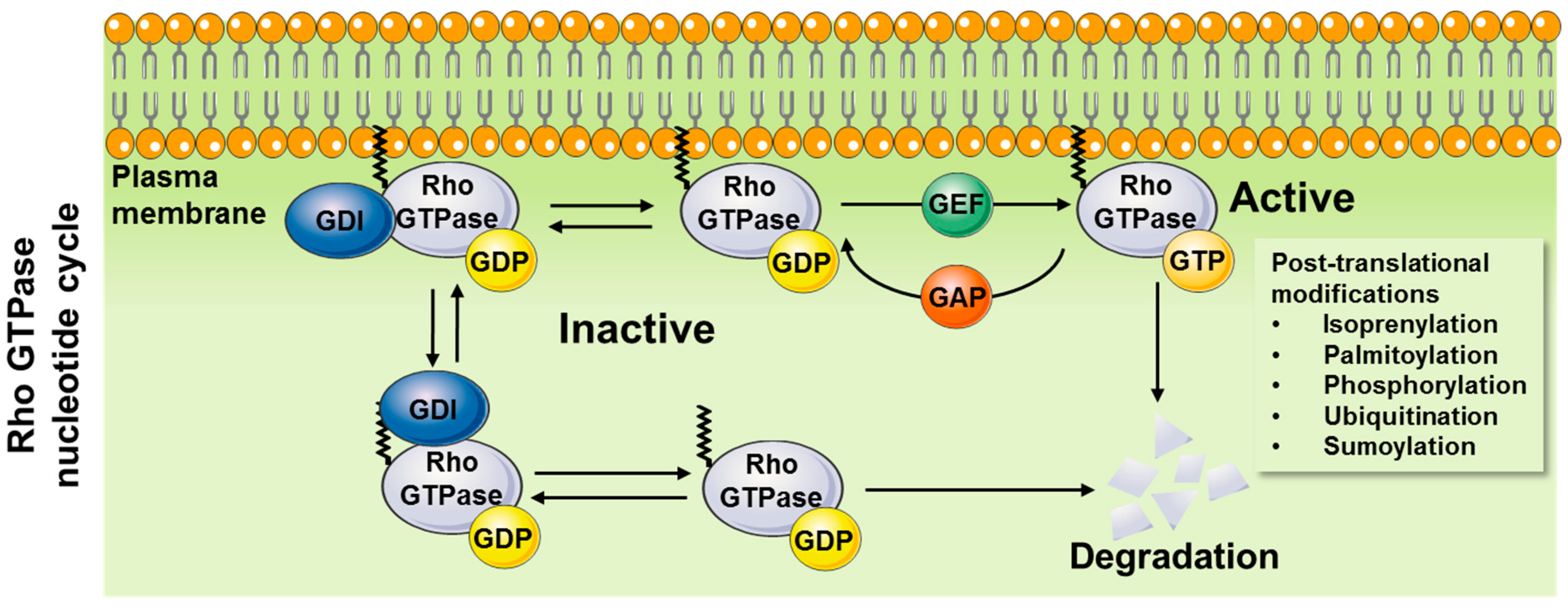
1.1. Mechanisms of Action

1.1.1. General Mechanisms of Activation

Wide interest in small GTPases was triggered by the discovery of the Ras oncogenes in 1982 [8] and with its mutations a few years later proven to cause human cancers [9,10]. Those discoveries were soon followed by the identification of related proteins now forming the Ras superfamily [11,12]. Rho GTPases function as molecular switches that cycle between an inactive guanosine diphosphate (GDP)-bound and an active guanosine triphosphate (GTP)-bound state (Figure 1). At least three major types of regulatory proteins/factors are described for Rho GTPases as illustrated in Figure 1. Activation of Rho GTPases is induced by guanine nucleotide exchange factors (GEFs), which facilitate switching from a GDP- to a GTP-bound state. Inactivation results from GTPase-activating proteins (GAPs) that promote the intrinsic GTPase activity mediating GTP to GDP hydrolysis [13,14]. Additionally, Rho guanine dissociation inhibitors (RhoGDIs) bind to the inactive, GDP-bound form of Rho GTPases maintaining their inactive state [15,16]. Thus, in order for a biological event to activate Rho GTPases, the balance between the GEFs, GAPs, and RhoGDIs must be altered to promote the Rho GTPase GTP-bound state. Mammalian cells encode genes for numerous of GEFs (>80) and GAPs (>60), but only three RhoGDIs [17]. Depending on the biological mode of regulation, release from RhoGDI must occur and GEFs must be activated to interact with the Rho GTPase and promote GTP binding. Conversely, inactivation occurs when GAPs promote the GTP hydrolysis. Rho GTPases can then be extracted from the membrane by RhoGDIs.
In addition to the regulation of the GTP–GDP cycling, post-translational modifications (e.g., isoprenylation, palmitoylation, phosphorylation, ubiquitination, sumoylation) of Rho GTPases appear to be additionally required for optimal regulation of Rho GTPase function [18,19]. The intracellular spatiotemporal distribution of Rho GTPases is tightly controlled. In general, active Rho GTPases are targeted to the plasma membrane (or endosomal membranes) via a polybasic region and a prenyl group attached to a C-terminal cysteine residue, and several Rho GTPases are also palmitoylated [18,20,21]. Prenylated cytosolic Rho GTPases are unstable and rapidly degraded [22]. Additionally, active Rho GTPases can be targeted for proteasomal degradation by ubiquitination [19,23]. The molecular chaperone, RhoGDI, binds to prenylated Rho GTPases forming a cytosolic pool of mainly inactive GDP-loaded Rho GTPases and protects Rho GTPases from ubiquitination and degradation [22]. Thus, while release from RhoGDI is a necessary event for the Rho GTPases to be activated, the release may reduce Rho GTPase protein abundance. Like Rho GTPases, GEFs, GAPs, and RhoGDIs are also regulated by post-translational modifications, revealing the many modalities for regulation of these key molecular switches [19]. Yet another layer of complexity is added to the regulation of the spatiotemporal distribution and activation, as Rho GTPases can interact to regulate each other’s activity. For example, the Rho GTPases, Rac1/Cdc42, and RhoA have opposing roles and double-negative feedback loops exist between them [24,25,26]. The mechanisms underlying this feedback regulation between Rho GTPases are not resolved but might involve Rho GTPase competitive binding to RhoGDI [22]. One study found that in vascular smooth muscle cells, phosphorylation of RhoA on S188 induced dissociation of Rac1 from RhoGDIα, thereby activating Rac1 and vascular smooth muscle cell migration [27], while protecting RhoA from ubiquitin/proteasome-mediated degradation [28]. Moreover, feedback loops exist between downstream effector proteins and the Rho GTPases. The downstream effectors of Rac1/Cdc42, p21-activated kinases (PAKs), have been reported to negatively regulate GEFs [29,30], while PAK1 phosphorylation of RhoGDI at site S101 and S174 leads to RhoGDI dissociation from Rac1 [31]. Additionally, pharmacological inhibition (IPA-3) of PAK1-3 lowered Rac1 activity [24]. This also increased RhoA activity in agreement with the above mentioned negative regulation between the Rho GTPases.
As outlined above, the regulation of Rho GTPases is highly complex making it challenging to determine the exact regulatory mechanisms operating under any one circumstance.

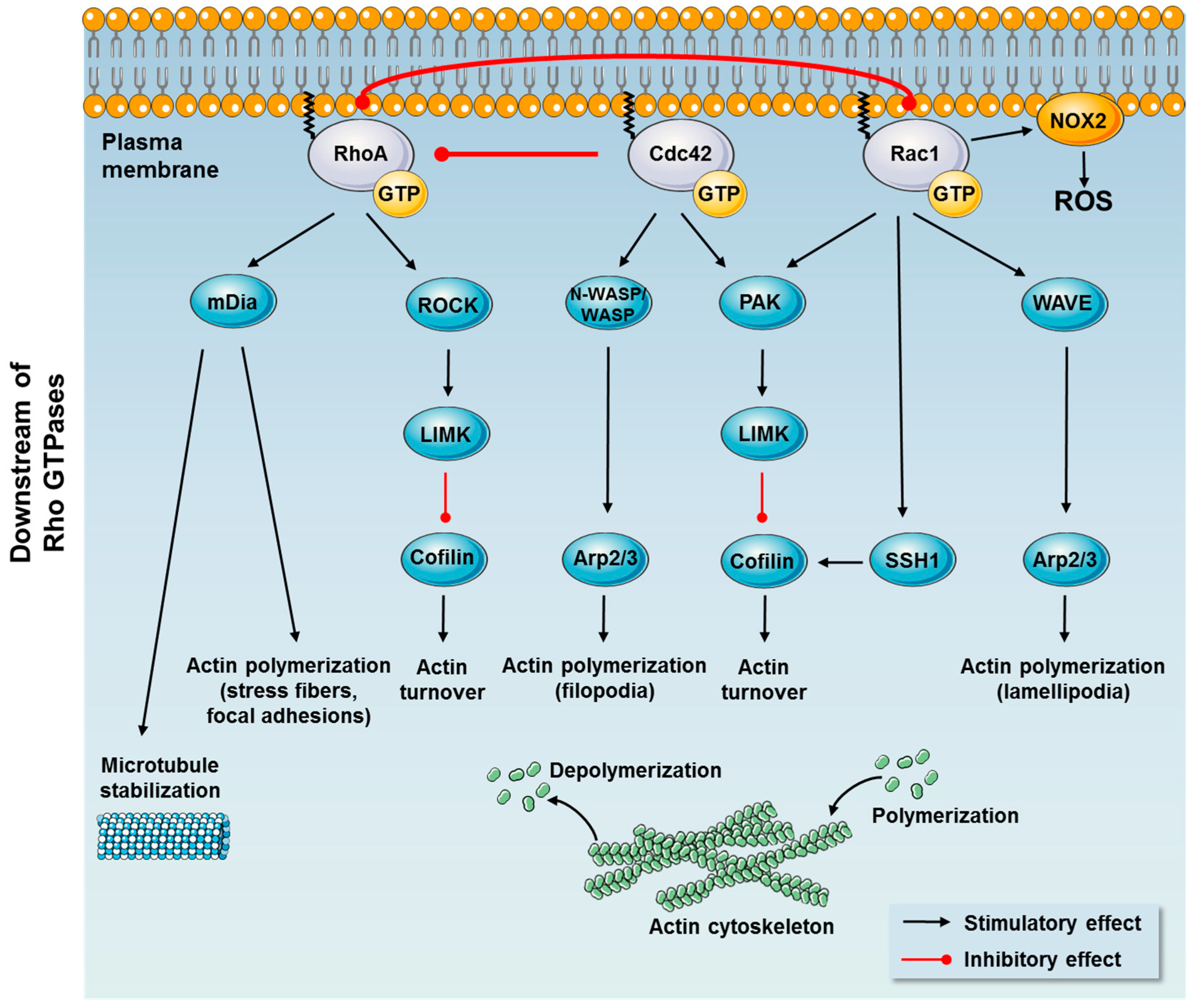
1.1.2. Downstream Effects

Active Rho GTPases recruit downstream effector molecules to the plasma membrane and trigger their activation [32]. Conserved structure and mechanism in multiple versions of Rho GTPases in bacteria, yeast, flies, and vertebrates, suggest that all derive from a single primordial protein, repeatedly modified in the course of evolution to perform a variety of functions. Each Rho GTPase affects numerous downstream proteins, all of which have specific roles in various cell processes [32,33]. Especially Rac1, Cdc42, and RhoA control pathways central to metabolic regulation. Rho GTPases are crucial organizers of the actin cytoskeleton (Figure 2). Active GTP-bound RhoA, Rac1, and Cdc42 promote the assembly of actin stress fibers and focal adhesions, thin sheet-like lamellipodial protrusions and finger-like filopodial protrusions, respectively [34,35,36,37,38,39]. The regulation of the actin cytoskeleton by Rho GTPases are extensively reviewed in [1,40,41] and only selected regulatory mechanisms investigated in relation to metabolic regulation will be presented here.
Actin remodeling is controlled through parallel actin polymerization and depolymerization. Generally, Rac1 and Cdc42 regulate comparable downstream events via activation of PAKs and activation of the Wiskott–Aldrich syndrome family of proteins including Wiskott-Aldrich Syndrome protein (WASP), neural-WASP (N-WASP), and WASP family verprolin homologous protein 1 or 2 (WAVE1/2). WASP and N-WASP are critical downstream effectors of Cdc42 that mediate the formation of filopodia through activation of the actin polymerization factor Arp2/3 [42]. Likewise, Rac1 exerts its effect on the cytoskeleton by binding to the WAVE complex, releasing WAVE and thereby promoting lamellipodia polymerization via activation of the Arp2/3 complex [43].
Among other downstream effectors, PAKs activate LIM domain kinases (LIMKs) promoting phosphorylation and inactivation of cofilin [42]. Active cofilin severs and depolymerizes actin filaments and leads to an increase in uncapped barbed ends promoting actin polymerization [42,44]. To counteract LIMK-dependent inactivation of cofilin, Rac1 activates the cofilin phosphatase, slingshot1 (SSH1). Thereby, phosphorylation of cofilin is tightly regulated by Rac1 by two opposite mechanisms, which results in cofilin dephosphorylation and activation [45,46,47]. Rac1 is also an essential component for the assembly of the NADPH oxidase complex (NOX2) by binding to p67-phox, and thereby facilitates the production of reactive oxygen species (ROS) at cell membranes [48,49].
Critical downstream effectors of RhoA are Rho-associated protein kinase (ROCK) and the Formin family of proteins that promotes the formation of straight, unbranched actin fibers. ROCK phosphorylates a large cohort of actin-binding proteins and intermediate filament proteins to modulate their functions. For example, ROCK phosphorylates LIMK leading to the phosphorylation and inactivation of the actin severing protein cofilin [50,51]. Formins capable of actin nucleation promote the elongation of pre-existing filaments by removing barbed end capping proteins [52]. Diaphanous formin (mDIa) is the most extensively studied and suggested to regulate microtubule stabilization in addition to its actin polymerization activity [53,54].
Whereas both downstream pathways of Rac1 and Cdc42 seem to be involved in metabolic control, RhoA may via downstream effectors be involved in muscle mass regulation and thereby play an important role in conditions of muscle mass loss. This will be reviewed below with emphasis on knowledge gained from in vivo studies and humans.

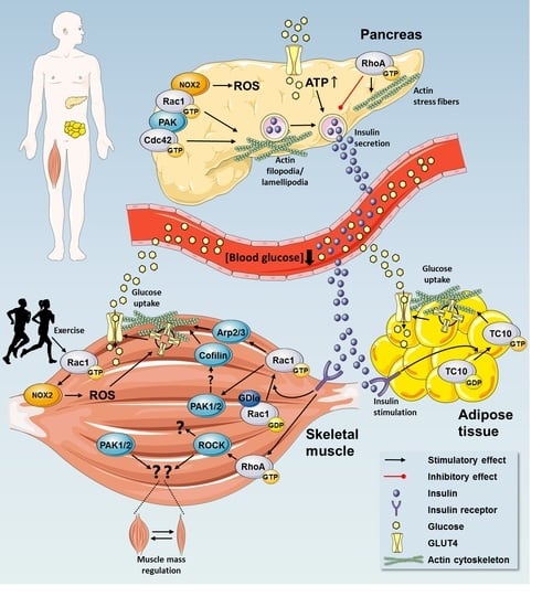
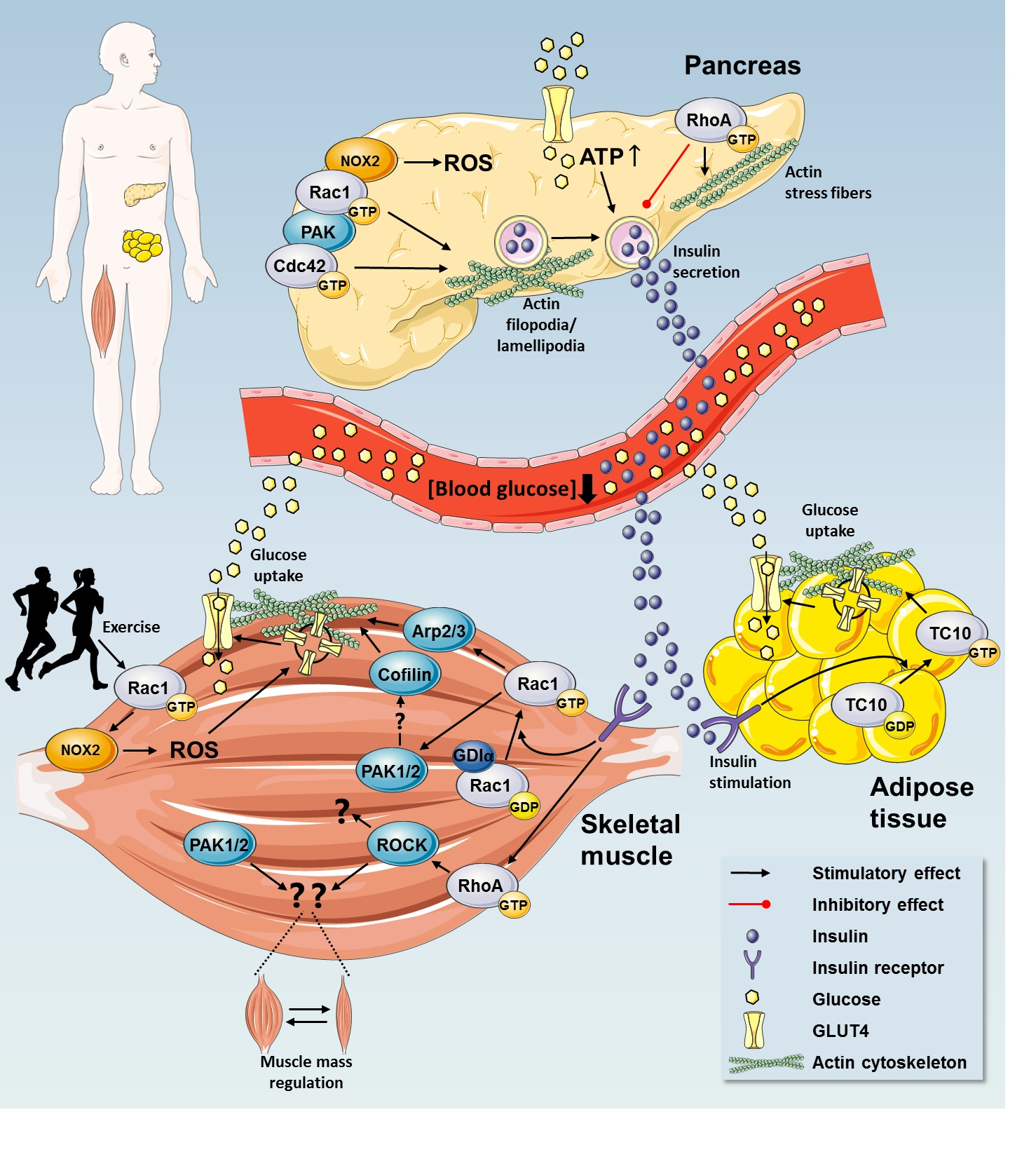
2. Rho GTPases in Regulation of Glucose Homeostasis

Rho GTPases have important functions during cell development and differentiation (reviewed in [1,55,56,57]). They also play crucial roles in fully differentiated tissues, including the pancreas, skeletal muscle, and adipose tissue (Figure 3). Following a meal, nutrients are absorbed in the intestines and enter the circulation. This results in a postprandial transient increase in blood glucose and free fatty acids. Blood glucose quickly returns to baseline because of an increased secretion of the hormone insulin from the pancreatic β cells. When in circulation, insulin stimulates glucose uptake into skeletal muscle and the adipose tissue together with inhibition of hepatic glucose production. Thereby insulin returns blood glucose back to baseline [58]. Those processes are significantly dysregulated in metabolically dysfunctional conditions, such as type 2 diabetes and obesity [59,60,61]. Both the pancreatic release of insulin and resulting insulin-stimulated uptake of glucose into skeletal muscle and adipose tissue are tightly regulated by complex processes that involve Rho GTPases. Rho GTPases likely also play insulin-independent roles in the maintenance of glucose homeostasis, including exercise-stimulated glucose uptake and beneficial adaptations to exercise training. In the following sections, we will delineate and discuss the current evidence for a role of Rho GTPases with a focus on their roles in maintaining glucose homeostasis.

2.1. Insulin Secretion by Pancreatic β Cells

The secretion of insulin is an essential everyday event that occurs following a meal to promote glucose disposition by the muscle and curb hepatic glucose output. Several lines of evidence place Rho GTPases, in particular Cdc42 and Rac1, as important players in glucose-stimulated insulin secretion via their roles in actin reorganization [62,63,64,65] as depicted in Figure 3. Glucose is a hydrophilic molecule that cannot cross the lipid bilayer membrane. High blood glucose is sensed by pancreatic β cells and the glucose taken up through GLUcose transporters (GLUTs; predominantly GLUT1 and GLUT3 in human islets, GLUT2 in rodents). In general terms, this rapidly translates into elevated intracellular ATP, which in turn closes the ATP-sensitive potassium ion channel, leading to depolarization of the cell membrane. Depolarization causes the opening of voltage-gated calcium channels, and insulin vesicles fuse with the cell membrane to release insulin (reviewed in [66]). During the fasting state when levels of blood glucose are low, the actin cytoskeleton restricts the access of secretory insulin-containing vesicles to their release sites. In response to a postprandial increase of blood glucose, the β cell’s actin cytoskeleton is reorganized, enabling insulin-containing vesicle translocation to the plasma membrane. Under normal circumstances, Cdc42 and Rac1 activation is needed for this process [58,63,67]. The precise mechanisms by which high glucose activates Rho GTPases are not clear but in vitro studies have shown that in response to glucose stimulation, Cdc42 is activated, which leads to Rac1 activation via PAK1 [68]. In agreement, isolated islets from pancreatic β cell-specific Rac1 knockout mice showed decreased glucose-stimulated insulin secretion in response to high glucose concentrations [65]. On the other hand, in vitro studies show that expression of constitutively active Cdc42 interferes with β cell differentiation leading to hyperglycemia [63]. Thus, tight regulation of the Rho GTPases must be in place. In vivo work from Thurmond’s group shows that the downstream effector of Cdc42 and Rac1, PAK1, is involved in insulin secretion, as whole body PAK1 knockout mice were glucose intolerant due to reduced insulin production [69].
Overall, it is well established that Rho GTPases play essential roles in normal β cell function and insulin secretion, primarily via the essential role of Cdc42 and Rac1 in insulin-vesicle translocation in response to increased glucose concentration in the circulation. However, the up- and downstream mechanisms remain to be clearly defined.

2.2. Rho GTPases are Implicated in β Cell Dysfunction in Metabolic Disease

Type 2 diabetes is associated with defects in insulin secretion whereby pancreatic β cells fail to compensate for peripheral insulin resistance (described below) so that hyperglycemia ensues [70]. Before the onset of type 2 diabetes, insulin resistance causes hyperproduction of insulin to overcome the insulin resistance of muscle, adipose tissue, and the liver. Accelerated insulin production is only tolerated for a limited period of time, after which the insulin-producing β cells dedifferentiate and insulin production is impaired or even stopped [71]. The stage of insulin hyperproduction has been associated with conditions of glucotoxicity and accelerated production of reactive oxygen species (ROS) in the islets. Rac1 is an important regulator of the assembly of the NADPH oxidase (NOX2) complex at the plasma membrane [49]. While ROS production is important for glucose-stimulated insulin secretion, uncontrolled Rac1-mediated superoxide hyperproduction is associated with β cell failure (reviewed in [72]). For example, hyperactivation of Rac1 has been demonstrable in human islets exposed to glucotoxic conditions and in islets derived from the Zucker diabetic fatty (ZDF) rat [73], a model of type 2 diabetes. Indeed, suppression of Rac1 activation protects β cells against noxious effects of glucolipotoxicity and cytokines [73,74]. Thus, Rac1 exerts damaging roles under pathological conditions by inducing NOX2 activity to create excessive oxidative stress, mitochondrial damage, and cell death [72,75]. In support of that, the expression and activation of Rac1 in pancreatic tissue from ob/ob mice, a model for obesity, is increased [76]. Interestingly, islets from donors with type 2 diabetes display an average PAK1 protein loss of 80% compared with non-diabetics [69] and recent work has shown that islets from donors with type 2 diabetes had profound defects in glucose-stimulated Cdc42 and PAK1 activation together with impaired glucose-stimulated insulin secretion [77]. Long-term hyperglycemia causing glucotoxicity has also been linked to RhoA-dependent stress fiber formation and diminished glucose-stimulated insulin secretion [78].
Thus, while the Rho GTPases Cdc42 and Rac1 are critical for insulin secretion, pathological hyperactivation of Rac1 and RhoA negatively affects β cell function during times of stress and may thus be targets for preventing β cell failure in diabetes.

2.3. Rho GTPases Regulate Glucose Uptake

The Rho GTPases Rac1, RhoA, and Cdc42 are highly expressed in skeletal muscle and adipose tissue and emerging evidence suggests that they have key functions in the regulation of glucose uptake and the maintenance of whole body glucose homeostasis.

2.3.1. Rho GTPases in Insulin Action in Skeletal Muscle

In response to insulin, the majority of dietary glucose is taken up by skeletal muscle [79,80]. Comprising 40–50% of the body, skeletal muscle is essential for maintaining whole body glucose homeostasis. Glucose uptake into skeletal muscle is an actively regulated process that necessitates mobilization and insertion of GLUT4 into the plasma membrane. This mechanism is primarily promoted by insulin and muscle contraction [81,82]. Perhaps not surprisingly due to their major role in vesicle traffic in other cell types, Rho GTPases are involved in the traffic of GLUT4-containing vesicles to the muscle plasma membrane (Figure 3).
Rac1, TC10, and RhoA are activated by insulin in skeletal muscle [83,84,85,86,87]. The first evidence that Rho GTPases are actively involved in the regulation of glucose uptake in skeletal muscle came nearly 20 years ago when the lack of functional Rac1 was observed to attenuate insulin-stimulated GLUT4 translocation in L6 muscle cells in vitro [88,89]. Additionally, constitutive active Rac1, but not Cdc42 or TC10, increased the amount of GLUT4 in the plasma membrane [83,90]. Subsequent studies also demonstrated an important role for Rac1 in insulin action in mature skeletal muscle in vivo. In mouse skeletal muscle, pharmacological inhibition and genetic ablation of Rac1 abolished insulin-stimulated GLUT4 translocation and reduced insulin-stimulated glucose uptake [86,87,91,92].
The involvement of Rac1 in insulin-stimulated GLUT4 translocation has been ascribed to a Rac1-mediated insulin-stimulated actin remodeling in skeletal muscle cells [83,88,89]. However, the relevance of a mobile actin cytoskeleton in fully mature skeletal muscle was recently questioned as mice that lack β-actin, one of the major actin cytoskeleton isoforms in skeletal muscle, displayed normal glucose transport [93]. On the other hand, in mature skeletal muscle, the actin depolymerizing agent, Latrunculin B reduced insulin-stimulated glucose uptake [87,91], suggesting participation of the actin cytoskeleton.
It is largely unknown how Rho GTPases are activated by insulin but it seems to occur downstream of phosphatidylinositol 3-kinase (PI3K). Skeletal muscle evidently expresses the following GEFs for Rac1: Tiam1 [94], Trio [95], Def6 [96], son of sevenless [97], Arf6 [98], Kalirin [99], Vav2, FLJ00068, DBl-I, α- and β-PIX (PAK-interacting exchange factor) [100], and switch-associated protein 70 (SWAP70) [90]. Among these, FLJ00068 has been suggested to regulate insulin-stimulated Rac1-dependent GLUT4 translocation in L6 myotubes [90] and mouse skeletal muscle [101]. Further research within this area will add to the understanding of the mechanism by which Rac1 regulates GLUT4 translocation.
RhoGDIα and RhoGDIβ are expressed in skeletal muscle [102,103] and RhoGDIα (but not RhoGDIβ) forms complexes with Rac1, RhoA, and Cdc42 [15]. Recent data from our laboratory has identified RhoGDIα as a negative regulator of Rac1 activity and GLUT4 translocation in skeletal muscle. In L6-GLUT4myc myotubes, siRNA-mediated RhoGDIα knockdown augments Rac1 activity and GLUT4 translocation both in the basal state and in response to insulin stimulation. Corroborating those in vitro results, RhoGDIα overexpression in mouse skeletal muscle in vivo decreased insulin-stimulated glucose uptake and impaired whole-body glucose tolerance (unpublished work, [104]). Thus, RhoGDIα is a novel key regulator of Rac1, important for maintaining the appropriate level of glucose uptake in muscle. The mechanisms by which insulin triggers Rac1 dissociation from RhoGDIα in skeletal muscle are currently unknown but it could involve phosphorylation of RhoGDIα by PAK or Src as described for other cell types [105,106].
Downstream of Rac1, PAK1 and PAK2 (PAK3 is not expressed in skeletal muscle) are activated in response to insulin [69,87,107] and are suggested to mediate Rac1-dependent insulin-stimulated reorganization of the actin cytoskeleton and GLUT4 translocation in skeletal muscle cells [108]. However, the role of group I PAKs in mature mouse skeletal muscle is debated. One study reported markedly impaired insulin-stimulated GLUT4 translocation in vivo in skeletal muscle lacking PAK1 [69], while recent work in our laboratory found no impairment in insulin-stimulated glucose uptake in PAK1 knockout mouse skeletal muscle in vivo or in isolated muscles in vitro [109]. In transgenic mice jointly lacking PAK1 and PAK2 only a mild reduction in insulin-stimulated glucose transport in muscle was evident [109], suggesting that group I PAKs play only a minor role in the regulation of glucose uptake. In support of this, constitutively activated Cdc42, an activator of group I PAKs, did not stimulate GLUT4 translocation in muscle cells in vitro [90]. The discrepancy between the PAK studies could perhaps be explained by the age of the mice, as double PAK1/2 knockout mice exhibit other age-related phenotypes [110]. Further downstream of Rac1, signaling through Arp2/3 and cofilin could mediate the cortical actin polymerization evoked by insulin (see Figure 2). In L6 myoblasts siRNA-mediated silencing of Arp3 abrogates actin remodeling and impairs GLUT4 translocation [47]. Moreover, cofilin knockdown causes overwhelming actin polymerization that subsequently inhibits GLUT4 translocation [47]. Those findings suggest that most distal regulators of actin cytoskeleton dynamics evoked by Rac1 are involved in insulin-stimulated glucose uptake, although those findings remain to be confirmed in differentiated muscle and in vivo. Thus, while the important role for Rac1 in insulin-stimulated glucose uptake is clear, Rac1 likely mediates glucose uptake via PAK-dependent, but also PAK-independent mechanisms that remain to be elucidated.
An input by RhoA in the regulation of insulin-stimulated glucose transport, has also been proposed, although the results are few and rather inconsistent [111]. Furthermore, all results are obtained by manipulating pathways downstream of RhoA and in several cases using pharmacological inhibitors reported to have unspecific effects [112]. Among the various effectors of RhoA, the kinase ROCK stands out. Two isoforms, ROCK1 and ROCK2, are expressed in skeletal muscle. ROCK1-deficient mice are insulin resistant [113] and siRNA-mediated silencing of ROCK1 reduced insulin-stimulated glucose transport in L6 myotubes [114], suggesting that ROCK1 is a positive regulator of insulin signaling. In addition, overexpression of a dominant negative ROCK2 or pharmacological inhibition of ROCKs impair insulin-stimulated GLUT4 translocation and glucose uptake in muscle in vivo [85].
Challenging a positive input of RhoA in the regulation of glucose uptake, membrane-bound (active) RhoA is elevated in skeletal muscles of obese Zucker rats [115]. In that model, jointly overexpressing a dominant-negative ROCK1 and 2 rescued diet-induced obesity and improved glucose tolerance [116], which contrasts with the findings in mice expressing only the dominant negative ROCK2 [85] or lacking ROCK1 [113] as described above. Interestingly, knockout of geranylgeranyl diphosphate synthase 1 (GGPPS; a branch point enzyme in the mevalonic acid pathway) specifically in skeletal muscle of mice decreased membrane-associated and prenylated RhoA (= active) and improved whole body glucose tolerance [117]. However, GGPPS might also affect the activity of other GTPases regulated by prenylations, including Rac1, which was not investigated. Thus, direct evidence for RhoA’s role in muscle insulin action is still lacking.
Collectively, there is ample support that Rac1, and possibly RhoA, but seemingly not Cdc42, participate in the insulin-dependent regulation of glucose uptake in skeletal muscle.

2.3.2. Cross-Talk Between Rho GTPases and Canonical Insulin Signaling

Canonical insulin signaling via Akt and TBC1D4 is a well-described signaling mechanism by which insulin increases muscle and adipose tissue glucose uptake [118,119]. In muscle and muscle cells in culture, both Akt/TBC1D4 and Rac1 are important regulators of insulin-stimulated glucose uptake. Studies from our laboratories have suggested that Rac1 and Akt mediate insulin-stimulated glucose uptake via parallel signaling mechanisms. This is because, in our hands, silencing Rac1 expression via siRNA [89] or knocking out the Rac1 gene [87] did not affect insulin-stimulated Akt phosphorylation although it abolished GLUT4 translocation [83,86,90]. Moreover, by inhibiting Rac1 or Akt2 separately, each partially reduced insulin-stimulated glucose transport, while joint inhibition of both pathways nearly fully blocked it [91]. Thus, the Rac1 and Akt pathways seem to act on GLUT4 translocation through distinct downstream processes. On the other hand, studies from the Satoh group point to cross-talk or feedback loops between the Rac1 and the Akt signaling pathways. For example, basal (but not insulin-stimulated) Akt activity was required for constitutively active Rac1 to induce GLUT4 translocation in L6 myotubes [90]. In agreement, Rac1 and PAK have been reported to be involved in the activation of Akt in various cell types [120,121] suggesting the possible existence of a feedback loop between Rac1 and Akt. Also, several publications from Satoh´s group suggest that Akt2 is involved in insulin-stimulated Rac1 activation leading to glucose uptake in muscle [101,122,123]. Most recently, this was proposed because knockdown of Akt2 by siRNA abolished Rac1 activation following intravenous administration of insulin or ectopic expression of a constitutively activated PI3K mutant in mouse muscle [124]. Similarly, we have observed reduced insulin-stimulated phosphorylation of PAKs in extensor digitorum longus, but not soleus muscle from Akt2 KO mice, although this was not observed in any of the muscles in response to pharmacological Akt inhibition using MK2206 [91]. Thus, currently the relative role of each pathway and how they interact to regulate insulin-stimulated glucose uptake is not fully understood. In our hands, a superactivation of Rac1 achieved by an acute overstimulation of the Rho GTPase lead to Akt phosphorylation, but the levels of Rac1 superactivation are far higher than those physiologically stimulated by insulin [125]. Based on these collective results we propose that, in muscle, the level of Rac1 activation achieved with insulin does not suffice to stimulate Akt2, however, it is possible to show a connection between these signals when Rac1 activation is supraphysiological. Moreover, while Akt might have some regulatory role over Rac1, Rac1 activity does not rely on Akt. Rac1 can be activated independent of Akt, since exercise and muscle contraction, which do not activate Akt, activate Rac1.

2.3.3. Exercise Elicits Metabolic Benefits via Regulation of Rho GTPases

It has been convincingly documented that regular exercise improves glycemic control and insulin action among healthy, as well as obese and type 2 diabetic subjects [126,127,128,129], and that this effect can be superior to those exerted by drugs or insulin therapy [130,131]. Physical activity causes a large increase in energy utilization [126,132,133]. Glucose is a major fuel source for the contracting muscles and glucose uptake acutely increases more than 50-fold during exercise [133]. Because glucose is taken up by the contracting muscles via insulin-independent mechanisms [134,135], it is effective in lowering blood glucose in insulin resistant subjects [136].
Rac1 is activated during exercise in mouse and human skeletal muscle [137] and Rac1 muscle-specific knockout mice exhibit reduced exercise-stimulated glucose uptake in muscle [138,139]. Likewise, GLUT4 translocation in response to electrical pulse stimulation (which mimics, but does not recapitulate muscle contraction) is reduced by inhibition of Rac1 in C2C12 myotubes [140].
Very recent results shed light onto the downstream events by which Rac1 may induce glucose uptake during exercise. Henriquez-Olguin et al. showed that Rac1 is implicated in the increase in ROS-production that occurs during exercise in muscle [141]. As described for pancreatic β cells, Rac1 is an integral part of the NOX2 complex in skeletal muscle. In skeletal muscle Rac1-mediated production of ROS contributes to increased glucose uptake in mouse skeletal muscle during exercise [141]. Because mice lacking PAK1 displayed normal contraction-stimulated glucose transport in skeletal muscle [109], one can assume that Rac1 does not mediate contraction-stimulated glucose uptake via PAK1. Rac1 would then be available for activation, contributing to NOX2 activation. The resulting ROS production would in turn contribute to the stimulation of muscle glucose uptake [141] by an unknown mechanism. Thus, Rac1 helps remove glucose from the blood via insulin independent mechanisms.
In addition to the acute stimulation of glucose uptake by a single bout of exercise, exercise training also induces remarkable skeletal muscle adaptations that benefit metabolic regulation, including the enhancement of insulin sensitivity (recently reviewed in [126]). Recent work has implicated RhoA in exercise-training adaptations, as seen by the increased expression of ROCK1, ROCK2, and RhoA in rat gastrocnemius muscle following short-term swimming exercise training [142]. Furthermore, ROCK1/2 inhibition by Y-27632 impaired the insulin sensitizing effect of exercise training [142]. Although non-specific effects of pharmacological inhibitors cannot be ruled out, that study suggests that the downstream effectors of RhoA and ROCK1/2 are involved in the metabolic benefits of exercise training.
Interestingly, exercise results in phosphorylation of RhoGDIα on serine 34 in human skeletal muscle [143]. Whether RhoGDIα serine 34 phosphorylation in muscle destabilizes its interaction with Rho GTPases to permit their activation during exercise will be interesting to determine.
In summary, exercise elicits metabolic benefits, several of which involve regulation of Rho GTPases. Future studies should delineate the mechanisms by which Rho GTPases may be influencing health with exercise and importantly if they can be explored pharmaceutically to harness some of the metabolic benefits of exercise in individuals that cannot adhere to an exercise program.

2.4. A Possible Role for Rho GTPases in Adipose Tissue Glucose Uptake

Although being mainly a fat storage tissue, adipose tissue is also essential for maintaining metabolic homeostasis and health. Especially in conditions of obesity, adipose tissue takes up a substantial amount of glucose following a meal, thereby significantly contributing to the clearance of glucose from the blood. Like in skeletal muscle, adipocyte glucose uptake relies on the translocation of GLUT4 to the plasma membrane for glucose to diffuse across the membrane lipid bilayer. Insulin activates TC10 [144,145], Cdc42 [146], Rac1 [147], and RhoA [148] in adipocytes but their involvement in adipose tissue glucose uptake is not clear.
An early investigation showed that transfection of a constitutively-active RhoA increased GLUT4 translocation in response to insulin, while dominant-negative RhoA significantly decreased it in 3T3-L1 and rat adipocytes [149]. Another study using siRNA-mediated Cdc42 knockdown reported that Cdc42 could mediate insulin signaling to glucose transport in 3T3-L1 adipocytes [146]. 3T3-L1 adipocytes seemingly respond normally to insulin after transfection of a dominant-negative or constitutively-active Rac1 [150]. However, Marcusohn et al. did not confirm that the mutants, transfected in the fibroblast stage of the culture, persisted in the adipocyte stage where insulin-stimulated glucose uptake was studied. Nevertheless, that study was supported by the finding that pharmacological Rac1 inhibition did not reduce insulin-stimulated glucose uptake in incubated fat pads from mice [87], although actual inhibition of Rac1-signaling was not confirmed in that study. Thus, despite methodological issues, the few studies to date suggest no role for Rac1 in insulin stimulated glucose uptake in adipocytes.
The Rho GTPase TC10 also seems relevant for regulating insulin-stimulated glucose uptake in adipose tissue as depicted in Figure 3. Dominant-negative TC10 prevents actin reorganization [151,152], an event that is necessary for adipocytes to translocate GLUT4 to the membrane. Accordingly, dominant-negative TC10 inhibits adipocyte glucose uptake and GLUT4 translocation [144]. That study [144] also reported no effect on insulin-stimulated GLUT4 translocation and glucose uptake of transfecting constitutively-active, or dominant-negative RhoA or Cdc42 in adipocytes (although data was not shown), contrasting with the other studies described above [146,149].
Taken together, Rho GTPases could play roles in adipocyte glucose uptake by regulating GLUT4 vesicle dynamics. Indeed, the fusion of GLUT4 vesicles with the adipocyte membrane requires dynamic insulin-induced actin polymerization, evincing the contribution of the actin cytoskeleton at different steps in the process of GLUT4 translocation [153]. However, the studies linking Rho GTPases to this event are few and the results conflicting with only TC10 consistently shown to regulate insulin-stimulated GLUT4 translocation in adipocytes. Importantly, mechanistic in vivo evidence is completely lacking and there is currently no human evidence in the literature. Thus, the involvement of Rho GTPases in adipose tissue function is largely unexplored and exciting discoveries lie ahead.

2.5. Rho GTPases are Implicated in Insulin Resistance—A Key Contributor to Metabolic Disease

In insulin resistant human subjects that are obese or have type 2 diabetes, insulin-stimulated glucose uptake by skeletal muscle is a primary site of dysfunction [79]. Due to their key role in skeletal muscle (and perhaps adipocyte) glucose uptake, Rho GTPases are relevant in metabolic diseases, including type 2 diabetes. Decreased skeletal muscle Rac1 signaling has been associated with insulin resistance in insulin resistant obese and type 2 diabetic human subjects [87], as well as in diabetic ob/ob mice and diet-induced obese insulin resistant rodents [91]. This has also been proposed by in vitro studies of muscle showing that ceramide-induced insulin resistance was associated with marked impairments in insulin-induced Rac1 activation [89]. However, in another in vitro model of insulin resistance, palmitate treatment did not impair Rac1 activity, although the phosphorylation of Rac1’s downstream target, PAK1 was reduced [154]. Interestingly, in a recent study from our laboratory, we observed that lack of Rac1 in muscles from diet-induced obese mice exacerbates insulin resistance [155], suggesting a relevance for Rac1 in counteracting metabolic dysfunctions. Those studies highlight the possibility that compromised Rac1 activity and/or downstream signaling contribute to the development of muscular insulin resistance.
RhoA might also regulate glucose homeostasis, as pharmacological inhibition of RhoA’s target, ROCK, using Fasudil prevented high-fat diet-induced hypercholesterolemia and glucose intolerance in mice [156]. Furthermore, body weight, serum lipid levels, and glucose metabolism were improved in mice with whole body overexpression of a dominant-negative ROCK, compared with littermate control mice [116]. In muscle cells in vitro, palmitic acid-induced insulin resistance was associated with increased expression of ROCK1 [157]. However, that ROCK would negatively affect glucose homeostasis contradicts the fact that lean ROCK1-deficient mice are insulin resistant [113] and lean mice with overexpression of a dominant negative ROCK2 display impaired insulin-stimulated GLUT4 translocation and glucose uptake in muscle in vivo [85]. Indeed, the RhoA-ROCK pathway seems to be clinically relevant, since insulin-stimulated muscle ROCK1/2 activity was attenuated in obese and type 2 diabetic subjects during an euglycemic hyperinsulinemic clamp compared to lean control [158]. Thus, the results on RhoA’s role in metabolic (dys)regulation warrant clarification.
Taken together, Rho GTPases, in particular Rac1 and RhoA, may be highly implicated in muscular insulin resistance and thereby contribute to metabolic dysregulation in metabolic diseases, such as type 2 diabetes.

3. Rho GTPases as Hitherto Unrecognized Regulators of Muscle Mass

Muscle mass is important for metabolic health because it increases the amount of tissue available for storing glucose. Although studies are few, there is emerging evidence that Rho GTPases may play hitherto unrecognized roles in muscle mass regulation. Indeed, Rho GTPases are highly regulated during conditions of muscle atrophy. For example, a marked reduction in levels of RhoA was noted in the muscles of unweighted hindlimbs (an experimental model simulating muscle atrophy under weightlessness) in mice [159] and in dystrophic mice [160] along with rapid atrophy. In male Sprague-Dawley rats, three days of hindlimb suspension-induced muscle atrophy decreased RhoA mRNA and protein expression [161]. In contrast, muscle mass loss caused by denervation, increased RhoA expression but returned to baseline when the decline in muscle mass ceased [162]. Thus, RhoA expression is highly regulated in models of muscle mass loss although this remains to be recapitulated in human skeletal muscle [163]. Conversely, increasing muscle mass using functional overload and anabolic steroid administration in mice [164], or hypertrophy-stimulating resistance training in humans elevated the expression of skeletal muscle RhoA [165]. Future studies should investigate muscle mass regulation in muscles that lack RhoA in order to directly determine the implications for RhoA.
Rho GTPases other than RhoA may also be involved in muscle mass regulation as proteomic analysis of mouse muscles following denervation-induced atrophy identified significant changes in RhoGDIα, PAK1-2, and Cdc42 expression [166]. Dominant-negative Cdc42, introduced with a retroviral vector, resulted in fibers that appeared atrophic [167] and blocking Rac1 function in precursors of the indirect flight muscle of Drosophila severely disrupted muscle formation. Thus, Rac1 is involved in the regulation of myoblast proliferation and segregation during adult myogenesis [168]. Downstream of Rac1 and Cdc42, PAK1 and PAK2 are activated during mammalian myoblast differentiation. Combined deletion of PAK1 and PAK2 results in reduced muscle mass, a phenotype that is exacerbated after repair to acute injury [110,169]. In support of that, pharmacological inhibition of group I PAKs (with IPA-3) delays skeletal muscle regeneration following cardiotoxin injury in vivo [170], suggesting that Rho GTPase-mediated signaling is important for muscle regeneration. Interestingly, insulin-stimulated phosphorylation of PAK1 at threonine 423 [171] and PAK1 protein content [171,172] were markedly increased in follistatin-induced hypertrophic mouse muscle compared to controls.
A role for Rho GTPases in muscle mass regulation is perhaps not surprising given their well-known requirement for tumor growth. Looking ahead, lessons from the tumor literature may help to understand the mechanisms by which Rho GTPases may be involved in muscle mass regulation in connection with metabolic regulation.

The Role of Rho GTPases in Muscle Wasting Conditions

Skeletal muscle atrophy is a severe consequence of ageing and many chronic diseases, including cancers. Muscle strength is inversely related to death from all causes [173] and is of the utmost importance for the preservation of mobility and quality of life. RhoA and RhoGDIα are both upregulated in mouse skeletal muscle with age-related muscle mass loss [174]. In agreement, single muscle fiber proteomics analysis showed that RhoGDIα protein expression increased with age in both slow and fast muscle fibers from human biopsy samples, while RhoA increased with age predominantly in fast muscle fibers [175]. Importantly, age-related muscle atrophy only occurred in the fast muscle fiber types. However, contradicting those two studies, a recent study found reduced RhoA protein expression in skeletal muscle of middle-aged rats together with diminished levels of ROCK proteins [176]. Thus, Rho GTPases might be differentially regulated at different ages and stages of sarcopenia, and this warrants further investigation.
Many cancers are associated with cachexia, a condition of involuntary body weight loss including severe muscle atrophy that is not due to anorexia [177]. Interestingly, PAK1 mRNA and protein expression are reduced in cancer-associated cachectic muscles from colon adenocarcinoma C26-bearing mice [170], although PAKs’ upstream Rho GTPases, Rac1, and Cdc42 were not examined. That is consistent with the role of group I PAKs in muscle mass regulation [110,169]. Indeed, PAK1 overexpression partly preserved fiber size in cachectic muscles [170], suggesting that the defect in PAK might be directly involved in the pathogenesis. From these collective studies, RhoA, Rac1 and Cdc42, and PAK emerge as candidate regulators of muscle mass. However, studies exploring a direct mechanistic role for the Rho GTPases in muscle mass regulation are completely lacking.
As muscle is the largest organ of the body, completely necessary for mobility and also responsible for the majority of glucose disposal, future studies should investigate the role for Rho GTPases in muscle wasting diseases.
Unresolved issues are showed in Box 1.
Box 1. Unresolved issues.

Unresolved Issues

  • Lack of in vivo experiments to support the in vitro literature on Rho GTPase regulation and in particular their role in metabolism.
  • Evidence on the regulatory functions of Rho GTPases in humans is limited.
  • Molecularly, the upstream activators and downstream effectors of Rho GTPases in different tissues and in response to different stimuli are poorly defined.
  • Cross-talk between Rho GTPases is poorly defined but important to delineate, as they challenge all interpretation of data using knockdown or overexpression of a single Rho GTPase.
  • High throughput methodological advances to directly measure in vivo GTP binding (fast hydrolysis) warranted.
  • Whether Rho GTPases can be targeted pharmacologically to benefit metabolic diseases remains to be determined.

4. Conclusions

In this review, we summarize evidence for the role of Rho GTPases in metabolic regulation in health and disease. We demonstrate that Rho GTPases may be hitherto overlooked players in glucose homeostasis by contributing to metabolically essential functions in skeletal muscle, adipose tissue, and the pancreas. However, this area of research is at its early stages and mechanistic in vivo insights are lacking. This will be an exciting area for future discoveries.

Author Contributions

Conceptualization, L.S., and L.L.V.M.; writing–original draft preparation, L.L.V.M and L.S.; writing–review and editing, L.L.V.M., A.K., and L.S. All authors contributed substantially to the work reported.

Funding

L.S. was supported by the Novo Nordisk Foundation, grant number NNF18OC0032082 and by Independent Research Fund Denmark, grant number DFF–4004-00233. A.K. was supported by the Canadian Institutes of Health Research Foundation Grant. L.LV.M. was supported by a PhD fellowship from The Lundbeck Foundation, grant number 2015-3388.

Acknowledgments

We thank our colleagues at the Section of Molecular Physiology, Department of Nutrition, Exercise, and Sports, Faculty of Science, University of Copenhagen for fruitful discussions on this topic. We apologize to those whose important work was not cited due to space limitations.

Conflicts of Interest

The authors declare no conflicts of interest.

References

  1. Jaffe, A.B.; Hall, A. Rho GTPases: Biochemistry and biology. Annu. Rev. Cell Dev. Biol. 2005, 21, 247–269. [Google Scholar] [CrossRef]
  2. Takai, Y.; Sasaki, T.; Matozaki, T. Small GTP-binding proteins. Physiol. Rev. 2001, 81, 153–208. [Google Scholar] [CrossRef]
  3. Boettner, B.; Van Aelst, L. The role of Rho GTPases in disease development. Gene 2002, 286, 155–174. [Google Scholar] [CrossRef]
  4. WHO Global status report on noncommunicable diseases 2014. Attaining the nine global noncommunicable diseases targets; a shared responsicility. World Heal. Organ. 2014, 176. [Google Scholar]
  5. Esposito, K.; Chiodini, P.; Colao, A.; Lenzi, A.; Giugliano, D. Metabolic Syndrome and Risk of Cancer: A systematic review and meta-analysis. Diabetes Care 2012, 35, 2402–2411. [Google Scholar] [CrossRef]
  6. Calle, E.E.; Rodriguez, C.; Walker-Thurmond, K.; Thun, M.J. Overweight, Obesity, and Mortality from Cancer in a Prospectively Studied Cohort of U.S. Adults. N. Engl. J. Med. 2003, 348, 1625–1638. [Google Scholar] [CrossRef]
  7. Samson, S.L.; Garber, A.J. Metabolic Syndrome. Endocrinol. Metab. Clin. North Am. 2014, 43, 1–23. [Google Scholar] [CrossRef]
  8. Parada, L.F.; Tabin, C.J.; Shih, C.; Weinberg, R.A. Human EJ bladder carcinoma oncogene is homologue of Harvey sarcoma virus ras gene. Nature 1982, 297, 474–478. [Google Scholar] [CrossRef]
  9. Santos, E.; Martin-Zanca, D.; Reddy, E.P.; Pierotti, M.A.; Della Porta, G.; Barbacid, M. Malignant activation of a K-ras oncogene in lung carcinoma but not in normal tissue of the same patient. Science 1984, 223, 661–664. [Google Scholar] [CrossRef]
  10. Bos, J.L.; Fearon, E.R.; Hamilton, S.R.; Vries, M.V.; van Boom, J.H.; van der Eb, A.J.; Vogelstein, B. Prevalence of ras gene mutations in human colorectal cancers. Nature 1987, 327, 293–297. [Google Scholar] [CrossRef]
  11. Bourne, H.R.; Sanders, D.A.; McCormick, F. The GTPase superfamily: A conserved switch for diverse cell functions. Nature 1990, 348, 125–132. [Google Scholar] [CrossRef]
  12. Cox, A.D.; Der, C.J. Ras history. Small GTPases 2010, 1, 22–27. [Google Scholar] [CrossRef]
  13. Bos, J.L.; Rehmann, H.; Wittinghofer, A. GEFs and GAPs: Critical Elements in the Control of Small G Proteins. Cell 2007, 129, 865–877. [Google Scholar] [CrossRef]
  14. Peurois, F.; Veyron, S.; Ferrandez, Y.; Ladid, I.; Benabdi, S.; Zeghouf, M.; Peyroche, G.; Cherfils, J. Characterization of the activation of small GTPases by their GEFs on membranes using artificial membrane tethering. Biochem. J. 2017, 474, 1259–1272. [Google Scholar] [CrossRef]
  15. Olofsson, B. Rho Guanine Dissociation Inhibitors. Cell. Signal. 1999, 11, 545–554. [Google Scholar] [CrossRef]
  16. Cherfils, J.; Zeghouf, M. Regulation of small GTPases by GEFs, GAPs, and GDIs. Physiol. Rev. 2013, 93, 269–309. [Google Scholar] [CrossRef]
  17. Rossman, K.L.; Der, C.J.; Sondek, J. GEF means go: Turning on RHO GTPases with guanine nucleotide-exchange factors. Nat. Rev. Mol. Cell Biol. 2005, 6, 167–180. [Google Scholar] [CrossRef]
  18. Wennerberg, K.; Der, C.J. Rho-family GTPases: It’s not only Rac and Rho (and I like it). J. Cell Sci. 2004, 117, 1301–1312. [Google Scholar] [CrossRef]
  19. Hodge, R.G.; Ridley, A.J. Regulating Rho GTPases and their regulators. Nat. Rev. Mol. Cell Biol. 2016, 17, 496–510. [Google Scholar] [CrossRef]
  20. Roberts, P.J.; Mitin, N.; Keller, P.J.; Chenette, E.J.; Madigan, J.P.; Currin, R.O.; Cox, A.D.; Wilson, O.; Kirschmeier, P.; Der, C.J. Rho Family GTPase modification and dependence on CAAX motif-signaled posttranslational modification. J. Biol. Chem. 2008, 283, 25150–25163. [Google Scholar] [CrossRef]
  21. Nishimura, A.; Linder, M.E. Identification of a Novel Prenyl and Palmitoyl Modification at the CaaX Motif of Cdc42 That Regulates RhoGDI Binding. Mol. Cell. Biol. 2013, 33, 1417–1429. [Google Scholar] [CrossRef]
  22. Boulter, E.; Garcia-Mata, R.; Guilluy, C.; Dubash, A.; Rossi, G.; Brennwald, P.J.; Burridge, K. Regulation of Rho GTPase crosstalk, degradation and activity by RhoGDI1. Nat. Cell Biol. 2010, 12, 477–483. [Google Scholar] [CrossRef]
  23. Garcia-Mata, R.; Boulter, E.; Burridge, K. The “invisible hand”: Regulation of RHO GTPases by RHOGDIs. Nat. Rev. Mol. Cell Biol. 2011, 12, 493–504. [Google Scholar] [CrossRef]
  24. Byrne, K.M.; Monsefi, N.; Dawson, J.C.; Degasperi, A.; Bukowski-Wills, J.-C.; Volinsky, N.; Dobrzyński, M.; Birtwistle, M.R.; Tsyganov, M.A.; Kiyatkin, A.; et al. Bistability in the Rac1, PAK, and RhoA Signaling Network Drives Actin Cytoskeleton Dynamics and Cell Motility Switches. Cell Syst. 2016, 2, 38–48. [Google Scholar] [CrossRef]
  25. Sander, E.E.; ten Klooster, J.P.; van Delft, S.; van der Kammen, R.A.; Collard, J.G. Rac downregulates Rho activity: Reciprocal balance between both GTPases determines cellular morphology and migratory behavior. J. Cell Biol. 1999, 147, 1009–1022. [Google Scholar] [CrossRef]
  26. Nimnual, A.S.; Taylor, L.J.; Bar-Sagi, D. Redox-dependent downregulation of Rho by Rac. Nat. Cell Biol. 2003, 5, 236–241. [Google Scholar] [CrossRef]
  27. Rolli-Derkinderen, M.; Toumaniantz, G.; Pacaud, P.; Loirand, G. RhoA phosphorylation induces Rac1 release from guanine dissociation inhibitor alpha and stimulation of vascular smooth muscle cell migration. Mol. Cell. Biol. 2010, 30, 4786–4796. [Google Scholar] [CrossRef]
  28. Rolli-Derkinderen, M.; Sauzeau, V.; Boyer, L.; Lemichez, E.; Baron, C.; Henrion, D.; Loirand, G.; Pacaud, P. Phosphorylation of Serine 188 Protects RhoA from Ubiquitin/Proteasome-Mediated Degradation in Vascular Smooth Muscle Cells. Circ. Res. 2005, 96, 1152–1160. [Google Scholar] [CrossRef]
  29. Rosenfeldt, H.; Castellone, M.D.; Randazzo, P.A.; Gutkind, J.S. Rac inhibits thrombin-induced Rho activation: Evidence of a Pak-dependent GTPase crosstalk. J. Mol. Signal. 2006, 1, 8. [Google Scholar] [CrossRef]
  30. Barac, A.; Basile, J.; Vázquez-Prado, J.; Gao, Y.; Zheng, Y.; Gutkind, J.S. Direct Interaction of p21-Activated Kinase 4 with PDZ-RhoGEF, a G Protein-linked Rho Guanine Exchange Factor. J. Biol. Chem. 2004, 279, 6182–6189. [Google Scholar] [CrossRef]
  31. DerMardirossian, C.; Schnelzer, A.; Bokoch, G.M. Phosphorylation of RhoGDI by Pak1 Mediates Dissociation of Rac GTPase. Mol. Cell 2004, 15, 117–127. [Google Scholar] [CrossRef]
  32. Bustelo, X.R.; Sauzeau, V.; Berenjeno, I.M. GTP-binding proteins of the Rho/Rac family: Regulation, effectors and functions in vivo. BioEssays 2007, 29, 356–370. [Google Scholar] [CrossRef]
  33. Bishop, A.L.; Hall, A. Rho GTPases and their effector proteins. Biochem. J. 2000, 348, 241–255. [Google Scholar] [CrossRef]
  34. Hall, A. Ras-related GTPases and the cytoskeleton. Mol. Biol. Cell 1992, 3, 475–479. [Google Scholar] [CrossRef]
  35. Nobes, C.; Hall, A. Regulation and function of the Rho subfamily of small GTPases. Curr. Opin. Genet. Dev. 1994, 4, 77–81. [Google Scholar] [CrossRef]
  36. Nobes, C.D.; Hall, A. Rho, rac and cdc42 GTPases: Regulators of actin structures, cell adhesion and motility. Biochem. Soc. Trans. 2015, 23, 456–459. [Google Scholar] [CrossRef]
  37. Nobes, C.D.; Hall, A. Rho, Rac, and Cdc42 GTPases regulate the assembly of multimolecular focal complexes associated with actin stress fibers, lamellipodia, and filopodia. Cell 1995, 81, 53–62. [Google Scholar] [CrossRef]
  38. Ridley, A.J.; Hall, A. The small GTP-binding protein rho regulates the assembly of focal adhesions and actin stress fibers in response to growth factors. Cell 1992, 70, 389–399. [Google Scholar] [CrossRef]
  39. Ridley, A.J.; Paterson, H.F.; Johnston, C.L.; Diekmann, D.; Hall, A. The small GTP-binding protein rac regulates growth factor-induced membrane ruffling. Cell 1992, 70, 401–410. [Google Scholar] [CrossRef]
  40. Spiering, D.; Hodgson, L. Dynamics of the Rho-family small GTPases in actin regulation and motility. Cell Adh. Migr. 2011, 5, 170–180. [Google Scholar] [CrossRef]
  41. Sit, S.-T.; Manser, E. Rho GTPases and their role in organizing the actin cytoskeleton. J. Cell Sci. 2011, 124, 679–683. [Google Scholar] [CrossRef]
  42. Ono, S. Mechanism of depolymerization and severing of actin filaments and its significance in cytoskeletal dynamics. Int. Rev. Cytol. 2007, 258, 1–82. [Google Scholar]
  43. Goley, E.D.; Welch, M.D. The ARP2/3 complex: An actin nucleator comes of age. Nat. Rev. Mol. Cell Biol. 2006, 7, 713–726. [Google Scholar] [CrossRef]
  44. Ghosh, M.; Song, X.; Mouneimne, G.; Sidani, M.; Lawrence, D.S.; Condeelis, J.S. Cofilin promotes actin polymerization and defines the direction of cell motility. Science 2004, 304, 743–746. [Google Scholar] [CrossRef]
  45. Kligys, K.; Claiborne, J.N.; DeBiase, P.J.; Hopkinson, S.B.; Wu, Y.; Mizuno, K.; Jones, J.C.R. The Slingshot Family of Phosphatases Mediates Rac1 Regulation of Cofilin Phosphorylation, Laminin-332 Organization, and Motility Behavior of Keratinocytes. J. Biol. Chem. 2007, 282, 32520–32528. [Google Scholar] [CrossRef]
  46. Cao, Z.; Li, X.; Li, J.; Kang, B.; Chen, J.; Luo, W.; Huang, C. SUMOylation of RhoGDIα is required for its repression of cyclin D1 expression and anchorage-independent growth of cancer cells. Mol. Oncol. 2014, 8, 285–296. [Google Scholar] [CrossRef]
  47. Chiu, T.T.; Patel, N.; Shaw, A.E.; Bamburg, J.R.; Klip, A. Arp2/3- and Cofilin-coordinated Actin Dynamics Is Required for Insulin-mediated GLUT4 Translocation to the Surface of Muscle Cells. Mol. Biol. Cell 2010, 21, 3529–3539. [Google Scholar] [CrossRef]
  48. Bedard, K.; Krause, K.-H. The NOX Family of ROS-Generating NADPH Oxidases: Physiology and Pathophysiology. Physiol. Rev. 2007, 87, 245–313. [Google Scholar] [CrossRef]
  49. Abo, A.; Pick, E.; Hall, A.; Totty, N.; Teahan, C.G.; Segal, A.W. Activation of the NADPH oxidase involves the small GTP-binding protein p21rac1. Nature 1991, 353, 668–670. [Google Scholar] [CrossRef]
  50. Maekawa, M.; Ishizaki, T.; Boku, S.; Watanabe, N.; Fujita, A.; Iwamatsu, A.; Obinata, T.; Ohashi, K.; Mizuno, K.; Narumiya, S. Signaling from Rho to the actin cytoskeleton through protein kinases ROCK and LIM-kinase. Science 1999, 285, 895–898. [Google Scholar] [CrossRef]
  51. Ohashi, K.; Nagata, K.; Maekawa, M.; Ishizaki, T.; Narumiya, S.; Mizuno, K. Rho-associated Kinase ROCK Activates LIM-kinase 1 by Phosphorylation at Threonine 508 within the Activation Loop. J. Biol. Chem. 2000, 275, 3577–3582. [Google Scholar] [CrossRef]
  52. Breitsprecher, D.; Goode, B.L. Formins at a glance. J. Cell Sci. 2013, 126, 1–7. [Google Scholar] [CrossRef]
  53. Copeland, J.W. The Diaphanous-related Formin mDia1 Controls Serum Response Factor Activity through its Effects on Actin Polymerization. Mol. Biol. Cell 2002, 13, 4088–4099. [Google Scholar] [CrossRef]
  54. Kühn, S.; Geyer, M. Formins as effector proteins of Rho GTPases. Small GTPases 2014, 5, e983876. [Google Scholar] [CrossRef] [PubMed]
  55. Etienne-Manneville, S.; Hall, A. Rho GTPases in cell biology. Nature 2002, 420, 629–635. [Google Scholar] [CrossRef]
  56. Hall, A. Rho family GTPases. Biochem. Soc. Trans. 2012, 40, 1378–1382. [Google Scholar] [CrossRef] [PubMed]
  57. Zegers, M.M.; Friedl, P. Rho GTPases in collective cell migration. Small GTPases 2014, 5, e983869. [Google Scholar] [CrossRef] [PubMed]
  58. Tokarz, V.L.; MacDonald, P.E.; Klip, A. The cell biology of systemic insulin function. J. Cell Biol. 2018, 217, 2273–2289. [Google Scholar] [CrossRef] [PubMed]
  59. Gandasi, N.R.; Yin, P.; Riz, M.; Chibalina, M.V.; Cortese, G.; Lund, P.-E.; Matveev, V.; Rorsman, P.; Sherman, A.; Pedersen, M.G.; et al. Ca2+ channel clustering with insulin-containing granules is disturbed in type 2 diabetes. J. Clin. Invest. 2017, 127, 2353–2364. [Google Scholar] [CrossRef]
  60. Zierath, J.R.; He, L.; Gumà, A.; Odegoard Wahlström, E.; Klip, A.; Wallberg-Henriksson, H. Insulin action on glucose transport and plasma membrane GLUT4 content in skeletal muscle from patients with NIDDM. Diabetologia 1996, 39, 1180–1189. [Google Scholar] [CrossRef]
  61. Czech, M.P. Insulin action and resistance in obesity and type 2 diabetes. Nat. Med. 2017, 23, 804–814. [Google Scholar] [CrossRef]
  62. Greiner, T.U.; Kesavan, G.; Ståhlberg, A.; Semb, H. Rac1 regulates pancreatic islet morphogenesis. BMC Dev. Biol. 2009, 9, 2. [Google Scholar] [CrossRef]
  63. Kesavan, G.; Lieven, O.; Mamidi, A.; Ohlin, Z.L.; Johansson, J.K.; Li, W.-C.; Lommel, S.; Greiner, T.U.; Semb, H. Cdc42/N-WASP signaling links actin dynamics to pancreatic cell delamination and differentiation. Development 2014, 141, 685–696. [Google Scholar] [CrossRef]
  64. Veluthakal, R.; Tunduguru, R.; Arora, D.K.; Sidarala, V.; Syeda, K.; Vlaar, C.P.; Thurmond, D.C.; Kowluru, A. VAV2, a guanine nucleotide exchange factor for Rac1, regulates glucose-stimulated insulin secretion in pancreatic beta cells. Diabetologia 2015, 58, 2573–2581. [Google Scholar] [CrossRef]
  65. Asahara, S.; Shibutani, Y.; Teruyama, K.; Inoue, H.Y.; Kawada, Y.; Etoh, H.; Matsuda, T.; Kimura-Koyanagi, M.; Hashimoto, N.; Sakahara, M.; et al. Ras-related C3 botulinum toxin substrate 1 (RAC1) regulates glucose-stimulated insulin secretion via modulation of F-actin. Diabetologia 2013, 56, 1088–1097. [Google Scholar] [CrossRef]
  66. Rorsman, P.; Braun, M. Regulation of Insulin Secretion in Human Pancreatic Islets. Annu. Rev. Physiol. 2013, 75, 155–179. [Google Scholar] [CrossRef]
  67. Huang, Q.-Y.; Lai, X.-N.; Qian, X.-L.; Lv, L.-C.; Li, J.; Duan, J.; Xiao, X.-H.; Xiong, L.-X. Cdc42: A Novel Regulator of Insulin Secretion and Diabetes-Associated Diseases. Int. J. Mol. Sci. 2019, 20, 179. [Google Scholar] [CrossRef]
  68. Kalwat, M.A.; Thurmond, D.C. Signaling mechanisms of glucose-induced F-actin remodeling in pancreatic islet β cells. Exp. Mol. Med. 2013, 45, e37. [Google Scholar] [CrossRef]
  69. Wang, Z.; Oh, E.; Clapp, D.W.; Chernoff, J.; Thurmond, D.C. Inhibition or ablation of p21-activated kinase (PAK1) disrupts glucose homeostatic mechanisms in vivo. J. Biol. Chem. 2011, 286, 41359–41367. [Google Scholar] [CrossRef]
  70. Prentki, M. Islet cell failure in type 2 diabetes. J. Clin. Invest. 2006, 116, 1802–1812. [Google Scholar] [CrossRef]
  71. Seino, S.; Shibasaki, T.; Minami, K. Dynamics of insulin secretion and the clinical implications for obesity and diabetes. J. Clin. Invest. 2011, 121, 2118–2125. [Google Scholar] [CrossRef]
  72. Kowluru, A. Friendly, and not so friendly, roles of Rac1 in islet β-cell function: Lessons learnt from pharmacological and molecular biological approaches. Biochem. Pharmacol. 2011, 81, 965–975. [Google Scholar] [CrossRef] [PubMed]
  73. Syed, I.; Kyathanahalli, C.N.; Jayaram, B.; Govind, S.; Rhodes, C.J.; Kowluru, R.A.; Kowluru, A. Increased Phagocyte-Like NADPH Oxidase and ROS Generation in Type 2 Diabetic ZDF Rat and Human Islets. Diabetes 2011, 60, 2843–2852. [Google Scholar] [CrossRef]
  74. Subasinghe, W.; Syed, I.; Kowluru, A. Phagocyte-like NADPH oxidase promotes cytokine-induced mitochondrial dysfunction in pancreatic β-cells: Evidence for regulation by Rac1. Am. J. Physiol. Integr. Comp. Physiol. 2011, 300, R12–R20. [Google Scholar] [CrossRef] [PubMed]
  75. Kowluru, A.; Kowluru, R.A. Phagocyte-like NADPH oxidase [Nox2] in cellular dysfunction in models of glucolipotoxicity and diabetes. Biochem. Pharmacol. 2014, 88, 275–283. [Google Scholar] [CrossRef] [PubMed]
  76. Zhou, S.; Yu, D.; Ning, S.; Zhang, H.; Jiang, L.; He, L.; Li, M.; Sun, M. Augmented Rac1 Expression and Activity are Associated with Oxidative Stress and Decline of β Cell Function in Obesity. Cell. Physiol. Biochem. 2015, 35, 2135–2148. [Google Scholar] [CrossRef]
  77. Veluthakal, R.; Chepurny, O.G.; Leech, C.A.; Schwede, F.; Holz, G.G.; Thurmond, D.C. Restoration of Glucose-Stimulated Cdc42-Pak1 Activation and Insulin Secretion by a Selective Epac Activator in Type 2 Diabetic Human Islets. Diabetes 2018, 67, 1999–2011. [Google Scholar] [CrossRef] [PubMed]
  78. Kong, X.; Yan, D.; Sun, J.; Wu, X.; Mulder, H.; Hua, X.; Ma, X. Glucagon-Like Peptide 1 Stimulates Insulin Secretion via Inhibiting RhoA/ROCK Signaling and Disassembling Glucotoxicity-Induced Stress Fibers. Endocrinology 2014, 155, 4676–4685. [Google Scholar] [CrossRef] [PubMed]
  79. DeFronzo, R.A.; Gunnarsson, R.; Björkman, O.; Olsson, M.; Wahren, J. Effects of insulin on peripheral and splanchnic glucose metabolism in noninsulin-dependent (type II) diabetes mellitus. J. Clin. Invest. 1985, 76, 149–155. [Google Scholar] [CrossRef]
  80. Lang, C.H. Rates and Tissue Sites of Noninsulin- and Insulin-Mediated Glucose Uptake in Diabetic Rats. Exp. Biol. Med. 2013, 199, 81–87. [Google Scholar] [CrossRef]
  81. Ploug, T.; van Deurs, B.; Ai, H.; Cushman, S.W.; Ralston, E. Analysis of GLUT4 Distribution in Whole Skeletal Muscle Fibers: Identification of Distinct Storage Compartments That Are Recruited by Insulin and Muscle Contractions. J. Cell Biol. 1998, 142, 1429–1446. [Google Scholar] [CrossRef] [PubMed]
  82. Lauritzen, H.P.M.M. Insulin- and Contraction-Induced Glucose Transporter 4 Traffic in Muscle. Exerc. Sport Sci. Rev. 2013, 41, 77–86. [Google Scholar] [CrossRef] [PubMed]
  83. JeBailey, L.; Rudich, A.; Huang, X.; Ciano-Oliveira, C.D.; Kapus, A.; Klip, A. Skeletal Muscle Cells and Adipocytes Differ in Their Reliance on TC10 and Rac for Insulin-Induced Actin Remodeling. Mol. Endocrinol. 2004, 18, 359–372. [Google Scholar] [CrossRef]
  84. Ishikura, S.; Koshkina, A.; Klip, A. Small G proteins in insulin action: Rab and Rho families at the crossroads of signal transduction and GLUT4 vesicle traffic. Acta Physiol. 2007, 192, 61–74. [Google Scholar] [CrossRef] [PubMed]
  85. Furukawa, N.; Ongusaha, P.; Jahng, W.J.; Araki, K.; Choi, C.S.; Kim, H.-J.; Lee, Y.H.; Kaibuchi, K.; Kahn, B.B.; Masuzaki, H.; et al. Role of Rho-kinase in regulation of insulin action and glucose homeostasis. Cell Metab. 2005, 2, 119–129. [Google Scholar] [CrossRef]
  86. Ueda, S.; Kitazawa, S.; Ishida, K.; Nishikawa, Y.; Matsui, M.; Matsumoto, H.; Aoki, T.; Nozaki, S.; Takeda, T.; Tamori, Y.; et al. Crucial role of the small GTPase Rac1 in insulin-stimulated translocation of glucose transporter 4 to the mouse skeletal muscle sarcolemma. FASEB J. 2010, 24, 2254–2261. [Google Scholar] [CrossRef]
  87. Sylow, L.; Jensen, T.E.; Kleinert, M.; Hojlund, K.; Kiens, B.; Wojtaszewski, J.; Prats, C.; Schjerling, P.; Richter, E.A. Rac1 Signaling Is Required for Insulin-Stimulated Glucose Uptake and Is Dysregulated in Insulin-Resistant Murine and Human Skeletal Muscle. Diabetes 2013, 62, 1865–1875. [Google Scholar] [CrossRef]
  88. Khayat, Z.A.; Tong, P.; Yaworsky, K.; Bloch, R.J.; Klip, A. Insulin-induced actin filament remodeling colocalizes actin with phosphatidylinositol 3-kinase and GLUT4 in L6 myotubes. J. Cell Sci. 2000, 113, 279–290. [Google Scholar]
  89. JeBailey, L.; Wanono, O.; Niu, W.; Roessler, J.; Rudich, A.; Klip, A. Ceramide- and oxidant-induced insulin resistance involve loss of insulin-dependent Rac-activation and actin remodeling in muscle cells. Diabetes 2007, 56, 394–403. [Google Scholar] [CrossRef]
  90. Ueda, S.; Kataoka, T.; Satoh, T. Activation of the small GTPase Rac1 by a specific guanine-nucleotide-exchange factor suffices to induce glucose uptake into skeletal-muscle cells. Biol. Cell 2008, 100, 645–661. [Google Scholar] [CrossRef] [PubMed]
  91. Sylow, L.; Kleinert, M.; Pehmøller, C.; Prats, C.; Chiu, T.T.; Klip, A.; Richter, E.A.; Jensen, T.E. Akt and Rac1 signaling are jointly required for insulin-stimulated glucose uptake in skeletal muscle and downregulated in insulin resistance. Cell. Signal. 2014, 26, 323–331. [Google Scholar] [CrossRef]
  92. Chiu, T.T.; Jensen, T.E.; Sylow, L.; Richter, E.A.; Klip, A. Rac1 signalling towards GLUT4/glucose uptake in skeletal muscle. Cell. Signal. 2011, 23, 1546–1554. [Google Scholar] [CrossRef]
  93. Madsen, A.B.; Knudsen, J.R.; Henriquez-Olguin, C.; Angin, Y.; Zaal, K.J.; Sylow, L.; Schjerling, P.; Ralston, E.; Jensen, T.E. β-Actin shows limited mobility and is required only for supraphysiological insulin-stimulated glucose transport in young adult soleus muscle. Am. J. Physiol. Metab. 2018, 315, E110–E125. [Google Scholar] [CrossRef]
  94. You, G.Y.; Lee, J.O.; Kim, J.H.; Kim, N.; Lee, S.K.; Moon, J.W.; Jie, S.; Lee, H.J.; Kim, S.J.; Park, S.H.; et al. Tiam-1, a GEF for Rac1, plays a critical role in metformin-mediated glucose uptake in C2C12 cells. Cell. Signal. 2013, 25, 2558–2565. [Google Scholar] [CrossRef]
  95. Charrasse, S.; Comunale, F.; Fortier, M.; Portales-Casamar, E.; Debant, A.; Gauthier-Rouviere, C. M-cadherin activates Rac1 GTPase through the Rho-GEF trio during myoblast fusion. Mol. Biol. Cell 2007, 18, 1734–1743. [Google Scholar] [CrossRef]
  96. Samson, T.; Will, C.; Knoblauch, A.; Sharek, L.; von der Mark, K.; Burridge, K.; Wixler, V. Def-6, a guanine nucleotide exchange factor for Rac1, interacts with the skeletal muscle integrin chain alpha7A and influences myoblast differentiation. J. Biol. Chem. 2007, 282, 15730–15742. [Google Scholar] [CrossRef]
  97. Kim, Y.B.; Inoue, T.; Nakajima, R.; Shirai-Morishita, Y.; Tokuyama, K.; Suzuki, M. Effect of long-term exercise on gene expression of insulin signaling pathway intermediates in skeletal muscle. Biochem. Biophys. Res. Commun. 1999, 254, 720–727. [Google Scholar] [CrossRef]
  98. Chen, E.H.; Pryce, B.A.; Tzeng, J.A.; Gonzalez, G.A.; Olson, E.N. Control of myoblast fusion by a guanine nucleotide exchange factor, loner, and its effector ARF6. Cell 2003, 114, 751–762. [Google Scholar] [CrossRef]
  99. Lee, H.J.; Lee, J.O.; Lee, Y.W.; Kim, S.A.; Park, S.H.; Kim, H.S. Kalirin, a GEF for Rac1, plays an important role in FSTL-1-mediated glucose uptake in skeletal muscle cells. Cell. Signal. 2017, 29, 150–157. [Google Scholar] [CrossRef]
  100. Kim, T.; Park, D. Molecular cloning and characterization of a novel mouse betaPix isoform. Mol. Cells 2001, 11, 89–94. [Google Scholar]
  101. Takenaka, N.; Nihata, Y.; Satoh, T. Rac1 Activation Caused by Membrane Translocation of a Guanine Nucleotide Exchange Factor in Akt2-Mediated Insulin Signaling in Mouse Skeletal Muscle. PLoS One 2016, 11, e0155292. [Google Scholar] [CrossRef] [PubMed]
  102. Zalcman, G.; Closson, V.; Camonis, J.; Honoré, N.; Rousseau-Merck, M.F.; Tavitian, A.; Olofsson, B. RhoGDI-3 is a new GDP dissociation inhibitor (GDI). Identification of a non-cytosolic GDI protein interacting with the small GTP-binding proteins RhoB and RhoG. J. Biol. Chem. 1996, 271, 30366–30374. [Google Scholar] [CrossRef]
  103. Adra, C.N.; Manor, D.; Ko, J.L.; Zhu, S.; Horiuchi, T.; Van Aelst, L.; Cerione, R.A.; Lim, B. RhoGDI: A GDP-dissociation inhibitor for Rho proteins with preferential expression in brain and pancreas. Proc. Natl. Acad. Sci. 1997, 94, 4279–4284. [Google Scholar] [CrossRef]
  104. Møller, L.L.V.; Davey, J.; Jedrychowski, M.; Qian, H.; Nielsen, J.; Ørtenblad, N.; Han, X.; Jensen, T.E.; Højlund, K.; Wojtaszewski, J.F.P.; et al. RhoGDIα is a negative regulator of Rac1 activity and glucose metabolism in skeletal muscle (Unpublished work).
  105. Wang, Z.; Thurmond, D.C. Differential phosphorylation of RhoGDI mediates the distinct cycling of Cdc42 and Rac1 to regulate second-phase insulin secretion. J. Biol. Chem. 2010, 285, 6186–6197. [Google Scholar] [CrossRef] [PubMed]
  106. DerMardirossian, C.; Rocklin, G.; Seo, J.-Y.; Bokoch, G.M. Phosphorylation of RhoGDI by Src regulates Rho GTPase binding and cytosol-membrane cycling. Mol. Biol. Cell 2006, 17, 4760–4768. [Google Scholar] [CrossRef] [PubMed]
  107. Tsakiridis, T.; Taha, C.; Grinsteinl, S.; Klip, A. Insulin activates a p21-activated kinase in muscle cells via phosphatidylinositol 3-kinase. J. Biol. Chem. 1996, 271, 19664–19667. [Google Scholar] [CrossRef]
  108. Tunduguru, R.; Chiu, T.T.; Ramalingam, L.; Elmendorf, J.S.; Klip, A.; Thurmond, D.C. Signaling of the p21-activated kinase (PAK1) coordinates insulin-stimulated actin remodeling and glucose uptake in skeletal muscle cells. Biochem. Pharmacol. 2014, 92, 380–388. [Google Scholar] [CrossRef] [PubMed]
  109. Moller, L.L.V.; Jaurji, M.; Nielsen, I.L.; Joseph, G.A.; Kjobsted, R.; Knudsen, J.R.; Madsen, A.B.; Schjerling, P.; Jensen, T.E.; Krauss, R.S.; et al. The role of p-21 activated kinases (PAKs) in glucose homeostasis and skeletal muscle glucose uptake. bioRxiv 2019, 543736. [Google Scholar]
  110. Joseph, G.A.; Hung, M.; Goel, A.J.; Hong, M.; Rieder, M.-K.; Beckmann, N.D.; Serasinghe, M.N.; Chipuk, J.E.; Devarakonda, P.M.; Goldhamer, D.J.; et al. Late-onset megaconial myopathy in mice lacking group I Paks. Skelet. Muscle 2019, 9, 5. [Google Scholar] [CrossRef] [PubMed]
  111. Huang, H.; Lee, D.-H.; Zabolotny, J.M.; Kim, Y.-B. Metabolic actions of Rho-kinase in periphery and brain. Trends Endocrinol. Metab. 2013, 24, 506–514. [Google Scholar] [CrossRef] [PubMed]
  112. Davies, S.P.; Reddy, H.; Caivano, M.; Cohen, P. Specificity and mechanism of action of some commonly used protein kinase inhibitors. Biochem. J 2000, 351, 95–105. [Google Scholar] [CrossRef]
  113. Lee, D.H.; Shi, J.; Jeoung, N.H.; Kim, M.S.; Zabolotny, J.M.; Lee, S.W.; White, M.F.; Wei, L.; Kim, Y.-B. Targeted Disruption of ROCK1 Causes Insulin Resistance in Vivo. J. Biol. Chem. 2009, 284, 11776–11780. [Google Scholar] [CrossRef]
  114. Chun, K.-H.; Araki, K.; Jee, Y.; Lee, D.-H.; Oh, B.-C.; Huang, H.; Park, K.S.; Lee, S.W.; Zabolotny, J.M.; Kim, Y.-B. Regulation of Glucose Transport by ROCK1 Differs from That of ROCK2 and Is Controlled by Actin Polymerization. Endocrinology 2012, 153, 1649–1662. [Google Scholar] [CrossRef]
  115. Kanda, T.; Wakino, S.; Homma, K.; Yoshioka, K.; Tatematsu, S.; Hasegawa, K.; Takamatsu, I.; Sugano, N.; Hayashi, K.; Saruta, T. Rho-kinase as a molecular target for insulin resistance and hypertension. FASEB J. 2006, 20, 169–171. [Google Scholar] [CrossRef]
  116. Noda, K.; Nakajima, S.; Godo, S.; Saito, H.; Ikeda, S.; Shimizu, T.; Enkhjargal, B.; Fukumoto, Y.; Tsukita, S.; Yamada, T.; et al. Rho-Kinase Inhibition Ameliorates Metabolic Disorders through Activation of AMPK Pathway in Mice. PLoS One 2014, 9, e110446. [Google Scholar] [CrossRef]
  117. Tao, W.; Wu, J.; Xie, B.X.; Zhao, Y.Y.; Shen, N.; Jiang, S.; Wang, X.X.; Xu, N.; Jiang, C.; Chen, S.; et al. Lipid-induced muscle insulin resistance is mediated by GGPPS via modulation of the RhoA/Rho kinase signaling pathway. J. Biol. Chem. 2015, 290, 20086–20097. [Google Scholar] [CrossRef]
  118. Kramer, H.F.; Witczak, C.A.; Taylor, E.B.; Fujii, N.; Hirshman, M.F.; Goodyear, L.J. AS160 Regulates Insulin- and Contraction-stimulated Glucose Uptake in Mouse Skeletal Muscle. J. Biol. Chem. 2006, 281, 31478–31485. [Google Scholar] [CrossRef] [PubMed]
  119. Cho, H.; Mu, J.; Kim, J.K.; Thorvaldsen, J.L.; Chu, Q.; Crenshaw, E.B.; Kaestner, K.H.; Bartolomei, M.S.; Shulman, G.I.; Birnbaum, M.J. Insulin resistance and a diabetes mellitus-like syndrome in mice lacking the protein kinase Akt2 (PKB beta). Science 2001, 292, 1728–1731. [Google Scholar] [CrossRef]
  120. Delaney, M.K.; Liu, J.; Zheng, Y.; Berndt, M.C.; Du, X. The role of Rac1 in glycoprotein Ib-IX-mediated signal transduction and integrin activation. Arterioscler. Thromb. Vasc. Biol. 2012, 32, 2761–2768. [Google Scholar] [CrossRef]
  121. Ijuin, T.; Takenawa, T. Regulation of Insulin Signaling by the Phosphatidylinositol 3,4,5-Triphosphate Phosphatase SKIP through the Scaffolding Function of Pak1. Mol. Cell. Biol. 2012, 32, 3570–3584. [Google Scholar] [CrossRef]
  122. Takenaka, N.; Yasuda, N.; Nihata, Y.; Hosooka, T.; Noguchi, T.; Aiba, A.; Satoh, T. Role of the guanine nucleotide exchange factor in Akt2-mediated plasma membrane translocation of GLUT4 in insulin-stimulated skeletal muscle. Cell. Signal. 2014, 26, 2460–2469. [Google Scholar] [CrossRef] [PubMed]
  123. Takenaka, N.; Izawa, R.; Wu, J.; Kitagawa, K.; Nihata, Y.; Hosooka, T.; Noguchi, T.; Ogawa, W.; Aiba, A.; Satoh, T. A critical role of the small GTPase Rac1 in Akt2-mediated GLUT4 translocation in mouse skeletal muscle. FEBS J. 2014, 281, 1493–1504. [Google Scholar] [CrossRef] [PubMed]
  124. Takenaka, N.; Araki, N.; Satoh, T. Involvement of the protein kinase Akt2 in insulin-stimulated Rac1 activation leading to glucose uptake in mouse skeletal muscle. PLoS One 2019, 14, e0212219. [Google Scholar] [CrossRef]
  125. Chiu, T.T.; Sun, Y.; Koshkina, A.; Klip, A. Rac-1 superactivation triggers insulin-independent glucose transporter 4 (GLUT4) translocation that bypasses signaling defects exerted by c-Jun N-terminal kinase (JNK)- and ceramide-induced insulin resistance. J. Biol. Chem. 2013, 288, 17520–17531. [Google Scholar] [CrossRef]
  126. Sylow, L.; Richter, E.S. Current advances in our understanding of exercise as medicine in metabolic disease. Curr. Opin. Physiol. 2019. [Google Scholar] [CrossRef]
  127. Boulé, N.G.; Haddad, E.; Kenny, G.P.; Wells, G.A.; Sigal, R.J. Effects of Exercise on Glycemic Control and Body Mass in Type 2 Diabetes Mellitus. JAMA 2001, 286, 1218. [Google Scholar] [CrossRef] [PubMed]
  128. Dela, F.; Larsen, J.J.; Mikines, K.J.; Ploug, T.; Petersen, L.N.; Galbo, H. Insulin-Stimulated Muscle Glucose Clearance in Patients With NIDDM: Effects of One-Legged Physical Training. Diabetes 1995, 44, 1010–1020. [Google Scholar] [CrossRef] [PubMed]
  129. Fatone, C.; Guescini, M.; Balducci, S.; Battistoni, S.; Settequattrini, A.; Pippi, R.; Stocchi, L.; Mantuano, M.; Stocchi, V.; De Feo, P. Two weekly sessions of combined aerobic and resistance exercise are sufficient to provide beneficial effects in subjects with Type 2 diabetes mellitus and metabolic syndrome. J. Endocrinol. Invest. 2010, 33, 489–495. [Google Scholar] [CrossRef] [PubMed]
  130. Knowler, W.C.; Barrett-Connor, E.; Fowler, S.E.; Hamman, R.F.; Lachin, J.M.; Walker, E.A.; Nathan, D.M. Diabetes Prevention Program Research Group Reduction in the Incidence of Type 2 Diabetes with Lifestyle Intervention or Metformin. N. Engl. J. Med. 2002, 346, 393–403. [Google Scholar] [PubMed]
  131. Snowling, N.J.; Hopkins, W.G. Effects of Different Modes of Exercise Training on Glucose Control and Risk Factors for Complications in Type 2 Diabetic Patients: A meta-analysis. Diabetes Care 2006, 29, 2518–2527. [Google Scholar] [CrossRef]
  132. Kiens, B. Skeletal Muscle Lipid Metabolism in Exercise and Insulin Resistance. Physiol. Rev. 2006, 86, 205–243. [Google Scholar] [CrossRef]
  133. Sylow, L.; Kleinert, M.; Richter, E.A.; Jensen, T.E. Exercise-stimulated glucose uptake — regulation and implications for glycaemic control. Nat. Rev. Endocrinol. 2017, 13, 133–148. [Google Scholar] [CrossRef]
  134. Ploug, T.; Galbo, H.; Richter, E.A. Increased muscle glucose uptake during contractions: No need for insulin. Am. J. Physiol. Metab. 1984, 247, E726–E731. [Google Scholar] [CrossRef]
  135. Wallberg-Henriksson, H.; Holloszy, J.O. Contractile activity increases glucose uptake by muscle in severely diabetic rats. J. Appl. Physiol. 1984, 57, 1045–1049. [Google Scholar] [CrossRef]
  136. Martin, I.K.; Katz, A.; Wahren, J. Splanchnic and muscle metabolism during exercise in NIDDM patients. Am. J. Physiol. Metab. 1995, 269, E583–E590. [Google Scholar] [CrossRef]
  137. Sylow, L.; Jensen, T.E.; Kleinert, M.; Mouatt, J.R.; Maarbjerg, S.J.; Jeppesen, J.; Prats, C.; Chiu, T.T.; Boguslavsky, S.; Klip, A.; et al. Rac1 Is a Novel Regulator of Contraction-Stimulated Glucose Uptake in Skeletal Muscle. Diabetes 2013, 62, 1139–1151. [Google Scholar] [CrossRef]
  138. Sylow, L.; Nielsen, I.L.; Kleinert, M.; Møller, L.L.V.; Ploug, T.; Schjerling, P.; Bilan, P.J.; Klip, A.; Jensen, T.E.; Richter, E.A. Rac1 governs exercise-stimulated glucose uptake in skeletal muscle through regulation of GLUT4 translocation in mice. J. Physiol. 2016, 594, 4997–5008. [Google Scholar] [CrossRef]
  139. Sylow, L.; Møller, L.L.V.; Kleinert, M.; D’Hulst, G.; De Groote, E.; Schjerling, P.; Steinberg, G.R.; Jensen, T.E.; Richter, E.A. Rac1 and AMPK Account for the Majority of Muscle Glucose Uptake Stimulated by Ex Vivo Contraction but Not In Vivo Exercise. Diabetes 2017, 66, 1548–1559. [Google Scholar] [CrossRef]
  140. Hu, F.; Li, N.; Li, Z.; Zhang, C.; Yue, Y.; Liu, Q.; Chen, L.; Bilan, P.J.; Niu, W. Electrical pulse stimulation induces GLUT4 translocation in a Rac-Akt-dependent manner in C2C12 myotubes. FEBS Lett. 2018, 592, 644–654. [Google Scholar] [CrossRef]
  141. Henriquez-Olguin, C.; Knudsen, J.R.; Raun, S.H.; Li, Z.; Dalbram, E.; Treebak, J.T.; Sylow, L.; Holmdahl, R.; Richter, E.A.; Jaimovich, E.; Jensen, T. Exercise-stimulated muscle ROS production and glucose uptake requires NADPH oxidase 2. bioRxiv 2019. [Google Scholar] [CrossRef]
  142. Muñoz, V.R.; Gaspar, R.C.; Kuga, G.K.; da Rocha, A.L.; Crisol, B.M.; Botezelli, J.D.; Baptista, I.L.; Mekary, R.A.; da Silva, A.S.R.; Cintra, D.E.; et al. Exercise increases Rho-kinase activity and insulin signaling in skeletal muscle. J. Cell. Physiol. 2018, 233, 4791–4800. [Google Scholar] [CrossRef]
  143. Hoffman, N.J.; Parker, B.L.; Chaudhuri, R.; Fisher-Wellman, K.H.; Kleinert, M.; Humphrey, S.J.; Yang, P.; Holliday, M.; Trefely, S.; Fazakerley, D.J.; et al. Global Phosphoproteomic Analysis of Human Skeletal Muscle Reveals a Network of Exercise-Regulated Kinases and AMPK Substrates. Cell Metab. 2015, 22, 922–935. [Google Scholar] [CrossRef]
  144. Chiang, S.-H.; Baumann, C.A.; Kanzaki, M.; Thurmond, D.C.; Watson, R.T.; Neudauer, C.L.; Macara, I.G.; Pessin, J.E.; Saltiel, A.R. Insulin-stimulated GLUT4 translocation requires the CAP-dependent activation of TC10. Nature 2001, 410, 944–948. [Google Scholar] [CrossRef]
  145. Watson, R.T.; Shigematsu, S.; Chiang, S.H.; Mora, S.; Kanzaki, M.; Macara, I.G.; Saltiel, A.R.; Pessin, J.E. Lipid raft microdomain compartmentalization of TC10 is required for insulin signaling and GLUT4 translocation. J. Cell Biol. 2001, 154, 829–840. [Google Scholar] [CrossRef]
  146. Usui, I.; Imamura, T.; Huang, J.; Satoh, H.; Olefsky, J.M. Cdc42 Is a Rho GTPase Family Member That Can Mediate Insulin Signaling to Glucose Transport in 3T3-L1 Adipocytes. J. Biol. Chem. 2003, 278, 13765–13774. [Google Scholar] [CrossRef]
  147. Takenaka, N.; Nihata, Y.; Ueda, S.; Satoh, T. In situ detection of the activation of Rac1 and RalA small GTPases in mouse adipocytes by immunofluorescent microscopy following in vivo and ex vivo insulin stimulation. Cell. Signal. 2017, 39, 108–117. [Google Scholar] [CrossRef]
  148. Karnam, P.; Standaert, M.L.; Galloway, L.; Farese, R. V Activation and translocation of Rho (and ADP ribosylation factor) by insulin in rat adipocytes: Apparent involvement of phosphatidylinositol 3- kinase. J. Biol. Chem. 1997, 272, 6136–6140. [Google Scholar] [CrossRef]
  149. Standaert, M.; Bandyopadhyay, G.; Galloway, L.; Ono, Y.; Mukai, H.; Farese, R. Comparative effects of GTPgammaS and insulin on the activation of Rho, phosphatidylinositol 3-kinase, and protein kinase N in rat adipocytes. Relationship to glucose transport. J. Biol. Chem. 1998, 273, 7470–7477. [Google Scholar] [CrossRef]
  150. Marcusohn, J.; Isakoff, S.J.; Rose, E.; Symons, M.; Skolnik, E.Y. The GTP-binding protein Rac does not couple PI 3-kinase to insulin-stimulated glucose transport in adipocytes. Curr. Biol. 1995, 5, 1296–1302. [Google Scholar] [CrossRef]
  151. Jiang, Z.Y.; Chawla, A.; Bose, A.; Way, M.; Czech, M.P. A Phosphatidylinositol 3-Kinase-independent Insulin Signaling Pathway to N-WASP/Arp2/3/F-actin Required for GLUT4 Glucose Transporter Recycling. J. Biol. Chem. 2002, 277, 509–515. [Google Scholar] [CrossRef]
  152. Kanzaki, M.; Watson, R.T.; Hou, J.C.; Stamnes, M.; Saltiel, A.R.; Pessin, J.E. Small GTP-binding Protein TC10 Differentially Regulates Two Distinct Populations of Filamentous Actin in 3T3L1 Adipocytes. Mol. Biol. Cell 2002, 13, 2334–2346. [Google Scholar] [CrossRef]
  153. Lopez, J.A.; Burchfield, J.G.; Blair, D.H.; Mele, K.; Ng, Y.; Vallotton, P.; James, D.E.; Hughes, W.E. Identification of a Distal GLUT4 Trafficking Event Controlled by Actin Polymerization. Mol. Biol. Cell 2009, 20, 3918–3929. [Google Scholar] [CrossRef]
  154. Stierwalt, H.D.; Ehrlicher, S.E.; Bergman, B.C.; Robinson, M.M.; Newsom, S.A. Insulin-stimulated Rac1-GTP binding is not impaired by palmitate treatment in L6 myotubes. Physiol. Rep. 2018, 6, e13956. [Google Scholar] [CrossRef]
  155. Raun, S.H.; Ali, M.; Kjøbsted, R.; Møller, L.L.V.; Federspiel, M.A.; Richter, E.A.; Jensen, T.E.; Sylow, L. Rac1 muscle knockout exacerbates the detrimental effect of high-fat diet on insulin-stimulated muscle glucose uptake independently of Akt. J. Physiol. 2018, 596, 2283–2299. [Google Scholar] [CrossRef]
  156. Noda, K.; Godo, S.; Saito, H.; Tsutsui, M. Opposing Roles of Nitric Oxide and Rho-Kinase in Lipid Metabolism in Mice. Tohoku J. Exp. Med. 2015, 235, 171–183. [Google Scholar] [CrossRef]
  157. Tang, S.; Wu, W.; Tang, W.; Ge, Z.; Wang, H.; Hong, T.; Zhu, D.; Bi, Y. Suppression of Rho-kinase 1 is responsible for insulin regulation of the AMPK/SREBP-1c pathway in skeletal muscle cells exposed to palmitate. Acta Diabetol. 2017, 54, 635–644. [Google Scholar] [CrossRef]
  158. Chun, K.-H.; Choi, K.-D.; Lee, D.-H.; Jung, Y.; Henry, R.R.; Ciaraldi, T.P.; Kim, Y.-B. In vivo activation of ROCK1 by insulin is impaired in skeletal muscle of humans with type 2 diabetes. Am. J. Physiol. Endocrinol. Metab. 2011, 300, E536–E542. [Google Scholar] [CrossRef]
  159. Gordon, S.E.; Flück, M.; Booth, F.W. Selected Contribution: Skeletal muscle focal adhesion kinase, paxillin, and serum response factor are loading dependent. J. Appl. Physiol. 2001, 90, 1174–1183. [Google Scholar] [CrossRef]
  160. Sakuma, K.; Nakao, R.; Inashima, S.; Hirata, M.; Kubo, T.; Yasuhara, M. Marked reduction of focal adhesion kinase, serum response factor and myocyte enhancer factor 2C, but increase in RhoA and myostatin in the hindlimb dy mouse muscles. Acta Neuropathol. 2004, 108, 241–249. [Google Scholar] [CrossRef]
  161. McClung, J.M.; Thompson, R.W.; Lowe, L.L.; Carson, J.A. RhoA expression during recovery from skeletal muscle disuse. J. Appl. Physiol. 2004, 96, 1341–1348. [Google Scholar] [CrossRef]
  162. Tsai, F.C.; Pai, M.H.; Chiu, C.C.; Chou, C.M.; Hsieh, M.S. Denervation dynamically regulates integrin α7 signaling pathways and microscopic structures in rats. J. Trauma 2011, 70, 220–227. [Google Scholar] [CrossRef]
  163. Sakuma, K.; Watanabe, K.; Hotta, N.; Koike, T.; Ishida, K.; Katayama, K.; Akima, H. The adaptive responses in several mediators linked with hypertrophy and atrophy of skeletal muscle after lower limb unloading in humans. Acta Physiol. 2009, 197, 151–159. [Google Scholar] [CrossRef]
  164. McClung, J.M.; Lee, W.J.; Thompson, R.W.; Lowe, L.L.; Carson, J.A. RhoA induction by functional overload and nandrolone decanoate administration in rat skeletal muscle. Pflugers Arch. Eur. J. Physiol. 2003, 447, 345–355. [Google Scholar] [CrossRef]
  165. Lamon, S.; Wallace, M.A.; Léger, B.; Russell, A.P. Regulation of STARS and its downstream targets suggest a novel pathway involved in human skeletal muscle hypertrophy and atrophy. J. Physiol. 2009, 587, 1795–1803. [Google Scholar] [CrossRef]
  166. Lang, F.; Aravamudhan, S.; Nolte, H.; Türk, C.; Hölper, S.; Müller, S.; Günther, S.; Blaauw, B.; Braun, T.; Krüger, M. Dynamic changes in the mouse skeletal muscle proteome during denervation-induced atrophy. Dis. Model. Mech. 2017, 10, 881–896. [Google Scholar] [CrossRef]
  167. Chockalingam, P.S.; Cholera, R.; Oak, S.A.; Zheng, Y.; Jarrett, H.W.; Thomason, D.B. Dystrophin-glycoprotein complex and Ras and Rho GTPase signaling are altered in muscle atrophy. Am. J. Physiol. 2002, 283, C500–C511. [Google Scholar] [CrossRef]
  168. Fernandes, J.J.; Atreya, K.B.; Desai, K.M.; Hall, R.E.; Patel, M.D.; Desai, A.A.; Benham, A.E.; Mable, J.L.; Straessle, J.L. A dominant negative form of Rac1 affects myogenesis of adult thoracic muscles in Drosophila. Dev. Biol. 2005, 285, 11–27. [Google Scholar] [CrossRef]
  169. Joseph, G.A.; Lu, M.; Radu, M.; Lee, J.K.; Burden, S.J.; Chernoff, J.; Krauss, R.S. Group I Paks Promote Skeletal Myoblast Differentiation In Vivo and In Vitro. Mol. Cell. Biol. 2017, 37, e00222-16. [Google Scholar] [CrossRef]
  170. Cerquone Perpetuini, A.; Re Cecconi, A.D.; Chiappa, M.; Martinelli, G.B.; Fuoco, C.; Desiderio, G.; Castagnoli, L.; Gargioli, C.; Piccirillo, R.; Cesareni, G. Group I Paks support muscle regeneration and counteract cancer-associated muscle atrophy. J. Cachexia Sarcopenia Muscle 2018. [Google Scholar] [CrossRef]
  171. Han, X.; Møller, L.L.V.; Groote, E.D.; Bojsen-Møller, K.N.; Davey, J.; Henríquez-Olguin, C.; Li, Z.; Knudsen, J.R.; Jensen, T.E.; Madsbad, S.; et al. Mechanisms involved in follistatin-induced increased insulin action in skeletal muscle. bioRxiv 2019, 568097. [Google Scholar] [CrossRef]
  172. Barbé, C.; Bray, F.; Gueugneau, M.; Devassine, S.; Lause, P.; Tokarski, C.; Rolando, C.; Thissen, J.-P. Comparative Proteomic and Transcriptomic Analysis of Follistatin-Induced Skeletal Muscle Hypertrophy. J. Proteome Res. 2017, 16, 3477–3490. [Google Scholar] [CrossRef]
  173. Leong, D.P.; Teo, K.K.; Rangarajan, S.; Lopez-Jaramillo, P.; Avezum, A.; Orlandini, A.; Seron, P.; Ahmed, S.H.; Rosengren, A.; Kelishadi, R.; et al. Prognostic value of grip strength: Findings from the Prospective Urban Rural Epidemiology (PURE) study. Lancet 2015, 386, 266–273. [Google Scholar] [CrossRef]
  174. Sakuma, K.; Akiho, M.; Nakashima, H.; Akima, H.; Yasuhara, M. Age-related reductions in expression of serum response factor and myocardin-related transcription factor A in mouse skeletal muscles. Biochim. Biophys. Acta 2008, 1782, 453–461. [Google Scholar] [CrossRef]
  175. Murgia, M.; Toniolo, L.; Nagaraj, N.; Ciciliot, S.; Vindigni, V.; Schiaffino, S.; Reggiani, C.; Mann, M. Single Muscle Fiber Proteomics Reveals Fiber-Type-Specific Features of Human Muscle Aging. Cell Rep. 2017, 19, 2396–2409. [Google Scholar] [CrossRef]
  176. Muñoz, V.R.; Gaspar, R.C.; Kuga, G.K.; Pavan, I.C.B.; Simabuco, F.M.; da Silva, A.S.R.; de Moura, L.P.; Cintra, D.E.; Ropelle, E.R.; Pauli, J.R. The Effects of Aging on Rho-Kinase and Insulin Signaling in Skeletal Muscle and White Adipose Tissue of Rats. J. Gerontol. A. Biol. Sci. Med. Sci 2018. [Google Scholar] [CrossRef]
  177. Baracos, V.E.; Martin, L.; Korc, M.; Guttridge, D.C.; Fearon, K.C.H. Cancer-associated cachexia. Nat. Rev. Dis. Prim. 2018, 4, 17105. [Google Scholar] [CrossRef]
Figure 1. Rho guanosine triphosphatase (GTPase) nucleotide cycle. Activation of Rho GTPases require: i) release from Rho guanine dissociation inhibitors (GDIs) and ii) activation of guanine nucleotide exchange factors (GEFs), which facilitate switching from an inactive guanosine diphosphate (GDP)-bound to an active guanosine triphosphate (GTP)-bound state and translocation to the plasma membrane. Inactivation occurs when GTPase-activating proteins (GAPs) stimulate GTP to GDP hydrolysis and the Rho GTPase re-binds to RhoGDI. Free prenylated Rho GTPases are unstable and degraded and also active Rho GTPases can be targeted to degradation.
Cells 08 00434 g001
Figure 2. Downstream effector proteins of the major Rho GTPases, Rac1, Cdc42, and RhoA. Downstream effector proteins of Rac1 and Cdc42 include the Wiskott–Aldrich syndrome family of proteins (Wiskott-Aldrich Syndrome protein (WASP), neural-WASP (N-WASP), WASP family verprolin homologous protein 1 or 2 (WAVE1/2)) and p21-activated kinases (PAKs). WASP, N-WASP, and WAVE promote actin polymerization via activation of the Arp2/3 complex. PAKs activate LIM domain kinases (LIMKs) promoting phosphorylation and inactivation of the actin severing protein cofilin. Rac1-dependent activation of slingshot1 (SSH1) promotes cofilin dephosphorylation and actin depolymerization (actin turnover). Downstream effector proteins of Rac1 also include the NADPH oxidase complex (NOX2) to produce reactive oxygen species (ROS) at the cellular membrane. Downstream effector proteins of RhoA include Rho-associated protein kinase (ROCK) and the formin family of proteins (mDia). Among other downstream effector proteins, ROCK regulates the actin turnover via the LIMK–Cofilin pathway.
Cells 08 00434 g002
Figure 3. Rho GTPases play tissue-specific crucial roles in fully differentiated tissues, including the pancreas, skeletal muscle, and adipose tissue with the main physiological outcome of lowering blood glucose.
Cells 08 00434 g003

