Review

Thoughts and Therapies: Melanoma Brain Metastases

1
Arizona College of Osteopathic Medicine, Midwestern University, Glendale, AZ 85308, USA
2
College of Medicine Phoenix, University of Arizona, Phoenix, AZ 85004, USA
3
Department of Neurosurgery, University of Florida, Gainesville, FL 32611, USA
*
Author to whom correspondence should be addressed.
Cells 2026, 15(9), 758; https://doi.org/10.3390/cells15090758
Submission received: 12 December 2025 / Revised: 20 April 2026 / Accepted: 21 April 2026 / Published: 23 April 2026

Abstract

Brain metastases are the third most common metastatic site in melanoma patients, with 40% of melanoma patients developing melanoma brain metastasis (MBM). Symptomology of MBM ranges from headaches, neurological deficits, cognitive changes, and seizures, resulting from MBM embedding in areas of highest blood flow following the breakdown of the blood–brain barrier (BBB) via genetic, cytokine, and molecular processes. The BBB is highly restrictive, making MBM difficult to treat. Challenges in MBM treatment are evident in adverse therapeutic effects, such as neurocognitive decline with whole-brain radiation therapy (WBRT), increased risk of radiation necrosis with stereotactic radiosurgery (SRS), and reduced penetration into the brain, which can lead to drug resistance with prolonged use of MAPK inhibitors. This review investigates current and novel treatments against MBM, including radiotherapy, chemotherapy, targeted therapies such as BRAF/MAPK inhibitors, and immunotherapy.

Graphical Abstract

1. Introduction

1.1. Melanoma

Melanoma is a cancer of the skin affecting melanocytes. Melanocytes produce melanin, a crucial pigment for photoprotection that gives color to the skin, eyes, and hair [1]. Ultraviolet (UV) light exposure damages melanocyte DNA, mutating genes necessary for its growth and division [2]. Increased exposure to UV radiation, such as from tanning beds and lamps, is a strong contributor to melanoma pathogenesis in affected patients [1].
Melanoma accounts for about 1% of skin cancers but is responsible for the majority of skin cancer-related deaths [2]. Melanoma incidence is affected by age, gender, and socioeconomic factors with the highest incidence in males over the age of 60 [3]. Alarmingly, over the past 30 years, young adults have had the highest melanoma mortality rate [3,4].

1.2. Melanoma Brain Metastasis

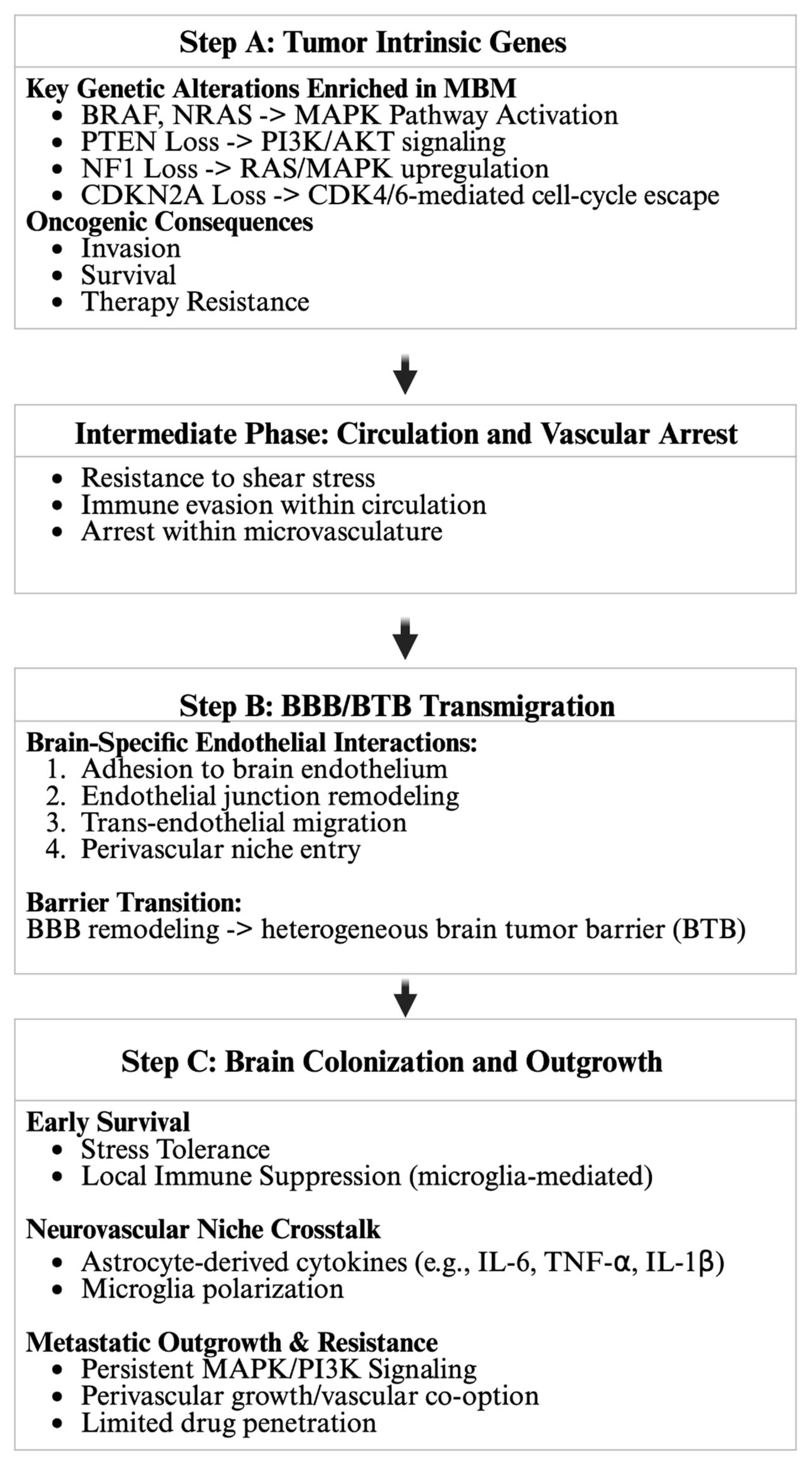
Metastatic melanoma commonly involves the GI tract, lung, and bone [3]. Brain metastasis represents one of the most clinically challenging and lethal disease manifestations [3,5,6,7,8,9]. Figure 1 outlines the pathogenesis of melanoma brain metastasis (MBM). MBM occurs through hematogenous dissemination of tumor cells and is mediated by tumor-intrinsic genes that enable survival in circulation [10]. Survival of metastatic melanoma in the circulation depends on interactions with vascular tissue, including the formation of the blood–tumor barrier (BTB) and transmigration across the blood–brain barrier (BBB) [11]. The BBB is maintained by glial cells, specifically astrocytes, which develop a tumor microenvironment by secreting IL-6, TNF-⍺, NF-κB, and IL-1β [6,7,9].
Following dissemination into the brain, the malignant cells tend to embed in areas with the highest blood flow, specifically the cerebral hemispheres (80%), brainstem (5%), and cerebellum (15%), with a historically poor prognosis of 4–6 months before the development of more novel therapies [9,12,13]. Initial presentation of MBM includes headache, neurological impairment, and seizures [13,14]. These symptoms are generated due to risk factors such as male gender, primary disease site in the head or neck, the presence of visceral or nodal metastases, metastases at three or more sites, and elevated LDH levels [13].
MBM is aggressive, therapeutically limited in crossing the BBB, and has numerous complications contributing to its deleterious nature [13]. Cutaneous melanoma is present in more than 50% of stage IV patients. The cancer has the highest brain metastasis rate among other solid tumors [9,13]. Approximately 5% of cutaneous melanomas are diagnosed at stage IV, which has a relapse rate of 29% [15]. Stage IV melanoma is lethal due to the complications of metastasis to the brain, lungs, liver, and bone, with a 28% 5-year overall survival [16]. MBM complications include hemorrhage and focal, global, or functional neurologic symptoms. Focal symptoms are related to paresis, global symptoms are related to intracranial hypertension, and functional symptoms are related to encephalopathy caused by therapeutics or stemming from seizures [13]. The incidence of seizures is higher for MBM, at 19.8%, compared to melanoma patients, at 8.62%, likely due to interactions between tumor cells and the central nervous system [13,14]. MBM has a high hemorrhage risk leading to intracerebral hemorrhage. Basic fibroblast growth factor (bFGF) expression increases the density of immature blood vessels, contributing to MBM-associated hemorrhage neurological sequelae [17,18].
MBM is challenging to treat due to its historical exclusion from clinical trials, despite being estimated to occur in more than 30–75% of patients with melanoma [12,19,20]. Multiple therapeutic options are being developed and utilized for MBM, including combination therapies such as BRAF + MEK inhibitors, which have shown high rates of rapid response and improved overall MBM survival. Other therapeutics include immune checkpoint inhibitors (ICIs), such as the anti-CTLA-4/anti-PD-1 combination [13], as well as surgical resection, whole-brain radiotherapy (WBRT), and stereotactic radiosurgery (SRS) [21]. These therapeutic advances directly target the tumor-intrinsic pathways and immune mechanisms highlighted in Figure 1. In this review, we aim to describe the pathogenesis and treatment of MBM due to mutations such as BRAF, MAPK, PTEN, and CDKN2A.
Figure 1. Genetic drivers and multistep mechanisms of melanoma brain metastasis (MBM). Step A highlights tumor-intrinsic alterations enriched in MBM, including activation of MAPK signaling (BRAF and NRAS), dysregulation of the PI3K/AKT pathway via PTEN loss, and cell-cycle deregulation associated with NF1 and CDKN2A loss, which collectively promote invasion, survival, and therapeutic resistance. The intermediate circulatory phase depicts hematogenous dissemination, during which circulating tumor cells resist hemodynamic shear stress, evade immune surveillance, and undergo vascular arrest within the microvasculature [22]. Step B illustrates brain-specific endothelial interactions required for blood–brain barrier (BBB) transmigration, including endothelial adhesion, junctional remodeling, trans-endothelial migration, and perivascular niche entry, which result in BBB remodeling and the evolution toward a heterogeneous brain–tumor barrier (BTB) [22,23]. Step C demonstrates early metastatic survival and outgrowth within the neurovascular niche, supported by astrocyte-derived pro-inflammatory cytokines (e.g., IL-6, TNF-α, and IL-1β), microglial polarization, sustained MAPK/PI3K signaling, vascular co-option, and limited therapeutic penetration [24,25,26]. Together, these processes define a gene–microenvironment interplay that drives MBM establishment and therapeutic resistance.
Cells 15 00758 g001

2. Pathogenesis of Melanoma Brain Metastasis

2.1. Key Mutations in MBM

MBMs are associated with mutations in key oncogenic pathways, including activating mutations in BRAF, NRAS, and the mitogen-activated protein kinase (MAPK) pathway, as well as loss of tumor suppressors such as phosphatase and tensin homolog (PTEN), NF1, and CDKN2A. These mutations cause dysregulation of the cell cycle, increasing cell survival, invasion, and metastatic competence, thereby supporting survival, proliferation, and adaptive processes associated with integration into the brain’s microenvironment [27,28].

2.2. BRAF

BRAF is a serine/threonine kinase in the RAF family that is mutated in nearly 50% of melanomas [29]. RAF proteins dimerize to become active, with RAS accelerating dimerization, while ERK provides negative feedback on the RAF dimerization pathway [30,31,32]. The most common BRAF mutation is BRAFV600E, which causes unregulated downstream MEK/ERK activation [33]. The mutation leads to uncontrolled cell proliferation and tumor development. The most common BRAF mutation, V600E, leads to the constitutive activation of the MAPK signaling pathway, promoting cell growth and inhibiting apoptosis, thereby driving malignancy and reactive astrocytes [28,34].

2.3. NRAS

NRAS is the second most common oncogene in metastatic melanoma [35]. RAS proteins are small GTPases implicated in oncogenesis, including KRAS, HRAS, and NRAS [36]. RAS-guanine nucleotide exchange factors (GEFs) catalyze GDP-to-GTP exchange, leading RAS proteins to bind RAS-binding domains, which include BRAF [37]. Most RAS mutations increase the GTP:GDP-bound RAS ratio, thereby markedly increasing basal activation of downstream pathways [38]. In addition to promoting tumor growth via BRAF signaling, NRAS mutations mediate immune suppression via PD-L1 upregulation, cGAS-STING suppression, and reduction of antigen presentation [39,40,41]. RAS also suppresses PTEN, which is a negative regulator of MAPKs [42]. Despite success in targeting KRAS for other solid tumors, targeting NRAS has had little success, and more research is needed to target this important mediator in melanoma growth [43,44].

2.4. MAPKs

MAPKs are a family of serine/threonine kinases that act as a convergence point for signal transduction pathways [45]. MAP kinase-ERK kinase (MEK) and extracellular regulated MAP kinase (ERK) are two MAPKs, with mutations present in 3–8% of melanomas [46]. MEK phosphorylates ERK, which stimulates protein synthesis for growth [47]. Mutations in these proteins are not as common as BRAF (a MAP kinase kinase kinase) or NRAS (a MAP kinase kinase); targeting MAPKs is still actionable because they are downstream effectors of more common mutations [47].

2.5. NF1

NF1 encodes neurofibromin, a GTPase that downregulates RAS activation [48]. NF1 mutations are present in 12–18% of melanomas and occur more frequently in older adults with significant UV exposure [48]. This mutation is also present in 45–95% of desmoplastic melanomas, a painful melanoma subtype that often develops on sun-damaged skin [48]. NF1 mutations are often the driver in melanomas lacking BRAF or NRAS mutations, making NF1 a target for resistant MBM [49].

2.6. CDKN2A

CDKN2A is a tumor suppressor gene mutated in 60–70% of melanomas [50]. This gene encodes p16 and p14. Canonically, p16 inhibits cyclin-dependent kinases (CDKs), preventing Rb phosphorylation, which arrests the cell cycle at the G1/S phase [51]. Recent evidence, cogently reviewed by Buj & Aird in 2019, has also implicated p16 in a variety of other pathways, including mTORC1, p65, ROS, PRC, TFAM, JNK1/3, and eEF1A2 [52].
The role of p14 is to prevent ubiquitination and degradation of p53, a key tumor suppressor that also arrests the cell cycle at G1/S phase [53]. Mutations in CDKN2A are mechanistically distinct from those in MAPK pathways, making them a potential alternative target for MBM therapies.

2.7. Chromosomal Instability

Another factor that influences MBM development is chromosomal instability. Duplications, deletions, and translocations alter chromosome number and integrity, increasing the probability of aggressive tumors crossing the BBB [17]. Disruption of chromosomal integrity can also cause melanoma cells to adapt to the brain’s isolated environment, helping tumors evade immune detection. Chromosomal instability can also increase MBM drug resistance [17,54].

2.8. Blood–Brain Barrier and Blood–Tumor Barrier

A key challenge in MBM is the presence of the restrictive BBB and BTB. The presence of these barriers limits the efficacy of chemotherapeutic agents in the brain, preventing them from achieving a meaningful pharmacodynamic effect on their target of interest [9].
The BBB is a semi-permeable structure that prevents the diffusion of most molecules into the brain parenchyma unless the molecule is lipid-soluble and less than 400–600 Da, or has a BBB-specific receptor or transporter [55]. These parameters prevent most molecules and cells, including tumor cells, from entering the brain [56]. This barrier is composed of endothelial cells with a negatively charged glycocalyx, which repels molecules and astrocytes, which regulate BBB function; pericytes, which enhance communication with growth factors; and cell junction proteins, which promote adhesion [56]. MBM cells initially overcome this barrier to metastasize to the brain by upregulating and anchoring to vascular adhesion proteins such as selectins and integrins [57]. Once anchored, these cells secrete extracellular vesicles which deliver miRNAs that break down the cell membrane, such as miR-181c and cytokines via NF-κB [58,59]. After this initial degradation, MMPs derived from MBM cells help degrade the BBB further while transmigrating via a paracellular route to enter the brain parenchyma [60,61].
When tumor cells metastasize to the brain, elevated VEGF levels stimulate angiogenesis, remodeling the BBB. This disrupts the integrity of the BBB, creating a nuanced vascular unit termed the BTB. The BTB disrupts tight junctions, thickens basement membranes, increases fenestrae, and promotes the formation of abnormal pinocytic vesicles, leading to abnormally leaky blood vessels with non-uniform permeability [54,62,63].
Chemotherapy delivery is limited by BBB penetration; however, successful agents have been shown to have limited therapeutic response [64,65]. Astrocytes promote chemotherapy resistance through sequestration of intracellular calcium and increased gap junction communication [7,66].

3. Radiotherapy

3.1. Background on Radiotherapy

Radiotherapy plays a central role in treating brain metastases because it can quickly palliate neurologic symptoms, decrease edema-related mass effect, and improve intracranial control [67]. Current practice typically favors focal approaches such as stereotactic radiosurgery (SRS) for limited-volume disease due to strong local control and better cognitive preservation, while whole-brain radiotherapy (WBRT) remains important for patients with extensive intracranial tumor burden, lesions unsuitable for SRS/surgery, or when a comprehensive whole-brain approach is needed [68]. WBRT delivers radiation to the entire brain and targets both macroscopic lesions and microscopic disease, while SRS administers highly conformal radiation limited to the surgical cavity, thereby sparing normal brain tissue [69].

3.2. Whole-Brain Radiotherapy

WBRT is most often used as palliative therapy for symptomatic patients with diffuse intracranial metastases who are not candidates for focal therapy, with goals of symptom relief and stabilization of neurologic function [68,69]. Conceptually, WBRT treats visible metastases while also addressing potential microscopic/subclinical disease beyond what is detectable on imaging [68]. However, WBRT has historically been limited by neurocognitive toxicity, and guideline syntheses of randomized evidence support that adding WBRT to focal strategies increases cognitive decline without a consistent overall survival advantage in many settings—driving the modern preference for SRS when feasible. SRS provides comparable survival and local control to WBRT while more effectively preserving neurocognitive function [68,70].
In melanoma brain metastases (MBM), WBRT is now generally reserved for selected palliative scenarios (e.g., extensive intracranial disease not amenable to SRS/surgery or progression despite systemic therapy) [71]. Outcomes with WBRT alone are typically modest; a systematic review of radiotherapy in MBM reported median survival ~3.5 months with WBRT alone versus ~7.5 months with SRS alone (noting strong selection effects and heterogeneity across studies) [71]. More broadly, many MBM patients ultimately die from extracranial progression, so WBRT often cannot meaningfully change long-term survival even when it provides short-term intracranial palliation [71].
When WBRT is necessary, modern practice increasingly incorporates neuroprotection strategies. Major guidelines recommend hippocampal-avoidance WBRT (when feasible) with memantine for appropriate patients. The final phase III results of NRG Oncology CC001 show better preservation of cognition and patient-reported symptoms with hippocampal avoidance plus memantine compared with standard WBRT plus memantine [68,72].

3.3. Stereotactic Radiosurgery

SRS, an alternative to WBRT, delivers high-dose radiation to discrete MRI-defined targets with a steep dose falloff, limiting dose to the surrounding brain tissue and reducing the likelihood of global neurocognitive decline compared with WBRT [68,70]. This makes SRS well-suited to MBM, where lesions are typically well visualized on contrast-enhanced MRI and can often be treated focally [71].
Important SRS toxicities include radiation necrosis (RN) and intracranial hemorrhage. RN rates vary by dose, volume, and fractionation; contemporary clinical series continue to document clinically meaningful RN in a subset of patients following SRS [73,74,75]. Hemorrhage is particularly relevant for melanoma metastases, with hemorrhage risk increasing over time post-Gamma Knife resection of MBM [76]. For this reason, it is important to monitor and manage hematologic modulators, including platelets, anticoagulation, and corticosteroids.
Novel preclinical trials have shown the efficacy of Novel Microbeam Radiation Therapy (MRT) on radioresistant murine melanoma and its metastases in mouse models. MRT delivers X-rays at a high rate into micrometer-range high-dose areas, separated by a few hundred micrometers of low-dose regions. The spatial dose separation enables high peak doses while mitigating neurotoxicity. MRT’s reduced neurotoxicity and effectiveness warrant future investigation as an MBM treatment [77].

4. Immunotherapy

4.1. Monoimmunotherapy for Melanoma Brain Metastases

Immunotherapy harnesses the body’s immune system to target cancer cells, providing a systemic approach that differs from traditional tumor-directed therapies. In MBM, immune checkpoint inhibitors targeting CTLA-4 and PD-1 pathways have emerged as critical therapeutic agents [78,79]. Ipilimumab (anti-CTLA-4) was among the first to demonstrate intracranial efficacy, enhancing T-cell activation and resulting in notable tumor regression in select patients, with an intracranial response rate (ICRR) of approximately 16% and a median survival of approximately 7 months [78]. Pembrolizumab and nivolumab (anti-PD-1 inhibitors) further advance this approach by reversing T-cell exhaustion, achieving an ICRR of 20% to 22%, and prolonging median survival to approximately 18.5 months in asymptomatic MBM patients [79].
Immune-related adverse events (irEAs) have been observed after immunotherapy, with irEAs appearing more frequently after combination therapy as opposed to monotherapy [80,81]. It is hypothesized that irAEs arise from cross-reactivity to shared antigens between tumor and normal cells, leading to T-cell-mediated responses at sites outside the tumor. Alternatively, decreased systemic tolerance to self-antigens induced by immunotherapy could be the source of irAEs [82]. Clinical trials, including CheckMate-204, reported ICRR up to 51% and a two-year survival rate of nearly 72% in asymptomatic patients, representing a substantial advancement over monotherapy [79,83]. This approach has significantly transformed MBM management and outcomes [84]. However, for symptomatic patients, ICRR was only around 22.2%. In these patients, radiation therapy or surgery may be necessary before immunotherapeutic treatment, as steroids may potentially interfere with their efficacy [85,86].

4.2. Combination Immunotherapy

Combination therapy using dual checkpoint inhibitors (nivolumab + ipilimumab) has significantly improved outcomes [87]. This combination leverages a “priming and boosting” mechanism: CTLA-4 blockade broadens T-cell activation, while PD-1 blockade sustains effector T-cell function, resulting in higher intracranial response rates (46–57%) and durable disease control, even in the CNS [88]. The American Society of Clinical Oncology, in collaboration with the Society for Neuro-Oncology (SNO) and the Society for Neuro-Oncology (ASTRO), recommends ipilimumab with nivolumab as a standard first-line option for patients with asymptomatic melanoma brain metastases, with monotherapy (nivolumab or pembrolizumab) as an alternative [89].
Immunotherapy is also increasingly combined with stereotactic radiosurgery (SRS). The concurrent use of immune checkpoint inhibitors and SRS leverages radiation-induced tumor antigen release to potentiate systemic immune responses (the abscopal effect), enhancing ICRR and tumor control compared to either therapy alone [90,91]. Emerging data suggest optimal timing, generally within two weeks of SRS, may maximize therapeutic synergy without significantly increasing toxicity [92]. Surgery remains important for selected patients with limited, symptomatic, or surgically accessible lesions.
While combination immunotherapy has been noted to improve the survival rate in a number of MBM patients, it can also come at the cost of acquired resistance mechanisms. Acquired resistance can occur through mutations in the Janus Kinase 1 and 2 enzymes (JAK1/2), which has the effect of reducing expression of PD-L1 and lowering antigen presentation to immune cells. Additionally, mutations of the β2-microglobulin can result in loss of MHC I expression on the cell surface and decreased recognition by immune cells [87].

5. Targeted Therapies

5.1. MAPK Inhibitors

Targeted therapies against MAPK pathway components such as BRAF and MEK are standard for treating BRAF V600 melanoma brain metastases. BRAF inhibitors targeting the V600E variant are effective in treating melanoma due to the prevalence of the mutation [93]. Notable FDA-approved BRAF inhibitors include dabrafenib, encorafenib, and vemurafenib [94]. The MEK inhibitors trametinib and cobimetinib target MEK1/2 and reduce their downstream targets [93]. These drugs bind to the ATP-binding site of the BRAF V600E kinase domain and MEK, respectively, preventing downstream signaling [93,94]. ERK inhibitors are in clinical development, with the most advanced ERK1/2 inhibitor ulixertinib currently in phase I and II trials [95]. Combination targeted therapy has shown greater efficacy,
MEK/BRAF inhibitor combination therapy is superior to monotherapy because it delays resistance and mitigates toxicities specific to BRAF inhibitor therapy. A real-world multicenter respective cohort study of 65 patients used three MEK/BRAF therapies, showing a progression-free survival from the start of therapy of 5.3 months with a median overall survival of 9.5 months [96]. Combination therapy with MEK and BRAF inhibitors is a viable treatment option for patients dealing with BRAF-mutant MBMs [96,97].
Secondary mutations cause therapeutic resistance in MAPK inhibitor therapy after 6–9 months. According to a novel study by De et al., machine learning algorithms found drugs that interact with BRAF V600E could circumvent limitations of traditional therapies [94]. Additionally, MAPK inhibitors increase T cell activity and tumor antigen expression, allowing for adjunct immunotherapy [98]. These inhibitors may be sequenced or combined with immunotherapy to treat BRAF-mutant melanomas, and the optimal strategy is currently under investigation [98,99]. While RAS mutations have been historically difficult to target, daraxonrasib is a novel RAS inhibitor that may have future applications to MBM [41,100].

5.2. Other Targeted Therapies

Targeted therapies for MBM outside the MAPK pathway are under investigation for MBM and other tumor types, but have yet to be approved. Direct PTEN inhibitors, such as bisperoxovanadium compounds, vanadyl-hydroxypicolinic acid, and SF1670 are small molecule inhibitors in preclinical development [101,102,103]. Tumor suppressor genes, such as NF1 and CDKN2A, are more difficult to target pharmacologically because they require recovery rather than inhibition of a molecular pathway. Novel drug discovery methods using artificial intelligence (AI) may uncover more therapeutic tools against MBM. Further research is needed to validate the involvement of AI in MBM treatment. Table 1 compares different treatment modalities for MBM. Table 2 lists targetable mutations in MBM, molecules inhibiting these targets, and the clinical status of these molecules.

6. Discussion

Melanoma brain metastasis is the third most common brain metastasis, driven by tumor-intrinsic genetic alterations and supportive inflammatory signaling [104,105]. Treatment for MBM has evolved over the years to include radio therapies such as SRS and WBRT [68,69,70,71]. The BBB and cancer-protective properties of astrocytes limit chemotherapy efficacy against MBM despite its use in other cancers [66]. More novel therapies, such as immunotherapy and MAPK-targeted therapies, aim to reduce toxicity and improve treatment outcomes [78,79,88].
WBRT has historically been used as a palliative external-beam option for patients with diffuse or symptomatic brain metastases because it treats both visible lesions and potential micrometastatic intracranial disease [73,74,75]. However, in melanoma brain metastases, WBRT is now generally de-emphasized because durable intracranial control is limited and the risk of neurocognitive decline and quality-of-life deterioration is substantial. Current practice guidelines increasingly recommend stereotactic radiosurgery (SRS) (and postoperative cavity SRS, when appropriate) as the preferred radiation strategy for many patients, often including those with multiple metastases, to maximize local control while minimizing cognitive toxicity [89]. WBRT is typically reserved for carefully selected circumstances such as widespread intracranial disease not amenable to SRS, poor prognosis, limited life expectancy, where short-course palliation is the goal, or salvage scenarios. When WBRT is used, cognitive-sparing approaches such as hippocampal avoidance and memantine are recommended when feasible to reduce treatment-related neurocognitive morbidity [69,99].
Another treatment modality targeting cancer cells is immunotherapy that blocks CTLA-4 and PD-1, which are inhibitory receptors that allow cancer cells to evade detection by the immune system. Immune checkpoint inhibitors like ipilimumab (anti-CTLA-4) enhance T-cell activation and proliferation, prolonging them and achieving an intracranial response rate (ICRR) [78]. Both pembrolizumab and nivolumab (anti-PD-1 inhibitors) have shown even greater efficacy [79,88]. Additionally, checkpoint inhibitor combination therapy (nivolumab and ipilimumab) yields significantly improved outcomes, including a higher ICRR and more robust disease control, even in the CNS. The ASCO recommends the combination of ipilimumab and nivolumab as the first-line treatment for patients with asymptomatic melanoma brain metastases, with monotherapy (nivolumab or pembrolizumab) as an alternative [87,89]. Furthermore, the administration of immunotherapy alongside SRS is becoming increasingly common. Concurrent use of immune checkpoint inhibitors and SRS leverages radiation-induced tumor antigen release to enhance systemic immune responses [90,91]. Current data suggest optimal timing, generally within two weeks of SRS, can potentially enhance therapeutic synergy without significantly increasing toxicity [92].
BRAF and MEK inhibitors treat melanoma by reducing activation of downstream targets [93,94,97]. Combination therapy mitigates BRAF-inhibitor-specific toxicities [96,97]. However, these MAPK inhibitors can induce resistance through secondary mutations, a risk that may be mitigated by using AI algorithms to select drugs that best target the chosen receptors [99,106].

7. Future Directions

Many novel cancer therapeutics are applicable to MBM. Therapies that bypass the BBB involve novel routes of administration, including intranasal drug delivery via the olfactory and trigeminal nerve pathways, as well as regulating the non-uniform permeability of the blood vessels, are especially applicable to MBM. The latter has been explored in which tumor markers have been found against the BTB, including ATP-sensitive K+ channels, Ca2+-activated K+ channels, and ATP-sensitive Ca2+ channels, thus allowing the possibility of modulating BTB-associated permeability. Also, components of the tight junctions, such as claudin-5 and angulin-1, have been shown to be promising drug targets that could be delivered against the BBB [107].
Other researchers have explored additional treatment models applicable to MBM, in addition to metastases to other organs. One such strategy leveraged the protective glial machinery to create a glial cancer cell co-culture that identified targetable astrocyte-driven brain metastases associated with melanoma, as well as lung and breast cancer [7]. Another study investigated the use of fluoxetine to inhibit MBM, reporting that it achieved effective growth inhibition across melanoma cell lines without major effects on healthy tissue [108].
These studies demonstrate that mitigating the dire effects of MBM is an ongoing endeavor for researchers. That said, formulating a therapeutic standard for MBM may be on the horizon, but it may only materialize if there is continued interest in clinical research aimed at a therapeutic solution.

8. Conclusions

MBM therapy has progressed from WBRT and SRS to newer therapies such as immunotherapy and targeted therapy. Overcoming the BBB and BTB is a key difficulty for delivering therapy. Targeted therapies aim to combat mutations implicated in tumor growth and metastasis, like BRAF. Immunotherapies reduce immune-dampening to leverage the immune system for tumor destruction. Researchers are still refining these treatments to improve the standard of care for patients.

Author Contributions

C.S.: Conceptualization, Writing—Original Draft Preparation, Writing—Review and Editing, Project Administration. M.N.: Conceptualization, Writing—Original Draft Preparation, Writing—Review and Editing C.C.: Writing—Original Draft Preparation N.T.: Visualization, Writing—Original Draft Preparation, Writing—Review and Editing S.K.: Visualization, Writing—Original Draft Preparation, Writing—Review and Editing J.R.: Writing—Original Draft Preparation, Writing—Review and Editing V.P.: Writing—Original Draft Preparation, Writing—Review and Editing A.N.: Writing—Original Draft Preparation, Writing—Review and Editing T.C.: Writing—Original Draft Preparation Z.R.: Writing—Original Draft Preparation, Writing—Review and Editing M.L.: Writing—Original Draft S.B.: Writing—Original Draft Preparation, Writing—Review and Editing H.H.: Writing—Review and Editing B.L.-W.: Conceptualization, Supervision, Writing—Review and Editing. All authors have read and agreed to the published version of the manuscript.

Funding

This research received no external funding.

Institutional Review Board Statement

Not applicable.

Informed Consent Statement

Not applicable.

Data Availability Statement

No new data were created or analyzed in this study.

Acknowledgments

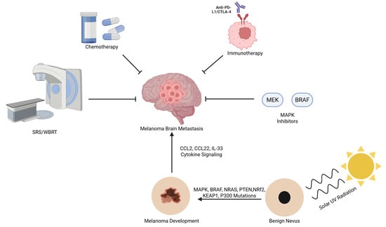
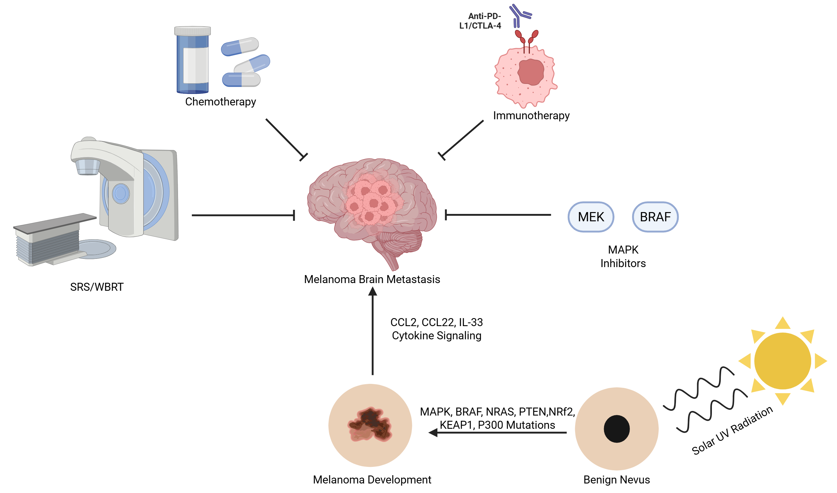
We would like to acknowledge BioRender in helping create the graphical abstract and Figure 1. Created in BioRender. Kazmi, S. (2026) https://BioRender.com/iuymqpq (accessed on 20 April 2026). Created in BioRender. Sanghadia, C. (2025) https://BioRender.com/twzqso9 (accessed on 20 April 2026).

Conflicts of Interest

The authors declare no conflicts of interest.

References

  1. Schadendorf, D.; Fisher, D.E.; Garbe, C.; Gershenwald, J.E.; Grob, J.-J.; Halpern, A.; Herlyn, M.; Marchetti, M.A.; McArthur, G.; Ribas, A.; et al. Melanoma. Nat. Rev. Dis. Primers 2015, 1, 15003. [Google Scholar] [CrossRef] [PubMed]
  2. Sample, A.; He, Y.-Y. Mechanisms and Prevention of UV-Induced Melanoma. Photodermatol. Photoimmunol. Photomed. 2018, 34, 13–24. [Google Scholar] [CrossRef] [PubMed]
  3. Janka, E.A.; Ványai, B.; Dajnoki, Z.; Szabó, I.L.; Reibl, D.; Komka, I.; Blasszauer, C.; Várvölgyi, T.; Szegedi, A.; Emri, G. Regional Variability of Melanoma Incidence and Prevalence in Hungary. Epidemiological Impact of Ambient UV Radiation and Socioeconomic Factors. Eur. J. Cancer Prev. 2022, 31, 377–384. [Google Scholar] [CrossRef] [PubMed]
  4. De Pinto, G.; Mignozzi, S.; La Vecchia, C.; Levi, F.; Negri, E.; Santucci, C. Global Trends in Cutaneous Malignant Melanoma Incidence and Mortality. Melanoma Res. 2024, 34, 265–275. [Google Scholar] [CrossRef]
  5. Tasoudis, P.; Manaki, V.; Parness, S.; Khoury, A.L.; Agala, C.B.; Haithcock, B.E.; Mody, G.N.; Long, J.M. The Role of Metastasectomies and Immunotherapy in the Management of Melanoma Lung Metastases: An Analysis of the National Cancer Database. Cancers 2025, 17, 206. [Google Scholar] [CrossRef]
  6. Tas, F.; Erturk, K. Brain Metastases during Follow-up of Patients with Resected Cutaneous Melanoma. Melanoma Res. 2025, 35, 187–191. [Google Scholar] [CrossRef]
  7. Rone, J.M.; Faust Akl, C.; Quintana, F.J. Astrocyte Control of Brain Metastasis. Dev. Cell 2024, 59, 559–560. [Google Scholar] [CrossRef]
  8. Shimizu, M.R.; Van De Langerijt, O.N.; Torres, D.; De Groot, T.M.; Groot, O.Q. Incidence, Risk Factors, and Survival of Bone Metastases and Skeletal-Related Events in Melanoma Patients: A Systematic Review and Quality Assessment of 29 Studies. J. Bone Oncol. 2024, 46, 100603. [Google Scholar] [CrossRef]
  9. Hasanov, M.; Acikgoz, Y.; Davies, M.A. Melanoma Brain Metastasis. Hematol./Oncol. Clin. N. Am. 2024, 38, 1027–1043. [Google Scholar] [CrossRef]
  10. Fazakas, C.; Wilhelm, I.; Nagyoszi, P.; Farkas, A.E.; Haskó, J.; Molnár, J.; Bauer, H.; Bauer, H.-C.; Ayaydin, F.; Dung, N.T.K.; et al. Transmigration of Melanoma Cells through the Blood-Brain Barrier: Role of Endothelial Tight Junctions and Melanoma-Released Serine Proteases. PLoS ONE 2011, 6, e20758. [Google Scholar] [CrossRef]
  11. Strozyk, E.A.; Desch, A.; Poeppelmann, B.; Magnolo, N.; Wegener, J.; Huck, V.; Schneider, S.W. Melanoma-Derived IL-1 Converts Vascular Endothelium to a Proinflammatory and Procoagulatory Phenotype via NFκB Activation. Exp. Dermatol. 2014, 23, 670–676. [Google Scholar] [CrossRef] [PubMed]
  12. Amouzegar, A.; Tawbi, H.A. Local and Systemic Management Options for Melanoma Brain Metastases. Cancer J. 2024, 30, 102–107. [Google Scholar] [CrossRef] [PubMed]
  13. Phadke, M.; Ozgun, A.; Eroglu, Z.; Smalley, K.S.M. Melanoma Brain Metastases: Biological Basis and Novel Therapeutic Strategies. Exp. Dermatol. 2022, 31, 31–42. [Google Scholar] [CrossRef] [PubMed]
  14. Huntoon, K.; Musgrave, N.; Shaikhouni, A.; Elder, J. Frequency of Seizures in Patients with Metastatic Brain Tumors. Neurol. Sci. 2023, 44, 2501–2507. [Google Scholar] [CrossRef]
  15. Wolchok, J.D.; Chiarion-Sileni, V.; Rutkowski, P.; Cowey, C.L.; Schadendorf, D.; Wagstaff, J.; Queirolo, P.; Dummer, R.; Butler, M.O.; Hill, A.G.; et al. Final, 10-Year Outcomes with Nivolumab plus Ipilimumab in Advanced Melanoma. N. Engl. J. Med. 2025, 392, 11–22. [Google Scholar] [CrossRef]
  16. Adcock, B.; Dhakal, A.; Rehman, N.; Alsabbagh Alchirazi, M.; Albliwi, M.; Mushtaq, A.; Zureigat, H.; Lee, M.; Mohamed Hassan, A.N.; Haddad, S.F.; et al. Real-World Survival Outcomes of Patients with de Novo Stage IV Melanoma: A 13-Year Single Center Experience. J. Clin. Oncol. 2025, 43, e21526. [Google Scholar] [CrossRef]
  17. Lukow, D.A.; Sheltzer, J.M. Chromosomal Instability and Aneuploidy as Causes of Cancer Drug Resistance. Trends Cancer 2022, 8, 43–53. [Google Scholar] [CrossRef]
  18. Trembath, D.G.; Davis, E.S.; Rao, S.; Bradler, E.; Saada, A.F.; Midkiff, B.R.; Snavely, A.C.; Ewend, M.G.; Collichio, F.A.; Lee, C.B.; et al. Brain Tumor Microenvironment and Angiogenesis in Melanoma Brain Metastases. Front. Oncol. 2021, 10, 604213. [Google Scholar] [CrossRef]
  19. Le Rhun, E.; Weller, M.; Anders, C.; Larkin, J.; Li, J.; Moss, N.S.; Tawbi, H.; Dummer, R. “Symptomatic” Melanoma Brain Metastases: A Call for Clear Definitions and Adoption of Standardized Tools. Eur. J. Cancer 2024, 208, 114202. [Google Scholar] [CrossRef]
  20. Pedersen, S.; Møller, S.; Donia, M.; Persson, G.F.; Svane, I.M.; Ellebaek, E. Real-World Data on Melanoma Brain Metastases and Survival Outcome. Melanoma Res. 2022, 32, 173–182. [Google Scholar] [CrossRef]
  21. Knox, A.; Wang, T.; Shackleton, M.; Ameratunga, M. Symptomatic Brain Metastases in Melanoma. Exp. Dermatol. 2024, 33, e15075. [Google Scholar] [CrossRef]
  22. Micalizzi, D.S.; Maheswaran, S.; Haber, D.A. A Conduit to Metastasis: Circulating Tumor Cell Biology. Genes. Dev. 2017, 31, 1827–1840. [Google Scholar] [CrossRef] [PubMed]
  23. Wilhelm, I.; Molnár, J.; Fazakas, C.; Haskó, J.; Krizbai, I. Role of the Blood-Brain Barrier in the Formation of Brain Metastases. Int. J. Mol. Sci. 2013, 14, 1383–1411. [Google Scholar] [CrossRef] [PubMed]
  24. Schreurs, L.D.; Vom Stein, A.F.; Jünger, S.T.; Timmer, M.; Noh, K.-W.; Buettner, R.; Kashkar, H.; Neuschmelting, V.; Goldbrunner, R.; Nguyen, P.-H. The Immune Landscape in Brain Metastasis. Neuro-Oncology 2025, 27, 50–62. [Google Scholar] [CrossRef] [PubMed]
  25. Patabendige, A.; Singh, A.; Jenkins, S.; Sen, J.; Chen, R. Astrocyte Activation in Neurovascular Damage and Repair Following Ischaemic Stroke. Int. J. Mol. Sci. 2021, 22, 4280. [Google Scholar] [CrossRef] [PubMed]
  26. Redmer, T.; Schumann, E.; Peters, K.; Weidemeier, M.E.; Nowak, S.; Schroeder, H.W.S.; Vidal, A.; Radbruch, H.; Lehmann, A.; Kreuzer-Redmer, S.; et al. MET Receptor Serves as a Promising Target in Melanoma Brain Metastases. Acta Neuropathol. 2024, 147, 44. [Google Scholar] [CrossRef]
  27. Yavuz, B.R.; Jang, H.; Nussinov, R. Cellular Mechanisms Underlying Melanoma Brain Metastasis. bioRxiv 2025. [Google Scholar] [CrossRef]
  28. Rodriguez-Baena, F.J.; Marquez-Galera, A.; Ballesteros-Martinez, P.; Castillo, A.; Diaz, E.; Moreno-Bueno, G.; Lopez-Atalaya, J.P.; Sanchez-Laorden, B. Microglial Reprogramming Enhances Antitumor Immunity and Immunotherapy Response in Melanoma Brain Metastases. Cancer Cell 2025, 43, 413–427.e9. [Google Scholar] [CrossRef]
  29. Long, G.V.; Menzies, A.M.; Nagrial, A.M.; Haydu, L.E.; Hamilton, A.L.; Mann, G.J.; Hughes, T.M.; Thompson, J.F.; Scolyer, R.A.; Kefford, R.F. Prognostic and Clinicopathologic Associations of Oncogenic BRAF in Metastatic Melanoma. J. Clin. Oncol. 2011, 29, 1239–1246. [Google Scholar] [CrossRef]
  30. Weber, C.K.; Slupsky, J.R.; Kalmes, H.A.; Rapp, U.R. Active Ras Induces Heterodimerization of cRaf and BRaf. Cancer Res. 2001, 61, 3595–3598. [Google Scholar]
  31. Rajakulendran, T.; Sahmi, M.; Lefrançois, M.; Sicheri, F.; Therrien, M. A Dimerization-Dependent Mechanism Drives RAF Catalytic Activation. Nature 2009, 461, 542–545. [Google Scholar] [CrossRef] [PubMed]
  32. Ritt, D.A.; Monson, D.M.; Specht, S.I.; Morrison, D.K. Impact of Feedback Phosphorylation and Raf Heterodimerization on Normal and Mutant B-Raf Signaling. Mol. Cell. Biol. 2010, 30, 806–819. [Google Scholar] [CrossRef] [PubMed]
  33. Maurer, G.; Tarkowski, B.; Baccarini, M. Raf Kinases in Cancer-Roles and Therapeutic Opportunities. Oncogene 2011, 30, 3477–3488. [Google Scholar] [CrossRef] [PubMed]
  34. Kolch, W.; Berta, D.; Rosta, E. Dynamic Regulation of RAS and RAS Signaling. Biochem. J. 2023, 480, 1–23. [Google Scholar] [CrossRef]
  35. Fedorenko, I.V.; Gibney, G.T.; Smalley, K.S.M. NRAS Mutant Melanoma: Biological Behavior and Future Strategies for Therapeutic Management. Oncogene 2013, 32, 3009–3018. [Google Scholar] [CrossRef]
  36. Punekar, S.R.; Velcheti, V.; Neel, B.G.; Wong, K.-K. The Current State of the Art and Future Trends in RAS-Targeted Cancer Therapies. Nat. Rev. Clin. Oncol. 2022, 19, 637–655. [Google Scholar] [CrossRef]
  37. Cargnello, M.; Roux, P.P. Activation and Function of the MAPKs and Their Substrates, the MAPK-Activated Protein Kinases. Microbiol. Mol. Biol. Rev. 2011, 75, 50–83. [Google Scholar] [CrossRef]
  38. Hobbs, G.A.; Der, C.J.; Rossman, K.L. RAS Isoforms and Mutations in Cancer at a Glance. J. Cell Sci. 2016, 129, 1287–1292. [Google Scholar] [CrossRef]
  39. Coelho, M.A.; de Carné Trécesson, S.; Rana, S.; Zecchin, D.; Moore, C.; Molina-Arcas, M.; East, P.; Spencer-Dene, B.; Nye, E.; Barnouin, K.; et al. Oncogenic RAS Signaling Promotes Tumor Immunoresistance by Stabilizing PD-L1 mRNA. Immunity 2017, 47, 1083–1099.e6. [Google Scholar] [CrossRef]
  40. Shan, J.-L.; Zhang, K.-M.; Zhong, W.-Q.; Yang, X.-Y.; Li, Z.-L.; Huang, Y.; Du, T.; Yang, D.; Tang, J.-H.; Chen, Y.-H.; et al. Reactivating cGAS-STING Signaling by Targeting SOS1 Enhances Antitumor Immunity in NRAS-Mutant Tumors. Cancer Res. 2025, 85, 3015–3031. [Google Scholar] [CrossRef]
  41. Anastacio Da Costa Carvalho, L.; Tovbis Shifrin, N.; Phadke, M.S.; Emmons, M.F.; Pechuan-Jorge, X.; Mbuga, F.; Ospina, O.E.; Chow, C.; Seu, L.; Rose, O.L.; et al. RAS(ON) Multiselective Inhibition Drives Antitumor Immunity in Preclinical Models of NRAS-Mutant Melanoma. Cancer Immunol. Res. 2026, 14, 90–106. [Google Scholar] [CrossRef] [PubMed]
  42. Zhang, J.; Xiang, Z.; Malaviarachchi, P.A.; Yan, Y.; Baltz, N.J.; Emanuel, P.D.; Liu, Y.L. PTEN Is Indispensable for Cells to Respond to MAPK Inhibitors in Myeloid Leukemia. Cell. Signal. 2018, 50, 72–79. [Google Scholar] [CrossRef] [PubMed]
  43. Ostrem, J.M.; Peters, U.; Sos, M.L.; Wells, J.A.; Shokat, K.M. K-Ras(G12C) Inhibitors Allosterically Control GTP Affinity and Effector Interactions. Nature 2013, 503, 548–551. [Google Scholar] [CrossRef] [PubMed]
  44. Hong, D.S.; Fakih, M.G.; Strickler, J.H.; Desai, J.; Durm, G.A.; Shapiro, G.I.; Falchook, G.S.; Price, T.J.; Sacher, A.; Denlinger, C.S.; et al. KRASG12C Inhibition with Sotorasib in Advanced Solid Tumors. N. Engl. J. Med. 2020, 383, 1207–1217. [Google Scholar] [CrossRef]
  45. Braicu, C.; Buse, M.; Busuioc, C.; Drula, R.; Gulei, D.; Raduly, L.; Rusu, A.; Irimie, A.; Atanasov, A.G.; Slaby, O.; et al. A Comprehensive Review on MAPK: A Promising Therapeutic Target in Cancer. Cancers 2019, 11, 1618. [Google Scholar] [CrossRef]
  46. Burotto, M.; Chiou, V.L.; Lee, J.-M.; Kohn, E.C. The MAPK Pathway across Different Malignancies: A New Perspective. Cancer 2014, 120, 3446–3456. [Google Scholar] [CrossRef]
  47. Morante, M.; Pandiella, A.; Crespo, P.; Herrero, A. Immune Checkpoint Inhibitors and RAS–ERK Pathway-Targeted Drugs as Combined Therapy for the Treatment of Melanoma. Biomolecules 2022, 12, 1562. [Google Scholar] [CrossRef]
  48. Kiuru, M.; Busam, K.J. The NF1 Gene in Tumor Syndromes and Melanoma. Lab. Investig. 2017, 97, 146–157. [Google Scholar] [CrossRef]
  49. Nissan, M.H.; Pratilas, C.A.; Jones, A.M.; Ramirez, R.; Won, H.; Liu, C.; Tiwari, S.; Kong, L.; Hanrahan, A.J.; Yao, Z.; et al. Loss of NF1 in Cutaneous Melanoma Is Associated with RAS Activation and MEK Dependence. Cancer Res. 2014, 74, 2340–2350. [Google Scholar] [CrossRef]
  50. Cancer Genome Atlas Network. Genomic Classification of Cutaneous Melanoma. Cell 2015, 161, 1681–1696. [Google Scholar] [CrossRef]
  51. Serrano, M.; Lin, A.W.; McCurrach, M.E.; Beach, D.; Lowe, S.W. Oncogenic Ras Provokes Premature Cell Senescence Associated with Accumulation of P53 and p16INK4a. Cell 1997, 88, 593–602. [Google Scholar] [CrossRef]
  52. Buj, R.; Aird, K.M. P16: Cycling off the Beaten Path. Mol. Cell. Oncol. 2019, 6, e1677140. [Google Scholar] [CrossRef] [PubMed]
  53. Kreuger, I.Z.M.; Slieker, R.C.; van Groningen, T.; van Doorn, R. Therapeutic Strategies for Targeting CDKN2A Loss in Melanoma. J. Investig. Dermatol. 2023, 143, 18–25.e1. [Google Scholar] [CrossRef] [PubMed]
  54. Fares, J.; Kanojia, D.; Rashidi, A.; Ulasov, I.; Lesniak, M.S. Genes That Mediate Metastasis across the Blood–Brain Barrier. Trends Cancer 2020, 6, 660–676. [Google Scholar] [CrossRef] [PubMed]
  55. Wu, D.; Chen, Q.; Chen, X.; Han, F.; Chen, Z.; Wang, Y. The Blood–Brain Barrier: Structure, Regulation and Drug Delivery. Signal Transduct. Target. Ther. 2023, 8, 217. [Google Scholar] [CrossRef]
  56. Santa-Maria, A.R.; Walter, F.R.; Figueiredo, R.; Kincses, A.; Vigh, J.P.; Heymans, M.; Culot, M.; Winter, P.; Gosselet, F.; Dér, A.; et al. Flow Induces Barrier and Glycocalyx-Related Genes and Negative Surface Charge in a Lab-on-a-Chip Human Blood-Brain Barrier Model. J. Cereb. Blood Flow. Metab. 2021, 41, 2201–2215. [Google Scholar] [CrossRef]
  57. Malhotra, J.; Mambetsariev, I.; Gilmore, G.; Fricke, J.; Nam, A.; Gallego, N.; Chen, B.T.; Chen, M.; Amini, A.; Lukas, R.V.; et al. Targeting CNS Metastases in Non–Small Cell Lung Cancer with Evolving Approaches Using Molecular Markers: A Review. JAMA Oncol. 2025, 11, 60–69. [Google Scholar] [CrossRef]
  58. Tominaga, N.; Kosaka, N.; Ono, M.; Katsuda, T.; Yoshioka, Y.; Tamura, K.; Lötvall, J.; Nakagama, H.; Ochiya, T. Brain Metastatic Cancer Cells Release microRNA-181c-Containing Extracellular Vesicles Capable of Destructing Blood-Brain Barrier. Nat. Commun. 2015, 6, 6716. [Google Scholar] [CrossRef]
  59. Di Russo, S.; Borsatti, G.E.; Bouzidi, A.; Liberati, F.R.; Riva, A.; Tripodi, F.; Rolfi, L.R.; Spizzichino, S.; Tramutola, A.; Perluigi, M.; et al. NF-κB-Mediated Cytokine Secretion and Glutamate Metabolic Reprogramming Converge in Breast Cancer Brain Tropism. Cancer Lett. 2025, 630, 217907. [Google Scholar] [CrossRef]
  60. Karreman, M.A.; Bauer, A.T.; Solecki, G.; Berghoff, A.S.; Mayer, C.D.; Frey, K.; Hebach, N.; Feinauer, M.J.; Schieber, N.L.; Tehranian, C.; et al. Active Remodeling of Capillary Endothelium via Cancer Cell-Derived MMP9 Promotes Metastatic Brain Colonization. Cancer Res. 2023, 83, 1299–1314. [Google Scholar] [CrossRef]
  61. Herman, H.; Fazakas, C.; Haskó, J.; Molnár, K.; Mészáros, Á.; Nyúl-Tóth, Á.; Szabó, G.; Erdélyi, F.; Ardelean, A.; Hermenean, A.; et al. Paracellular and Transcellular Migration of Metastatic Cells through the Cerebral Endothelium. J. Cell. Mol. Med. 2019, 23, 2619–2631. [Google Scholar] [CrossRef]
  62. Steeg, P.S. The Blood–Tumour Barrier in Cancer Biology and Therapy. Nat. Rev. Clin. Oncol. 2021, 18, 696–714. [Google Scholar] [CrossRef]
  63. Zhou, T.; Yan, Y.; Lin, M.; Zeng, C.; Mao, X.; Zhu, Y.; Han, J.; Li, D.-D.; Zhang, J. Astrocyte Involvement in Brain Metastasis: From Biological Mechanisms to Therapeutic Strategies. Cancer Metastasis Rev. 2025, 44, 60. [Google Scholar] [CrossRef] [PubMed]
  64. Anand, U.; Dey, A.; Chandel, A.K.S.; Sanyal, R.; Mishra, A.; Pandey, D.K.; De Falco, V.; Upadhyay, A.; Kandimalla, R.; Chaudhary, A.; et al. Cancer Chemotherapy and beyond: Current Status, Drug Candidates, Associated Risks and Progress in Targeted Therapeutics. Genes Dis. 2023, 10, 1367–1401. [Google Scholar] [CrossRef] [PubMed]
  65. Maleki, E.H.; Bahrami, A.R.; Matin, M.M. Cancer Cell Cycle Heterogeneity as a Critical Determinant of Therapeutic Resistance. Genes Dis. 2024, 11, 189–204. [Google Scholar] [CrossRef] [PubMed]
  66. Westerlund, L.H.; Bergström, C.K.; Laakkonen, P.M.; Le Joncour, V. Deciphering the Dialogue between Brain Tumors, Neurons, and Astrocytes. Am. J. Pathol. 2025, 195, 1193–1208. [Google Scholar] [CrossRef]
  67. Vlachos, N.; Lampros, M.G.; Filis, P.; Voulgaris, S.; Alexiou, G.A. Stereotactic Radiosurgery versus Whole-Brain Radiotherapy after Resection of Solitary Brain Metastasis: A Systematic Review and Meta-Analysis. World Neurosurg. X 2023, 18, 100170. [Google Scholar] [CrossRef]
  68. Gondi, V.; Bauman, G.; Bradfield, L.; Burri, S.H.; Cabrera, A.R.; Cunningham, D.A.; Eaton, B.R.; Hattangadi-Gluth, J.A.; Kim, M.M.; Kotecha, R.; et al. Radiation Therapy for Brain Metastases: An ASTRO Clinical Practice Guideline. Pract. Radiat. Oncol. 2022, 12, 265–282. [Google Scholar] [CrossRef]
  69. Rahimy, E.; Soltys, S.G. Stereotactic Radiosurgery for Brain Metastases: Review of Existing Data and Future Directions. Appl. Rad. Oncol. 2023, 12, 5–14. [Google Scholar] [CrossRef]
  70. Amdur, R.J. Recognition of PRO Reviewers and Reviewer Apprentices in 2020. Pract. Radiat. Oncol. 2021, 11, 236–237. [Google Scholar] [CrossRef]
  71. Thompson, J.F.; Williams, G.J.; Hong, A.M. Radiation Therapy for Melanoma Brain Metastases: A Systematic Review. Radiol. Oncol. 2022, 56, 267–284. [Google Scholar] [CrossRef]
  72. Gondi, V.; Deshmukh, S.; Brown, P.D.; Wefel, J.S.; Armstrong, T.S.; Tome, W.A.; Gilbert, M.R.; Konski, A.; Robinson, C.G.; Bovi, J.A.; et al. Sustained Preservation of Cognition and Prevention of Patient-Reported Symptoms With Hippocampal Avoidance During Whole-Brain Radiation Therapy for Brain Metastases: Final Results of NRG Oncology CC001. Int. J. Radiat. Oncol. Biol. Phys. 2023, 117, 571–580. [Google Scholar] [CrossRef]
  73. Williams, G.J.; Hong, A.M.; Thompson, J.F. Treatment of Melanoma Brain Metastases with Radiation and Immunotherapy or Targeted Therapy: A Systematic Review with Meta-Analysis. Crit. Rev. Oncol./Hematol. 2024, 202, 104462. [Google Scholar] [CrossRef] [PubMed]
  74. Nijboer, C.B.; Piersma, D.; Sijben, A.E.J.; Hoti, B.; Van Der Meulen, M. The Effect of Concomitant Immunotherapy and Stereotactic Radiotherapy, and of Location on Survival in Patients With Brain Metastases From Melanoma. Cancer Med. 2025, 14, e70923. [Google Scholar] [CrossRef]
  75. Thomson, H.M.; Fortin Ensign, S.P.; Edmonds, V.S.; Sharma, A.; Butterfield, R.J.; Schild, S.E.; Ashman, J.B.; Zimmerman, R.S.; Patel, N.P.; Bryce, A.H.; et al. Clinical Outcomes of Stereotactic Radiosurgery-Related Radiation Necrosis in Patients with Intracranial Metastasis from Melanoma. Clin. Med. Insights Oncol. 2023, 17, 11795549231161878. [Google Scholar] [CrossRef] [PubMed]
  76. Hong, S.; Bouchal, S.M.; Bauman, M.M.J.; Riviere-Cazaux, C.; Pumford, A.D.; Brown, P.D.; Yan, E.S.; Stafford, S.L.; Markovic, S.N.; Link, M.J.; et al. The Longitudinal Risk of Hemorrhage of Melanoma Brain Metastases after Gamma Knife Radiosurgery. J. Neurosurg. 2024, 140, 938–948. [Google Scholar] [CrossRef] [PubMed]
  77. Trappetti, V.; Potez, M.; Fernandez-Palomo, C.; Volarevic, V.; Shintani, N.; Pellicioli, P.; Ernst, A.; Haberthür, D.; Fazzari, J.M.; Krisch, M.; et al. Microbeam Radiation Therapy Controls Local Growth of Radioresistant Melanoma and Treats Out-of-Field Locoregional Metastasis. Int. J. Radiat. Oncol. *Biol. *Phys. 2022, 114, 478–493. [Google Scholar] [CrossRef]
  78. Tawbi, H.A.; Forsyth, P.A.; Hodi, F.S.; Algazi, A.P.; Hamid, O.; Lao, C.D.; Moschos, S.J.; Atkins, M.B.; Lewis, K.; Postow, M.A.; et al. Long-Term Outcomes of Patients with Active Melanoma Brain Metastases Treated with Combination Nivolumab plus Ipilimumab (CheckMate 204): Final Results of an Open-Label, Multicentre, Phase 2 Study. Lancet Oncol. 2021, 22, 1692–1704. [Google Scholar] [CrossRef]
  79. Long, G.V.; Atkinson, V.; Lo, S.N.; Guminski, A.D.; Sandhu, S.K.; Brown, M.P.; Gonzalez, M.; McArthur, G.A.; Menzies, A.M. Ipilimumab plus Nivolumab versus Nivolumab Alone in Patients with Melanoma Brain Metastases (ABC): 7-Year Follow-up of a Multicentre, Open-Label, Randomised, Phase 2 Study. Lancet Oncol. 2025, 26, 320–330. [Google Scholar] [CrossRef]
  80. Nowacka, A.; Fajkiel-Madajczyk, A.; Ohla, J.; Woźniak-Dąbrowska, K.; Liss, S.; Gryczka, K.; Smuczyński, W.; Ziółkowska, E.; Bożiłow, D.; Śniegocki, M.; et al. Current Treatment of Melanoma Brain Metastases. Cancers 2023, 15, 4088. [Google Scholar] [CrossRef]
  81. Esfahani, K.; Roudaia, L.; Buhlaiga, N.; Del Rincon, S.V.; Papneja, N.; Miller, W.H. A Review of Cancer Immunotherapy: From the Past, to the Present, to the Future. Curr. Oncol. 2020, 27, 87–97. [Google Scholar] [CrossRef] [PubMed]
  82. Huo, C.W.; Gunadasa, I.; Gkolia, P.; Shackleton, M.; Hunn, M. Immune-Related Adverse Events Are Associated with Therapeutic Efficacy of Immunotherapy in Patients with Melanoma Brain Metastases. Melanoma Res. 2023, 33, 58–65. [Google Scholar] [CrossRef] [PubMed]
  83. Benghiat, H.; Hodson, J.; Hickman, M.; Meade, S.; Hussein, S.; Stange, R.; Heyes, G.; Jackson, T.; Augustus, H.; Chavda, S.; et al. Outcomes of Patients with Five or More Brain Metastases Treated with Stereotactic Radiosurgery from 2014 to 2019: A UK Series. Clin. Oncol. 2025, 38, 103697. [Google Scholar] [CrossRef]
  84. Casagrande, S.; Sopetto, G.B.; Bertalot, G.; Bortolotti, R.; Racanelli, V.; Caffo, O.; Giometto, B.; Berti, A.; Veccia, A. Immune-Related Adverse Events Due to Cancer Immunotherapy: Immune Mechanisms and Clinical Manifestations. Cancers 2024, 16, 1440. [Google Scholar] [CrossRef] [PubMed]
  85. Zheng, S.; Lin, Z.; Zhang, R.; Cheng, Z.; Li, K.; Gu, C.; Chen, Y.; Lin, J. Progress in Immunotherapy for Brain Metastatic Melanoma. Front. Oncol. 2025, 14, 1485532. [Google Scholar] [CrossRef]
  86. Eroglu, Z.; Topcu, T.O.; Yu, H.M.; Margolin, K.A. How I Treat Brain Metastases of Melanoma. ESMO Open 2022, 7, 100598. [Google Scholar] [CrossRef]
  87. Curti, B.D.; Faries, M.B. Recent Advances in the Treatment of Melanoma. N. Engl. J. Med. 2021, 384, 2229–2240. [Google Scholar] [CrossRef]
  88. Kattenhøj, K.D.; Møberg, C.L.; Guldbrandt, L.M.; Friis, R.B.; Mapendano, C.K.; Petersen, S.K.; Ruhlmann, C.H.B.; Svane, I.M.; Donia, M.; Ellebaek, E.; et al. Efficacy of Ipilimumab and Nivolumab in Patients with Melanoma and Brain Metastases—A Danish Real-World Cohort. Cancers 2024, 16, 2559. [Google Scholar] [CrossRef]
  89. Vogelbaum, M.A.; Brown, P.D.; Messersmith, H.; Brastianos, P.K.; Burri, S.; Cahill, D.; Dunn, I.F.; Gaspar, L.E.; Gatson, N.T.N.; Gondi, V.; et al. Treatment for Brain Metastases: ASCO-SNO-ASTRO Guideline. J. Clin. Oncol. 2022, 40, 492–516. [Google Scholar] [CrossRef]
  90. Tian, W.; Chu, X.; Tanzhu, G.; Zhou, R. Optimal Timing and Sequence of Combining Stereotactic Radiosurgery with Immune Checkpoint Inhibitors in Treating Brain Metastases: Clinical Evidence and Mechanistic Basis. J. Transl. Med. 2023, 21, 244. [Google Scholar] [CrossRef]
  91. Amjad, M.T.; Chidharla, A.; Kasi, A. Cancer Chemotherapy. In StatPearls; StatPearls Publishing: Treasure Island, FL, USA, 2026. [Google Scholar]
  92. Lehrer, E.J.; Kowalchuk, R.O.; Gurewitz, J.; Bernstein, K.; Kondziolka, D.; Niranjan, A.; Wei, Z.; Lunsford, L.D.; Fakhoury, K.R.; Rusthoven, C.G.; et al. Concurrent Administration of Immune Checkpoint Inhibitors and Single Fraction Stereotactic Radiosurgery in Patients With Non-Small Cell Lung Cancer, Melanoma, and Renal Cell Carcinoma Brain Metastases. Int. J. Radiat. Oncol. *Biol. *Phys. 2023, 116, 858–868. [Google Scholar] [CrossRef] [PubMed]
  93. Lau, P.K.H.; Feran, B.; Smith, L.; Lasocki, A.; Molania, R.; Smith, K.; Weppler, A.; Angel, C.; Kee, D.; Bhave, P.; et al. Melanoma Brain Metastases That Progress on BRAF-MEK Inhibitors Demonstrate Resistance to Ipilimumab-Nivolumab That Is Associated with the Innate PD-1 Resistance Signature (IPRES). J. Immunother. Cancer 2021, 9, e002995. [Google Scholar] [CrossRef] [PubMed]
  94. De, A.; Kouznetsova, V.L.; Tsigelny, I.F. Machine Learning Enabled Elucidation of Novel BRAF V600E Inhibitors for Metastatic Melanoma Treatment. BMC Methods 2025, 2, 31. [Google Scholar] [CrossRef]
  95. Sullivan, R.J.; Infante, J.R.; Janku, F.; Wong, D.J.L.; Sosman, J.A.; Keedy, V.; Patel, M.R.; Shapiro, G.I.; Mier, J.W.; Tolcher, A.W.; et al. First-in-Class ERK1/2 Inhibitor Ulixertinib (BVD-523) in Patients with MAPK Mutant Advanced Solid Tumors: Results of a Phase I Dose-Escalation and Expansion Study. Cancer Discov. 2018, 8, 184–195. [Google Scholar] [CrossRef]
  96. Pedersen, S.; Johansen, E.L.; Højholt, K.L.; Pedersen, M.W.; Mogensen, A.M.; Petersen, S.K.; Haslund, C.A.; Donia, M.; Schmidt, H.; Bastholt, L.; et al. Survival Improvements in Patients with Melanoma Brain Metastases and Leptomeningeal Disease in the Modern Era: Insights from a Nationwide Study (2015–2022). Eur. J. Cancer 2025, 217, 115253. [Google Scholar] [CrossRef]
  97. Drago, J.Z.; Lawrence, D.; Livingstone, E.; Zimmer, L.; Chen, T.; Giobbie-Hurder, A.; Amann, V.C.; Mangana, J.; Siano, M.; Zippelius, A.; et al. Clinical Experience with Combination BRAF/MEK Inhibitors for Melanoma with Brain Metastases: A Real-Life Multicenter Study. Melanoma Res. 2019, 29, 65–69. [Google Scholar] [CrossRef]
  98. Tang, H.K.C.; Rao, A.; Peters, C.; Ambulkar, T.; Ho, M.F.; Wang, B.; Patel, P. Immunotherapeutic Strategies for Intra-Cranial Metastatic Melanoma—A Meta-Analysis and Systematic Review. J. Cancer 2024, 15, 3495–3509. [Google Scholar] [CrossRef]
  99. Brown, P.D.; Gondi, V.; Pugh, S.; Tome, W.A.; Wefel, J.S.; Armstrong, T.S.; Bovi, J.A.; Robinson, C.; Konski, A.; Khuntia, D.; et al. Hippocampal Avoidance During Whole-Brain Radiotherapy Plus Memantine for Patients with Brain Metastases: Phase III Trial NRG Oncology CC001. J. Clin. Oncol. 2020, 38, 1019–1029. [Google Scholar] [CrossRef]
  100. Cregg, J.; Edwards, A.V.; Chang, S.; Lee, B.J.; Knox, J.E.; Tomlinson, A.C.A.; Marquez, A.; Liu, Y.; Freilich, R.; Aay, N.; et al. Discovery of Daraxonrasib (RMC-6236), a Potent and Orally Bioavailable RAS(ON) Multi-Selective, Noncovalent Tri-Complex Inhibitor for the Treatment of Patients with Multiple RAS-Addicted Cancers. J. Med. Chem. 2025, 68, 6064–6083. [Google Scholar] [CrossRef]
  101. Augello, G.; Puleio, R.; Emma, M.R.; Cusimano, A.; Loria, G.R.; McCubrey, J.A.; Montalto, G.; Cervello, M. A PTEN Inhibitor Displays Preclinical Activity against Hepatocarcinoma Cells. Cell Cycle 2016, 15, 573–583. [Google Scholar] [CrossRef]
  102. Premeti, K.; Tsipa, D.; Nadalis, A.E.; Papanikolaou, M.G.; Syropoulou, V.; Karagkiozeli, K.-D.; Aggelis, G.; Iordanidou, E.; Labrakakis, C.; Pappas, P.; et al. First Generation Vanadium-Based PTEN Inhibitors: Comparative Study in Vitro and in Vivo and Identification of a Novel Mechanism of Action. Biochem. Pharmacol. 2025, 233, 116756. [Google Scholar] [CrossRef]
  103. Spinelli, L.; Lindsay, Y.E.; Leslie, N.R. PTEN Inhibitors: An Evaluation of Current Compounds. Adv. Biol. Regul. 2015, 57, 102–111. [Google Scholar] [CrossRef]
  104. Radke, J.; Schumann, E.; Onken, J.; Koll, R.; Acker, G.; Bodnar, B.; Senger, C.; Tierling, S.; Möbs, M.; Vajkoczy, P.; et al. Decoding Molecular Programs in Melanoma Brain Metastases. Nat. Commun. 2022, 13, 7304. [Google Scholar] [CrossRef]
  105. Tehranian, C.; Fankhauser, L.; Harter, P.N.; Ratcliffe, C.D.H.; Zeiner, P.S.; Messmer, J.M.; Hoffmann, D.C.; Frey, K.; Westphal, D.; Ronellenfitsch, M.W.; et al. The PI3K/Akt/mTOR Pathway as a Preventive Target in Melanoma Brain Metastasis. Neuro-Oncology 2022, 24, 213–225. [Google Scholar] [CrossRef]
  106. Grant, K.-G.; Gillespie, Y.; Karamian, A.; Lewin, I.; Patel, S.; Quigley, A.; Lucke-Wold, B. Evolving Treatment Paradigms for Melanoma Brain Metastases: A Systematic Review of Current Modalities. Clin. Neurol. Neurosurg. 2025, 257, 109025. [Google Scholar] [CrossRef]
  107. Ding, J.; Jiang, Y.; Jiang, N.; Xing, S.; Ge, F.; Ma, P.; Tang, Q.; Miao, H.; Zhou, J.; Fang, Y.; et al. Bridging the Gap: Unlocking the Potential of Emerging Drug Therapies for Brain Metastasis. Brain 2025, 148, 702–722. [Google Scholar] [CrossRef]
  108. He, A.; Wu, M.; Pu, Y.; Li, R.; Zhang, Y.; He, J.; Xia, Y.; Ma, Y. Fluoxetine as a Potential Therapeutic Agent for Inhibiting Melanoma Brain and Lung Metastasis: Induction of Apoptosis, G0/G1 Cell Cycle Arrest, and Disruption of Autophagy Flux. J. Cancer 2024, 15, 3825–3840. [Google Scholar] [CrossRef] [PubMed]

Table 1. Comparison of treatment modalities for melanoma brain metastases. WBRT = whole-brain radiotherapy; SRS = stereotactic radio surgery; CTLA-4 = cytotoxic T-lymphocyte associated protein 4; PD-1 = programmed cell death protein 1; MBM = melanoma brain metastasis; MAPK = mitogen-activated protein kinases; irAE = immune-related adverse events.
FeatureWBRTSRSImmunotherapy (Monotherapy)Immunotherapy (Combination)Targeted Therapy (BRAF/MEK)
Treatment TypeGlobal radiotherapyFocal radiotherapySystemicSystemicSystemic
MechanismWhole-brain radiation treats macro- and microscopic diseaseHigh-dose conformal radiation to discrete lesionsImmune checkpoint inhibition (CTLA-4 or PD-1)Dual checkpoint blockade (CTLA-4 + PD-1)MAPK pathway inhibition (BRAF/MEK)
Typical IndicationDiffuse intracranial disease; palliativeLimited number of lesions; focal controlAsymptomatic MBMFirst-line for asymptomatic MBMBRAF V600–mutant MBM
Intracranial Response Rate LowModerate–high16–22%46–57%Not explicitly stated (rapid responses observed)
Median Overall Survival3.5 months7.5 months7–18.5 monthsImproved; 72% 2-year survival (asymptomatic)~9.5 months
Onset of EffectRapidRapidDelayedModerateRapid
Durability of ResponseLimitedModerateModerateHighLimited (resistance develops)
Key ToxicitiesNeurocognitive declineRadiation necrosis, hemorrhageImmune-related adverse events Increased irAEsResistance, class-specific toxicities
Special ConsiderationsHippocampal avoidance + memantine reduces cognitive declineSpares normal brain tissueSteroids may reduce efficacySynergistic with SRS; higher efficacyResistance after ~6–9 months
Role in Current PracticeReserved for select/palliative casesPreferred radiotherapy modalityAlternative if combo not toleratedStandard first-line (asymptomatic MBM)Option for BRAF-mutant disease; may be sequenced with immunotherapy
Table 2. Status of druggable targets for melanoma brain metastases. “Phase” in status denotes the United States Federal Drug Administration trial phase for any solid tumor type.
MutationDrugsStatusReferences
BRAFdabrafenib; encorafenib; vemurafenib Phase IV[93,94]
NRASdaraxonrasibPhase III[41,100]
MEKtrametinib; cobimetinib Phase IV[93]
ERKulixertinibPhase I/II[95]
PTENbisperoxovanadium compounds; vanadyl-hydroxypicolinic acid; SF1670Preclinical[101,102,103]
Disclaimer/Publisher’s Note: The statements, opinions and data contained in all publications are solely those of the individual author(s) and contributor(s) and not of MDPI and/or the editor(s). MDPI and/or the editor(s) disclaim responsibility for any injury to people or property resulting from any ideas, methods, instructions or products referred to in the content.