1. Introduction
Acute myocardial infarction (AMI) remains a leading cause of global mortality [
1]. Myocardial apoptosis is a critical pathological process contributing to AMI-related cardiac damage, and its inhibition represents a promising strategy for improving functional recovery [
2,
3]. However, limitations in our understanding of the underlying molecular determinants constrain the development of effective anti-apoptotic therapies.
Circular RNAs (circRNAs), derived from eukaryotic protein-coding genes and predominantly localized in the cytoplasm, exhibit greater stability than their linear counterparts [
4,
5]. Evidence indicates that cardiomyocyte-expressed circRNAs can modulate cellular apoptosis, often through interactions with microRNAs (miRNAs) [
4,
5]. Despite this, the specific pathophysiological role of circRERE in AMI, as well as the potential functional crosstalk between circRNAs and Caspase signaling pathways during AMI, remains largely unexplored.
Epigallocatechin gallate (EGCG) exhibits potent cardioprotective effects, but its clinical application is limited by poor bioavailability and instability [
6,
7]. Developing advanced delivery systems or exploiting its active derivatives to enhance its circulation stability and targeted accumulation is essential for improving therapeutic efficacy [
8,
9].
Exosomes are extracellular vesicles (30–150 nm in diameter) that carry diverse cargo, including proteins, lipids and nucleic acids, facilitating intercellular communication [
10]. They perform a vital role in numerous physiological and pathological processes, serving as natural nanoplatforms for bioactive molecule delivery [
10,
11]. Critically, recent research substantiates that pretreatment with chemical compounds can modify exosomal content and function, offering significant potential for exosome engineering and therapeutic development [
12,
13]. We also reported that EGCG enhances exosome secretion from hypoxic cardiomyocytes [
13]. However, the functional significance and precise mechanisms of EGCG-induced exosomes (Exo
EGCG) require further elucidation.
In this study, we first employed bioinformatics tools to identify circRNAs with putative regulatory functions in AMI. Subsequently, leveraging the circRNA/miRNA/mRNA axis framework, we aimed to delineate the therapeutic effects of ExoEGCG and clarify its underlying molecular mechanisms.
Importantly, our circRNA selection criteria prioritized conservation between humans and mice, alongside robust expression and junction ratios in the human heart. This ensured that candidate circRNAs possessed fundamental characteristics conducive to future clinical therapeutic translation. Our findings are anticipated to provide innovative therapeutic strategies for AMI.
2. Materials and Methods
2.1. Chemicals and Reagents
EGCG (E4143, CAS No.989-51-5, purity ≥95%) and captopril (C4042, CAS No. 62571-86-2, purity ≥98%) were obtained from Sigma-Aldrich Inc. (St. Louis, MO, USA). ExoEasy Maxi Kit (76064) was acquired from QIAGEN (Hilden, Germany). CCK-8 (BS350C) was obtained from Biosharp Biotechnology Co., Ltd. (Guangzhou, China). Invitrogen Lipofectamine 3000 reagent (L3000015) was acquired from Life Technologies (Carlsbad, CA, USA). TUNEL staining kit (E-CK-A320) and cTn-I ELISA kit (E-EL-M1203) were obtained from Elabscience Biotechnology Co., Ltd. (Wuhan, China). Trizol reagent (DP424) was obtained from Tiangen Biotechnology Co., Ltd. (Beijing, China). Primary antibodies against Caspase9 (ab202068), cleaved Caspase3 (ab184787), and GAPDH (ab9485) were acquired from Abcam (Cambridge, UK). Reverse transcription system (AT311, AT351) and QPCR Master Mix (AQ601) were purchased from Trans Gen Biotech Biosharp Biotechnology Co., Ltd. (Beijing, China). RIP kit (Bes5101) was obtained from BersinBio Co., Ltd. (Guangzhou, China).
2.2. Cell Culture and Hypoxia Model Establishment
HL-1 cardiomyocytes or 293T cells were obtained from iCell Bioscience Inc. (Shanghai, China) or the National Collection of Authenticated Cell Cultures (Shanghai, China). Prior to hypoxia induction, HL-1 cells were co-cultured with the relevant exosomes for 24 h, according to the experimental design (
Table S1). Subsequently, the cells were maintained in serum-free medium under hypoxic conditions (anaerobic incubator) for 12 h.
2.3. Exosome Extraction and Identification
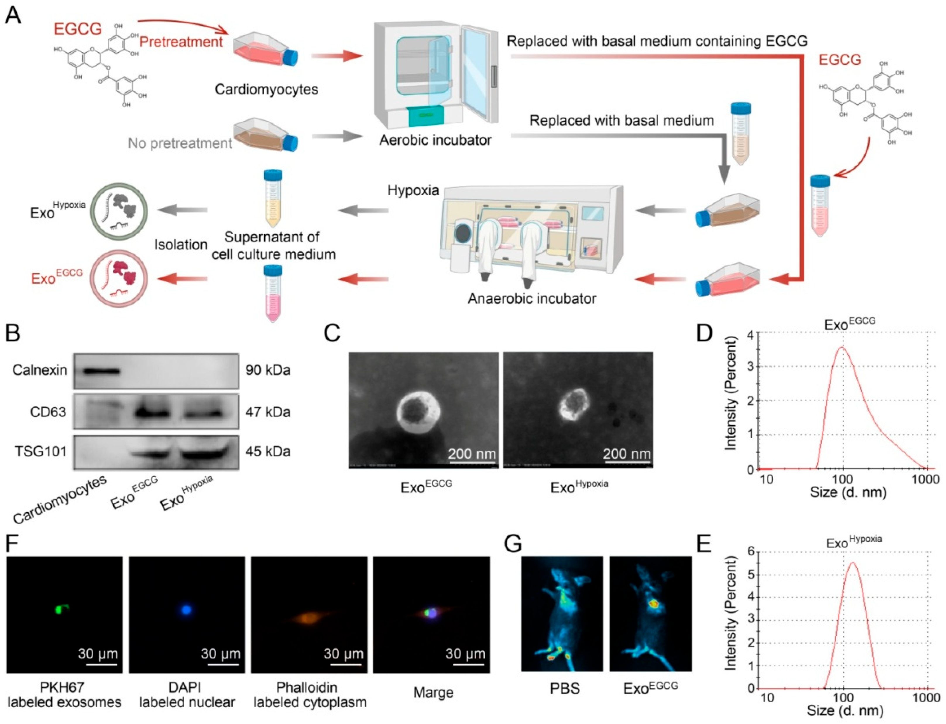
Exosomes were isolated from the culture supernatants of hypoxic HL-1 cardiomyocytes, either pretreated with EGCG or not (recorded as ExoEGCG or ExoHypoxia, respectively), using the exoEasy Maxi Kit. Exosomes were characterized by Western blotting, nanoparticle tracking analysis (NTA, Malvern Instruments, Malvern, UK) and transmission electron microscope (TEM, HITACHI, HT7700, Tokyo, Japan). To assess exosome uptake, exosomes were labeled with PKH67 or DiR in vitro or in vivo.
2.4. Transient Transfection
All plasmids were constructed by Jikai Gene Technology (Shanghai, China). Plasmid or corresponding NC was transfected according to the instructions of Invitrogen Lipofectamine 3000.
2.5. Animal Treatment and AMI Model Establishment
The animal procedures were conducted in compliance with the Guide for the Care and Use of Laboratory Animals and approved by the Animal Ethics Committee of Guilin Medical University (GLMC 202003304). Male C57BL/6 mice (22–25 g) were randomly assigned to experimental groups. Based on the study design, Exo
EGCG, si-circRERE, antagomiR-27a-3p or their corresponding negative control (NC) was administered prior to ischemia (
Table S2). The AMI model was induced by ligating the left anterior descending coronary artery for 12 h. While the AMI model was established, the success criteria were determined by ST-segment elevation, tall or inverted T waves in lead II electrocardiogram manifestations. After the ischemic period, mice were anesthetized with isoflurane, after which myocardial tissues and serum were collected and stored at −80 °C for subsequent analysis.
2.6. Cell Viability Assay
CCK-8 assay kit was used following instruction.
2.7. ELISA
CTn-I levels were measured according to the technical manual.
2.8. TUNEL Staining
The apoptosis rate of myocardia or cardiomyocytes was detected by TUNEL staining based on the experiment protocol.
2.9. RT-qPCR Analysis
Total RNA was isolated using Trizol reagent. cDNA was produced with transcript cDNA synthesis system and then amplified using QPCR Master Mix. All procedures were implemented strictly following manufacturers’ manuals. For mRNA or miRNA quantification,
GAPDH or U6 was performed as internal reference gene, respectively. Data analysis was conducted via 2
−ΔΔCt method. Primer sequences are listed in
Table S3.
2.10. Western Blotting Analysis
Protein extracts from cardiomyocytes or myocardium were separated by SDS-PAGE, then transferred to a membrane, and blocked. The membrane was then incubated sequentially with primary and secondary antibodies. Protein bands were observed and documented with gel imaging system (Syngene, Cambridge, UK), then band intensity was quantitated through ImageJ 1.8.0 software (Rawak Software Inc., Germany).
2.11. Echocardiography Detection
After 12 h of ischemia, mice were anesthetized with isoflurane. Left ventricular ejection fraction (EF) and fractional shortening (FS) were automatically calculated from three consecutive cardiac cycles to evaluated cardiac function with echocardiography (VINNO X6, Suzhou, China).
2.12. circRERE Circularization and Cell Localization Detection
Convergent primer and divergent primer of circRERE were designed to amplify genomic DNA (gDNA) and complementary DNA (cDNA), then gel electrophoresis and Sanger sequencing were implemented. To conduct circRERE’s subcellular localization, cytoplasmic and nuclear RNA fractions were isolated from HL-1 cells. RT-qPCR was then executed to assess the relative enrichment of circRERE in each compartment.
2.13. Dual-Luciferase Reporter Assay
Respective wild-type (WT) and mutant (MUT) reporter vector plasmids were constructed by Sangon Biotech (Shanghai, China). The miR-27a-3p mimic or NC was co-transfected with the WT or MUT into 293T cells. The dual-luciferase activity was then measured following the manufacturer’s instructions.
2.14. RNA Immunoprecipitation (RIP) Test
Following transfection with either miR-27a-3p mimic or NC plasmid, immunoprecipitation was performed. RNA was extracted according to the manual’s instructions. Then, the binding efficiency of circRERE was detected by RT-qPCR.
2.15. Bioinformatics Analyses
By analyzing the sequencing data of the GSE24548 dataset in the GEO database, differentially expressed miRNAs in AMI were identified [
14]. To identify circRNAs interacting with the objective miRNA, we queried the ENCORI database, extracting the number of supporting AGO CLIP-seq experiments (AgoExpNum) for each interaction [
15]. Subsequently, the circBase database was utilized to filter for circRNAs exhibiting high sequence conservation between humans and mice [
16]. Finally, the circAtlas database was employed to obtain the expression levels (circExp) and junction ratios (JncRto) of these conserved circRNAs across various human tissues [
17].
2.16. Statistical Analyses
Data were presented as means±SD. Comparison between two groups was tested by t-test. Variations among multiple groups were tested using one-way ANOVA after confirming normality and homogeneity of variances, followed by Tukey’s post hoc comparisons. p < 0.05 was considered as statistical significance. All calculations were performed in GraphPad Prism 5 (San Diego, CA, USA).
4. Discussion
CircRNAs are increasingly acknowledged as pivotal regulators in the modulation of apoptosis and AMI [
4,
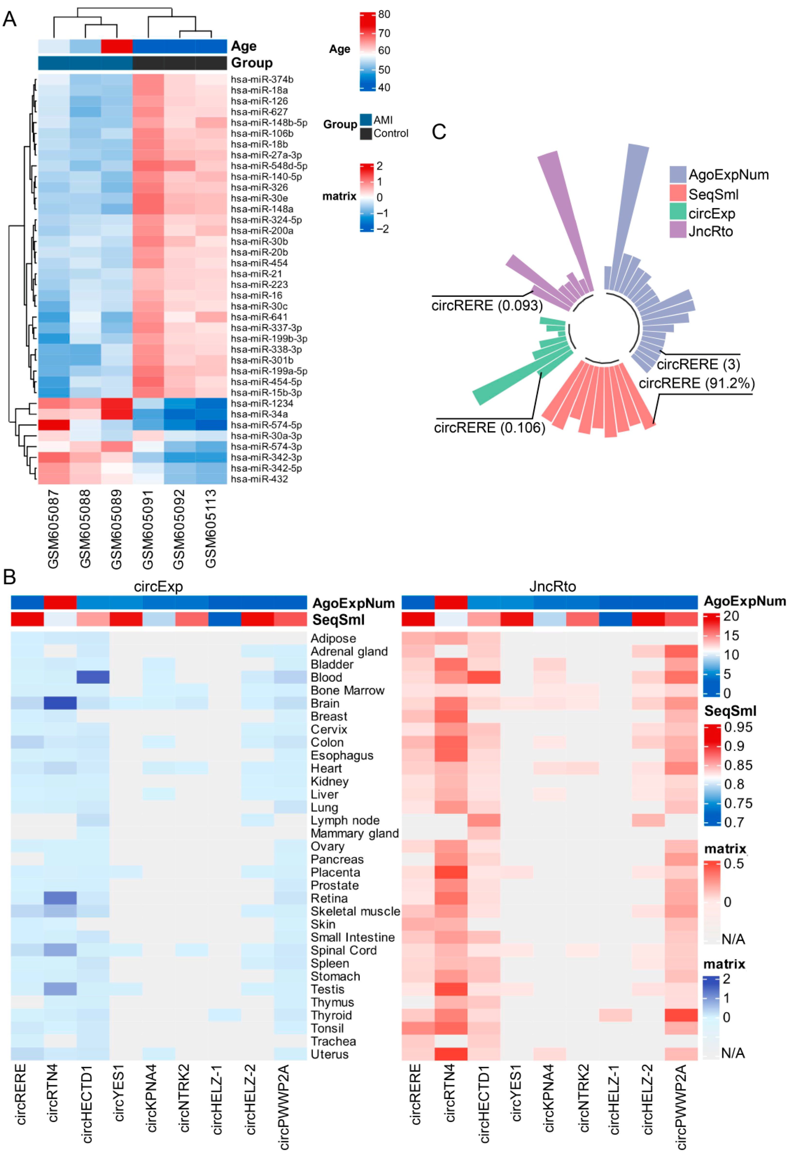
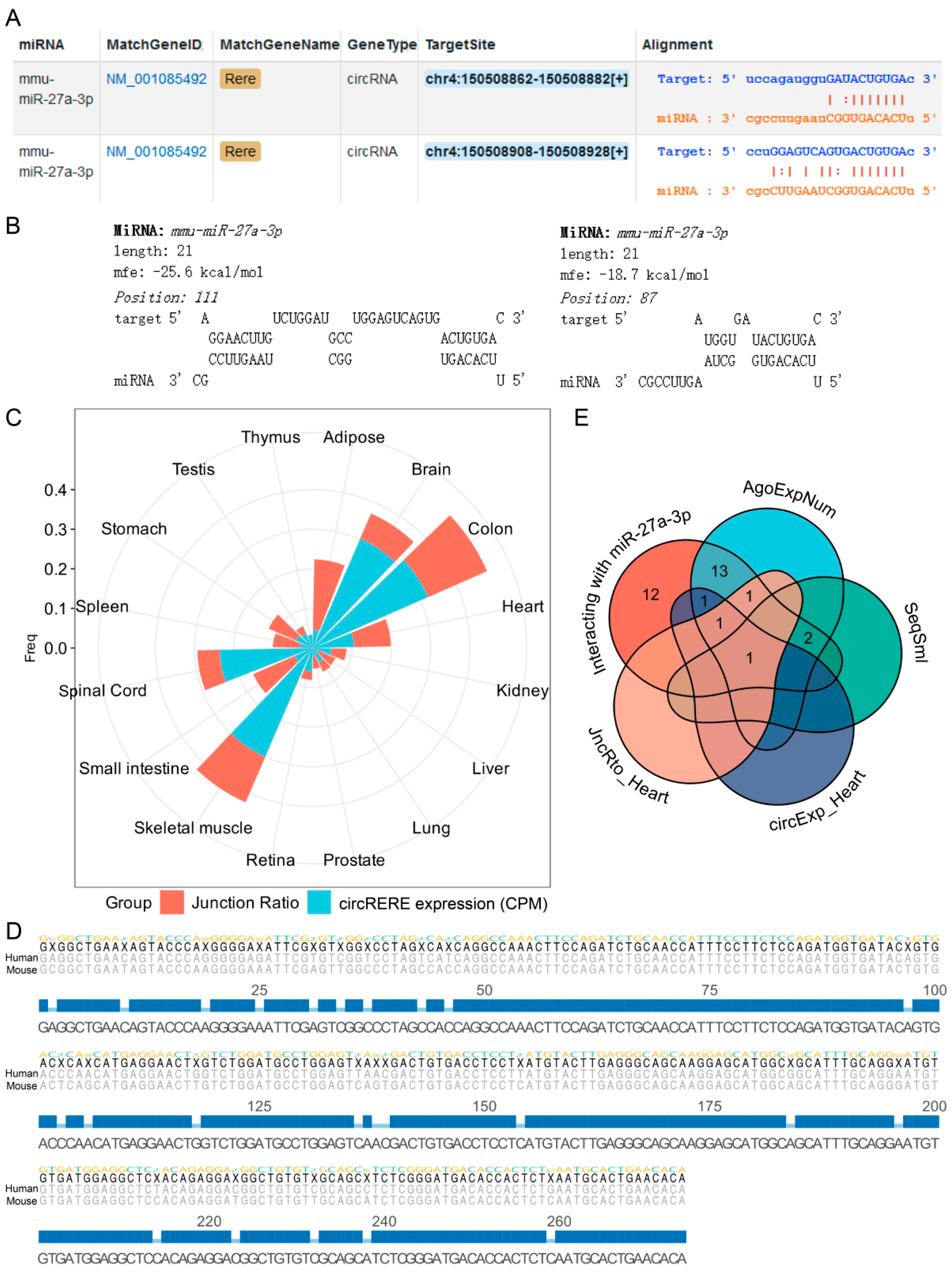
5]. Using the ENCORI database, we first analyzed the differentially expressed miRNAs in AMI patients and initially screened out the candidate circRNAs that may have important functions. Next, to identify a candidate circRNA for further investigation, we evaluated four key metrics: the number of supporting AGO CLIP-seq experiments (AgoExpNum), human–mouse sequence similarity (SeqSml), expression level (circExp), and junction ratio (JncRto) of each circRNA. Specifically, high AgoExpNum indicates that its binding to miRNAs is reproducible and stable, which eliminates accidental experimental errors and significantly enhances the credibility of the circRNA possessing biological functions [
15,
19,
20]. SeqSml reflects the evolutionary conservation and functional necessity of circRNAs. A high sequence similarity of homologous circRNAs between humans and mice suggests that these circRNAs have been retained during mammalian evolution, implying that their functions are indispensable for life activities. Nucleic acid sequences with high evolutionary conservation usually exert critical biological functions, and this indicator helps screen circRNAs with cross-species functional potential, making it more likely for the research conclusions obtained from mouse models to be extrapolated to humans [
19,
20]. In addition, highly expressed circRNAs enhance the operability and feasibility of downstream experiments. Their robust expression facilitates verification and quantification across diverse experimental approaches, which simplifies subsequent functional studies [
17,
19,
20]. As shown in
Figure 2 and
Figure 3, the high circExp score of circRERE, in particular, overcomes the common limitation of tissue-specific expression, confirming its stable presence in cardiac tissue. JncRto is a core indicator for judging the actual existence of circRNAs. A high junction ratio demonstrates that the formation of the circular structure of the circRNA is specific and stable, which can effectively exclude false-positive circRNAs caused by RNA degradation and sequencing errors, thus endowing them with substantial research value [
17,
19,
20]. Notably, unlike other candidates that exhibited distinct deficiencies in the above-mentioned metrics, circRERE (circBase ID: mmu_circ_0001305) achieved excellent performance in every single dimension without any critical weaknesses that might compromise subsequent experiments. This well-defined expression pattern makes it an ideal candidate for investigating the functional roles and regulatory networks of circRNAs in AMI.
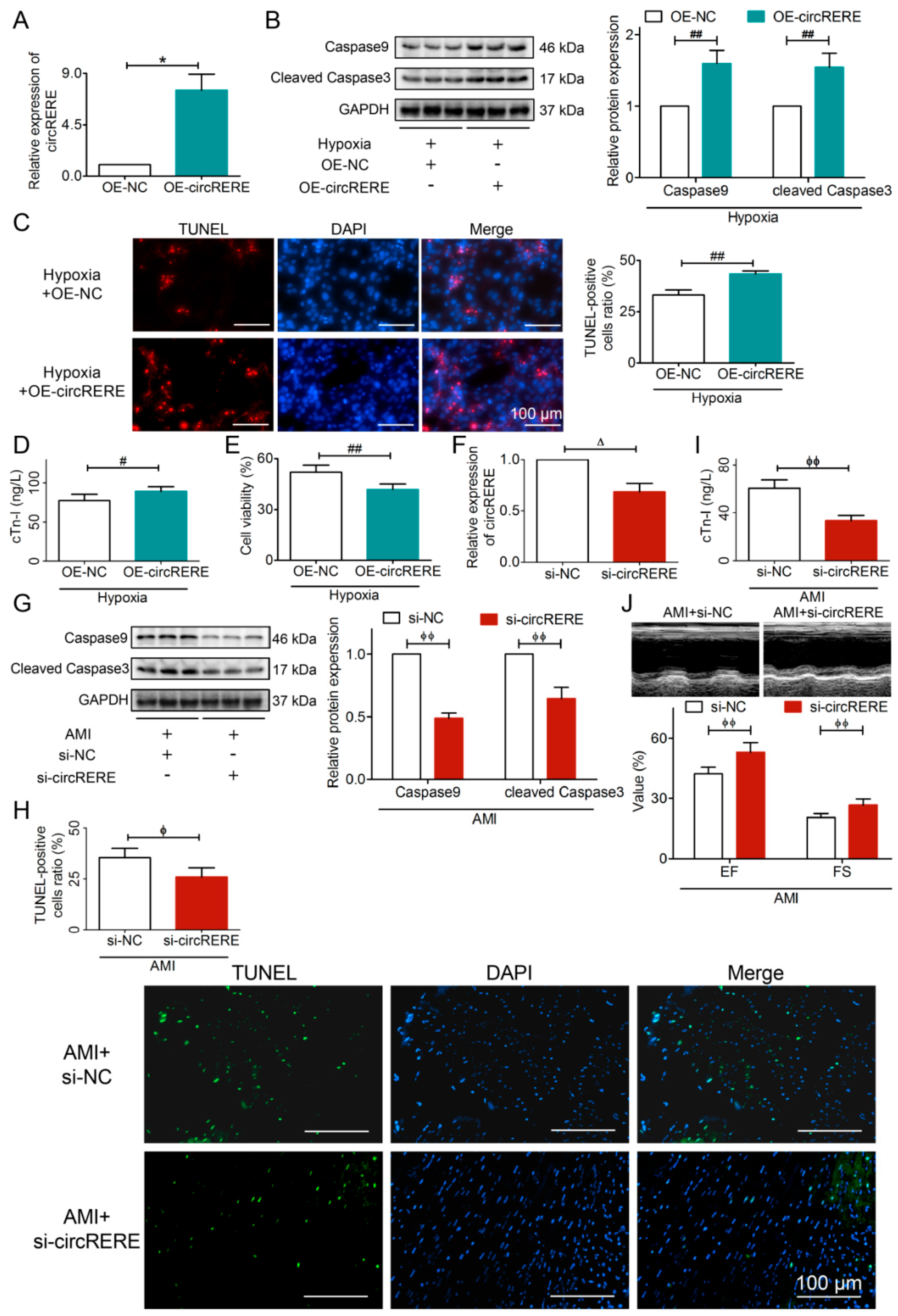
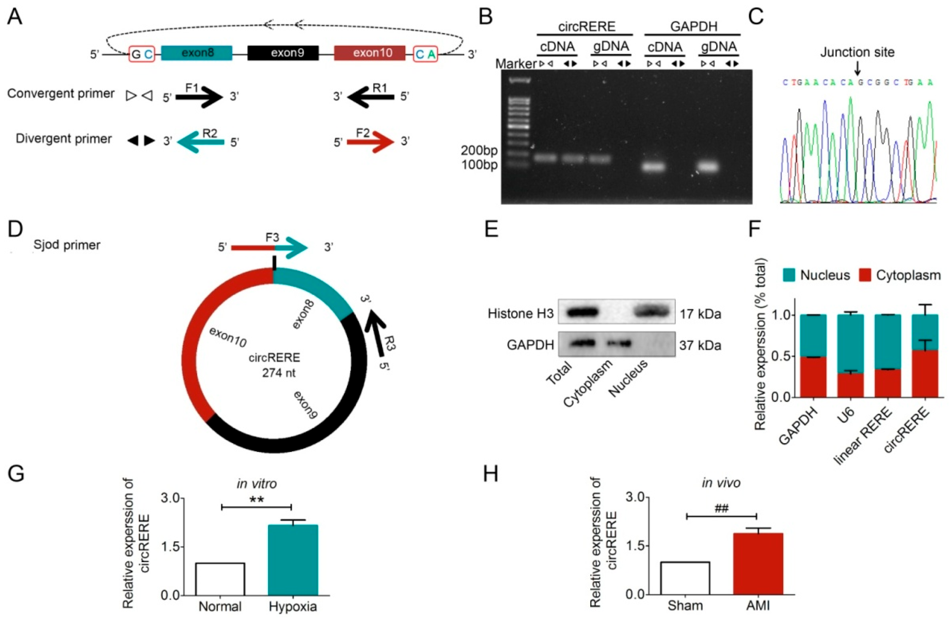
We first investigated the expression pattern and function of circRERE in AMI. Experimental results showed that the expression level of circRERE was significantly upregulated (
Figure 4G,H). Given that circRNAs typically exert their miRNA-sponge functions in the cytoplasm [
5,
21], we then verified the subcellular localization of circRERE. As shown in
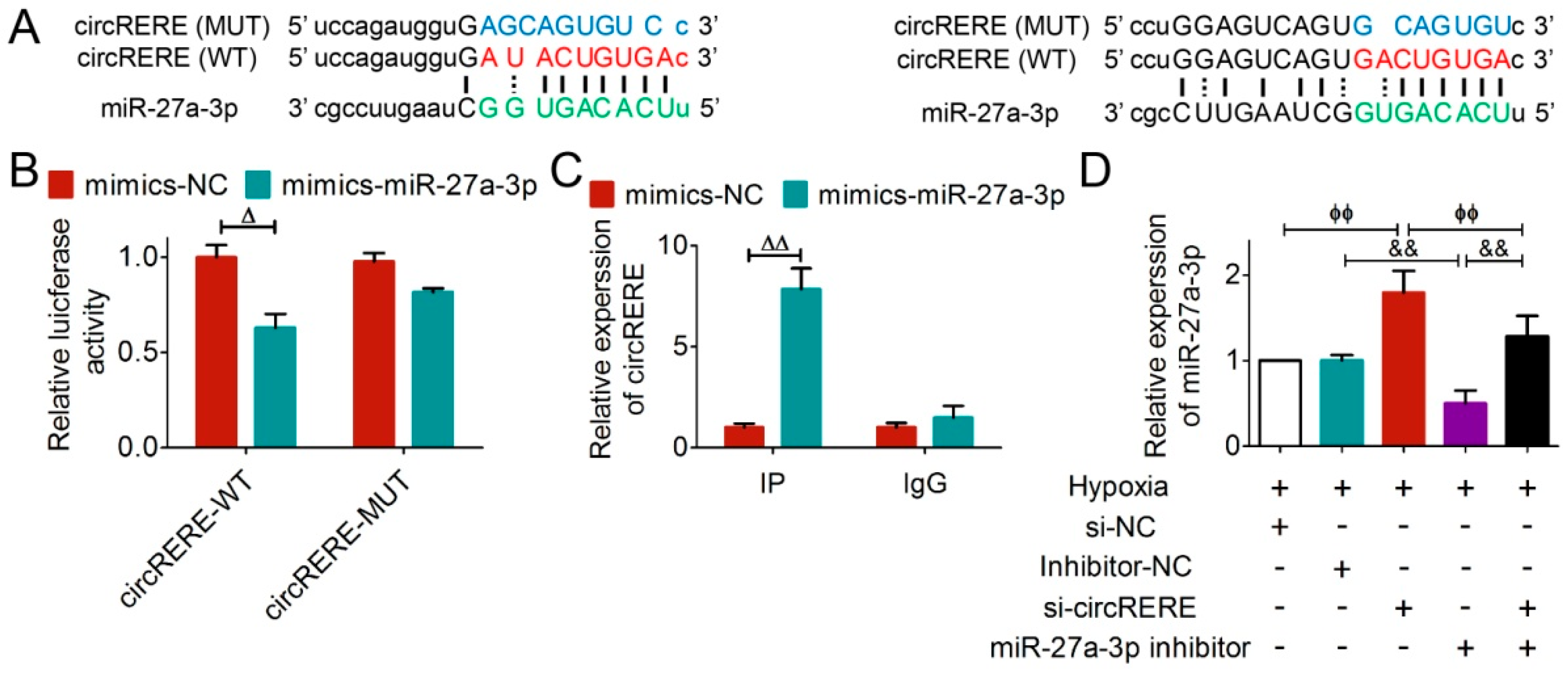
Figure 4, circRERE was predominantly cytoplasmic, fulfilling the spatial requirement. With the help of RNAhybrid software and the ENCORI database [
15,
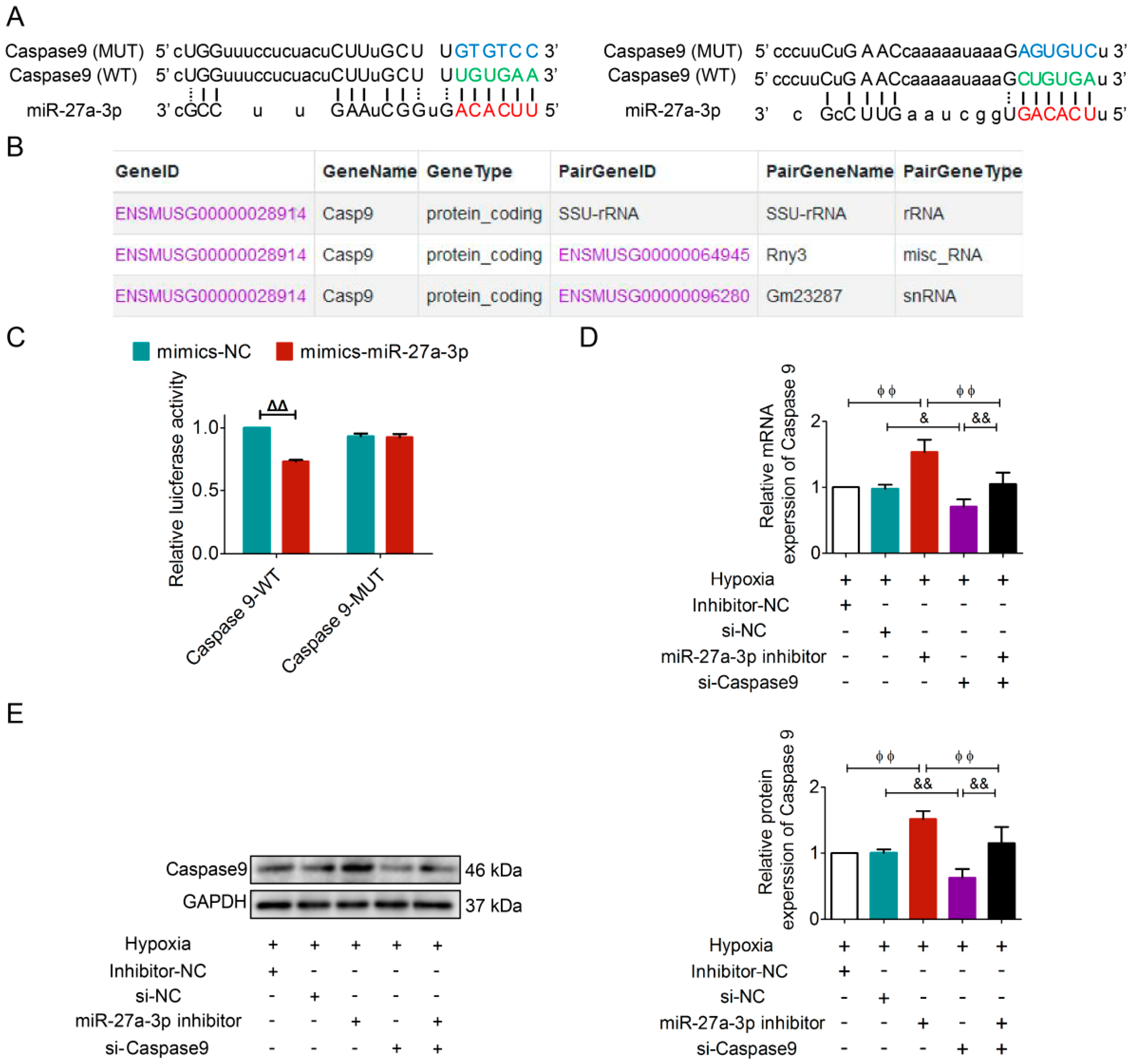
18], we predicted that miR-27a-3p and circRERE might have interactions. Subsequent luciferasereporter assay and RIP test validated that circRERE acts as a competitive endogenous RNA (ceRNA). We further discovered that miR-27a-3p and circRERE had direct interactions in mouse cardiomyocytes.
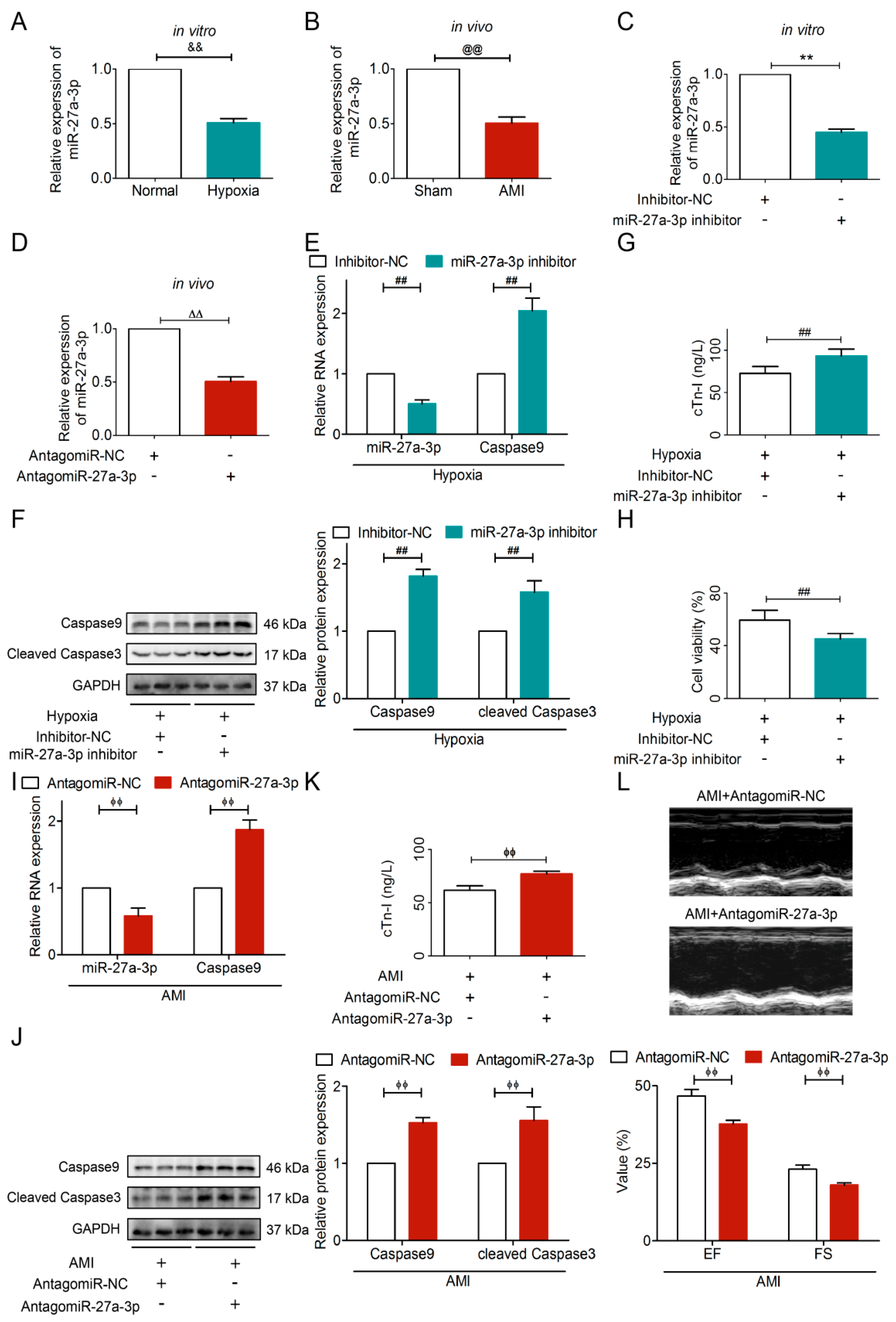
To better understand the role of circRERE on AMI, we constructed a plasmid for circRERE overexpression in vitro and a siRNA for circRERE silencing in vivo. Subsequent gain-of-function and loss-of-function experiments revealed that circRERE could suppress cell viability, aggravate the myocardial injury, and promote cell apoptosis by upregulating Caspase9 and cleaved Caspase3. Additionally, knockdown of circRERE upregulated miR-27a-3p, which in turn suppressed Caspase9 expression. These findings suggested that circRERE and miR-27a-3p may serve as upstream regulators of Caspase9-mediated apoptosis. Accumulatively, our studies established that circRERE acts as a molecular sponge for miR-27a-3p, thus promoting apoptosis in ischemic/hypoxic cardiomyocytes. For the first time, we clearly elucidated the unreported pathophysiological function of circRERE in AMI and identified a regulatory axis involving circRERE, miR-27a-3p, and Caspase9. Notably, circRERE shows remarkable cross-species conservation, with >90% sequence homology between humans and mice, which strongly highlights its translational relevance.
Cardiomyocytes, although not conventional secretory cells, could release exosomes under stressful conditions such as hypoxia and ischemia [
13]. Notably, external stimuli, including pharmacological agents and alterations in the cellular microenvironment, can significantly modulate exosome biogenesis and cellular uptake [
12,
13,
22,
23]. Therefore, investigating natural bioactive compounds that modulate exosome-mediated cardiac communication offers a promising avenue for developing novel multi-target therapies against AMI.
EGCG, the bioactive polyphenol in green tea, exhibits potent antioxidant activity [
7]. Current research primarily emphasizes EGCG’s applications in oncology, while evidence remains scarce regarding its cardioprotective effects mediated by non-coding RNA regulation, particularly via the exosomal pathway [
6,
7]. As shown in
Figure S2, compared to Exo
Hypoxia, Exo
EGCG markedly increased cell viability and reduced cTn-I level, indicating that EGCG markedly improves the cardioprotective efficacy of cardiomyocyte-secreted exosomes. Further mechanistic studies confirmed that Exo
EGCG exerts opposing biological effects to circRERE in AMI. Then, we investigated whether Exo
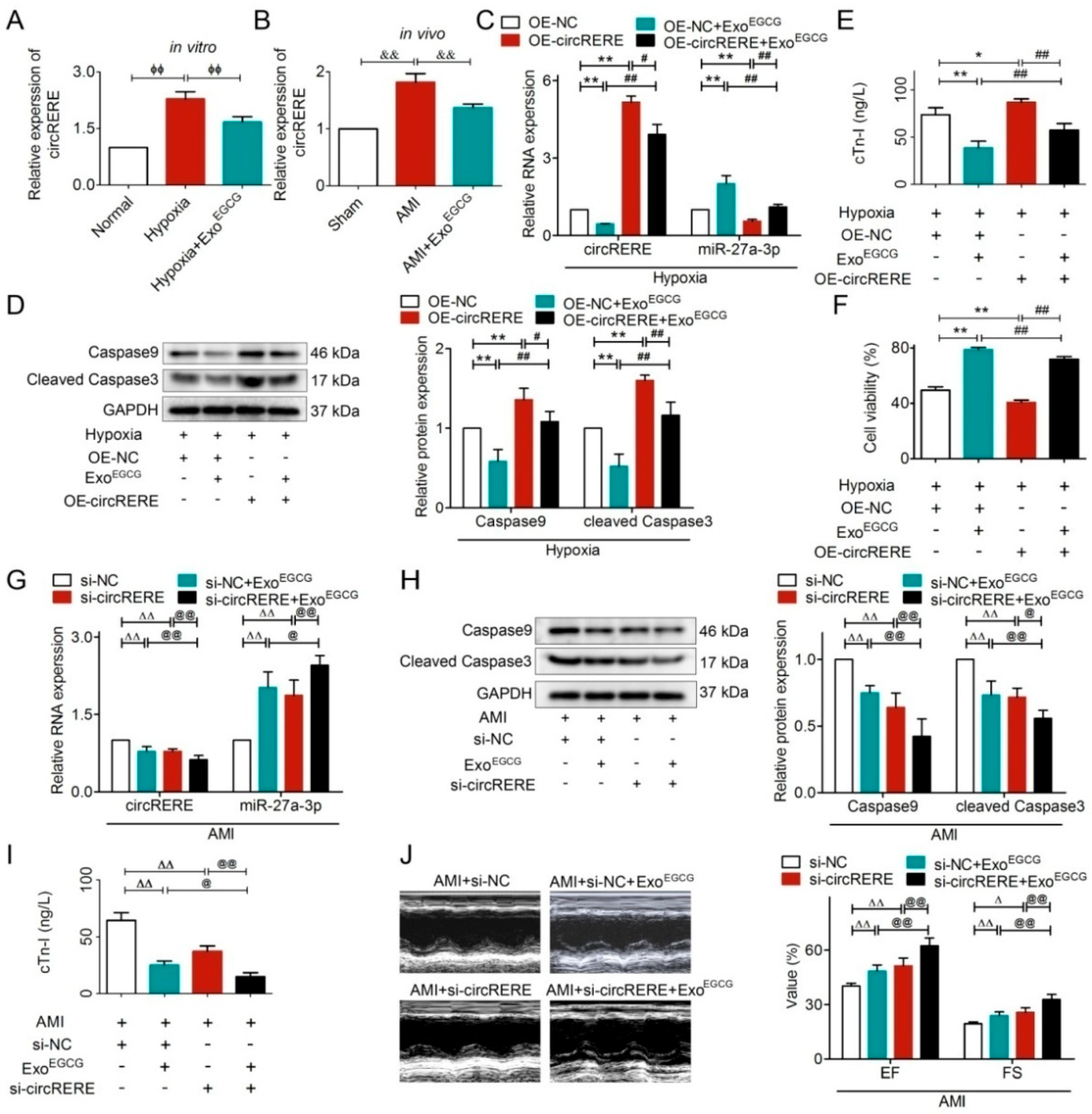
EGCG could regulate the functions of circRERE in AMI. As shown in
Figure 9A,B, Exo
EGCG downregulated the expression level of circRERE. Further mechanistic studies confirmed that Exo
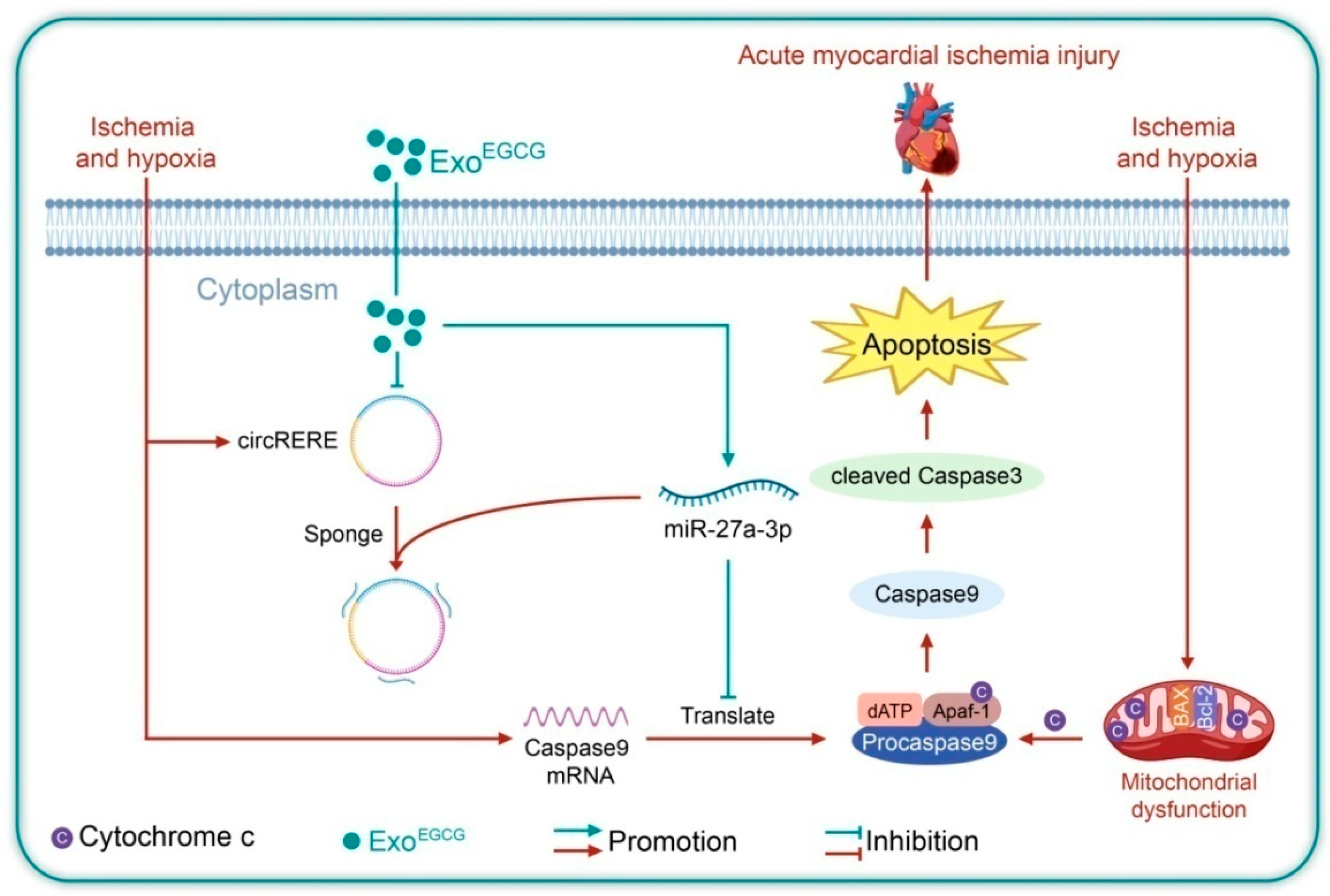
EGCG attenuated AMI injury via the circRERE/miR-27a-3p/Caspase9 regulatory axis.
Our research proposes a novel strategy to improve the therapeutic efficacy of EGCG using an exosome-based nanodelivery platform. As established in the literature, exosomes offer distinct advantages as natural nanocarriers, including enhanced cellular uptake, excellent biocompatibility, and greater stability in vivo [
10,
11]. Their lipid bilayer membrane provides a protective barrier that shields encapsulated therapeutics—such as nucleic acids and small molecules—from enzymatic degradation and chemical inactivation, thereby preserving bioactivity during systemic circulation [
11]. From a drug delivery perspective, exosomes represent an endogenous nanoplatform with low immunogenicity and inherent targeting potential, which can be further engineered for precision therapy [
10,
11]. As shown in
Figure 9, the combined delivery of Exo
EGCG and si-circRERE markedly improved therapeutic outcomes, highlighting the synergistic potential of co-encapsulating a small-molecule inhibitor and an RNA-based therapy within a unified exosomal system. Therefore, utilizing exosomes to simultaneously deliver si-circRERE and EGCG not only leverages the inherent benefits of nanodelivery—such as extended circulation time, targeted accumulation, and reduced off-target effects—but also establishes a promising combinatorial nanotherapeutic strategy for the treatment of AMI.
From a translational perspective, several key pathophysiological factors should be considered for future clinical application. First, the net effect on cardiomyocyte survival reflects the dynamic coexistence and crosstalk between pro-apoptotic and anti-apoptotic pathways rather than a single signaling cascade; the balance between Caspase-driven apoptosis and survival signals such as PI3K/Akt and Bcl-2 determines the ultimate fate of ischemic myocardium [
2,
3]. Second, cardiac responses undergo time-dependent transitions from acute protection to adaptive compensation and eventually maladaptive remodeling [
2,
3], meaning the efficacy of Exo
EGCG and circRERE inhibition may differ across the ischemic, reperfusion, and post-infarction repair phases. Third, the energetic state (mitochondrial function, ATP depletion) and microcirculatory status (capillary perfusion, no-reflow phenomenon) of ischemic/reperfused tissue directly affect exosome uptake, circRNA stability, and miRNA activity, which are critical for in vivo therapeutic outcomes [
2,
3,
24].
Future investigations are warranted to explore whether the circRERE/miR-27a-3p/Caspase9 axis remains dysregulated in chronic myocardial remodeling and chronic heart failure following acute ischemic injury. Given the central role of sustained cardiomyocyte apoptosis in progressive ventricular dysfunction, it is worthwhile to investigate whether targeted inhibition of circRERE combined with Exo
EGCG administration holds potential for long-term intervention to limit pathological deterioration and preserve cardiac function during the chronic phase after myocardial infarction. In addition, engineered exosomes represent a novel and promising delivery platform for cardiovascular diseases, with excellent biocompatibility, low immunogenicity, and tunable cargo-loading capacity [
10,
11,
12,
13]. Therefore, further attempts can be made to apply Exo
EGCG in precise nucleic acid-based therapy.
There are certain limitations in the present study. First, HL-1 cells were used for in vitro experiments, which are immortalized cells rather than primary cardiomyocytes. Compared with primary cardiomyocytes, HL-1 cells may differ in metabolic characteristics and contractile function under hypoxic stress [
25]. Future studies should validate our key findings using primary cardiomyocytes to enhance physiological relevance. Furthermore, the key regulatory factors governing circRERE biogenesis in AMI remain unclear. In addition, further systematic investigation is required to fully elucidate the precise mechanism by which Exo
EGCG modulates circRERE.
In conclusion, our study has identified the novel role of circRERE in myocardial ischemia, revealing its mechanism as a molecular sponge for miR-27a-3p to suppress apoptosis. Additionally, we delineated the signaling cascades and effector targets through which ExoEGCG affords protection against AMI injury. The works provide innovative insights for the development of nanodrug delivery platforms and gene therapy strategies against AMI, and open up new avenues for the interventional treatment of cardiovascular diseases.