Bridging Regenerative and Restorative Stem Cell Therapies in Parkinson’s Disease

Highlights
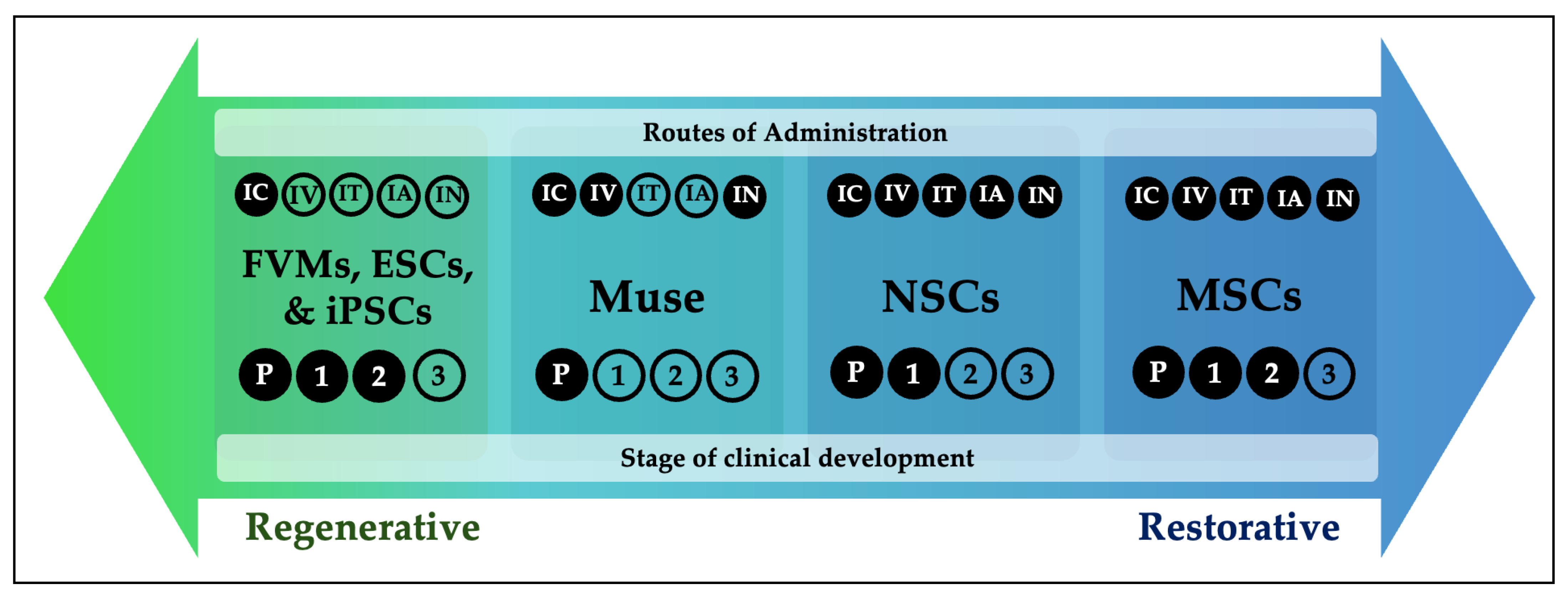
- Stem cell therapies for Parkinson’s disease can be conceptualized along a regenerative-restorative spectrum, determined by cell type and administration route.
- Regenerative approaches (e.g., fetal ventral mesencephalic stem cells, embryonic stem cells, induced pluripotent stem cells) primarily aim to replace lost dopaminergic neurons, while restorative approaches (mesenchymal stem cells) mainly provide neuroprotective and immunomodulatory effects. Some cell types, such as Multilineage-differentiating stress-enduring cells and neural stem cells have both regenerative and restorative features.
- Distinguishing regenerative from restorative mechanisms helps explain variability in clinical outcomes across stem cell trials and clarifies the interpretation of therapeutic efficacy.
- This framework can guide rational trial design and exposes the potential for combined or sequential strategies that may yield more durable disease-modifying benefits in Parkinson’s disease.
Abstract
1. Introduction
2. Regenerative Therapies
2.1. Fetal Ventral Mesencephalic Stem Cells
2.2. Human Pluripotent Stem Cells
2.2.1. Embryonic Stem Cells
2.2.2. Induced Pluripotent Stem Cells
2.3. Alternative Administration Routes
3. Restorative Therapies
3.1. Mesenchymal Stem Cells

| First Author (Year) | Stem Cell Type | Delivery Route | Study Design | Sample Size | Follow-Up (Months) | Age a | Disease Duration (Years) a | Clinical Outcomes | Safety/Adverse Events |
|---|---|---|---|---|---|---|---|---|---|
| Brazzini et al. (2010) [79] | autologous BM-MSCs | IA | phase I | 53 | 12 | 62.5 ± 10.4 | 9.3 | Improvement in UPDRS, QoL, and ADL scores | Psychosis, common colds |
| Venkataramana et al. (2010) [80] | autologous BM-MSCs | IC | phase I | 7 | 10–36 | 55.4 ± 15.4 | 14.7 ± 7.56 | Improvement in UPDRS III, ADL scores seen in 3 patients | NR |
| Yun et al. (2011) [81] | UC-MSCs | IA | case series | 8 | 1 | 58.4 ± 9.3 | 10 ± 6.9 | Improvement in UPDRS and ADL scores | Fatigue, fever, euphoria |
| Venkataramana et al. (2012) [82] | allogeneic BM-MSCs | IC | phase I | 12 (8 PwPD) | 12 | 58.8 ± 10.2 | 7.75 ± 3.5 | Improvement in UPDRS in PwPD, early disease showed more improvement | NR |
| Aili et al. (2013) [83] | UC-MSCs | IT | phase I | 9 | 6 | 67.1 ± 5.3 | NR | Improvement in UPDRS | fever |
| Dapeng et al. (2013) [84] | UC-MSCs | IT | phase I | 30 | 3 | 56 | NR | Improvement in UPDRS total, II, and III scores | NR |
| Xiaoquin et al. (2013) [85] | NSC | IT | phase I | 22 | 2 | 62 ± 4.2 | NR | Improvement in UPDRS | NR |
| Lige et al. (2014) [86] | NSCs | IV | phase I | 21 | 24 | 57.3 ± 9.1 | NR | Improvement in UPDRS, H&Y and QoL measures | NR |
| Yan et al. (2014) [87] | UC-MSCs | IA | case series | 15 | 1 | 63.4 ± 7.9 | NR | Improvement in UPDRS | Fever, fatigue |
| Boika et al. (2020) [88] | autologous BM-MSCs | IN, IV | pilot study | 23 | 3 | 52 (39.5, 59) intervention; 52 (47.5, 62.5) placebo | 7 (5, 8) intervention; 6 (4, 7) placebo | Improvement in MDS-UPDRS and nonmotor scores, reduced OFF time | NR |
| Schiess et al. (2021) [89] | allogeneic BM-MSCs | IV | phase I | 20 | 12 | 66 | 4–6 | Dose dependent improvement in UPDRS total, III scores, and H&Y in OFF state | No serious AEs related to the product; most AEs were mild and transient |
| Shigematsu et al. (2021) [90] | autologous Ad-MSCs | IV | case series | 3 | 6 | 75 ± 4.4 | 9.7 ± 6.4 | Gradual improvement in UPDRS I—III that plateaued after 3rd infusion | NR |
| Vij et al. (2023) [91] | autologous Ad-MSCs | IV | case report | 1 | 30 | 77 | >17 years | Improvement in UPDRS, decrease in dyskinesias and LEDD | NR |
| Jiang et al. (2024) [92] | NSCs | IN | phase I | 18 | 12 | 61.8 ± 7.6 | 10.7 ± 3.9 | Non-dose-dependent improvement in MDS-UPDRS total and II, III score; improvement in H&Y and LEDD; no changes in non-motor symptoms | No significant difference in severity of AE between groups; non-serious AEs were mild and transient |
| Schiess et al. (2025) [93] | allogeneic BM-MSCs | IV | phase II | 45 | 22 | 66.5 | 3 | 3 infusion arm improved MDS-UPDRS more than 2 infusion arm and placebo; improvement in QoL measures in treatment arms | AEs were mild and transient; no severe AEs; general malaise, flu-like symptoms, vomiting, headache |
| Vij et al. (2025) [94] | autologous Ad-MSCs | IV | phase I | 10 | 6 | 79.4 ± 3.95 | NR | No statistical improvement in MDS-UPDRS; significant improvement in PDQ-39 | Most AEs were mild; none of the severe AEs were due to the product; flu-like symptoms, fatigue |
3.2. Mechanism of Action
3.3. Evaluation of Administration Routes
4. Dual-Effect Cell Populations
4.1. Multilineage-Differentiating Stress Enduring Cells
4.2. Neural Stem Cells
5. Current Obstacles and Advancements in Regenerative Therapies
5.1. Graft Induced Dyskinesias from Serotonergic Excess
5.2. Lack of Clinical Benefit Due to Batch Heterogeneity and Patient Selection
5.3. Decreased Cell Survival Due to Unfavorable Host Microenvironment
5.4. Accumulation of Alpha Synuclein Pathology in Grafted Cells
6. Current Obstacles and Advancements in Restorative Therapies
6.1. Decreased Cell Survivability
6.2. Necessity of Repeated Dosing
6.3. Decreased Penetrance of the Blood–Brain Barrier
7. Conclusions
Author Contributions
Funding
Institutional Review Board Statement
Informed Consent Statement
Data Availability Statement
Conflicts of Interest
Abbreviations
| 6-OHDA | 6-hydroxydopamine |
| Ad-MSC | adipose mesenchymal stem cell |
| -syn | alpha synuclein |
| BBB | blood–brain barrier |
| BM-MSC | bone marrow mesenchymal stem cell |
| ESC | embryonic stem cell |
| FVM | fetal ventral mesencephalic |
| GID | graft induced dyskinesia |
| HIE | hypoxic ischemic encephalopathy |
| IA | intra-arterial |
| IC | intracerebral |
| ICH | intracerebral hemorrhage |
| IN | Intranasal |
| iPSC | induced pluripotent stem cell |
| IT | intrathecal |
| IV | intravenous |
| MSC | mesenchymal stem cell |
| Muse | multilineage differentiating stress-enduring |
| NSC | neural stem cell |
| PD | Parkinson’s Disease |
| ROS | reactive oxygen species |
| SDF-1 | stromal- cell derived factor 1 |
| SNpc | substantia nigra pars compacta |
| TH | tyrosine hydroxylase |
| UC-MSC | umbilical cord derived mesenchymal stem cell |
| UPDRS | Unified Parkinson’s Disease Rating Scale |
References
- Gammon, K. Neurodegenerative disease: Brain windfall. Nature 2014, 515, 299–300. [Google Scholar] [CrossRef] [PubMed]
- Li, M.; Ye, X.; Huang, Z.; Ye, L.; Chen, C. Global burden of Parkinson’s disease from 1990 to 2021: A population-based study. BMJ Open 2025, 15, e095610. [Google Scholar] [CrossRef]
- Su, D.; Cui, Y.; He, C.; Yin, P.; Bai, R.; Zhu, J.; Lam, J.S.T.; Zhang, J.; Yan, R.; Zheng, X.; et al. Projections for prevalence of Parkinson’s disease and its driving factors in 195 countries and territories to 2050: Modelling study of Global Burden of Disease Study 2021. BMJ 2025, 388, e080952. [Google Scholar] [CrossRef] [PubMed]
- Lai, T.T.; Kim, Y.J.; Ma, H.I.; Kim, Y.E. Evidence of Inflammation in Parkinson’s Disease and Its Contribution to Synucleinopathy. J. Mov. Disord. 2022, 15, 1–14. [Google Scholar] [CrossRef]
- Trounson, A.; McDonald, C. Stem Cell Therapies in Clinical Trials: Progress and Challenges. Cell Stem Cell 2015, 17, 11–22. [Google Scholar] [CrossRef] [PubMed]
- Lindvall, O.; Rehncrona, S.; Brundin, P.; Gustavii, B.; Astedt, B.; Widner, H.; Lindholm, T.; Bjorklund, A.; Leenders, K.L.; Rothwell, J.C.; et al. Human fetal dopamine neurons grafted into the striatum in two patients with severe Parkinson’s disease. A detailed account of methodology and a 6-month follow-up. Arch. Neurol. 1989, 46, 615–631. [Google Scholar] [CrossRef]
- Hitchcock, E.R.; Kenny, B.G.; Clough, C.G.; Hughes, R.C.; Henderson, B.T.; Detta, A. Stereotactic implantation of foetal mesencephalon (STIM): The UK experience. Prog. Brain Res. 1990, 82, 723–728. [Google Scholar] [CrossRef]
- Madrazo, I.; Franco-Bourland, R.; Ostrosky-Solis, F.; Aguilera, M.; Cuevas, C.; Alvarez, F.; Magallon, E.; Zamorano, C.; Morelos, A. Neural transplantation (auto-adrenal, fetal nigral and fetal adrenal) in Parkinson’s disease: The Mexican experience. Prog. Brain Res. 1990, 82, 593–602. [Google Scholar] [CrossRef]
- Henderson, B.T.; Clough, C.G.; Hughes, R.C.; Hitchcock, E.R.; Kenny, B.G. Implantation of human fetal ventral mesencephalon to the right caudate nucleus in advanced Parkinson’s disease. Arch. Neurol. 1991, 48, 822–827. [Google Scholar] [CrossRef]
- Freed, C.R.; Breeze, R.E.; Rosenberg, N.L.; Schneck, S.A.; Kriek, E.; Qi, J.X.; Lone, T.; Zhang, Y.B.; Snyder, J.A.; Wells, T.H.; et al. Survival of implanted fetal dopamine cells and neurologic improvement 12 to 46 months after transplantation for Parkinson’s disease. N. Engl. J. Med. 1992, 327, 1549–1555. [Google Scholar] [CrossRef]
- Widner, H.; Tetrud, J.; Rehncrona, S.; Snow, B.; Brundin, P.; Gustavii, B.; Bjorklund, A.; Lindvall, O.; Langston, J.W. Bilateral fetal mesencephalic grafting in two patients with parkinsonism induced by 1-methyl-4-phenyl-1,2,3,6-tetrahydropyridine (MPTP). N. Engl. J. Med. 1992, 327, 1556–1563. [Google Scholar] [CrossRef]
- Molina, H.; Quinones, R.; Ortega, I.; Alvarez, L.; Munoz, J.; Gonzalez, C.; Suarez, C. Computer assisted CT-guided stereotactic transplantation of foetal ventral mesencephalon to the caudate nucleus and putamen in Parkinson’s disease. Acta Neurochir. Suppl. 1993, 58, 17–19. [Google Scholar] [CrossRef]
- Peschanski, M.; Defer, G.; N’Guyen, J.P.; Ricolfi, F.; Monfort, J.C.; Remy, P.; Geny, C.; Samson, Y.; Hantraye, P.; Jeny, R.; et al. Bilateral motor improvement and alteration of L-dopa effect in two patients with Parkinson’s disease following intrastriatal transplantation of foetal ventral mesencephalon. Brain 1994, 117, 487–499. [Google Scholar] [CrossRef]
- Freeman, T.B.; Olanow, C.W.; Hauser, R.A.; Nauert, G.M.; Smith, D.A.; Borlongan, C.V.; Sanberg, P.R.; Holt, D.A.; Kordower, J.H.; Vingerhoets, F.J.; et al. Bilateral fetal nigral transplantation into the postcommissural putamen in Parkinson’s disease. Ann. Neurol. 1995, 38, 379–388. [Google Scholar] [CrossRef]
- Defer, G.L.; Geny, C.; Ricolfi, F.; Fenelon, G.; Monfort, J.C.; Remy, P.; Villafane, G.; Jeny, R.; Samson, Y.; Keravel, Y.; et al. Long-term outcome of unilaterally transplanted parkinsonian patients. I. Clinical approach. Brain 1996, 119, 41–50. [Google Scholar] [CrossRef]
- Kopyov, O.V.; Jacques, D.; Lieberman, A.; Duma, C.M.; Rogers, R.L. Clinical study of fetal mesencephalic intracerebral transplants for the treatment of Parkinson’s disease. Cell Transplant. 1996, 5, 327–337. [Google Scholar] [CrossRef] [PubMed]
- Levivier, M.; Dethy, S.; Rodesch, F.; Peschanski, M.; Vandesteene, A.; David, P.; Wikler, D.; Goldman, S.; Claes, T.; Biver, F.; et al. Intracerebral transplantation of fetal ventral mesencephalon for patients with advanced Parkinson’s disease. Methodology and 6-month to 1-year follow-up in 3 patients. Stereotact. Funct. Neurosurg. 1997, 69, 99–111. [Google Scholar] [CrossRef] [PubMed]
- Hauser, R.A.; Freeman, T.B.; Snow, B.J.; Nauert, M.; Gauger, L.; Kordower, J.H.; Olanow, C.W. Long-term evaluation of bilateral fetal nigral transplantation in Parkinson disease. Arch. Neurol. 1999, 56, 179–187. [Google Scholar] [CrossRef] [PubMed]
- Brundin, P.; Pogarell, O.; Hagell, P.; Piccini, P.; Widner, H.; Schrag, A.; Kupsch, A.; Crabb, L.; Odin, P.; Gustavii, B.; et al. Bilateral caudate and putamen grafts of embryonic mesencephalic tissue treated with lazaroids in Parkinson’s disease. Brain 2000, 123, 1380–1390. [Google Scholar] [CrossRef]
- Freed, C.R.; Greene, P.E.; Breeze, R.E.; Tsai, W.Y.; DuMouchel, W.; Kao, R.; Dillon, S.; Winfield, H.; Culver, S.; Trojanowski, J.Q.; et al. Transplantation of embryonic dopamine neurons for severe Parkinson’s disease. N. Engl. J. Med. 2001, 344, 710–719. [Google Scholar] [CrossRef]
- Mendez, I.; Dagher, A.; Hong, M.; Gaudet, P.; Weerasinghe, S.; McAlister, V.; King, D.; Desrosiers, J.; Darvesh, S.; Acorn, T.; et al. Simultaneous intrastriatal and intranigral fetal dopaminergic grafts in patients with Parkinson disease: A pilot study. Report of three cases. J. Neurosurg. 2002, 96, 589–596. [Google Scholar] [CrossRef]
- Olanow, C.W.; Goetz, C.G.; Kordower, J.H.; Stoessl, A.J.; Sossi, V.; Brin, M.F.; Shannon, K.M.; Nauert, G.M.; Perl, D.P.; Godbold, J.; et al. A double-blind controlled trial of bilateral fetal nigral transplantation in Parkinson’s disease. Ann. Neurol. 2003, 54, 403–414. [Google Scholar] [CrossRef] [PubMed]
- Schweitzer, J.S.; Song, B.; Herrington, T.M.; Park, T.Y.; Lee, N.; Ko, S.; Jeon, J.; Cha, Y.; Kim, K.; Li, Q.; et al. Personalized iPSC-Derived Dopamine Progenitor Cells for Parkinson’s Disease. N. Engl. J. Med. 2020, 382, 1926–1932. [Google Scholar] [CrossRef] [PubMed]
- Barker, R.A.; Lao-Kaim, N.P.; Guzman, N.V.; Athauda, D.; Bjartmarz, H.; Bjorklund, A.; Church, A.; Cutting, E.; Daft, D.; Dayal, V.; et al. The TransEuro open-label trial of human fetal ventral mesencephalic transplantation in patients with moderate Parkinson’s disease. Nat. Biotechnol. 2025, 44, 70–78. [Google Scholar] [CrossRef]
- Chang, J.W.; Na, H.K.; Chang, K.W.; Park, C.W.; Kim, D.H.; Park, S.; Park, C.Y.; Eom, J.H.; Nam, S.T.; Jo, K.S.; et al. Phase 1/2a clinical trial of hESC-derived dopamine progenitors in Parkinson’s disease. Cell 2025, 188, 7036–7048.e11. [Google Scholar] [CrossRef]
- Sawamoto, N.; Doi, D.; Nakanishi, E.; Sawamura, M.; Kikuchi, T.; Yamakado, H.; Taruno, Y.; Shima, A.; Fushimi, Y.; Okada, T.; et al. Phase I/II trial of iPS-cell-derived dopaminergic cells for Parkinson’s disease. Nature 2025, 641, 971–977. [Google Scholar] [CrossRef]
- Tabar, V.; Sarva, H.; Lozano, A.M.; Fasano, A.; Kalia, S.K.; Yu, K.K.H.; Brennan, C.; Ma, Y.; Peng, S.; Eidelberg, D.; et al. Phase I trial of hES cell-derived dopaminergic neurons for Parkinson’s disease. Nature 2025, 641, 978–983. [Google Scholar] [CrossRef]
- Lindvall, O.; Rehncrona, S.; Gustavii, B.; Brundin, P.; Astedt, B.; Widner, H.; Lindholm, T.; Bjorklund, A.; Leenders, K.L.; Rothwell, J.C.; et al. Fetal dopamine-rich mesencephalic grafts in Parkinson’s disease. Lancet 1988, 2, 1483–1484. [Google Scholar] [CrossRef]
- Lindvall, O.; Brundin, P.; Widner, H.; Rehncrona, S.; Gustavii, B.; Frackowiak, R.; Leenders, K.L.; Sawle, G.; Rothwell, J.C.; Marsden, C.D.; et al. Grafts of fetal dopamine neurons survive and improve motor function in Parkinson’s disease. Science 1990, 247, 574–577. [Google Scholar] [CrossRef]
- Lindvall, O.; Widner, H.; Rehncrona, S.; Brundin, P.; Odin, P.; Gustavii, B.; Frackowiak, R.; Leenders, K.L.; Sawle, G.; Rothwell, J.C.; et al. Transplantation of fetal dopamine neurons in Parkinson’s disease: One-year clinical and neurophysiological observations in two patients with putaminal implants. Ann. Neurol. 1992, 31, 155–165. [Google Scholar] [CrossRef] [PubMed]
- Lindvall, O.; Sawle, G.; Widner, H.; Rothwell, J.C.; Bjorklund, A.; Brooks, D.; Brundin, P.; Frackowiak, R.; Marsden, C.D.; Odin, P.; et al. Evidence for long-term survival and function of dopaminergic grafts in progressive Parkinson’s disease. Ann. Neurol. 1994, 35, 172–180. [Google Scholar] [CrossRef] [PubMed]
- Freed, C.R.; Breeze, R.E.; Rosenberg, N.L.; Schneck, S.A.; Wells, T.H.; Barrett, J.N.; Grafton, S.T.; Huang, S.C.; Eidelberg, D.; Rottenberg, D.A. Transplantation of human fetal dopamine cells for Parkinson’s disease. Results at 1 year. Arch. Neurol. 1990, 47, 505–512. [Google Scholar] [CrossRef] [PubMed]
- Spencer, D.D.; Robbins, R.J.; Naftolin, F.; Marek, K.L.; Vollmer, T.; Leranth, C.; Roth, R.H.; Price, L.H.; Gjedde, A.; Bunney, B.S.; et al. Unilateral transplantation of human fetal mesencephalic tissue into the caudate nucleus of patients with Parkinson’s disease. N. Engl. J. Med. 1992, 327, 1541–1548. [Google Scholar] [CrossRef]
- Wenning, G.K.; Odin, P.; Morrish, P.; Rehncrona, S.; Widner, H.; Brundin, P.; Rothwell, J.C.; Brown, R.; Gustavii, B.; Hagell, P.; et al. Short- and long-term survival and function of unilateral intrastriatal dopaminergic grafts in Parkinson’s disease. Ann. Neurol. 1997, 42, 95–107. [Google Scholar] [CrossRef] [PubMed]
- Hagell, P.; Schrag, A.; Piccini, P.; Jahanshahi, M.; Brown, R.; Rehncrona, S.; Widner, H.; Brundin, P.; Rothwell, J.C.; Odin, P.; et al. Sequential bilateral transplantation in Parkinson’s disease: Effects of the second graft. Brain 1999, 122, 1121–1132. [Google Scholar] [CrossRef]
- Perlow, M.J.; Freed, W.J.; Hoffer, B.J.; Seiger, A.; Olson, L.; Wyatt, R.J. Brain grafts reduce motor abnormalities produced by destruction of nigrostriatal dopamine system. Science 1979, 204, 643–647. [Google Scholar] [CrossRef]
- Fjodorova, M.; Torres, E.M.; Dunnett, S.B. Transplantation site influences the phenotypic differentiation of dopamine neurons in ventral mesencephalic grafts in Parkinsonian rats. Exp. Neurol. 2017, 291, 8–19. [Google Scholar] [CrossRef]
- Grealish, S.; Jonsson, M.E.; Li, M.; Kirik, D.; Bjorklund, A.; Thompson, L.H. The A9 dopamine neuron component in grafts of ventral mesencephalon is an important determinant for recovery of motor function in a rat model of Parkinson’s disease. Brain 2010, 133, 482–495. [Google Scholar] [CrossRef]
- Wuerthele, S.M.; Freed, W.J.; Olson, L.; Morihisa, J.; Spoor, L.; Wyatt, R.J.; Hoffer, B.J. Effect of dopamine agonists and antagonists on the electrical activity of substantia nigra neurons transplanted into the lateral ventricle of the rat. Exp. Brain Res. 1981, 44, 1–10. [Google Scholar] [CrossRef] [PubMed]
- Bjorklund, A.; Stenevi, U. Reconstruction of the nigrostriatal dopamine pathway by intracerebral nigral transplants. Brain Res. 1979, 177, 555–560. [Google Scholar] [CrossRef]
- Bankiewicz, K.S.; Plunkett, R.J.; Jacobowitz, D.M.; Porrino, L.; di Porzio, U.; London, W.T.; Kopin, I.J.; Oldfield, E.H. The effect of fetal mesencephalon implants on primate MPTP-induced parkinsonism. Histochemical and behavioral studies. J. Neurosurg. 1990, 72, 231–244. [Google Scholar] [CrossRef]
- Hagell, P.; Piccini, P.; Bjorklund, A.; Brundin, P.; Rehncrona, S.; Widner, H.; Crabb, L.; Pavese, N.; Oertel, W.H.; Quinn, N.; et al. Dyskinesias following neural transplantation in Parkinson’s disease. Nat. Neurosci. 2002, 5, 627–628. [Google Scholar] [CrossRef] [PubMed]
- Parish, C.L.; Thompson, L.H. Embryonic stem cells go from bench to bedside for Parkinson’s disease. Cell Rep. Med. 2021, 2, 100251. [Google Scholar] [CrossRef]
- Goodarzi, P.; Aghayan, H.R.; Larijani, B.; Soleimani, M.; Dehpour, A.R.; Sahebjam, M.; Ghaderi, F.; Arjmand, B. Stem cell-based approach for the treatment of Parkinson’s disease. Med. J. Islam. Repub. Iran 2015, 29, 168. [Google Scholar]
- Cerneckis, J.; Cai, H.; Shi, Y. Induced pluripotent stem cells (iPSCs): Molecular mechanisms of induction and applications. Signal Transduct. Target. Ther. 2024, 9, 112. [Google Scholar] [CrossRef]
- Choi, J.; Lee, S.; Mallard, W.; Clement, K.; Tagliazucchi, G.M.; Lim, H.; Choi, I.Y.; Ferrari, F.; Tsankov, A.M.; Pop, R.; et al. A comparison of genetically matched cell lines reveals the equivalence of human iPSCs and ESCs. Nat. Biotechnol. 2015, 33, 1173–1181. [Google Scholar] [CrossRef]
- Stoddard-Bennett, T.; Reijo Pera, R. Treatment of Parkinson’s Disease through Personalized Medicine and Induced Pluripotent Stem Cells. Cells 2019, 8, 26. [Google Scholar] [CrossRef] [PubMed]
- Sanders, L.H.; Laganiere, J.; Cooper, O.; Mak, S.K.; Vu, B.J.; Huang, Y.A.; Paschon, D.E.; Vangipuram, M.; Sundararajan, R.; Urnov, F.D.; et al. LRRK2 mutations cause mitochondrial DNA damage in iPSC-derived neural cells from Parkinson’s disease patients: Reversal by gene correction. Neurobiol. Dis. 2014, 62, 381–386. [Google Scholar] [CrossRef] [PubMed]
- Deacon, T.; Dinsmore, J.; Costantini, L.C.; Ratliff, J.; Isacson, O. Blastula-stage stem cells can differentiate into dopaminergic and serotonergic neurons after transplantation. Exp. Neurol. 1998, 149, 28–41. [Google Scholar] [CrossRef]
- Brederlau, A.; Correia, A.S.; Anisimov, S.V.; Elmi, M.; Paul, G.; Roybon, L.; Morizane, A.; Bergquist, F.; Riebe, I.; Nannmark, U.; et al. Transplantation of human embryonic stem cell-derived cells to a rat model of Parkinson’s disease: Effect of in vitro differentiation on graft survival and teratoma formation. Stem Cells 2006, 24, 1433–1440. [Google Scholar] [CrossRef]
- Takagi, Y.; Takahashi, J.; Saiki, H.; Morizane, A.; Hayashi, T.; Kishi, Y.; Fukuda, H.; Okamoto, Y.; Koyanagi, M.; Ideguchi, M.; et al. Dopaminergic neurons generated from monkey embryonic stem cells function in a Parkinson primate model. J. Clin. Investig. 2005, 115, 102–109. [Google Scholar] [CrossRef] [PubMed]
- Zhang, S.C.; Wernig, M.; Duncan, I.D.; Brustle, O.; Thomson, J.A. In vitro differentiation of transplantable neural precursors from human embryonic stem cells. Nat. Biotechnol. 2001, 19, 1129–1133. [Google Scholar] [CrossRef] [PubMed]
- Kriks, S.; Shim, J.W.; Piao, J.; Ganat, Y.M.; Wakeman, D.R.; Xie, Z.; Carrillo-Reid, L.; Auyeung, G.; Antonacci, C.; Buch, A.; et al. Dopamine neurons derived from human ES cells efficiently engraft in animal models of Parkinson’s disease. Nature 2011, 480, 547–551. [Google Scholar] [CrossRef]
- Kirkeby, A.; Grealish, S.; Wolf, D.A.; Nelander, J.; Wood, J.; Lundblad, M.; Lindvall, O.; Parmar, M. Generation of regionally specified neural progenitors and functional neurons from human embryonic stem cells under defined conditions. Cell Rep. 2012, 1, 703–714. [Google Scholar] [CrossRef]
- Grealish, S.; Diguet, E.; Kirkeby, A.; Mattsson, B.; Heuer, A.; Bramoulle, Y.; Van Camp, N.; Perrier, A.L.; Hantraye, P.; Bjorklund, A.; et al. Human ESC-derived dopamine neurons show similar preclinical efficacy and potency to fetal neurons when grafted in a rat model of Parkinson’s disease. Cell Stem Cell 2014, 15, 653–665. [Google Scholar] [CrossRef]
- Kim, T.W.; Piao, J.; Koo, S.Y.; Kriks, S.; Chung, S.Y.; Betel, D.; Socci, N.D.; Choi, S.J.; Zabierowski, S.; Dubose, B.N.; et al. Biphasic Activation of WNT Signaling Facilitates the Derivation of Midbrain Dopamine Neurons from hESCs for Translational Use. Cell Stem Cell 2021, 28, 343–355.e5. [Google Scholar] [CrossRef]
- Ståhl, K. Update on STEM-PD Clinical Trial—Stem Cell-Based Transplant for Parkinson’s Disease. Available online: https://www.stemcellcenter.lu.se/article/update-stem-pd-clinical-trial-stem-cell-based-transplant-parkinsons-disease?utm_source=chatgpt.com (accessed on 22 November 2025).
- Stem-Cell Therapy is a ‘Big Leap’ for Parkinson’s Treatment. Available online: https://www.ucihealth.org/about-us/news/2025/04/parkinsons-study-nature (accessed on 10 November 2025).
- Kikuchi, T.; Morizane, A.; Doi, D.; Magotani, H.; Onoe, H.; Hayashi, T.; Mizuma, H.; Takara, S.; Takahashi, R.; Inoue, H.; et al. Human iPS cell-derived dopaminergic neurons function in a primate Parkinson’s disease model. Nature 2017, 548, 592–596. [Google Scholar] [CrossRef]
- Rouhani, F.; Kumasaka, N.; de Brito, M.C.; Bradley, A.; Vallier, L.; Gaffney, D. Genetic background drives transcriptional variation in human induced pluripotent stem cells. PLoS Genet. 2014, 10, e1004432. [Google Scholar] [CrossRef] [PubMed]
- Khelifi, G.; Chow, T.; Whiteley, J.; Fort, V.; Humphreys, B.D.; Hussein, S.M.I.; Rogers, I.M. Determining epigenetic memory in kidney proximal tubule cell derived induced pluripotent stem cells using a quadruple transgenic reprogrammable mouse. Sci. Rep. 2022, 12, 20340. [Google Scholar] [CrossRef]
- Hallett, P.J.; Deleidi, M.; Astradsson, A.; Smith, G.A.; Cooper, O.; Osborn, T.M.; Sundberg, M.; Moore, M.A.; Perez-Torres, E.; Brownell, A.L.; et al. Successful function of autologous iPSC-derived dopamine neurons following transplantation in a non-human primate model of Parkinson’s disease. Cell Stem Cell 2015, 16, 269–274. [Google Scholar] [CrossRef]
- Tao, Y.; Vermilyea, S.C.; Zammit, M.; Lu, J.; Olsen, M.; Metzger, J.M.; Yao, L.; Chen, Y.; Phillips, S.; Holden, J.E.; et al. Autologous transplant therapy alleviates motor and depressive behaviors in parkinsonian monkeys. Nat. Med. 2021, 27, 632–639. [Google Scholar] [CrossRef] [PubMed]
- iPS Cell-Based Parkinson’s Disease Therapy Administered to First Patient. Available online: https://www.kyoto-u.ac.jp/en/news/2018-12-10?utm_source=chatgpt.com (accessed on 22 November 2025).
- Jaslow, R. Clinical Trial Tests Novel Stem-Cell Treatment for Parkinson’s Disease. Available online: https://www.massgeneralbrigham.org/en/about/newsroom/press-releases/clinical-trial-novel-stem-cell-treatment-for-parkinsons?utm_source=chatgpt.com (accessed on 22 November 2025).
- Kauhausen, J.; Thompson, L.H.; Parish, C.L. Cell intrinsic and extrinsic factors contribute to enhance neural circuit reconstruction following transplantation in Parkinsonian mice. J. Physiol. 2013, 591, 77–91. [Google Scholar] [CrossRef]
- Barker, R.A.; Barrett, J.; Mason, S.L.; Bjorklund, A. Fetal dopaminergic transplantation trials and the future of neural grafting in Parkinson’s disease. Lancet Neurol. 2013, 12, 84–91. [Google Scholar] [CrossRef]
- Achon Buil, B.; Tackenberg, C.; Rust, R. Editing a gateway for cell therapy across the blood-brain barrier. Brain 2023, 146, 823–841. [Google Scholar] [CrossRef]
- Kot, M.; Baj-Krzyworzeka, M.; Szatanek, R.; Musial-Wysocka, A.; Suda-Szczurek, M.; Majka, M. The Importance of HLA Assessment in “Off-the-Shelf” Allogeneic Mesenchymal Stem Cells Based-Therapies. Int. J. Mol. Sci. 2019, 20, 5680. [Google Scholar] [CrossRef]
- Jin, H.J.; Bae, Y.K.; Kim, M.; Kwon, S.J.; Jeon, H.B.; Choi, S.J.; Kim, S.W.; Yang, Y.S.; Oh, W.; Chang, J.W. Comparative analysis of human mesenchymal stem cells from bone marrow, adipose tissue, and umbilical cord blood as sources of cell therapy. Int. J. Mol. Sci. 2013, 14, 17986–18001. [Google Scholar] [CrossRef]
- Mendes Filho, D.; Ribeiro, P.D.C.; Oliveira, L.F.; de Paula, D.R.M.; Capuano, V.; de Assuncao, T.S.F.; da Silva, V.J.D. Therapy With Mesenchymal Stem Cells in Parkinson Disease: History and Perspectives. Neurologist 2018, 23, 141–147. [Google Scholar] [CrossRef] [PubMed]
- Unnisa, A.; Dua, K.; Kamal, M.A. Mechanism of Mesenchymal Stem Cells as a Multitarget Disease- Modifying Therapy for Parkinson’s Disease. Curr. Neuropharmacol. 2023, 21, 988–1000. [Google Scholar] [CrossRef]
- Friedenstein, A.J.; Chailakhjan, R.K.; Lalykina, K.S. The development of fibroblast colonies in monolayer cultures of guinea-pig bone marrow and spleen cells. Cell Tissue Kinet. 1970, 3, 393–403. [Google Scholar] [CrossRef]
- Li, Y.; Chen, J.; Wang, L.; Zhang, L.; Lu, M.; Chopp, M. Intracerebral transplantation of bone marrow stromal cells in a 1-methyl-4-phenyl-1,2,3,6-tetrahydropyridine mouse model of Parkinson’s disease. Neurosci. Lett. 2001, 316, 67–70. [Google Scholar] [CrossRef] [PubMed]
- Jamali, F.; Aldughmi, M.; Khasawneh, M.W.; Dahbour, S.; Salameh, A.A.; Awidi, A. A New Tool for Safety Evaluation and a Combination of Measures for Efficacy Assessment of Cotransplanting Human Allogenic Neuronal Stem Cells and Mesenchymal Stem Cells for the Treatment of Parkinson Disease: Protocol for an Interventional Study. JMIR Res. Protoc. 2021, 10, e29695. [Google Scholar] [CrossRef]
- Tashima, T. Mesenchymal Stem Cell (MSC)-Based Drug Delivery into the Brain across the Blood-Brain Barrier. Pharmaceutics 2024, 16, 289. [Google Scholar] [CrossRef]
- Wragg, A.; Mellad, J.A.; Beltran, L.E.; Konoplyannikov, M.; San, H.; Boozer, S.; Deans, R.J.; Mathur, A.; Lederman, R.J.; Kovacic, J.C.; et al. VEGFR1/CXCR4-positive progenitor cells modulate local inflammation and augment tissue perfusion by a SDF-1-dependent mechanism. J. Mol. Med. 2008, 86, 1221–1232. [Google Scholar] [CrossRef] [PubMed]
- Han, X.; Liao, R.; Li, X.; Zhang, C.; Huo, S.; Qin, L.; Xiong, Y.; He, T.; Xiao, G.; Zhang, T. Mesenchymal stem cells in treating human diseases: Molecular mechanisms and clinical studies. Signal Transduct. Target. Ther. 2025, 10, 262. [Google Scholar] [CrossRef] [PubMed]
- Brazzini, A.; Cantella, R.; De la Cruz, A.; Yupanqui, J.; Leon, C.; Jorquiera, T.; Brazzini, M.; Ortega, M.; Saenz, L.N. Intraarterial autologous implantation of adult stem cells for patients with Parkinson disease. J. Vasc. Interv. Radiol. 2010, 21, 443–451. [Google Scholar] [CrossRef] [PubMed]
- Venkataramana, N.K.; Kumar, S.K.; Balaraju, S.; Radhakrishnan, R.C.; Bansal, A.; Dixit, A.; Rao, D.K.; Das, M.; Jan, M.; Gupta, P.K.; et al. Open-labeled study of unilateral autologous bone-marrow-derived mesenchymal stem cell transplantation in Parkinson’s disease. Transl. Res. 2010, 155, 62–70. [Google Scholar] [CrossRef]
- Qiu, Y.; Wang, Z.; Lu, H. Umbilical cord mesenchymal stem cell transplantation for treatment of Parkinson’s disease in 8 cases. Chin. J. Tissue Eng. Res. 2011, 15, 6833–6836. [Google Scholar] [CrossRef]
- Venkataramana, N.K.; Pal, R.; Rao, S.A.; Naik, A.L.; Jan, M.; Nair, R.; Sanjeev, C.C.; Kamble, R.B.; Murthy, D.P.; Chaitanya, K. Bilateral transplantation of allogenic adult human bone marrow-derived mesenchymal stem cells into the subventricular zone of Parkinson’s disease: A pilot clinical study. Stem Cells Int. 2012, 2012, 931902. [Google Scholar] [CrossRef]
- Aili, L. Efficacy of umbilical cord mesenchymal stem cell transplantation for treatment of Parkinson’s disease. Chin. J. Gerontol. 2013, 33, 5245–5247. [Google Scholar] [CrossRef]
- Dapeng, S.; Yan, W. Repair and reconstruction of neurons by umbilical cord mesenchymal stem cells. Guide China Med. 2013, 11, 419–420+422. [Google Scholar] [CrossRef]
- Xiaoquin, B.; Yabin, S.; Songtao, L.; Hong, Y. Neural stem cell transplantation for Parkinson’s disease. Chin. J. Gerontol. 2013, 33, 3591–3592. [Google Scholar] [CrossRef]
- Lige, L.; Zengmin, T. Treatment of Parkinson disease by implantation of neural stem cells into stratums (report of 21 cases). Chin. J. Clin. Neurosurg. 2014, 19, 412–413+417. [Google Scholar] [CrossRef]
- Yan, W.; Xinli, Z.; Junyan, Z.; Jun, T. Therapeutic applications of umbilical cord mesenchymal stem cells in Parkinson’s disease. Chin. J. Tissue Eng. Res. 2014, 18, 6. [Google Scholar] [CrossRef]
- Boika, A.; Aleinikava, N.; Chyzhyk, V.; Zafranskaya, M.; Nizheharodava, D.; Ponomarev, V. Mesenchymal stem cells in Parkinson’s disease: Motor and nonmotor symptoms in the early posttransplant period. Surg. Neurol. Int. 2020, 11, 380. [Google Scholar] [CrossRef] [PubMed]
- Schiess, M.; Suescun, J.; Doursout, M.F.; Adams, C.; Green, C.; Saltarrelli, J.G.; Savitz, S.; Ellmore, T.M. Allogeneic Bone Marrow-Derived Mesenchymal Stem Cell Safety in Idiopathic Parkinson’s Disease. Mov. Disord. 2021, 36, 1825–1834. [Google Scholar] [CrossRef]
- Shigematsu, K.; Komori, N.; Tahara, K.; Yamagishi, H. Repeated infusion of autologous adipose tissue-derived stem cells for Parkinson’s disease. Acta Neurol. Scand. 2022, 145, 119–122. [Google Scholar] [CrossRef]
- Vij, R.; Prossin, A.; Tripathy, M.; Kim, H.; Park, H.; Cheng, T.; Lotfi, D.; Chang, D. Long-term, repeated doses of intravenous autologous mesenchymal stem cells for a patient with Parkinson’s disease: A case report. Front. Neurol. 2023, 14, 1257080. [Google Scholar] [CrossRef] [PubMed]
- Jiang, S.; Wang, H.; Yang, C.; Feng, F.; Xu, D.; Zhang, M.; Xie, M.; Cui, R.; Zhu, Z.; Jia, C.; et al. Phase 1 study of safety and preliminary efficacy of intranasal transplantation of human neural stem cells (ANGE-S003) in Parkinson’s disease. J. Neurol. Neurosurg. Psychiatry 2024, 95, 1102–1111. [Google Scholar] [CrossRef]
- Schiess, M.C.; Suescun, J.; Martinez-Lemus, J.D.; Green, C.; Thomas, T.S.; Shahnawaz, M.; Tharp, E.; Satani, N.B.; Saltarrelli, J.G.; Adams, C.; et al. Allogeneic Bone Marrow-Derived Mesenchymal Stem Cells for Parkinson’s Disease: A Randomized Trial. Mov. Disord. 2025, 40, 2688–2699. [Google Scholar] [CrossRef]
- Vij, R.; Kim, H.; Park, H.; Cheng, T.; Lotfi, D.; Chang, D. Safety and efficacy of adipose-derived mesenchymal stem cell therapy in elderly Parkinson’s disease patients: An intermediate-size expanded access program. Cytotherapy 2025, 27, 181–187. [Google Scholar] [CrossRef]
- Guo, L.; Yin, F.; Meng, H.Q.; Ling, L.; Hu-He, T.N.; Li, P.; Zhang, C.X.; Yu, S.; Duan, D.S.; Fan, H.X. Differentiation of mesenchymal stem cells into dopaminergic neuron-like cells in vitro. Biomed. Environ. Sci. 2005, 18, 36–42. [Google Scholar] [PubMed]
- Dezawa, M.; Kanno, H.; Hoshino, M.; Cho, H.; Matsumoto, N.; Itokazu, Y.; Tajima, N.; Yamada, H.; Sawada, H.; Ishikawa, H.; et al. Specific induction of neuronal cells from bone marrow stromal cells and application for autologous transplantation. J. Clin. Investig. 2004, 113, 1701–1710. [Google Scholar] [CrossRef] [PubMed]
- Trzaska, K.A.; Kuzhikandathil, E.V.; Rameshwar, P. Specification of a dopaminergic phenotype from adult human mesenchymal stem cells. Stem Cells 2007, 25, 2797–2808. [Google Scholar] [CrossRef]
- Chen, D.; Fu, W.; Zhuang, W.; Lv, C.; Li, F.; Wang, X. Therapeutic effects of intranigral transplantation of mesenchymal stem cells in rat models of Parkinson’s disease. J. Neurosci. Res. 2017, 95, 907–917. [Google Scholar] [CrossRef]
- Shetty, P.; Ravindran, G.; Sarang, S.; Thakur, A.M.; Rao, H.S.; Viswanathan, C. Clinical grade mesenchymal stem cells transdifferentiated under xenofree conditions alleviates motor deficiencies in a rat model of Parkinson’s disease. Cell Biol. Int. 2009, 33, 830–838. [Google Scholar] [CrossRef]
- Chao, Y.X.; He, B.P.; Tay, S.S. Mesenchymal stem cell transplantation attenuates blood brain barrier damage and neuroinflammation and protects dopaminergic neurons against MPTP toxicity in the substantia nigra in a model of Parkinson’s disease. J. Neuroimmunol. 2009, 216, 39–50. [Google Scholar] [CrossRef]
- Teixeira, F.G.; Carvalho, M.M.; Panchalingam, K.M.; Rodrigues, A.J.; Mendes-Pinheiro, B.; Anjo, S.; Manadas, B.; Behie, L.A.; Sousa, N.; Salgado, A.J. Impact of the Secretome of Human Mesenchymal Stem Cells on Brain Structure and Animal Behavior in a Rat Model of Parkinson’s Disease. Stem Cells Transl. Med. 2017, 6, 634–646. [Google Scholar] [CrossRef]
- Abrishamdar, M.; Jalali, M.S.; Yazdanfar, N. The role of exosomes in pathogenesis and the therapeutic efficacy of mesenchymal stem cell-derived exosomes against Parkinson’s disease. Neurol. Sci. 2023, 44, 2277–2289. [Google Scholar] [CrossRef]
- Giunti, D.; Parodi, B.; Usai, C.; Vergani, L.; Casazza, S.; Bruzzone, S.; Mancardi, G.; Uccelli, A. Mesenchymal stem cells shape microglia effector functions through the release of CX3CL1. Stem Cells 2012, 30, 2044–2053. [Google Scholar] [CrossRef]
- Musial-Wysocka, A.; Kot, M.; Majka, M. The Pros and Cons of Mesenchymal Stem Cell-Based Therapies. Cell Transplant. 2019, 28, 801–812. [Google Scholar] [CrossRef] [PubMed]
- Kim, Y.J.; Park, H.J.; Lee, G.; Bang, O.Y.; Ahn, Y.H.; Joe, E.; Kim, H.O.; Lee, P.H. Neuroprotective effects of human mesenchymal stem cells on dopaminergic neurons through anti-inflammatory action. Glia 2009, 57, 13–23. [Google Scholar] [CrossRef] [PubMed]
- Angeloni, C.; Gatti, M.; Prata, C.; Hrelia, S.; Maraldi, T. Role of Mesenchymal Stem Cells in Counteracting Oxidative Stress-Related Neurodegeneration. Int. J. Mol. Sci. 2020, 21, 3299. [Google Scholar] [CrossRef]
- Moon, H.E.; Yoon, S.H.; Hur, Y.S.; Park, H.W.; Ha, J.Y.; Kim, K.H.; Shim, J.H.; Yoo, S.H.; Son, J.H.; Paek, S.L.; et al. Mitochondrial dysfunction of immortalized human adipose tissue-derived mesenchymal stromal cells from patients with Parkinson’s disease. Exp. Neurobiol. 2013, 22, 283–300. [Google Scholar] [CrossRef]
- Essawy Essawy, A.; Abou-ElNaga, O.A.; Mehanna, R.A.; Badae, N.M.; Elsawy, E.S.; Soffar, A.A. Comparing the effect of intravenous versus intracranial grafting of mesenchymal stem cells against parkinsonism in a rat model: Behavioral, biochemical, pathological and immunohistochemical studies. PLoS ONE 2024, 19, e0296297. [Google Scholar] [CrossRef]
- Choi, H.S.; Kim, H.J.; Oh, J.H.; Park, H.G.; Ra, J.C.; Chang, K.A.; Suh, Y.H. Therapeutic potentials of human adipose-derived stem cells on the mouse model of Parkinson’s disease. Neurobiol. Aging 2015, 36, 2885–2892. [Google Scholar] [CrossRef]
- Velarde, F.; Ezquerra, S.; Delbruyere, X.; Caicedo, A.; Hidalgo, Y.; Khoury, M. Mesenchymal stem cell-mediated transfer of mitochondria: Mechanisms and functional impact. Cell. Mol. Life Sci. 2022, 79, 177. [Google Scholar] [CrossRef]
- Wang, F.; Yasuhara, T.; Shingo, T.; Kameda, M.; Tajiri, N.; Yuan, W.J.; Kondo, A.; Kadota, T.; Baba, T.; Tayra, J.T.; et al. Intravenous administration of mesenchymal stem cells exerts therapeutic effects on parkinsonian model of rats: Focusing on neuroprotective effects of stromal cell-derived factor-1alpha. BMC Neurosci. 2010, 11, 52. [Google Scholar] [CrossRef]
- Chi, H.; Guan, Y.; Li, F.; Chen, Z. The Effect of Human Umbilical Cord Mesenchymal Stromal Cells in Protection of Dopaminergic Neurons from Apoptosis by Reducing Oxidative Stress in the Early Stage of a 6-OHDA-Induced Parkinson’s Disease Model. Cell Transplant. 2019, 28, 87S–99S. [Google Scholar] [CrossRef] [PubMed]
- Riecke, J.; Johns, K.M.; Cai, C.; Vahidy, F.S.; Parsha, K.; Furr-Stimming, E.; Schiess, M.; Savitz, S.I. A Meta-Analysis of Mesenchymal Stem Cells in Animal Models of Parkinson’s Disease. Stem Cells Dev. 2015, 24, 2082–2090. [Google Scholar] [CrossRef] [PubMed]
- Cerri, S.; Greco, R.; Levandis, G.; Ghezzi, C.; Mangione, A.S.; Fuzzati-Armentero, M.T.; Bonizzi, A.; Avanzini, M.A.; Maccario, R.; Blandini, F. Intracarotid Infusion of Mesenchymal Stem Cells in an Animal Model of Parkinson’s Disease, Focusing on Cell Distribution and Neuroprotective and Behavioral Effects. Stem Cells Transl. Med. 2015, 4, 1073–1085. [Google Scholar] [CrossRef]
- Giordano, R.; Canesi, M.; Isalberti, M.; Marfia, G.; Campanella, R.; Vincenti, D.; Cereda, V.; Ranghetti, A.; Palmisano, C.; Isaias, I.U.; et al. Safety and Effectiveness of Cell Therapy in Neurodegenerative Diseases: Take-Home Messages from a Pilot Feasibility Phase I Study of Progressive Supranuclear Palsy. Front. Neurosci. 2021, 15, 723227. [Google Scholar] [CrossRef]
- Lee, P.H.; Lee, J.E.; Kim, H.S.; Song, S.K.; Lee, H.S.; Nam, H.S.; Cheong, J.W.; Jeong, Y.; Park, H.J.; Kim, D.J.; et al. A randomized trial of mesenchymal stem cells in multiple system atrophy. Ann. Neurol. 2012, 72, 32–40. [Google Scholar] [CrossRef] [PubMed]
- Danielyan, L.; Schafer, R.; von Ameln-Mayerhofer, A.; Bernhard, F.; Verleysdonk, S.; Buadze, M.; Lourhmati, A.; Klopfer, T.; Schaumann, F.; Schmid, B.; et al. Therapeutic efficacy of intranasally delivered mesenchymal stem cells in a rat model of Parkinson disease. Rejuvenation Res. 2011, 14, 3–16. [Google Scholar] [CrossRef] [PubMed]
- Salama, M.; Sobh, M.; Emam, M.; Abdalla, A.; Sabry, D.; El-Gamal, M.; Lotfy, A.; El-Husseiny, M.; Sobh, M.; Shalash, A.; et al. Effect of intranasal stem cell administration on the nigrostriatal system in a mouse model of Parkinson’s disease. Exp. Ther. Med. 2017, 13, 976–982. [Google Scholar] [CrossRef]
- Aguilera, Y.; Mellado-Damas, N.; Olmedo-Moreno, L.; Lopez, V.; Panadero-Moron, C.; Benito, M.; Guerrero-Cazares, H.; Marquez-Vega, C.; Martin-Montalvo, A.; Capilla-Gonzalez, V. Preclinical Safety Evaluation of Intranasally Delivered Human Mesenchymal Stem Cells in Juvenile Mice. Cancers 2021, 13, 1169. [Google Scholar] [CrossRef] [PubMed]
- Baak, L.M.; Wagenaar, N.; van der Aa, N.E.; Groenendaal, F.; Dudink, J.; Tataranno, M.L.; Mahamuud, U.; Verhage, C.H.; Eijsermans, R.; Smit, L.S.; et al. Feasibility and safety of intranasally administered mesenchymal stromal cells after perinatal arterial ischaemic stroke in the Netherlands (PASSIoN): A first-in-human, open-label intervention study. Lancet Neurol. 2022, 21, 528–536. [Google Scholar] [CrossRef]
- Wagenaar, N.; Baak, L.M.; van der Aa, N.E.; Groenendaal, F.; Dudink, J.; Tataranno, M.L.; Koopman, C.; Verhage, C.H.; Eijsermans, R.; van Teeseling, H.C.; et al. Perinatal Arterial Stroke Treated With Stromal Cells Intranasally: 2-Year Safety and Neurodevelopment. Stroke 2025, 56, 2410–2418. [Google Scholar] [CrossRef]
- Zhang, Y.T.; He, K.J.; Zhang, J.B.; Ma, Q.H.; Wang, F.; Liu, C.F. Advances in intranasal application of stem cells in the treatment of central nervous system diseases. Stem Cell Res. Ther. 2021, 12, 210. [Google Scholar] [CrossRef]
- Maric, D.M.; Velikic, G.; Maric, D.L.; Supic, G.; Vojvodic, D.; Petric, V.; Abazovic, D. Stem Cell Homing in Intrathecal Applications and Inspirations for Improvement Paths. Int. J. Mol. Sci. 2022, 23, 4290. [Google Scholar] [CrossRef]
- Mesa Bedoya, L.E.; Camacho Barbosa, J.C.; Lopez Quiceno, L.; Barrios Arroyave, F.; Halpert, K.; Espana Pena, J.A.; Salazar Uribe, J.C. The safety profile of mesenchymal stem cell therapy administered through intrathecal injections for treating neurological disorders: A systematic review and meta-analysis of randomised controlled trials. Stem Cell Res. Ther. 2024, 15, 146. [Google Scholar] [CrossRef]
- Stoker, T.B.; Andresen, K.E.R.; Barker, R.A. Hydrocephalus Complicating Intrathecal Antisense Oligonucleotide Therapy for Huntington’s Disease. Mov. Disord. 2021, 36, 263–264. [Google Scholar] [CrossRef]
- Zhao, J.; Qu, K.; Jia, S.; Yang, R.; Cui, Z.; Li, J.; Yu, P.; Dong, M. Efficacy and efficacy-influencing factors of stem cell transplantation on patients with Parkinson’s disease: A systematic review and meta-analysis. Front. Neurol. 2024, 15, 1329343. [Google Scholar] [CrossRef]
- Kuroda, Y.; Kitada, M.; Wakao, S.; Dezawa, M. Bone marrow mesenchymal cells: How do they contribute to tissue repair and are they really stem cells? Arch. Immunol. Ther. Exp. 2011, 59, 369–378. [Google Scholar] [CrossRef] [PubMed]
- Reddy, A.P.; Ravichandran, J.; Carkaci-Salli, N. Neural regeneration therapies for Alzheimer’s and Parkinson’s disease-related disorders. Biochim. Biophys. Acta Mol. Basis Dis. 2020, 1866, 165506. [Google Scholar] [CrossRef] [PubMed]
- Studer, L.; Tabar, V.; McKay, R.D. Transplantation of expanded mesencephalic precursors leads to recovery in parkinsonian rats. Nat. Neurosci. 1998, 1, 290–295. [Google Scholar] [CrossRef] [PubMed]
- Zhang, Z.; Wang, Y.; Zhang, Z.; Qi, Z. Network Meta-Analysis of Stem Cell Therapies for Parkinson’s Disease: Exploring the Optimal Strategy Based on Animal Models. Stem Cells Dev. 2025, 34, 227–239. [Google Scholar] [CrossRef]
- Shimamura, N.; Kakuta, K.; Wang, L.; Naraoka, M.; Uchida, H.; Wakao, S.; Dezawa, M.; Ohkuma, H. Neuro-regeneration therapy using human Muse cells is highly effective in a mouse intracerebral hemorrhage model. Exp. Brain Res. 2017, 235, 565–572. [Google Scholar] [CrossRef]
- Yamauchi, T.; Kuroda, Y.; Morita, T.; Shichinohe, H.; Houkin, K.; Dezawa, M.; Kuroda, S. Therapeutic effects of human multilineage-differentiating stress enduring (MUSE) cell transplantation into infarct brain of mice. PLoS ONE 2015, 10, e0116009. [Google Scholar] [CrossRef]
- Uchida, H.; Niizuma, K.; Kushida, Y.; Wakao, S.; Tominaga, T.; Borlongan, C.V.; Dezawa, M. Human Muse Cells Reconstruct Neuronal Circuitry in Subacute Lacunar Stroke Model. Stroke 2017, 48, 428–435. [Google Scholar] [CrossRef]
- Yasuhara, T.; Matsukawa, N.; Hara, K.; Maki, M.; Ali, M.M.; Yu, S.J.; Bae, E.; Yu, G.; Xu, L.; McGrogan, M.; et al. Notch-induced rat and human bone marrow stromal cell grafts reduce ischemic cell loss and ameliorate behavioral deficits in chronic stroke animals. Stem Cells Dev. 2009, 18, 1501–1514. [Google Scholar] [CrossRef]
- Suzuki, T.; Sato, Y.; Kushida, Y.; Tsuji, M.; Wakao, S.; Ueda, K.; Imai, K.; Iitani, Y.; Shimizu, S.; Hida, H.; et al. Intravenously delivered multilineage-differentiating stress enduring cells dampen excessive glutamate metabolism and microglial activation in experimental perinatal hypoxic ischemic encephalopathy. J. Cereb. Blood Flow. Metab. 2021, 41, 1707–1720. [Google Scholar] [CrossRef]
- Abe, T.; Aburakawa, D.; Niizuma, K.; Iwabuchi, N.; Kajitani, T.; Wakao, S.; Kushida, Y.; Dezawa, M.; Borlongan, C.V.; Tominaga, T. Intravenously Transplanted Human Multilineage-Differentiating Stress-Enduring Cells Afford Brain Repair in a Mouse Lacunar Stroke Model. Stroke 2020, 51, 601–611. [Google Scholar] [CrossRef]
- Kajitani, T.; Endo, T.; Iwabuchi, N.; Inoue, T.; Takahashi, Y.; Abe, T.; Niizuma, K.; Tominaga, T. Association of intravenous administration of human Muse cells with deficit amelioration in a rat model of spinal cord injury. J. Neurosurg. Spine 2021, 34, 648–655. [Google Scholar] [CrossRef]
- Yamada, Y.; Wakao, S.; Kushida, Y.; Minatoguchi, S.; Mikami, A.; Higashi, K.; Baba, S.; Shigemoto, T.; Kuroda, Y.; Kanamori, H.; et al. S1P-S1PR2 Axis Mediates Homing of Muse Cells Into Damaged Heart for Long-Lasting Tissue Repair and Functional Recovery After Acute Myocardial Infarction. Circ. Res. 2018, 122, 1069–1083. [Google Scholar] [CrossRef]
- Yamashita, T.; Kushida, Y.; Wakao, S.; Tadokoro, K.; Nomura, E.; Omote, Y.; Takemoto, M.; Hishikawa, N.; Ohta, Y.; Dezawa, M.; et al. Therapeutic benefit of Muse cells in a mouse model of amyotrophic lateral sclerosis. Sci. Rep. 2020, 10, 17102. [Google Scholar] [CrossRef] [PubMed]
- Aprile, D.; Patrone, D.; Al Sammarraie, S.H.A.; Alessio, N.; Peluso, G.; Di Bernardo, G.; Galderisi, U. Lineage specification into GABAergic, glutamatergic, dopaminergic, and astrocytic phenotypes using MUSE stem cells: A novel approach for modeling neurodegenerative and psychiatric disorders. Mol. Psychiatry 2025, 31, 1372–1384. [Google Scholar] [CrossRef] [PubMed]
- Li, H.; Wei, J.; Liu, X.; Zhang, P.; Lin, J. Muse cells: Ushering in a new era of stem cell-based therapy for stroke. Stem Cell Res. Ther. 2022, 13, 421. [Google Scholar] [CrossRef]
- Alanazi, R.F.; Alhwity, B.S.; Almahlawi, R.M.; Alatawi, B.D.; Albalawi, S.A.; Albalawi, R.A.; Albalawi, A.A.; Abdel-Maksoud, M.S.; Elsherbiny, N. Multilineage Differentiating Stress Enduring (Muse) Cells: A New Era of Stem Cell-Based Therapy. Cells 2023, 12, 1676. [Google Scholar] [CrossRef]
- Yin, X.Y.; Wang, C.C.; Du, P.; Wang, X.S.; Lu, Y.C.; Sun, Y.W.; Sun, Y.H.; Hu, Y.M.; Chen, X. Muse cells decrease the neuroinflammatory response by modulating the proportion of M1 and M2 microglia in vitro. Neural Regen. Res. 2023, 18, 213–218. [Google Scholar] [CrossRef] [PubMed]
- Nitobe, Y.; Nagaoki, T.; Kumagai, G.; Sasaki, A.; Liu, X.; Fujita, T.; Fukutoku, T.; Wada, K.; Tanaka, T.; Kudo, H.; et al. Neurotrophic Factor Secretion and Neural Differentiation Potential of Multilineage-differentiating Stress-enduring (Muse) Cells Derived from Mouse Adipose Tissue. Cell Transplant. 2019, 28, 1132–1139. [Google Scholar] [CrossRef]
- Valentin-Torres, A.; Day, C.; Taggart, J.M.; Williams, N.; Stubblefield, S.R.; Roobrouck, V.D.; Beyens, J.; Ting, A.E. Multipotent adult progenitor cells induce regulatory T cells and promote their suppressive phenotype via TGFbeta and monocyte-dependent mechanisms. Sci. Rep. 2021, 11, 13549. [Google Scholar] [CrossRef]
- Zhang, L.; Yang, H. Research progress of neural stem cells as a source of dopaminergic neurons for cell therapy in Parkinson’s disease. Mol. Biol. Rep. 2024, 51, 347. [Google Scholar] [CrossRef] [PubMed]
- Nelke, A.; Garcia-Lopez, S.; Caso, J.R.; Pereira, M.P. The therapeutic use of clonal neural stem cells in experimental Parkinson s disease. Stem Cell Res. Ther. 2024, 15, 356. [Google Scholar] [CrossRef] [PubMed]
- Mine, Y.; Momiyama, T.; Hayashi, T.; Kawase, T. Grafted Miniature-Swine Neural Stem Cells of Early Embryonic Mesencephalic Neuroepithelial Origin can Repair the Damaged Neural Circuitry of Parkinson’s Disease Model Rats. Neuroscience 2018, 386, 51–67. [Google Scholar] [CrossRef]
- Yasuhara, T.; Matsukawa, N.; Hara, K.; Yu, G.; Xu, L.; Maki, M.; Kim, S.U.; Borlongan, C.V. Transplantation of human neural stem cells exerts neuroprotection in a rat model of Parkinson’s disease. J. Neurosci. 2006, 26, 12497–12511. [Google Scholar] [CrossRef]
- Zuo, F.X.; Bao, X.J.; Sun, X.C.; Wu, J.; Bai, Q.R.; Chen, G.; Li, X.Y.; Zhou, Q.Y.; Yang, Y.F.; Shen, Q.; et al. Transplantation of Human Neural Stem Cells in a Parkinsonian Model Exerts Neuroprotection via Regulation of the Host Microenvironment. Int. J. Mol. Sci. 2015, 16, 26473–26492. [Google Scholar] [CrossRef] [PubMed]
- Zuo, F.; Xiong, F.; Wang, X.; Li, X.; Wang, R.; Ge, W.; Bao, X. Intrastriatal Transplantation of Human Neural Stem Cells Restores the Impaired Subventricular Zone in Parkinsonian Mice. Stem Cells 2017, 35, 1519–1531. [Google Scholar] [CrossRef]
- Kim, H.W.; Lee, H.S.; Kang, J.M.; Bae, S.H.; Kim, C.; Lee, S.H.; Schwarz, J.; Kim, G.J.; Kim, J.S.; Cha, D.H.; et al. Dual Effects of Human Placenta-Derived Neural Cells on Neuroprotection and the Inhibition of Neuroinflammation in a Rodent Model of Parkinson’s Disease. Cell Transplant. 2018, 27, 814–830. [Google Scholar] [CrossRef]
- Napoli, E.; Borlongan, C.V. Cell Therapy in Parkinson’s Disease: Host Brain Repair Machinery Gets a Boost from Stem Cell Grafts. Stem Cells 2017, 35, 1443–1445. [Google Scholar] [CrossRef]
- Goldberg, N.R.S.; Marsh, S.E.; Ochaba, J.; Shelley, B.C.; Davtyan, H.; Thompson, L.M.; Steffan, J.S.; Svendsen, C.N.; Blurton-Jones, M. Human Neural Progenitor Transplantation Rescues Behavior and Reduces alpha-Synuclein in a Transgenic Model of Dementia with Lewy Bodies. Stem Cells Transl. Med. 2017, 6, 1477–1490. [Google Scholar] [CrossRef]
- Revazova, E.S.; Turovets, N.A.; Kochetkova, O.D.; Agapova, L.S.; Sebastian, J.L.; Pryzhkova, M.V.; Smolnikova, V.I.; Kuzmichev, L.N.; Janus, J.D. HLA homozygous stem cell lines derived from human parthenogenetic blastocysts. Cloning Stem Cells 2008, 10, 11–24. [Google Scholar] [CrossRef]
- Gonzalez, R.; Garitaonandia, I.; Crain, A.; Poustovoitov, M.; Abramihina, T.; Noskov, A.; Jiang, C.; Morey, R.; Laurent, L.C.; Elsworth, J.D.; et al. Proof of concept studies exploring the safety and functional activity of human parthenogenetic-derived neural stem cells for the treatment of Parkinson’s disease. Cell Transplant. 2015, 24, 681–690. [Google Scholar] [CrossRef]
- Kern, R.; Garitaonandia, I.; Gonzalez, R.; Sherman, G.; Semechkin, A.; Braine, E.; Nair, G.; Evans, A. Results of an Open Label, Dose Escalating, Phase 1 Clinical Trial Evaluating the Safety of a Human Neural Stem Cell Based Therapy in Parkinson’s Disease (P1.8-016). Neurology 2019, 92, P1.8-016. [Google Scholar] [CrossRef]
- Garitaonandia, I.; Gonzalez, R.; Christiansen-Weber, T.; Abramihina, T.; Poustovoitov, M.; Noskov, A.; Sherman, G.; Semechkin, A.; Snyder, E.; Kern, R. Neural Stem Cell Tumorigenicity and Biodistribution Assessment for Phase I Clinical Trial in Parkinson’s Disease. Sci. Rep. 2016, 6, 34478. [Google Scholar] [CrossRef]
- Lundberg, J.; Sodersten, E.; Sundstrom, E.; Le Blanc, K.; Andersson, T.; Hermanson, O.; Holmin, S. Targeted intra-arterial transplantation of stem cells to the injured CNS is more effective than intravenous administration: Engraftment is dependent on cell type and adhesion molecule expression. Cell Transplant. 2012, 21, 333–343. [Google Scholar] [CrossRef]
- Bjugstad, K.B.; Teng, Y.D.; Redmond, D.E., Jr.; Elsworth, J.D.; Roth, R.H.; Cornelius, S.K.; Snyder, E.Y.; Sladek, J.R., Jr. Human neural stem cells migrate along the nigrostriatal pathway in a primate model of Parkinson’s disease. Exp. Neurol. 2008, 211, 362–369. [Google Scholar] [CrossRef]
- Lu, M.H.; Ji, W.L.; Chen, H.; Sun, Y.Y.; Zhao, X.Y.; Wang, F.; Shi, Y.; Hu, Y.N.; Liu, B.X.; Wu, J.W.; et al. Intranasal Transplantation of Human Neural Stem Cells Ameliorates Alzheimer’s Disease-Like Pathology in a Mouse Model. Front. Aging Neurosci. 2021, 13, 650103. [Google Scholar] [CrossRef] [PubMed]
- Olanow, C.W.; Gracies, J.M.; Goetz, C.G.; Stoessl, A.J.; Freeman, T.; Kordower, J.H.; Godbold, J.; Obeso, J.A. Clinical pattern and risk factors for dyskinesias following fetal nigral transplantation in Parkinson’s disease: A double blind video-based analysis. Mov. Disord. 2009, 24, 336–343. [Google Scholar] [CrossRef] [PubMed]
- Politis, M.; Wu, K.; Loane, C.; Quinn, N.P.; Brooks, D.J.; Rehncrona, S.; Bjorklund, A.; Lindvall, O.; Piccini, P. Serotonergic neurons mediate dyskinesia side effects in Parkinson’s patients with neural transplants. Sci. Transl. Med. 2010, 2, 38ra46. [Google Scholar] [CrossRef] [PubMed]
- Carta, M.; Carlsson, T.; Kirik, D.; Bjorklund, A. Dopamine released from 5-HT terminals is the cause of L-DOPA-induced dyskinesia in parkinsonian rats. Brain 2007, 130, 1819–1833. [Google Scholar] [CrossRef]
- Politis, M.; Oertel, W.H.; Wu, K.; Quinn, N.P.; Pogarell, O.; Brooks, D.J.; Bjorklund, A.; Lindvall, O.; Piccini, P. Graft-induced dyskinesias in Parkinson’s disease: High striatal serotonin/dopamine transporter ratio. Mov. Disord. 2011, 26, 1997–2003. [Google Scholar] [CrossRef]
- Braak, H.; Del Tredici, K. Assessing fetal nerve cell grafts in Parkinson’s disease. Nat. Med. 2008, 14, 483–485. [Google Scholar] [CrossRef] [PubMed]
- Sawamoto, K.; Nakao, N.; Kakishita, K.; Ogawa, Y.; Toyama, Y.; Yamamoto, A.; Yamaguchi, M.; Mori, K.; Goldman, S.A.; Itakura, T.; et al. Generation of dopaminergic neurons in the adult brain from mesencephalic precursor cells labeled with a nestin-GFP transgene. J. Neurosci. 2001, 21, 3895–3903. [Google Scholar] [CrossRef]
- Politis, M. Dyskinesias after neural transplantation in Parkinson’s disease: What do we know and what is next? BMC Med. 2010, 8, 80. [Google Scholar] [CrossRef] [PubMed]
- Bjorklund, L.M.; Sanchez-Pernaute, R.; Chung, S.; Andersson, T.; Chen, I.Y.; McNaught, K.S.; Brownell, A.L.; Jenkins, B.G.; Wahlestedt, C.; Kim, K.S.; et al. Embryonic stem cells develop into functional dopaminergic neurons after transplantation in a Parkinson rat model. Proc. Natl. Acad. Sci. USA 2002, 99, 2344–2349. [Google Scholar] [CrossRef]
- Aslam, S.; Manfredsson, F.; Stokes, A.; Shill, H. “Advanced” Parkinson’s disease: A review. Park. Relat. Disord. 2024, 123, 106065. [Google Scholar] [CrossRef]
- Hall, H.; Reyes, S.; Landeck, N.; Bye, C.; Leanza, G.; Double, K.; Thompson, L.; Halliday, G.; Kirik, D. Hippocampal Lewy pathology and cholinergic dysfunction are associated with dementia in Parkinson’s disease. Brain 2014, 137, 2493–2508. [Google Scholar] [CrossRef]
- Huynh, B.; Fu, Y.; Kirik, D.; Shine, J.M.; Halliday, G.M. Comparison of Locus Coeruleus Pathology with Nigral and Forebrain Pathology in Parkinson’s Disease. Mov. Disord. 2021, 36, 2085–2093. [Google Scholar] [CrossRef]
- Yang, G.; Xu, X.; Gao, W.; Wang, X.; Zhao, Y.; Xu, Y. Microglia-orchestrated neuroinflammation and synaptic remodeling: Roles of pro-inflammatory cytokines and receptors in neurodegeneration. Front. Cell. Neurosci. 2025, 19, 1700692. [Google Scholar] [CrossRef] [PubMed]
- Antonini, A.; Emmi, A.; Campagnolo, M. Beyond the Dopaminergic System: Lessons Learned from levodopa Resistant Symptoms in Parkinson’s Disease. Mov. Disord. Clin. Pract. 2023, 10, S50–S55. [Google Scholar] [CrossRef]
- Freed, C.R.; Zhou, W.; Breeze, R.E. Dopamine cell transplantation for Parkinson’s disease: The importance of controlled clinical trials. Neurotherapeutics 2011, 8, 549–561. [Google Scholar] [CrossRef]
- Ferrucci, L.; Fabbri, E. Inflammageing: Chronic inflammation in ageing, cardiovascular disease, and frailty. Nat. Rev. Cardiol. 2018, 15, 505–522. [Google Scholar] [CrossRef]
- Branton, R.L.; Clarke, D.J. Apoptosis in primary cultures of E14 rat ventral mesencephala: Time course of dopaminergic cell death and implications for neural transplantation. Exp. Neurol. 1999, 160, 88–98. [Google Scholar] [CrossRef]
- Brundin, P.; Karlsson, J.; Emgard, M.; Schierle, G.S.; Hansson, O.; Petersen, A.; Castilho, R.F. Improving the survival of grafted dopaminergic neurons: A review over current approaches. Cell Transplant. 2000, 9, 179–195. [Google Scholar] [CrossRef] [PubMed]
- Yang, S.; Golkaram, M.; Oh, S.; Oh, Y.; Cho, Y.; Yoe, J.; Ju, S.; Lalli, M.A.; Park, S.Y.; Lee, Y.; et al. ETV4 is a mechanical transducer linking cell crowding dynamics to lineage specification. Nat. Cell Biol. 2024, 26, 903–916. [Google Scholar] [CrossRef] [PubMed]
- Song, B.; Cha, Y.; Ko, S.; Jeon, J.; Lee, N.; Seo, H.; Park, K.J.; Lee, I.H.; Lopes, C.; Feitosa, M.; et al. Human autologous iPSC-derived dopaminergic progenitors restore motor function in Parkinson’s disease models. J. Clin. Investig. 2020, 130, 904–920. [Google Scholar] [CrossRef]
- Kim, J.; Jeon, J.; Song, B.; Lee, N.; Ko, S.; Cha, Y.; Leblanc, P.; Seo, H.; Kim, K.S. Spotting-based differentiation of functional dopaminergic progenitors from human pluripotent stem cells. Nat. Protoc. 2022, 17, 890–909. [Google Scholar] [CrossRef] [PubMed]
- Park, T.Y.; Jeon, J.; Lee, N.; Kim, J.; Song, B.; Kim, J.H.; Lee, S.K.; Liu, D.; Cha, Y.; Kim, M.; et al. Co-transplantation of autologous T(reg) cells in a cell therapy for Parkinson’s disease. Nature 2023, 619, 606–615. [Google Scholar] [CrossRef]
- Park, T.Y.; Jeon, J.; Cha, Y.; Kim, K.S. Past, present, and future of cell replacement therapy for parkinson’s disease: A novel emphasis on host immune responses. Cell Res. 2024, 34, 479–492. [Google Scholar] [CrossRef]
- Olanow, C.W. An introduction to the free radical hypothesis in Parkinson’s disease. Ann. Neurol. 1992, 32, S2–S9. [Google Scholar] [CrossRef]
- Kim, S.W.; Woo, H.J.; Kim, E.H.; Kim, H.S.; Suh, H.N.; Kim, S.H.; Song, J.J.; Wulansari, N.; Kang, M.; Choi, S.Y.; et al. Neural stem cells derived from human midbrain organoids as a stable source for treating Parkinson’s disease: Midbrain organoid-NSCs (Og-NSC) as a stable source for PD treatment. Prog. Neurobiol. 2021, 204, 102086. [Google Scholar] [CrossRef]
- Sinclair, S.R.; Fawcett, J.W.; Dunnett, S.B. Delayed implantation of nigral grafts improves survival of dopamine neurones and rate of functional recovery. Neuroreport 1999, 10, 1263–1267. [Google Scholar] [CrossRef] [PubMed]
- Schierle, G.S.; Hansson, O.; Leist, M.; Nicotera, P.; Widner, H.; Brundin, P. Caspase inhibition reduces apoptosis and increases survival of nigral transplants. Nat. Med. 1999, 5, 97–100. [Google Scholar] [CrossRef] [PubMed]
- Nakao, N.; Frodl, E.M.; Duan, W.M.; Widner, H.; Brundin, P. Lazaroids improve the survival of grafted rat embryonic dopamine neurons. Proc. Natl. Acad. Sci. USA 1994, 91, 12408–12412. [Google Scholar] [CrossRef]
- Kaminski Schierle, G.S.; Hansson, O.; Brundin, P. Flunarizine improves the survival of grafted dopaminergic neurons. Neuroscience 1999, 94, 17–20. [Google Scholar] [CrossRef] [PubMed]
- Song, J.J.; Oh, S.M.; Kwon, O.C.; Wulansari, N.; Lee, H.S.; Chang, M.Y.; Lee, E.; Sun, W.; Lee, S.E.; Chang, S.; et al. Cografting astrocytes improves cell therapeutic outcomes in a Parkinson’s disease model. J. Clin. Investig. 2018, 128, 463–482. [Google Scholar] [CrossRef]
- Yang, Y.; Song, J.J.; Choi, Y.R.; Kim, S.H.; Seok, M.J.; Wulansari, N.; Darsono, W.H.W.; Kwon, O.C.; Chang, M.Y.; Park, S.M.; et al. Therapeutic functions of astrocytes to treat alpha-synuclein pathology in Parkinson’s disease. Proc. Natl. Acad. Sci. USA 2022, 119, e2110746119. [Google Scholar] [CrossRef]
- Zeng, B.Y.; Jenner, P.; Marsden, C.D. Altered motor function and graft survival produced by basic fibroblast growth factor in rats with 6-OHDA lesions and fetal ventral mesencephalic grafts are associated with glial proliferation. Exp. Neurol. 1996, 139, 214–226. [Google Scholar] [CrossRef]
- Gantner, C.W.; de Luzy, I.R.; Kauhausen, J.A.; Moriarty, N.; Niclis, J.C.; Bye, C.R.; Penna, V.; Hunt, C.P.J.; Ermine, C.M.; Pouton, C.W.; et al. Viral Delivery of GDNF Promotes Functional Integration of Human Stem Cell Grafts in Parkinson’s Disease. Cell Stem Cell 2020, 26, 511–526.E5. [Google Scholar] [CrossRef]
- Deng, X.; Liang, Y.; Lu, H.; Yang, Z.; Liu, R.; Wang, J.; Song, X.; Long, J.; Li, Y.; Lei, D.; et al. Co-transplantation of GDNF-overexpressing neural stem cells and fetal dopaminergic neurons mitigates motor symptoms in a rat model of Parkinson’s disease. PLoS ONE 2013, 8, e80880. [Google Scholar] [CrossRef]
- Zawada, W.M.; Zastrow, D.J.; Clarkson, E.D.; Adams, F.S.; Bell, K.P.; Freed, C.R. Growth factors improve immediate survival of embryonic dopamine neurons after transplantation into rats. Brain Res. 1998, 786, 96–103. [Google Scholar] [CrossRef]
- Comini, G.; Kelly, R.; Jarrin, S.; Patton, T.; Narasimhan, K.; Pandit, A.; Drummond, N.; Kunath, T.; Dowd, E. Survival and maturation of human induced pluripotent stem cell-derived dopaminergic progenitors in the parkinsonian rat brain is enhanced by transplantation in a neurotrophin-enriched hydrogel. J. Neural Eng. 2024, 21, 024002. [Google Scholar] [CrossRef]
- Moriarty, N.; Cabre, S.; Alamilla, V.; Pandit, A.; Dowd, E. Encapsulation of young donor age dopaminergic grafts in a GDNF-loaded collagen hydrogel further increases their survival, reinnervation, and functional efficacy after intrastriatal transplantation in hemi-Parkinsonian rats. Eur. J. Neurosci. 2019, 49, 487–496. [Google Scholar] [CrossRef]
- Tatard, V.M.; Sindji, L.; Branton, J.G.; Aubert-Pouessel, A.; Colleau, J.; Benoit, J.P.; Montero-Menei, C.N. Pharmacologically active microcarriers releasing glial cell line—Derived neurotrophic factor: Survival and differentiation of embryonic dopaminergic neurons after grafting in hemiparkinsonian rats. Biomaterials 2007, 28, 1978–1988. [Google Scholar] [CrossRef]
- Rodriguez, A.L.; Bruggeman, K.F.; Wang, Y.; Wang, T.Y.; Williams, R.J.; Parish, C.L.; Nisbet, D.R. Using minimalist self-assembling peptides as hierarchical scaffolds to stabilise growth factors and promote stem cell integration in the injured brain. J. Tissue Eng. Regen. Med. 2018, 12, e1571–e1579. [Google Scholar] [CrossRef] [PubMed]
- Aguado, B.A.; Mulyasasmita, W.; Su, J.; Lampe, K.J.; Heilshorn, S.C. Improving viability of stem cells during syringe needle flow through the design of hydrogel cell carriers. Tissue Eng. Part A 2012, 18, 806–815. [Google Scholar] [CrossRef] [PubMed]
- Vissers, C.; Ming, G.L.; Song, H. Nanoparticle technology and stem cell therapy team up against neurodegenerative disorders. Adv. Drug Deliv. Rev. 2019, 148, 239–251. [Google Scholar] [CrossRef] [PubMed]
- Gelain, F.; Bottai, D.; Vescovi, A.; Zhang, S. Designer self-assembling peptide nanofiber scaffolds for adult mouse neural stem cell 3-dimensional cultures. PLoS ONE 2006, 1, e119. [Google Scholar] [CrossRef]
- Xu, J.; Chen, T.Y.; Tai, C.H.; Hsu, S.H. Bioactive self-healing hydrogel based on tannic acid modified gold nano-crosslinker as an injectable brain implant for treating Parkinson’s disease. Biomater. Res. 2023, 27, 8. [Google Scholar] [CrossRef]
- Francis, N.L.; Bennett, N.K.; Halikere, A.; Pang, Z.P.; Moghe, P.V. Self-Assembling Peptide Nanofiber Scaffolds for 3-D Reprogramming and Transplantation of Human Pluripotent Stem Cell-Derived Neurons. ACS Biomater. Sci. Eng. 2016, 2, 1030–1038. [Google Scholar] [CrossRef]
- Saraiva, C.; Paiva, J.; Santos, T.; Ferreira, L.; Bernardino, L. MicroRNA-124 loaded nanoparticles enhance brain repair in Parkinson’s disease. J. Control. Release 2016, 235, 291–305. [Google Scholar] [CrossRef]
- Moayeri, A.; Darvishi, M.; Amraei, M. Homing of Super Paramagnetic Iron Oxide Nanoparticles (SPIONs) Labeled Adipose-Derived Stem Cells by Magnetic Attraction in a Rat Model of Parkinson’s Disease. Int. J. Nanomed. 2020, 15, 1297–1308. [Google Scholar] [CrossRef] [PubMed]
- Li, J.Y.; Englund, E.; Holton, J.L.; Soulet, D.; Hagell, P.; Lees, A.J.; Lashley, T.; Quinn, N.P.; Rehncrona, S.; Bjorklund, A.; et al. Lewy bodies in grafted neurons in subjects with Parkinson’s disease suggest host-to-graft disease propagation. Nat. Med. 2008, 14, 501–503. [Google Scholar] [CrossRef]
- Chu, Y.; Kordower, J.H. Lewy body pathology in fetal grafts. Ann. N. Y. Acad. Sci. 2010, 1184, 55–67. [Google Scholar] [CrossRef]
- Kefalopoulou, Z.; Politis, M.; Piccini, P.; Mencacci, N.; Bhatia, K.; Jahanshahi, M.; Widner, H.; Rehncrona, S.; Brundin, P.; Bjorklund, A.; et al. Long-term clinical outcome of fetal cell transplantation for Parkinson disease: Two case reports. JAMA Neurol. 2014, 71, 83–87. [Google Scholar] [CrossRef]
- Kordower, J.H.; Goetz, C.G.; Chu, Y.; Halliday, G.M.; Nicholson, D.A.; Musial, T.F.; Marmion, D.J.; Stoessl, A.J.; Sossi, V.; Freeman, T.B.; et al. Robust graft survival and normalized dopaminergic innervation do not obligate recovery in a Parkinson disease patient. Ann. Neurol. 2017, 81, 46–57. [Google Scholar] [CrossRef]
- Li, W.; Englund, E.; Widner, H.; Mattsson, B.; van Westen, D.; Latt, J.; Rehncrona, S.; Brundin, P.; Bjorklund, A.; Lindvall, O.; et al. Extensive graft-derived dopaminergic innervation is maintained 24 years after transplantation in the degenerating parkinsonian brain. Proc. Natl. Acad. Sci. USA 2016, 113, 6544–6549. [Google Scholar] [CrossRef]
- Kordower, J.H.; Chu, Y.; Hauser, R.A.; Freeman, T.B.; Olanow, C.W. Lewy body-like pathology in long-term embryonic nigral transplants in Parkinson’s disease. Nat. Med. 2008, 14, 504–506. [Google Scholar] [CrossRef] [PubMed]
- Duan, X.; Lu, L.; Wang, Y.; Zhang, F.; Mao, J.; Cao, M.; Lin, B.; Zhang, X.; Shuai, X.; Shen, J. The long-term fate of mesenchymal stem cells labeled with magnetic resonance imaging-visible polymersomes in cerebral ischemia. Int. J. Nanomed. 2017, 12, 6705–6719. [Google Scholar] [CrossRef]
- Eggenhofer, E.; Benseler, V.; Kroemer, A.; Popp, F.C.; Geissler, E.K.; Schlitt, H.J.; Baan, C.C.; Dahlke, M.H.; Hoogduijn, M.J. Mesenchymal stem cells are short-lived and do not migrate beyond the lungs after intravenous infusion. Front. Immunol. 2012, 3, 297. [Google Scholar] [CrossRef] [PubMed]
- Amadeo, F.; Hanson, V.; Liptrott, N.J.; Wilm, B.; Murray, P.; Taylor, A. Fate of intravenously administered umbilical cord mesenchymal stromal cells and interactions with the host’s immune system. Biomed. Pharmacother. 2023, 159, 114191. [Google Scholar] [CrossRef]
- Lee, S.; Choi, E.; Cha, M.J.; Hwang, K.C. Cell adhesion and long-term survival of transplanted mesenchymal stem cells: A prerequisite for cell therapy. Oxid. Med. Cell. Longev. 2015, 2015, 632902. [Google Scholar] [CrossRef]
- Shin, J.Y.; Kim, D.Y.; Lee, J.; Shin, Y.J.; Kim, Y.S.; Lee, P.H. Priming mesenchymal stem cells with alpha-synuclein enhances neuroprotective properties through induction of autophagy in Parkinsonian models. Stem Cell Res. Ther. 2022, 13, 483. [Google Scholar] [CrossRef]
- Payne, N.L.; Pang, S.H.M.; Freeman, A.J.; Ozkocak, D.C.; Limar, J.W.; Wallis, G.; Zheng, D.; Mendonca, S.; O’Reilly, L.A.; Gray, D.H.D.; et al. Proinflammatory cytokines sensitise mesenchymal stromal cells to apoptosis. Cell Death Discov. 2025, 11, 121. [Google Scholar] [CrossRef]
- Wang, Y.; Yang, J.; Li, H.; Wang, X.; Zhu, L.; Fan, M.; Wang, X. Hypoxia promotes dopaminergic differentiation of mesenchymal stem cells and shows benefits for transplantation in a rat model of Parkinson’s disease. PLoS ONE 2013, 8, e54296. [Google Scholar] [CrossRef]
- Bhatti, F.U.; Mehmood, A.; Latief, N.; Zahra, S.; Cho, H.; Khan, S.N.; Riazuddin, S. Vitamin E protects rat mesenchymal stem cells against hydrogen peroxide-induced oxidative stress in vitro and improves their therapeutic potential in surgically-induced rat model of osteoarthritis. Osteoarthr. Cartil. 2017, 25, 321–331. [Google Scholar] [CrossRef]
- Ekrani, S.T.; Mahmoudi, M.; Haghmorad, D.; Kheder, R.K.; Hatami, A.; Esmaeili, S.A. Manipulated mesenchymal stem cell therapy in the treatment of Parkinson’s disease. Stem Cell Res. Ther. 2024, 15, 476. [Google Scholar] [CrossRef] [PubMed]
- Ocansey, D.K.W.; Pei, B.; Yan, Y.; Qian, H.; Zhang, X.; Xu, W.; Mao, F. Improved therapeutics of modified mesenchymal stem cells: An update. J. Transl. Med. 2020, 18, 42. [Google Scholar] [CrossRef] [PubMed]
- Leibacher, J.; Dauber, K.; Ehser, S.; Brixner, V.; Kollar, K.; Vogel, A.; Spohn, G.; Schafer, R.; Seifried, E.; Henschler, R. Human mesenchymal stromal cells undergo apoptosis and fragmentation after intravenous application in immune-competent mice. Cytotherapy 2017, 19, 61–74. [Google Scholar] [CrossRef] [PubMed]
- Rodriguez-Pallares, J.; Garcia-Garrote, M.; Parga, J.A.; Labandeira-Garcia, J.L. Combined cell-based therapy strategies for the treatment of Parkinson’s disease: Focus on mesenchymal stromal cells. Neural Regen. Res. 2023, 18, 478–484. [Google Scholar] [CrossRef]
- Li, Y.; Li, F.; Qin, D.; Chen, H.; Wang, J.; Wang, J.; Song, S.; Wang, C.; Wang, Y.; Liu, S.; et al. The role of brain derived neurotrophic factor in central nervous system. Front. Aging Neurosci. 2022, 14, 986443. [Google Scholar] [CrossRef]
- Lee, J.; Chang, W.S.; Shin, J.; Seo, Y.; Kong, C.; Song, B.W.; Na, Y.C.; Kim, B.S.; Chang, J.W. Non-invasively enhanced intracranial transplantation of mesenchymal stem cells using focused ultrasound mediated by overexpression of cell-adhesion molecules. Stem Cell Res. 2020, 43, 101726. [Google Scholar] [CrossRef] [PubMed]
- Wu, S.K.; Tsai, C.L.; Mir, A.; Hynynen, K. MRI-guided focused ultrasound for treating Parkinson’s disease with human mesenchymal stem cells. Sci. Rep. 2025, 15, 2029. [Google Scholar] [CrossRef] [PubMed]
- Chung, T.H.; Hsu, S.C.; Wu, S.H.; Hsiao, J.K.; Lin, C.P.; Yao, M.; Huang, D.M. Dextran-coated iron oxide nanoparticle-improved therapeutic effects of human mesenchymal stem cells in a mouse model of Parkinson’s disease. Nanoscale 2018, 10, 2998–3007. [Google Scholar] [CrossRef] [PubMed]
- Wei, M.; Yang, Z.; Li, S.; Le, W. Nanotherapeutic and Stem Cell Therapeutic Strategies in Neurodegenerative Diseases: A Promising Therapeutic Approach. Int. J. Nanomed. 2023, 18, 611–626. [Google Scholar] [CrossRef]

| First Author (Year) | Stem Cell Type | Study Design | Sample Size | Follow-Up (Months) | Age a | Disease Duration (Years) a | Clinical Outcomes | Safety/Adverse Events |
|---|---|---|---|---|---|---|---|---|
| Lindvall et al. b (1989) [6] | FVM | case series | 2 | 6 | 48 and 55 | 15 | Minor motor improvement; no changes in daily ON time | Fever |
| Hitchcock et al. (1990) [7] | FVM | case series | 12 | 12 | 55.8 | 16 | Clinical motor improvement; decrease in LEDD and dyskinesias | NR |
| Madrazo et al. (1990) [8] | FVM | case series | 4 | 11 | 48.5 ± 3.1 | 11.8 ± 3.4 | Motor and QoL improvement | Bone flap infection, brain abscess, thrombophlebitis |
| Henderson et al. (1991) [9] | FVM | case series | 12 | 12 | 56 ± 6.2 | 18 | Varied UPDRS changes (improvement vs. unchanged vs. deterioration) | Depression |
| Freed et al. c (1992) [10] | FVM | case series | 7 | 11–46 | 56.3 ± 9.3 | 16.2 ± 10 | UPDRS and QoL improvement; decreased motor fluctuations; reduced dyskinesias | NR |
| Widner et al. (1992) [11] | FVM | case series | 2 | 24 | 43 and 30 | 10 | Total UPDRS improvement; reduced dyskinesias | Bronchitis |
| Molina et al. (1993) [12] | FVM | case series | 5 | 3 | 51 | 7–14 | Significant clinical improvement; reduced LEDD | NR |
| Peschanski et al. (1994) [13] | FVM | case series | 2 | 10 and 17 | 63 and 49 | 17 and 10 | Motor improvement; decreased motor fluctuations; reduced dyskinesias | OCD, depression, anxiety, euphoria, UTI |
| Freeman et al. (1995) [14] | FVM | case series | 4 | 6 | 52.25 ± 10 | 14.25 ± 6 | Improvement in total UPDRS; increase in ON time | Superficial cortical hemorrhage, confusion, hallucinations |
| Defer et al. (1996) [15] | FVM | case series | 5 | 15–36 | 57.4 ± 8 | 17.2 ± 3 | Total and motor UPDRS improvement | GID, transient frontal lobe syndrome |
| Kopyov et al. (1996) [16] | FVM | case series | 22 | 6–24 | 55.2 | NR | Improvement in UPDRS, ADL and dyskinesia scores; 4 non-responders | NR |
| Levivier et al. (1997) [17] | FVM | case series | 3 | 12 | NR | NR | UPDRS improvement; increased daily ON time | Cushing syndrome, confusion, dyskinesias |
| Hauser et al. (1999) [18] | FVM | case series | 6 | 24 | 55.5 ± 9.3 | 18.2 ± 7.6 | Improvement in UPDRS and ADL scores; increase in ON time; reduced dyskinesias | Cortical hemorrhage, nausea and dehydration |
| Brundin et al. (2000) [19] | FVM | case series | 5 | 18–24 | 53 ± 9.8 | 12.6 years | Improvement in UPDRS motor score, decrease in OFF time; 1 non-responder | Peripheral thrombophlebitis, nocturnal confusion, depression, personality change, apathy |
| Freed et al. (2001) [20] | FVM | Phase II | 40 | 12 | 57 ± 10 | 14 ± 6 | No significant difference in UPDRS in whole cohort; significant improvement of UPDRS in younger patients | No significant difference in severity of AE between groups |
| Mendez et al. (2002) [21] | FVM | case series | 3 | 13 | 53 ± 5.6 | 11.7 ± 2 | Improvement in motor UPDRS and ADL scores in OFF; variable change in LEDD | Cerebral hemorrhage |
| Olanow et al. (2003) [22] | FVM | Phase II | 34 | 24 | 58.5 ± 8.4 | 10.9 | No significant difference in UPDRS in whole cohort; significant improvement of UPDRS in milder patients | More frequent AEs in transplantation group; GID |
| Schweitzer et al. (2020) [23] | iPSC | case report | 1 | 24 | 69 | 10 | Motor and QoL improvement | NR |
| Barker et al. (2025) [24] | FVM | Phase II | 27 | 36 | 51.8 ± 9.2 (transplantation); 54.6 ± 4.7 (control) | NR | No significant difference in UPDRS in whole cohort; decreased OFF time in transplant group | More frequent AEs in transplantation group; GID |
| Chang et al. (2025) [25] | ESC | phase I/IIa | 12 | 12 | 60.3 ± 5 | 10.5 ± 2.5 | Dose dependent improvement in OFF MDS-UPDRS III and H&Y scores; decrease in daily OFF | Cerebral hemorrhage, new onset DM, idiopathic thrombocytopenia; no product related AEs |
| Sawamoto et al. (2025) [26] | iPSC | phase I/II trial | 7 | 24 | NR | NR | Improvement in MDS-UPDRS and H&Y; no change in PDQ-39 | No difference in severity and amount of AE based on dosage; dyskinesias |
| Tabar et al. (2025) [27] | ESC | phase I | 12 | 18 | 67 (64.5, 70) | 9 (5.9, 11.5) | Dose dependent improvement in OFF MDS-UPDRS III and ON time | COVID, seizure, GI hemorrhage; no product related AEs |
Disclaimer/Publisher’s Note: The statements, opinions and data contained in all publications are solely those of the individual author(s) and contributor(s) and not of MDPI and/or the editor(s). MDPI and/or the editor(s) disclaim responsibility for any injury to people or property resulting from any ideas, methods, instructions or products referred to in the content. |
© 2026 by the authors. Licensee MDPI, Basel, Switzerland. This article is an open access article distributed under the terms and conditions of the Creative Commons Attribution (CC BY) license.
Share and Cite
Onuigbo, C.; Martinez-Lemus, J.; Tharp, E.; Schiess, M. Bridging Regenerative and Restorative Stem Cell Therapies in Parkinson’s Disease. Cells 2026, 15, 578. https://doi.org/10.3390/cells15070578
Onuigbo C, Martinez-Lemus J, Tharp E, Schiess M. Bridging Regenerative and Restorative Stem Cell Therapies in Parkinson’s Disease. Cells. 2026; 15(7):578. https://doi.org/10.3390/cells15070578
Chicago/Turabian StyleOnuigbo, Chiamaka, Juan Martinez-Lemus, Emily Tharp, and Mya Schiess. 2026. "Bridging Regenerative and Restorative Stem Cell Therapies in Parkinson’s Disease" Cells 15, no. 7: 578. https://doi.org/10.3390/cells15070578
APA StyleOnuigbo, C., Martinez-Lemus, J., Tharp, E., & Schiess, M. (2026). Bridging Regenerative and Restorative Stem Cell Therapies in Parkinson’s Disease. Cells, 15(7), 578. https://doi.org/10.3390/cells15070578