Review

Beyond Macromolecules: Extracellular Vesicles as Regulators of Inflammatory Diseases

1
Department of Cellular and Molecular Biology, The University of Texas at Tyler Health Science Center, Tyler, TX 75708, USA
2
School of Biological Sciences, Indian Association for the Cultivation of Science, Jadavpur, Kolkata 700032, India
3
School of Medicine, The University of Texas at Tyler Health Science Center, Tyler, TX 75708, USA
4
Department of Molecular, Cell and Cancer Biology, University of Massachusetts Medical School, Worcester, MA 01605, USA
5
Department of Neurobiology, The University of Texas Medical Branch, 301 University Blvd, Galveston, TX 77555, USA
6
Department of Biotechnology, GLA University, Mathura 281406, India
7
Department of Medicine, Division of Hematology & Oncology, Medical College of Wisconsin, Milwaukee, WI 53226, USA
*
Authors to whom correspondence should be addressed.
These authors contributed equally to this work.
Cells 2023, 12(15), 1963; https://doi.org/10.3390/cells12151963
Submission received: 25 June 2023 / Revised: 26 July 2023 / Accepted: 27 July 2023 / Published: 29 July 2023
(This article belongs to the Special Issue Role of Extracellular Vesicles in Inflammatory Diseases)

Abstract

:
Inflammation is the defense mechanism of the immune system against harmful stimuli such as pathogens, toxic compounds, damaged cells, radiation, etc., and is characterized by tissue redness, swelling, heat generation, pain, and loss of tissue functions. Inflammation is essential in the recruitment of immune cells at the site of infection, which not only aids in the elimination of the cause, but also initiates the healing process. However, prolonged inflammation often brings about several chronic inflammatory disorders; hence, a balance between the pro- and anti-inflammatory responses is essential in order to eliminate the cause while producing the least damage to the host. A growing body of evidence indicates that extracellular vesicles (EVs) play a major role in cell–cell communication via the transfer of bioactive molecules in the form of proteins, lipids, DNA, RNAs, miRNAs, etc., between the cells. The present review provides a brief classification of the EVs followed by a detailed description of how EVs contribute to the pathogenesis of various inflammation-associated diseases and their implications as a therapeutic measure. The latter part of the review also highlights how EVs act as a bridging entity in blood coagulation disorders and associated inflammation. The findings illustrated in the present review may open a new therapeutic window to target EV-associated inflammatory responses, thereby minimizing the negative outcomes.

Graphical Abstract

1. Introduction

The ability of cells to communicate with each other holds an important step in the differentiation and development of multicellular organisms. Numerous mechanisms govern how cells interact with each other, such as cellular secreted molecules, direct interaction between the adjacent cells through the cell-adhesion molecules, and the formation of cytoplasmic bridges or nanotubules [1]. However, a growing body of evidence identifies a unique mechanism by which cells convey signals between one another, the release of extracellular vesicles (EVs) [2,3,4]. EVs are membrane-enclosed nano-sized bodies, shown to be released from almost every cell type [5,6]. As EVs are derived from cells, they often carry cellular components such as proteins, lipids, and genetic materials in the form of DNA, RNA, microRNA (miRNA), etc. [7], and upon transferring these bioactive molecules, EVs generally modulate the function of the target recipient cells [8,9,10]. A wide variety of non-coding RNAs (ncRNAs) including miRNAs regulate the fundamental cellular processes which can be therapeutically targeted in the context of cancer [11,12]. The uptake mechanisms of EVs by the recipient cells include the direct fusion of EVs with the plasma membrane or endocytosis [4,8,13]. EVs are readily detected in every biological fluids including blood, urine, saliva, synovial fluid, sputum, breast milk, bronchoalveolar lavage fluid (BALF), and cerebrospinal fluid (CSF) and even in interstitial spaces between the cells [6,14,15,16,17,18]. Based on the biogenesis, content, size, and function, EVs are extensively categorized into three major groups, microvesicles, exosomes, and apoptotic bodies (Figure 1) [5,6].
Microvesicles. Microvesicles (MVs) or microparticles (MPs) or ectosomes are recognized as plasma membrane ‘buds’ of the cells [7,19]. The crosstalk among cytoskeletal components such as actin and microtubules, molecular motor proteins such as kinesin and myosin, fusion machineries such as soluble N-ethylmaleimide-sensitive factor activating protein receptor (SNARE), and tethering factors essentially regulates the formation and release of MVs from the cells [2,3,20,21,22,23]. The size of MVs is believed to range from 100 nm to 1 µm in diameter [5,6,14]. MVs, because of being generated by plasma membrane outward budding, are shown to carry cytosolic and plasma membrane-associated proteins such as tetraspanins, which often serve as a universal marker for the MVs, regardless of the cells’ origin [24]. Moreover, cytoskeletal proteins such as heat shock proteins, integrins, and proteins associated with posttranslational modifications including glycosylation, phosphorylation, etc., are, sometimes, found to be enriched in MVs [25].
Exosomes. Exosomes, the smaller EV class having a diameter of 30–150 nm [26], are generated by the endocytic mechanism [25]. Typically, invagination of the early endosomal membrane produces these exosomes which are matured into multivesicular bodies (MVBs) [25]. MVBs are eventually fused with the plasma membrane, thereby releasing the exosomes outside the cells [25]. Exosomes’ biogenesis often requires the active involvement of endosomal sorting complexes required for the transport (ESCRT) pathway [25]; therefore, ESCRT pathway-associated molecules including TSG101, Alix, HSP90β, and HSC70 are shown to be present in the exosomes [27,28], which are also used as exosomal markers. However, ESCRT-independent exosomal biogenesis also occurs, which is reported to be associated with sphingolipid ceramide [29].
Recently, a unique exosomal release mechanism has been identified which involves the autophagic pathway. Autophagy is the process of eliminating non-functional and futile components of the cells depending on lysosomal mechanisms [30]. The sequestration of a cytoplasmic portion by a membranous organelle, called a phagophore, generates autophagosomes, which in turn fuse with the MVBs to produce the amphisomes [31,32]. Amphisomes are often found to be enriched with endosomes as well as autophagosome markers, LC3 and CD63, respectively. Moreover, cytosolic DNA and nucleosomes are also present in the amphisomes. Amphisomes are either fused with the plasma membrane, resulting in the release of amphisomal content including the exosomes outside the cell, a phenomenon called ‘exophagy’, or their fusion with the lysosomes leads to the degradation of the amphisomal components by lysosomal enzymes.
Apoptotic bodies. In contrast to MVs and exosomes, apoptotic bodies are larger in size, ~50 nm to 5 µm in diameter [33]. These are released from the apoptotic cells via the separation of the plasma membrane from the cytoskeleton due to immense hydrostatic pressure, generated during the cell contraction [34]. Apoptotic bodies are often found to contain cell organelles, nuclear chromatin, and a few glycosylated proteins; therefore, mitochondrial proteins, such as HSP60, Golgi, and endoplasmic reticular proteins, such as GRP78, and nuclear histones appear to be markers for apoptotic bodies [33,35,36,37]. A basic comparison among different classes of EVs is shown in Table 1.
EV isolation procedures: A comparative analysis. The present section briefly discusses different techniques of EVs isolation in a comparative approach. Currently, the widely accepted procedures for the isolation of EVs include centrifugation, precipitation, size exclusion, affinity purification, and micro-/nano-fluidics or chips [38]. Table 2 briefly summarizes the purity, yield, time consumption, and sample volume required for the isolation of EVs by different procedures in a comparative manner [38,39].
Centrifugation. This is the most commonly used method for isolating EVs by several research groups, principally based on the particle size, density, shape, and viscosity of the medium. This is further classified into differential ultracentrifugation, density-gradient centrifugation, and rate-zonal centrifugation [39,40]. (1) Differential centrifugation separates the EVs based on the size, shape, and density [39,41]. The influencing factors in this method include temperature, sample dilution, and duration of centrifugation [42,43]. Although the procedure is easy, has average yield, and needs no additional steps for the preparation of samples, it is time-consuming, laborious [39,44,45,46], and incapable of differentiating between different EVs types [47]. In addition, protein contaminants are the major issue in this EV isolation procedure [46]. (2) In contrast to differential centrifugation, density-gradient centrifugation employs a preconstructed density-gradient medium such as sucrose and iodixanol for the isolation of EVs [39,48]. This method has the advantage of separating EVs from the contaminating proteins [39], and different types of EVs can be separated according to their density [49]. However, average yield and the need for longer isolation time are the two major pitfalls of this approach [50,51]. (3) Rate-zonal centrifugation, on the other hand, utilizes the combined principle of density-gradient and sedimentation in which the sample is loaded on top of the tube, and following centrifugation, EVs with higher density are shown to pass through the dense layer as compared to lighter EVs [38]. The additional advantages of this technique over the other centrifugation procedures are that EVs with same density but different size can be separated [52] and the high yield recovery of the EVs [39].
Precipitation. This method employs the use of a water-excluding compound, such as Polyethylene glycol (PEG), which is mixed with the EV sample, followed by centrifugation or filtration. PEG dries up the sample, leading to the precipitation of the other molecules [53,54,55]. Although this method is easy and applicable for both small and larger volume of samples, more often it results in the co-precipitation of the non-EV components. Therefore, precipitation is always combined with other techniques to improve the quality and selectivity [39,54,56,57].
Size exclusion. This procedure explores the different size distributions of EVs for their isolation. Size exclusion techniques include ultrafiltration, sequential filtration, isolation kits, field-flow fractionation, size-exclusion chromatography, and hydrostatic filtration dialysis. (1) In ultrafiltration, the EVs samples pass through different pore-sized membrane filters, leading to the separation of the EVs based on their size and molecular weight [39,54]. Despite the fast and inexpensive separation of the EVs [39,56], this method has several disadvantages. Often, the EVs become entrapped in the membrane [39,56]. Moreover, poor efficiency and EVs’ deformation due to membrane pressure further lead to the lower efficiency of the process [39,56,58,59]. (2) Sequential filtration, a semi-automated technique, is basically a system composed of multiple filters of different sizes. When an EV sample is loaded, the larger particles are trapped in the filters, and the smaller ones pass through. Although this technique is fast [58], it often results in membrane plugging and hence low yield [58,60,61]. (3) Recently, isolation kits have been developed which also separate EVs based on their size. For example, Exomir Kits are composed of two membranes: the upper one is of a higher pore size (200 nm), whereas the bottom one has a lower pore size (20 nm) [39]. Another isolation kit, ExoTIC, contains multiple filters, and the EV samples, when applied to it, are separated according to their size. These kits often produce high yield EVs [62]. (4) In field-flow fractionation, the EV samples are loaded into a chamber in which a crossflow is generated. The larger particles, due to the cross-flow, are positioned on the chamber wall, whereas the smaller particles are eluted first [39,63]. This technique is fast, is efficient, provides higher recovery, and facilitates the isolation of EVs from a very small sample volume [64]. (5) Size-exclusion chromatography allows the elution of larger particles from the column followed by the release of smaller particles through the pores [39,58,65,66]. This not only obtains the biological integrity of the EVs but also offers no damage of sample pre-treatment [58,65]. (6) Hydrostatic filtration dialysis employs hydrostatic pressure for the isolation of EVs. It is a tube-based technique in which the small particles are diffused through the membrane, whereas the larger ones are retained in the tube [39,67].
Affinity purification. Affinity purification of EVs involves antibody-mediated purification of the EVs against surface antigens [39]. In this technique, the purity of the EVs is shown to be the highest [39]; however, at the same time, poor yield limits the efficiency of the method [39,57]. Also, the availability of antibodies against unique antigens on the EVs further adds to the difficulties of affinity purification [38]. However, combinational techniques, in association with affinity purification, are found to be quite effective [68].
Micro/nano fluidics or chips. Biochemical features such as electrophoretic, acoustic, and electromagnetic properties of the EVs are often explored to develop micro-/nano-chips for the isolation of EVs [39,54]. For example, the development of micro-chips is based on the size, immunoaffinity, and density of the EVs [38]. Nanowires, viscoelastic flow, and nano-sized deterministic lateral displacement (nano-DLD) are the other techniques that fall into this category. The nanowires’ principle is very similar to size-exclusion chromatography, which contain silicon micropores [38]. The elastic lift forces of different sized EVs vary in a viscoelastic medium, which is utilized in EV isolation by the viscoelastic flow [69,70]. On the other hand, nano-DLD utilizes the pillar-array-based microfluidic mechanism for the isolation and analysis of the EVs [69]. The acoustic separation method employs the ultrasonic radiation, in which the EVs are exposed, for the separation of the EVs. Based on their size, the frequency of the waves is controlled to separate the EVs. The larger particles, influenced by the heavier waves, move to the pressure node at a faster rate [39,71]. This often leads to the yield of highly purified EVs [72].
Table 2. Purity, yield, time consumption, and sample volume required for different EVs’ isolation procedures in a comparative approach [38,39].
Isolation MethodPurityYieldTime ConsumptionSample Volume Needed
Centrifugation
Differential ultracentrifugationLowLow-moderate8 h100 mL
Density-gradient centrifugation>Differential ultracentrifugationLow-moderate20 h1 mL
Rate-zonal CentrifugationHigh>Density-gradient centrifugation2 h0.5 mL
PrecipitationLow 16 h1 mL
Size exclusion
Ultrafiltration>Differential ultracentrifugationVery high18 h0.5 mL
Sequential filtrationHigh<Differential Ultracentrifugation-150 mL
Isolation kitsHighHigh-10–100 µL
Field-flow fractionationHighHigh<1 h100 µL
Size-exclusion ChromatographyHighHigh~1.5 h50 mL
Hydrostatic Filtration dialysis->Differential ultracentrifugation9 h15–200 mL
Affinity purificationVery highPoor~45 min~100 µL
Micro/nano-fluidics or chips
Immune microfluidic-Almost 100%~100 min30 µL
Viscoelastic flowVery highVery high5–25 min<100 µL
Acoustic separationVery highVery high25 min100 µL
Heterogeneity in EV preparations: Minimal Information for Studies of Extracellular Vesicles 2018 (MISEV2018). In the past three decades, the advancement of EV research has also increased the complexity of EV characterization. Depending on the cells of origin, biogenetic mechanisms, and various physiological and pathological functions, different research groups apply different terminology for the EVs, such as exosomes, microparticles, microvesicles, ectosomes, apoptotic bodies, oncosomes, and many others. However, the disparity in size within different methods of EV preparation often turns out to be the primary limitation for EV characterization. In this regard, the International Society for Extracellular Vesicles (ISEV) proposed a guideline for the isolation and characterization of EVs, termed as ‘Minimal Information for Studies of Extracellular Vesicles’ (MISEV), in 2014 which was further updated in 2018 [73]. A worldwide ISEV survey from 2015 [74] indicates that the differential ultracentrifugation was the most frequently used technique for separating and concentrating EVs over the other conventional methods, such as density-gradient centrifugation, precipitation, filtration, size-exclusion chromatography, affinity purification, etc., with moderate purity and recovery. However, for better specificity and recovery, several other techniques were further used which are mentioned in MISEV2018 [73]. These include tangential flow filtration and variations thereon, asymmetric flow field-flow fractionation, field-flow fractionation, field-free viscoelastic flow, variations on size exclusion chromatography (SEC), acoustics, alternating current electrophoretics, ion exchange chromatography, fluorescence-activated sorting, microfiltration, DLD arrays, novel precipitation/combination techniques, novel immunoisolation or other affinity isolation technologies, hydrostatic filtration dialysis, a wide variety of microfluidics devices which combine one or more principles, as mentioned above, and high-throughput/high-pressure methods including fast protein liquid chromatography/high performance liquid chromatography (FPLC/HPLC) involving some chromatography techniques [73]. Table 3 briefly describes the differences in purity and recovery among various EV isolation procedures in accordance with MISEV2018 [73].
Selectivity of EVs in the uptake by target cells. There is mixed evidence which indicates the movement of EVs towards specific target cells. Although EVs are shown to be non-selectively taken up by a wide variety of recipient cells [75], at times, the release of specific morphogens by the target recipient cells may guide the EVs towards them [76]. However, an interesting study by Sharif et al. demonstrates that Wharton’s jelly-mesenchymal stem cell (WJ-MSC)-derived EVs specifically deliver miR-124 to glioblastoma multiforme (GBM), resulting in the down-regulation of GBM migration while increasing its chemosensitivity [77]. This indicates the possibility of a ligand–receptor interaction in the specific uptake of EVs by the target recipient cells. In this context, the role of EVs’ membrane proteins, lipids, and glycans becomes indispensable. Table 4 briefly summarizes the role of EVs’ membrane components in the uptake of EVs by target recipient cells.
EVs’ membrane proteins. EVs’ membrane proteins play a major role in their uptake by specific target cells. Tetraspanins (CD63, CD9, CD82, and CD81), the abundantly expressed molecules on the surface of EVs [78], in association with other adhesion molecules such as intercellular adhesion molecule (ICAM) [79] essentially mediate the docking and uptake of EVs by the recipient cells upon interacting with cellular integrins and other adhesion molecules [78]. Hoshino et al. further demonstrate that α6β4- and α6β1-integrin + EVs are associated with lung metastasis, whereas αvβ5-integrin + EVs are involved in liver metastasis, and targeting the EVs’ integrins not only interferes with the EVs’ uptake but also decreases the EV-associated metastasis [80].
Lipids of EVs’ membrane. EVs are enriched with a negatively charged phospholipid, phosphatidylserine (PS), which is indirectly identified by the growth arrest-specific protein 6, Gas6, leading to the activation of Mer receptor tyrosine kinase (MERTK) on the surface of macrophages, thereby facilitating the EVs’ uptake and associated anti-inflammatory response [81].
EVs’ membrane glycans. In most cases, glycans are abundantly found on the surface of the EVs, and targeting glycans, more specifically proteoglycans, is believed to reduce EVs’ uptake by interfering with the glycans–lectin interaction [82]. Moreover, mannose-containing glycoproteins are glycan structures that are often found on the EVs’ membrane whose inhibition significantly down-regulates the uptake of the EVs by ovarian cancer cells [36].
Table 4. The role of EVs’ membrane components in the uptake of EVs by target recipient cells.
Membrane ComponentTypeSpecific NameFunctionReference
Membrane proteinsTetraspaninsCD63, CD9, CD82, CD81EVs’ tetraspanins, in association with adhesion molecules, such as ICAM, bind to cellular integrins and other adhesion molecules, hence promoting EVs uptake by target recipient cells[78]
Integrinsα6β4, α6β1, αvβ5Targeting α6β4- and α6β1-integrins on the EVs decreases lung metastasis, whereas αvβ5-integrin targeting of EVs reduces liver metastasis, via interfering with the uptake of the EVs[80]
Membrane lipidsGlycero-PhospholipidsPSEVs-PS is indirectly recognized by Gas6, leading to MERTK activation in the recipient macrophages, thereby facilitating EVs’ uptake and associated anti-inflammatory response[81]
Membrane glycansProteoglycans-Proteoglycans are abundant on the EVs’ surface, and targeting proteoglycans would reduce EVs’ uptake by inhibiting the glycan–lectin interaction [82]
Mannose-containing glycoproteins-Mannose-containing glycoproteins are enriched on the EVs’ surface, the blocking of which significantly attenuates EVs’ uptake by ovarian cancer cells[36]
Abbreviations: CD, cluster of differentiation; ICAM, intercellular adhesion molecule; PS, phosphatidylserine; Gas6, growth arrest-specific protein 6; MERTK, Mer receptor tyrosine kinase.
EVs in various diseases. The abundance and heterogeneity of different cargoes entrapped within EVs often turn out to be important biomarkers in various pathophysiological conditions. For example, the level of pro-coagulant tissue factor (TF) expression is shown to be well-elevated on the plasma EVs of Gram-negative sepsis-induced urinary tract infection (UTI) patients, which often contributes significantly to the hyper-coagulative responses [83]. In contrast, EVs derived from activated platelets are believed to confer anti-coagulative effects [84]. In the case of atherosclerosis, the plaque-derived EVs transport ICAM-1 to the endothelial cells depending on the PS, thereby leading to the recruitment of inflammatory cells to promote atherosclerotic plaque progression [85]. Moreover, in acute kidney injury (AKI), fetuin-A and AQP1 + EVs may be used as diagnostic biomarkers. The level of fetuin-A is significantly up-regulated in the urinary EVs, whereas EVs’ AQP1 expression is shown to be down-regulated in AKI [86]. Furthermore, ten signature miRNA molecules (miR-199a-5p, miR-143-3p, miR-4532, miR-193b-3p, miR-199b-3p, miR-199a-3p, miR-629-5p, miR-25-3p, miR-4745-3p, and miR-6087) are found to be up-regulated, whereas another ten miRNAs (miR-23b-3p, miR-10a-5p, miR-141-3p, miR-98-5p, miR-382-5p, miR-200a-3p, miR-200c-3p, miR-483-5p, miR-483-3p, and miR-3911) are significantly down-regulated in the human follicular fluid (HFF)-derived EVs of polycystic ovary syndrome (PCOS) patients, which can serve as PCOS biomarkers [87]. The cerebrospinal fluid (CSF) of patients with Parkinson’s disease (PD) is shown to be enriched with α-synuclein + EVs which facilitate the aggregation of α-synuclein in healthy cells, leading to the progression of PD [88]. Circulating EVs from the differentiating myoblasts actively participate in the enhancement of muscle regeneration during congenital myopathies, and thus the elevated level of circulating EVs could be considered as the biomarker for congenital myopathy progression [89]. The composition of microbial EVs in the feces, blood, and urine of patients with gastrointestinal tract disease is significantly altered as compared to healthy individuals, and hence these EVs have the potential of being recognized as a diagnostic biomarker for microbial infection [90]. Numerous studies have mentioned the important contributions of EVs in the progression of cancer. For example, breast cancer cell-derived EVs transfer miR-125b to the normal fibroblasts in the tumor microenvironment (TME) rendering their transformation into cancer-associated fibroblasts (CAFs) [91]. Moreover, the population of triple-negative breast cancer (TNBC) cell-secreted EVs is shown to be significantly increased in the presence of FVIIa, which imparts epithelial to mesenchymal transition (EMT) to the EMT-negative cells via miR-221 transfer, leading to the progression of TNBC [9]. Again, the level of miR-144 is shown to be well-elevated in the EVs derived from nasopharyngeal carcinoma (NPC) which is readily transferred to the endothelial cells following EVs uptake, thereby resulting in the enhanced migration, invasion, and angiogenesis of the endothelial cells [92]. In the majority of instances, EVs have been associated with the modulation of inflammatory responses in different ways and are often considered an important regulator in various inflammation-associated diseases.
The role of EVs in inflammatory diseases. Inflammation, the defense mechanism of the immune system against harmful stimuli [93] such as radiation [94], toxic compounds [95], damaged cells [96,97,98,99], and most importantly pathogens [100], is characterized by tissue redness, swelling, heat, pain, loss of tissue functions, and recruitment of the immune cells at the site of infection [101,102,103], the results of which help eliminating the harmful cause and initiate the healing process [104,105,106]. However, just as ‘too much of anything is bad’, prolonged inflammation often gives rise to several chronic disorders [107,108,109,110]. Therefore, a balance between pro- and anti-inflammatory responses is a prerequisite in the removal of injurious stimuli with minimal damage to the host. Inflammation is often shown to play a pivotal role in various pathophysiological anomalies such as neurological disorders, cardiovascular diseases, respiratory syndrome, defects in the digestive and integumentary systems, disease associated with musculoskeletal, urinary, and reproductive systems, and endocrine as well as lymphatic disorders. The emerging role of EV-associated inflammatory responses in various diseased conditions is briefly illustrated in the present review (Table 5). Moreover, it has been established that inflammation and blood coagulation are intrinsically related: the activation of one process often leads to the activation of the other [111,112,113]. The latter part of the review focuses on how EVs influence the inflammatory responses in various coagulation-associated disorders.
Evs in neuroinflammatory diseases. Emerging evidence implicates the active involvement of Evs in various neuroinflammatory diseases. For example, the concentration of plasma Evs is shown to be significantly up regulated in the central nervous system (CNS). Autoimmune disease, multiple sclerosis (MS) [114], and EVs of endothelial as well as platelet origin from the plasma of MS patients have been revealed to induce blood–brain barrier (BBB) permeability, leading to the transmigration of myeloid- and T-cells into the CNS, thereby contributing to the neuropathology in MS [115,116,117]. Moreover, EVs in the plasma and CSF of patients suffering from neurodegenerative disorders such as Alzheimer’s disease (AD), PD, etc., are enriched with neurotoxic molecules including β-amyloid (Aβ), α-synuclein, and tau, whose origin are believed to be microglia and neuronal cells, and the uptake of toxic molecule-laden EVs to the local and distant neurons contributes to the neuronal loss, the characteristic feature of neurodegenerative disorders [118,119,120,121]. Another neurodegenerative disease, Creutzfeldt–Jakob disease (CJD), is caused by the misfolded and transmissible form of the prion protein (PrP) PrPSc. PrPSc is readily detected in the plasma EVs of CJD patients [122], and the selective packaging of PrPSc into the neuronal EVs often contributes to the EV-associated pathogenetic spread of CJD [123]. EVs often contribute to CNS infection. For example, JC polyomavirus (JCPyV), the causative agent of progressive multifocal leukoencephalopathy (PML), is shown to be transferred via serum EVs between glial cells and is highly infectious and leads to the pathogenesis of PML [124]. Furthermore, Plasmodium-infected red blood cells and other host cells have been demonstrated to release a significant amount of EVs in circulation [125] which contribute to the pathogenesis of cerebral malaria (CM), the most severe form of malaria, and targeting EV biogenesis has proven to be highly effective against CM in an animal model system [126]. In contrast to the above, EVs have also proved to be beneficial in a few instances.
In stroke, MSC-derived EVs have been reported to perturb the microglial differentiation of pro-inflammatory M1 phenotypes, thereby prohibiting neuroinflammation and brain injury following middle cerebral artery occlusion (MCAO) in rats [127]. Again, during spinal cord injury (SCI), infiltrating macrophages release NADPH oxidase 2 (NOX2)-loaded EVs which are readily taken up by the injured neuronal axons, and inside the neurons, NOX2 inactivates PTEN, thereby stimulating the PI3K-AKT pathway to regenerate neuronal outgrowth [128]. In addition, microglial EVs are shown to be enriched with miR-124-3p in conditions such as traumatic brain injury (TBI), which not only inhibits neuronal inflammation but also induces neurite outgrowth via PDE4B-targeted down-regulation of the mTOR signaling pathway [129]. In the majority of the above-mentioned studies, differential centrifugation techniques have been employed to isolate the EVs, which often reduces the purity, always leaving behind the possibilities of protein contaminants’ presence in the EV preparation which could affect the inflammatory responses of the EVs. However, Asai et al. [118] and Robertson et al. [122] used ultracentrifugation followed by density-gradient centrifugation for isolating the EVs, which improves the purity of the EVs markedly. In addition to this, Guo et al. utilized ExoQuick-TC PLUS followed by ultracentrifugation for EVs isolation which also yields highly purified EVs [121]. Figure 2 illustrates how EVs contribute to the progression of different neuroinflammatory diseases via different mechanisms.
EVs and cardiovascular inflammatory responses. Inflammation plays a key role in the pathogenesis of various cardiovascular diseases such as atherosclerosis, myocardial infarction and ischemic heart disease, heart failure, aneurysms, etc.
A growing body of evidence highlights the active participation of EVs in these inflammation-associated cardiovascular anomalies. For example, during initial atherogenic stages, EVs from atherogenic plaque, circulating monocytes, and neutrophils induce the endothelial expression of ICAM-1. This facilitates leukocyte recruitment, adhesion, and trans-endothelial migration, mostly via the activation of pro-inflammatory signaling pathways [85,130,131]. This is followed by the plaque maturation stages, wherein EVs from platelets and adipose cells play a pivotal role by enhancing the formation of foam cells depending on the pro-inflammatory signaling. Platelet-derived EVs trigger the macrophages’ phagocytosis of oxidized LDL (ox-LDL) [132]. Adipose cell-derived EVs, on the other hand, perturb the cholesterol efflux of macrophages [133]. Both the platelet- and adipose cell-derived EVs are shown to stimulate the formation of foam cells. In the final stage, atherosclerotic plaque progression essentially requires calcification, and EVs from pro-inflammatory macrophages are shown to induce microcalcification both in human and murine systems [134,135]. EVs also play a pivotal role in inflammation-associated myocardial infarction (MI) and ischemic heart disease. For example, EVs in the myocardium, originating from cardiomyocytes and endothelial cells, trigger the secretion of pro-inflammatory cytokines and chemokines from infiltrating monocytes. These pro-inflammatory molecules contribute to the pathogenesis of MI and ischemic heart disease [136]. On the other hand, EVs’ miR-155 is reported to be transferred from activated macrophages to cardiac fibroblasts. This leads to the inhibition of fibroblast proliferation and triggers the inflammatory responses, thereby contributing to the cardiac rupture [137]. EVs are also demonstrated to be involved in heart failure (HF)-associated inflammatory responses. For example, cardiac fibroblasts are well known for releasing miR-27a*- and miR-21*-laden EVs, capable enough of promoting cardiac hypertrophy [138,139]. On the other hand, cardiac hypertrophy is also driven by cardiomyocytes which promote fibroblast proliferation via the release of miR-217-laden EVs [140]. Moreover, the role of EVs in aneurysm is widely documented. For example, neutrophil EVs, in the intraluminal thrombus of aortic aneurysms, are known for carrying ADAM10 and ADAM17 which, due to their proteolytic activities, cause the degradation of aortic walls [141]. Additionally, ficolin-3 + platelet-derived EVs are well elevated in the plasma of aortic aneurysms patients which contribute to the progression of aneurysms [142]. In the above-mentioned studies, the authors used either differential centrifugation or ultracentrifugation for EVs’ isolation, which reduces EVs’ purity and hence could influence the inflammatory behavior of the EVs. Figure 3 briefly summarizes the role of EVs in the progression of different cardiovascular inflammatory diseases.
EVs in respiratory inflammatory diseases. EVs often influence inflammation-associated respiratory diseases, such as acute lung injury (ALI) and acute respiratory distress syndrome (ARDS), chronic obstructive pulmonary disease (COPD), pulmonary hypertension (PH), idiopathic lung fibrosis (ILF), asthma, etc. In ALI and ARDS, EVs are released into the BALF upon infection (LPS or Gram-negative bacteria) or sterile stimuli (acid aspiration or oxidative stress) from alveolar macrophages or alveolar type-I epithelial cells, respectively. These EVs trigger the release of pro-inflammatory cytokines and mediators from naïve alveolar macrophages, leading to the development of lung inflammation [143]. In the case of COPD, bronchial epithelial cell-derived EVs are shown to be enriched with miR-210. These miR-210-laden EVs are associated with autophagy functions and myofibroblasts differentiation, the dysregulation of which leads to the pathogenesis of COPD [144]. Furthermore, in PH, more specifically pulmonary arterial hypertension (PAH), miR-143-laden EVs from pulmonary arterial smooth muscle cells (PASMCs) promote migration and angiogenesis of pulmonary arterial endothelial cells (PAECs) [145]. These contribute to the pathogenesis of PH. BALF-EVs of ILF patients have an abundance of WNT5A, believed to originate from the lung fibroblasts, and are shown to promote fibroblast proliferation and the pathology of ILF [146]. In asthma, plasma EV-associated miR-145 plays a crucial role in epithelial and smooth muscle cell functions [147] related to inflammation. The inhibition of miR-145 is often observed during asthma which is accompanied by low eosinophilic inflammation, Th2 cytokine production, airway hyperresponsiveness, and hypersecretion of mucous, characteristic features of asthma-induced bronchial stress [148]. The use of ultracentrifugation in EV isolation, in the above-mentioned studies, limits the purity of the EVs except for Martin-Medina et al. who used highly purified EVs, isolated by ExoQuick followed by ultracentrifugation, in their study [146]. Figure 4 briefly demonstrates how EVs play their part in various respiratory inflammatory syndromes.
EVs in inflammatory diseases of the digestive system. A growing body of evidence indicates that EVs also play important roles in the inflammatory diseases of the digestive system, such as necrotizing enterocolitis (NEC) and inflammatory bowel disease (IBD). Numerous studies have demonstrated the active involvement of EVs in influencing NEC and IBD; however, the present review highlights a few of them. NEC is considered to be one of the catastrophic diseases of newborns with mortality rates of ~20–30% [149]. EVs from stem cells often show protective responses against NEC, indicating the therapeutic potential of the stem cell-derived EVs in NEC. Pisano et al., in a recent study, demonstrated that pre-treatment of intestinal epithelial cells (IEC) with bone marrow (BM)-derived EVs, which are abundant in the breast milk, rescues IEC against hypoxia/reoxygenation (H/R)-triggered inhibition of proliferation and induction of apoptosis in a rat model [149]. Furthermore, amniotic fluid stem cell (AFSM)-derived EVs are shown to promote epithelial proliferation and anti-inflammation, leading to the regeneration of normal intestinal epithelium, ultimately contributing to the intestinal recovery following NEC [150]. IBD, the other inflammatory disease of the gastrointestinal tract, is caused by the dysbiosis of the intestinal microenvironment, currently affecting more than 3.5 million people worldwide [151]. IECs, under physiological conditions, produce TGF-β1-laden EVs which induce regulatory T-cells (Treg) and immunosuppressive dendritic cells, thereby decreasing the severity of IBD [152]. Moreover, mast cell (MC)-derived EVs transfer miR-223 to the IECs, hence targeting IECs’ Claudin 8 (CLDN8), resulting in the loss of intestinal epithelial tight junctions which leads to increased intestinal epithelial permeability, the characteristic feature of IBD [153]. IBD-induced injury to the epithelial barrier triggers the release of annexin A1 (ANXA1) + EVs from the IECs, which is associated with the activation of the wound repair process [154]. Unlike others, Li et al. [150] and Jiang et al. [152] used the ExoQuick kit for the isolation of EVs in their studies, which not only improves the yield as compared to conventional ultracentrifugation but also consumes less time. However, ExoQuick-purified EVs without subsequent centrifugation steps may result in a high degree of lipoprotein contamination.
The role of EVs in integumentary inflammatory diseases. EVs are sometimes shown to be involved in the inflammatory responses of various integumentary diseases such as systemic lupus erythematosus (SLE), psoriasis, atopic dermatitis (AD), etc. In SLE, the number of circulating EVs is found to be well-elevated, and those EVs target the endothelial cells leading to the secretion of pro-inflammatory cytokines, induction of endothelial apoptosis, and enhancement of vascular permeability, ultimately contributing to secondary tissue leukocyte infiltration [155]. In psoriasis, interferon α (IFN-α)-induced mast cell-derived EVs transfer cytoplasmic phospholipase A2 (PLA2) to nearby CD1a-expressing cells, thereby generating neo lipid antigens and their recognition by CD1a-reactive T-cells to induce the release of IL-22 and IL-17A, ultimately leading to skin inflammation [156]. Furthermore, in AD patients, Staphylococcus aureus-derived EVs (SEVs) trigger dermal microvascular endothelial cells (DMECs) to induce the expression of E-selectin, ICAM-1, VCAM-1, and IL-6 release via TLR4-NF-ĸB signaling, thereby promoting leukocytes’ adhesion to the endothelium and their subsequent transmigration to promote AD progression [157]. In the above-mentioned studies, the isolation of EVs was carried out through differential or ultracentrifugation. However, it is important to note that these methods leave behind the possibility of soluble protein contaminants, which can have a significant impact on the inflammatory responses under investigation.
EVs’ role in musculoskeletal inflammatory diseases. An increasing body of evidence indicates that EVs also play a crucial role in inflammatory responses associated with musculoskeletal diseases, which include osteoporosis (OP), osteoarthritis (OA), etc. For example, oxidative stress and aging result in the elevated expression of miR-183-5p in the EVs isolated from bone marrow interstitial fluid (BMIF). miR-183-5p is shown to arrive from aged bone marrow stromal cells (aBMSCs) and is capable of targeting heme oxygenase-1 (Hmox1) in young BMSCs (yBMSCs), thereby not only inhibiting the proliferation and osteogenic differentiation of yBMSCs but also promoting yBMSCs senescence, the characteristic features of OP [158]. In OA, EVs from IL-1β-stimulated synovial fibroblasts (SFBs) are observed to induce MMP-13 and ADAMTS-5, whereas inhibiting COL2A1 and ACAN expression in articular chondrocytes contributes to the pathogenesis of OA [159]. Unlike others, Kato et al. [159] used both ultracentrifugation and ExoQuick for the isolation of EVs in their studies. As stated before, the use of ExoQuick without subsequent ultracentrifugation improves the yield significantly but leaves behind the possibility of lipoprotein contamination. Figure 5 briefly illustrates the role of EVs in different inflammation-associated diseases of the digestive system, integumentary system, and musculoskeletal system.
The role of EVs in urinary inflammatory diseases. EVs also play critical roles in the progression of several urinary inflammatory diseases. For example, the level of plasma or urine-derived EVs is often used as a predictive biomarker for the progression of acute kidney injury (AKI) [160]. Guan et al. showed that hypoxia or ischemia-reperfusion (I/R)-induced injured tubular epithelial cells (TECs) release a significant amount of miR-150-laden EVs which develop profibrotic manifestations to renal fibroblasts. Moreover, the expression of urinary EVs’ chemokine (C-C motif) ligand 2 (CCL2) mRNA is shown to be significantly higher in IgA nephropathy (IgAN) patients as compared to other glomerulopathy controls, which is correlated with the tubular interstitial inflammation and C3 deposition, reflecting renal injury and impaired renal functions [161]. Again, as in the majority of cases, using ultracentrifugation to isolate EVs frequently results in a drop in EV purity.
EVs’ role in inflammatory diseases of the reproductive system. In the uterine microenvironment (UME), EVs play a crucial role in maternal–embryo interaction by promoting implantation defects which often lead to several pregnancy-related disorders. Maternal immune macrophage-derived EVs are shown to be endocytosed by placental trophoblasts, resulting in the release of pro-inflammatory cytokines, thereby contributing to the maternal inflammatory responses to protect the fetus [162]. On the other hand, placental trophoblast-derived EVs are loaded with chromosome 19 miRNA cluster (C19MC) which attenuates autophagy-mediated virus replication in non-placental cells, thereby protecting the embryo from viral infections [163]. Delorme-Axford [163], unlike others, employed ultracentrifugation followed by density-gradient centrifugation in their EV preparation which is shown to yield highly purified EVs.
The role of EVs in inflammatory diseases of the endocrine system. A few studies indicate the active participation of the EVs in inflammatory responses of the endocrine system. For example, EVs derived from obese adipose tissues and plasma show a significantly lower expression of miR-141-3p, which is associated with glucose intolerance and insulin resistance [164,165]. EVs released into the serum from brown adipocytes contain a significant level of miR-99b, which targets FGF21 in the liver, thereby contributing to metabolic dysfunctions such as glucose intolerance in obesity [166]. Adipose tissue macrophage (ATM)-EVs are shown to be over-expressed with miR-155 under obese conditions, which targets PPARγ in adipocytes, myocytes, and primary hepatocytes, leading to glucose intolerance and insulin resistance [167]. As with most cases, the use of ultracentrifugation to isolate EVs in the mentioned studies raises questions about the presence of protein contaminants in the EV suspension.
EVs of the lymphatic system in inflammatory diseases. EVs of the lymphatic system often influence various inflammation-associated diseases. For example, the concentration of EVs derived from the lymph is shown to be well-elevated in atherosclerotic conditions as compared to healthy controls, which is believed to contribute to lymphatic dysfunction and associated-inflammatory disease progression [168]. Pronounced inflammation-induced vascular leakage promotes the egress of platelet-derived EVs into the lymphatic system, which is shown to contribute to the pathogenesis of rheumatoid arthritis (RA) [169]. Figure 6 demonstrates the role of EVs in inflammation-related diseases of the urinary system, reproductive system, endocrine system, and lymphatic system.
Apart from the above-mentioned conditions, inflammation is shown to play a major role in the pathogenesis of diseases, associated with memory T-cells. Inflammation is often controlled by the memory T-cells during repeated exposure to infectious agents. The duration of their existence is significantly enhanced by the telomeres, which are shown to be transferred via the EVs in immunological synapse, as discovered recently by Lanna et al. [170]. The intriguing discovery by the group indicates that the interaction of T-cells with the antigen-presenting cells results in the cleavage of telomeres in the antigen-presenting cells and their subsequent incorporation into the EVs at the immunological synapse. These EVs are positive for recombination factor Rad51, which is readily transferred to the T-cells following EV fusion. Inside the T-cells, Rad51-mediated recombination enables the fusion of EVs-carried telomeres with the T-cells’ chromosome ends, leading to an increase in chromosome length. This further contributes to the protection of T-cells from senescence, ultimately imparting long-lasting immune protection [170].
Table 5. The role of EVs in various inflammatory diseases.
DiseaseEVs Found inFunctionReference/s
Neuroinflammatory disease
MSCSF and PlasmaEndothelial- or platelet-EVs from MS patients’ plasma promote BBB leakage, resulting in myeloid- and T-cells’ transmigration into CNS contributing to MS neuropathology[115,116,117]
AD and PDCSF and PlasmaMicroglia and neuronal-EVs from AD or PD patients transport Aβ, α-synuclein, and tau to the local/distant neurons, leading to neuronal loss[118,119,120,121]
CJDPlasmaPrPSc is selectively packaged into neuronal EVs and EV-mediated transfer of PrPSc contributes to the pathogenetic spread of CJD[123]
PMLSerumJCPyV transfer via the serum EVs of PML patients between the glial cells is infectious and contributes to PML pathogenesis[124]
CMPlasmaPlasmodium-infected red blood cell-derived EVs are implicated in the pathogenesis of CM, and blocking EV biogenesis shows protection against CM[126]
Stroke-MSC-EVs inhibit pro-inflammatory M1 microglial differentiation, preventing neuroinflammation and brain injury following MCAO[127]
SCI-EVs released from infiltrating macrophages are loaded with NOX2 which targets PTEN in the recipient neurons and promotes PI3K-AKT-driven outgrowth[128]
TBISerumMicroglial EVs transfer miR-124-3p to the neurons and target PDE4B to down-regulate the mTOR pathway leading to inhibition of neuronal inflammation and thus promoting neurite growth[129]
Cardiovascular inflammatory diseases
AtherosclerosisPlaque and plasmaEVs from atherogenic plaque, monocytes, and neutrophils trigger the endothelial ICAM-1 expression leading to leukocyte recruitment, adhesion, and trans-endothelial migration via pro-inflammatory signaling mechanisms[85,130,131]
PlasmaDuring plaque maturation stages, platelet-EVs trigger the phagocytosis of ox-LDL by macrophages, and adipose cell-derived EVs stimulate cholesterol efflux by macrophages via pro-inflammatory signaling, both of which lead to the formation of foam cells[132,133]
PlasmaDuring plaque progression, EVs from inflammatory macrophages promote microcalcification[134,135]
MI and ischemic heart diseaseMyocardiumCardiomyocytes and endothelial-EVs induce the release of pro-inflammatory cytokines and chemokines from infiltrating monocytes, thereby contributing to MI and ischemic heart disease progression[136]
MyocardiumActivated macrophage-derived miR-155-enriched EVs are incorporated into cardiac fibroblasts and promote inflammation while suppressing fibroblast proliferation, leading to cardiac rupture[137]
HFPlasmaCardiac fibroblast-derived EVs are enriched with miR27a* and miR-21*, promoting cardiac hypertrophy[138,139]
PlasmaCardiomyocyte-derived EVs promote fibroblast proliferation depending on miR-217 transfer[140]
AneurysmsIntraluminal thrombus of aortic aneurysmNeutrophil-EVs carry proteases ADAM10 and ADAM17 which degrade aortic walls[141]
PlasmaFicolin-3 + platelet-EVs often contribute to the progression of aortic aneurysms[142]
Respiratory inflammatory diseases
ALI or ARDSBALFEVs from alveolar macrophages or alveolar type-I epithelial cells upon infection or sterile stimulation, respectively, trigger pro-inflammatory cytokines and mediators’ release from naïve alveolar macrophages, contributing to the lung inflammation[143]
COPD EVs from bronchial epithelial cells are enriched with miR-210, which regulates autophagy functions and myofibroblast differentiation, the dysregulation of which leads to COPD pathogenesis[144]
PH miR-143-loaded EVs from PASMCs promote migration and differentiation of PAECs, leading to PH pathogenesis[145]
ILFBALFBALF-EVs, loaded with WNT5A, trigger the proliferation of lung fibroblasts, leading to ILF pathogenesis[146]
AsthmaPlasmaEVs derived from the plasma of asthma patients are related to epithelial and smooth muscle cell functions[147]
Inflammatory diseases of the digestive system
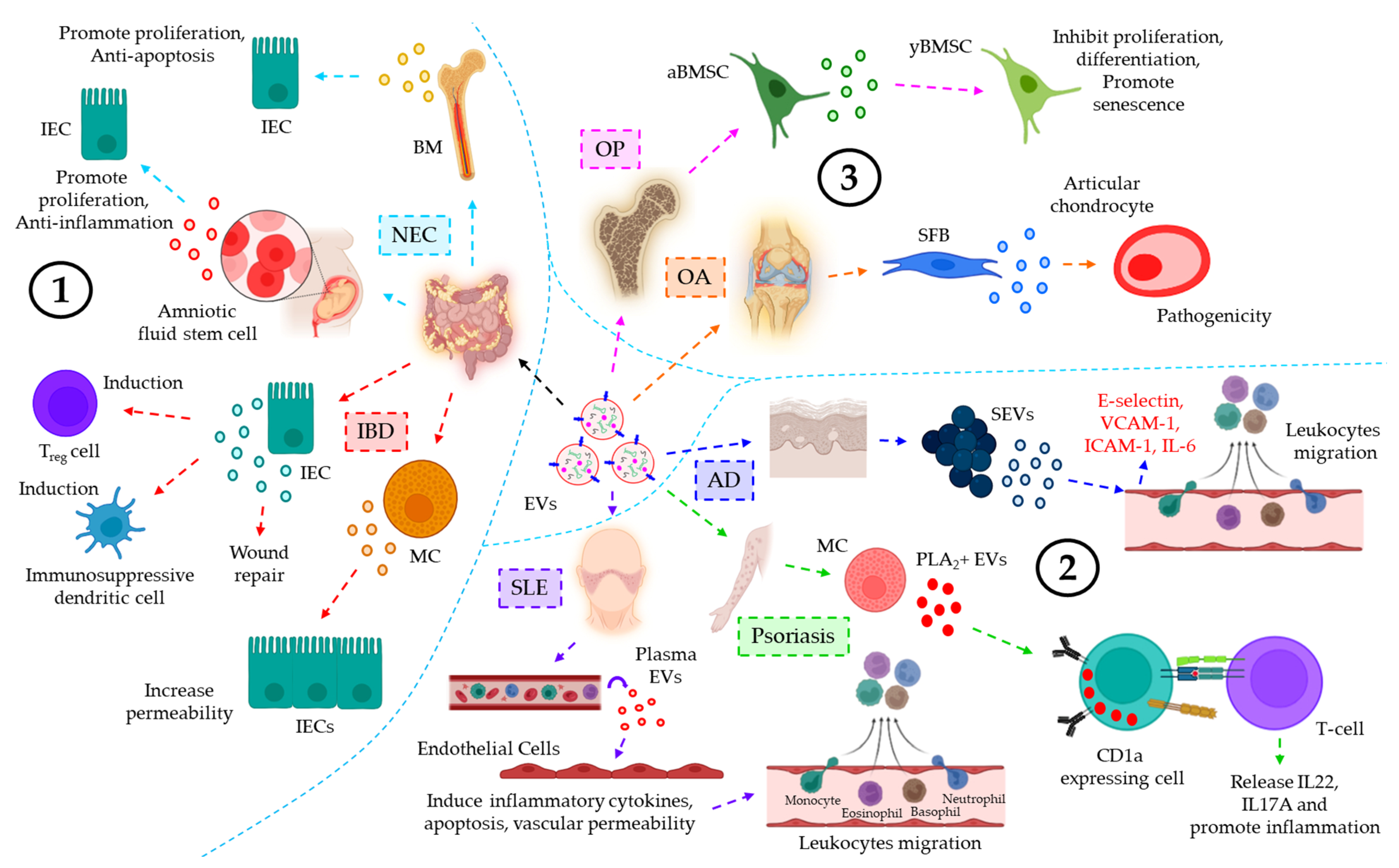
NECBreast milkBM-EVs protect IEC against H/R-induced apoptosis and loss of proliferation[149]
Amniotic fluidAFSC-EVs promote intestinal epithelial proliferation and anti-inflammation, leading to epithelial regeneration to help intestinal recovery from NEC[150]
IBDIntestinal luminal fluidTGF-β1+ EVs from IECs under physiological conditions induce Treg and immunosuppressive dendritic cells, leading to the downregulation of IBD severity[152]
Intestinal mucosamiR-223+ EVs from MCs target CLDN8 in the IECs, resulting in the loss of intestinal epithelial tight junctions and increased epithelial permeability[153]
SerumANXA1+ EVs from injury induced IECs help in the activation of the wound repair process[154]
Integumentary inflammatory diseases
SLEPlasmaSLE plasma-EVs promote endothelial release of pro-inflammatory cytokines, endothelial apoptosis, and increased vascular permeability, contributing to secondary tissue leukocyte infiltration[155]
PsoriasisPlasmaIFN-α-triggered mast cell-derived cytoplasmic PLA2+ EVs promote neo-lipid antigen presentation by CD1a+ cells and their concomitant recognition by CD1a-reactive T-cells, leading to IL22 and IL17A release and skin inflammation[156]
ADPlasmaSEVs trigger DMECs to induce the expression of E-selectin, VCAM-1, and ICAM-1 as well as IL-6 release to promote endothelial adhesion and subsequent transmigration of leukocytes, leading to AD progression[157]
Musculoskeletal inflammatory diseases
OPBMIFaBMSCs, under oxidative stress, release miR-183-5p-laden EVs which target Hmox1 in yBMSCs, thereby leading to the inhibition of proliferation and osteogenic differentiation as well as senescence induction of yBMSCs[158]
OA-IL-1β-stimulated SFB-derived EVs promote MMP-13 and ADAMTS-5 expression while inhibiting COL2A1 and ACAN expression in articular chondrocytes, leading to OA pathology[159]
Urinary inflammatory diseases
AKIPlasma and UrineHypoxia or I/R-induced injured TECs release miR-150-loaded EVs which trigger profibrotic manifestations in renal fibroblasts[171]
IgANUrineCCL2 mRNA expression in urinary EVs of IgAN is significantly higher as compared to controls, which is correlated with tubular interstitial inflammation and C3 deposition, reflecting renal injury and impaired renal functions[161]
Reproductive system inflammatory diseases
Pregnancy disordersPlasmaMaternal macrophage derived EVs induce the release of pro-inflammatory cytokines from placental trophoblasts, contributing to maternal inflammatory responses to protect the fetus[162]
Amniotic fluidPlacental trophoblast derived EVs, via the transfer of C19MC, prevent virus replication in non-placental cells, leading to embryonic protection against viral infections[163]
Inflammatory diseases of the endocrine system
ObesityAdipose tissue and PlasmaObese adipose tissue or plasma EVs show a significant down-regulation of miR-141-3p expression which contributes to glucose intolerance and insulin resistance[164,165]
SerumBrown adipocyte-derived miR-99b-laden EVs target FGF21 in the liver, leading to metabolic dysfunctions such as glucose intolerance[166]
SerumATM-EVs are highly expressed with miR-155, which targets PPARγ in adipocytes, myocytes, and primary hepatocytes, leading to glucose intolerance and insulin resistance[167]
EVs of the lymphatic system in inflammatory diseases
AtherosclerosisLymphLymph-derived EVs in atherosclerotic conditions influence lymphatic dysfunction and associated inflammatory disease progression[168]
RALymphIn RA, prolonged inflammation-induced vascular leakage promotes the egress of platelet-derived EVs in the lymphatic system, contributing to the pathogenesis of RA[169]
Abbreviations: MS, multiple sclerosis; CSF, cerebrospinal fluid; BBB, blood–brain barrier; CNS, central nervous system; AD, Alzheimer’s disease; PD, Parkinson’s disease; Aβ, β-amyloid; CJD, Creutzfeldt–Jakob disease; PrPSc, transmissible prion proteins; PML, progressive multifocal leukoencephalopathy; JCPyV, JC polyomavirus; CM, cerebral malaria; MSC, mesenchymal stem cell; MCAO, middle cerebral artery occlusion; SCI, spinal cord injury; NOX2, NADPH oxidase 2; PTEN, phosphatase and tensin homolog; PI3K, phosphatidylinositol 3-kinase; TBI, traumatic brain injury; PDE4B, phosphodiesterase 4B; mTOR, the mammalian target of rapamycin; ICAM-1, intercellular adhesion molecule 1; ox-LDL, oxidized low-density lipoprotein; MI, myocardial infarction; HF, heart failure; ADAM, a disintegrin and metalloproteinase; ALI, acute lung injury; ARDS, acute respiratory distress syndrome; BALF, bronchoalveolar lavage fluid; COPD, chronic obstructive pulmonary disease; PH, pulmonary hypertension; PASMC, pulmonary arterial smooth muscle cell; PAEC, pulmonary arterial endothelial cell; ILF, idiopathic lung fibrosis; WNT5A, Wnt family member 5A; NEC, necrotizing enterocolitis; BM, bone marrow; IEC, intestinal epithelial cell; H/R, hypoxia/reoxygenation; AFSC, amniotic fluid stem cell; IBD, inflammatory bowel disease; TGF-β1, transforming growth factor β1; Treg cell, regulatory T-cell; MC, mast cell; CLDN8, claudin 8; ANXA1, annexin A1; SLE, systemic lupus erythematosus; IFN, interferon; PLA2, phospholipase A2; CD, cluster of differentiation; IL, interleukin; AD, atopic dermatitis; SEV, Staphylococcus aureus-derived EV; DMEC, dermal microvascular endothelial cell; VCAM-1, vascular cell adhesion molecule 1; OP, osteoporosis; BMIF, bone marrow interstitial fluid; aBMSC, aged BM stromal cell; yBMSC, young BMSC; Hmox1, heme oxygenase-1; OA, osteoarthritis; SFB, synovial fibroblast; MMP, matrix metalloproteinase; ADAMTS-5, ADAM metalloproteinase with thrombospondin motifs 5; COL2A1, collagen type II α1; ACAN, aggrecan; AKI, acute kidney injury; I/R, ischemia-reperfusion; TEC, tubular epithelial cell; IgAN, IgA neuropathy; CCL2, (C-C motif) ligand 2; C3, complement component 3; C19MC, chromosome 19 miRNA cluster; FGF21, fibroblast growth factor 21; ATM, adipocyte tissue macrophage; PPARγ, peroxisome proliferator activated receptor γ; RA, rheumatoid arthritis.
The role of EVs in coagulation-associated inflammatory diseases. Blood coagulation is a tightly regulated biological process which prevents excessive bleeding when a blood vessel is injured [172]. Blood coagulation and inflammation are intrinsically related; the activation of one process often leads to the activation of the other [111,112,113]. Vessel injury results in the outburst of thrombin, the central key molecule of the coagulation system, which acts on vascular endothelium to induce the release of pro-inflammatory cytokines [173,174]. Inflammation, on the other hand, often leads to endothelial barrier leakage which further enhances the coagulation process [175,176,177]. A recent study delineates the active involvement of Grb2-associated binder 2 (Gab2) in IL-1β-induced exocytosis of P-selectin and von Willebrand factor (vWF) as well as expression of tissue factor (TF) and VCAM-1, which together often results in the pro-coagulant functions [178]. In the past two decades, EVs have slowly emerged as a key molecule which not only influence the coagulation process but also influence both pro- and anti-inflammatory responses. In most of the cases, the EVs are believed to enhance the coagulation process, due to the presence of pro-coagulant protein TF [179] and negatively charged phospholipid PS [180] on the surface. Although EVs’ TF directly activates the coagulation cascade, PS-dependent activation of the coagulation system requires the assembly of factor VIIIa, IXa, and X (tenase complex) as well as factors Va, Xa, and thrombin (prothrombinase complex) in the presence of Ca2+ [181]. In contrast to the above, EVs also exert anticoagulant properties. For example, EVs released from the endothelial cells upon exposure with anticoagulant protease activated protein C (APC) turn out to be anti-coagulant [182]; however, in this case, the anticoagulant activity is largely due to the bound APC on the EVs’ surface [182]. Similar to coagulation, EVs also confer both pro- and anti-inflammatory responses in the context of clotting. The pro-inflammatory effects of the platelet-derived EVs are well-established, which prevent their clinical use as a pro-coagulant factor against hypo-coagulable conditions, such as trauma-induced coagulopathy (TIC) [183,184]. On the other hand, Njock et al. demonstrated that EVs released from unperturbed endothelium confer anti-inflammatory responses via the enrichment of anti-inflammatory miRNAs [185]. Despite the advancement of EV research, it was still unknown how EVs generated from the unperturbed vascular endothelium upon exposure of coagulation proteases contribute to the inflammatory responses, until the recent intriguing discovery by Das et al. who demonstrated, for the first time, that FVIIa-triggered endothelial EVs (EEVs) suppress monocytic inflammation against bacterial-LPS-induced sepsis (Figure 7) [8,19]. The study delineates the fact that FVIIa triggers the endothelial release of EVs by endothelial cell protein C receptor (EPCR)-driven activation of protease activated receptor 1 (PAR1) both in vitro and in vivo [19]. Unlike FVIIa-TF-PAR2 signaling, observed predominantly in cancer [186], FVIIa-induced EV generation from unperturbed endothelial cells is shown to be independent of both TF and PAR2 [19]. FVIIa-EEVs are enriched with anti-inflammatory miRNAs, the predominant being miR-10a, and the transfer of EVs-miR-10a to monocytes confers anti-inflammatory responses against LPS-induced sepsis [8,19]. Furthermore, FVIIa infusion into hemophilia patients increases the level of plasma EEVs enriched with miR-10a [4], and these EEVs also impart miR-10a-dependent anti-inflammatory responses [4].
EVs in inflammation therapy. EVs are known for transporting bioactive cargoes, such as proteins, nucleic acids, lipids, etc., between the cells, thereby playing an important role in cell–cell communication. EVs could be engineered at the surface and bestowed with target-specific moiety, rendering the target-specific therapeutic applications of the EVs in various inflammation-associated diseases. For example, therapeutic drugs entrapped within the EVs often reach the target-specific sites with higher efficacy through EVs. The present section provides a brief overview of how EVs could be used as a potential therapeutic agent in the context of various inflammatory diseases (Table 6).
Therapeutic roles of EVs in neuroinflammatory diseases. MSC-derived EVs are often used as a promising therapeutic mode in various neuroinflammatory diseases. For example, the activation of infiltrating leukocytes, astrocytes, and microglial cells has been shown to be attenuated upon intra-arterial injection of MSC-EVs in an ischemic stroke-induced rat model [187]. Moreover, T-lymphoblast-derived EVs, packaged with a neuroprotective drug, curcumin, are efficiently taken up by the inflamed brain microglial cells, thereby triggering apoptosis, when administered intranasally in a LPS-induced brain inflammation murine model [188]. Furthermore, in a cocaine-induced brain inflammation murine model, DC-secreted EVs, further engineered to over-express miR-124, are shown to attenuate microglial activation and the expression of pro-inflammatory molecules, TLR4, MYD88, STAT3, and NF-ĸB p65 [189].
Therapeutic roles of EVs in cardiovascular inflammatory diseases. EVs often exert beneficial roles in cardiovascular inflammatory diseases in the context of post-MI cardiac repair processes. For example, in an I/R-induced cardiac inflammation rat model, cardiosphere-derived cell (CDC)-derived EVs, laden with Y-RNA fragments, promote the release of IL-10 in the infarcted myocardium, thereby contributing to post-MI cardiac repair [190]. Moreover, DC-EVs are shown to activate CD4 + T-cells, leading to the perturbation of pro-inflammatory cytokines’ release and improvement of cardiac functions in a cardiac MI mice model [191]. Again, adipose-derived stem cell (ADSC)-derived EVs are found to over-express miR-93-5p, which down-regulates autophagy and pro-inflammation by targeting Atg7 and TLR4, respectively, thereby showing protection against infarction-induced myocardial damage in an ischemia-induced cardiac injury rat model [192].
EVs’ therapeutic roles in respiratory inflammatory diseases. MSC-EVs often show promising effects against respiratory inflammatory diseases, which render them to be considered an effective therapeutic entity. For example, MSC-EVs, via delivering miR-21-5p, protect the epithelial cells from reactive oxygen species (ROS)-induced apoptotic damage in asthma and COPD [193,194]. Moreover, MSC-EVs also trigger the polarization of alveolar macrophages into M2 phenotypes, thereby inducing the release of anti-inflammatory cytokines and promoting wound healing [195]. MSC-EVs also produce promising therapeutic outcomes in ALI/ARDS by inhibiting the proliferation and differentiation of B-cells as well as promoting the differentiation of TH-cells to Treg cells, leading to the down-regulation of pro-inflammatory cytokines TNF-α, IL-1β, and IFN-γ and up-regulation of anti-inflammatory cytokines PEG2, IL-10, and TGF-β [196].
Therapeutic roles of EVs in integumentary inflammatory diseases. Research indicates that EVs also confer therapeutic potential in several integumentary inflammatory diseases. For example, human keratinocyte (HK)-derived EVs are shown to carry miR-21, which not only promotes angiogenesis but also facilitates fibroblast functions, leading to skin wound healing in diabetic rats [197]. Murine intraperitoneal injection of the EVs, engineered with a super repressor of IĸB (srIĸB, the dominant active form) by the optogenetic method, are shown to inhibit the NF-ĸB pro-inflammatory signaling pathway in liver and spleen neutrophils and monocytes, leading to the attenuation of sepsis-induced inflammation and associated mortality [198].
EVs’ therapeutic roles in autoimmune inflammatory diseases. EVs also play their therapeutic roles in autoimmune inflammatory disorders. For example, EVs from IL-10-treated DCs are associated with the inhibition of arthritis onset as well as the already-established severity [199]. Furthermore, in a collagen-induced arthritis (CIA) model of RA, MSC-EVs exert anti-inflammatory effects on B- and T-lymphocytes, thus demonstrating the therapeutic potential of MSC-EVs in RA [200].
Due to commendable success in preclinical studies, EVs have now mostly reached phase I and phase II clinical trials as briefly mentioned in Table 7 [201].
Artificial EVs and disease therapy. So far, the present review discussed how EVs influence different inflammation-associated diseases and their potential use in therapeutic purposes. The natural tropism [202], fine biodistribution and less clearance from the system [203], ability to transfer bioactive cargoes efficiently [39], biocompatibility [204], and most importantly, the extraordinary capacity to cross blood–brain barriers (BBB) [205] render EVs to be an excellent means in various disease therapies. However, the natural EV-based therapies also have their limitations: (1) the heterogeneity of EVs makes EV isolation and purification difficult [206]; (2) although less, EVs still show immunogenic responses [207]; and (3) as this review already discussed, most of the conventional methods of EV isolation are time-consuming, with the yield and purity always remaining a concern. However, in the past decade, the concept of bioengineered EVs has evolved, which includes the isolation of natural EVs followed by some modifications to generate the biomimetic nanocarriers which are not only being used as an efficient drug delivery system but also improved the target specificity significantly [208,209,210]. Recently, two unique mechanisms have evolved. The first one, termed as top-down method, employs the disruption of membranes into small fragments which reassembles automatically to form nano- or microvesicles [211,212]. In the second approach, molecular components such as synthetic lipids are used to generate the artificial lipid bilayers which mimic the EVs [213]. These recently developed artificial EVs have several advantages over the natural EVs [214] which include the fact that (1) the size of the EVs can be easily controlled, and in this sense these EVs reduce the heterogeneity unlike natural EVs, (2) the ingredients, such as synthetic lipids, are commercially available, (3) more standardized and high scale production can be achieved, and (4) these EVs are safe to be used and are highly reproducible. However, additional bioengineering on these artificial EVs could improve the target specificity which is essential in the delivery of EV-based therapeutic drugs against various diseases, including inflammation-associated disorders.

2. Conclusions and Future Directions

There is much evidence suggesting that submicron-sized EVs are crucial and have an immense therapeutic approach because of their biocompatibility, at the experimental and clinical level, against several inflammation-associated diseases such as neurological disorders, cardiovascular anomalies, respiratory syndromes, integumentary disorders, and autoimmune diseases and in regenerative medicine. EVs play a vital role in innate and adaptive immunity, including inflammation, as they carry autoantigens, cytokines, lipid mediators, tissue-degrading enzymes, etc. As a non-cellular membrane structure, EVs can regulate immune cell activity, suppress excessive inflammation, and promote immune tolerance. This suggests that EV-based therapies may hold promise for the treatment of inflammatory diseases, providing a targeted and personalized approach. EVs are safe, are efficient, and have many advantages in clinical application due to low immunogenicity, flexibility in engineering the membrane or cargo, and potential for tissue-specific targeting, long half-life, in vivo stability, and high delivery efficiency. Now, several EV-based treatments are being studied in phase I and II clinical trials. EVs’ research is rapidly advancing, and several directions are being explored in the future. Although EVs hold great promise for clinical applications, the toxicity, long-term safety, and immunoregulatory functions of EVs in the human body need to be further evaluated. Additionally, there are several challenges to overcome, including standardization of isolation and characterization, cost-effective production methods with consistent quality, developing robust cargo loading strategies and improving cargo stability within the EVs and biodistribution, targeted delivery and tissue specificity, and establishing clear regulatory guidelines and frameworks to ensure safety, quality, and efficacy for gaining regulatory approvals. To address these challenges, researchers, clinicians, regulators, and industry partners will have to collaborate. In order to overcome these hurdles and unlock the full therapeutic potential of EVs in clinical settings, further research and clinical trials are required to fully explore the therapeutic efficacy and safety of EV-based therapies. Nevertheless, the potential impact of EVs in revolutionizing disease treatment is undeniable. However, if the aforementioned difficulties are addressed, EVs could be used as an excellent mode of delivery system in the therapeutic implications of various inflammation-associated diseases. In addition to these, the present review also delineates how EVs play their part in promoting inflammation-associated diseases. In these conditions, the therapeutic approach includes targeting the EVs. Numerous studies indicate that intracellular signaling mechanisms, leading to the actomyosin reorganization, play a critical role in the biogenesis of EVs. Therefore, targeting EVs’ formation could be a potential therapeutic means to limit EV-associated promotion of inflammatory diseases.

Author Contributions

K.D. contributed to the conceptualization, literature search, experimental data collection, study design, critical review, and preparation of the final version of the manuscript. S.P. contributed to the literature search, data collection, and preparation of the manuscript. T.M. contributed to the literature search and experimental data collection and wrote the initial draft of the manuscript. A.G. contributed to the literature search, data collection, and preparation of the manuscript. A.S. contributed to the literature search and manuscript editing. P.S. contributed to the literature search and experimental data collection. S.G. contributed to the study design, critical review, and preparation of the final version of the manuscript. S.K. contributed to literature search, critical review, and preparation of the revised version of the manuscript. D.P. contributed to the study design, critical review, and preparation of the final version of the manuscript. All authors have read and agreed to the published version of the manuscript.

Funding

This research received no external funding.

Institutional Review Board Statement

Not applicable.

Informed Consent Statement

Not applicable.

Data Availability Statement

Not applicable.

Acknowledgments

All the images in the manuscript were created with BioRender.com. We acknowledge BioRender for providing us with the platform for the preparation of images. The authors thank Sara Rose for editing the manuscript for the English language.

Conflicts of Interest

The authors declare no conflict of interest.

Abbreviations

β-amyloid
aBMSCaged BM stromal cell
ACANaggrecan
ADAlzheimer’s disease
ADAMa disintegrin and metalloproteinase
ADAMTS-5ADAM metallopeptidase with thrombospondin type 1 motif 5
ADSCadipose-derived stem cell
AFSMamniotic fluid stem cell
AKIacute kidney injury
ALIacute lung injury
AlixALG-2-interacting protein X
ANXA1annexin A1
APCactivated protein C
AQP1aquaporin 1
ARDSacute respiratory distress syndrome
Atg7autophagy related 7
ATMadipose tissue macrophage
BALFbronchoalveolar lavage fluid
BBBblood brain barrier
BMbone marrow
BMIFBM interstitial fluid
C3complement component 3
CAFcancer-associated fibroblasts
CCL2chemokine (C-C motif) ligand 2
CDcluster of differentiation
CDCcardiosphere-derived cell
CJDCreutzfeldt-Jacob disease
CLDN8Claudin 8
CMcerebral malaria
C19MCchromosome 19 miRNA cluster
CNScentral nervous system
COL2A1collagen type II alpha 1 chain
COPDchronic obstructive pulmonary disease
CSFcerebrospinal fluid
CIAcollagen-induced arthritis
COVID-19coronavirus 19
DLDdeterministic later displacement
DMECdermal microvascular endothelial cell
DNAdeoxyribonucleic acid
EEVendothelial EV
EMTepithelial to mesenchymal transition
EPCRendothelial cell protein C receptor
ESCRTendosomal sorting complexes required for transport
EVextracellular vesicle
FGF21fibroblast growth factor 21
FPLCfast protein liquid chromatography
FVIIaactivated factor VII
Gab2Grb2-associated binder 2
Gas6growth arrest-specific protein 6
GBMglioblastoma multiforme
GRP78glucose-regulated protein 78
HFheart failure
HFFhuman follicular fluid
HKhuman keratinocyte
Hmox1heme oxygenase 1
HPLChigh performance liquid chromatography
H/Rhypoxia/reoxygenation
HSC70heat shock cognate 70 kDa protein
HSPheat shock protein
ICAM-1intercellular adhesion molecule 1
IBDinflammatory bowel disease
IECintestinal epithelial cell
IFNinterferon
IgANimmunoglobulin A nephropathy
ILinterleukin
ILFidiopathic lung fibrosis
I/Rischemia-reperfusion
ISEVinternational society for extracellular vesicles
JCPyVJC polyomavirus
LC3microtubule-associated proteins 1A/1B light chain 3B
LPSlipopolysaccharide
MCmast cell
MCAOmiddle cerebral artery occlusion
MERTRMer receptor tyrosine kinase
MImyocardial infarction
miRNAmicroRNA
MMPmatrix metalloproteinase
MISEVminimal information for studies of extracellular vesicles
MPmicroparticle
MSmultiple sclerosis
MSCmesenchymal stem cell
mTORmammalian target of rapamycin
MVmicrovesicle
MVBmultivesicular body
MYD88myeloid differentiation primary response 88
NECnecrotizing enterocolitis
NF-ĸBnuclear factor kappa-light-chain-enhancer of activated B-cells
NOX2reduced nicotinamide adenine dinucleotide phosphate (NADPH) oxidase 2
NPCnasopharyngeal carcinoma
OAosteoarthritis
OPosteoporosis
ox-LDLoxidized low density lipoprotein
PAECpulmonary arterial endothelial cell
PAHpulmonary arterial hypertension
PARprotease-activated receptor
PASMCpulmonary arterial smooth muscle cell
PBMCperipheral blood mononuclear cell
PCOSpolycystic ovary syndrome
PDParkinson’s disease
PDE4Bphosphodiesterase 4B
PEGpolyethylene glycol
PHpulmonary hypertension
PI3Kphosphoinositide 3-kinase
PLA2phospholipase A2
PMLprogressive multifocal leukoencephalopathy
PPARγperoxisome proliferator-activated receptor gamma
PrPprion protein
PrPScmisfolded and transmissible form of PrP
PSphosphatidylserine
PTENphosphatase and tensin homolog
RArheumatoid arthritis
ROSreactive oxygen species
RNAribonucleic acid
SCIspinal cord injury
SECsize exclusion chromatography
SEVStaphylococcus aureus-derived EV
SFBsynovial fibroblast
SLEsystemic lupus erythematosus
SNAREsoluble N-ethylmaleimide-sensitive factor activating protein receptor
srIĸBsuper repressor NF-kappa-B inhibitor alpha (I-kappa-B)
STAT3signal transducer and activator of transcription 3
TAK1TGF-β-activated kinase 1
TBItraumatic brain injury
TECtubular endothelial cell
TFtissue factor
TGF-β1transforming growth factor β1
THhelper T-cell
TICtrauma-induced coagulopathy
TLR4toll-like receptor 4
TMEtumor microenvironment
TNBCtriple-negative breast cancer
TNFtumor necrosis factor
Tregregulatory T-cell
TSG101tumor susceptibility gene 101
UMEuterine microenvironment
UTIurinary tract infection
VCAM-1vascular cell adhesion molecule 1
vWFvon Willebrand factor
WNT5AWnt family member 5A
yBMSCyoung BMSC

References

  1. Combarnous, Y.; Nguyen, T.M.D. Cell Communications among Microorganisms, Plants, and Animals: Origin, Evolution, and Interplays. Int. J. Mol. Sci. 2020, 21, 8052. [Google Scholar] [CrossRef] [PubMed]
  2. Das, K.; Prasad, R.; Singh, A.; Bhattacharya, A.; Roy, A.; Mallik, S.; Mukherjee, A.; Sen, P. Protease-activated receptor 2 promotes actomyosin dependent transforming microvesicles generation from human breast cancer. Mol. Carcinog. 2018, 57, 1707–1722. [Google Scholar] [CrossRef] [PubMed]
  3. Das, K.; Prasad, R.; Roy, S.; Mukherjee, A.; Sen, P. The Protease Activated Receptor2 Promotes Rab5a Mediated Generation of Pro-metastatic Microvesicles. Sci. Rep. 2018, 8, 7357. [Google Scholar] [CrossRef]
  4. Das, K.; Pendurthi, U.R.; Manco-Johnson, M.; Martin, E.J.; Brophy, D.F.; Rao, L.V.M. Factor VIIa treatment increases circulating extracellular vesicles in hemophilia patients: Implications for the therapeutic hemostatic effect of FVIIa. J. Thromb. Haemost. 2022, 20, 1928–1933. [Google Scholar] [CrossRef] [PubMed]
  5. Zaborowski, M.P.; Balaj, L.; Breakefield, X.O.; Lai, C.P. Extracellular Vesicles: Composition, Biological Relevance, and Methods of Study. Bioscience 2015, 65, 783–797. [Google Scholar] [CrossRef] [PubMed] [Green Version]
  6. Yanez-Mo, M.; Siljander, P.R.-M.; Andreu, Z.; Zavec, A.B.; Borras, F.E.; Buzas, E.I.; Buzas, K.; Casal, E.; Cappello, F.; Carvalho, J.; et al. Biological properties of extracellular vesicles and their physiological functions. J. Extracell. Vesicles 2015, 4, 27066. [Google Scholar] [CrossRef] [Green Version]
  7. Van Niel, G.; D’Angelo, G.; Raposo, G. Shedding light on the cell biology of extracellular vesicles. Nat. Rev. Mol. Cell Biol. 2018, 19, 213–228. [Google Scholar] [CrossRef]
  8. Das, K.; Keshava, S.; Pendurthi, U.R.; Rao, L.V.M. Factor VIIa suppresses inflammation and barrier disruption through the release of EEVs and transfer of microRNA 10a. Blood 2022, 139, 118–133. [Google Scholar] [CrossRef]
  9. Das, K.; Paul, S.; Singh, A.; Ghosh, A.; Roy, A.; Ansari, S.A.; Prasad, R.; Mukherjee, A.; Sen, P. Triple-negative breast cancer-derived microvesicles transfer microRNA221 to the recipient cells and thereby promote epithelial-to-mesenchymal transition. J. Biol. Chem. 2019, 294, 13681–13696. [Google Scholar] [CrossRef]
  10. Das, K.; Rao, L.V.M. The Role of microRNAs in Inflammation. Int. J. Mol. Sci. 2022, 23, 15479. [Google Scholar] [CrossRef]
  11. Parashar, D.; Singh, A.; Gupta, S.; Sharma, A.; Sharma, M.K.; Roy, K.K.; Chauhan, S.C.; Kashyap, V.K. Emerging Roles and Potential Applications of Non-Coding RNAs in Cervical Cancer. Genes 2022, 13, 1254. [Google Scholar] [CrossRef]
  12. Parashar, D.; Geethadevi, A.; Aure, M.R.; Mishra, J.; George, J.; Chen, C.; Mishra, M.K.; Tahiri, A.; Zhao, W.; Nair, B.; et al. miRNA551b-3p Activates an Oncostatin Signaling Module for the Progression of Triple-Negative Breast Cancer. Cell Rep. 2019, 29, 4389–4406.e4310. [Google Scholar] [CrossRef] [Green Version]
  13. Mulcahy, L.A.; Pink, R.C.; Carter, D.R. Routes and mechanisms of extracellular vesicle uptake. J. Extracell. Vesicles 2014, 3, 24641. [Google Scholar] [CrossRef] [PubMed] [Green Version]
  14. Raposo, G.; Stoorvogel, W. Extracellular vesicles: Exosomes, microvesicles, and friends. J. Cell Biol. 2013, 200, 373–383. [Google Scholar] [CrossRef] [PubMed]
  15. Neven, K.Y.; Nawrot, T.S.; Bollati, V. Extracellular Vesicles: How the External and Internal Environment Can Shape Cell-To-Cell Communication. Curr. Environ. Health Rep. 2017, 4, 30–37. [Google Scholar] [CrossRef] [PubMed]
  16. Pulliero, A.; Pergoli, L.; Maestra, S.L.A.; Micale, R.T.; Camoirano, C.; Bollati, V.; Izzotti, A.; Flora, S.D.E. Extracellular vesicles in biological fluids. A biomarker of exposure to cigarette smoke and treatment with chemopreventive drugs. J. Prev. Med. Hyg. 2019, 60, E327–E336. [Google Scholar] [CrossRef]
  17. Admyre, C.; Johansson, S.M.; Qazi, K.R.; Filen, J.-J.; Lahesmaa, R.; Norman, M.; Neve, E.P.A.; Scheynius, A.; Gabrielsson, S. Exosomes with immune modulatory features are present in human breast milk. J. Immunol. 2007, 179, 1969–1978. [Google Scholar] [CrossRef] [Green Version]
  18. Carnino, J.M.; Lee, H.; Jin, Y. Isolation and characterization of extracellular vesicles from Broncho-alveolar lavage fluid: A review and comparison of different methods. Respir. Res. 2019, 20, 240. [Google Scholar] [CrossRef] [Green Version]
  19. Das, K.; Keshava, S.; Ansari, S.A.; Kondreddy, V.; Esmon, C.T.; Griffin, J.H.; Pendurthi, U.R.; Rao, L.V.M. Factor VIIa induces extracellular vesicles from the endothelium: A potential mechanism for its hemostatic effect. Blood 2021, 137, 3428–3442. [Google Scholar] [CrossRef]
  20. Ratajczak, M.Z.; Ratajczak, J. Extracellular microvesicles/exosomes: Discovery, disbelief, acceptance, and the future? Leukemia 2020, 34, 3126–3135. [Google Scholar] [CrossRef]
  21. Holliday, L.S.; Faria, L.P.; Rody, W.J., Jr. Actin and Actin-Associated Proteins in Extracellular Vesicles Shed by Osteoclasts. Int. J. Mol. Sci. 2019, 21, 158. [Google Scholar] [CrossRef] [PubMed] [Green Version]
  22. Muralidharan-Chari, V.; Clancy, J.; Plou, C.; Romao, M.; Chavrier, P.; Raposo, G.; D’Souza-Schorey, C. ARF6-regulated shedding of tumor cell-derived plasma membrane microvesicles. Curr. Biol. 2009, 19, 1875–1885. [Google Scholar] [CrossRef] [PubMed] [Green Version]
  23. Cai, H.; Reinisch, K.; Ferro-Novick, S. Coats, tethers, Rabs, and SNAREs work together to mediate the intracellular destination of a transport vesicle. Dev. Cell 2007, 12, 671–682. [Google Scholar] [CrossRef] [Green Version]
  24. Escola, J.M.; Kleijmeer, M.J.; Stoorvogel, W.; Griffith, J.M.; Yoshie, O.; Geuze, H.J. Selective enrichment of tetraspan proteins on the internal vesicles of multivesicular endosomes and on exosomes secreted by human B-lymphocytes. J. Biol. Chem. 1998, 273, 20121–20127. [Google Scholar] [CrossRef] [Green Version]
  25. Das, K.; Mukherjee, T.; Shankar, P. The Role of Extracellular Vesicles in the Pathogenesis of Hematological Malignancies: Interaction with Tumor Microenvironment; a Potential Biomarker and Targeted Therapy. Biomolecules 2023, 13, 897. [Google Scholar]
  26. Kowal, J.; Tkach, M.; Thery, C. Biogenesis and secretion of exosomes. Curr. Opin. Cell Biol. 2014, 29, 116–125. [Google Scholar] [CrossRef] [Green Version]
  27. Geminard, C.; De Gassart, A.; Blanc, L.; Vidal, M. Degradation of AP2 during reticulocyte maturation enhances binding of hsc70 and Alix to a common site on TFR for sorting into exosomes. Traffic 2004, 5, 181–193. [Google Scholar] [CrossRef] [PubMed]
  28. van Niel, G.; Porto-Carreiro, I.; Simoes, S.; Raposo, G. Exosomes: A common pathway for a specialized function. J. Biochem. 2006, 140, 13–21. [Google Scholar] [CrossRef]
  29. Trajkovic, K.; Hsu, C.; Chiantia, S.; Rajendran, L.; Wenzel, D.; Wieland, F.; Schwille, P.; Brügger, B.; Simons, M. Ceramide triggers budding of exosome vesicles into multivesicular endosomes. Science 2008, 319, 1244–1247. [Google Scholar] [CrossRef]
  30. Glick, D.; Barth, S.; Macleod, K.F. Autophagy: Cellular and molecular mechanisms. J. Pathol. 2010, 221, 3–12. [Google Scholar] [CrossRef] [Green Version]
  31. Fader, C.M.; Colombo, M.I. Autophagy and multivesicular bodies: Two closely related partners. Cell Death Differ. 2009, 16, 70–78. [Google Scholar] [CrossRef] [PubMed] [Green Version]
  32. Xu, J.; Camfield, R.; Gorski, S.M. The interplay between exosomes and autophagy-partners in crime. J. Cell Sci. 2018, 131, jcs215210. [Google Scholar] [CrossRef] [PubMed] [Green Version]
  33. Borges, F.T.; Reis, L.A.; Schor, N. Extracellular vesicles: Structure, function, and potential clinical uses in renal diseases. Braz. J. Med. Biol. Res. 2013, 46, 824–830. [Google Scholar] [CrossRef] [PubMed] [Green Version]
  34. Wickman, G.; Julian, L.; Olson, M.F. How apoptotic cells aid in the removal of their own cold dead bodies. Cell Death Differ. 2012, 19, 735–742. [Google Scholar] [CrossRef] [PubMed] [Green Version]
  35. Thery, C.; Boussac, M.; Véron, P.; Ricciardi-Castagnoli, P.; Raposo, G.; Garin, J.; Amigorena, S. Proteomic analysis of dendritic cell-derived exosomes: A secreted subcellular compartment distinct from apoptotic vesicles. J. Immunol. 2001, 166, 7309–7318. [Google Scholar] [CrossRef]
  36. Escrevente, C.; Keller, S.; Altevogt, P.; Costa, J. Interaction and uptake of exosomes by ovarian cancer cells. BMC Cancer 2011, 11, 108. [Google Scholar] [CrossRef] [Green Version]
  37. Kerr, J.F.; Wyllie, A.H.; Currie, A.R. Apoptosis: A basic biological phenomenon with wide-ranging implications in tissue kinetics. Br. J. Cancer 1972, 26, 239–257. [Google Scholar] [CrossRef] [Green Version]
  38. Akbar, A.; Malekian, F.; Baghban, N.; Kodam, S.P.; Ullah, M. Methodologies to Isolate and Purify Clinical Grade Extracellular Vesicles for Medical Applications. Cells 2022, 11, 186. [Google Scholar] [CrossRef]
  39. Doyle, L.M.; Wang, M.Z. Overview of Extracellular Vesicles, Their Origin, Composition, Purpose, and Methods for Exosome Isolation and Analysis. Cells 2019, 8, 727. [Google Scholar] [CrossRef] [Green Version]
  40. Pillalamarri, N.; Abdullah; Ren, G.; Khan, L.; Ullah, A.; Jonnakuti, S.; Ullah, M. Exploring the utility of extracellular vesicles in ameliorating viral infection-associated inflammation, cytokine storm and tissue damage. Transl. Oncol. 2021, 14, 101095. [Google Scholar] [CrossRef]
  41. Muller, L.; Hong, C.S.; Stolz, D.B.; Watkins, S.C.; Whiteside, T.L. Isolation of biologically-active exosomes from human plasma. J. Immunol. Methods 2014, 411, 55–65. [Google Scholar] [CrossRef]
  42. Baranyai, T.; Herczeg, K.; Onódi, Z.; Voszka, I.; Módos, K.; Marton, N.; Nagy, G.; Mäger, I.; Wood, M.J.; El Andaloussi, S.; et al. Isolation of Exosomes from Blood Plasma: Qualitative and Quantitative Comparison of Ultracentrifugation and Size Exclusion Chromatography Methods. PLoS ONE 2015, 10, e0145686. [Google Scholar] [CrossRef] [Green Version]
  43. Ullah, M.; Kodam, S.P.; Mu, Q.; Akbar, A. Microbubbles versus Extracellular Vesicles as Therapeutic Cargo for Targeting Drug Delivery. ACS Nano 2021, 15, 3612–3620. [Google Scholar] [CrossRef] [PubMed]
  44. Soares Martins, T.; Catita, J.; Martins Rosa, I.; AB da Cruz e Silva, O.; Henriques, A.G. Exosome isolation from distinct biofluids using precipitation and column-based approaches. PLoS ONE 2018, 13, e0198820. [Google Scholar] [CrossRef] [Green Version]
  45. Serrano-Pertierra, E.; Oliveira-Rodríguez, M.; Rivas, M.; Oliva, P.; Villafani, J.; Navarro, A. Blanco-López, M.C.; Cernuda-Morollón, E. Characterization of Plasma-Derived Extracellular Vesicles Isolated by Different Methods: A Comparison Study. Bioengineering 2019, 6, 8. [Google Scholar] [CrossRef] [PubMed] [Green Version]
  46. Zhao, Z.; Wijerathne, H.; Godwin, A.K.; Soper, S.A. Isolation and analysis methods of extracellular vesicles (EVs). Extracell. Vesicles Circ. Nucl. Acids 2021, 2, 80–103. [Google Scholar] [CrossRef]
  47. Livshits, M.A.; Khomyakova, E.; Evtushenko, E.G.; Lazarev, V.N.; Kulemin, N.A.; Semina, S.E.; Generozov, E.V.; Govorun, V.M. Isolation of exosomes by differential centrifugation: Theoretical analysis of a commonly used protocol. Sci. Rep. 2015, 5, 17319. [Google Scholar] [CrossRef] [Green Version]
  48. Zarovni, N.; Corrado, A.; Guazzi, P.; Zocco, D.; Lari, E.; Radano, G.; Muhhina, J.; Fondelli, C.; Gavrilova, J.; Chiesi, A. Integrated isolation and quantitative analysis of exosome shuttled proteins and nucleic acids using immunocapture approaches. Methods 2015, 87, 46–58. [Google Scholar] [CrossRef]
  49. Taylor, D.D.; Shah, S. Methods of isolating extracellular vesicles impact down-stream analyses of their cargoes. Methods 2015, 87, 3–10. [Google Scholar] [CrossRef]
  50. Salih, M.; Zietse, R.; Hoorn, E.J. Urinary extracellular vesicles and the kidney: Biomarkers and beyond. Am. J. Physiol. Renal Physiol. 2014, 306, F1251–F1259. [Google Scholar] [CrossRef] [Green Version]
  51. Van Deun, J.; Mestdagh, P.; Sormunen, R.; Cocquyt, V.; Vermaelen, K.; Vandesompele, J.; Bracke, M.; De Wever, O.; Hendrix, A. The impact of disparate isolation methods for extracellular vesicles on downstream RNA profiling. J. Extracell. Vesicles 2014, 3, 24858. [Google Scholar] [CrossRef] [Green Version]
  52. Rikkert, L.G.; Engelaer, M.; Hau, C.M.; Terstappen, L.W.M.M.; Nieuwland, R.; Coumans, F.A.W. Rate zonal centrifugation can partially separate platelets from platelet-derived vesicles. Res. Pract. Thromb. Haemost. 2020, 4, 1053–1059. [Google Scholar] [CrossRef] [PubMed]
  53. Ullah, A.; Mabood, N.; Maqbool, M.; Khan, L.; Khan, M.; Ullah, M. SAR-CoV-2 infection, emerging new variants and the role of activation induced cytidine deaminase (AID) in lasting immunity. Saudi Pharm. J. 2021, 29, 1181–1184. [Google Scholar] [CrossRef] [PubMed]
  54. Li, P.; Kaslan, M.; Lee, S.H.; Yao, J.; Gao, Z. Progress in Exosome Isolation Techniques. Theranostics 2017, 7, 789–804. [Google Scholar] [CrossRef]
  55. Kim, D.K.; Nishida, H.; An, S.Y.; Shetty, A.K.; Bartosh, T.J.; Prockop, D.J. Chromatographically isolated CD63+CD81+ extracellular vesicles from mesenchymal stromal cells rescue cognitive impairments after TBI. Proc. Natl. Acad. Sci. USA 2016, 113, 170–175. [Google Scholar] [CrossRef]
  56. Zeringer, E.; Barta, T.; Li, M.; Vlassov, A.V. Strategies for isolation of exosomes. Cold Spring Harb. Protoc. 2015, 2015, 319–323. [Google Scholar] [CrossRef] [Green Version]
  57. Batrakova, E.V.; Kim, M.S. Using exosomes, naturally-equipped nanocarriers, for drug delivery. J. Control. Release 2015, 219, 396–405. [Google Scholar] [CrossRef] [Green Version]
  58. Yang, D.; Zhang, W.; Zhang, H.; Zhang, F.; Chen, L.; Ma, L.; Larcher, L.M.; Chen, S.; Liu, N.; Zhao, Q.; et al. Progress, opportunity, and perspective on exosome isolation-efforts for efficient exosome-based theranostics. Theranostics 2020, 10, 3684–3707. [Google Scholar] [CrossRef] [PubMed]
  59. Lobb, R.J.; Becker, M.; Wen, S.W.; Wong, C.S.; Wiegmans, A.P.; Leimgruber, A.; Möller, A. Optimized exosome isolation protocol for cell culture supernatant and human plasma. J. Extracell. Vesicles 2015, 4, 27031. [Google Scholar] [CrossRef]
  60. Soda, N.; Rehm, B.H.A.; Sonar, P.; Nguyen, N.T.; Shiddiky, M.J.A. Advanced liquid biopsy technologies for circulating biomarker detection. J. Mater. Chem. B 2019, 7, 6670–6704. [Google Scholar] [CrossRef]
  61. Peterson, M.F.; Otoc, N.; Sethi, J.K.; Gupta, A.; Antes, T.J. Integrated systems for exosome investigation. Methods 2015, 87, 31–45. [Google Scholar] [CrossRef] [PubMed]
  62. Liu, F.; Vermesh, O.; Mani, V.; Ge, T.J.; Madsen, S.J.; Sabour, A.; Hsu, E.C.; Gowrishankar, G.; Kanada, M.; Jokerst, J.V.; et al. The Exosome Total Isolation Chip. ACS Nano 2017, 11, 10712–10723. [Google Scholar] [CrossRef] [PubMed]
  63. Kang, D.; Oh, S.; Ahn, S.M.; Lee, B.H.; Moon, M.H. Proteomic analysis of exosomes from human neural stem cells by flow field-flow fractionation and nanoflow liquid chromatography-tandem mass spectrometry. J. Proteome Res. 2008, 7, 3475–3480. [Google Scholar] [CrossRef] [PubMed]
  64. Zhang, H.; Lyden, D. Asymmetric-flow field-flow fractionation technology for exomere and small extracellular vesicle separation and characterization. Nat. Protoc. 2019, 14, 1027–1053. [Google Scholar] [CrossRef]
  65. Gamez-Valero, A.; Monguió-Tortajada, M.; Carreras-Planella, L.; Franquesa, M.l.; Beyer, K.; Borràs, F.E. Size-Exclusion Chromatography-based isolation minimally alters Extracellular Vesicles’ characteristics compared to precipitating agents. Sci. Rep. 2016, 6, 33641. [Google Scholar] [CrossRef] [Green Version]
  66. Gheinani, A.H.; Vögeli, M.; Baumgartner, U.; Vassella, E.; Draeger, A.; Burkhard, F.C.; Monastyrskaya, K. Improved isolation strategies to increase the yield and purity of human urinary exosomes for biomarker discovery. Sci. Rep. 2018, 8, 3945. [Google Scholar] [CrossRef]
  67. Musante, L.; Tataruch, D.; Gu, D.; Benito-Martin, A.; Calzaferri, G.; Aherne, S.; Holthofer, H. A simplified method to recover urinary vesicles for clinical applications, and sample banking. Sci. Rep. 2014, 4, 7532. [Google Scholar] [CrossRef] [Green Version]
  68. Zhang, J.; Nguyen, L.T.H.; Hickey, R.; Walters, N.; Wang, X.; Kwak, K.J.; Lee, L.J.; Palmer, A.F.; Reátegui, E. Immunomagnetic sequential ultrafiltration (iSUF) platform for enrichment and purification of extracellular vesicles from biofluids. Sci. Rep. 2021, 11, 8034. [Google Scholar] [CrossRef]
  69. Guo, S.C.; Tao, S.C.; Dawn, H. Microfluidics-based on-a-chip systems for isolating and analysing extracellular vesicles. J. Extracell. Vesicles 2018, 7, 1508271. [Google Scholar] [CrossRef] [Green Version]
  70. Liu, C.; Guo, J.; Tian, F.; Yang, N.; Yan, F.; Ding, Y.; Wei, J.; Hu, G.; Nie, G.; Sun, J. Field-Free Isolation of Exosomes from Extracellular Vesicles by Microfluidic Viscoelastic Flows. ACS Nano 2017, 11, 6968–6976. [Google Scholar] [CrossRef] [Green Version]
  71. Lee, K.; Shao, H.; Weissleder, R.; Lee, H. Acoustic purification of extracellular microvesicles. ACS Nano 2015, 9, 2321–2327. [Google Scholar] [CrossRef] [Green Version]
  72. Wu, M.; Chen, C.; Wang, Z.; Bachman, H.; Ouyang, Y.; Huang, P.H.; Sadovsky, Y.; Huang, T.J. Separating extracellular vesicles and lipoproteins via acoustofluidics. Lab Chip 2019, 19, 1174–1182. [Google Scholar] [CrossRef] [PubMed]
  73. Thery, C.; Witwer, K.W.; Aikawa, E.; Alcaraz, M.J.; Anderson, J.D.; Andriantsitohaina, R.; Antoniou, A.; Arab, T.; Archer, F.; Atkin-Smith, G.K.; et al. Minimal information for studies of extracellular vesicles 2018 (MISEV2018): A position statement of the International Society for Extracellular Vesicles and update of the MISEV2014 guidelines. J. Extracell Vesicles 2018, 7, 1535750. [Google Scholar] [CrossRef] [Green Version]
  74. Gardiner, C.; Di Vizio, D.; Sahoo, S.; Théry, C.; Witwer, K.W.; Wauben, M.; Hill, A.F. Techniques used for the isolation and characterization of extracellular vesicles: Results of a worldwide survey. J. Extracell. Vesicles 2016, 5, 32945. [Google Scholar] [CrossRef]
  75. Horibe, S.; Tanahashi, T.; Kawauchi, S.; Murakami, Y.; Rikitake, Y. Mechanism of recipient cell-dependent differences in exosome uptake. BMC Cancer 2018, 18, 47. [Google Scholar] [CrossRef] [Green Version]
  76. Takahashi, Y.; Nishikawa, M.; Shinotsuka, H.; Matsui, Y.; Ohara, S.; Imai, T.; Takakura, Y. Visualization and in vivo tracking of the exosomes of murine melanoma B16-BL6 cells in mice after intravenous injection. J. Biotechnol. 2013, 165, 77–84. [Google Scholar] [CrossRef]
  77. Sharif, S.; Ghahremani, M.H.; Soleimani, M. Delivery of Exogenous miR-124 to Glioblastoma Multiform Cells by Wharton’s Jelly Mesenchymal Stem Cells Decreases Cell Proliferation and Migration, and Confers Chemosensitivity. Stem Cell Rev. Rep. 2018, 14, 236–246. [Google Scholar] [CrossRef] [PubMed]
  78. Andreu, Z.; Yanez-Mo, M. Tetraspanins in extracellular vesicle formation and function. Front. Immunol. 2014, 5, 442. [Google Scholar] [CrossRef] [PubMed] [Green Version]
  79. Liangsupree, T.; Multia, E.; Forssen, P.; Fornstedt, T.; Riekkola, M.L. Kinetics and interaction studies of anti-tetraspanin antibodies and ICAM-1 with extracellular vesicle subpopulations using continuous flow quartz crystal microbalance biosensor. Biosens. Bioelectron. 2022, 206, 114151. [Google Scholar] [CrossRef]
  80. Hoshino, A.; Costa-Silva, B.; Shen, T.L.; Rodrigues, G.; Hashimoto, A.; Tesic Mark, M.; Molina, H.; Kohsaka, S.; Di Giannatale, A.; Ceder, S.; et al. Tumour exosome integrins determine organotropic metastasis. Nature 2015, 527, 329–335. [Google Scholar] [CrossRef] [Green Version]
  81. Graham, D.K.; DeRyckere, D.; Davies, K.D.; Earp, H.S. The TAM family: Phosphatidylserine sensing receptor tyrosine kinases gone awry in cancer. Nat. Rev. Cancer 2014, 14, 769–785. [Google Scholar] [CrossRef] [PubMed]
  82. Liu, Y.J.; Wang, C. A review of the regulatory mechanisms of extracellular vesicles-mediated intercellular communication. Cell Commun. Signal 2023, 21, 77. [Google Scholar] [CrossRef]
  83. Woei, A.J.F.J.; van der Starre, W.E.; Tesselaar, M.E.; Garcia Rodriguez, P.; van Nieuwkoop, C.; Bertina, R.M.; van Dissel, J.T.; Osanto, S. Procoagulant tissue factor activity on microparticles is associated with disease severity and bacteremia in febrile urinary tract infections. Thromb. Res. 2014, 133, 799–803. [Google Scholar] [CrossRef] [PubMed]
  84. Tans, G.; Rosing, J.; Thomassen, M.C.; Heeb, M.J.; Zwaal, R.F.; Griffin, J.H. Comparison of anticoagulant and procoagulant activities of stimulated platelets and platelet-derived microparticles. Blood 1991, 77, 2641–2648. [Google Scholar] [CrossRef] [Green Version]
  85. Rautou, P.E.; Leroyer, A.S.; Ramkhelawon, B.; Devue, C.; Duflaut, D.; Vion, A.C.; Nalbone, G.; Castier, Y.; Leseche, G.; Lehoux, S.; et al. Microparticles from human atherosclerotic plaques promote endothelial ICAM-1-dependent monocyte adhesion and transendothelial migration. Circ. Res. 2011, 108, 335–343. [Google Scholar] [CrossRef]
  86. Zhang, W.; Zhou, X.; Zhang, H.; Yao, Q.; Liu, Y.; Dong, Z. Extracellular vesicles in diagnosis and therapy of kidney diseases. Am. J. Physiol. Ren. Physiol. 2016, 311, F844–F851. [Google Scholar] [CrossRef] [Green Version]
  87. Hu, J.; Tang, T.; Zeng, Z.; Wu, J.; Tan, X.; Yan, J. The expression of small RNAs in exosomes of follicular fluid altered in human polycystic ovarian syndrome. PeerJ 2020, 8, e8640. [Google Scholar] [CrossRef]
  88. Herman, S.; Djaldetti, R.; Mollenhauer, B.; Offen, D. CSF-derived extracellular vesicles from patients with Parkinson’s disease induce symptoms and pathology. Brain 2023, 146, 209–224. [Google Scholar] [CrossRef] [PubMed]
  89. Murphy, C.; Withrow, J.; Hunter, M.; Liu, Y.; Tang, Y.L.; Fulzele, S.; Hamrick, M.W. Emerging role of extracellular vesicles in musculoskeletal diseases. Mol. Asp. Med. 2018, 60, 123–128. [Google Scholar] [CrossRef]
  90. Yang, J.; Shin, T.S.; Kim, J.S.; Jee, Y.K.; Kim, Y.K. A new horizon of precision medicine: Combination of the microbiome and extracellular vesicles. Exp. Mol. Med. 2022, 54, 466–482. [Google Scholar] [CrossRef]
  91. Vu, L.T.; Peng, B.; Zhang, D.X.; Ma, V.; Mathey-Andrews, C.A.; Lam, C.K.; Kiomourtzis, T.; Jin, J.; McReynolds, L.; Huang, L.; et al. Tumor-secreted extracellular vesicles promote the activation of cancer-associated fibroblasts via the transfer of microRNA-125b. J. Extracell. Vesicles 2019, 8, 1599680. [Google Scholar] [CrossRef] [PubMed] [Green Version]
  92. Tian, X.; Liu, Y.; Wang, Z.; Wu, S. miR-144 delivered by nasopharyngeal carcinoma-derived EVs stimulates angiogenesis through the FBXW7/HIF-1alpha/VEGF-A axis. Mol. Ther. Nucleic Acids 2021, 24, 1000–1011. [Google Scholar] [CrossRef] [PubMed]
  93. Nathan, C.; Ding, A. Nonresolving inflammation. Cell 2010, 140, 871–882. [Google Scholar] [CrossRef] [Green Version]
  94. Schaue, D.; Micewicz, E.D.; Ratikan, J.A.; Xie, M.W.; Cheng, G.; McBride, W.H. Radiation and inflammation. Semin. Radiat. Oncol. 2015, 25, 4–10. [Google Scholar] [CrossRef] [PubMed] [Green Version]
  95. Parke, D.V.; Parke, A.L. Chemical-induced inflammation and inflammatory diseases. Int. J. Occup. Med. Environ. Health 1996, 9, 211–217. [Google Scholar]
  96. Medzhitov, R. Inflammation 2010: New adventures of an old flame. Cell 2010, 140, 771–776. [Google Scholar] [CrossRef] [Green Version]
  97. Chen, X.; Bi, M.; Yang, J.; Cai, J.; Zhang, H.; Zhu, Y.; Zheng, Y.; Liu, Q.; Shi, G.; Zhang, Z. Cadmium exposure triggers oxidative stress, necroptosis, Th1/Th2 imbalance and promotes inflammation through the TNF-alpha/NF-kappaB pathway in swine small intestine. J. Hazard. Mater. 2022, 421, 126704. [Google Scholar] [CrossRef]
  98. Li, Y.; Yuan, Y.; Huang, Z.X.; Chen, H.; Lan, R.; Wang, Z.; Lai, K.; Chen, H.; Chen, Z.; Zou, Z.; et al. GSDME-mediated pyroptosis promotes inflammation and fibrosis in obstructive nephropathy. Cell Death Differ. 2021, 28, 2333–2350. [Google Scholar] [CrossRef]
  99. Zeng, Z.; Huang, H.; Zhang, J.; Liu, Y.; Zhong, W.; Chen, W.; Lu, Y.; Qiao, Y.; Zhao, H.; Meng, X.; et al. HDM induce airway epithelial cell ferroptosis and promote inflammation by activating ferritinophagy in asthma. FASEB J. 2022, 36, e22359. [Google Scholar] [CrossRef]
  100. Cronkite, D.A.; Strutt, T.M. The Regulation of Inflammation by Innate and Adaptive Lymphocytes. J. Immunol. Res. 2018, 2018, 1467538. [Google Scholar] [CrossRef]
  101. Miyauchi, M.; Ito, Y.; Nakahara, F.; Hino, T.; Nakamura, F.; Iwasaki, Y.; Kawagoshi, T.; Koya, J.; Yoshimi, A.; Arai, S.; et al. Efficient production of human neutrophils from iPSCs that prevent murine lethal infection with immune cell recruitment. Blood 2021, 138, 2555–2569. [Google Scholar] [CrossRef] [PubMed]
  102. Puschel, F.; Favaro, F.; Redondo-Pedraza, J.; Lucendo, E.; Iurlaro, R.; Marchetti, S.; Majem, B.; Eldering, E.; Nadal, E.; Ricci, J.E.; et al. Starvation and antimetabolic therapy promote cytokine release and recruitment of immune cells. Proc. Natl. Acad. Sci. USA 2020, 117, 9932–9941. [Google Scholar] [CrossRef] [PubMed] [Green Version]
  103. Chua, R.L.; Lukassen, S.; Trump, S.; Hennig, B.P.; Wendisch, D.; Pott, F.; Debnath, O.; Thürmann, L.; Kurth, F.; Völker, M.T.; et al. COVID-19 severity correlates with airway epithelium-immune cell interactions identified by single-cell analysis. Nat. Biotechnol. 2020, 38, 970–979. [Google Scholar] [CrossRef] [PubMed]
  104. Tu, C.; Lu, H.; Zhou, T.; Zhang, W.; Deng, L.; Cao, W.; Yang, Z.; Wang, Z.; Wu, X.; Ding, J.; et al. Promoting the healing of infected diabetic wound by an anti-bacterial and nano-enzyme-containing hydrogel with inflammation-suppressing, ROS-scavenging, oxygen and nitric oxide-generating properties. Biomaterials 2022, 286, 121597. [Google Scholar] [CrossRef] [PubMed]
  105. Siddiqui, Y.D.; Omori, K.; Ito, T.; Yamashiro, K.; Nakamura, S.; Okamoto, K.; Ono, M.; Yamamoto, T.; Van Dyke, T.E.; Takashiba, S. Resolvin D2 Induces Resolution of Periapical Inflammation and Promotes Healing of Periapical Lesions in Rat Periapical Periodontitis. Front. Immunol. 2019, 10, 307. [Google Scholar] [CrossRef]
  106. Xu, X.; Zeng, Y.; Chen, Z.; Yu, Y.; Wang, H.; Lu, X.; Zhao, J.; Wang, S. Chitosan-based multifunctional hydrogel for sequential wound inflammation elimination, infection inhibition, and wound healing. Int. J. Biol. Macromol. 2023, 235, 123847. [Google Scholar] [CrossRef]
  107. Pahlavani, N.; Malekahmadi, M.; Firouzi, S.; Rostami, D.; Sedaghat, A.; Moghaddam, A.B.; Ferns, G.A.; Navashenaq, J.G.; Reazvani, R.; Safarian, M.; et al. Molecular and cellular mechanisms of the effects of Propolis in inflammation, oxidative stress and glycemic control in chronic diseases. Nutr. Metab. (Lond) 2020, 17, 65. [Google Scholar] [CrossRef]
  108. Gomez-Pastora, J.; Weigand, M.; Kim, J.; Wu, X.; Strayer, J.; Palmer, A.F.; Zborowski, M.; Yazer, M.; Chalmers, J.J. Hyperferritinemia in critically ill COVID-19 patients-Is ferritin the product of inflammation or a pathogenic mediator? Clin. Chim. Acta 2020, 509, 249–251. [Google Scholar] [CrossRef]
  109. Ranneh, Y.; Akim, A.M.; Hamid, H.A.; Khazaai, H.; Fadel, A.; Mahmoud, A.M. Stingless bee honey protects against lipopolysaccharide induced-chronic subclinical systemic inflammation and oxidative stress by modulating Nrf2, NF-kappaB and p38 MAPK. Nutr. Metab. (Lond) 2019, 16, 15. [Google Scholar] [CrossRef] [Green Version]
  110. Braun, T.P.; Zhu, X.; Szumowski, M.; Scott, G.D.; Grossberg, A.J.; Levasseur, P.R.; Graham, K.; Khan, S.; Damaraju, S.; Colmers, W.F. Central nervous system inflammation induces muscle atrophy via activation of the hypothalamic-pituitary-adrenal axis. J. Exp. Med. 2011, 208, 2449–2463. [Google Scholar] [CrossRef]
  111. Foley, J.H.; Conway, E.M. Cross Talk Pathways Between Coagulation and Inflammation. Circ. Res. 2016, 118, 1392–1408. [Google Scholar] [CrossRef] [PubMed]
  112. Wu, Y. Contact pathway of coagulation and inflammation. Thromb. J. 2015, 13, 17. [Google Scholar] [CrossRef] [PubMed] [Green Version]
  113. Levi, M.; Keller, T.T.; van Gorp, E.; ten Cate, H. Infection and inflammation and the coagulation system. Cardiovasc. Res. 2003, 60, 26–39. [Google Scholar] [CrossRef]
  114. Saenz-Cuesta, M.; Osorio-Querejeta, I.; Otaegui, D. Extracellular Vesicles in Multiple Sclerosis: What are They Telling Us? Front. Cell Neurosci. 2014, 8, 100. [Google Scholar] [CrossRef] [PubMed]
  115. Saenz-Cuesta, M.; Irizar, H.; Castillo-Triviño, T.; Muñoz-Culla, M.; Osorio-Querejeta, I.; Prada, A.; Sepúlveda, L.; López-Mato, M.P.; López de Munain, A.; Comabella, M.; et al. Circulating microparticles reflect treatment effects and clinical status in multiple sclerosis. Biomark. Med. 2014, 8, 653–661. [Google Scholar] [CrossRef]
  116. Jimenez, J.; Jy, W.; Mauro, L.M.; Horstman, L.L.; Ahn, E.R.; Ahn, Y.S.; Minagar, A. Elevated endothelial microparticle-monocyte complexes induced by multiple sclerosis plasma and the inhibitory effects of interferon-beta 1b on release of endothelial microparticles, formation and transendothelial migration of monocyte-endothelial microparticle complexes. Mult. Scler. 2005, 11, 310–315. [Google Scholar] [CrossRef]
  117. Marcos-Ramiro, B.; Oliva Nacarino, P.; Serrano-Pertierra, E.; Blanco-Gelaz, M.A.; Weksler, B.B.; Romero, I.A.; Couraud, P.O.; Tuñón, A.; López-Larrea, C.; Millán, J.; et al. Microparticles in multiple sclerosis and clinically isolated syndrome: Effect on endothelial barrier function. BMC Neurosci. 2014, 15, 110. [Google Scholar] [CrossRef] [Green Version]
  118. Asai, H.; Ikezu, S.; Tsunoda, S.; Medalla, M.; Luebke, J.; Haydar, T.; Wolozin, B.; Butovsky, O.; Kügler, S.; Ikezu, T. Depletion of microglia and inhibition of exosome synthesis halt tau propagation. Nat. Neurosci. 2015, 18, 1584–1593. [Google Scholar] [CrossRef] [Green Version]
  119. Joshi, P.; Turola, E.; Ruiz, A.; Bergami, A.; Libera, D.D.; Benussi, L.; Giussani, P.; Magnani, G.; Comi, G.; Legname, G.; et al. Microglia convert aggregated amyloid-beta into neurotoxic forms through the shedding of microvesicles. Cell Death Differ. 2014, 21, 582–593. [Google Scholar] [CrossRef] [Green Version]
  120. Agosta, F.; Dalla Libera, D.; Spinelli, E.G.; Finardi, A.; Canu, E.; Bergami, A.; Bocchio Chiavetto, L.; Baronio, M.; Comi, G.; Martino, G.; et al. Myeloid microvesicles in cerebrospinal fluid are associated with myelin damage and neuronal loss in mild cognitive impairment and Alzheimer disease. Ann. Neurol. 2014, 76, 813–825. [Google Scholar] [CrossRef]
  121. Guo, M.; Wang, J.; Zhao, Y.; Feng, Y.; Han, S.; Dong, Q.; Cui, M.; Tieu, K. Microglial exosomes facilitate alpha-synuclein transmission in Parkinson’s disease. Brain 2020, 143, 1476–1497. [Google Scholar] [CrossRef] [PubMed]
  122. Robertson, C.; Booth, S.A.; Beniac, D.R.; Coulthart, M.B.; Booth, T.F.; McNicol, A. Cellular prion protein is released on exosomes from activated platelets. Blood 2006, 107, 3907–3911. [Google Scholar] [CrossRef] [PubMed]
  123. Guo, B.B.; Bellingham, S.A.; Hill, A.F. The neutral sphingomyelinase pathway regulates packaging of the prion protein into exosomes. J. Biol. Chem. 2015, 290, 3455–3467. [Google Scholar] [CrossRef] [PubMed] [Green Version]
  124. Morris-Love, J.; Gee, G.V.; O’Hara, B.A.; Assetta, B.; Atkinson, A.L.; Dugan, A.S.; Haley, S.A.; Atwood, W.J. JC Polyomavirus Uses Extracellular Vesicles To Infect Target Cells. mBio 2019, 10, e00379-19. [Google Scholar] [CrossRef] [Green Version]
  125. Opadokun, T.; Rohrbach, P. Extracellular vesicles in malaria: An agglomeration of two decades of research. Malar. J. 2021, 20, 442. [Google Scholar] [CrossRef]
  126. Debs, S.; Cohen, A.; Hosseini-Beheshti, E.; Chimini, G.; Hunt, N.H.; Grau, G.E.R. Interplay of extracellular vesicles and other players in cerebral malaria pathogenesis. Biochim. Biophys. Acta Gen. Subj. 2019, 1863, 325–331. [Google Scholar] [CrossRef]
  127. Zhao, Y.; Gan, Y.; Xu, G.; Yin, G.; Liu, D. MSCs-Derived Exosomes Attenuate Acute Brain Injury and Inhibit Microglial Inflammation by Reversing CysLT2R-ERK1/2 Mediated Microglia M1 Polarization. Neurochem. Res. 2020, 45, 1180–1190. [Google Scholar] [CrossRef]
  128. Hervera, A.; De Virgiliis, F.; Palmisano, I.; Zhou, L.; Tantardini, E.; Kong, G.; Hutson, T.; Danzi, M.C.; Perry, R.B.; Santos, C.X.C.; et al. Publisher Correction: Reactive oxygen species regulate axonal regeneration through the release of exosomal NADPH oxidase 2 complexes into injured axons. Nat. Cell Biol. 2018, 20, 1098. [Google Scholar] [CrossRef] [Green Version]
  129. Huang, S.; Ge, X.; Yu, J.; Han, Z.; Yin, Z.; Li, Y.; Chen, F.; Wang, H.; Zhang, J.; Lei, P. Increased miR-124-3p in microglial exosomes following traumatic brain injury inhibits neuronal inflammation and contributes to neurite outgrowth via their transfer into neurons. FASEB J. 2018, 32, 512–528. [Google Scholar] [CrossRef] [Green Version]
  130. Gomez, I.; Ward, B.; Souilhol, C.; Recarti, C.; Ariaans, M.; Johnston, J.; Burnett, A.; Mahmoud, M.; Luong, L.A.; West, L.; et al. Neutrophil microvesicles drive atherosclerosis by delivering miR-155 to atheroprone endothelium. Nat. Commun. 2020, 11, 214. [Google Scholar] [CrossRef] [Green Version]
  131. Tang, N.; Sun, B.; Gupta, A.; Rempel, H.; Pulliam, L. Monocyte exosomes induce adhesion molecules and cytokines via activation of NF-kappaB in endothelial cells. FASEB J. 2016, 30, 3097–3106. [Google Scholar] [CrossRef] [PubMed] [Green Version]
  132. Feng, C.; Chen, Q.; Fan, M.; Guo, J.; Liu, Y.; Ji, T.; Zhu, J.; Zhao, X. Platelet-derived microparticles promote phagocytosis of oxidized low-density lipoprotein by macrophages, potentially enhancing foam cell formation. Ann. Transl. Med. 2019, 7, 477. [Google Scholar] [CrossRef] [PubMed]
  133. Xie, Z.; Wang, X.; Liu, X.; Du, H.; Sun, C.; Shao, X.; Tian, J.; Gu, X.; Wang, H.; Tian, J.; et al. Adipose-Derived Exosomes Exert Proatherogenic Effects by Regulating Macrophage Foam Cell Formation and Polarization. J. Am. Heart Assoc. 2018, 7, e007442. [Google Scholar] [CrossRef] [PubMed]
  134. New, S.E.; Goettsch, C.; Aikawa, M.; Marchini, J.F.; Shibasaki, M.; Yabusaki, K.; Libby, P.; Shanahan, C.M.; Croce, K.; Aikawa, E. Macrophage-derived matrix vesicles: An alternative novel mechanism for microcalcification in atherosclerotic plaques. Circ. Res. 2013, 113, 72–77. [Google Scholar] [CrossRef]
  135. Kawakami, R.; Katsuki, S.; Travers, R.; Romero, D.C.; Becker-Greene, D.; Passos, L.S.A.; Higashi, H.; Blaser, M.C.; Sukhova, G.K.; Buttigieg, J.; et al. S100A9-RAGE Axis Accelerates Formation of Macrophage-Mediated Extracellular Vesicle Microcalcification in Diabetes Mellitus. Arterioscler. Thromb. Vasc. Biol. 2020, 40, 1838–1853. [Google Scholar] [CrossRef]
  136. Loyer, X.; Zlatanova, I.; Devue, C.; Yin, M.; Howangyin, K.Y.; Klaihmon, P.; Guerin, C.L.; Kheloufi, M.; Vilar, J.; Zannis, K.; et al. Intra-Cardiac Release of Extracellular Vesicles Shapes Inflammation Following Myocardial Infarction. Circ. Res. 2018, 123, 100–106. [Google Scholar] [CrossRef]
  137. Wang, C.; Zhang, C.; Liu, L.; A, X.; Chen, B.; Li, Y.; Du, J. Macrophage-Derived mir-155-Containing Exosomes Suppress Fibroblast Proliferation and Promote Fibroblast Inflammation during Cardiac Injury. Mol. Ther. 2017, 25, 192–204. [Google Scholar] [CrossRef] [Green Version]
  138. Tian, C.; Hu, G.; Gao, L.; Hackfort, B.T.; Zucker, I.H. Extracellular vesicular MicroRNA-27a* contributes to cardiac hypertrophy in chronic heart failure. J. Mol. Cell Cardiol. 2020, 143, 120–131. [Google Scholar] [CrossRef]
  139. Bang, C.; Batkai, S.; Dangwal, S.; Gupta, S.K.; Foinquinos, A.; Holzmann, A.; Just, A.; Remke, J.; Zimmer, K.; Zeug, A.; et al. Cardiac fibroblast-derived microRNA passenger strand-enriched exosomes mediate cardiomyocyte hypertrophy. J. Clin. Investig. 2014, 124, 2136–2146. [Google Scholar] [CrossRef] [PubMed] [Green Version]
  140. Nie, X.; Fan, J.; Li, H.; Yin, Z.; Zhao, Y.; Dai, B.; Dong, N.; Chen, C.; Wang, D.W. miR-217 Promotes Cardiac Hypertrophy and Dysfunction by Targeting PTEN. Mol. Ther. Nucleic Acids 2018, 12, 254–266. [Google Scholar] [CrossRef] [Green Version]
  141. Folkesson, M.; Li, C.; Frebelius, S.; Swedenborg, J.; Wågsäter, D.; Williams, K.J.; Eriksson, P.; Roy, J.; Liu, M.L. Proteolytically active ADAM10 and ADAM17 carried on membrane microvesicles in human abdominal aortic aneurysms. Thromb. Haemost. 2015, 114, 1165–1174. [Google Scholar] [CrossRef] [PubMed]
  142. Fernandez-Garcia, C.E.; Burillo, E.; Lindholt, J.S.; Martinez-Lopez, D.; Pilely, K.; Mazzeo, C.; Michel, J.B.; Egido, J.; Garred, P.; Blanco-Colio, L.M.; et al. Association of ficolin-3 with abdominal aortic aneurysm presence and progression. J. Thromb. Haemost. 2017, 15, 575–585. [Google Scholar] [CrossRef] [PubMed] [Green Version]
  143. Lee, H.; Zhang, D.; Laskin, D.L.; Jin, Y. Functional Evidence of Pulmonary Extracellular Vesicles in Infectious and Noninfectious Lung Inflammation. J. Immunol. 2018, 201, 1500–1509. [Google Scholar] [CrossRef] [PubMed] [Green Version]
  144. Fujita, Y.; Araya, J.; Ito, S.; Kobayashi, K.; Kosaka, N.; Yoshioka, Y.; Kadota, T.; Hara, H.; Kuwano, K.; Ochiya, T. Suppression of autophagy by extracellular vesicles promotes myofibroblast differentiation in COPD pathogenesis. J. Extracell. Vesicles 2015, 4, 28388. [Google Scholar] [CrossRef] [PubMed]
  145. Deng, L.; Blanco, F.J.; Stevens, H.; Lu, R.; Caudrillier, A.; McBride, M.; McClure, J.D.; Grant, J.; Thomas, M.; Frid, M.; et al. MicroRNA-143 Activation Regulates Smooth Muscle and Endothelial Cell Crosstalk in Pulmonary Arterial Hypertension. Circ. Res. 2015, 117, 870–883. [Google Scholar] [CrossRef]
  146. Martin-Medina, A.; Lehmann, M.; Burgy, O.; Hermann, S.; Baarsma, H.A.; Wagner, D.E.; De Santis, M.M.; Ciolek, F.; Hofer, T.P.; Frankenberger, M.; et al. Increased Extracellular Vesicles Mediate WNT5A Signaling in Idiopathic Pulmonary Fibrosis. Am. J. Respir. Crit. Care Med. 2018, 198, 1527–1538. [Google Scholar] [CrossRef]
  147. Nagano, T.; Katsurada, M.; Dokuni, R.; Hazama, D.; Kiriu, T.; Umezawa, K.; Kobayashi, K.; Nishimura, Y. Crucial Role of Extracellular Vesicles in Bronchial Asthma. Int. J. Mol. Sci. 2019, 20, 2589. [Google Scholar] [CrossRef] [Green Version]
  148. Collison, A.; Mattes, J.; Plank, M.; Foster, P.S. Inhibition of house dust mite-induced allergic airways disease by antagonism of microRNA-145 is comparable to glucocorticoid treatment. J. Allergy Clin. Immunol. 2011, 128, 160–167.e4. [Google Scholar] [CrossRef]
  149. Pisano, C.; Galley, J.; Elbahrawy, M.; Wang, Y.; Farrell, A.; Brigstock, D.; Besner, G.E. Human Breast Milk-Derived Extracellular Vesicles in the Protection Against Experimental Necrotizing Enterocolitis. J. Pediatr. Surg. 2020, 55, 54–58. [Google Scholar] [CrossRef] [Green Version]
  150. Li, B.; Lee, C.; O’Connell, J.S.; Antounians, L.; Ganji, N.; Alganabi, M.; Cadete, M.; Nascimben, F.; Koike, Y.; Hock, A.; et al. Activation of Wnt signaling by amniotic fluid stem cell-derived extracellular vesicles attenuates intestinal injury in experimental necrotizing enterocolitis. Cell Death Dis. 2020, 11, 750. [Google Scholar] [CrossRef]
  151. Shen, Q.; Huang, Z.; Yao, J.; Jin, Y. Extracellular vesicles-mediated interaction within intestinal microenvironment in inflammatory bowel disease. J. Adv. Res. 2022, 37, 221–233. [Google Scholar] [CrossRef] [PubMed]
  152. Jiang, L.; Shen, Y.; Guo, D.; Yang, D.; Liu, J.; Fei, X.; Yang, Y.; Zhang, B.; Lin, Z.; Yang, F.; et al. EpCAM-dependent extracellular vesicles from intestinal epithelial cells maintain intestinal tract immune balance. Nat. Commun. 2016, 7, 13045. [Google Scholar] [CrossRef] [PubMed] [Green Version]
  153. Li, M.; Zhao, J.; Cao, M.; Liu, R.; Chen, G.; Li, S.; Xie, Y.; Xie, J.; Cheng, Y.; Huang, L.; et al. Mast cells-derived MiR-223 destroys intestinal barrier function by inhibition of CLDN8 expression in intestinal epithelial cells. Biol. Res. 2020, 53, 12. [Google Scholar] [CrossRef] [PubMed] [Green Version]
  154. Leoni, G.; Neumann, P.A.; Kamaly, N.; Quiros, M.; Nishio, H.; Jones, H.R.; Sumagin, R.; Hilgarth, R.S.; Alam, A.; Fredman, G.; et al. Annexin A1-containing extracellular vesicles and polymeric nanoparticles promote epithelial wound repair. J. Clin. Investig. 2015, 125, 1215–1227. [Google Scholar] [CrossRef] [Green Version]
  155. Atehortua, L.; Rojas, M.; Vásquez, G.; Muñoz-Vahos, C.H.; Vanegas-García, A.; Posada-Duque, R.A.; Castaño, D. Endothelial activation and injury by microparticles in patients with systemic lupus erythematosus and rheumatoid arthritis. Arthritis Res. Ther. 2019, 21, 34. [Google Scholar] [CrossRef] [Green Version]
  156. Cheung, K.L.; Jarrett, R.; Subramaniam, S.; Salimi, M.; Gutowska-Owsiak, D.; Chen, Y.L.; Hardman, C.; Xue, L.; Cerundolo, V.; Ogg, G. Psoriatic T cells recognize neolipid antigens generated by mast cell phospholipase delivered by exosomes and presented by CD1a. J. Exp. Med. 2016, 213, 2399–2412. [Google Scholar] [CrossRef]
  157. Kim, J.; Bin, B.H.; Choi, E.J.; Lee, H.G.; Lee, T.R.; Cho, E.G. Staphylococcus aureus-derived extracellular vesicles induce monocyte recruitment by activating human dermal microvascular endothelial cells in vitro. Clin. Exp. Allergy 2019, 49, 68–81. [Google Scholar] [CrossRef] [Green Version]
  158. Davis, C.; Dukes, A.; Drewry, M.; Helwa, I.; Johnson, M.H.; Isales, C.M.; Hill, W.D.; Liu, Y.; Shi, X.; Fulzele, S.; et al. MicroRNA-183-5p Increases with Age in Bone-Derived Extracellular Vesicles, Suppresses Bone Marrow Stromal (Stem) Cell Proliferation, and Induces Stem Cell Senescence. Tissue Eng. Part. A 2017, 23, 1231–1240. [Google Scholar] [CrossRef]
  159. Kato, T.; Miyaki, S.; Ishitobi, H.; Nakamura, Y.; Nakasa, T.; Lotz, M.K.; Ochi, M. Exosomes from IL-1beta stimulated synovial fibroblasts induce osteoarthritic changes in articular chondrocytes. Arthritis Res. Ther. 2014, 16, R163. [Google Scholar] [CrossRef] [Green Version]
  160. Souza, A.C.; Yuen, P.S.; Star, R.A. Microparticles: Markers and mediators of sepsis-induced microvascular dysfunction, immunosuppression, and AKI. Kidney Int. 2015, 87, 1100–1108. [Google Scholar] [CrossRef] [Green Version]
  161. Feng, Y.; Lv, L.L.; Wu, W.J.; Li, Z.L.; Chen, J.; Ni, H.F.; Zhou, L.T.; Tang, T.T.; Wang, F.M.; Wang, B.; et al. Urinary Exosomes and Exosomal CCL2 mRNA as Biomarkers of Active Histologic Injury in IgA Nephropathy. Am. J. Pathol. 2018, 188, 2542–2552. [Google Scholar] [CrossRef] [Green Version]
  162. Holder, B.; Jones, T.; Sancho Shimizu, V.; Rice, T.F.; Donaldson, B.; Bouqueau, M.; Forbes, K.; Kampmann, B. Macrophage Exosomes Induce Placental Inflammatory Cytokines: A Novel Mode of Maternal-Placental Messaging. Traffic 2016, 17, 168–178. [Google Scholar] [CrossRef] [Green Version]
  163. Delorme-Axford, E.; Donker, R.B.; Mouillet, J.F.; Chu, T.; Bayer, A.; Ouyang, Y.; Wang, T.; Stolz, D.B.; Sarkar, S.N.; Morelli, A.E.; et al. Human placental trophoblasts confer viral resistance to recipient cells. Proc. Natl. Acad. Sci. USA 2013, 110, 12048–12053. [Google Scholar] [CrossRef] [PubMed]
  164. Dang, S.Y.; Leng, Y.; Wang, Z.X.; Xiao, X.; Zhang, X.; Wen, T.; Gong, H.Z.; Hong, A.; Ma, Y. Exosomal transfer of obesity adipose tissue for decreased miR-141-3p mediate insulin resistance of hepatocytes. Int. J. Biol. Sci. 2019, 15, 351–368. [Google Scholar] [CrossRef] [PubMed]
  165. Castano, C.; Kalko, S.; Novials, A.; Parrizas, M. Obesity-associated exosomal miRNAs modulate glucose and lipid metabolism in mice. Proc. Natl. Acad. Sci. USA 2018, 115, 12158–12163. [Google Scholar] [CrossRef] [PubMed] [Green Version]
  166. Thomou, T.; Mori, M.A.; Dreyfuss, J.M.; Konishi, M.; Sakaguchi, M.; Wolfrum, C.; Rao, T.N.; Winnay, J.N.; Garcia-Martin, R.; Grinspoon, S.K.; et al. Adipose-derived circulating miRNAs regulate gene expression in other tissues. Nature 2017, 542, 450–455. [Google Scholar] [CrossRef] [Green Version]
  167. Ying, W.; Riopel, M.; Bandyopadhyay, G.; Dong, Y.; Birmingham, A.; Seo, J.B.; Ofrecio, J.M.; Wollam, J.; Hernandez-Carretero, A.; Fu, W.; et al. Adipose Tissue Macrophage-Derived Exosomal miRNAs Can Modulate In Vivo and In Vitro Insulin Sensitivity. Cell 2017, 171, 372–384.e312. [Google Scholar] [CrossRef] [PubMed] [Green Version]
  168. Milasan, A.; Tessandier, N.; Tan, S.; Brisson, A.; Boilard, E.; Martel, C. Extracellular vesicles are present in mouse lymph and their level differs in atherosclerosis. J. Extracell. Vesicles 2016, 5, 31427. [Google Scholar] [CrossRef] [PubMed] [Green Version]
  169. Tessandier, N.; Melki, I.; Cloutier, N.; Allaeys, I.; Miszta, A.; Tan, S.; Milasan, A.; Michel, S.; Benmoussa, A.; Lévesque, T.; et al. Platelets Disseminate Extracellular Vesicles in Lymph in Rheumatoid Arthritis. Arterioscler. Thromb. Vasc. Biol. 2020, 40, 929–942. [Google Scholar] [CrossRef]
  170. Lanna, A.; Vaz, B.; D’Ambra, C.; Valvo, S.; Vuotto, C.; Chiurchiù, V.; Devine, O.; Sanchez, M.; Borsellino, G.; Akbar, A.N.; et al. An intercellular transfer of telomeres rescues T cells from senescence and promotes long-term immunological memory. Nat. Cell Biol. 2022, 24, 1461–1474. [Google Scholar] [CrossRef]
  171. Guan, H.; Peng, R.; Mao, L.; Fang, F.; Xu, B.; Chen, M. Injured tubular epithelial cells activate fibroblasts to promote kidney fibrosis through miR-150-containing exosomes. Exp. Cell Res. 2020, 392, 112007. [Google Scholar] [CrossRef]
  172. Macfarlane, R.G. An Enzyme Cascade in the Blood Clotting Mechanism, and Its Function as a Biochemical Amplifier. Nature 1964, 202, 498–499. [Google Scholar] [CrossRef]
  173. Coughlin, S.R. How the protease thrombin talks to cells. Proc. Natl. Acad. Sci. USA 1999, 96, 11023–11027. [Google Scholar] [CrossRef] [PubMed]
  174. Jackson, S.P.; Darbousset, R.; Schoenwaelder, S.M. Thromboinflammation: Challenges of therapeutically targeting coagulation and other host defense mechanisms. Blood 2019, 133, 906–918. [Google Scholar] [CrossRef] [Green Version]
  175. Wang, M.; Hao, H.; Leeper, N.J.; Zhu, L.; Early Career, C. Thrombotic Regulation From the Endothelial Cell Perspectives. Arterioscler. Thromb. Vasc. Biol. 2018, 38, e90–e95. [Google Scholar] [CrossRef] [PubMed] [Green Version]
  176. Dumnicka, P.; Maduzia, D.; Ceranowicz, P.; Olszanecki, R.; Drożdż, R.; Kuśnierz-Cabala, B. The Interplay between Inflammation, Coagulation and Endothelial Injury in the Early Phase of Acute Pancreatitis: Clinical Implications. Int. J. Mol. Sci. 2017, 18, 354. [Google Scholar] [CrossRef] [Green Version]
  177. Garcia, J.G.; Pavalko, F.M.; Patterson, C.E. Vascular endothelial cell activation and permeability responses to thrombin. Blood Coagul. Fibrinolysis 1995, 6, 609–626. [Google Scholar] [CrossRef]
  178. Kondreddy, V.; Keshava, S.; Das, K.; Magisetty, J.; Rao, L.V.M.; Pendurthi, U.R. The Gab2-MALT1 axis regulates thromboinflammation and deep vein thrombosis. Blood 2022, 140, 1549–1564. [Google Scholar] [CrossRef] [PubMed]
  179. Giesen, P.L.; Rauch, U.; Bohrmann, B.; Kling, D.; Roqué, M.; Fallon, J.T.; Badimon, J.J.; Himber, J.; Riederer, M.A.; Nemerson, Y. Blood-borne tissue factor: Another view of thrombosis. Proc. Natl. Acad. Sci. USA 1999, 96, 2311–2315. [Google Scholar] [CrossRef]
  180. Reddy, E.C.; Rand, M.L. Procoagulant Phosphatidylserine-Exposing Platelets in vitro and in vivo. Front. Cardiovasc. Med. 2020, 7, 15. [Google Scholar] [CrossRef] [Green Version]
  181. Heemskerk, J.W.; Bevers, E.M.; Lindhout, T. Platelet activation and blood coagulation. Thromb. Haemost. 2002, 88, 186–193. [Google Scholar] [PubMed]
  182. Perez-Casal, M.; Downey, C.; Fukudome, K.; Marx, G.; Toh, C.H. Activated protein C induces the release of microparticle-associated endothelial protein C receptor. Blood 2005, 105, 1515–1522. [Google Scholar] [CrossRef]
  183. Oggero, S.; Austin-Williams, S.; Norling, L.V. The Contrasting Role of Extracellular Vesicles in Vascular Inflammation and Tissue Repair. Front. Pharmacol. 2019, 10, 1479. [Google Scholar] [CrossRef]
  184. Chatterjee, V.; Yang, X.; Ma, Y.; Wu, M.H.; Yuan, S.Y. Extracellular vesicles: New players in regulating vascular barrier function. Am. J. Physiol. Heart Circ. Physiol. 2020, 319, H1181–H1196. [Google Scholar] [CrossRef] [PubMed]
  185. Njock, M.S.; Cheng, H.S.; Dang, L.T.; Nazari-Jahantigh, M.; Lau, A.C.; Boudreau, E.; Roufaiel, M.; Cybulsky, M.I.; Schober, A.; Fish, J.E. Endothelial cells suppress monocyte activation through secretion of extracellular vesicles containing antiinflammatory microRNAs. Blood 2015, 125, 3202–3212. [Google Scholar] [CrossRef] [Green Version]
  186. Das, K.; Prasad, R.; Ansari, S.A.; Roy, A.; Mukherjee, A.; Sen, P. Matrix metalloproteinase-2: A key regulator in coagulation proteases mediated human breast cancer progression through autocrine signaling. Biomed. Pharmacother. 2018, 105, 395–406. [Google Scholar] [CrossRef] [PubMed]
  187. Dabrowska, S.; Andrzejewska, A.; Lukomska, B.; Janowski, M. Neuroinflammation as a target for treatment of stroke using mesenchymal stem cells and extracellular vesicles. J. Neuroinflamm. 2019, 16, 178. [Google Scholar] [CrossRef] [PubMed] [Green Version]
  188. Zhuang, X.; Xiang, X.; Grizzle, W.; Sun, D.; Zhang, S.; Axtell, R.C.; Ju, S.; Mu, J.; Zhang, L.; Steinman, L.; et al. Treatment of brain inflammatory diseases by delivering exosome encapsulated anti-inflammatory drugs from the nasal region to the brain. Mol. Ther. 2011, 19, 1769–1779. [Google Scholar] [CrossRef] [PubMed]
  189. Chivero, E.T.; Liao, K.; Niu, F.; Tripathi, A.; Tian, C.; Buch, S.; Hu, G. Engineered Extracellular Vesicles Loaded With miR-124 Attenuate Cocaine-Mediated Activation of Microglia. Front. Cell Dev. Biol. 2020, 8, 573. [Google Scholar] [CrossRef]
  190. Cambier, L.; de Couto, G.; Ibrahim, A.; Echavez, A.K.; Valle, J.; Liu, W.; Kreke, M.; Smith, R.R.; Marbán, L.; Marbán, E. Y RNA fragment in extracellular vesicles confers cardioprotection via modulation of IL-10 expression and secretion. EMBO Mol. Med. 2017, 9, 337–352. [Google Scholar] [CrossRef]
  191. Liu, H.; Gao, W.; Yuan, J.; Wu, C.; Yao, K.; Zhang, L.; Ma, L.; Zhu, J.; Zou, Y.; Ge, J. Exosomes derived from dendritic cells improve cardiac function via activation of CD4(+) T lymphocytes after myocardial infarction. J. Mol. Cell Cardiol. 2016, 91, 123–133. [Google Scholar] [CrossRef] [PubMed]
  192. Liu, J.; Jiang, M.; Deng, S.; Lu, J.; Huang, H.; Zhang, Y.; Gong, P.; Shen, X.; Ruan, H.; Jin, M.; et al. miR-93-5p-Containing Exosomes Treatment Attenuates Acute Myocardial Infarction-Induced Myocardial Damage. Mol. Ther. Nucleic Acids 2018, 11, 103–115. [Google Scholar] [CrossRef] [Green Version]
  193. Lanyu, Z.; Feilong, H. Emerging role of extracellular vesicles in lung injury and inflammation. Biomed. Pharmacother. 2019, 113, 108748. [Google Scholar] [CrossRef] [PubMed]
  194. Pinkerton, M.; Chinchilli, V.; Banta, E.; Craig, T.; August, A.; Bascom, R.; Cantorna, M.; Harvill, E.; Ishmael, F.T. Differential expression of microRNAs in exhaled breath condensates of patients with asthma, patients with chronic obstructive pulmonary disease, and healthy adults. J. Allergy Clin. Immunol. 2013, 132, 217–219. [Google Scholar] [CrossRef]
  195. Worthington, E.N.; Hagood, J.S. Therapeutic Use of Extracellular Vesicles for Acute and Chronic Lung Disease. Int. J. Mol. Sci. 2020, 21, 2318. [Google Scholar] [CrossRef] [Green Version]
  196. Liu, A.; Zhang, X.; He, H.; Zhou, L.; Naito, Y.; Sugita, S.; Lee, J.W. Therapeutic potential of mesenchymal stem/stromal cell-derived secretome and vesicles for lung injury and disease. Expert. Opin. Biol. Ther. 2020, 20, 125–140. [Google Scholar] [CrossRef]
  197. Li, Q.; Zhao, H.; Chen, W.; Huang, P.; Bi, J. Human keratinocyte-derived microvesicle miRNA-21 promotes skin wound healing in diabetic rats through facilitating fibroblast function and angiogenesis. Int. J. Biochem. Cell Biol. 2019, 114, 105570. [Google Scholar] [CrossRef]
  198. Choi, H.; Kim, Y.; Mirzaaghasi, A.; Heo, J.; Kim, Y.N.; Shin, J.H.; Kim, S.; Kim, N.H.; Cho, E.S.; In Yook, J.; et al. Exosome-based delivery of super-repressor IkappaBalpha relieves sepsis-associated organ damage and mortality. Sci. Adv. 2020, 6, eaaz6980. [Google Scholar] [CrossRef] [PubMed] [Green Version]
  199. Kim, S.H.; Lechman, E.R.; Bianco, N.; Menon, R.; Keravala, A.; Nash, J.; Mi, Z.; Watkins, S.C.; Gambotto, A.; Robbins, P.D. Exosomes derived from IL-10-treated dendritic cells can suppress inflammation and collagen-induced arthritis. J. Immunol. 2005, 174, 6440–6448. [Google Scholar] [CrossRef]
  200. Cosenza, S.; Toupet, K.; Maumus, M.; Luz-Crawford, P.; Blanc-Brude, O.; Jorgensen, C.; Noël, D. Mesenchymal stem cells-derived exosomes are more immunosuppressive than microparticles in inflammatory arthritis. Theranostics 2018, 8, 1399–1410. [Google Scholar] [CrossRef]
  201. Hwang, H.S.; Kim, H.; Han, G.; Lee, J.W.; Kim, K.; Kwon, I.C.; Yang, Y.; Kim, S.H. Extracellular Vesicles as Potential Therapeutics for Inflammatory Diseases. Int. J. Mol. Sci. 2021, 22, 5487. [Google Scholar] [CrossRef] [PubMed]
  202. Garofalo, M.; Villa, A.; Crescenti, D.; Marzagalli, M.; Kuryk, L.; Limonta, P.; Mazzaferro, V.; Ciana, P. Heterologous and cross-species tropism of cancer-derived extracellular vesicles. Theranostics 2019, 9, 5681–5693. [Google Scholar] [CrossRef] [PubMed]
  203. Antimisiaris, S.G.; Mourtas, S.; Marazioti, A. Exosomes and Exosome-Inspired Vesicles for Targeted Drug Delivery. Pharmaceutics 2018, 10, 218. [Google Scholar] [CrossRef] [Green Version]
  204. Meng, W.; He, C.; Hao, Y.; Wang, L.; Li, L.; Zhu, G. Prospects and challenges of extracellular vesicle-based drug delivery system: Considering cell source. Drug Deliv. 2020, 27, 585–598. [Google Scholar] [CrossRef] [PubMed] [Green Version]
  205. Alvarez-Erviti, L.; Seow, Y.; Yin, H.; Betts, C.; Lakhal, S.; Wood, M.J. Delivery of siRNA to the mouse brain by systemic injection of targeted exosomes. Nat. Biotechnol. 2011, 29, 341–345. [Google Scholar] [CrossRef]
  206. Gudbergsson, J.M.; Jonsson, K.; Simonsen, J.B.; Johnsen, K.B. Systematic review of targeted extracellular vesicles for drug delivery—Considerations on methodological and biological heterogeneity. J. Control. Release 2019, 306, 108–120. [Google Scholar] [CrossRef]
  207. Stritzke, F.; Poeck, H.; Heidegger, S. In Vivo Immunogenicity Screening of Tumor-Derived Extracellular Vesicles by Flow Cytometry of Splenic T Cells. J. Vis. Exp. 2021, 175, e62811. [Google Scholar] [CrossRef]
  208. Kim, M.S.; Haney, M.J.; Zhao, Y.; Yuan, D.; Deygen, I.; Klyachko, N.L.; Kabanov, A.V.; Batrakova, E.V. Engineering macrophage-derived exosomes for targeted paclitaxel delivery to pulmonary metastases: In vitro and in vivo evaluations. Nanomedicine 2018, 14, 195–204. [Google Scholar] [CrossRef]
  209. Stickney, Z.; Losacco, J.; McDevitt, S.; Zhang, Z.; Lu, B. Development of exosome surface display technology in living human cells. Biochem. Biophys. Res. Commun. 2016, 472, 53–59. [Google Scholar] [CrossRef]
  210. Kooijmans, S.A.A.; Fliervoet, L.A.L.; van der Meel, R.; Fens, M.H.A.M.; Heijnen, H.F.G.; van Bergen En Henegouwen, P.M.P.; Vader, P.; Schiffelers, R.M. PEGylated and targeted extracellular vesicles display enhanced cell specificity and circulation time. J. Control. Release 2016, 224, 77–85. [Google Scholar] [CrossRef]
  211. Jang, S.C.; Kim, O.Y.; Yoon, C.M.; Choi, D.S.; Roh, T.Y.; Park, J.; Nilsson, J.; Lötvall, J.; Kim, Y.K.; Gho, Y.S. Bioinspired exosome-mimetic nanovesicles for targeted delivery of chemotherapeutics to malignant tumors. ACS Nano 2013, 7, 7698–7710. [Google Scholar] [CrossRef] [PubMed]
  212. Garcia-Manrique, P.; Gutierrez, G.; Blanco-Lopez, M.C. Fully Artificial Exosomes: Towards New Theranostic Biomaterials. Trends Biotechnol. 2018, 36, 10–14. [Google Scholar] [CrossRef] [Green Version]
  213. Kooijmans, S.A.; Vader, P.; van Dommelen, S.M.; van Solinge, W.W.; Schiffelers, R.M. Exosome mimetics: A novel class of drug delivery systems. Int. J. Nanomed. 2012, 7, 1525–1541. [Google Scholar] [CrossRef] [Green Version]
  214. Rosso, G.; Cauda, V. Biomimicking Extracellular Vesicles with Fully Artificial Ones: A Rational Design of EV-BIOMIMETICS toward Effective Theranostic Tools in Nanomedicine. ACS Biomater. Sci. Eng. 2022. [Google Scholar] [CrossRef] [PubMed]
Figure 1. Biogenesis and uptake of EVs. EVs are composed of microvesicles (MVs), exosomes, and apoptotic bodies. MVs are produced by outward budding of the plasma membrane, whereas exosomes are generated by endocytic mechanism. Invagination of the early endosomal membrane produces the exosomes inside the endocytic vesicles which mature into multivesicular bodies (MVBs). MVBs are eventually fused directly with the plasma membrane to release the exosomes outside the cells. Sometimes, MVBs also fuse with the autophagosomes to form amphisomes. Amphisomes, in turn, fuse with the plasma membrane to release their content including the exosomes outside the cells. Apoptotic bodies, on the other hand, are generated during the contraction of the cells, leading to the dissociation of plasma membrane from the cytoskeleton. The induction of apoptosis often results in the fragmentation of DNA which is incorporated into the apoptotic bodies. Both MVs and exosomes, which carry cargoes in the form of RNA, miRNA, proteins, etc., are readily taken up by the recipient cells via either direct fusion with the plasma membrane or endocytosis. In the case of endocytosis, inside the recipient cells, the EVs are further fused with the membrane of endocytic vesicles, thereby releasing the cargoes into the recipient cells’ cytosol. In contrast, direct fusion of EVs with the target cells’ plasma membrane results in the release of the EVs’ cargoes in the cytosol of the recipient cells.
Cells 12 01963 g001
Figure 2. The role of EVs in different forms of neuroinflammatory diseases. EVs from multiple sclerosis (MS) patients induce BBB permeability, leading to the transmigration of T-cells and myeloid cells into the CNS, contributing to MS neuropathology. In the case of Alzheimer’s disease (AD) and Parkinson’s disease (PD), microglial EVs, enriched with neurotoxic molecules, are incorporated into the neurons, leading to neuronal loss. EVs, loaded with infectious PrPSc, are released from infected neurons and are readily incorporated into healthy neurons, leading to pathogenic spread of Creutzfeldt–Jakob disease (CJD). In progressive multifocal leukoencephalopathy (PML), JCPyV-laden EVs are transferred between the glial cells which contribute to the pathogenesis of PML. Plasmodium-infected RBC-derived EVs are also known for increasing the pathogenesis of cerebral malaria (CM). In stroke, MSCs-EVs perturb microglial neuroinflammatory responses and the subsequent brain injury. Again, in spinal cord injury (SCI), EVs from infiltrating macrophages transport NOX2 to the neuronal cells which leads to the regeneration of neuronal outgrowth. Similarly, microglial EVs carried miR-124-3p, which not only prohibits neuronal inflammation but also induces neurite growth in the context of traumatic brain injury (TBI).
Cells 12 01963 g002
Figure 3. The role of EVs in cardiovascular inflammatory diseases. EVs are shown to play important roles in the progression of various cardiovascular inflammatory diseases including atherosclerosis, myocardial infarction (MI) and ischemic heart disease, heart failure (HF), and aneurysm. In atherosclerosis, during initial atherogenic stages, monocyte-, neutrophil-, and plaque-derived EVs interact with the endothelium, leading to transendothelial migration of leukocytes. During plaque maturation stages, platelet- and adipose cell-derived EVs convert macrophages into foam cells. Furthermore, during plaque progression stages, inflammatory macrophage derived EVs promote calcification of the plaque. In MI and ischemic heart disease, endothelial cell- and cardiomyocyte-derived EVs trigger macrophage pro-inflammatory responses. On the other hand, macrophage-EVs induce cardiac fibroblasts’ pro-inflammatory responses. In the case of HF, cardiomyocyte derived EVs promote the proliferation of cardiac fibroblasts, thereby contributing to cardiac hypertrophy. Moreover, EVs generated from cardiac fibroblasts also trigger cardiac hypertrophy. In aortic aneurysms, neutrophil-derived EVs cause degradation of aortic walls whereas platelet-derived EVs contribute to the progression of aneurysms.
Cells 12 01963 g003
Figure 4. The role of EVs in various respiratory inflammatory diseases. EVs’ role is well-established in various inflammation-associated respiratory diseases, such as acute lung injury (ALI) or acute respiratory distress syndrome (ARDS), chronic obstructive pulmonary disease (COPD), pulmonary hypertension (PH), idiopathic lung fibrosis (ILF), and asthma. In ALI/ARDS, infected alveolar macrophage- and oxidative stress-induced alveolar type-I epithelial cell-derived EVs trigger pro-inflammatory cytokines’ release from naïve alveolar macrophages. In COPD, bronchial epithelial cell secreted EVs promote myofibroblast differentiation and influence autophagy functions. In PH, PASMC-released EVs promote migration and angiogenesis of PAECs. ILF-infected lung fibroblast-derived EVs promote proliferation of normal lung fibroblasts. EVs from the plasma of asthma patients influence the functions of lung epithelial and smooth muscle cells.
Cells 12 01963 g004
Figure 5. The role of EVs in various inflammatory diseases associated with the digestive system, integumentary system, and musculoskeletal system. (1) The role of EVs in digestive inflammatory diseases. In necrotizing enterocolitis (NEC) (sky dotted arrows), BM-EVs promote IEC proliferation and anti-apoptosis. Moreover, amniotic fluid stem cell derived EVs promote the proliferation and anti-inflammation of IEC. In inflammatory bowel disease (IBD) (red dotted arrows), IEC-EVs promote the induction of Treg and immunosuppressive dendritic cells, as well as wound repair. MC-derived EVs increase IEC permeability. (2) EVs’ roles in integumentary inflammatory diseases. In systemic lupus erythematosus (SLE) (violet dotted arrows), plasma EVs promote endothelial apoptosis, permeability, and release of pro-inflammatory cytokines, leading to leukocytes transmigration. In psoriasis (green dotted arrows), MC-EVs containing PLA2 are taken up by CD1a-expressing cells which present a lipid antigen (red dot) to the CD1a-reactive T-cell, leading to the release of pro-inflammatory cytokines IL22 and IL17A. In atopic dermatitis (AD) (blue dotted arrows), SEVs trigger the expression of E-selectin, VCAM-1, and ICAM-1 and the release of IL-6, thereby promoting vascular permeability to induce leukocytes transendothelial migration. (3) EVs in musculoskeletal diseases. In osteoporosis (OP) (pink dotted arrows), aBMSC-derived EVs inhibit proliferation and differentiation, while promoting senescence of yBMSC. In the case of osteoarthritis (OA) (orange dotted arrows), SFB-EVs induce pathogenicity to articular chondrocytes.
Cells 12 01963 g005
Figure 6. The role of EVs in various inflammatory diseases associated with the urinary, reproductive, endocrine, and lymphatic system. (1) EVs and urinary inflammatory diseases. In acute kidney injury (AKI), I/R-induced TEC-derived miR-150-laden EVs promote profibrotic manifestations to renal fibroblasts. In IgA nephropathy (IgAN), CCL2 mRNA-loaded EVs promote inflammation-induced renal injury and impaired renal functions. (2) EVs’ roles in reproductive inflammatory diseases. In pregnancy-related diseases, maternal macrophage-derived EVs trigger inflammatory responses in the placental trophoblast. On the other hand, EVs from placental trophoblasts prevent virus replication of non-placental cells, thereby protecting the embryo from viral infections. (3) EVs and endocrine inflammatory responses. In obesity, adipose tissue derived EVs influence glucose intolerance and insulin resistance. Again, brown adipocyte derived EVs promote glucose intolerance after migrating to the liver tissues. Furthermore, ATM-EVs target adipocytes, myocytes, and primary hepatocytes leading to glucose intolerance and insulin resistance. (4) EVs of the lymphatic system influencing various inflammatory diseases. In atherosclerosis, EVs’ level in the lymph is significantly increased, contributing to inflammation and associated disease progression. On the other hand, in rheumatoid arthritis (RA), inflammation-induced vascular leakage renders the transmigration of platelet-EVs into the lymph, thereby contributing to the pathogenesis of RA.
Cells 12 01963 g006
Figure 7. Schematic representation showing the release of miR-10a-enriched EVs from endothelial cells and their uptake by the recipient monocytes, leading to the phenotypic alteration. FVIIa binding to the endothelial EPCR leads to the cleavage of PAR1 at R41 which not only triggers the induction of miR-10a expression but also promotes the release of miR-10a-laden EVs. The FVIIa-released EVs are readily taken up by the recipient monocytes via endocytic mechanism, ultimately resulting in the release of EVs’ miR-10a into the recipient cell’s cytosol. The released cytosolic miR-10a targets TAK1, thereby preventing LPS-induced activation of the NF-ĸB pathway to increase the release of TNF-α, IL-1β, and IL-6 and subsequent pro-inflammatory responses.
Cells 12 01963 g007
Table 1. The size, marker, and biogenetic mechanism of different forms of EVs.
EVs’ TypeSize (Diameter)Marker/sBiogenetic Mechanism
Microvesicles100 nm to 1 µmTetraspaninsMicrovesicles are generated by outward budding of the plasma membrane of the cell
Exosomes30 nm to 150 nmTSG101, Alix,
HSP90β, HSC70
Invagination of the early endosomal membrane produces exosomes which mature into multivesicular bodies (MVBs). MVBs fuse with the plasma membrane to release the exosomes outside the cells. MVBs can also fuse with auto-phagosomes to form amphisomes which eventually fuse with the plasma membrane to release the exosomes from the cells
Apoptotic bodies50 nm to 5 µmHSP60, GRP78,
Histones
Increased hydrodynamic forces, generated during apoptosis-induced cell contraction, segregate the plasma membrane from cytoskeleton to release such bodies
Abbreviations: TSG101, tumor susceptibility gene 101; Alix, ALG-2-interacting protein X; HSP, heat shock protein; HSC70, heat shock cognate protein 70; GRP78, glucose-regulated protein 78.
Table 3. Purity and recovery of the EVs among different EV isolation techniques according to MISEV2018 [73].
EVs Isolation TechniquePurityRecovery
Precipitation kits or polymer (PEG or others)LowHigh
Low molecular weight cut off centrifugal filters with no further separation stepsLowHigh
High speed ultracentrifugation with no previous lower speed stepsLowHigh
Size-exclusion chromatographyModerateModerate
High molecular weight centrifugal filtersModerateModerate
Differential ultracentrifugation with intermediate time/speed with/without washModerateModerate
Tangential flow filtrationModerateModerate
Membrane affinity columnsModerateModerate
Filtration combined with size-exclusion chromatographyHighLow
Immuno- or other affinity isolation with flow cytometryHighLow
Surface charge-based isolation techniquesHighLow
Note: High purity with high recovery is difficult to achieve as per MISEV2018 [73].
Table 6. Therapeutic roles of EVs in various inflammatory diseases.
DiseaseModelEVs’ SourceFunctionReference
Neuroinflammatory diseasesIschemic strokeMSCMSC-EVs prevent activation of astrocytes, infiltrating leukocytes, and microglial cells[187]
LPS-induced brain inflammationT-lymphoblastCurcumin-laden EVs induce apoptosis of inflamed brain microglial cells[188]
Cocaine-induced brain inflammationDCmiR-124-laden EVs attenuate microglial activation and expression of pro-inflammatory mediators, TLR4, MYD88, STAT3, and NF-ĸB p65[189]
Cardiovascular inflammatory diseasesI/R-induced cardiacCDCCDC-EVs with Y-RNA fragments promote IL-10 release in the infarcted myocardium and trigger inflammation post-MI cardiac repair[190]
Cardiac MI inflammationDCDC-EVs promote IL-10 release from CD4+T-cells, reducing inflammation and improved cardiac functions[191]
Ischemia-induced cardiac injuryADSCADSC-EVs with miR-93-5p target Atg7 and TLR4, thereby attenuating autophagy and inflammation to protect against infarction-induced myocardial damage[192]
Respiratory inflammatory diseasesAsthma and COPDMSCMSC-EVs’ miR-21-5p targets ROS-triggered apoptotic pathway in epithelial cells[193,194]
Lung inflammationMSCMSC-EVs promote the conversion of alveolar macrophages into M2 phenotypes, leading to anti-inflammation and would healing[195]
ALI/ARDSMSCMSC-EVs inhibit proliferation and differentiation of B-cells and promote differentiation of TH-cells to Treg cells, leading to anti-inflammatory cytokines release while attenuating pro-inflammatory cytokines[196]
Integumentary inflammatory diseaseDiabetesHKmiR-21+ HK-EVs promote angiogenesis and facilitate fibroblast function, leading to skin wound healing[197]
Sepsis-induced inflammation-srIĸB-EVs inhibit the NF-ĸB pathway in neutrophils and monocytes, alleviating sepsis-induced inflammatory responses[198]
Autoimmune inflammatory diseasesArthritisDCIL-10-treated DC-EVs not only inhibit the onset of arthritis but also lower the severity of already-established disease[199]
Collagen-induced RAMSCMSC-EVs exert anti-inflammatory effects on B- and T-lymphocytes[200]
Abbreviations: MSC, mesenchymal stem cell; LPS, lipopolysaccharide; DC, dendritic cell; TLR4, Toll-like receptor 4; MYD88, myeloid differentiation primary response 88; STAT3, signal transducer and activator of transcription 3; NF-ĸB, nuclear factor kappa-light-chain-enhancer of activated B-cells; I/R, ischemic-reperfusion; CDC, cardiosphere-derived cell; IL, interleukin; MI, myocardial infarction; CD, cluster of differentiation; ADSC, adipose-derived stem cell; Atg7, autophagy related 7; COPD, chronic obstructive pulmonary disease; ROS, reactive oxygen species; ALI, acute lung injury; ARDS, acute respiratory distress syndrome; TH, helper T; Treg, regulatory T; HK, human keratinocyte; srIĸB, super repressor of IĸB; RA, rheumatoid arthritis.
Table 7. EVs, associated with inflammatory diseases in human clinical trials.
NTA NumberDiseasePhaseEVs’ SourceAge and SexNo. of ParticipantsRecruitment Status
NCT03384433Cerebrovascular diseaseI and IIAllogenic MSCs40–80 years, both M and F5Unknown by 15 January 2021
NCT04602104ARDSI and IIAllogenic human MSCs18–70 years, both M and F169Unknown by 2 November 2021
NCT04493242COVID-19, ARDSIIBM-MSCs18–85 years102Completed by 11 April 2023
NCT04602442SARS-CoV-2 pneumoniaIIMSCs18–65 years both M and F90Unknown by 26 October 2020
NCT04276987CoronavirusIAllogenic adipose MSCs18–75 years both M and F24Completed by 7 September 2020
NCT02565264UlcerEarly IPlateletsChild, adult, Older adult5Unknown by 9 September 2020
NCT04664738Skin graftIPlatelets18–75 years both M and F37Enrolling by invitation by 27 June 2023
NCT02138331Diabetes Mellitus type 1II and IIIMSCs18–60 years both M and F20Unknown by 14 May 2014
Abbreviations: MSC, mesenchymal stem cell; ARDS, acute respiratory distress syndrome; COVID-19, Coronavirus disease 2019; SARS-CoV-2, severe acute respiratory syndrome coronavirus 2; BM, bone marrow; M, male; F, female; Note: Recruitment status; ‘Unknown’, A study on Clinical Trials.gov whose last known status was recruiting; not yet recruiting; or active, not recruiting but that has passed its completion date, and the status has not been last verified within the past 2 years; ‘Completed’, The study has ended normally, and participants are no longer being examined or treated (that is, the last participant’s last visit has occurred); ‘Enrolling by invitation’, The study is selecting its participants from a population, or group of people, decided on by the researchers in advance. These studies are not open to everyone who meets the eligibility criteria but only to people in that particular population, who are specifically invited to participate.
Disclaimer/Publisher’s Note: The statements, opinions and data contained in all publications are solely those of the individual author(s) and contributor(s) and not of MDPI and/or the editor(s). MDPI and/or the editor(s) disclaim responsibility for any injury to people or property resulting from any ideas, methods, instructions or products referred to in the content.

