Review

Structural and (Pseudo-)Enzymatic Properties of Neuroglobin: Its Possible Role in Neuroprotection

1
Dipartimento di Scienze, Università Roma Tre, Viale Marconi 446, 00146 Roma, Italy
2
IRCCS Fondazione Bietti, 00198 Roma, Italy
3
Dipartimento di Fisica, Università di Genova, Via Dodecaneso 33, 16100 Genova, Italy
4
Dipartmento di Scienze Cliniche e Medicina Traslazionale, Università di Roma “Tor Vergata”, Via Montpellier 1, 00133 Roma, Italy
5
Accademia Nazionale dei Lincei, Via della Lungara 10, 00165 Roma, Italy
6
Unità di Neuroendocrinologia, Metabolismo e Neurofarmacologia, IRCSS Fondazione Santa Lucia, 00179 Roma, Italy
*
Authors to whom correspondence should be addressed.
These authors contributed equally to this study.
Cells 2021, 10(12), 3366; https://doi.org/10.3390/cells10123366
Submission received: 18 October 2021 / Revised: 20 November 2021 / Accepted: 23 November 2021 / Published: 30 November 2021
(This article belongs to the Special Issue Neuroglobin from Brain Protection to Cancer Progression)

Abstract

:
Neuroglobin (Ngb), the third member of the globin family, was discovered in human and murine brains in 2000. This monomeric globin is structurally similar to myoglobin (Mb) and hemoglobin (Hb) α and β subunits, but it hosts a bis-histidyl six-coordinated heme-Fe atom. Therefore, the heme-based reactivity of Ngb is modulated by the dissociation of the distal HisE7-heme-Fe bond, which reflects in turn the redox state of the cell. The high Ngb levels (~100–200 μM) present in the retinal ganglion cell layer and in the optic nerve facilitate the O2 buffer and delivery. In contrast, the very low levels of Ngb (~1 μM) in most tissues and organs support (pseudo-)enzymatic properties including NO/O2 metabolism, peroxynitrite and free radical scavenging, nitrite, hydroxylamine, hydrogen sulfide reduction, and the nitration of aromatic compounds. Here, structural and (pseudo-)enzymatic properties of Ngb, which are at the root of tissue and organ protection, are reviewed, envisaging a possible role in the protection from neuronal degeneration of the retina and the optic nerve.

1. Introduction

Mammalian tetrameric hemoglobin (Hb) and monomeric myoglobin (Mb) are generally taken as macromolecular models, having been among the first proteins that were characterized from the structural and functional viewpoints. Over the last 30 years, the increase of genomic information and the availability of expression systems opened new horizons in the distribution, folding, and function of all α-helical globins [1,2,3,4,5,6,7,8].
All α-helical globins are expressed in all living organisms, with putative globin genes occurring in many prokaryotes and eukaryotes. In prokaryotes, about 65% of the known bacterial genomes contain one or more globin gene, whereas globin genes have been found in the genome of more than 90% of the eukaryote organisms classified so far [9,10,11,12,13]. In addition to classical 3/3 all α-helical Hb and Mb, several 3/3 and 2/2 α-helical globins have been discovered over the last two decades in vertebrates: (i) neuroglobin (Ngb) in neurons and glial cells, (ii) cytoglobin (Cygb) in fibroblasts, (iii) globin E (GbE) in avian eyes, (iv) globin X (GbX) in many metazoans but not in birds and mammals, (v) globin Y (GbY) in amphibians and monotreme mammals, and (vi) the chimeric heme-protein androglobin (Adgb) [8,10,11,12,13,14,15,16,17,18,19,20]. In humans, Adgb, Cygb, Hb, Mb, and Ngb are expressed in canonical and non-canonical sites fulfilling organ-specific (un)common functions [13].
In 3/3 all α-helical globins, present in unicellular and multicellular organisms, the heme is sandwiched between A-B-E α-helices on one side and F-G-H α-helices on the other side, with the proximal HisF8 residue forming the fifth coordination bond of the heme-Fe atom [2,4,8]. Only in unicellular organisms, all α-helical globins characterized by the 2/2-fold representing a subset of the 3/3 α-helical classical globin fold have been discovered [6,21]. Instead of the six to eight α-helical segments (lettered A to H) observed in the 3/3 structural organization, the 2/2 globin fold is composed of four α-helices (named B, E, G, and H), which are arranged around the heme in a sort of bundle composed of the antiparallel pairs B/E and G/H connected by an extended polypeptide loop. The most striking differences between the 2/2 and the 3/3 globin folds are: (i) the drastically shortened or absent A-helix, (ii) the alteration of the C-E region, and (iii) the presence of a long polypeptide segment, which precedes the short α-helix F where the proximal HisF8 residue is located coordinating the heme-Fe atom [2,4,7,8,21]. Over the last two decades, all β-barrel non-canonical globins (i.e., nitrophorins and nitrobindins) mainly devoted to NO metabolism have been reported [22,23,24,25,26,27,28,29]. Of note, human nitrobindin, corresponding to the C-terminal domain of the nuclear protein THAP4, has been hypothesized to act as a NO sensor modulating the transcriptional activity residing at the N-terminus [25,26,27,28,29].
Interestingly, cavities and/or tunnels within the protein matrix have been recognized in several members of all α-helical and all β-barrel globins [25,30,31,32,33,34,35,36,37,38]. Although internal cavity/tunnel systems reduce the stability of globins, they play a pivotal role in controlling the diffusion of ligands and possibly substrates to the metal center and the reshaping of the heme pocket [39,40,41].
Beyond the well-known O2 transport and storage in Metazoa, globins have been reported to display (pseudo-)enzymatic properties including NO/O2 metabolism, gas sensing, peroxidase activity, signaling functions, oxidative stress protection, fatty acid metabolism, anti-apoptotic effects, anti-proliferative and cytoprotective effects in cancer cells, anti-microbial and anti-inflammatory functions, and tissue repair and regeneration [8,10,11,12,13]. Here, the structural and (pseudo-)enzymatic properties of multifaced Ngb along with the possible protective role in the retina and the optic nerve are reviewed [13,42,43,44,45,46,47,48,49].

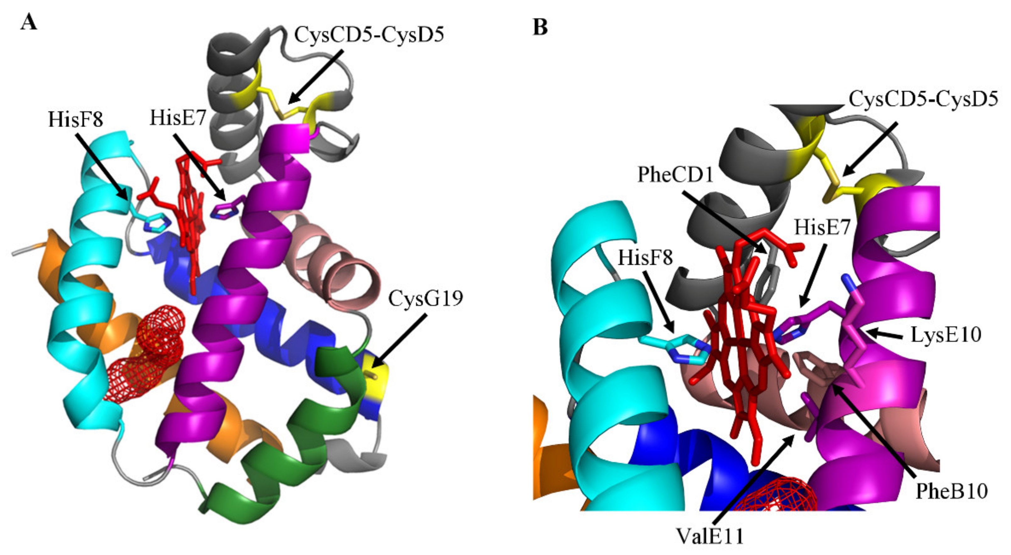
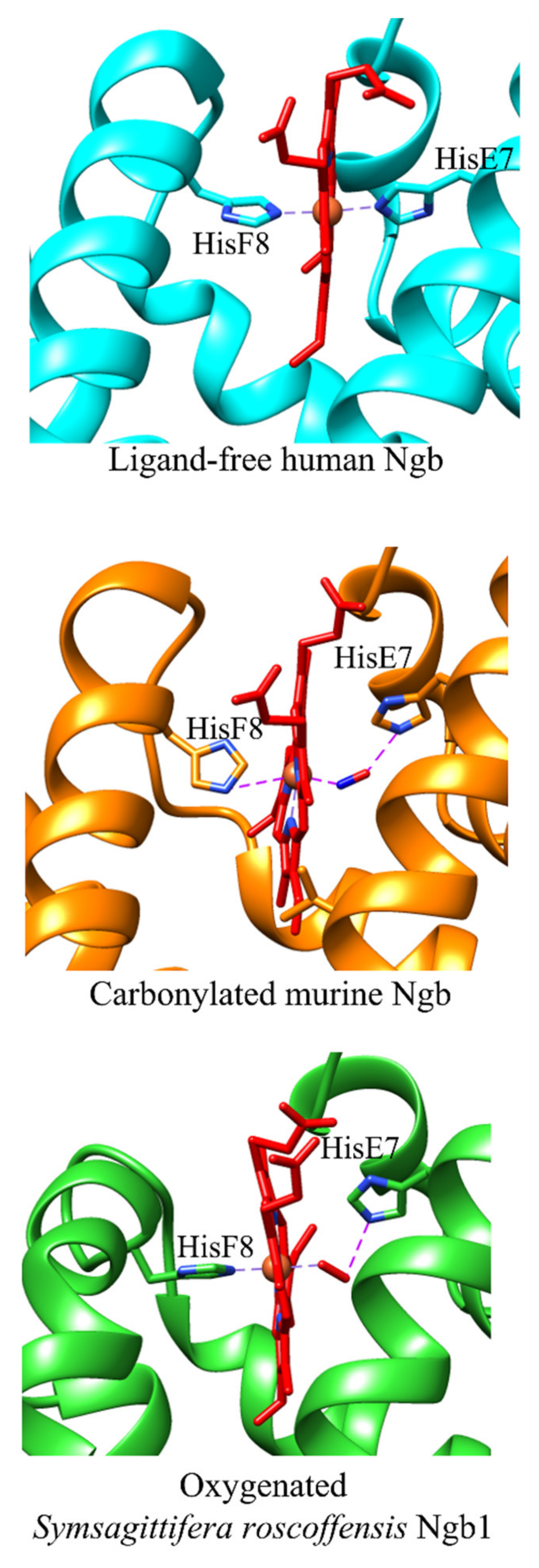
2. The Ngb Structure

Only the three-dimensional structures of Homo sapiens, Mus musculus, and Symsagittifera roscoffensis Ngb have been solved. Ngbs from the marine flat worm and mammals display the classical 3/3 globin fold, hosting the heme-group with α-helices A, B, E, F, G, and H, which are organized into a two-layer structure (A-B-E and F-G-H) [32,34,50] superimposable to that of Mb and Hb chains [2,4,8,51]. In ligand-free human and murine Ngb, the heme-Fe atom is six-coordinated by four N atoms belonging to the heme plane; the proximal (or fifth) and the distal (or sixth) ligands of the metal center are HisF8 and HisE7 residues, respectively [32,34]. In human and murine Ngb, the HisE7 imidazole ring is staggered relative to the heme pyrrole N atoms, being oriented toward the methinic bridge CHA and CHC atoms of the porphyrin ring. As a result, hexa-coordination is facilitated, since the α-helix E is pulled toward the heme by about 3.0 Å, relative to sperm whale Mb, which instead displays a five-coordinated heme-Fe atom [2,32,34] (Figure 1A).
The heme distal site of human and murine Ngb is crowded by the apolar residues (PheB10, PheCD1, and ValE11) trapping the porphyrin ring. LysE10 participates as well to the stabilization of macrocycles by electrostatic interactions with heme propionates [32,34]. Both in the crystalline state and in solution, the heme of mutant human and murine ferric Ngb (Ngb(III)) is accommodated in its pocket in two different geometries being rotated by 180° about the α–γ-meso axis [32,34]. The equilibrium constant for the two heme orientations in murine Ngb changes from ~2:1 in ligand-free Ngb(III) to ~1:1 in the Ngb(III)–cyanide complex, suggesting a role in the modulation of heme reactivity [52]. However, the structure of wild-type human Ngb reveals neither the occurrence of the heme isomerization nor the presence of multiple conformations of HisE7 [53]. Of note, the HisE7 and HisF8 imidazole rings of murine Ngb(III) show different orientations in the crystalline state and in solution, reflecting either the high exposure of the heme group to the bulk solvent or the absence of hydrogen-bond acceptors for the His NH protons provided by the heme-protein [54] (Figure 1B).
The overall fold of Symsagittifera roscoffensis Ngb1 matches well to that of murine ferrous carbonylated Ngb [35,50]. However, TrpCD3 is hydrogen bonded to one of the heme propionates and may represent a barrier for ligand access to the heme pocket. In addition, a water molecule is hydrogen-bonded to the heme distal HisE7 residue and to the second propionate of the heme group [50]. Unlike most globins, Symsagittifera roscoffensis Ngb displays a unique feature in that the α-helix F is bent by the presence of ProEF10, possibly reflecting the heme-protein flexibility [50].
A large cavity/tunnel system, encompassing part of the heme distal and proximal sites, is present in the protein matrix of human and murine Ngb, representing a pivotal determinant for the function and dynamics of these globins. In wild-type human Ngb, the cavity/tunnel system is shielded from the bulk solvent and is lined with hydrophobic residues, including ValE14, IleE15, ValG8, LeuG12, LeuH11, and ValH15. The volume of this cavity is 89 Å3 and 225 Å3 in molecules A and B present in the asymmetric unit, respectively. The different cavity size observed in A and B monomers of wild-type human Ngb is not a crystallographic packing artifact but reflects the high flexibility of the CD region that is crucial in the modulation of the heme reactivity and in the reshaping of the internal cavities [53]. In addition, a solvent-inaccessible internal cavity connected to the heme distal pocket (~60 Å3) occurs in the neighborhood of the CD loop/α-helix D region [53]. In the ligand-free CysCD5Gly/CysD5Ser/CysG19Ser mutant of human Ngb, a protein cavity (~120 Å3) connects the heme distal pocket to the EF inter-helical hinge. This cavity is lined with hydrophobic residues provided by the B, E, G, and H α-helices. Although in the crystals the cavity appears to be shielded from the external solvent, the intramolecular contacts indicate that, in a dynamic context, solvent access may be gained through side chain fluctuations [32]. In the ligand-free CysD5Ser/CysG19Ser mutant of murine Ngb, two small cavities (16 Å3 and 11 Å3), are present on the heme distal side. Moreover, a highly hydrophobic cavity (287 Å3), responsible for the unique CO binding mode [34,35], connects the heme distal and proximal sides [34], being also connected with the external solvent through a channel. In fact, upon CO binding, heme slides towards the interior of the protein matrix, decreasing the structural disorder especially in the EF-F-FG region, and inducing peripheral structural changes, which may affect Ngb actions and partner recognition. The repositioning of the heme-Fe group affects the geometry of HisF8, always bound to the heme-Fe(II) atom, and of the PheG5 [35]. The unusual CO-dependent repositioning of the heme group avoids the necessity of a large HisE7 swinging motion to permit ligand entry in and exit from the heme distal pocket [35], which is instead observed in Mb and Hb (i.e., the so-called “E7 gate” mechanism) [55,56,57]. Although the orifice of the channel is narrow, diatomic ligands can nonetheless access the metal center by virtue of the flexibility of the EF corner contributing to the channel external wall [34,35,58,59,60]. Due to the larger internal packing defects and the more pronounced active site fluctuations, the CO transfer barriers are considerably lower in Ngb(II) as compared to Mb(II) [61,62,63,64,65,66] (Figure 1).
In ferrous carbonylated murine Ngb, the CO molecule is bound to the heme-Fe(II) atom in a bent configuration, the Fe–C–O angle ranging between 100 and 156° and the Fe–C and Fe–O distances range between 1.8 to 2.0 Å and 2.5 to 3.0 Å, respectively. Moreover, the O atom of CO is placed at 2.8–3.2 Å from the NE atom of HisE7, possibly being stabilized by hydrogen bonding [35,58,60,67].
In ferrous oxygenated Symsagittifera roscoffensis Ngb1, O2 is bound to the distal site of the heme-Fe atom in a bent configuration, the Fe–O1–O2 angle being 136°. Moreover, the Fe–O1 and Fe–O2 distances are 1.96 and 3.05 Å, respectively, and the O1 and O2 atoms of O2 are located at 3.00 and 3.25 Å, respectively, from the NE atom of HisE7, which in turn stabilizes the heme-Fe-bound O2 by hydrogen bonding [50].
Mutagenesis on the structure of human and murine Ngb was relevant to understand at the atomic level the reactivity properties of this monomeric globin and to highlight the potential in vivo role of the redox modulation of the CD5–D5 disulfide bond.
Figure 1. (A) Three-dimensional structure of wild-type human Ngb (ID PDB code: 4MPM) [53]. A, B, E, F, G, and H α-helices, forming the classical 3/3 sandwich, are in forest green, salmon, purple, cyan, blue, and orange, respectively. Mesh surfaces of cavity/tunnel systems are highlighted in red. The CysCD5–CysD5 bridge and the CysG19 residue are highlighted in yellow. The proximal and distal amino acid residues HisF8 and HisE7, respectively, axially coordinating the heme-Fe atom through the Nε atom, are shown as balls and sticks. Tunnel/cavity systems have been determined by the interactive software CAVER Web 1.1 [68]. (B) Close-up of the heme pocket showing relevant residues cited in the text, in ball and stick representation. Figures drawn by PyMOL package [69].
Cells 10 03366 g001

3. Ngb (Pseudo-)Enzymatic Properties

Ngb is present at a relatively low concentration (~1 μM) compared to that of Mb (~200–400 μM) in most tissues and organs; this suggests that the role of Ngb in O2 supply and/or facilitated diffusion may become significant only in the retinal ganglion cell (RGC) layer and in the optic nerve where the Ngb concentration is up to ~100–200 μM [18,70,71,72,73]. Despite its low concentration, Ngb displays (pseudo-)enzymatic properties including NO/O2, peroxynitrite, and free radical scavenging; nitrite, hydroxylamine, and hydrogen sulfide reduction; and the nitration of aromatic compounds [44].
Of note, the heme-based reactivity of human Ngb is allosterically modulated by the redox state of the cell. In fact, the six-to-five-coordination transition (i.e., the inactive-to-active state) of the heme-Fe atom depends on the cleavage or the formation of the CysCD5–CysD5 bond, belonging to the so-called “dynamic loop” (CD loop/α-helix D region), and the (de-)phosphorylation of SerA7, SerA12, SerAB2, SerCD10, SerCD11, and SerDE3 [32,53,74,75,76,77].

4. The Cleavage of the HisE7-Heme-Fe Bond Modulates the Ngb Reactivity

The reactivity of six-coordinated human and murine Ngb is modulated through the minimum three-state mechanism encompassing (i) the dissociation of the endogenous ligand HisE7 from the HisF8-Fe-HisE7 complex, (ii) the formation of the transient five-coordinated HisF8-Fe adduct, and (iii) the association of the exogenous ligand (i.e., L) leading to the formation of the HisF8-Fe-L complex [35,74,78,79,80] (Figure 1 and Figure 2). The rate of HisE7 dissociation (i.e., k−1) limits the heme reactivity only under conditions where the formation of the transient five-coordinated HisF8-Fe adduct is slower than the rate of exogenous ligand binding to the metal center [8,44,81,82,83]. Of note, the five-coordinated HisF8-Fe adduct of Symsagittifera roscoffensis Ngb1 may represent the transient reactive adduct of six-coordinated human and murine Ngb (Figure 1 and Figure 2) [50].
Values of k+1 for HisE7 binding to human Ngb(II) range from 2.0 × 10³ s−1 to 9.8 × 10³ s−1 (pH 7.0 and 20.0–25.0 °C), and the value of k+1 for HisE7 binding to murine Ngb(II) is 2.0 × 10³ s−1 (pH 7.0 and 20.0 °C) [85]. Values of k−1 for HisE7 dissociation from human Ngb(II) range between 1.0 × 10⁻¹ s−1 and 8.2 × 10³ s−1 (pH 7.0 and 20.0–25.0 °C), and k−1 values of murine Ngb(II) range between 1.5 × 10⁻¹ s−1 and 1.2 s−1 (pH 7.0 and 20.0–25.0 °C) [16,44,78,79,80,85,86,87,88,89,90,91]. In the case of Ngb(III) the value of k−1 for the dissociation of HisE7 is >4.5 × 10⁻¹ s−1 (pH 7.2 and room temperature) [92]. The different values of k+1 and k−1 of human and murine Ngb [32,34,35] reflect, among others, the heme isomerization, the coordination state of the heme-Fe atom, and/or the chemical modifications affecting the heme-Fe reactivity [74,77,93,94]. In particular, the k−1 value of human Ngb(II) changes from 6.0 × 10⁻¹ s−1 to 7.0 s−1, reflecting the cleavage and the occurrence of the CysCD5–CysD5 bridge, respectively [74]. In contrast, the k−1 value of murine Ngb(II) (1.0 × 10⁻¹ s−1; pH 7.0 and 25.0 °C) [76] is unaffected by the redox state of the cell since the CD5 residue is Gly [44]. However, since in many of the following reactions the reduced Ngb(II) species is the most active form, a very important role for the Ngb metabolic function is played by the disulfide redox balance as well as by the kinetics of distal histidine coordination with the heme’s iron (Figure 3) [95].
Interestingly, the heme-Fe reactivity of six-coordinated Ngb is similar to that of five-coordinated globins (e.g., Mb), although it is modulated by the strength of the HisE7-Fe bond (about −3 kcal mol−1) rather than by the ligand accessibility to the heme pocket through the E7 gate, possibly reflecting a case of convergent evolution [8,32,34,35,37,38,44,50,53,55,56,57,58,59,67,76,96,97,98,99,100,101,102,103].

5. Ngb(II) Oxygenation

The brain is the most energetically demanding organ, but it displays a limited capacity to store energy; therefore, it is highly dependent on O2 and glucose supply from the blood stream [8,44,104,105,106,107]. Human and murine Ngb(II) binds reversibly to O2 with values of the Hill coefficient n close to unity, as expected for monomeric globins [8,44]. Values of P50 for human Ngb(II) oxygenation range between 0.9 mmHg and 10 mmHg (pH 7.0 and 25.0 °C), thus reflecting the different globin activation states, whereas the P50 value for murine Ngb(II) oxygenation is 2.2 mmHg (pH 7.0 and 25.0 °C). Since human and murine Ngb(II) oxygenation is limited by the cleavage of the heme-Fe-HisE7 bond, values of the intrinsic dissociation equilibrium constant for O2 binding to the transient five-coordinated species are lower than those of the six-coordinated state by about three orders of magnitude [8,14,44,74,78,80,85,86,108,109,110,111,112].
The rate of O2 binding to six-coordinated Ngb(II) (i.e., k+2), as measured by rapid-mixing, is independent of the ligand (e.g., O2, CO, and NO) and of the O2 concentration, being limited by the cleavage of the heme-Fe-HisE7 bond (i.e., k−1). Values of k+2 for human Ngb(II) oxygenation range from 6.0 × 10⁻¹ s−1 to 7.0 s−1 (pH 7.0 and 25.0 °C), reflecting the redox state of the CysCD5/CysD5 residue pair [74]. Since this disulfide bridge is absent in murine Ngb(II), the value of k+2 for the heme-protein oxygenation (1.5 × 10⁻¹ s−1; pH 7.0 and 25.0 °C) is independent of the cell redox properties [74,79,80]. The value of the second-order rate constant for the oxygenation of five-coordinated human and murine Ngb(II) (k+3), as measured after flash and laser photolysis, is ~2.0 × 10⁸ M−1 s−1 (pH 7.0 and 25.0 °C) and the first-order rate constant for Ngb(II)-O2 deoxygenation (i.e., k−3) is ~6.0 × 10⁻¹ s−1 (pH 7.0 and 25.0 °C) [74,78,86,113,114] (Figure 3).

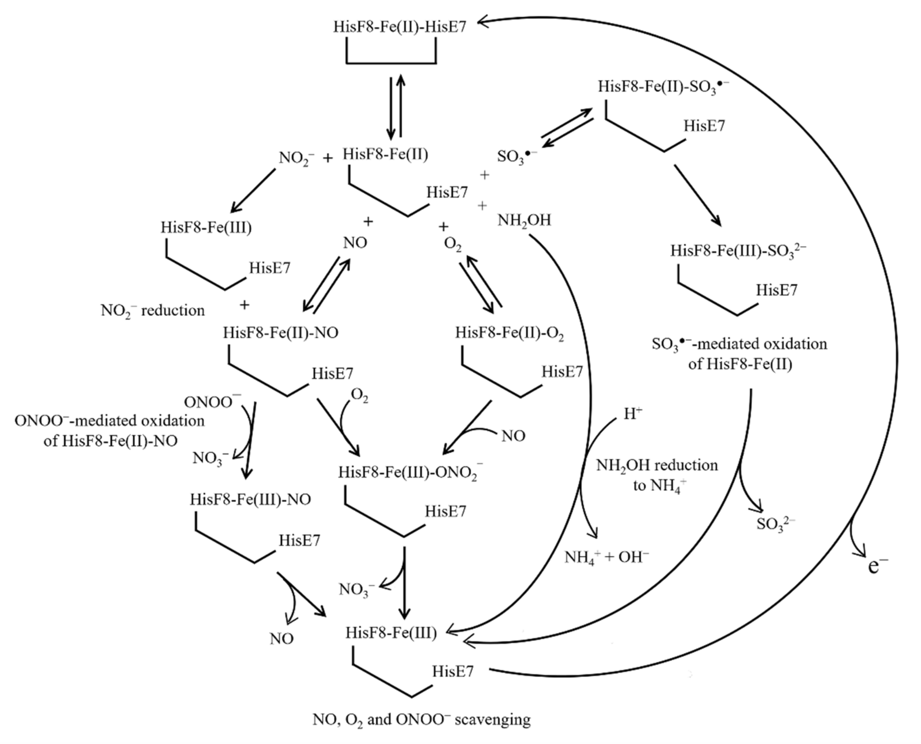
6. Ngb(II) Nitrosylation

The ubiquitous messenger NO plays pivotal roles in several regulatory functions, including vasodilatation, neurotransmission, and host-defense mechanisms; however, it also displays cytotoxic properties and is involved in a variety of neurodegenerative diseases [115,116]. The reaction of NO with murine Ngb(II), measured by rapid-mixing (i.e., k+4), is slow (~4.0 × 10⁻¹ s−1; pH 7.0 and 20.0 °C) and independent of the ligand concentration because it can only occur upon cleavage of the heme-Fe(II)-HisE7 bond limiting the association of the external ligand [79]. Flash photolysis study shows a fast recombination rate (i.e., k+5 = 1.5 × 10⁸ M−1 s−1; pH 7.0 and 25.0 °C) and a low dissociation rate (k−5 = 2.0 × 10⁻⁴ s−1; pH 7.0 and 25.0 °C) for murine Ngb(II) (de)nitrosylation, indicating a high intrinsic affinity for the ligand (K5 = 1.3 × 10⁻¹² M; pH 7.0 and 25.0 °C). However, since NO binding to murine Ngb(II) is affected by competitive inhibition of the HisE7 residue (K1 ~ 1.0 × 10⁻³; pH 7.0 and 25.0 °C) [74,78], the observed NO affinity decreases by about three orders of magnitude. As expected, NO affinity for ferrous five-coordinated HisE7Val, HisE7Gln, and HisE7Leu mutants corresponds to the intrinsic affinity of the wild-type heme-protein [113] (Figure 3).

7. NO and O2 Scavenging from Ngb(II)–O2 and Ngb(II)–NO

Ngb could be involved in the protection of the brain form damage, either scavenging O2 or detoxifying NO [79,92]. Human and murine Ngb(II)–O2 and Ngb(II)–NO react with NO and O2, respectively, yielding Ngb(III) and NO3 as the final products and preventing, among others, protein nitration [79,92,117,118]. The reaction of human and murine Ngb(II)–O2 with NO proceeds via the transient intermediate Ngb(III)–ONO2, which dissociates mono-exponentially with values of the first-order rate constant (i.e., k−6) of ~3.6 × 10² s−1 and ~3.0 × 10² s−1, respectively (pH 7.0 and 20.0 °C) [79,118]. O2 detoxification from human Ngb(II)-NO is a biphasic process, with values of the second-order rate constant (k+7) being 1.6 × 101 M−1 s−1 and 4.0 × 10⁻¹ M−1 s−1 (pH 7.0 and 25.0 °C) [92]. Since values of the bimolecular rate constant for O2 and NO binding to five-coordinated human Ngb(II) are almost identical (k+3 ~ k+5 ~ 108 M−1 s−1), the in vivo formation of Ngb(II)–NO is unlikely, occurring only under conditions where NO levels exceed those of O2 [79] (Figure 3).

8. NO2 Reduction by Ngb(II)

Human Ngb(II) catalyzes the reduction of NO2, leading to the formation of Ngb(III), Ngb(II)-NO, and NO. This process is modulated by the redox-sensitive CysCD5 and CysD5 residues regulating the fraction of the highly reactive five-coordinated state of the heme-Fe(II) atom. Therefore, the value of the second-order rate constant (k+8) for NO2 reduction by human Ngb(II) increases from 6.2 × 10⁻² M−1 s−1 to 1.2 × 10⁻¹ M−1 s−1 (pH 7.4 and 25.0 °C) upon formation of the CysCD5–CysD5 disulfide bond. This small effect was observed in vitro and may not be significant in vivo.
Investigations based on the PheB10Val, PheB10His, PheB10Trp, PheB10Leu, HisE7Ala, HisE7Gln, HisE7Trp, ValE11Ala, ValE11Phe, and ValE11Ile mutants indicate that the reductase activity of human Ngb(II) depends on the heme accessibility of NO2 [119,120]. Moreover, phosphorylation at putative sites SerA7, SerA12, SerAB2, SerCD10, SerCD11, and SerDE3 by intracellular kinases (e.g., ERK and PKA) increases the nitrite reductase activity of human Ngb(II) by stabilizing the five-coordinated heme-Fe(II) atom. Of note, the binding of the scaffold protein 14-3-3 at putative sites ArgA9-ProAB3 and ArgCD7-ProD1 of human Ngb stabilizes the phosphorylated derivative, enhancing the nitrite reductase activity [77]
The value of k+8 for NO2 reduction by murine Ngb(II) is 5.1 M−1 s−1 (pH 7.4 and 25.0 °C). Ngb(III) is the main product of the reaction, Ngb(II)–NO being lower than 20%. Upon reaction with nitrite, murine Ngb undergoes S-nitrosylation, the CysD5 residue representing the primary site. However, the overall intracellular physiological concentration of NO2 (<1 × 10⁻⁵ M) appears too low to convert Ngb to its S-nitrosylated derivative acting as a NO depot, but it may play a role in redox signaling pathways [121] (Figure 3).

9. NH2OH Reduction by Ngb(II)

Hbs reduce nitrite and hydroxylamine, an intermediate of nitrite reductase, to nitric oxide under anaerobic conditions. However, plant and cyanobacterial Hbs catalyze the reduction of hydroxylamine to ammonium at rates 100–2500 times faster than animal globins, including Ngb. These results support the view that plant and cyanobacterial hemoglobins contribute to anaerobic nitrogen metabolism in support of anaerobic respiration and survival during hypoxia [122,123,124]. Under anaerobic conditions, human Ngb(II) reduces NH2OH to NH4+, being converted to Ngb(III). The second-order rate constant for hydroxylamine reduction to ammonium by human Ngb(II) (k+8) is <2.5 × 101 M−1 s−1 [122] (Figure 3).

10. Ngb(II) Oxidation by ONOO and SO3●−

Peroxynitrite induces the oxidation of human Ngb(II)–NO by means of the Ngb(III)–NO transient species that decays to Ngb(III) and NO. The oxidation of Ngb(II)-NO to Ngb(III)–NO depends linearly on the ONOO concentration, whereas the dissociation of the Ngb(III)-NO complex is a monomolecular process (Figure 3). The value of the second-order rate constant for the ONOO-dependent oxidation of Ngb(III)-NO (k+7) is 1.3 × 10⁵ M−1 s−1 and the value of the first-order rate constant of the Ngb(III)–NO dissociation (k−7) is 1.2 × 10⁻¹ s−1 (pH 7.2 and 20.0 °C) [92]. Murine Ngb(II) undergoes oxidation by SO3●− sulfur trioxide, yielding Ngb(III) and SO3 as final products; the bimolecular rate constant (i.e., k+8) is 2.6 × 10⁹ M−1 s−1 (pH 7.2 and 37.0 °C). Since SO3●− scavenging by Ngb(II) is inhibited by glutathione, this reaction may take place in the retina where Ngb(II) levels are exceeding those of glutathione [125] (Figure 3).

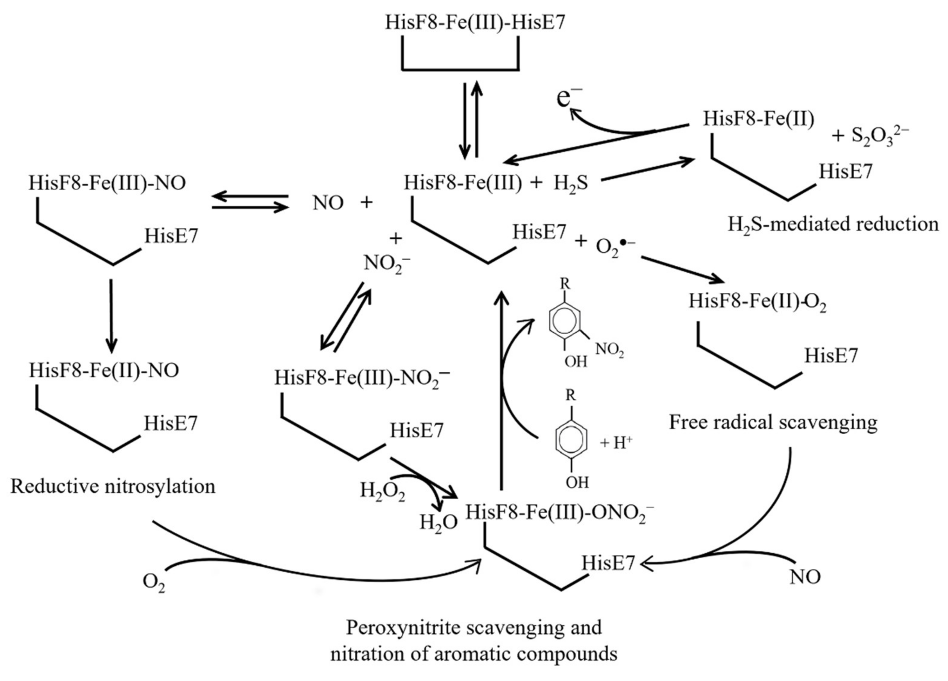
11. Reductive Nitrosylation of Ngb(III)

The reaction of human Ngb(III) with an excess of NO leads to Ngb(II)–NO via the formation of the transient Ngb(III)–NO adduct. Values of the dissociation equilibrium constant (K9) and of the second-order rate constant (k+9) for NO binding to human Ngb(III) are 7.5 × 10⁻⁵ M and 2.0 × 10³ M−1 s−1, respectively (pH 7.0 and 20.0 °C) [118]. The value of the first-order rate constant for the dissociation of NO from Fe(III)–NO (i.e., k−9) is 1.2 × 10⁻¹ s−1 [92]. The reductive nitrosylation of human Ngb(III) is a biphasic process; values of the overall second-order rate constant of the fast and the slow components (i.e., k+10) are 2.1 × 101 M−1 s−1 and 2.9 M−1 s−1 (pH 7.0 and 25.0 °C), respectively. The transient Fe(III)–NO species converts to Fe(II)–NO with a mono-exponential process, the value of the first-order rate constant (i.e., k+11) being > 2.4 × 10⁻² s−1 (pH 7.0 and 25.0 °C) [92] (Figure 4).

12. H2S-Mediated Reduction of Ngb(III)

Hydrogen sulfide H2S is a potential signaling molecule produced by cystathionine β-synthase, cystathionine γ-lyase, and 3-mercaptopyruvate sulfurtransferase. H2S affects neurotransmission in the brain and relaxes vascular smooth muscle in synergy with NO. Moreover, it has been reported that H2S acts as an O2 sensor, influences autophagy, displays a cytoprotective and angioprotective effect during the evolution of myocardial infarction, and has anti- inflammatory activity [126]. In addition, the hydrogen polysulfides (H2Sn, n ≥ 2), produced by 3-mercaptopyruvate sulfurtransferase, are potential signaling molecules able to modulate the activity of ion channels and enzymes as well as the tumor growth by S-sulfuration of target proteins [127] (Figure 4).
Under anaerobic conditions, human Ngb(III) catalyzes the oxidation of HS to S2O32− via the formation of the transient Ngb(II)–HS adduct; this reaction exhibits biphasic kinetics. For the fast phase, values of the second-order rate constant for the formation of the Ngb(II)–HS complex (k+12), of the first-order rate constant for the Ngb(II)–HS adduct dissociation (k−12), and of the equilibrium dissociation constant for H2S binding to Ngb(III) (K12) are 1.4 × 101 M−1 s−1, 5.1 × 10⁻³ s−1, and 3.7 × 10⁻⁴ M (pH 7.4 and 25.0 °C), respectively. On the other hand, the rate constant of the slow step for Ngb(II)–HS complexation (k+13 = 5.6 × 10⁻⁴ s−1; pH 7.4 and 25.0 °C) is independent of the ligand concentration. The Ngb(III) mutant HisE7Ala was more active for H2S oxidation (k+14 = 1.4 × 101 M−1 s−1, k−14 = 5.1 × 10⁻³ s−1, and K14 = 3.7 × 10⁻⁴ M; pH 7.4 and 25.0 °C), yielding the formation of 2–6 catenated sulfur atoms with or without oxygen insertion. This highlights the role of the heme distal residue in modulating the reactivity of the metal center and the different reaction options [127,128,129,130,131] (Figure 4).

13. Free Radical Scavenging from Ngb(III)

Human Ngb(III) scavenges a variety of free radicals including O2●−. The value of the affinity constant (K15) for O2●− binding to Ngb(III) is 7.4 × 10⁻⁶ M. The CysCD5–CysD5 bond is not necessary to scavenge superoxide, but it plays a pivotal role in stabilizing the heme-Fe(II)–O2 complex [132]. Wild-type human Ngb(III) and the PheB10Leu mutant are much more stable in the presence of H2O2 than the HisE7Leu mutant, which undergoes tyrosyl radical formation. This indicates that (i) the PheB10 residue is essentially insensitive to the redox state of Ngb and (ii) the five-coordination of the heme-Fe atom, occurring in the HisE7Leu mutant, induces the destabilization of the globin fold [133]. As a whole, the redox changes of the heme-Fe atom and of the CysCD5/CysD5 residue pair could regulate neuroprotective functions of human Ngb, protecting against hypoxic and ischemic stress in the brain [132,134] (Figure 4). Of note, human Ngb(III) does not produce the oxidizing oxoFe(IV)=O species when treated with H2O2, in contrast to Hb and Mb [92].

14. Nitration of Aromatic Compounds by Ngb(III)-NO2

Nitrosylated tyrosines play a role in the redox regulation of metabolism; however, their increase occurs in multiple neurodegenerative diseases (e.g., Parkinson’s and Alzheimer’s diseases) [135]. Human Ngb(III)–NO2 catalyzes the nitration of phenolic substrates in the presence of H2O2 via the transient formation of the highly reactive Ngb(III)–ONOO species (Figure 4). The two human Ngb forms, with and without the internal CysCD5–CysD5 disulfide bridge, reflecting the five- and six-coordination of the heme-Fe(III) atom, respectively, exhibit very different reactivities. In fact, values of the dissociation equilibrium constant (i.e., K16) and of the second-order rate constant (i.e., k+16) for the nitration of aromatic compounds by the highly reactive Ngb(III)–ONO2 complex are 4.3 × 10² M and 3.3 M1 s1, respectively, whereas those of the slowly reactive form are 2.2 × 101 M and 6.3 × 101 M1 s1, respectively [136]. This evidence reinforces the relevance of the cysteine redox equilibrium in Ngb, which plays an important role in addressing its reactivity toward alternative metabolic pathways (Figure 3 and Figure 4).

15. Possible Patho-Physiological Roles of Neuroglobin

Up to now, Ngb-based reactions have been investigated separately without attempting to link them to each other. Here, this limitation has been tentatively overcome to support the view that Ngb plays multiple roles in health and disease [44,137,138,139,140].
Ngb was first documented to be almost exclusively expressed in the brain, displaying high concentrations (up to 100–200 μM) in the retina layers, not only in the RGC layer, but also in astrocytes and in Müller cells (i.e., a specialized microglial lineage which spans across the layers, serving protective and nourishing activities). In this last cell type, the Ngb expression is further significantly raised during reactive gliosis in response to eye injury [141].
Ngb actions in vivo depend on its concentration and on an efficient Ngb(III) reductase system, still unknown, restoring Ngb(II) [44,79,80,118,142]. Thus, when present at high concentrations (~100–200 μM) Ngb may facilitate O2 buffer and transport in the RGC layer and the optic nerve, whereas at low concentrations (~1 μM) Ngb can display only potential enzymatic activities and cell signaling in most tissues and organs, including resting neurons [43,44,143].
Systems biology investigations and numerical simulations confirm that Ngb should play an important role in O2 transport rather than in storage; thus, since the retina is most susceptible to hypoxia in the regions of the photoreceptor inner segment and inner plexiform layers, high concentrations of Ngb have the potential to prevent hypoxia and increase the O2 uptake by 30–40% [144]. A mutual regulation of Ngb and hypoxia was envisaged by studies on Ngb expression after the induction of ocular hypertension in Wistar rats. Ngb was quickly transcriptionally upregulated in rat retinal layers in response to the acute retinal ischemia injury induced by the elevation of intraocular pressure, suggesting that the globin might play an important role in the metabolic adaptation to hypoxia [145].
Furthermore, decreased survival of Ngb-deficient primary cortical neurons along with increased viability of the same cells in the presence of Ngb overexpression were originally reported in an experimental model of hypoxia in vitro, where Ngb immunostaining was significantly increased in the surviving infarcted area [146]. In accordance with these findings, the intra-cerebro-ventricular administration of a Ngb antisense, but not sense, oligo-deoxy-nucleotide increased infarct volume, worsening the neurological outcome of stroke [147].
The hypothesis of Ngb having a role in reducing the focal ischemia and infarct size was further pushed forward by the development of transgenic mice overexpressing this globin (Ngb-Tg mice). A reduced infarct size was observed in these animals after transient cerebral arterial ligation followed by reperfusion [148]. Surprisingly, in this transgenic animal, Ngb expression was induced also in non-neuronal cells, such as in heart cells, and this was apparently enough to limit the infarct size area after heart arterial ligation and reperfusion, also in an organ which physiologically does not appreciably express the protein [148]. The reduced infarct size in the same Ngb-Tg animal was further confirmed by other authors [149].
Furthermore, the specific effect of increasing the amount of Ngb in retinal cells has been demonstrated by the injection of exogenous Ngb after a transient hypoxia, which turned out to be effective in decreasing the levels of inflammatory chemokines (IL-6, TNFα, IL-1B, RANTES, MCP-1, and VEGF) and microglia activation [150]. This effect appears to be linked to the regulatory effect of Ngb on the Wnt/β-catenin and NF-kB signaling pathways, which is exerted mostly through the induction of the proteasomal degradation of Dishevelled-1, a key hub protein eliciting these two processes [151].
Interestingly, endogenous, and exogenous compounds (e.g., hormones and phytochemicals) as well as injuries (e.g., oxidative stress, hypoxia, epilepsy, and ischemia) increase Ngb levels in the brain, allowing it to carry out functions directed to neuroprotection [152,153,154,155,156]. As a matter of fact, the enhanced expression of Ngb has been observed in RGCs after optical nerve injury either through exposure to intense led light [157] or to an oxidative stress [158].
Clues for interpreting the biological role of Ngb come from zebrafish retina, which, unlike mammals, shows regenerative properties of the optic nerve. In this small fish, Ngb mRNA was found to be expressed in amacrine cells, whereas Ngb protein was also detected in the inner plexiform layer. This discrepancy was attributable to the presence of a membrane-penetrating module of zebrafish Ngb which allowed the protein to cross the plasma membrane. Immuno-histochemical studies after optic nerve injury highlighted that Ngb protein levels were increased in both amacrine cells, in correspondence to the nerve ending processes, and presynaptic regions in the inner plexiform layer, suggesting that Ngb might be released by the former cells and picked up by the ganglion cells, wherein it participates in the regeneration of the axon [159].
Although most of details of the mechanisms through which Ngb exerts its neuroprotective activity are still unclear, co-localization studies in the retina have confirmed a strong interaction of the globin with mitochondria, envisaging a role in assisting energy metabolism and in patrolling apoptosis upon release of mitochondrial factors, such as cytochrome c. In this respect, a very important role seems to be played by Ngb in the protection of nerve cells from apoptosis, mainly through its interaction with cytochrome c [118,160], which can block the activation of caspase 9 by ferric cytochrome c [161,162]. This anti-apoptotic function requires a reduced Ngb(II) which interacts with ferric cytochrome c through negatively charged residues (mostly Asp73 and to a reduced extent Glu60, Asp63, and Glu87); this complex quickly reduces cytochrome c, impairing its pro-apoptotic role [163].
Some clues strengthening the contribution of Ngb in serving mitochondria functionality come from studies on the Harlequin mouse phenotype, which finds its genetic roots in the depletion of the apoptosis-inducing factor, and recapitulates hallmarks of human neurodegenerative mitochondrial pathologies, such as the age-dependent degeneration of the retina, optic nerve, cerebellum, and cortical regions. Rescue of Ngb expression in the retina, which significantly drops in the brain of the Harlequin mouse upon delivery of an adenovirus associated vector (AAV), improved RGC survival and overall retina neurodegeneration, suggesting that restoring adequate Ngb levels was enough to improve the mitochondrial chain respiration and energy homeostasis [43].
Similarly, the development of Ngb-transgenic (Ngb-Tg) mice has provided a significant technological advancement to point out the effect of Ngb overexpression on mitochondrial dynamics during retinal ischemia in vivo. Remarkably, compared with wild-type animals, Ngb-Tg mice showed a relevant decrease of mitochondrial DNA damage after 7 days of reperfusion following ischemia induction. In accordance with the general neuroprotective activity of Ngb, apoptosis markers, such as the release of the caspase-3 active fragment, were decreased in Ngb-Tg mice and TUNEL assay showed a reduced frequency of apoptotic cells [152].
Further, several studies have suggested that mitochondrial dysfunction may induce retinal degenerative disorders, such as glaucoma, and that Ngb is among the genes transcriptionally downregulated during the glaucoma onset [143]. For this, the role of Ngb has been investigated in a well-established model of retinal neurodegeneration, such as the DBA/2J mice (which develop spontaneous intraocular pressure and RGC degeneration, eventually inducing glaucoma), following the effect of Ngb upregulation in RGCs via a single intravitreal injection of an AAV vector. In these animals, because of decreased enzymatic activities of mitochondrial chain complexes, mitochondrial dysfunction precedes RGC loss by about 5 months, but AAV-Ngb administration led to a general and robust improvement of RGC functionality. Furthermore, immuno-histochemical studies of retina sections indicated the presence of conserved RGC morphology, suggesting neuronal remodeling and synaptic plasticity. Moreover, overexpression of Ngb attenuated ocular hypertension-induced superoxide production and the associated decrease of ATP levels in mice, suggesting that Ngb acts as an endogenous neuroprotectant to reduce oxidative stress and improve mitochondrial function, thereby promoting RGC survival. Thus, Ngb may modulate RGC susceptibility to glaucomatous neural damage [42].

16. Conclusions and Perspectives

The neuroprotective role of Ngb is openly debated since differences in transcript and protein Ngb levels have been reported [149,164,165,166,167,168]. Several studies in Ngb-null mice reported that Ngb expression in the central nervous system and in the retina, rather than diffuse, is restricted to specific brain regions and to two out of the ten layers forming the retina [149,169].
The use of Ngb-null mice turned out to be important to highlight some important Ngb functions, since they appear to have a reduced activity of respiratory chain complexes I and III, the degeneration of primary rat RGCs, and the impairment of visual functions [50]. In addition, a 56–60% increase of the infarct volume after focal cerebral ischemia has been observed in Ngb-null mice compared to wild-type animals [146]. In turn, the reduction in infarct size induced by ischemia has been obtained by increasing the Ngb levels before the onset of stroke [170]. These data highlight the role of Ngb as a compensatory protein (e.g., stress-sensor and stress-inducible macromolecule) responding to hypoxic/ischemic/oxidative injuries by activating survival/antiapoptotic pathways [171,172].
In the future, a better understanding of the molecular mechanisms at the root of the biological functions of Ngb will bear fundamental and translational significance, especially in the development of therapeutics against stroke and neurological disorders. Ngb-based therapeutic approaches to neurological disorders have started, and the results obtained are promising since no side effects of Ngb overexpression in transgenic mice have been observed [148,173]. However, since Ngb may act as a compensatory protein even in cancer cells, the possibility that systemic Ngb overexpression could enhance cancer cell survival should be taken into account considered; in this respect, the possibility of Ngb gene therapy is being considered realistically [172,174].

Author Contributions

Conceptualization, P.A. and M.C.; software, G.D.S. and A.P.; investigation, F.O.; data curation, F.O.; writing—original draft preparation, G.D.S. and D.S.; writing—review and editing, P.A. and M.C.; visualization, A.P.; supervision, P.A. and M.C.; funding acquisition, P.A. and M.C. All authors have read and agreed to publish this manuscript.

Funding

This work has been supported by the Italian Ministry of Education, Universities and Research (PRIN 2017SNRXH3 and Dipartimenti di Eccellenza; Legge 232/2016, Articolo 1, Comma 314–337) and Fondazione Roma.

Institutional Review Board Statement

Not applicable.

Informed Consent Statement

Not applicable.

Data Availability Statement

Not applicable.

Acknowledgments

The authors wish to thank Maria Marino for helpful discussions. The authors apologize for not having quoted several outstanding papers due to space limitations.

Conflicts of Interest

The authors declare no conflict of interest.

Abbreviations

AAVAdenovirus associated vector
Adgbandroglobin
Cygbcytoglobin
Hbhemoglobin
Mbmyoglobin
Ngb(II)ferrous Ngb
Ngb(III)ferric Ngb
GbEglobin E
GbXglobin X
GbYglobin Y
Mbmyoglobin
Ngbneuroglobin
mice Ngb-Tgtransgenic mice overexpressing Ngb
RGCretinal ganglion cell

References

  1. Antonini, E.; Brunori, M. Hemoglobin and Myoglobin in Their Reactions with Ligands; North-Holland Publishing, Co.: Amsterdam, The Netherlands, 1971. [Google Scholar]
  2. Perutz, M.F. Regulation of oxygen affinity of hemoglobin: Influence of structure of the globin on the heme iron. Annu. Rev. Biochem. 1979, 48, 327–386. [Google Scholar] [CrossRef]
  3. Bunn, H.F.; Forget, B.G. Hemoglobin: Molecular, Genetic and Clinical Aspects; W.B. Saunders Co.: Philadelphia, PA, USA, 1986. [Google Scholar]
  4. Bolognesi, M.; Bordo, D.; Rizzi, M.; Tarricone, C.; Ascenzi, P. Nonvertebrate hemoglobins: Structural bases for reactivity. Progr. Biophys. Mol. Biol. 1997, 68, 29–68. [Google Scholar] [CrossRef]
  5. Gilles-Gonzalez, M.A.; Gonzalez, G. Heme-based sensors: Defining characteristics, recent developments, and regulatory hypotheses. J. Inorg. Biochem. 2005, 99, 1–22. [Google Scholar] [CrossRef]
  6. Pesce, A.; Bolognesi, M.; Nardini, M. The diversity of 2/2 (truncated) globins. Adv. Microb. Physiol. 2013, 63, 49–78. [Google Scholar] [CrossRef]
  7. Pesce, A.; Bolognesi, M.; Nardini, M. Protoglobin: Structure and ligand-binding properties. Adv. Microb. Physiol. 2013, 63, 79–96. [Google Scholar] [CrossRef] [PubMed]
  8. Ascenzi, P.; Brunori, M. A molecule for all seasons: The heme. J. Porphyr. Phthalocyanines 2016, 29, 1–16. [Google Scholar] [CrossRef]
  9. Vinogradov, S.N.; Hoogewijs, D.; Bailly, X.; Arredondo-Peter, R.; Gough, J.; Dewilde, S.; Moens, L.; Vanfleteren, J.R. A phylogenomic profile of globins. BMC Evol. Biol. 2006, 6, 31–47. [Google Scholar] [CrossRef] [PubMed] [Green Version]
  10. Vinogradov, S.N.; Moens, L. Diversity of globin function: Enzymatic, transport, storage, and sensing. J. Biol. Chem. 2008, 283, 8773–8777. [Google Scholar] [CrossRef] [Green Version]
  11. Wajcman, H.; Kiger, L.; Marden, M.C. Structure and function evolution in the superfamily of globins. Crit. Rev. Biol. 2009, 332, 273–282. [Google Scholar] [CrossRef] [PubMed]
  12. Burmester, T.; Hankeln, T. Function and evolution of vertebrate globins. Acta Physiol. 2014, 211, 501–514. [Google Scholar] [CrossRef]
  13. Keppner, A.; Maric, D.; Correia, M.; Koay, T.W.; Orlando, I.M.C.; Vinogradov, S.N.; Hoogewijs, D. Lessons from the post-genomic era: Globin diversity beyond oxygen binding and transport. Redox Biol. 2020, 37, 101687–101705. [Google Scholar] [CrossRef] [PubMed]
  14. Burmester, T.; Weich, B.; Reinhardt, S.; Hankeln, T. A vertebrate globin expressed in the brain. Nature 2000, 407, 520–523. [Google Scholar] [CrossRef]
  15. Burmester, T.; Ebner, B.; Weich, B.; Hankeln, T. Cytoglobin: A novel globin type ubiquitously expressed in vertebrate tissues. Mol. Biol. Evol. 2002, 19, 416–421. [Google Scholar] [CrossRef] [Green Version]
  16. Trent, J.T., III; Hargrove, M.S. A ubiquitously expressed human hexacoordinate hemoglobin. J. Biol. Chem. 2002, 277, 19538–19545. [Google Scholar] [CrossRef] [Green Version]
  17. Fuchs, C.; Burmester, T.; Hankeln, T. The amphibian globin gene repertoire as revealed by the Xenopus genome. Genome Res. 2006, 112, 296–306. [Google Scholar] [CrossRef] [PubMed]
  18. Kugelstadt, D.; Haberkamp, M.; Hankeln, T.; Burmester, T. Neuroglobin, cytoglobin, and a novel, eye-specific globin from chicken. Biochem. Biophys. Res. Commun. 2004, 325, 719–725. [Google Scholar] [CrossRef]
  19. Roesner, A.; Fuchs, C.; Hankeln, T.; Burmester, T. A globin gene of ancient evolutionary origin in lower vertebrates: Evidence for two distinct globin families in animals. Mol. Biol. Evol. 2005, 22, 12–20. [Google Scholar] [CrossRef]
  20. Hoogewijs, D.; Ebner, B.; Germani, F.; Hoffmann, F.G.; Fabrizius, A.; Moens, L.; Burmester, T.; Dewilde, S.; Storz, J.F.; Vinogradov, S.N.; et al. Androglobin: A chimeric globin in metazoans that is preferentially expressed in mammalian testes. Mol. Biol. Evol. 2012, 29, 1105–1114. [Google Scholar] [CrossRef] [PubMed] [Green Version]
  21. Pesce, A.; Couture, M.; Dewilde, S.; Guertin, M.; Yamauchi, K.; Ascenzi, P.; Moens, L.; Bolognesi, M. A novel two-over-two alpha-helical sandwich fold is characteristic of the truncated hemoglobin family. EMBO J. 2000, 19, 2424–2434. [Google Scholar] [CrossRef] [Green Version]
  22. Montfort, W.R.; Weichsel, A.; Andersen, J.F. Nitrophorins and related antihemostatic lipocalins from Rhodnius prolixus and other blood-sucking arthropods. Biochim. Biophys. Acta 2000, 1482, 110–118. [Google Scholar] [CrossRef]
  23. Andersen, J.F. Structure and mechanism in salivary proteins from blood-feeding arthropods. Toxicon 2010, 56, 1120–1129. [Google Scholar] [CrossRef] [Green Version]
  24. Champagne, D.E. Antihemostatic molecules from saliva of blood-feeding arthropods. Pathophysiol. Haemos. Thromb. 2005, 34, 221–227. [Google Scholar] [CrossRef]
  25. De Simone, G.; Ascenzi, P.; Polticelli, F. Nitrobindin: An ubiquitous family of all β-barrel heme-proteins. IUBMB Life 2016, 68, 423–428. [Google Scholar] [CrossRef] [PubMed] [Green Version]
  26. De Simone, G.; Ascenzi, P.; di Masi, A.; Polticelli, F. Nitrophorins and nitrobindins: Structure and function. Biomol. Concepts 2017, 8, 105–118. [Google Scholar] [CrossRef]
  27. De Simone, G.; di Masi, A.; Vita, G.M.; Polticelli, F.; Pesce, A.; Nardini, M.; Bolognesi, M.; Ciaccio, C.; Coletta, M.; Turilli, E.S.; et al. Mycobacterial and human nitrobindins: Structure and function. Antioxid. Redox Signal. 2020, 33, 229–246. [Google Scholar] [CrossRef]
  28. De Simone, G.; di Masi, A.; Ciaccio, C.; Coletta, M.; Ascenzi, P. NO Scavenging through reductive nitrosylation of ferric Mycobacterium tuberculosis and Homo sapiens nitrobindins. Int. J. Mol. Sci. 2021, 21, 9395. [Google Scholar] [CrossRef]
  29. De Simone, G.; di Masi, A.; Pesce, A.; Bolognesi, M.; Ciaccio, C.; Tognaccini, L.; Smulevich, G.; Abbruzzetti, S.; Viappiani, C.; Bruno, S.; et al. Mycobacterial and human ferrous nitrobindins: Spectroscopic and reactivity properties. Int. J. Mol. Sci. 2021, 22, 1674. [Google Scholar] [CrossRef] [PubMed]
  30. Tilton, R.F.; Kuntz, I.D.; Petsko, G.A. Cavities in proteins: Structure of a metmyoglobin xenon complex solved at 1.9 Å. Biochemistry 1984, 23, 2849–2857. [Google Scholar] [CrossRef]
  31. Milani, M.; Pesce, A.; Ouellet, Y.; Ascenzi, P.; Guertin, M.; Bolognesi, M. Mycobacterium tuberculosis hemoglobin N displays a protein tunnel suited for O2 diffusion to the heme. EMBO J. 2001, 20, 3902–3909. [Google Scholar] [CrossRef] [Green Version]
  32. Pesce, A.; Dewilde, S.; Nardini, M.; Moens, L.; Ascenzi, P.; Hankeln, T.; Burmester, T.; Bolognesi, M. Human brain neuroglobin structure reveals a distinct mode of controlling oxygen affinity. Structure 2003, 11, 1087–1095. [Google Scholar] [CrossRef]
  33. De Sanctis, D.; Dewilde, S.; Pesce, A.; Moens, L.; Ascenzi, P.; Hankeln, T.; Burmester, T.; Bolognesi, M. Mapping protein matrix cavities in human cytoglobin through Xe atom binding. Biochem. Biophys. Res. Commun. 2004, 316, 1217–1221. [Google Scholar] [CrossRef] [PubMed]
  34. Vallone, B.; Nienhaus, K.; Brunori, M.; Nienhaus, G.U. The structure of murine neuroglobin: Novel pathways for ligand migration and binding. Proteins 2004, 56, 85–92. [Google Scholar] [CrossRef]
  35. Vallone, B.; Nienhaus, K.; Matthes, A.; Brunori, M.; Nienhaus, G.U. The structure of carbonmonoxy neuroglobin reveals a heme-sliding mechanism for control of ligand affinity. Proc. Natl. Acad. Sci. USA 2004, 101, 17351–17356. [Google Scholar] [CrossRef] [Green Version]
  36. Nardini, M.; Pesce, A.; Thijs, L.; Saito, J.A.; Dewilde, S.; Alam, M.; Ascenzi, P.; Coletta, M.; Ciaccio, C.; Moens, L.; et al. Archaeal protoglobin structure indicates new ligand diffusion paths and modulation of haem-reactivity. EMBO Rep. 2008, 9, 157–163. [Google Scholar] [CrossRef] [Green Version]
  37. Arroyo-Mañez, P.; Bikiel, D.E.; Boechi, L.; Capece, L.; Di Lella, S.; Estrin, D.A.; Martí, M.A.; Moreno, D.M.; Nadra, A.D.; Petruk, A.A. Protein dynamics and ligand migration interplay as studied by computer simulation. Biochim. Biophys. Acta 2011, 1814, 1054–1064. [Google Scholar] [CrossRef]
  38. De Simone, G.; di Masi, A.; Fattibene, P.; Ciaccio, C.; Platas-Iglesias, C.; Coletta, M.; Pesce, A.; Ascenzi, P. Oxygen-mediated oxidation of ferrous nitrosylated Nitrobindins. J. Inorg. Biochem. 2021, 224, 111579–111588. [Google Scholar] [CrossRef]
  39. Karplus, M.; Petsko, G.A. Molecular dynamics simulations in biology. Nature 1990, 347, 631–639. [Google Scholar] [CrossRef] [PubMed]
  40. Cui, Q.; Karplus, M. Allostery and cooperativity revisited. Protein Sci. 2008, 17, 1295–1307. [Google Scholar] [CrossRef] [Green Version]
  41. Elber, R. Ligand diffusion in globins: Simulations versus experiment. Curr. Opin. Struct. Biol. 2010, 20, 162–167. [Google Scholar] [CrossRef] [PubMed] [Green Version]
  42. Wei, X.; Yu, Z.; Cho, K.-S.; Chen, H.; Malik, M.T.A.; Chen, X.; Lo, E.H.; Wang, X.; Chen, D.F. Neuroglobin is an endogenous neuroprotectant for retinal ganglion cells against glaucomatous damage. Am. J. Pathol. 2011, 179, 2788–2797. [Google Scholar] [CrossRef] [PubMed]
  43. Lechauve, C.; Augustin, S.; Cwerman-Thibault, H.; Bouaita, A.; Forster, V.; Célier, C.; Rustin, P.; Marden, M.C.; Sahel, J.-A.; Corral-Debrinski, M. Neuroglobin involvement in respiratory chain function and retinal ganglion cell integrity. Biochim. Biophys. Acta 2012, 1823, 2261–2273. [Google Scholar] [CrossRef] [Green Version]
  44. Ascenzi, P.; di Masi, A.; Leboffe, L.; Fiocchetti, M.; Nuzzo, M.T.; Brunori, M.; Marino, M. Neuroglobin: From structure to function in health and disease. Mol. Aspects Med. 2016, 52, 1–48. [Google Scholar] [CrossRef] [PubMed]
  45. Fiocchetti, M.; Cipolletti, M.; Marino, M. Compensatory role of neuroglobin in nervous and non-nervous cancer cells in response to the nutrient deprivation. PLoS ONE 2017, 12, e0189179. [Google Scholar] [CrossRef] [Green Version]
  46. Fiocchetti, M.; Fernandez, V.S.; Montalesi, E.; Marino, M. Neuroglobin: A novel player in the oxidative stress response of cancer cells. Oxid. Med. Cell. Longev. 2019, 2019, 6315034. [Google Scholar] [CrossRef] [Green Version]
  47. Van Acker, Z.P.; Van Raemdonck, G.A.; Logie, E.; Van Acker, S.I.; Baggerman, G.; Van den Berghe, W.; Ponsaerts, P.; Dewilde, S. Connecting the dots in the neuroglobin-protein interaction network of an unstressed and ferroptotic cell death neuroblastoma model. Cells 2019, 8, 873. [Google Scholar] [CrossRef] [Green Version]
  48. Fiocchetti, M.; Cracco, P.; Montalesi, E.; Solar Fernandez, V.; Stuart, J.A.; Marino, M. Neuroglobin and mitochondria: The impact on neurodegenerative diseases. Arch. Biochem. Biophys. 2021, 701, 10882–108831. [Google Scholar] [CrossRef] [PubMed]
  49. Gorabi, A.M.; Aslani, S.; Barreto, G.E.; Báez-Jurado, E.; Kiaie, N.; Jamialahmadi, T.; Sahebkar, A. The potential of mitochondrial modulation by neuroglobin in treatment of neurological disorders. Free Radic. Biol. Med. 2021, 162, 471–477. [Google Scholar] [CrossRef] [PubMed]
  50. Lechauve, C.; Jager, M.; Laguerre, L.; Kiger, L.; Correc, G.; Leroux, C.; Vinogradov, S.; Czjzek, M.; Marden, M.C.; Bailly, X. Neuroglobins, pivotal proteins associated with emerging neural systems and precursors of metazoan globin diversity. J. Biol. Chem. 2013, 288, 6957–6967. [Google Scholar] [CrossRef] [Green Version]
  51. Bashford, D.; Chothia, C.; Lesk, A.M. Determinants of a protein fold. Unique features of the globin amino acid sequences. J. Mol. Biol. 1987, 196, 199–216. [Google Scholar] [CrossRef]
  52. Du, W.; Syvitski, R.; Dewilde, S.; Moens, L.; La Mar, G.N. Solution 1H NMR characterization of equilibrium heme orientational disorder with functional consequences in mouse neuroglobin. J. Am. Chem. Soc. 2003, 125, 8080–8081. [Google Scholar] [CrossRef]
  53. Guimarães, B.G.; Hamdane, D.; Lechauve, C.; Marden, M.C.; Golinelli-Pimpaneau, B. The crystal structure of wild-type human brain neuroglobin reveals flexibility of the disulfide bond that regulates oxygen affinity. Acta Crystallogr. D. Biol. Crystallogr. 2014, 70, 1005–1014. [Google Scholar] [CrossRef]
  54. Walker, F.A. The heme environment of mouse Neuroglobin: Histidine imidazole plane orientations obtained from solution NMR and EPR spectroscopy as compared with X-ray crystallography. J. Biol. Inorg. Chem. 2006, 11, 391–397. [Google Scholar] [CrossRef]
  55. Perutz, M.F. Myoglobin and haemoglobin: Role of distal residues in reactions with haem ligands. Trends Biochem. Sci. 1989, 14, 42–44. [Google Scholar] [CrossRef]
  56. Brancaccio, A.; Cutruzzola, F.; Allocatelli, C.T.; Brunori, M.; Smerdon, S.J.; Wilkinson, A.J.; Dou, Y.; Keenan, D.; Ikeda-Saito, M.; Brantley, R.E., Jr.; et al. Structural factors governing azide and cyanide binding to mammalian metmyoglobins. J. Biol. Chem. 1994, 269, 13843–13853. [Google Scholar] [CrossRef]
  57. Draghi, F.; Miele, A.E.; Travaglini-Allocatelli, C.; Vallone, B.; Brunori, M.; Gibson, Q.H.; Olson, J.S. Controlling ligand binding in myoglobin by mutagenesis. J. Biol. Chem. 2002, 277, 7509–7519. [Google Scholar] [CrossRef] [PubMed] [Green Version]
  58. Moschetti, T.; Mueller, U.; Schulze, J.; Brunori, M.; Vallone, B. The structure of neuroglobin at high Xe and Kr pressure reveals partial conservation of globin internal cavities. Biophys. J. 2009, 97, 1700–1708. [Google Scholar] [CrossRef] [Green Version]
  59. Abraini, J.H.; Marassio, G.; David, H.N.; Vallone, B.; Prangé, T.; Colloc’h, N. Crystallographic studies with xenon and nitrous oxide provide evidence for protein-dependent processes in the mechanisms of general anesthesia. Anesthesiology 2014, 121, 1018–1027. [Google Scholar] [CrossRef] [PubMed] [Green Version]
  60. Avella, G.; Ardiccioni, C.; Scaglione, A.; Moschetti, T.; Rondinelli, C.; Montemiglio, L.C.; Savino, C.; Giuffrè, A.; Brunori, M.; Vallone, B. Engineering the internal cavity of Neuroglobin demonstrates the role of haem-sliding mechanism. Acta Crystallogr. Sect. D Biol. Crystallogr. 2014, 70, 1640–1648. [Google Scholar] [CrossRef]
  61. Anselmi, M.; Brunori, M.; Vallone, B.; Di Nola, A. Molecular dynamics simulation of deoxy and carboxy murine Neuroglobin in water. Biophys. J. 2007, 93, 434–441. [Google Scholar] [CrossRef] [PubMed] [Green Version]
  62. Anselmi, M.; Brunori, M.; Vallone, B.; Di Nola, A. Molecular dynamics simulation of the neuroglobin crystal: Comparison with the simulation in solution. Biophys. J. 2008, 95, 4157–4162. [Google Scholar] [CrossRef] [Green Version]
  63. Lutz, S.; Nienhaus, K.; Nienhaus, G.U.; Meuwly, M. Ligand migration between internal docking sites in photodissociated carbonmonoxy Neuroglobin. J. Phys. Chem. B 2009, 113, 15334–15343. [Google Scholar] [CrossRef]
  64. Nienhaus, K.; Lutz, S.; Meuwly, M.; Nienhaus, G.U. Structural identification of spectroscopic substates in neuroglobin. Chem. Phys. Chem. 2010, 11, 119–129. [Google Scholar] [CrossRef]
  65. Anselmi, M.; Di Nola, A.; Amadei, A. Kinetics of carbon monoxide migration and binding in solvated Neuroglobin as revealed by molecular dynamics simulations and quantum mechanical calculations. J. Phys. Chem. B 2011, 115, 2436–2446. [Google Scholar] [CrossRef]
  66. Nienhaus, K.; Lutz, S.; Meuwly, M.; Nienhaus, G.U. Reaction-pathway selection in the structural dynamics of a heme protein. Chemistry 2013, 19, 3558–3562. [Google Scholar] [CrossRef]
  67. Lutz, S.; Meuwly, M. Structural characterization of spectroscopic substates in carbonmonoxy neuroglobin. Faraday Discuss. 2011, 150, 375–390. [Google Scholar] [CrossRef]
  68. Stourac, J.; Vavra, O.; Kokkonen, P.; Filipovic, J.; Pinto, G.; Brezovsky, J.; Damborsky, J.; Bednar, D. Caver Web 1.0: Identification of tunnels and channels in proteins and analysis of ligand transport. Nucleic Acids Res. 2019, 47, W414–W422. [Google Scholar] [CrossRef] [PubMed] [Green Version]
  69. Lilkova, E.; Petkov, P.; Ilieva, N.; Litov, L. The PyMOL molecular graphics system version 2.0, Software. Schrödinger LLC.: New York, NY, USA, 2015. [Google Scholar]
  70. Brunori, M. Nitric oxide, cytochrome-c oxidase and myoglobin. Trends Biochem. Sci. 2001, 26, 21–23. [Google Scholar] [CrossRef]
  71. Schmidt, M.; Giessl, A.; Laufs, T.; Hankeln, T.; Wolfrum, U.; Burmester, T. How does the eye breathe? Evidence for neuroglobin-mediated oxygen supply in the mammalian retina. J. Biol. Chem. 2003, 278, 1932–1935. [Google Scholar] [CrossRef] [PubMed] [Green Version]
  72. Brunori, M.; Vallone, B. A globin for the brain. FASEB J. 2006, 20, 192–197. [Google Scholar] [CrossRef] [PubMed]
  73. Ascenzi, P.; Gustincich, S.; Marino, M. Mammalian nerve globins in search of functions. IUBMB Life 2014, 66, 268–276. [Google Scholar] [CrossRef] [PubMed]
  74. Hamdane, D.; Kiger, L.; Dewilde, S.; Green, B.N.; Pesce, A.; Uzan, J.; Burmester, T.; Hankeln, T.; Bolognesi, M.; Moens, L.; et al. The redox state of the cell regulates the ligand binding affinity of human neuroglobin and cytoglobin. J. Biol. Chem. 2003, 278, 51713–51721. [Google Scholar] [CrossRef] [Green Version]
  75. Vinck, E.; Van Doorslaer, S.; Dewilde, S.; Moens, L. Structural change of the heme pocket due to disulfide bridge formation is significantly larger for neuroglobin than for cytoglobin. J. Am. Chem. Soc. 2004, 126, 4516–4517. [Google Scholar] [CrossRef]
  76. Hamdane, D.; Kiger, L.; Hoa, G.H.B.; Dewilde, S.; Uzan, J.; Burmester, T.; Hankeln, T.; Moens, L.; Marden, M.C. High pressure enhances hexacoordination in neuroglobin and other globins. J. Biol. Chem. 2005, 280, 36809–36814. [Google Scholar] [CrossRef] [PubMed] [Green Version]
  77. Jayaraman, T.; Tejero, J.; Chen, B.B.; Blood, A.B.; Frizzell, S.; Shapiro, C.; Tiso, M.; Hood, B.L.; Wang, X.; Zhao, X.; et al. 14-3-3 binding and phosphorylation of neuroglobin during hypoxia modulate six-to-five heme pocket coordination and rate of nitrite reduction to nitric oxide. J. Biol. Chem. 2011, 286, 42679–42689. [Google Scholar] [CrossRef] [PubMed] [Green Version]
  78. Kiger, L.; Uzan, J.; Dewilde, S.; Burmester, T.; Hankeln, T.; Moens, L.; Hamdane, D.; Baudin-Creuza, V.; Marden, M.C. Neuroglobin ligand binding kinetics. IUBMB Life 2004, 56, 709–719. [Google Scholar] [CrossRef] [PubMed]
  79. Brunori, M.; Giuffrè, A.; Nienhaus, K.; Nienhaus, G.U.; Scandurra, F.M.; Vallone, B. Neuroglobin, nitric oxide, and oxygen: Functional pathways and conformational changes. Proc. Natl. Acad. Sci. USA 2005, 102, 8483–8488. [Google Scholar] [CrossRef] [Green Version]
  80. Giuffrè, A.; Moschetti, T.; Vallone, B.; Brunori, M. Neuroglobin: Enzymatic reduction and oxygen affinity. Biochem. Biophys. Res. Commun. 2008, 367, 893–898. [Google Scholar] [CrossRef] [PubMed]
  81. De Sanctis, D.; Pesce, A.; Nardini, M.; Bolognesi, M.; Bocedi, A.; Ascenzi, P. Structure-function relationships in the growing hexa-coordinate hemoglobin sub-family. IUBMB Life 2004, 56, 643–651. [Google Scholar] [CrossRef]
  82. Pesce, A.; De Sanctis, D.; Nardini, M.; Dewilde, S.; Moens, L.; Hankeln, T.; Burmester, T.; Ascenzi, P.; Bolognesi, M. Reversible hexa- to penta-coordination of the heme Fe atom modulates ligand binding properties of neuroglobin and cytoglobin. IUBMB Life 2004, 56, 657–664. [Google Scholar] [CrossRef]
  83. Kakar, S.; Hoffman, F.G.; Storz, J.F.; Fabian, M.; Hargrove, M.S. Structure and reactivity of hexacoordinate hemoglobins. Biophys. Chem. 2010, 152, 1–14. [Google Scholar] [CrossRef] [PubMed] [Green Version]
  84. Pettersen, E.F.; Goddard, T.D.; Huang, C.C.; Couch, G.S.; Greenblatt, D.M.; Meng, E.C.; Ferrin, T.E. UCSF Chimera—A visualization system for exploratory research and analysis. J. Comput. Chem. 2004, 25, 1605–1612. [Google Scholar] [CrossRef] [PubMed] [Green Version]
  85. Dewilde, S.; Kiger, L.; Burmester, T.; Hankeln, T.; Baudin-Creuza, V.; Aerts, T.; Marden, M.C.; Caubergs, R.; Moens, L. Biochemical characterization and ligand binding properties of neuroglobin, a novel member of the globin family. J. Biol. Chem. 2001, 276, 38949–38955. [Google Scholar] [CrossRef] [PubMed] [Green Version]
  86. Trent, J.T., III; Watts, R.A.; Hargrove, M.S. Human neuroglobin, a hexacoordinate hemoglobin that reversibly binds oxygen. J. Biol. Chem. 2001, 276, 30106–30110. [Google Scholar] [CrossRef] [Green Version]
  87. Fago, A.; Mathews, A.J.; Dewilde, S.; Moens, L.; Brittain, T. The reactions of Neuroglobin with CO: Evidence for two forms of the ferrous protein. J. Inorg. Biochem. 2006, 100, 1339–1343. [Google Scholar] [CrossRef]
  88. Smagghe, B.J.; Sarath, G.; Ross, E.; Hilbert, J.; Hargrove, M.S. Slow ligand binding kinetics dominate ferrous hexacoordinate hemoglobin reactivities and reveal differences between plants and other species. Biochemistry 2006, 45, 561–570. [Google Scholar] [CrossRef] [PubMed] [Green Version]
  89. Vos, M.H.; Battistoni, A.; Lechauve, C.; Marden, M.C.; Kiger, L.; Desbois, A.; Pilet, E.; de Rosny, E.; Liebl, U. Ultrafast heme-residue bond formation in six-coordinate heme proteins: Implications for functional ligand exchange. Biochemistry 2008, 47, 5718–5723. [Google Scholar] [CrossRef]
  90. Abbruzzetti, S.; Faggiano, S.; Bruno, S.; Spyrakis, F.; Mozzarelli, A.; Dewilde, S.; Moens, L.; Viappiani, C. Ligand migration through the internal hydrophobic cavities in human neuroglobin. Proc. Natl. Acad. Sci. USA 2009, 106, 18984–18989. [Google Scholar] [CrossRef] [PubMed] [Green Version]
  91. Skommer, J.; Helbo, S.; Henty, K.; Brittain, T. Ligand binding, reactivity and biological activity of a distal pocket mutant of neuroglobin. Int. J. Biol. Macromol. 2012, 51, 284–290. [Google Scholar] [CrossRef]
  92. Herold, S.; Fago, A.; Weber, R.E.; Dewilde, S.; Moens, L. Reactivity studies of the Fe(III) and Fe(II)NO forms of human neuroglobin reveal a potential role against oxidative stress. J. Biol. Chem. 2004, 279, 22841–22847. [Google Scholar] [CrossRef] [PubMed] [Green Version]
  93. Bocahut, A.; Derrien, V.; Bernad, S.; Sebban, P.; Sacquin-Mora, S.; Guittet, E.; Lescop, E. Heme orientation modulates histidine dissociation and ligand binding kinetics in the hexacoordinated human neuroglobin. J. Biol. Inorg. Chem. 2013, 18, 111–122. [Google Scholar] [CrossRef] [PubMed] [Green Version]
  94. Danielsson, J.; Meuwly, M. Atomistic simulation of adiabatic reactive processes based on multi-state potential energy surfaces. J. Chem. Theory Comput. 2008, 4, 1083–1093. [Google Scholar] [CrossRef] [PubMed]
  95. Trashin, S.; de Jong, M.; Luyckx, E.; Dewilde, S.; De Wael, K. Electrochemical evidence for neuroglobin activity on NO at physiological concentrations. J. Biol. Chem. 2016, 291, 18959–18966. [Google Scholar] [CrossRef] [PubMed] [Green Version]
  96. Ascenzi, P.; Giacometti, G.M.; Antonini, E.; Rotilio, G.; Brunori, M. Equilibrium and kinetic evidence for a transition between six- and five-coordinate ferrous heme in the nitric oxide derivative of Aplysia myoglobin. J. Biol. Chem. 1981, 256, 5383–5386. [Google Scholar] [CrossRef]
  97. Giacometti, G.M.; Ascenzi, P.; Bolognesi, M.; Brunori, M. Reactivity of ferric Aplysia myoglobin towards anionic ligands in the acidic region. Proposal for a structural model. J. Mol. Biol. 1981, 146, 363–374. [Google Scholar] [CrossRef]
  98. Orlowski, S.; Nowak, W. Topology and thermodynamics of gaseous ligands diffusion paths in human neuroglobin. Biosystems 2008, 94, 263–266. [Google Scholar] [CrossRef] [PubMed]
  99. Bocahut, A.; Bernad, S.; Sebban, P.; Sacquin-Mora, S. Relating the diffusion of small ligands in human Neuroglobin to its structural and mechanical properties. J. Phys. Chem. B 2009, 113, 16257–16267. [Google Scholar] [CrossRef] [PubMed]
  100. Capece, L.; Marti, M.A.; Bidon-Chanal, A.; Nadra, A.; Luque, F.J.; Estrin, D.A. High pressure reveals structural determinants for globin hexacoordination: Neuroglobin and myoglobin cases. Proteins 2009, 75, 885–894. [Google Scholar] [CrossRef]
  101. Astudillo, L.; Bernad, S.; Derrien, V.; Sebban, P.; Miksovska, J. Probing the role of the internal disulfide bond in regulating conformational dynamics in neuroglobin. Biophys. J. 2010, 99, L16–L18. [Google Scholar] [CrossRef] [PubMed] [Green Version]
  102. Bocahut, A.; Bernad, S.; Sebban, P.; Sacquin-Mora, S. Frontier residues lining globin internal cavities present specific mechanical properties. J. Am. Chem. Soc. 2011, 133, 8753–8761. [Google Scholar] [CrossRef]
  103. Yang, Y.; Allemand, F.; Guca, E.; Vallone, B.; Delbecq, S.; Roumestand, C. 1H, 15N and 13C backbone resonance assignments of murine met-neuroglobin, free and in complex with cyanide. Biomol. NMR Assign. 2015, 9, 153–156. [Google Scholar] [CrossRef]
  104. Andreone, B.J.; Lacoste, B.; Gu, C. Neuronal and vascular interactions. Annu. Rev. Neurosci. 2015, 38, 25–46. [Google Scholar] [CrossRef] [Green Version]
  105. McBryde, F.D.; Malpas, S.C.; Paton, J.F. Intracranial mechanisms for preserving brain blood flow in health and disease. Acta Physiol. 2017, 219, 274–287. [Google Scholar] [CrossRef] [Green Version]
  106. Dienel, G.A. Brain glucose metabolism: Integration of energetics with function. Physiol. Rev. 2019, 99, 949–1045. [Google Scholar] [CrossRef]
  107. Tang, B.L. Glucose, glycolysis, and neurodegenerative diseases. J. Cell. Physiol. 2020, 235, 7653–7662. [Google Scholar] [CrossRef]
  108. Fago, A.; Hundahl, C.; Malte, H.; Weber, R.E. Functional properties of neuroglobin and cytoglobin. Insights into the ancestral physiological roles of globins. IUBMB Life 2001, 56, 689–696, Erratum in IUBMB Life 2005, 57, 461. [Google Scholar] [CrossRef]
  109. Fago, A.; Hundahl, C.; Dewilde, S.; Gilany, K.; Moens, L.; Weber, R.E. Allosteric regulation and temperature dependence of oxygen binding in human neuroglobin and cytoglobin: Molecular mechanisms and physiological significance. J. Biol. Chem. 2004, 279, 44417–44426. [Google Scholar] [CrossRef] [Green Version]
  110. Uzan, J.; Dewilde, S.; Burmester, T.; Hankeln, T.; Moens, L.; Hamdane, D.; Marden, M.C.; Kiger, L. Neuroglobin and other hexacoordinated hemoglobins show a weak temperature dependence of oxygen binding. Biophys. J. 2004, 87, 1196–1204. [Google Scholar] [CrossRef] [PubMed] [Green Version]
  111. Hundahl, C.; Fago, A.; Dewilde, S.; Moens, L.; Hankeln, T.; Burmester Weber, R.E. Oxygen binding properties of non-mammalian nerve globins. FEBS J. 2006, 273, 1323–1329. [Google Scholar] [CrossRef]
  112. Hundahl, C.; Kelsen, J.; Kjaer, K.; Rønn, L.C.B.; Weber, R.E.; Geuens, E.; Hay-Schmidt, A.; Nyengaard, A.J.R. Does neuroglobin protect neurons from ischemic insult? A quantitative investigation of neuroglobin expression following transient MCA in spontaneously hypertensive rats. Brain Res. 2006, 1085, 19–27. [Google Scholar] [CrossRef] [PubMed]
  113. Van Doorslaer, S.; Dewilde, S.; Kiger, L.; Nistor, S.V.; Goovaerts, E.; Marden, M.C.; Moens, L. Nitric oxide binding properties of neuroglobin: A characterization by EPR and flash photolysis. J. Biol. Chem. 2003, 278, 4919–4925. [Google Scholar] [CrossRef] [Green Version]
  114. Nienhaus, K.; Kriegl, J.M.; Nienhaus, G.U. Structural dynamics in the active site of murine neuroglobin and its effects on ligand binding. J. Biol. Chem. 2004, 279, 22944–22952. [Google Scholar] [CrossRef] [PubMed] [Green Version]
  115. Džoljić, E.; Grbatinić, I.; Kostić, V. Why is nitric oxide important for our brain? Funct. Neurol. 2015, 30, 159–163. [Google Scholar] [CrossRef]
  116. Garry, P.S.; Ezra, M.; Rowland, M.J.; Westbrook, J.; Pattinson, K.T.S. The role of the nitric oxide pathway in brain injury and its treatment—From bench to bedside. Exp. Neurol. 2015, 263, 235–243. [Google Scholar] [CrossRef] [Green Version]
  117. Jin, K.; Mao, X.O.; Xie, L.; Khan, A.A.; Greenberg, D.A. Neuroglobin protects against nitric oxide toxicity. Neurosci. Lett. 2008, 430, 135–137. [Google Scholar] [CrossRef] [PubMed] [Green Version]
  118. Smagghe, B.J.; Trent, J.T., 3rd; Hargrove, M.S. NO dioxygenase activity in hemoglobins is ubiquitous in vitro but limited by reduction in vivo. PLoS ONE 2008, 30, e2039. [Google Scholar] [CrossRef] [PubMed] [Green Version]
  119. Tiso, M.; Tejero, J.; Basu, S.; Azarov, I.; Wang, X.; Simplaceanu, V.; Frizzell, S.; Jayaraman, T.; Geary, L.; Shapiro, C.; et al. Human neuroglobin functions as a redox-regulated nitrite reductase. J. Biol. Chem. 2011, 286, 18277–18289. [Google Scholar] [CrossRef] [PubMed] [Green Version]
  120. Tejero, J.; Sparacino-Watkins, C.E.; Ragireddy, V.; Frizzell, S.; Gladwin, M.T. Exploring the mechanisms of the reductase activity of neuroglobin by site-directed mutagenesis of the heme distal pocket. Biochemistry 2015, 54, 722–733, Erratum in Biochemistry 2015, 54, 3716. [Google Scholar] [CrossRef] [PubMed] [Green Version]
  121. Petersen, M.G.; Dewilde, S.; Fago, A. Reactions of ferrous neuroglobin and cytoglobin with nitrite under anaerobic conditions. J. Inorg. Biochem. 2008, 102, 1777–1782. [Google Scholar] [CrossRef] [PubMed]
  122. Sturms, R.; DiSpirito, A.A.; Fulton, D.B.; Hargrove, M.S. Hydroxylamine reduction to ammonium by plant and cyanobacterial hemoglobins. Biochemistry 2011, 50, 10829–10835. [Google Scholar] [CrossRef]
  123. Athwal, N.S.; Alagurajan, J.; Andreotti, A.H.; Hargrove, M.S. Role of reversible histidine coordination in hydroxylamine reduction by plant hemoglobins (Phytoglobins). Biochemistry 2016, 55, 5809–5817. [Google Scholar] [CrossRef] [PubMed]
  124. Alagurajan, J.; Singh Athwal, N.; Hargrove, M.S. Steady-state kinetics of phytoglobin-catalyzed reduction of hydroxylamine to ammonium. Biochemistry 2018, 57, 4824–4832. [Google Scholar] [CrossRef] [PubMed]
  125. Gardner, P.R.; Gardner, D.P.; Gardner, A.P. Globins scavenge sulfur trioxide anion radical. J. Biol. Chem. 2015, 290, 27204–27214. [Google Scholar] [CrossRef] [PubMed] [Green Version]
  126. Sen, N. Functional and molecular insights of hydrogen sulfide signaling and protein sulfhydration. J. Mol. Biol. 2017, 429, 543–561. [Google Scholar] [CrossRef] [PubMed] [Green Version]
  127. Kimura, H. Hydrogen Sulfide (H2S) and polysulfide (H2Sn) signaling: The first 25 years. Biomolecules 2021, 11, 896. [Google Scholar] [CrossRef]
  128. Rizzi, M.; Wittenberg, J.B.; Coda, A.; Fasano, M.; Ascenzi, P.; Bolognesi, M. Structure of the sulfide-reactive hemoglobin from the clam Lucina pectinata. Crystallographic analysis at 1.5 Å resolution. J. Mol. Biol. 1994, 244, 86–99. [Google Scholar] [CrossRef] [PubMed]
  129. Rizzi, M.; Wittenberg, J.B.; Coda, A.; Ascenzi, P.; Bolognesi, M. Structural bases for sulfide recognition in Lucina pectinata hemoglobin I. J. Mol. Biol. 1996, 258, 1–5. [Google Scholar] [CrossRef]
  130. Brittain, T.; Yosaatmadja, Y.; Henty, K. The interaction of human neuroglobin with hydrogen sulphide. IUBMB Life 2008, 60, 135–138. [Google Scholar] [CrossRef]
  131. Ruetz, M.; Kumutima, J.; Lewis, B.E.; Filipovic, M.R.; Lehnert, N.; Stemmler, T.L.; Banerjee, R. A distal ligand mutes the interaction of hydrogen sulfide with human neuroglobin. J. Biol. Chem. 2017, 292, 6512–6528. [Google Scholar] [CrossRef] [Green Version]
  132. Yamashita, T.; Hafsi, L.; Masuda, E.; Tsujino, H.; Uno, T. Ferric human neuroglobin scavenges superoxide to form oxy adduct. Chem. Pharm. Bull. 2014, 62, 613–615. [Google Scholar] [CrossRef] [PubMed] [Green Version]
  133. Ezhevskaya, M.; Trandafir, F.; Moens, L.; Dewilde, S.; Van Doorslaer, S. EPR investigation of the role of B10 phenylalanine in neuroglobin: Evidence that B10Phe mediates structural changes in the heme region upon disulfide-bridge formation. J. Inorg. Biochem. 2011, 105, 1131–1137. [Google Scholar] [CrossRef]
  134. Li, W.; Wu, Y.; Ren, C.; Lu, Y.; Gao, Y.; Zheng, X.; Zhang, C. The activity of recombinant human neuroglobin as an antioxidant and free radical scavenger. Proteins 2011, 79, 115–125. [Google Scholar] [CrossRef]
  135. Sacksteder, C.A.; Qian, W.-J.; Knyushko, T.V.; Wang, H.; Chin, M.H.; Lacan, G.; Melega, W.P.; Camp, D.G., II; Smith, R.D.; Smith, D.J.; et al. Endogenously nitrated proteins in mouse brain: Links to neurodegenerative diseases. Biochemistry 2006, 45, 8009–8022. [Google Scholar] [CrossRef]
  136. Nicolis, S.; Monzani, E.; Ciaccio, C.; Ascenzi, P.; Moens, L.; Casella, L. Reactivity and endogenous modification by nitrite and hydrogen peroxide: Does human neuroglobin act only as a scavenger? Biochem. J. 2007, 407, 89–99. [Google Scholar] [CrossRef] [PubMed] [Green Version]
  137. Fordel, E.; Thijs, L.; Martinet, W.; Lenjou, M.; Laufs, T.; Van Bockstaele, D.; Moens, L.; Dewilde, S. Neuroglobin and Cytoglobin overexpression protects human SH-SY5Y neuroblastoma cells against oxidative stress-induced cell death. Neurosci. Lett. 2006, 410, 146–151. [Google Scholar] [CrossRef] [PubMed]
  138. Li, Y.; Dai, Y.; Sun, J.; Xiang, Y.; Yang, J.; Dai, S.; Zhang, X. Neuroglobin attenuates beta amyloid-induced apoptosis through inhibiting caspases activity by activating PI3K/Akt signaling pathway. J. Mol. Neurosci. 2016, 58, 28–38. [Google Scholar] [CrossRef] [PubMed]
  139. Yu, Z.; Cheng, C.; Liu, Y.; Liu, N.; Lo, E.H.; Wang, X. Neuroglobin promotes neurogenesis through Wnt signaling pathway. Cell Death Dis. 2018, 9, 945–956. [Google Scholar] [CrossRef]
  140. De Vidania, S.; Palomares-Perz, I.; Frank-Garcia, A.; Saito, T.; Saido, T.C.; Draffin, J.; Szaruga, M.; Chavez-Gutierrez, L.; Calero, M.; Medina, M.; et al. Prodromal Alzheimer’s disease: Constitutive upregulation of Neuroglobin prevents the initiation of Alzheimer’s pathology. Front. Neurosci. 2020, 14, 562581–562598. [Google Scholar] [CrossRef] [PubMed]
  141. Lechauve, C.; Augustin, S.; Roussel, D.; Sahel, J.-A.; Corral-Debrinski, M. Neuroglobin involvement in visual pathways through the optic nerve. Bochim. Biophys. Acta 2013, 1834, 1772–1778. [Google Scholar] [CrossRef]
  142. Moschetti, T.; Giuffrè, A.; Ardiccioni, C.; Vallone, B.; Modjtahedi, N.; Kroemer, G.; Brunori, M. Failure of apoptosis-inducing factor to act as neuroglobin reductase. Biochem. Biophys. Res. Commun. 2009, 390, 121–124. [Google Scholar] [CrossRef] [PubMed]
  143. Cwerman-Thibault, H.; Lechauve, C.; Augustin, S.; Roussel, D.; Reboussin, E.; Mohammad, A.; Degardin-Chicaud, J.; Simonutti, M.; Liang, H.; Brignole-Baudouin, F.; et al. Neuroglobin can prevent or reverse glaucomatous progression in DBA/2J mice. Mol. Ther. Meth. Clin. Dev. 2017, 5, 200–220. [Google Scholar] [CrossRef] [PubMed] [Green Version]
  144. Roberts, P.A.; Gaffney, E.A.; Luthert, P.J.; Foss, A.J.E.; Byrne, H.M. Retinal oxygen distribution and the role of neuroglobin. J. Math. Biol. 2016, 73, 1179–1204. [Google Scholar] [CrossRef]
  145. Shi, S.-Y.; Feng, X.-M.; Li, Y.; Li, X.; Chen, X.-L. Expression of neuroglobin in ocular hypertension induced acute hypoxic-ischemic retinal injury in rats. Int. J. Ophthalmol. 2011, 4, 393–395. [Google Scholar] [CrossRef] [PubMed]
  146. Sun, Y.; Jin, K.; Mao, X.O.; Zhu, Y.; Greenberg, D.A. Neuroglobin is up-regulated by and protects neurons from hypoxic-ischemic injury. Proc. Natl. Acad. Sci. USA 2001, 98, 15306–15311. [Google Scholar] [CrossRef] [Green Version]
  147. Sun, Y.; Jin, K.; Peel, A.; Mao, X.O.; Xie, L.; Greenberg, D.A. Neuroglobin protects the brain from experimental stroke in vivo. Proc. Natl. Acad. Sci. USA 2003, 100, 3497–3500. [Google Scholar] [CrossRef] [PubMed] [Green Version]
  148. Khan, A.A.; Wang, Y.; Sun, Y.; Mao, X.O.; Xie, L.; Miles, E.; Graboski, J.; Chen, S.; Ellerby, L.M.; Jin, K.; et al. Neuroglobin-overexpressing transgenic mice aare resistant to cerebral and myocardial ischemia. Proc. Natl. Acad. Sci. USA 2006, 103, 17944–17948. [Google Scholar] [CrossRef] [PubMed] [Green Version]
  149. Raida, Z.; Hundahl, C.A.; Nyengaard, J.R.; Hay-Schmidt, A. Neuroglobin over expressing mice: Expression pattern and effect on brain ischemic infarct size. PLoS ONE 2013, 8, e76565. [Google Scholar] [CrossRef] [Green Version]
  150. Tun, S.B.B.; Barathi, V.A.; Luu, C.D.; Lynn, M.N.; Chan, A.S.Y. Effects of exogenous neuroglobin (Ngb) on retinal inflammatory chemokines and microglia in a rat model of transient hypoxia. Sci. Rep. 2019, 9, 18799–18885. [Google Scholar] [CrossRef] [Green Version]
  151. Xu, Y.; Li, Z.; Tang, Y.; Yang, M.; Long, S.; Shu, P.; Li, J.; Xiao, Y.; Tang, F.; Wei, C.; et al. Neuroglobin regulates Wnt/β-catenin and NFkB signaling pathway through Dvl1. Int. J. Mol. Sci. 2018, 19, 2133. [Google Scholar] [CrossRef] [Green Version]
  152. Chan, A.S.Y.; Saraswathy, S.; Rehak, M.; Ueki, M.; Rao, N.A. Neuroglobin protection in retinal ischemia. Invest. Ophtalmol. Visual. Sci. 2012, 53, 704–711. [Google Scholar] [CrossRef] [Green Version]
  153. Singh, S.; Zhuo, M.; Gorgun, F.M.; Englander, E.W. Overexpressed neuroglobin raises threshold for nitric oxide-induced impairment of mitochondrial respiratory activities and stress signaling in primary cortical neurons. Nitric Oxide 2013, 32, 21–28. [Google Scholar] [CrossRef] [PubMed] [Green Version]
  154. Yu, Z.; Liu, N.; Li, Y.; Xu, J.; Wang, X. Neuroglobin overexpression inhibits oxygen-glucose deprivation-induced mitochondrial permeability transition pore opening in primary cultured mouse cortical neurons. Neurobiol. Dis. 2013, 56, 95–103. [Google Scholar] [CrossRef] [PubMed] [Green Version]
  155. Della Valle, B.; Hempel, C.; Kurtzhals, J.A.; Penkowa, M. In vivo expression of neuroglobin in reactive astrocytes during neuropathology in murine models of traumatic brain injury, cerebral malaria, and autoimmune encephalitis. Glia 2010, 58, 1220–1227. [Google Scholar] [CrossRef] [PubMed]
  156. Lima, D.C.; Cossa, A.C.; Perosa, S.R.; de Oliveira, E.M.; da Silva, J.A., Jr.; da Silva Fernandes, M.J.; da Silva, I.R.; Higa, E.M.; da Graça Naffah-Mazzacoratti, M.; Cavalheiro, E.A.; et al. Neuroglobin is up-regulated in the cerebellum of pups exposed to maternal epileptic seizures. Int. J. Dev. Neurosci. 2011, 29, 891–897. [Google Scholar] [CrossRef] [PubMed]
  157. Yu, Z.-L.; Qiu, S.; Chen, X.-C.; Dai, Z.-H.; Huang, Y.-C.; Li, Y.-N.; Cai, R.-H.; Lei, H.-T.; Gu, H.-Y. Neuroglobin-A potential biological marker of retinal damage induced by led light. Neuroscience 2014, 270, 158–167. [Google Scholar] [CrossRef]
  158. Sugitani, K.; Koriyama, Y.; Sera, M.; Arai, K.; Ogai, K.; Wakasugi, K. A novel function of neuroglobin for neurodegeneration in mice after optic nerve injury. Biochem. Biophys. Res. Comm. 2017, 493, 1254–1259. [Google Scholar] [CrossRef] [Green Version]
  159. Kamioka, Y.; Fujikawa, C.; Ogai, K.; Sugitani, K.; Watanabe, S.; Kato, S.; Wakasugi, K. Functional characterization of fish neuroglobin: Zebrafish neuroglobin is highly expressed in amacrine cells after optic nerve injury and can translocate into ZF4 cells. Biochim. Biophys. Acta 2013, 1834, 1779–1788. [Google Scholar] [CrossRef]
  160. Bønding, S.H.; Henty, K.; Dingley, A.J.; Brittain, T. The binding of cytochrome c to neuroglobin: A docking and surface plasmon resonance study. Int. J. Biol. Macromol. 2008, 43, 295–299. [Google Scholar] [CrossRef] [PubMed]
  161. De Marinis, E.; Fiocchetti, M.; Acconcia, F.; Ascenzi, P.; Marino, M. Neuroglobin upregulation induced by 17β-estradiol sequesters cytocrome c in the mitochondria preventing H2O2-induced apoptosis of neuroblastoma cells. Cell Death Dis. 2013, 4, e508. [Google Scholar] [CrossRef]
  162. Raychaudhuri, S.; Skommer, J.; Henty, K.; Birch, N.; Brittain, T. Neuroglobin protects nerve cells from apoptosis by inhibiting the intrinsic pathway of cell death. Apoptosis 2010, 15, 401–411. [Google Scholar] [CrossRef] [Green Version]
  163. Tejero, J. Negative surface charges in neuroglobin modulate the interaction with cytochrome c. Biochem. Biophys. Res. Comm. 2020, 523, 567–572. [Google Scholar] [CrossRef] [PubMed]
  164. Schmidt-Kastner, R.; Haberkamp, M.; Schmitz, C.; Hankeln, T.; Burmester, T. Neuroglobin mRNA expression after transient global brain ischemia and prolonged hypoxia in cell culture. Brain Res. 2006, 1103, 173–180. [Google Scholar] [CrossRef]
  165. Kelsen, J.; Hundahl, C.A.; Hay-Schmidt, A. Neuroglobin: Endogenous neuroprotectant or maintenance of homeostasis? Stroke 2008, 39, e177–e178. [Google Scholar] [CrossRef] [Green Version]
  166. Raida, Z.; Hundahl, C.A.; Kelsen, J.; Nyengaard, J.R.; Hay-Schmidt, A. Reduced infarct size in neuroglobin-null mice after experimental stroke in vivo. Exp. Transl. Stroke Med. 2012, 4, 15–16. [Google Scholar] [CrossRef] [Green Version]
  167. Di Pietro, V.; Lazzarino, G.; Amorini, A.M.; Tavazzi, B.; D’Urso, S.; Longo, S.; Vagnozzi, R.; Signoretti, S.; Clementi, E.; Giardina, B.; et al. Neuroglobin expression and oxidant/antioxidant balance after graded traumatic brain injury in the rat. Free Radic. Biol. Med. 2014, 69, 258–264. [Google Scholar] [CrossRef] [PubMed]
  168. Fabrizius, A.; Andre, D.; Laufs, T.; Bicker, A.; Reuss, S.; Porto, E.; Burmester, T.; Hankeln, T. Critical re-evaluation of neuroglobin expression reveals conserved patterns among mammals. Neuroscience 2016, 337, 339–354. [Google Scholar] [CrossRef] [PubMed]
  169. Hundahl, C.A.; Fahrenkrug, J.; Luuk, H.; Hay-Schmidt, A.; Hannibal, J. Restricted expression of neuroglobin in the mouse retina and co-localization with melanopsin and tyrosine hydroxylase. Biochem. Biophys. Res. Commun. 2012, 425, 100–106. [Google Scholar] [CrossRef]
  170. Cai, B.; Lin, Y.; Xue, X.H.; Fang, L.; Wang, N.; Wu, Z.Y. TAT-mediated delivery of neuroglobin protects against focal cerebral ischemia in mice. Exp. Neurol. 2011, 227, 224–231. [Google Scholar] [CrossRef]
  171. Khan, A.A.; Mao, X.O.; Banwait, S.; DerMardirossian, C.M.; Bokoch, G.M.; Jin, K.; Greenberg, D.A. Regulation of hypoxic neuronal death signaling by neuroglobin. FASEB J. 2008, 22, 1737–1747. [Google Scholar] [CrossRef] [PubMed] [Green Version]
  172. Solar Fernandez, V.; Fiocchetti, M.; Cipolletti, M.; Segatto, M.; Cercola, P.; Massari, A.; Ghinassi, S.; Cavaliere, F.; Marino, M. Neuroglobin: A new possible marker of estrogen-responsive breast cancer. Cells 2021, 10, 1986. [Google Scholar] [CrossRef] [PubMed]
  173. Khan, A.A.; Sun, Y.; Jin, K.; Mao, X.O.; Chen, S.; Ellerby, L.M.; Greenberg, D.A. A neuroglobin-overexpressing transgenic mouse. Gene 2007, 398, 172–176. [Google Scholar] [CrossRef] [PubMed] [Green Version]
  174. Lechauve, C.; Augustin, S.; Cwerman-Thibault, H.; Reboussin, E.; Roussel, D.; Lai-Kuen, R.; Saubamea, B.; Sahel, J.-A.; Debeir, T.; Corral-Debrinski, M. Neuroglobin gene therapy prevents optic atrophy and preserves durably visual function in Harlequin mice. Mol. Ther. 2014, 22, 1096–1109. [Google Scholar] [CrossRef] [PubMed] [Green Version]
Figure 2. Heme cavities of six-coordinated ligand-free human Ngb (ID PDB code: 1OJ6) [32], carbonylated murine Ngb (ID PDB code: 1Q1F) [35], and oxygenated Symsagittifera roscoffensis Ngb1 (ID PDB code: 4B4Y) [50]. All pictures have been drawn by UCSF-Chimera package [84].
Cells 10 03366 g002
Figure 3. (Pseudo-)enzymatic properties of Ngb(II). The Ngb unreactive hexacoordinated HisF8 Fe(II)-HisE7 species and the reactive pentacoordinated HisF8-Fe(II) conformation are highlighted.
Cells 10 03366 g003
Figure 4. (Pseudo-)enzymatic properties of Ngb(III). The Ngb unreactive hexacoordinated HisF8-Fe(III)-HisE7 species and the reactive pentacoordinated HisF8-Fe(III) conformation are highlighted.
Cells 10 03366 g004
Publisher’s Note: MDPI stays neutral with regard to jurisdictional claims in published maps and institutional affiliations.

