Article

Functional Characterization of Dual-Initiation Codon-Derived V2 Proteins in Tomato Yellow Leaf Curl Virus

1
State Key Laboratory of Rice Biology, Institute of Biotechnology, Zhejiang University, Hangzhou 310058, China
2
State Key Laboratory for Biology of Plant Diseases and Insect Pests, Institute of Plant Protection, Chinese Academy of Agricultural Sciences, Beijing 100193, China
*
Authors to whom correspondence should be addressed.
Agronomy 2025, 15(7), 1726; https://doi.org/10.3390/agronomy15071726
Submission received: 11 June 2025 / Revised: 8 July 2025 / Accepted: 15 July 2025 / Published: 17 July 2025
(This article belongs to the Section Pest and Disease Management)

Abstract

Tomato yellow leaf curl virus (TYLCV) is a highly destructive pathogen of global tomato crops. The open reading frame (ORF) of TYLCV V2 contains two initiation codons (ATG1/V2-1 and ATG2/V2-2), producing distinct protein isoforms. Using custom antibodies, we confirmed V2-1 and V2-2 expression in infected Nicotiana benthamiana and tomato plants. Deletion mutants revealed their specialized roles: V2-1 was indispensable for viral replication and systemic spread—its loss severely reduced pathogenicity and genome accumulation. V2-2 acted as an auxiliary factor, and its deletion attenuated symptoms but kept the virus infection. Host-specific effects were observed—V2-1 deletion led to lower viral DNA/coat protein levels in N. benthamiana than in tomato, suggesting host-dependent regulation. Mutant viruses declined progressively in tomato, indicating host defense clearance. Heterologous co-expression of both isoforms via potato virus X induced systemic necrosis in N. benthamiana, demonstrating functional synergy between isoforms. Both initiation codons were essential for V2-mediated suppression of transcriptional gene silencing (TGS) and post-transcriptional gene silencing (PTGS). This study uncovers the mechanistic divergence of V2 isoforms in TYLCV infection, highlighting their collaborative roles in virulence and host manipulation. The findings advance understanding of geminivirus coding complexity and offer potential targets for resistance strategies.

1. Introduction

Plant pathogenic viruses, bacteria, fungi, and oomycetes cause devastating diseases in agricultural production, posing a significant threat to global food security [1]. Among these pathogens, the Geminiviridae family stands out as one of the most important and largest groups of plant viruses, inflicting severe economic losses on agricultural and horticultural crops worldwide [2,3]. Notably, species within the genus Begomovirus (family Geminiviridae) represent the largest group of plant viruses and are responsible for destructive crop diseases across the globe. These viruses are vectored exclusively by whitefly (Bemisia tabaci, Hemiptera: Aleyrodidae), with current taxonomic records documenting over 460 formally described species in this genus [4,5]. Phylogenetic analyses combined with biogeographic patterns reveal two principal evolutionary clusters: Old World (Eastern hemisphere) and New World (Western hemisphere) begomovirus groups [6]. Classification based on genomic architecture and nucleotide sequence homology distinguishes monopartite from bipartite begomoviruses [7]. It is generally accepted that the monopartite variants possess a singular circular ssDNA genome (~2.7 kb) containing six open reading frames that encode key viral proteins: coat protein (CP), V2 protein, replication-associated protein (Rep/C1), transcription activator protein (TrAP/C2), replication enhancer protein (REn/C3), and C4 protein [8,9,10,11,12]. Bipartite species exhibit a dichotomous genome organization comprising two ~2.6 kb DNA components (DNA-A and DNA-B), where DNA-A mirrors the monopartite genome structure while DNA-B specifically encodes nuclear shuttle protein (NSP) and movement protein (MP) [13,14]. Meanwhile, geminiviral infections are commonly found in association with diverse satellite ssDNA elements that contribute to viral pathogenesis and symptom modulation [15,16]. To avoid spurious annotation, earlier studies on ORF-encoded proteins in geminiviral genomes primarily focused on those exceeding 10 kDa, while ORFs encoding proteins below this threshold remained overlooked. With advancing research on the functional roles of small-molecular-weight proteins and peptides in broad biological pathways [17], the protein-coding potential of geminiviral genomes is now being re-evaluated. Functional characterization of mungbean yellow mosaic India virus (MYMIV) AC5 protein established it as a pathogenicity determinant suppressing both post-transcriptional and transcriptional gene silencing defenses [18]. Similarly, a monopartite begomovirus, ageratum leaf curl Sichuan virus (ALCScV) C5 protein enhances viral accumulation through RNA silencing suppression [19]. Similar advances extend to geminiviral satellites, with tomato yellow leaf curl China betasatellite (TYLCCNB) βV1 promoting systemic infection [20] and betasatellite-encoded proteins expanding viral virulence strategies, exemplified by radish leaf curl betasatellite (RaLCB) βV1 inducing hypersensitive response [21].
Tomato yellow leaf curl virus (TYLCV), recognized as one of the most devastating pathogens in the family Geminiviridae, causes catastrophic crop diseases worldwide, resulting in annual economic losses exceeding billions of dollars [22]. Its single-stranded circular DNA genome utilizes a highly compact genetic architecture to encode multiple pathogenic determinants. Recent genomic analyses employing the cap-snatching technique of rice streak virus (RSV) in Nicotiana benthamiana have identified 21 transcription start sites (TSSs) across the TYLCV genome, suggesting the potential existence of novel open reading frames (ORFs) beyond the six canonical ones [23]. Functional studies further identified the V3 protein as a critical factor for viral cell-to-cell movement through Golgi- and ER-mediated trafficking to plasmodesmata (PD), while also suppressing both post-transcriptional gene silencing (PTGS) and transcriptional gene silencing (TGS) [24,25]. This genomic complexity is further evidenced by the dual-function C5 protein, which acts as a pathogenicity determinant and RNA silencing suppressor [26]. Comparative studies have revealed that TYLCV C7 exhibits a nucleocytoplasmic distribution, interacts with viral proteins C2 and V2, and critically contributes to the virus’s virulence [27]. Recent advances in virology have uncovered that plant viruses employ non-canonical translational strategies to maximize coding potential, including the exploitation of cryptic initiation codons to generate multiple protein isoforms [28]. A recent study on tomato yellow leaf curl Thailand virus (TYLCTHV) identified two in-frame AUG initiation codons within the AV2 gene. Intriguingly, dual mutagenesis of these translation initiation codons markedly attenuates viral pathogenicity [29], indicating that plant viruses may utilize multiple translation strategies to maximize viral protein production.
The V2 protein encoded by geminiviruses plays a crucial role in viral infection, counteracting host defense mechanisms and facilitating viral replication, movement, and systemic spread [10,30,31,32]. As a multifunctional protein, V2 contributes to viral pathogenicity through several mechanisms, including suppression of RNA silencing [33], modulation of host gene expression, interference with DNA methylation [34,35,36], and association with the endoplasmic reticulum to facilitate the accumulation of the viral C4 protein [29]. For example, one of the primary functions of V2 is its role as a viral suppressor of RNA silencing (VSR). RNA silencing is a key plant defense mechanism that targets viral RNA for degradation [37,38,39]. V2 binds to and inhibits host suppressor of gene silencing 3 (SGS3), a critical component and a plant-specific RNA-binding protein that cooperates with RDR6 to trigger geminivirus-induced gene silencing and suppress several geminivirus infections, thereby blocking the amplification of antiviral small interfering RNAs (siRNAs) [33,40,41,42]. Additionally, V2 disrupts DNA methylation by interacting with histone deacetylases (HDAs) and AGO4 [34,35,36], preventing the methylation of viral DNA, to facilitate virus infection. V2 forms small punctate granules at plasmodesmata (PD) when co-expressed with C5 or in TYLCV-infected cells. The interaction of V2 and C5 facilitates their nuclear export, and C5-mediated PD localization of V2 is conserved in two other geminiviruses [43], indicating the contribution of V2 to the localization of PD and geminiviral movement. Wang and her colleagues recently showed that a calmodulin-binding transcription factor links calcium signaling to antiviral RNAi defense in plants, and the geminiviral V2 protein can disrupt the calmodulin-CAMTA3 interaction to counteract RNAi defense [44]. Thus, the V2 protein is a key virulence factor that enables geminiviruses to evade host defenses, enhance viral protein accumulation, and ensure viral systemic infection. Its multifunctional nature makes it a potential target for antiviral strategies, such as CRISPR-based gene editing or small-molecule inhibitors, to disrupt its interactions with host proteins.
Building on these insights, this study focuses on elucidating the dual initiation codon motif in the TYLCV V2 gene, systematically investigating the differential roles of its two encoded protein variants during viral infection. Our findings redefine how plant viruses exploit alternative translation to balance replication efficiency with evolutionary flexibility, offering novel targets for pathogen-informed crop protection strategies.

2. Materials and Methods

2.1. Plasmids and Constructs

The coding sequences of TYLCV-mV2-1 and TYLCV-mV2-2 were cloned into Stu I/Kpn I-linearized pCambia2300-TYLCV—an infectious clone of the tomato yellow leaf curl virus BJ isolate previously constructed in our laboratory—via homologous recombination to generate mutant infectious clones. For transient expression, TYLCV-V2, TYLCV-V2-2, TYLCV-V2mV2-1, and TYLCV-V2mV2-2 were inserted into Sal I/Kpn I-digested 2 × 35S-MCS-Flag vector. Heterologous expression constructs utilized Not I/Sal I-linearized pGR106. Bimolecular fluorescence complementation (BiFC) assays employed TYLCV-mV2-1/mV2-2 in Mlu I/Sal I-treated pCAMBIA1300-N-3×HA-MCS-YN and Kpn I/Sal I-digested pCAMBIA1300-N-Flag-MCS-YC. All homologous recombination constructs were generated using the ClonExpress II One Step Cloning Kit (Vazyme, Nanjing, China, Cat# C112). The plasmids used for subcellular localization and yeast two-hybrid analysis were constructed using Gateway® cloning with pDonr221 entry vector and destination vectors pEarleyGate101, pEarleyGate104, pADT7, and pGBKT7 using the Gateway® BP Clonase™ II Enzyme Mix (Invitrogen/Thermo Fisher Scientific, Waltham, MA, USA, Cat# 11789020) for BP reactions and Gateway® LR Clonase™ II Enzyme Mix (Invitrogen/Thermo Fisher Scientific, Waltham, MA, USA, Cat# 11791020) for LR reactions. Primers are listed in Table A2 in Appendix A.

2.2. Virus Genome Sequence Analysis

The sequence information for geminivirus species was obtained from the following two databases accessed on 15 October 2023: (1) International Committee on Taxonomy of Viruses (ICTV): https://ictv.global/report/chapter/geminiviridae/geminiviridae/begomovirus. (2) GenBank nucleotide database: https://www.ncbi.nlm.nih.gov/genbank. Amino acid multiple sequence alignment was conducted using the MUSCLE algorithm integrated into SnapGene software (version 6.0.2). The GenBank accession numbers of sequences analyzed in the study are listed in Table A3 in Appendix B.

2.3. Agroinfiltration and Virus Inoculation

Binary plant expression constructs were introduced into Agrobacterium tumefaciens strains EHA105 (applied for transient expression and viral infection) and GV3101(pSoup) (employed for PVX-mediated heterologous expression). For viral inoculation, A. tumefaciens cultures harboring infectious clones of wild-type TYLCV, TYLCVmV2-1, TYLCVmV2-2, or empty vector (EV) control (OD600 = 0.6) were infiltrated into the abaxial surface of five-leaf-stage N. benthamiana (LAB strain) or two-leaf-stage Solanum lycopersicum cv. Aishenghong plants. For functional analysis of viral proteins, recombinant Potato virus X (PVX) vectors expressing TYLCV-V2, TYLCV-V2mV2-1, or TYLCV-V2mV2-2 were similarly delivered into five-leaf-stage N. benthamiana via agroinfiltration. All inoculated plants were maintained under controlled conditions (25 °C, 14-h light/10-h dark photoperiod) throughout the experimental period.

2.4. DNA Extraction, qPCR, and Southern Blot

Genomic DNA was isolated from infected plant tissues using the cetyltrimethylammonium bromide (CTAB) method. For Southern blot analysis, DNA samples (20 μg) were resolved on 1.5% agarose gels in 1× TAE buffer at 80 V for 6 h. Post-electrophoresis, gels underwent sequential treatments: (1) denaturation in 0.5 M NaOH/1.5 M NaCl (30 min × 2), (2) neutralization in 0.5 M Tris-HCl (pH 7.5) containing 1.5 M NaCl (40 min followed by 20 min). DNA was transferred to Hybond-N+ membranes (GE Healthcare, Mascot, NSW, Australia) via capillary transfer using 20× SSC buffer overnight, followed by UV cross-linking (1200 μJ/cm2, UVP, Upland, CA, USA, CL-1000 Ultraviolet Crosslinker). Probe labeling, hybridization, and detection were performed using the DIG-High Prime DNA Labeling and Detection Starter Kit II (Roche, Sydney, NSW, Australia, Cat# 11585614910). Quantitative PCR (qPCR) was performed with Taq Pro Universal SYBR qPCR Master Mix (Vazyme, Nanjing, China, Cat# Q712-02), using the NbActin and SlActin genes as internal controls for DNA normalization. Primers are listed in Table A1 in Appendix A.

2.5. Protein Extraction and Western Blot

Total protein was extracted from agroinfiltrated leaf tissues using lysis buffer (50 mM Tris-HCl [pH 6.8], 4.5% [w/v] SDS, 7.5% [v/v] β-mercaptoethanol, 9 M urea). Proteins were subjected to immunoblot analysis with the following antibodies: mouse polyclonal anti-GFP (1:5000; Roche, #11814460001), polyclonal anti-PVX CP (1:5000), anti-TYLCV CP (1:5000) [45,46], polyclonal anti-TYLCV-V2-M (1:1000) and polyclonal anti-TYLCV-V2-C (1:1000). Polyclonal anti-TYLCV-V2-M and polyclonal anti-TYLCV-V2-C were generated by Huabio Biotechnology (Hangzhou, China). Membranes were probed with horseradish peroxidase (HRP)-conjugated anti-mouse IgG secondary antibody (1:5000; Cell Signaling Technology, Danvers, MA, USA, #7076), with chemiluminescent detection performed using SuperSignal™ West Pico substrate (Thermo Fisher Scientific, Waltham, MA, USA).

3. Results

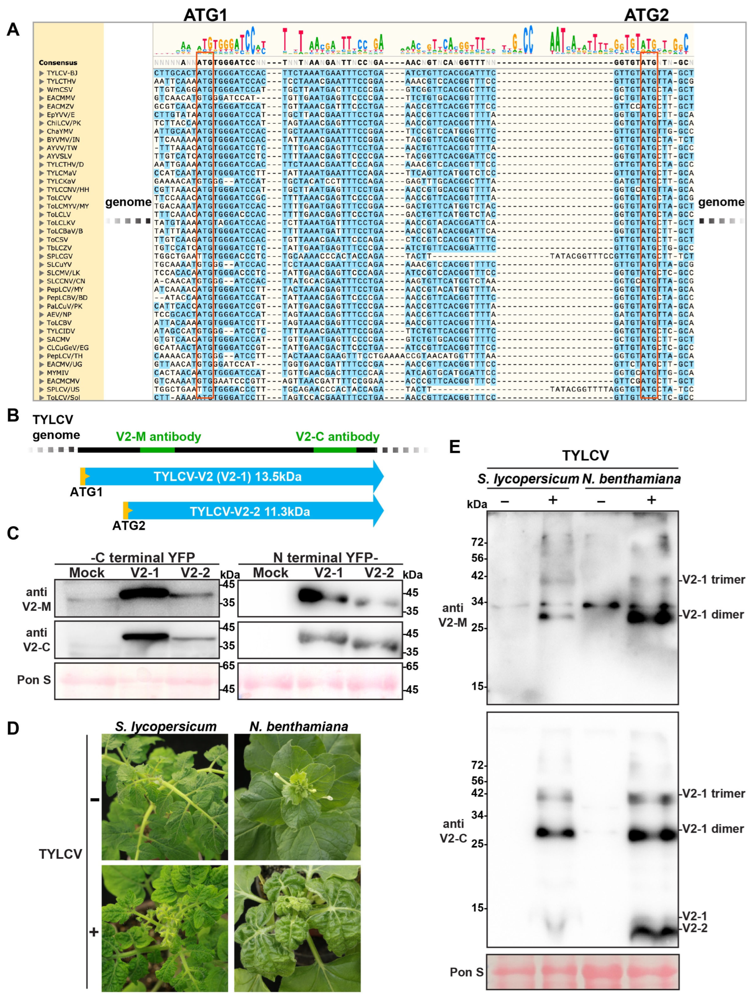
3.1. TYLCV Encodes Two Distinct V2 Protein Variants

Previous report showed TYLCTHV, a begomovirus, has two in-frame AUG initiation codons within the AV2 ORF, and both of them were required for viral full virulence [32]. To investigate whether the presence of two AUG codons in the AV2 ORF is conserved among begomoviruses, we analyzed representative Begomovirus species through sequence alignments. The results revealed widespread conservation of dual initiation codons in V2 proteins across different begomoviruses (Figure 1A), suggesting the evolutionary conservation of this molecular feature and providing a foundation for functional characterization. Consistent with the previous finding in the TYLCTHV AV2 ORFs [29], the TYLCV V2 ORF also encodes two in-frame isoforms: V2-1 (the full-length isoform initiating from the first ATG, 13.5 kDa) and V2-2 (initiating from the second ATG, 11.3 kDa) (Figure 1B).
To determine whether TYLCV encodes both V2 proteins during infection, two custom-made antibodies targeting distinct regions were generated: V2-M targets the central region of the V2, while V2-C specifically recognizes the C-terminal region. Antibody effectiveness was validated via Western blot using N-terminal/C-terminal YFP fusion proteins of V2-1 and V2-2 transiently expressed in N. benthamiana (Figure 1C). Systemic leaves showing severe symptoms were collected at 30 days post-inoculation (dpi) from TYLCV-infected tomato and N. benthamiana plants (Figure 1D). Western blot analysis of total proteins revealed distinct detection patterns: the absence of monomer bands using the V2-M antibody reflects selective recognition of highly accumulated multimeric complexes, whereas concurrent detection of V2-1 and V2-2-specific bands by the V2-C antibody in TYLCV-infected N. benthamiana samples demonstrates that TYLCV encodes two V2 isoforms during infection (Figure 1E). Although N. benthamiana data confirmed the formation of V2-1 and V2-2, the monomeric forms of V2 were not detected in tomato samples under the experimental conditions employed (Figure 1E). Under the current experimental conditions, the lack of detectable signal could be attributed to either lower accumulation of the monomers in tomato or potential differences in antibody detection performance across plant species.
Analyses of yeast two-hybrid and BiFC assays (Figure S1) confirmed that V2-1 and V2-2 are both capable of self-interaction and mutual interaction. These observations were further supported by the granular aggregates observed during subcellular localization of V2-1 and V2-2 (Figure S2). However, Western blot analysis failed to detect polymeric forms of V2-2 (Figure 1E). We speculate this may be attributed to the disruption of its comparatively weaker self-interaction bonds under denaturing gel conditions, contrasting with the stable interactions of V2-1 itself. Notably, V2-1 consistently formed nuclear aggregates regardless of YFP fusion at its N- or C-terminus, whereas V2-2 exhibited diffuse nuclear distribution without aggregation (Figure S2).

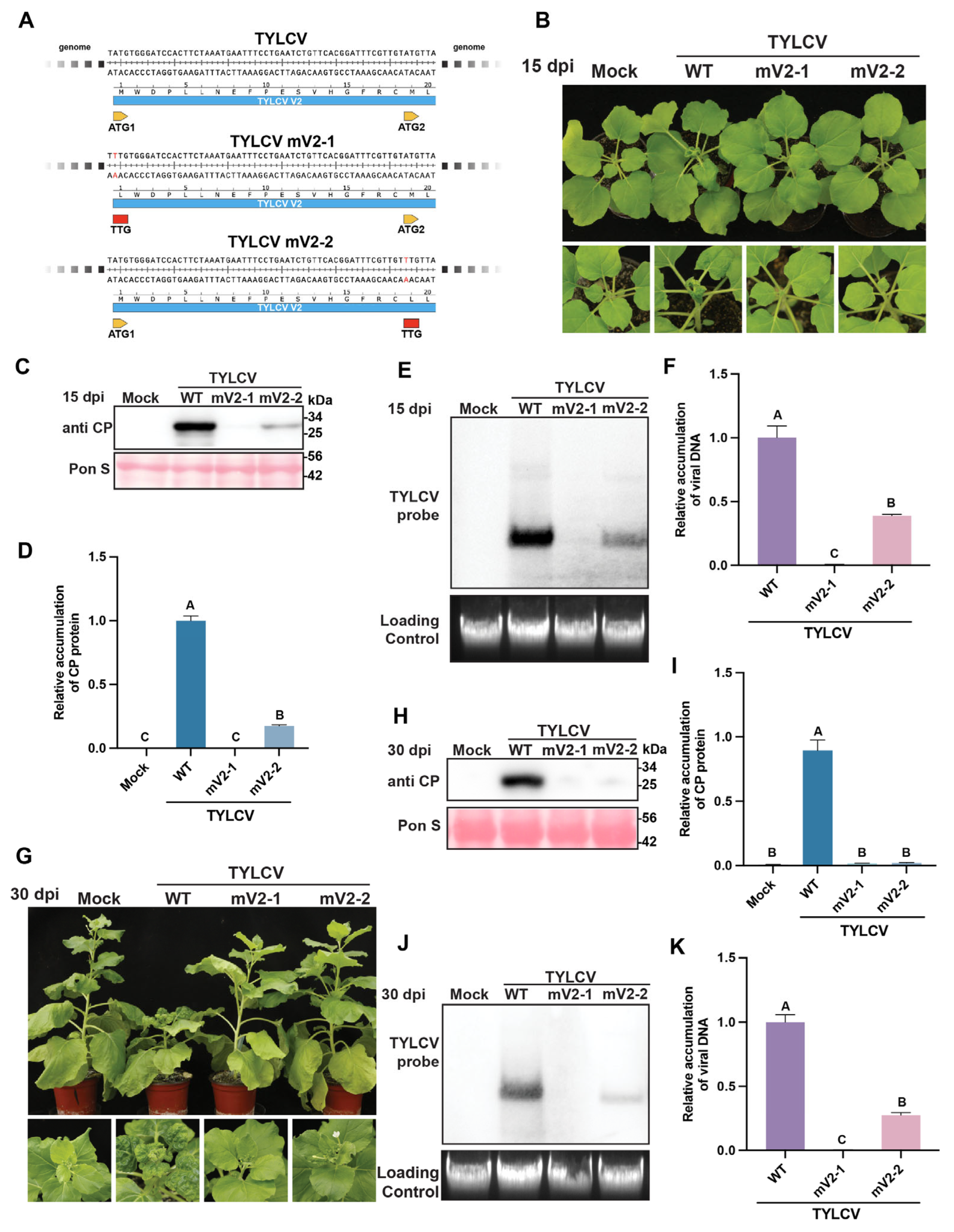
3.2. Mutations in Both ATG Codons of the V2 Protein Impair TYLCV Infectivity in N. benthamiana

To delineate the roles of two V2 isoforms encoded by the TYLCV genome [V2-1 (translated from ATG1 initiation codon) and V2-2 (translated from the second ATG, ATG2 initiation codon)] during viral infection, we engineered V2-specific knockout mutants using site-directed mutagenesis. Specifically, nucleotide substitutions were introduced at the first base of ATG1 (A→T) and ATG2 (A→T) in the V2 coding sequence of the TYLCV infectious clone. These mutations abolish methionine initiation codons while substituting methionine (M/Met) with leucine (L/Leu), which has comparable hydrophobicity and side-chain volume [47,48], thereby blocking isoform-specific translation while minimizing structural perturbations from radical amino acid substitutions. The mutagenesis strategy was meticulously designed to avoid interference with overlapping open reading frames (ORFs). Sequence-verified mutants included TYLCV-mV2-1 (V2-1 isoform knock-out) and TYLCV-mV2-2 (V2-2 isoform knock-out) (Figure 2A). This conservative substitution approach ensures the specific ablation of individual V2 isoforms without compromising global protein folding parameters, thereby enabling the precise functional dissection of each translation variant. To evaluate the roles of V2-1/V2-2 proteins in viral infection, the model plant N. benthamiana was first selected as the experimental system. A. tumefaciens suspensions (OD600 = 0.6) carrying the EV (pCambia1300), wild-type TYLCV, TYLCV mV2-1, or TYLCV mV2-2 were infiltrated into healthy 4–5 leaf-stage N. benthamiana plants. At 15 days post-inoculation (15 dpi), wild-type TYLCV-infected plants exhibited characteristic leaf curling (Figure 2B), while both TYLCV mV2-1- and mV2-2-infected groups showed no visible symptoms, with leaf morphology and growth states indistinguishable from the EV control. Western blot detection of TYLCV CP showed strong signals in the WT-infected group (Figure 2C). ImageJ quantification revealed the CP accumulation levels drastically reduced to 0.08% (mV2-1) and 17.54% (mV2-2) of WT levels (Figure 2D), with both mutants showing highly significant differences compared to WT. Southern blot analysis using digoxigenin-labeled probes detected clear viral genomic signals in the TYLCV-WT group, while the mV2-1 group showed near-background signals and the mV2-2 group exhibited faint but detectable viral DNA (Figure 2E). qPCR further confirmed viral genomic copy numbers at 0.60% (mV2-1) and 38.98% (mV2-2) of WT levels (Figure 2F). These data indicate that at 15 dpi in N. benthamiana, the absence of V2-1 resulted in >99% loss of viral replication capacity, while V2-2 deletion caused ~60% replication suppression. This suggests that V2-1 deficiency nearly abolishes viral colonization in the host, whereas V2-2 plays a substantial yet non-decisive role in viral infection and proliferation.
Further phenotypic and molecular analyses were conducted at 30 dpi to assess viral pathogenicity. Wild-type TYLCV-infected plants displayed aggravated symptoms including severe stunting and pronounced apical leaf curling, while both mutant-infected groups remained asymptomatic, maintaining phenotypic stability consistent with 15 dpi observations (Figure 2G). Quantitative Western blot analysis showed CP accumulation levels of 0.88% (mV2-1) and 3.35% (mV2-2) relative to WT (Figure 2H,I). Although mV2-2 exhibited marginally higher viral protein accumulation than mV2-1, the difference was not statistically significant. Southern blot analysis at 30 dpi failed to detect viral DNA in the mV2-1 group but identified significantly attenuated genomic signals in the mV2-2 group compared to TYLCV-WT (Figure 2J). qPCR validation revealed viral genomic accumulation levels of 0.49% (mV2-1) and 27.38% (mV2-2) relative to WT (Figure 2K). Both mutants showed highly significant reductions compared to WT, with significant inter-group differences, demonstrating that both V2-1 and V2-2 are critical for TYLCV infection in N. benthamiana, while V2-1 exerts a stronger functional impact than V2-2—a conclusion fully consistent with 15 dpi results.

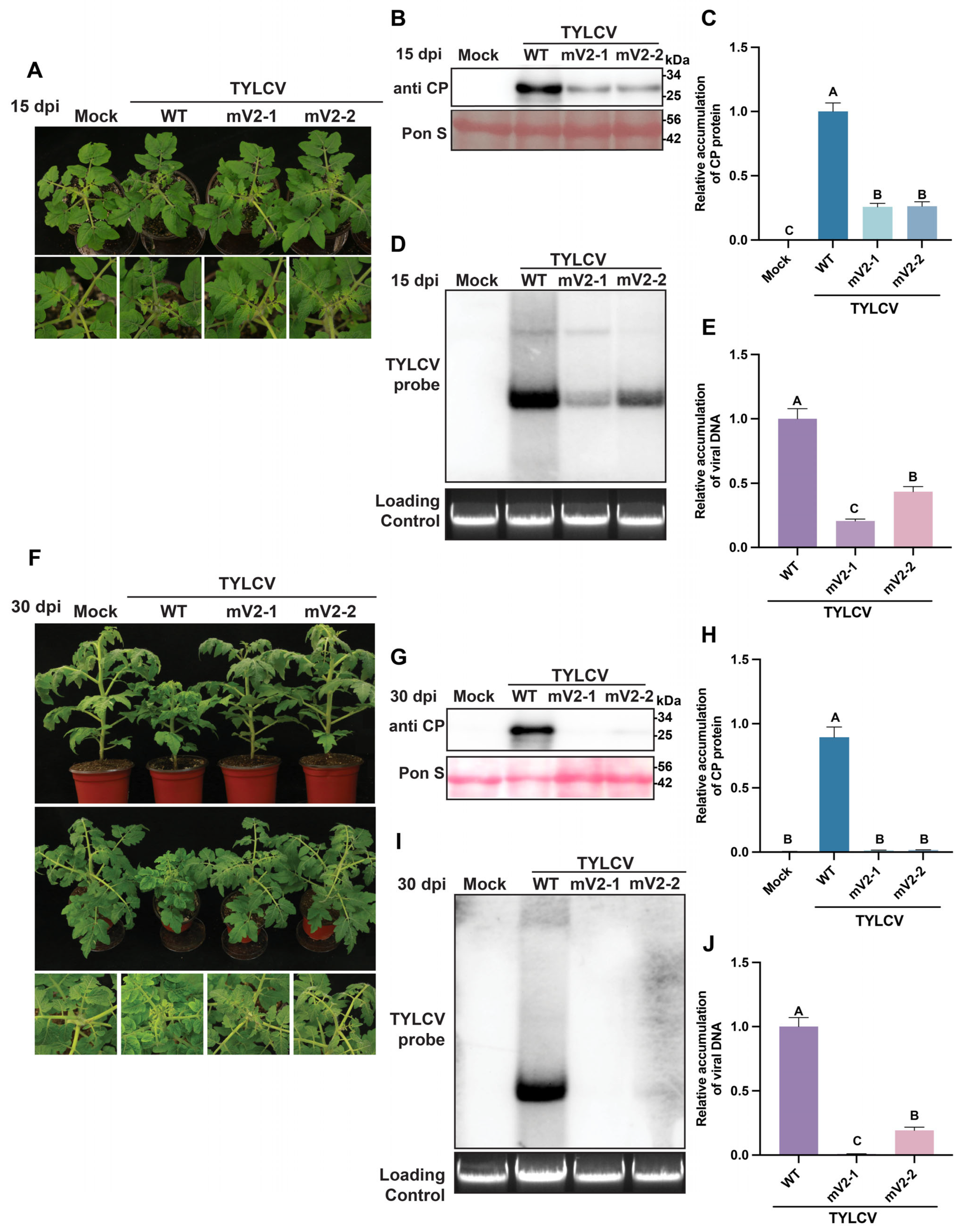
3.3. Mutations in Both ATG Codons of the V2 Protein Impair TYLCV Infectivity in S. lycopersicum

To validate the functional conservation of V2 isoforms in natural hosts, this study further evaluated the infectivity of TYLCV V2 mutants (mV2-1 and mV2-2) using tomato as the experimental system. A. tumefaciens suspensions (OD600 = 0.6) carrying EV (Mock), wild-type (WT), or mutants were infiltrated into true-leaf-stage tomato plants (8 biological replicates per treatment), with phenotypic observations and sampling conducted at 15 dpi and 30 dpi. At 15 dpi, TYLCV-WT-infected tomato plants exhibited characteristic apical leaf curling symptoms, including leaf yellowing, marginal curling, and growth retardation (Figure 3A). In contrast, both mV2-1- and mV2-2-infected groups showed no significant phenotypic differences from the Mock control, with no visible symptoms, indicating that deletion of either V2 protein severely compromises viral pathogenicity. Western blot analysis of viral CP accumulation revealed CP signal intensities reduced to 25.82% (mV2-1) and 26.29% (mV2-2) of WT levels (Figure 3B,C). Southern blot analysis further demonstrated a greater reduction in ssDNA/dsDNA replicative intermediate abundance in the mV2-1 group compared to mV2-2 (Figure 3D). qPCR quantification confirmed a 79.46% reduction in viral genomic accumulation for mV2-1 and 56.62% for mV2-2 compared to WT (Figure 3E). Notably, both mutants retained partial replication capacity in tomato (particularly mV2-2), despite significantly lower CP/viral DNA accumulation than WT, suggesting potential host factor-mediated compensation for V2 functions in natural hosts. At 30 dpi, TYLCV-WT-infected plants displayed aggravated symptoms including pronounced stunting alongside leaf curling (Figure 3F), while both mutant groups remained asymptomatic, consistent with 15 dpi observations. Quantitative Western blot showed CP levels further declining to 1.62% (mV2-1) and 1.86% (mV2-2) of WT at 30 dpi (Figure 3G,H), while Southern blot and qPCR analyses showed undetectable viral genomes for mV2-1 and merely 18.65% genomic retention in mV2-2 at the same time point (Figure 3I,J), indicating progressive impairment of viral replication/packaging efficiency over time. Compared to 15 dpi, mV2-1 and mV2-2 infectious clones-infected plants exhibited significantly reduced viral CP levels and DNA accumulation relative to wild-type TYLCV at 30 dpi—a time-dependent reduction that may suggest active host defenses selectively clearing replication-defective virions.
The experimental findings reveal three fundamental insights into V2 isoform functionality. Firstly, V2-1 exhibits host-dependent functional divergence, as evidenced by its retention of 20.54% viral DNA and 25.82% CP accumulation in tomato (S. lycopersicum) at 15 dpi, markedly higher than the 0.60% genomic and 0.08% CP levels observed in N. benthamiana, suggesting potential compensation through host-specific molecular interactions. Secondly, a conserved functional hierarchy positions V2-1 as the dominant isoform across both plant systems, with mV2-2-infected plants consistently maintaining significantly higher viral genomic accumulation than mV2-1 at both 15 dpi and 30 dpi. Thirdly, temporal analysis uncovers progressive attenuation dynamics in tomato, where mutant viral loads declined from 20.54% (mV2-1) and 43.38% (mV2-2) at 15 dpi to undetectable and 18.65% levels, respectively, by 30 dpi, indicative of active host defense mechanisms selectively eliminating replication-compromised virions. These collective observations establish V2-1 as the principal virulence determinant while highlighting host-pathogen coevolutionary adaptations modulating viral persistence.

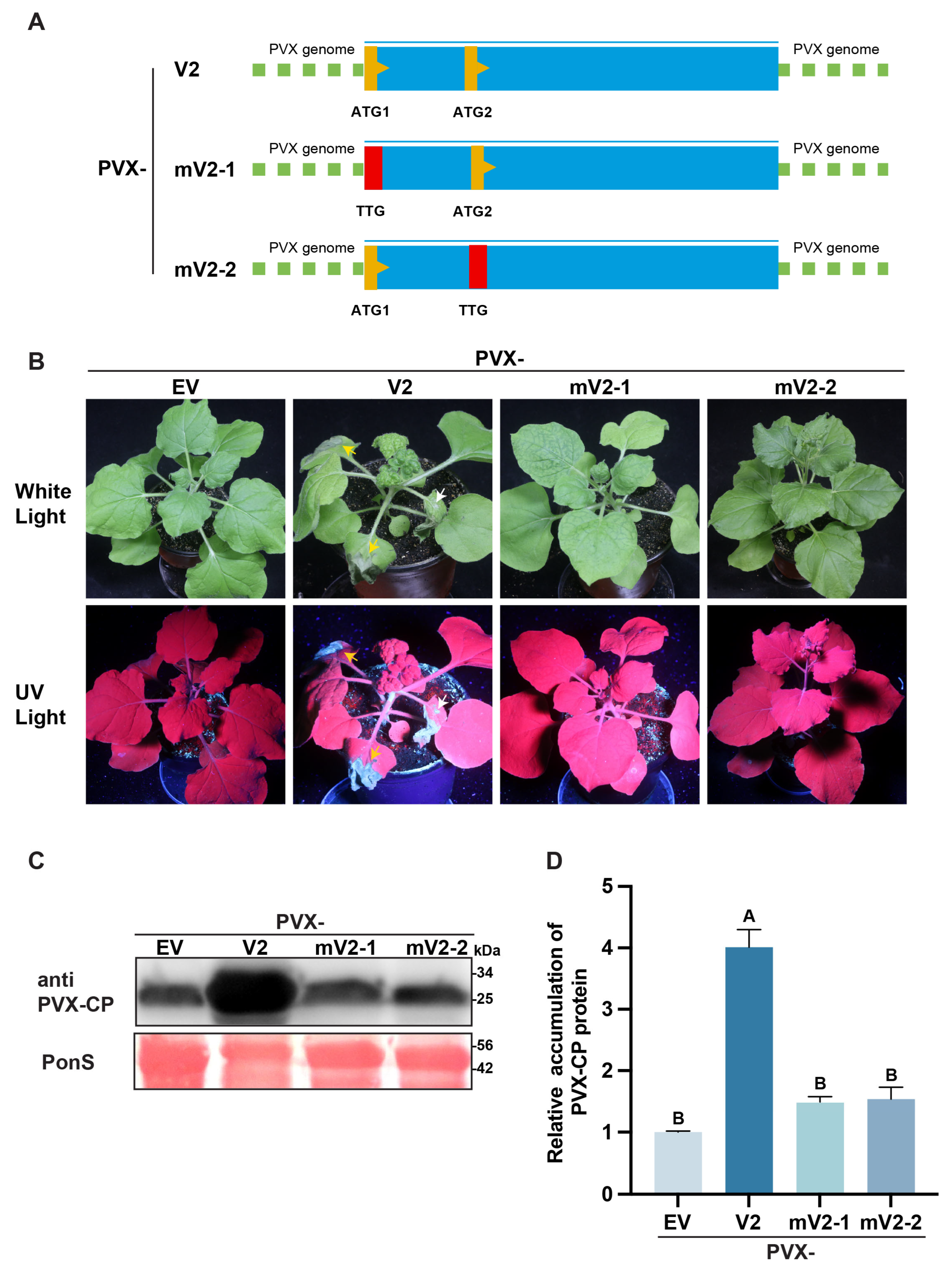
3.4. Dual ATG Mutations in the V2 Protein Compromise Its Ability to Potentiate Potato Virus X (PVX) Infection

Previous results demonstrated that full-length V2 expression via the PVX vector induces necrosis in N. benthamiana [49,50,51], indicating that the V2 protein enhances PVX virulence and suppresses the immune defense in plants. To dissect the roles of two distinct translation initiation products of TYLCV V2 (V2-1 and V2-2) in potentiating PVX infection, we conducted systematic functional analyses using the PVX heterologous expression system. The V2 coding region of TYLCV was cloned and mutagenized as follows: the full-length open reading frame (ORF) from the first initiation codon (ATG1) to the stop codon was defined as V2 (full-length protein). To investigate the functional independence of the two initiation sites, site-directed mutagenesis was performed: the first initiation codon (ATG→TTG) was mutated to generate mV2-1, and the second initiation codon (ATG→TTG) was mutated to generate mV2-2. These substitutions replaced the initiator methionine (Met) with leucine (Leu), thereby abolishing translation of the corresponding viral proteins, consistent with the mutagenesis strategy for infectious clones (Figure 2A and Figure 4A). The constructs (V2, mV2-1, mV2-2) and the PVX EV control were agroinfiltrated (OD600 = 0.6) into 4–5 leaf-stage N. benthamiana plants. Phenotypic observations were conducted at 9 dpi. Results showed necrosis in both inoculated and systemic leaves of PVX-V2-infected plants, whereas no necrosis was observed in PVX-mV2-1 and PVX-mV2-2 treated groups (Figure 4B). Western blot analysis revealed significantly reduced PVX CP accumulation in PVX-mV2-1 and PVX-mV2-2-infected plants compared to PVX-V2-infected plants, indicating that both initiation products are essential for V2-mediated enhancement of the PVX CP accumulation (Figure 4C,D). These results collectively demonstrate that V2-dependent potentiation of PVX-induced necrosis requires the cooperative functions of both V2-1 and V2-2.

3.5. Mutations at Both ATG Sites of the V2 Protein Attenuate Its Gene Silencing Suppression Activity

To investigate the mechanistic contributions of two TYLCV V2 translation initiation products (V2-1 and V2-2) to TGS suppression, this study employed the 16-TGS transgenic N. benthamiana system for systematic functional analysis. The 16-TGS line functions as a TGS model because its genomically integrated GFP gene undergoes endogenous RNA silencing-mediated mRNA degradation, resulting in no detectable fluorescence under UV excitation. Delivery of exogenous TGS suppressor proteins via the PVX vector restores GFP expression (detectable fluorescence) by inhibiting host transcriptionally silencing pathways, enabling functional identification of suppressors. PVX constructs (V2, mV2-1, mV2-2) and the positive control PVX-βC1 (a known TGS suppressor) were agroinfiltrated (OD600 = 0.6) into 4–5 leaf-stage 16-TGS N. benthamiana plants. Fluorescence phenotyping and Western blot analysis were performed at 7 dpi. PVX-βC1 (positive control) showed strong GFP fluorescence, confirming efficient TGS suppression. PVX-V2 (wild-type) exhibited comparable fluorescence intensity to βC1, validating potent TGS suppression by V2. PVX-mV2-1 and PVX-mV2-2 mutants displayed significantly attenuated fluorescence, indicating that loss of either V2-1 or V2-2 severely compromises TGS suppression, with both isoforms being indispensable for full V2 activity (Figure 5A). The Western blot analysis and quantitative data on GFP accumulation correlate with the observed phenotypes (Figure 5B,C). Collectively, phenotypic and molecular data demonstrate that V2-mediated TGS suppression requires the cooperative action of V2-1 and V2-2, potentially involving complex regulatory networks spanning translation initiation sites.
Given the established roles of V2-1/V2-2 in TGS suppression, parallel analysis of PTGS suppression was conducted using the 16c transgenic N. benthamiana system. Constructs (V2, mV2-1, mV2-2), negative control (EV), and positive control (P19) were co-infiltrated (1:1 ratio) with 35S-GFP-carrying A. tumefaciens into 4–5 leaf-stage 16c N. benthamiana. Fluorescence phenotyping and western blot analysis were performed at 4 dpi. Fluorescence results (Figure 5D,E) and quantification analyses (Figure 5F) showed that P19 (positive control) induced strong GFP fluorescence via PTGS suppression. V2 exhibited comparable fluorescence to P19, confirming robust PTGS suppression. However, mV2-1 and mV2-2 groups showed significantly reduced fluorescence, indicating that both isoforms are critical for full PTGS suppression activity. In conclusion, phenotypic and molecular analyses demonstrate that V2-mediated PTGS suppression requires synergistic contributions from both V2-1 and V2-2, with neither isoform being dispensable.

4. Discussion

Increasing evidence shows that plant and animal viruses encode additional small proteins, which are required for viral pathogenicity and suppression of host defenses [17,52,53,54,55,56,57,58]. Geminiviruses, the largest group of plant viruses, have recently also drawn attention due to their encoding of additional small proteins [58,59]. Our several reports showed that TYLCV encodes more than the previously described 6 canonical proteins and that at least some small proteins from TYLCV and ToLCCNV displayed specific subcellular localization patterns and had virulence functions [24,25,26,59]. Of note, Chiu and his colleagues also identified several hidden ORFs using TYLCTHV, a bipartite geminivirus as a model, and revealed that the translation of different protein isoforms was required for the viral pathogenesis in tomato plants [29]. They first identified genes beyond the annotated gene sets by experimentally profiling in vivo translation initiation sites (TISs), and they found that unanticipated AUG TISs were prevalent and determined. Two downstream in-frame TISs were identified in the viral gene AV2 that were conserved in the begomovirus lineage and led to the translation of different protein isoforms with different subcellular localizations. Mutations of AV2 isoforms significantly attenuated disease symptoms in tomato, suggesting the biological significance of these hidden open reading frames [29]. Consistent with this finding, we also revealed that the evolutionary conservation and functional roles of two initiation codons (ATG1 and ATG2) in the V2 protein of TYLCV, a monopartite geminivirus, during viral infection, through employing multidisciplinary approaches to elucidate their critical contributions to viral infection, pathogenicity, and suppression of host defense responses.
Firstly, by analysis of geminiviral coding sequences, we found widespread conservation of dual initiation codons (ATG1 and ATG2) in the geminiviral V2 ORFs across viral evolution (Figure 1A) as reported previously [29]. Such conservation likely reflects functional importance, potentially conferring selective advantages in viral survival or host adaptation, thereby providing an evolutionary framework for subsequent functional investigations. To functionally validate the expression of these conserved V2 isoforms, we generated antibodies targeting distinct V2 regions (Figure 1B). Western blot analysis of TYLCV-infected N. benthamiana confirmed concurrent expression of both V2-1 and V2-2 monomers (Figure 1C–E), demonstrating viral encoding of dual isoforms during infection. Surprisingly, monomeric V2 forms were not detected in tomato under identical experimental conditions (Figure 1E). This lack of detectable signals could be attributed to either lower monomer accumulation in tomato or potential limitations in antibody detection performance across plant species. Site-directed mutagenesis of ATG1 (V2-1 knockout, mV2-1) and ATG2 (V2-2 knockout, mV2-2) (Figure 2A) revealed divergent contributions: V2-1 serves as the core determinant of viral infection, with mV2-1 showing drastically reduced genomic copies and CP accumulation in N. benthamiana, and no viral DNA was detectable by Southern blot analysis (Figure 2). This demonstrates that V2-1, translated from ATG1, is indispensable for viral systemic infection. Compared to V2-1, V2-2 might play an auxiliary role, as TYLCV carrying mV2-2-infecting plants exhibited higher genomic copies than mV2-1. However, the V2-2 mutation still significantly attenuated viral pathogenicity, suggesting its importance in the virus infection cycle (Figure 2 and Figure 3). This may relate to V2-2′s involvement in non-core functions such as virion assembly or host defense suppression. Host-dependent functional divergence was also observed: mV2-1 retained higher viral accumulation in tomato than in N. benthamiana (Figure 2B–F and Figure 3A–E). This phenotype may involve partial functional compensation by tomato-specific host factors and/or compensatory activity from other viral ORFs that function more effectively in natural host plants. This highlights the critical influence of specific host-virus interactions on functional assessments. Notably, in tomato, viral accumulation in TYLCV carrying mV2-1 or mV2-2-infecting plants declined progressively from 15 to 30 dpi, with parallel reductions in CP levels (Figure 3). This time-dependent attenuation likely reflects host-mediated recognition, defense, and clearance of defective viruses, underscoring the necessity for intact V2 expression to counteract the host immunity response. Furthermore, lower viral accumulation in N. benthamiana mutants compared to tomato suggests an innate immune sensitivity to defective viruses. The PVX heterologous system revealed synergistic roles of V2-1/V2-2 in pathogenicity: (1) Systemic necrosis in N. benthamiana required both isoforms, as PVX-V2-induced necrosis was abolished when either ATG was mutated (Figure 4). ATG mutants (PVX-mV2-1/mV2-2) failed to induce necrosis, suggesting that V2-1 and V2-2 cooperatively activate host necrosis pathways via physical interaction or functional complementation. (2) TGS suppression by V2 in 16-TGS plants required both isoforms, as ATG mutations (mV2-1/mV2-2) significantly reduced fluorescence (Figure 5A–C), implicating their cooperation across distinct silencing suppression steps. (3) In 16c plants, both mV2-1 and mV2-2 severely impaired PTGS suppression (Figure 5D–F), confirming that neither isoform is indispensable for V2′s PTGS suppression activity. Collectively, this result delineates the functional synergy between dual initiation codons in geminiviral V2 proteins, establishing V2-1′s central role in replication and V2-2′s auxiliary contributions, while elucidating their coordinated actions in viral pathogenicity and virus-host interactions.

5. Conclusions

This study elucidates the functional divergence of dual-initiation codon-derived V2 isoforms (V2-1 and V2-2) in TYLCV pathogenesis. Through custom antibody validation and deletion mutant analyses, we demonstrate that V2-1 is indispensable for viral replication and systemic spread, while V2-2 serves as an auxiliary virulence factor. Host-specific regulation was evidenced by differential viral attenuation patterns in host tomato versus N. benthamiana species, suggesting host defense-mediated clearance. Crucially, heterologous co-expression confirmed synergistic interactions between isoforms in inducing systemic necrosis and suppressing transcriptional/post-transcriptional gene silencing. These findings significantly advance our understanding of geminivirus coding complexity, revealing how overlapping ORFs optimize viral genome efficiency and host manipulation. This work provides foundational insights for developing resistance strategies against globally destructive geminiviruses.

Supplementary Materials

The following supporting information can be downloaded at: https://www.mdpi.com/article/10.3390/agronomy15071726/s1, Figure S1: V2-1 and V2-2 exhibit self-interaction and mutual interaction; Figure S2: Subcellular localization of V2-1 and V2-2 in Nicotiana benthamiana epidermal cells.

Author Contributions

Conceptualization, F.L. and X.Z.; methodology, Z.W.; software, P.G.; validation, Z.W., P.G. and S.Z.; formal analysis, Z.W. and P.G.; investigation, Z.W. and P.G.; resources, F.L.; data curation, Z.W. and P.G.; writing—original draft preparation, Z.W. and F.L.; writing—review and editing, Z.W., F.L., P.G., X.Z. and S.Z.; visualization, Z.W.; supervision, F.L. and X.Z.; project administration, F.L. and X.Z.; funding acquisition, X.Z. and F.L. All authors have read and agreed to the published version of the manuscript.

Funding

This research was supported by grants from the National Key Research and Development Program of China (2022YFD1400800 and 2021YFD1400400) and the National Natural Science Foundation of China (32172385 and W2411024).

Data Availability Statement

Data are contained within the article and Supplementary Materials.

Acknowledgments

The authors thank David M. Bisaro for providing the 16-TGS seeds, David C. Baulcombe for the provision of the transgenic GFP 16c line and the PVX vector, and Michael M. Goodin for sharing the transgenic RFP-H2B line.

Conflicts of Interest

The authors declare no conflict of interest.

Abbreviations

The following abbreviations are used in this manuscript:
TYLCVTomato yellow leaf curl virus
ATGInitiation codon
TGSTranscriptional gene silencing
PTGSPost-transcriptional gene silencing
CPCoat protein
RepReplication-associated protein
TrAPTranscription activator protein
REnReplication enhancer protein
NSPNuclear shuttle protein
MPMovement protein
MYMIVmungbean yellow mosaic India virus
ALCScVageratum leaf curl Sichuan virus
TYLCCNBtomato yellow leaf curl China betasatellite
RaLCBradish leaf curl betasatellite
ORFOpen reading frame
RSVRice streak virus
TSSTranscription start site
PDPlasmodesmata
EREndoplasmic reticulum
TYLCTHVTomato yellow leaf curl Thailand virus
VSRViral suppressor of RNA silencing
SGS3Suppressor of gene silencing 3
RDR6RNA-dependent RNA polymerase 6
siRNASmall interfering RNA
HDAsHistone deacetylases
AGO4Argonaute 4
ToLCCNVTomato leaf curl China virus
TISTranslation initiation site
MCSMultiple cloning site
qPCRQuantitative polymerase chain reaction
CTABCetyltrimethylammonium bromide
TAETris-acetate-EDTA buffer
SSCSaline-sodium citrate buffer
DIGDigoxigenin
dpiDays post-inoculation
HRPHorseradish peroxidase
Y2HYeast two-hybrid
BiFCBimolecular fluorescence complementation
V2-1Full-length V2 isoform (initiated from ATG1)
V2-2Truncated V2 isoform (initiated from ATG2)
WTWild-type
mV2-1V2-1 isoform knockout mutant
mV2-2V2-2 isoform knockout mutant
PVXPotato virus X
ssDNASingle-stranded DNA
dsDNADouble-stranded DNA
EBEthidium bromide
RbcLRubisco large subunit
GFPGreen fluorescent protein
A. tumefaciensAgrobacterium tumefaciens
N. benthamianaNicotiana benthamiana
S. lycopersicumSolanum lycopersicum

Appendix A

Table A1. RT-qPCR primers used in this study.
NameSequence (5′ to 3′)
NbActinF: AAGCTGCAGGTATCCATGAGACTA
R: CAATCCAGACACTGTACTTTCTCTC
SlActinF: AAAGACCAGCTCATCTGTTGAGAAG
R: GTGGTTTCATGAATACCAGCAGC
TYLCV-DNAF: CACGCCCGTCTCGAAG
R: CATAAGACTGGACTTTACATGGGC
Table A2. Primers for vector construction.
Construct NameSequence (5′ to 3′)
pDonr221-TYLCV-V2-1F: GGGGACAAGTTTGTACAAAAAAGCAGGCTTAATGTGGGATCCACTTCTAAATGAATTTCCT
R: GGGGACCACTTTGTACAAGAAAGCTGGGTTGGGCTTCGATACATTCTGTATATTCTGGG
pDonr221-TYLCV-V2-2F: GGGGACAAGTTTGTACAAAAAAGCAGGCTTAATGTTAGCTATTAAATATTTGCAGTCCG
R: GGGGACCACTTTGTACAAGAAAGCTGGGTTGGGCTTCGATACATTCTGTATATTCTGGG
pDonr221-TYLCVmV2-1F: GGGGACAAGTTTGTACAAAAAAGCAGGCTTATTGTGGGATCCACTTCTAAATGAATTTCCT
R: GGGGACCACTTTGTACAAGAAAGCTGGGTTGGGCTTCGATACATTCTGTATATTCTGGG
pDonr221-TYLCVmV2-2F: GGGGACAAGTTTGTACAAAAAAGCAGGCTTATTGTTAGCTATTAAATATTTGCAGTCCG
R: GGGGACCACTTTGTACAAGAAAGCTGGGTTGGGCTTCGATACATTCTGTATATTCTGGG
2 × 35S-TYLCV-V2-FlagF: CTCAAGCTTCGAATTCCTGCAATGTGGGATCCACTTCTAAATGAATTTC
R: GTCCATGCCACCTCCGGGCTTCGATACATTCTGTATATTC
2 × 35S-TYLCV-mV2-1-FlagF: CTCAAGCTTCGAATTCCTGCATTGTGGGATCCACTTCTAAATGAATTTC
R: GTCCATGCCACCTCCGGGCTTCGATACATTCTGTATATTC
2 × 35S-TYLCV-mV2-2-FlagF: CTCAAGCTTCGAATTCCTGCATTGTGGGATCCACTTCTAAATGAATTTC
R: GTCCATGCCACCTCCGGGCTTCGATACATTCTGTATATTC
PVX-TYLCV-V2F: GTCAGCACCAGCTAGCATCGATATGTGGGATCCACTTCTAAATGAATTTC
R: TAACCGTTCATCGGCGGTCGACTCAGGGCTTCGATACATTCTGTATATTCTG
PVX-TYLCV-mV2-1F: GTCAGCACCAGCTAGCATCGATTTGTGGGATCCACTTCTAAATGAATTTC
R: TAACCGTTCATCGGCGGTCGACTCAGGGCTTCGATACATTCTGTATATTCTG
PVX-TYLCV-mV2-2F: CAGCACCAGCTAGCATCGATTTGTTAGCTATTAAATATTTGCAGTCCGTTG
R: TAACCGTTCATCGGCGGTCGACTCAGGGCTTCGATACATTCTGTATATTCTG
pCambia2300-TYLCV-mV2-1F: CTTGCACTTTGTGGGATCCACTTCTAAATGAATT
R: ATCCCACAAAGTGCAAGACAAACTACTTGGGG
pCambia2300-TYLCV-mV2-2F: CGGATTTCGTTGTTTGTTAGCTATTAAATATTTGCAGTCCG
R: ACAAACAACGAAATCCGTGAACAGATTCAGGA
pCAMBIA1300-N-3×HA-V2-1-YNF: TGCCGGACTACGCGGGAATGTGGGATCCACTTCTAAATGAATTTC
R: CGCCCTTGCTCACCATACTAGTATTTAAATGGGGCTTCGATACATTCTGTATATTCTGG
pCAMBIA1300-N-3×HA-V2-2-YNF: CATATGATGTGCCGGACTACGCGGGAATGTTAGCTATTAAATATTTGCAGTCCGTTG
R: CGCCCTTGCTCACCATACTAGTATTTAAATGGGGCTTCGATACATTCTGTATATTCTGG
pCAMBIA1300-N-Flag-V2-1-YCF: TGACAAGCTTGGAGGTGGGATGTGGGATCCACTTCTAAATGAATTTCC
R: GCCGTGGTTCATTCTGCAGGGCTTCGATACATTCTGTATATTCTGGG
pCAMBIA1300-N-Flag-V2-2-YCF: GACAAGCTTGGAGGTGGGATGTTAGCTATTAAATATTTGCAGTCCGTTGAG
R: GCCGTGGTTCATTCTGCAGGGCTTCGATACATTCTGTATATTCTGGG

Appendix B

Table A3. All begomoviruses included in the V2 sequence alignment in this study.
Virus SpeciesAcronymGenBank Accession Number
watermelon chlorotic stunt virusWmCSVAJ012081
East African cassava mosaic Malawi virusEACMMVAJ006460
East African cassava mosaic Zanzibar virusEACMZVAF422174
eupatorium yellow vein virusEpYVV/EAB433979
chilli leaf curl virusChiLCV/PKAF336806
chayote yellow mosaic virusChaYMVAJ223191
bhendi yellow vein mosaic virusBYVMV/INAF241479
ageratum yellow vein virusAYVV/TWAF307861
ageratum yellow vein Sri Lanka virusAYVSLVAF314144
tomato yellow leaf curl Thailand virusTYLCTHV/DAF206674
tomato yellow leaf curl Malaga virusTYLCMaVAF271234
tomato yellow leaf curl Kanchanaburi virusTYLCKaVAF511529;
tomato yellow leaf curl China virusTYLCCNV/HHAF311734
tomato leaf curl Vietnam virusToLCVVAF264063
tomato leaf curl Malaysia virusToLCMYV/MYAF327436
tomato leaf curl Laos virusToLCLVAF195782
tomato leaf curl Sri Lanka virusToLCLKVAF274349
tomato leaf curl Bangalore virusToLCBaV/BAF295401
tomato curly stunt virusToCSVAF261885
tobacco leaf curl Zimbabwe virusTbLCZVAF350330
sweet potato leaf curl Georgia virusSPLCGVAF326775
squash leaf curl Yunnan virusSLCuYVAJ420319
Sri Lankan cassava mosaic virusSLCMV/LKAJ314737
squash leaf curl China virusSLCCNV/CNAF509743
pepper leaf curl virusPepLCV/MYAF414287
pepper leaf curl Bangladesh virusPepLCBV/BDAF314531
papaya leaf curl virusPaLCuV/PKAJ436992
fiageratum enation virusAEV/NPAJ437618
tomato leaf curl Bangladesh virusToLCBVAF188481
tomato yellow leaf curl Indonesia virusTYLCIDVAF189018
South African cassava mosaic virusSACMVAF155806
cotton leaf curl Gezira virusCLCuGeV/EGAF155064
pepper leaf curl virusPepLCV/THAF134484
East African cassava mosaic virusEACMV/UGAF126806
mungbean yellow mosaic India virusMYMIVAF126406
East African cassava mosaic Cameroon virusEACMCMVAF112354
sweet potato leaf curl virusSPLCV/USAF104036
tomato leaf curl virusToLCV/SolAF084006

References

  1. Wang, Y.; Pruitt, R.N.; Nürnberger, T.; Wang, Y. Evasion of Plant Immunity by Microbial Pathogens. Nat. Rev. Microbiol. 2022, 20, 449–464. [Google Scholar] [CrossRef] [PubMed]
  2. Navas-Castillo, J.; Fiallo-Olivé, E.; Sánchez-Campos, S. Emerging Virus Diseases Transmitted by Whiteflies. Annu. Rev. Phytopathol. 2011, 49, 219–248. [Google Scholar] [CrossRef]
  3. Rojas, M.R.; Macedo, M.A.; Maliano, M.R.; Soto-Aguilar, M.; Souza, J.O.; Briddon, R.W.; Kenyon, L.; Rivera Bustamante, R.F.; Zerbini, F.M.; Adkins, S.; et al. World Management of Geminiviruses. Annu. Rev. Phytopathol. 2018, 56, 637–677. [Google Scholar] [CrossRef]
  4. Fiallo-Olivé, E.; Pan, L.-L.; Liu, S.-S.; Navas-Castillo, J. Transmission of Begomoviruses and Other Whitefly-Borne Viruses: Dependence on the Vector Species. Phytopathology 2020, 110, 10–17. [Google Scholar] [CrossRef]
  5. Brown, J.K.; Zerbini, F.M.; Navas-Castillo, J.; Moriones, E.; Ramos-Sobrinho, R.; Silva, J.C.F.; Fiallo-Olivé, E.; Briddon, R.W.; Hernández-Zepeda, C.; Idris, A.; et al. Revision of Begomovirus Taxonomy Based on Pairwise Sequence Comparisons. Arch. Virol. 2015, 160, 1593–1619. [Google Scholar] [CrossRef]
  6. Rybicki, E.P. A Phylogenetic and Evolutionary Justification for Three Genera of Geminiviridae. Arch. Virol. 1994, 139, 49–77. [Google Scholar] [CrossRef]
  7. Fiallo-Olivé, E.; Lett, J.-M.; Martin, D.P.; Roumagnac, P.; Varsani, A.; Zerbini, F.M.; Navas-Castillo, J. ICTV Virus Taxonomy Profile: Geminiviridae 2021: This Article Is Part of the ICTV Virus Taxonomy Profiles Collection. J. Gen. Virol. 2021, 102, 001696. [Google Scholar] [CrossRef]
  8. Pooma, W.; Petty, I.T.D. Tomato Golden Mosaic Virus Open Reading Frame Al4 Is Genetically Distinct from Its C4 Analogue in Monopartite Geminiviruses. J. Gen. Virol. 1996, 77, 1947–1951. [Google Scholar] [CrossRef]
  9. Sunter, G.; Bisaro, D.M. Transactivation in a Geminivirus: AL2 Gene Product Is Needed for Coat Protein Expression. Virology 1991, 180, 416–419. [Google Scholar] [CrossRef]
  10. Fondong, V.N. Geminivirus Protein Structure and Function. Mol. Plant Pathol. 2013, 14, 635–649. [Google Scholar] [CrossRef]
  11. Laufs, J. Geminivirus Replication: Genetic and Biochemical Characterization of Rep Protein Function, a Review. Biochimie 1995, 77, 765–773. [Google Scholar] [CrossRef] [PubMed]
  12. Rojas, M.R.; Jiang, H.; Salati, R.; Xoconostle-Cázares, B.; Sudarshana, M.R.; Lucas, W.J.; Gilbertson, R.L. Functional Analysis of Proteins Involved in Movement of the Monopartite Begomovirus, Tomato Yellow Leaf Curl Virus. Virology 2001, 291, 110–125. [Google Scholar] [CrossRef] [PubMed]
  13. Hanley-Bowdoin, L.; Settlage, S.B.; Orozco, B.M.; Nagar, S.; Robertson, D. Geminiviruses: Models for Plant DNA Replication, Transcription, and Cell Cycle Regulation. Crit. Rev. Plant Sci. 2000, 35, 105–140. [Google Scholar] [CrossRef] [PubMed]
  14. Lazarowitz, S.G.; Beachy, R.N. Viral Movement Proteins as Probes for Intracellular and Intercellular Trafficking in Plants. Plant Cell 1999, 11, 535–548. [Google Scholar] [CrossRef]
  15. Zhou, X. Advances in Understanding Begomovirus Satellites. Annu. Rev. Phytopathol. 2013, 51, 357–381. [Google Scholar] [CrossRef]
  16. Saunders, K.; Bedford, I.D.; Yahara, T.; Stanley, J. The Earliest Recorded Plant Virus Disease. Nature 2003, 422, 831. [Google Scholar] [CrossRef]
  17. Orr, M.W.; Mao, Y.; Storz, G.; Qian, S.-B. Alternative ORFs and Small ORFs: Shedding Light on the Dark Proteome. Nucleic Acids Res. 2020, 48, 1029–1042. [Google Scholar] [CrossRef]
  18. Li, F.; Xu, X.; Huang, C.; Gu, Z.; Cao, L.; Hu, T.; Ding, M.; Li, Z.; Zhou, X. The AC 5 Protein Encoded by Mungbean Yellow Mosaic India Virus Is a Pathogenicity Determinant That Suppresses RNA Silencing-based Antiviral Defenses. New Phytol. 2015, 208, 555–569. [Google Scholar] [CrossRef]
  19. Li, P.; Su, F.; Meng, Q.; Yu, H.; Wu, G.; Li, M.; Qing, L. The C5 Protein Encoded by Ageratum Leaf Curl Sichuan Virus Is a Virulence Factor and Contributes to the Virus Infection. Mol. Plant Pathol. 2021, 22, 1149–1158. [Google Scholar] [CrossRef]
  20. Hu, T.; Song, Y.; Wang, Y.; Zhou, X. Functional Analysis of a Novel βV1 Gene Identified in a Geminivirus Betasatellite. Sci. China Life Sci. 2020, 63, 688–696. [Google Scholar] [CrossRef]
  21. Gupta, N.; Reddy, K.; Gnanasekaran, P.; Zhai, Y.; Chakraborty, S.; Pappu, H.R. Functional Characterization of a New ORF βV1 Encoded by Radish Leaf Curl Betasatellite. Front. Plant Sci. 2022, 13, 972386. [Google Scholar] [CrossRef]
  22. Prasad, A.; Sharma, N.; Hari-Gowthem, G.; Muthamilarasan, M.; Prasad, M. Tomato Yellow Leaf Curl Virus: Impact, Challenges, and Management. Trends Plant Sci. 2020, 25, 897–911. [Google Scholar] [CrossRef]
  23. Lin, W.; Qiu, P.; Jin, J.; Liu, S.; Ul Islam, S.; Yang, J.; Zhang, J.; Kormelink, R.; Du, Z.; Wu, Z. The Cap Snatching of Segmented Negative Sense RNA Viruses as a Tool to Map the Transcription Start Sites of Heterologous Co-Infecting Viruses. Front. Microbiol. 2017, 8, 2519. [Google Scholar] [CrossRef]
  24. Gong, P.; Zhao, S.; Liu, H.; Chang, Z.; Li, F.; Zhou, X. Tomato Yellow Leaf Curl Virus V3 Protein Traffics along Microfilaments to Plasmodesmata to Promote Virus Cell-to-Cell Movement. Sci. China Life Sci. 2022, 65, 1046–1049. [Google Scholar] [CrossRef] [PubMed]
  25. Gong, P.; Tan, H.; Zhao, S.; Li, H.; Liu, H.; Ma, Y.; Zhang, X.; Rong, J.; Fu, X.; Lozano-Durán, R.; et al. Geminiviruses Encode Additional Small Proteins with Specific Subcellular Localizations and Virulence Function. Nat. Commun. 2021, 12, 4278. [Google Scholar] [CrossRef]
  26. Zhao, S.; Gong, P.; Ren, Y.; Liu, H.; Li, H.; Li, F.; Zhou, X. The Novel C5 Protein from Tomato Yellow Leaf Curl Virus Is a Virulence Factor and Suppressor of Gene Silencing. Stress Biol. 2022, 2, 19. [Google Scholar] [CrossRef]
  27. Liu, H.; Chang, Z.; Zhao, S.; Gong, P.; Zhang, M.; Lozano-Durán, R.; Yan, H.; Zhou, X.; Li, F. Functional Identification of a Novel C7 Protein of Tomato Yellow Leaf Curl Virus. Virology 2023, 585, 117–126. [Google Scholar] [CrossRef]
  28. Stern-Ginossar, N.; Ingolia, N.T. Ribosome Profiling as a Tool to Decipher Viral Complexity. Annu. Rev. Virol. 2015, 2, 335–349. [Google Scholar] [CrossRef]
  29. Chiu, C.-W.; Li, Y.-R.; Lin, C.-Y.; Yeh, H.-H.; Liu, M.-J. Translation Initiation Landscape Profiling Reveals Hidden Open-Reading Frames Required for the Pathogenesis of Tomato Yellow Leaf Curl Thailand Virus. Plant Cell 2022, 34, 1804–1821. [Google Scholar] [CrossRef]
  30. Hanley-Bowdoin, L.; Bejarano, E.R.; Robertson, D.; Mansoor, S. Geminiviruses: Masters at Redirecting and Reprogramming Plant Processes. Nat. Rev. Microbiol. 2013, 11, 777–788. [Google Scholar] [CrossRef]
  31. Poornima Priyadarshini, C.G.; Ambika, M.V.; Tippeswamy, R.; Savithri, H.S. Functional Characterization of Coat Protein and V2 Involved in Cell to Cell Movement of Cotton Leaf Curl Kokhran Virus-Dabawali. PLoS ONE 2011, 6, e26929. [Google Scholar] [CrossRef] [PubMed]
  32. Luna, A.P.; Rodríguez-Negrete, E.A.; Morilla, G.; Wang, L.; Lozano-Durán, R.; Castillo, A.G.; Bejarano, E.R. V2 from a Curtovirus Is a Suppressor of Post-Transcriptional Gene Silencing. J. Gen. Virol. 2017, 98, 2607–2614. [Google Scholar] [CrossRef] [PubMed]
  33. Glick, E.; Zrachya, A.; Levy, Y.; Mett, A.; Gidoni, D.; Belausov, E.; Citovsky, V.; Gafni, Y. Interaction with Host SGS3 Is Required for Suppression of RNA Silencing by Tomato Yellow Leaf Curl Virus V2 Protein. Proc. Natl. Acad. Sci. USA 2008, 105, 157–161. [Google Scholar] [CrossRef] [PubMed]
  34. Wang, L.; Ding, Y.; He, L.; Zhang, G.; Zhu, J.-K.; Lozano-Duran, R. A Virus-Encoded Protein Suppresses Methylation of the Viral Genome through Its Interaction with AGO4 in the Cajal Body. Elife 2020, 9, e55542. [Google Scholar] [CrossRef]
  35. Wang, B.; Yang, X.; Wang, Y.; Xie, Y.; Zhou, X. Tomato Yellow Leaf Curl Virus V2 Interacts with Host Histone Deacetylase 6 To Suppress Methylation-Mediated Transcriptional Gene Silencing in Plants. J. Virol. 2018, 92, e00036-18. [Google Scholar] [CrossRef]
  36. Wang, Y.; Wu, Y.; Gong, Q.; Ismayil, A.; Yuan, Y.; Lian, B.; Jia, Q.; Han, M.; Deng, H.; Hong, Y.; et al. Geminiviral V2 Protein Suppresses Transcriptional Gene Silencing through Interaction with AGO4. J. Virol. 2019, 93, e01675-18. [Google Scholar] [CrossRef]
  37. Baulcombe, D.C. The Role of Viruses in Identifying and Analyzing RNA Silencing. Annu. Rev. Virol. 2022, 9, 353–373. [Google Scholar] [CrossRef]
  38. Wu, Q.; Luo, Y.; Lu, R.; Lau, N.; Lai, E.C.; Li, W.-X.; Ding, S.-W. Virus Discovery by Deep Sequencing and Assembly of Virus-Derived Small Silencing RNAs. Proc. Natl. Acad. Sci. USA 2010, 107, 1606–1611. [Google Scholar] [CrossRef]
  39. Mlotshwa, S.; Pruss, G.J.; Vance, V. Small RNAs in Viral Infection and Host Defense. Trends Plant Sci. 2008, 13, 375–382. [Google Scholar] [CrossRef]
  40. Li, F.; Wang, Y.; Zhou, X. SGS3 Cooperates with RDR6 in Triggering Geminivirus-Induced Gene Silencing and in Suppressing Geminivirus Infection in Nicotiana benthamiana. Viruses 2017, 9, 247. [Google Scholar] [CrossRef]
  41. Yoshikawa, M.; Iki, T.; Tsutsui, Y.; Miyashita, K.; Poethig, R.S.; Habu, Y.; Ishikawa, M. 3′ Fragment of miR173-Programmed RISC-Cleaved RNA Is Protected from Degradation in a Complex with RISC and SGS3. Proc. Natl. Acad. Sci. USA 2013, 110, 4117–4122. [Google Scholar] [CrossRef]
  42. Kumakura, N.; Takeda, A.; Fujioka, Y.; Motose, H.; Takano, R.; Watanabe, Y. SGS3 and RDR6 Interact and Colocalize in Cytoplasmic SGS3/RDR6-Bodies. FEBS Lett. 2009, 583, 1261–1266. [Google Scholar] [CrossRef] [PubMed]
  43. Zhao, S.; Gong, P.; Liu, J.; Liu, H.; Lozano-Durán, R.; Zhou, X.; Li, F. Geminivirus C5 Proteins Mediate Formation of Virus Complexes at Plasmodesmata for Viral Intercellular Movement. Plant Physiol. 2023, 193, 322–338. [Google Scholar] [CrossRef] [PubMed]
  44. Wang, Y.; Gong, Q.; Wu, Y.; Huang, F.; Ismayil, A.; Zhang, D.; Li, H.; Gu, H.; Ludman, M.; Fátyol, K.; et al. A Calmodulin-Binding Transcription Factor Links Calcium Signaling to Antiviral RNAi Defense in Plants. Cell Host Microbe 2021, 29, 1393–1406.e7. [Google Scholar] [CrossRef]
  45. Wu, J.-X.; Shang, H.-L.; Xie, Y.; Shen, Q.-T.; Zhou, X.-P. Monoclonal Antibodies against the Whitefly-Transmitted Tomato Yellow Leaf Curl Virus and Their Application in Virus Detection. J. Integr. Agric. 2012, 11, 263–268. [Google Scholar] [CrossRef]
  46. Wu, J.; Zhou, X. Production and application of monoclonal antibodies against Potato virus X. J. Zhejiang Univ. B 2005, 31, 608–612. [Google Scholar]
  47. Machkovech, H.M.; Bloom, J.D.; Subramaniam, A.R. Comprehensive Profiling of Translation Initiation in Influenza Virus Infected Cells. PLOS Pathog. 2019, 15, e1007518. [Google Scholar] [CrossRef]
  48. Creixell, P.; Schoof, E.M.; Tan, C.S.H.; Linding, R. Mutational Properties of Amino Acid Residues: Implications for Evolvability of Phosphorylatable Residues. Trans. R. Soc. B Biol. Sci. 2012, 367, 2584–2593. [Google Scholar] [CrossRef]
  49. Zrachya, A.; Glick, E.; Levy, Y.; Arazi, T.; Citovsky, V.; Gafni, Y. Suppressor of RNA Silencing Encoded by Tomato Yellow Leaf Curl Virus-Israel. Virology 2007, 358, 159–165. [Google Scholar] [CrossRef]
  50. Sharma, P.; Ikegami, M. Tomato Leaf Curl Java Virus V2 Protein Is a Determinant of Virulence, Hypersensitive Response and Suppression of Posttranscriptional Gene Silencing. Virology 2010, 396, 85–93. [Google Scholar] [CrossRef]
  51. Luna, A.P.; Morilla, G.; Voinnet, O.; Bejarano, E.R. Functional Analysis of Gene-Silencing Suppressors from Tomato Yellow Leaf Curl Disease Viruses. Mol. Plant-Microbe Interact. 2012, 25, 1294–1306. [Google Scholar] [CrossRef]
  52. Li, F.; Jia, M.; Wang, A. Hidden Viral Proteins: How Powerful Are They? PLOS Pathog. 2024, 20, e1011905. [Google Scholar] [CrossRef]
  53. Dinan, A.M.; Lukhovitskaya, N.I.; Olendraite, I.; Firth, A.E. A Case for a Negative-Strand Coding Sequence in a Group of Positive-Sense RNA Viruses. Virus Evol. 2020, 6, veaa007. [Google Scholar] [CrossRef]
  54. Firth, A.E.; Brierley, I. Non-Canonical Translation in RNA Viruses. J. Gen. Virol. 2012, 93, 1385–1409. [Google Scholar] [CrossRef]
  55. Retallack, H.; Popova, K.D.; Laurie, M.T.; Sunshine, S.; DeRisi, J.L. Persistence of Ambigrammatic Narnaviruses Requires Translation of the Reverse Open Reading Frame. J. Virol. 2021, 95, 10–1128. [Google Scholar] [CrossRef] [PubMed]
  56. Zhang, Y.; Liang, X.; Zhao, M.; Qi, T.; Guo, H.; Zhao, J.; Zhao, J.; Zhan, G.; Kang, Z.; Zheng, L. A Novel Ambigrammatic Mycovirus, PsV5, Works Hand in Glove with Wheat Stripe Rust Fungus to Facilitate Infection. Plant Commun. 2023, 4, 100505. [Google Scholar] [CrossRef] [PubMed]
  57. Chen, Y.; Ge, L.; Li, Z.; Wang, A.; Zhou, X.; Li, F. Targeting of viral RNAs by nonsense-mediated decay is compromised by virus–plant interactions. New Plant Prot. 2024, 1, e8. [Google Scholar] [CrossRef]
  58. Tan, H.; Li, F.; Zhou, X.; Lozano-Durán, R. New Genes on the Block: Additional Open Reading Frames in the Genomes of Geminiviruses. Trends Microbiol. 2025. [Google Scholar] [CrossRef]
  59. Wang, Z.; Wang, Y.; Lozano-Duran, R.; Hu, T.; Zhou, X. Identification of a Novel C6 Protein Encoded by Tomato Leaf Curl China Virus. Phytopathol. Res. 2022, 4, 46. [Google Scholar] [CrossRef]
Figure 1. Evolutionary conservation and validation of dual V2 isoforms in TYLCV infection. (A) Phylogenetic conservation of dual initiation codons (ATG1/ATG2) in V2 proteins among begomoviruses. Analysis of representative and additional species within the genus Begomovirus is shown in the figure, with sequence information displayed for the V2 protein region. ATG1 and ATG2 loci are highlighted with red rectangular annotations. (B) Genomic organization of the TYLCV V2 open reading frame (ORF) encoding in-frame isoforms: V2-1 (initiated from ATG1, 13.5 kDa) and V2-2 (initiated from ATG2, 11.3 kDa). Solid and dashed black lines represent viral genomic components, green shaded areas indicate antibody-targeted regions, blue arrows denote ORFs for V2-1 (full-length V2) and V2-2, and yellow arrows mark ATG1/ATG2 initiation codons. (C) Antibody specificity validation: Western blot analysis of YFP-V2-1/V2-2 and V2-1/V2-2-YFP fusion proteins expressed in Nicotiana benthamiana at 2 days post-infiltration (dpi). Ponceau S (Pon S) staining of Rubisco large subunit (RbL) serves as a loading control. (D) S. lycopersicum and N. benthamiana plants inoculated with EV control or TYLCV infectious clone at 30 dpi. (E) Detection of viral proteins by Western Blot using 15% SDS-PAGE gel in tomato and N. benthamiana samples from panel D using V2-M and V2-C antibodies. Pon S staining of RbL confirms equal loading.
Agronomy 15 01726 g001
Figure 2. Infectivity analysis of TYLCV V2 mutants in N. benthamiana. (A) Schematic diagram of the construction of two V2 isoform protein deletion mutants. The blue rectangle represents the V2 coding region. The yellow arrow indicates the functional start codon (ATG). The red rectangle represents the mutated, nonfunctional start codon. The black, long-dashed line indicates the remaining genomic sequence of TYLCV. (B) Systemic symptoms of N. benthamiana plants at 15 dpi. From left to right: Mock (EV, negative control), wild-type TYLCV (WT), V2-1 knockout mutant (mV2-1), V2-2 knockout mutant (mV2-2). (C) Western blot analysis of TYLCV CP accumulation in systemic leaves of plants, shown in panel (B), Pon S staining of RbcL shows protein loading. (D) Quantification of band signal intensities from C (n = 3) using ImageJ software (version 1.53k). Statistical significance was assessed by One-Way ANOVA with Tukey’s HSD test. Columns labeled with distinct uppercase letters indicate extremely significant differences (p < 0.01). (E) Southern blot analysis of viral genomic DNA accumulation in systemic leaves sampled from plants in (B). Ethidium bromide (EB) nucleic acid staining confirmed uniform DNA loading amounts across all samples. (F) qPCR analysis of viral genomic DNA accumulation in systemic leaves sampled from plants in (B) (n = 3), normalized to the NbActin internal reference. Data normalized to the mean viral load of WT. Statistical significance was assessed by One-Way ANOVA with Tukey’s HSD test. Columns labeled with distinct uppercase letters indicate extremely significant differences (p < 0.01). (G) Systemic symptoms of N. benthamiana plants at 30 dpi. From left to right: Mock (EV, negative control), WT, mV2-1, mV2-2. (H) Western blot analysis of sustained TYLCV CP accumulation in systemic leaves, Pon S staining of RbcL shows protein loading. (I) Quantification of band signal intensities from (H) (n = 3) using ImageJ software. Statistical significance was assessed by One-Way ANOVA with Tukey’s HSD test. Columns labeled with distinct uppercase letters indicate extremely significant differences (p < 0.01). (J) Southern blot analysis of long-term viral genomic DNA replication in systemic leaves from plants in (G), EB nucleic acid staining confirmed uniform DNA loading amounts across all samples. (K) qPCR analysis of viral genomic DNA accumulation in systemic leaves from plants in (G) (n = 3), normalized to the NbActin internal reference. Statistical significance was assessed by One-Way ANOVA with Tukey’s HSD test. Columns labeled with distinct uppercase letters indicate extremely significant differences (p < 0.01).
Agronomy 15 01726 g002
Figure 3. Infectivity analysis of TYLCV V2 protein mutants in tomato (S. lycopersicum) (A) Systemic symptoms of tomato plants at 15 dpi. From left to right: Mock (EV, negative control), TYLCV-WT, -mV2-1, and -mV2-2 infecting tomato plants. (B) Western blot analysis of TYLCV CP accumulation in systemic leaves sampled from plants in (A), Pon S staining of RbcL shows protein loading. (C) Quantification of band signal intensities from (B) (n = 3) using ImageJ. Statistical significance was assessed by One-Way ANOVA with Tukey’s HSD test. Columns labeled with distinct uppercase letters indicate extremely significant differences (p < 0.01). (D) Southern blot analysis of viral genomic DNA replication in systemic leaves sampled from plants in (A), EB nucleic acid staining confirmed uniform DNA loading amounts across all samples. (E) qPCR analysis of viral genomic DNA accumulation in systemic leaves sampled from plants in (A) (n = 3), normalized to the SlActin internal reference. Statistical significance was assessed by One-Way ANOVA with Tukey’s HSD test. Columns labeled with distinct uppercase letters indicate extremely significant differences (p < 0.01). (F) Systemic symptoms of tomato plants at 30 dpi. From left to right: Mock (EV, negative control), TYLCV-WT, -mV2-1, and -mV2-2 infecting tomato plants. (G) Western blot analysis of TYLCV CP accumulation in systemic leaves sampled from plants in (F), Pon S staining of RbcL shows protein loading. (H) Quantification of band signal intensities from (G) (n = 3) using ImageJ. Statistical significance was assessed by One-Way ANOVA with Tukey’s HSD test. Columns labeled with distinct uppercase letters indicate extremely significant differences (p < 0.01). (I) Southern blot analysis of viral genomic DNA replication in systemic leaves sampled from plants in (F), EB nucleic acid staining confirmed uniform DNA loading amounts across all samples. (J) qPCR analysis of viral genomic DNA accumulation in systemic leaves sampled from plants in (F) (n = 3), normalized to the SlActin internal reference. Statistical significance was assessed by One-Way ANOVA with Tukey’s HSD test. Columns labeled with distinct uppercase letters indicate extremely significant differences (p < 0.01).
Agronomy 15 01726 g003
Figure 4. Disruption of either V2 initiation codon abrogates PVX-V2-induced necrosis in N. benthamiana. (A) Schematic diagram of PVX vector constructs. The blue rectangle represents the V2 coding region. The yellow rectangle-triangle composite symbol denotes the functional initiation codon (ATG), while the red rectangle indicates the mutated nonfunctional initiation codon. The green dashed line represents the remaining sequence of the PVX vector. (B) Phenotypic observation of N. benthamiana plants infiltrated with PVX (EV, negative control), PVX-V2, PVX-mV2-1, or PVX-mV2-2 under white light and UV light at 9 dpi. Necrotic lesions are more pronounced under UV illumination: white arrows indicate systemic leaf necrosis, and yellow arrows denote necrosis in infiltrated leaves. (C) Western blot analysis of PVX CP accumulation levels in the plants shown in panel (B), Pon S staining of RbcL shows protein loading. (D) Quantification of the Western blot results from panel (C) (n = 3). Statistical significance was assessed by One-Way ANOVA with Tukey’s HSD test. Columns labeled with distinct uppercase letters indicate extremely significant differences (p < 0.01).
Agronomy 15 01726 g004
Figure 5. Mutation of either V2 initiation codon attenuates the gene silencing suppressor activity of V2. (A) Phenotypic observation of 16-TGS plants infiltrated with PVX (EV, negative control), PVX-βC1 (positive control), PVX-V2, PVX-mV2-1, or PVX-mV2-2 under white light and UV illumination at 7 dpi. (B) Western blot analysis of GFP accumulation levels in the plants shown in panel (A). RbcL stained with Pon S serves as a loading control. (C) Quantification of GFP band intensity in panel (B) (n = 3). Statistical significance was assessed by One-Way ANOVA with Tukey’s HSD test. Columns labeled with distinct uppercase letters indicate extremely significant differences (p < 0.01). (D) Phenotype of N. benthamiana 16c plants under UV light at 4 dpi following co-infiltration with 35S-GFP and constructs expressing EV (negative control), P19 (positive control), V2, mV2-1, or mV2-2. (E) Western blot analysis of GFP accumulation in the infiltrated leaves shown in panel (A). RbcL stained with Pon S serves as a loading control. (F) Quantification of GFP band intensity in panel (B) (n = 3). Statistical significance was assessed by One-Way ANOVA with Tukey’s HSD test. Columns labeled with distinct uppercase letters indicate extremely significant differences (p < 0.01).
Agronomy 15 01726 g005
Disclaimer/Publisher’s Note: The statements, opinions and data contained in all publications are solely those of the individual author(s) and contributor(s) and not of MDPI and/or the editor(s). MDPI and/or the editor(s) disclaim responsibility for any injury to people or property resulting from any ideas, methods, instructions or products referred to in the content.

