Article

Silver Nanoparticles Embedded in Sodium Alginate: Antibacterial Efficacy and Effects on Red Cabbage Seedling Performance

by
Miłosz Rutkowski
1,2,3,
Wojciech Makowski
3,
Lidia Krzemińska-Fiedorowicz
2,
Karen Khachatryan
2,
Andrzej Kalisz
3,
Dagmara Malina
4,
Jarosław Chwastowski
4,
Zbigniew Wzorek
4,
Gohar Khachatryan
2,
Agnieszka Sękara
3 and
Anna Kołton
3,*
1
Centre for Innovation and Research on Prohealthy and Safe Food, University of Agriculture in Krakow, 30-149 Krakow, Poland
2
Faculty of Food Technology, University of Agriculture in Krakow, 31-120 Kraków, Poland
3
Faculty of Biotechnology and Horticulture, University of Agriculture in Krakow, 31-120 Krakow, Poland
4
Faculty of Chemical Engineering and Technology, Cracow University of Technology, 31-155 Krakow, Poland
*
Author to whom correspondence should be addressed.
Agronomy 2025, 15(7), 1640; https://doi.org/10.3390/agronomy15071640
Submission received: 20 May 2025 / Revised: 26 June 2025 / Accepted: 3 July 2025 / Published: 5 July 2025

Abstract

Innovative plant protection solutions are increasingly sought in modern agriculture. Rapid advances in nanotechnology offer promising opportunities to develop biodegradable, cost-effective composites containing silver nanoparticles (AgNPs) with well-documented antimicrobial properties. The aim of this study was to synthesize sodium alginate gels containing AgNPs, evaluate their physicochemical and antibacterial properties, and assess their effect on the growth of red cabbage (Brassica oleracea var. capitata f. rubra) seedlings. In accordance with the principles of green chemistry, AgNPs were chemically synthesized using sodium alginate as a stabilizer and fructose as a non-toxic reducing agent. The final composite contained 150 mg/L AgNPs and was diluted to 20 and 60 mg/L for biological tests. Antibacterial activity against Bacillus cereus, Enterococcus faecalis, Escherichia coli, and Pseudomonas aeruginosa was tested using agar diffusion assays. Seedling growth parameters and phytochemical content were assessed after 10 days of seedlings exposure to AgNPs. The results showed significant antibacterial activity in all tested strains. Crucially, neither AgNPs concentration negatively affected seedling development or phytochemical concentration. Application of AgNPs at concentration of 60 mg/L increased ascorbic acid and carotenoids content in comparison to control (deionized water). These results suggest that AgNPs-alginate composites may serve as sustainable antimicrobial agents in agriculture, inhibiting pathogens without compromising crop quality.

1. Introduction

The dynamic development of nanotechnology over recent decades has influenced intensive progress across various sectors of industry. Owing to economic, social and environmental benefits, the use of nanomaterials in the form of commercial, innovative solutions is of interest in such areas as cosmetology, medicine, biotechnology, materials technology and electronics [1,2]. Agriculture has likewise embraced nanotechnology: the large surface area and reactivity of nanoparticles promote increased biomass production. It is possible to use nanoparticles as substances that control the release of agricultural chemicals into the environment. This may therefore enable minimization of excessive soil fertilization. Nanoparticles increase the water retention process and increase the availability of nutrients in the soil. The use of nanoparticles as potential sensors can affect the early detection of microbial infections in plants [3]. Plant pathogens are a major constrain on agriculture. Bacterial diseases such as black rot cause losses in vegetable crops such as cabbage. One of the microorganisms causing black rot is the bacterium Xanthomonas campestris pv. campestris. Visible symptoms of infection are the formation of necrotic lesions on the leaves of the plant. In the field, infection occurs through stomata and wounds in the plant tissue. After entering the plant, the bacterium spreads rapidly to the tissues via the vascular system. Bacterial infections in plant production may also be caused by: Bacillus, Pseudomonas or Lactobacillus [4]. The consequences of crop infections do not refer only to the problem of reduced yields. Vegetables affected by bacterial disease are characterized by reduced quality and therefore lower value. Plant diseases also pose an economic problem. They cause increased labor costs, financial outlays on the agricultural sector and changes in farmers’ incomes [5]. Human pathogens can also infect vegetables during their production. The main sources of green food infections are contaminated irrigation water, agricultural waste, and soil fertilization with manure containing dangerous pathogens. Pathogens attach to the plant surface, penetrate tissues and multiply in the mesophyll. The process of plant disinfection is difficult and not always effective [6]. In particular, silver nanoparticles with well-known antimicrobial properties have attracted special attention from researchers in recent years. Their biological activity against pathogens such as Pseudomonas aeruginosa, Escherichia coli or Staphylococcus aureus is becoming a valuable aspect of their innovative use, especially in the context of the growing resistance of microorganisms to commonly used antimicrobial agents [7,8,9,10]. It is possible to obtain silver nanoparticles synthesized in a polysaccharide matrix. As a result of the reduction reaction of silver ions using a non-toxic reductant, nanoparticles can finally be deposited in the space of biodegradable carriers and stabilizers. A notable example of a biopolymer used in nanotechnology research is sodium alginate [11].
Sodium alginate is a biodegradable and biocompatible polysaccharide isolated from marine algae. It occurs in the form of a soluble salt of alginic acid. In molecular terms, it is composed of subunits of 1,4-β-d-mannuronic acid and α-l-guluronic acid, linked together by glycosidic bonds. Due to its physicochemical properties, alginate may be able to form flexible hydrogels significant in various fields of science and economy [12,13,14]. The process of obtaining hydrogels based on sodium alginate is an important area of research on modern forms of polysaccharide applications for creating innovative materials with bioactive effects. So far, the important issues have been the decomposition of these biomaterials, their safety for the environment, especially for agriculturally important crops such as cultivated vegetables [15].
Red cabbage (Brassica oleracea var. capitata f. rubra) is a widely cultivated, health-promoting vegetable and a valuable source of vitamins, fiber, polyphenols and fatty acid. Consequently, it has considerable dietary potential and is consumed raw or cooked [16,17,18,19]. Recent studies suggest that consuming red cabbage at various stages of its vegetative development may help reduce the risk of lifestyle diseases such as diabetes [20].
The aim of the conducted research was to synthesize alginate gels containing silver nanoparticles along with the evaluation of their physicochemical and antibacterial properties as well as to assess their effect on the growth of red cabbage seedlings (Brassica oleracea var. capitata f. rubra). The following hypotheses were verified through experimental work: (I) the use of fructose as a non-toxic reducing agent allows the synthesis of silver nanoparticles in a sodium alginate matrix, (II) the presence of silver nanoparticles synthesized within alginate composites effectively inhibits the growth of microorganisms, (III) applied silver nanoparticles do not exhibit toxicity to cabbage seedlings.

2. Materials and Methods

2.1. Reagents Used for the Synthesis of Polymer Composites with Silver Nanoparticles

Reagents used during synthesis: sodium alginate (weight ≈ 1.565 × 10⁵ Da); silver nitrate (99.99%); D(-)-fructose (≥99%); glycerin (99.5%). All reagents were purchased from Sigma-Aldrich (Poznan, Poland).

2.2. Synthesis of Polymer Gels with Silver Nanoparticles

The silver nanoparticles synthesis reaction was carried out in a manner similar to that described in the work’ of Rutkowski et al. [11,15] using the individual reagent contents presented in Table 1.
The polysaccharide gel (1.5%) was obtained by dissolving 1.50 g of sodium alginate in 95.50 g of distilled water until a homogeneous suspension was formed, at 60 °C for 24 h, adding glycerol as a plasticizer in a weight ratio of 1:2 to the weight of alginate. The gel obtained in this way was divided into two equal parts. To obtain the same concentration of alginate, 48.52 g of water was added to the first sample (AlgC), and a mixture consisting of 0.1 M silver nitrate solution, water and 20% ammonia as well as a reducing sugar solution (fructose) was added to the second sample (AlgAgNPs) and stirred on a magnetic stirrer at 60 °C until the tested suspension turned orange, characteristic of silver nanoparticles. In this way, an alginate gel with silver nanoparticles at an initial concentration of 150 mg/L and a gel without nanometals were obtained, which were then subjected to physicochemical analysis.

2.3. Physicochemical Analysis of Alginate Gels with Silver Nanoparticles

Morphological features of the obtained silver nanoparticles composite were determined using a high-resolution scanning electron microscope JEOL 7550 with a TEM (transmission electron microscope) detector (JEOL, Tokyo, Japan). Samples were prepared by depositing 10 µL of the sample on 200 Cu (100) mesh carbon-coated grids (TAAB Laboratories, Aldermaston, Berks, UK). The obtained pictures from TEM were used to the particle size distribution analysis conducted with ImageJ software (ImageJ, Wayne Rasband and contributors, National Institutes of Health, USA, version 1.54d).
The distribution of silver nanoparticles size was confirmed by the DLS (dynamic light scattering) technique using a Zetasizer Nano ZS (Malvern Instruments Ltd., Malvern, UK) which allows for determination of particle sizes in the range from 0.6 to 6000 nm. Scattered-light fluctuations were measured by positioning the detector at 173°. The polymer composite with silver nanoparticles was measured in triplicate with each measurement consisting of several 10 s runs to precisely determine mean values. The measurements were carried out at 25 °C.
UV-VIS (ultraviolet-visible) absorption spectra of the obtained composites were recorded using a Shimadzu 2101 scanning spectrophotometer (Shimadzu, Kyoto, Japan) in the range of 200–800 nm.
For Fourier Transform Infrared Attenuated Total Reflectance (FTIR-ATR) measurements, 25 g of gel from each sample were poured into Petri dishes and dried at 37 °C. Thin films were obtained and recorded in the range of 4000–700 cm−1 with a resolution of 4 cm−1 using a MATTSON 3000 FT-IR spectrophotometer (Mattson Technology, Inc., Madison, WI, USA) equipped with a 30SPEC 30° reflective attachment with a MIRacle ATR accessory from PIKE Technologies Inc., Madison, WI, USA.

2.4. Experimental Treatments

The initial alginate gels were diluted 7.5-fold and 2.5-fold. In this way, aqueous solutions with two silver nanoparticles concentration were obtained: 20 mg/L and 60 mg/L. Gels without silver nanoparticles were diluted in the same volumetric way: Alg(20) and Alg(60).
The following treatments were tested in the experiments with bacteria strain or plants:
  • C–control–sterile water (experiment with bacteria) or deionized water (experiment with plants)
  • Alg(20)–sodium alginate solution without silver nanoparticles diluted 7.5-fold
  • Alg(60)–sodium alginate solution without silver nanoparticles diluted 2.5-fold
  • 20 mg/L AgNPs–solution with silver nanoparticles at a concentration of 20 mg/L
  • 60 mg/L AgNPs–solution with silver nanoparticles at a concentration of 60 mg/L.

2.5. Antibacterial ActivityAssay of Silver Nanoparticles

The degree of sensitivity of selected Gram-positive (Bacillus cereus and Enterococcus faecalis) and Gram-negative (Escherichia coli and Pseudomonas aeruginosa) microorganisms to the test solutions was determined. The microbial strains used were purchased from the microbial bank (Leibniz Institute DSMZ). Before using the bacterial cell suspension, its optical density was determined at a wavelength of 540 nm. The number of cells per mL of solution used in the study was approximately 1.5 × 10⁶. A sterile culture medium (agar) was prepared and added to Petri dishes. Then, 100 μL of aqueous solutions of the tested alginate gels were added to the holes with a diameter of 1 cm in the agar medium. Then, 25 μL of bacterial cell suspension was added to the surface of the agar medium and spread over the entire surface of the medium in the conditions of a sterile laminar flow cabinet in triplicate. The samples prepared in this way were placed in an incubator at 30 °C. After 24 h, the values of the microorganism zone of growth inhibition were assessed [21].

2.6. Growth of Red Cabbage Seedlings in the Presence of Silver Nanoparticles

In this part of the experiment, a sample of 1 g of cabbage seeds (W. Legutko, Jutrosin, Poland) was sown in plastic containers containing cotton material (organic cotton) at the bottom, previously soaked in 20 mL of aqueous solutions of the tested gels. The control seeds were sown on material soaked in distilled water. Each treatment was performed in triplicate (3 containers per treatment). The plastic containers were stored for 10 days at 20 °C and illuminated 8 h a day with white light. After this time, the morphological and biochemical parameters of the obtained seedlings were assessed.
Seedling stem width and length measurements were taken using a caliper, and 10 randomly selected seedlings from each container were assessed. Cotyledon thickness was assessed using a dial thickness gauge (Mitutoyo, Kawasaki, Japan) in 10 replicates from each container. Fresh weight was assessed by weighing 10 randomly selected seedlings from each container. To determine the dry matter content of seedlings, the material was dried at 105 °C for 24 h. Dry matter measurements were performed using 10 randomly selected seedlings from each container.

2.7. Antioxidant Properties Assessment

All chemical analyzes were performed in triplicate laboratory replications. A Hitachi U2900 spectrophotometer (Hitachi, Tokyo, Japan) was used for analyses. Pigment compounds (carotenoids, chlorophyll a, chlorophyll b) were measured by extraction with 80% acetone. The absorbance of obtained solutions were measured at wavelengths of 470, 646, and 663 nm. The concentration of individual photosynthetic pigments were calculated using the formulas provided by Wellburn [22]. The anthocyanin content was determined according to the method presented by Lee et al. [23]. Ascorbic acid extraction was performed at 4 °C using 0.2 N HCl. The total ascorbate (both reduced and oxidized form) content was conducted according to method described by Queval and Noctor [24]. The ascorbic acid content was expressed as a percentage of the control, taking as 100% the ascorbic acid content for the control plants treated with distilled water (C). The antioxidant capacity of cabbage seedlings treated with silver nanoparticles were evaluated with three methods: radical scavenging activity with stable free radical 2,2-diphenyl-1-picrylhydrazyl (DPPH)—a DPPH assay; ferric reducing antioxidant power assay (FRAP) and cupric-ion-reducing antioxidant capacity assay (CUPRAC). After 5 min of reaction of plant-derived extracts with synthetic radical DPPH the absorbance was measured at 517 nm [25]. The FRAP method was based on the reduction in ferric–tripyridyl-s-triazine (Fe3+–TPTZ) complex to its ferrous derivative (Fe2+) [26]. The absorbance was measured at 595 nm after 5 min of reaction. The CUPRAC was performed according to Apak et al. [27]. The absorbance was measured at 450 nm after 5 min of reaction. The results of all antioxidant capacity assays were expressed as mMol Trolox (6-hydroxy-2,5,7,8-tetramethylchroman-2-carboxylic acid; Sigma-Aldrich) per 1 g of fresh weight. The determination of polyphenolic compounds was based on the Cicco et al. [28] work.

2.8. Statistical Analysis

Statistical significance of differences between experimental treatments was determined using one-way analysis of variance, and samples were separated into homogenous groups using Fisher’s LSD (Least Significant Difference) test, at α = 0.05 using Statistica software, version 13.3 (TIBCO Software Inc., Palo Alto, CA, USA). Data presented in bar charts and tables represent means and standard error.

3. Results

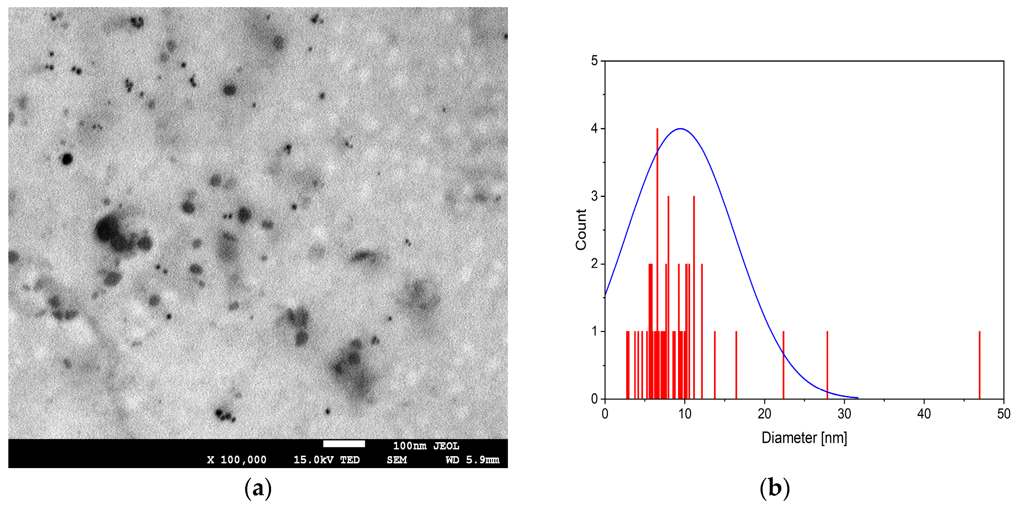
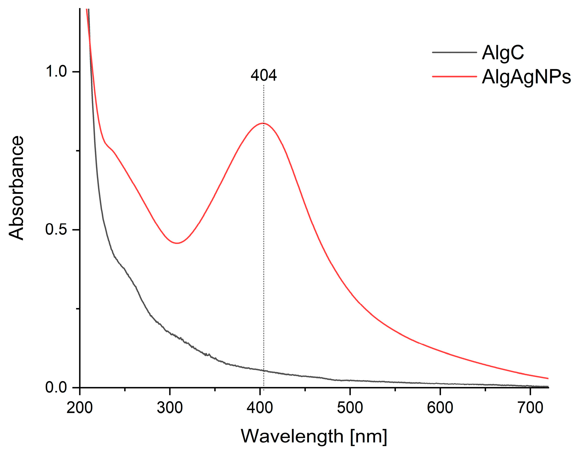
3.1. Physicochemical Characteristics of Alginate Gels with Silver Nanoparticles

A TEM electron microscope image shows the presence of uniformly distributed spherical silver nanoparticles less than 15 nm in size with individual aggregates (Figure 1a). The distribution of particles exhibits an average size of less than 10 nm, with the presence of larger particles and aggregates (Figure 1b).
The above observations were verified by the analysis of the particle size distribution determined by the dynamic light scattering technique, which showed the presence of particles in the AlgAgNPs sample with sizes ranging from a few to a dozen or so nanometers, with the vast majority of particles having sizes below 5 nm; however, a small fraction (0.1%) of slightly larger particles reaching a size of about 14 nm is also observed (Figure 2).
The UV-Vis spectrum of fructose-reduced silver alginate nanoparticles (AlgAgNPs) shows a distinct surface plasmon resonance (SPR) band at 404 nm (Figure 3), which was not observed in the case of the alginate gel without nanoparticles (AlgC) (Figure 3).
The FTIR-ATR spectrum of sodium alginate (AlgC) displays characteristic absorption bands associated with its polysaccharide structure, predominantly featuring guluronic and mannuronic acid units (Figure 4). A prominent symmetric stretching vibration of carboxylate groups (COO) is observed at ~1403 cm−1, a key identifier of alginate’s ionic nature. Additional bands in the range of 1290–1206 cm−1 correspond to C–O stretching vibrations within the pyranose rings and glycosidic linkages (C–O–C), while the peak at ~814 cm−1 is attributed to bending vibrations of glycosidic bonds or ring deformations in uronic acid residues. The absence of a distinct asymmetric COO stretch near 1600 cm−1 may reflect limitations in ATR sensitivity or spectral overlap. For the fructose-reduced alginate–silver nanoparticle composite (AlgAgNPs), the spectrum retains the majority of alginate-specific bands, indicating structural preservation of the polysaccharide matrix. The symmetric COO vibration at ~1403 cm−1 shows minimal shifts but slight intensity variations, suggesting potential interactions between carboxylate groups and silver nanoparticles, likely through physical encapsulation or weak coordination rather than covalent bonding. The persistence of C–O–C glycosidic vibrations at ~814 cm−1 and unchanged C–O stretching modes (1290–1206 cm−1) further confirms the stability of the alginate backbone during nanoparticle synthesis.

3.2. Antibacterial Activity of Silver Nanoparticles

Antibacterial activity was demonstrated for aqueous solutions of alginate gels with both concentrations of nanoparticles: 20 and 60 mg/L (Table 2). The size of the inhibition zone of microorganism growth depended on both the concentration of nanoparticles and the tested microorganism. Higher concentrations of silver nanoparticles suppressed bacterial colony growth more strongly than lower concentration in the case of: Escherichia coli, Bacillus cereus and Enterococcus. faecalis. The same level of growth inhibition was obtained for both concentrations of silver nanoparticles for P. aeruginosa (Table 2). Lack of microorganism growth inhibition was demonstrated by the use of both sodium alginate solutions without nanoparticles, as well as sterile water (Table 2).

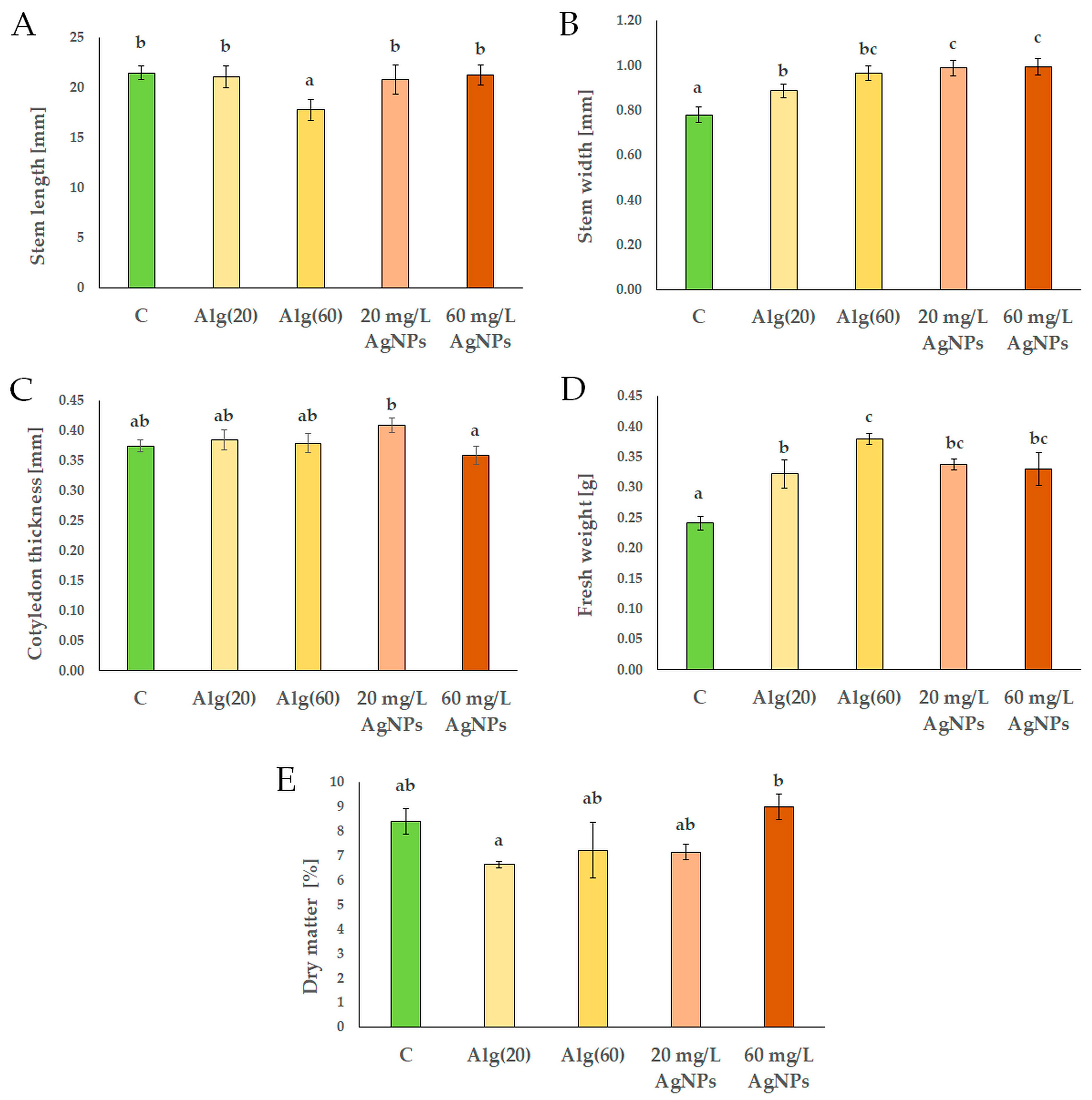
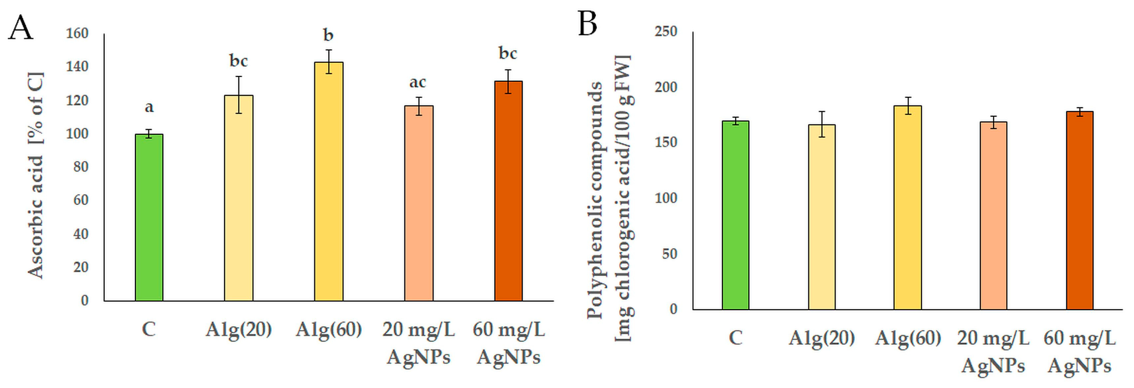
3.3. Features of Red Cabbage Seedlings in the Presence of Silver Nanoparticles

The stem length of red cabbage seedlings remained comparable to the controls after treatment with silver nanoparticles at 20 and 60 mg/L (Figure 5A). Only the seedlings treated with more concentrated sodium alginate solution (Alg(60)), were shorter than control ones. The stem width of seedlings treated with both concentrations of silver nanoparticles: 20 and 60 mg/L, and with both concentrations of sodium alginate: Alg(20) and Alg(60) were significantly greater than treated with water control (C) (Figure 5B). No statistically significant difference in stem width was observed between 20 and 60 mg/L of silver nanoparticles (Figure 5B). The thickness of cotyledons in red cabbage seedlings differed statistically only between the two tested concentrations of silver nanoparticles. Treatment with 60 mg/L of nanoparticles resulted with significant reduction in cotyledon thickness compared to the 20 mg/L nanoparticles treatment (Figure 5C). Every experimental treatment increased fresh weight of seedlings compared to control ones. Seedlings treated with Alg(60) produced a higher fresh weight than those treated with Alg(20) (Figure 5D). Dry matter content was similar in each treated seedling compared to control ones (Figure 5E). However, seedlings exposed to 60 mg/L of silver nanoparticles accumulated more dry matter than seedlings treated with Alg(20) (Figure 5E).
Red cabbage plants treated with sodium alginate solutions without nanoparticles: Alg(20) and Alg(60), accumulated higher carotenoid content than control plants (Table 3). However, only plants treated with the higher concentration of silver nanoparticles (60 mg/L) contained more carotenoids than control plants (C). Accumulation of chlorophylls was similar in treated and control plants, with the exception of effect of Alg(60) on chlorophyll a concentration and the effect of both alginate treatments on total chlorophylls content. Generally, chlorophylls accumulation in plant tissue was observed after alginate treatment in comparison with the silver nanoparticles solution. The ratio of both types of chlorophylls was similar in each sample. Conversely, the ratio of chlorophylls to carotenoids in cabbage seedlings was highest in the control plants, and both silver nanoparticles gels decreased this parameter in plant tissue compared with the control. In plants treated with higher concentration of both tested solutions: Alg(60) and 60 mg/L of silver nanoparticles, the content of anthocyanins was higher in those treated with the respective lower concentration.
Treatment of plants with both alginate solutions increased the content of ascorbic acid compared with control ones (Figure 6A). However, only the application of the higher concentration of silver nanoparticles resulted in an increase in this compound in cabbage seedlings. Plants affected by each tested solution accumulated similar amounts of polyphenolic compounds as the control ones (Figure 6B).
The ability of each plant extract to scavenge the free radical DPPH was similar (Table 4). Slightly elevated, but significant, antioxidant activity measured with the DPPH radical was observed in plants treated with the higher concentration of silver nanoparticles compared with plants treated with the lower concentration of this solution. The capacity for the reduction in ferric ions by plant extracts was similar in the case of each plant treatment. The cupric-reducing antioxidant capacity was similar in each treatment compared with control ones. However, plants treated with the higher concentrations of both tested solutions were characterized by higher antioxidant activity measured by CUPRAC method than plants treated with lower concentrations, respectively.

4. Discussion

The possibility of using the principles of green chemistry to obtain composites with silver nanoparticles exists [11]. The morphological features of nanocomposites depend on the reaction conditions [11,15]. After the synthesis, the formation of nanoparticles was confirmed using a TEM microscope method (Figure 1a,b). In addition, analyses were carried out with scanning spectrophotometer. The SPR phenomenon at 404 nm is attributable to the collective oscillation of conduction electrons at the nanoparticle surface when it is in interaction with incident light. The 404 nm absorption maximum corresponds to the literature reports for small, spherical AgNPs [15,29,30,31]. Smaller nanoparticles, defined as those with a diameter less than 50 nm, characteristically exhibit SPR bands at shorter wavelengths, typically between 400 and 420 nm. As presented in Figure 3, characteristic peaks at 404 nm comprise a size smaller than 50 nm. In contrast, larger particles, defined as those with a diameter greater than 50 nm, or anisotropic shapes, such as rods or triangles, exhibit a shift in the peak to longer wavelengths, ranging from 450 to 600 nm [15,29,30,31]. The absence of secondary peak in presented results at 500–600 nm (Figure 3) confirms the predominance of spherical morphology, as non-spherical particles often exhibit multiple or broadened SPR bands due to varied electron oscillations along different axes. The symmetry and narrow width of the SPR band at 404 nm suggest monodisperse nanoparticles with minimal size variation and uniform particle size distribution, a hallmark of effective stabilization by the alginate matrix [32,33]. This observation was also confirmed by DLS analysis (Figure 2). Alginate functions as both a stabilizer and a capping agent, thereby preventing aggregation via electrostatic repulsion (from carboxylate groups) and steric hindrance. It has been demonstrated that this contributes to the observed symmetry and stability of the SPR band. The symmetric SPR band at 404 nm confirms the successful synthesis of small, spherical and monodisperse AgNPs in alginate matrix. The narrow peak width indicates effective stabilization by alginate, while the absence of secondary bands excludes significant aggregation or shape anisotropy.
The results of the synthesis of nanoparticles described by Rutkowski et al. [15] correlate with the results described in this manuscript due to similar temperature conditions, the choice of the carrier as well as the similar chemical nature of the reducing agent used. On the other hand, Hovhannisyan et al. [31] documented the possibility of embedding silver nanoparticles in the structure of hyaluronic acid. The particles described by them were characterized by sizes around 25 nm. Possibility of obtaining silver nanoparticles in the starch structure was proven by Janik et al. [34]. The dimensions of the obtained particles were close to 20 nm, and their shape was described as spherical. In our study, the obtained silver nanoparticles in alginate, as well as the results of other authors, confirms that biopolymer materials are good carriers and stabilizers of particles in the nanoscale.
The activity of silver nanoparticles against microorganisms is widely tested. A limitation in the growth of bacterial cells of the following strains after silver nanoparticles application was demonstrated: Pseudomonas aeruginosa, Escherichia coli, Staphylococcus aureus, Staphylococcus epidermidis, Klebsiella pneumoniae, Acinetobacter baumannii and Enterococcus faecium [7,35,36,37]. The aforementioned antibacterial activity has been demonstrated with nanoparticles made using various biopolymers. In our study, we tested only four strains of bacteria, but against each of them the produced silver nanoparticles showed an inhibitory effect on colony growth (Table 2). This confirms the possibility of using silver nanoparticles embedded in the alginate matrix as a product that limits the growth of dangerous bacteria. However, before applying the product, it is also worth checking whether beneficial microorganisms, such as soil microorganisms, will also be inhibited in growth. So far, it has been reported that nanoparticles can cause changes in soil microflora. The research results remain ambiguous. Contradictions in the research results are probably due to the physical properties of the nanoparticles (size, shape). They may also result from the biological nature of the specific strain of microorganisms used [38].
Thinking about nanoparticles as potential plant protection agents against microorganisms, it is necessary to check their effects on plants. The inhibitory effect of the synthesized silver nanoparticles on the growth of cabbage seedlings, compared with plants treated with water alone, was not evidenced, and even increased fresh weight production was observed (Figure 5). However, tests conducted in other studies have sometimes shown inhibition of plant growth under the influence of silver nanoparticles (Raphanus sativus var. sativus [39], Brassica oleracea L. var. sabellica L. [39] and Brassica napus L. [40]). The concentrations used in our study seemed to be not toxic to cabbage plants. The authors indicated that a reduction in plant growth was observed with higher concentrations of nanoparticles. For example, Hasan et al. [41] found that after exposure to silver nanoparticles at a concentration of 25 mg/L, stimulation of stem length growth was observed in lettuce seedlings (Lactuca sativa L.). In the case of concentrations close to 100 mg/L, the presence of nanoparticles caused a reduction in stem length in seedlings [41]. Similarly, varied information can be found regarding the production of plant fresh and dry matter, some authors observed an accumulation, while others a decrease in these parameters, and this was again linked to the applied concentration of nanoparticles [39,40,41,42]. It was emphasized that the presence of silver nanoparticles at a concentration of 25 and 50 mg/L increased the fresh and dry weight of lettuce seedlings while the concentration of 100 mg/L reduced the mentioned parameters [41]. The specific effects of silver nanoparticles on the species cannot be ruled out either. In the presented study, silver nanoparticles were applied without demonstrating any toxic effects on plants in terms of their growth parameters. The mechanism of silver nanoparticle penetration into living organisms is not yet fully understood. Researchers suggest that there is probably a relationship between the bioactivity of nanoparticles and plant growth processes. Penetration of nanoparticles through the seed coat and increased water absorption by seeds contribute to the stimulation of seedlings growth and may have a direct impact on plants biochemical parameters [42,43,44]. In addition, alginate is a polysaccharide that can stimulate plant growth [45]. It has been proven that the decomposition of sodium alginate into simpler compounds induces a cellular signal. This resulted in the stimulation of processes at the morphological and intracellular levels in plants [46]. Therefore, in the presented work, not only were gels with nanoparticles used to treat plants but also gels with alginate alone. It is worth emphasizing that plants treated with alginate alone or alginate with the addition of silver nanoparticles did not differ in terms of the accumulation of fresh or dry mass (Figure 5). The increased accumulation of fresh mass in plants treated with solutions may be related to the use of alginate. However, comparing plants treated with alginate alone and alginate with nanoparticles, no toxic effect of the applied concentrations of nanoparticles was demonstrated.
The content of individual substances, such as pigments, is important for the proper functioning of plants, including the photosynthesis process [47]. The concentration of photosynthetic pigments may, however, depend on the exposure of plants to nanoparticles [48]. After the application of silver nanoparticles, both increases and decreases in the content of these important molecules were observed [39,41,48,49,50,51]. The authors noted that the response of plants depended on the concentration of nanoparticles but also on the species of the plant itself. Thus, Tymoszczuk [39] observed differences in the content of pigment compounds, such as chlorophylls and carotenoids in seedlings, depending on the specific concentration of silver nanoparticles (50 mg/L or 100 mg/L), as well as the species of experimental plant. Interestingly, in the case of radish (Raphanus sativus var. sativus), the accumulation of chlorophylls and carotenoids was documented after treatment with nanoparticles, while in the case of kale (Brassica oleracea L. var. sabellica L.) a decrease in the content of pigments was observed [39]. Alhammad et al. [42] showed that after treating seeds of Vicia faba L. with nanoparticles at concentration of 10 mg/L, an increase in the content of carotenoids, chlorophyll a, chlorophyll b and the sum of total chlorophylls occurred. However, an increase in nanoparticles to the concentration of 50 mg/L caused a decrease in the content of carotenoids compared to the control seedlings, despite the increase in the content of chlorophylls [42]. The treatment of oak-leaf lettuce (Lactuca sativa L.) with silver nanoparticles at concentrations 20 or 40 mg/L significantly increased the content of carotenoids and chlorophyll a [51]. In our study, silver nanoparticles only slightly affected the accumulation of chlorophylls or carotenoids (Table 3). The reduction in the accumulation of these particles in the plant tissues treated with the tested nanoparticles was not demonstrated. Treatment with sodium alginate solution increased the content of photosynthetic pigments in lettuce [52]. In our study, an increased number of carotenoids and the sum of chlorophylls was also obtained in the seedlings treated with alginate solution compared to the control ones (Table 3). The increased content of individual pigments may have resulted from increased access to nitrogen and carbon skeleton [52].
Nanoparticle treatment affected the accumulation of anthocyanins in Arabidopsis seedlings [49]. In the case of turnip (Brassica rapa ssp. rapa L.), the accumulation of anthocyanins was observed after the application of high concentrations of silver nanoparticles [50]. In our study (Table 3), the anthocyanin content in red cabbage seedlings after treatment with both tested concentrations of silver nanoparticles did not change significantly compared to the control plants. In response to abiotic stress, plant cells produce large amounts of reactive oxygen species. Stress factors affect antioxidant activity, contributing to the development of oxidative stress in plants [52]. In our study, see Table 4 and Figure 6B, no reduction in antioxidant activity parameters or polyphenols in plant extracts was observed after treatment with silver nanoparticles. On this basis, it can be presumed that the tested cabbage plants did not experience oxidative stress. Silver nanoparticles treatment increased polyphenolic compounds accumulation and antioxidant activity in lettuce [53]. However, an increase in the content of ascorbic acid in Asparagus officinalis L. after exposure to silver nanoparticles was reported [54]. Ascorbic acid accumulation in red cabbage seedlings was observed after treatment with both sodium alginate solution and the higher concentration of silver nanoparticles (Figure 6A). The collected reports confirmed that the content of individual bioactive substances depended significantly on the biological nature of the specific plant as well as the dose of silver nanoparticles. The application of the two tested concentrations of silver nanoparticles (20 and 60 mg/L) did not deteriorate the quality of the obtained seedlings of red cabbage. Further studies could be continued to obtain more information on the effect of other concentrations of silver nanoparticles in sodium alginate on vegetable plants. Undoubtedly, the phenomenon of phytotoxicity of higher doses of silver nanoparticles is closely related to the occurrence of oxidative stress [55].
Studies on the phytotoxicity of silver nanoparticles indicate that the interaction between plants and nanoparticles is dependent on several factors. Therefore, important aspects of nanoparticle phytotoxicity are the correlations between the morphological and physical properties of particles, the form, concentration and time of their application, as well as the species of the tested plant and its developmental stage.
The potential environmental toxicity of silver nanoparticles is still a poorly characterized topic. Challenges resulting from the development of various chemical technologies, including nanotechnology, determine the need to answer the question about the chemical safety of synthesized xenobiotics such as metal nanoparticles. Due to the assessed application potential, it is necessary to conduct further research. Analyses regarding the sensitivity of environmental microorganisms (including soil bacteria and fungi) to the effects of various metal nanoparticles seem to be important. It is necessary to conduct tests on plants at various growth stages (including pot studies). The introduction of new agrochemicals in the agricultural sector requires multi-stage and interdisciplinary studies that would allow for a holistic approach to the subject.

5. Conclusions

Spherical silver nanoparticles with a relatively uniform structure were successfully produced in a sodium alginate gel using fructose as a non-toxic reducing agent. The synthesized silver nanoparticles showed antibacterial activity against selected Gram-positive and Gram-negative strains of microorganisms, even at the low concentration of 20 mg/L, thus they can be considered effective against microorganisms. The application of the produced silver nanoparticles did not negatively affect the growth or quality of red cabbage seedlings, suggesting that the tested concentrations of silver nanoparticles (20 and 60 mg/L) are not phytotoxic.

Author Contributions

Conceptualization, M.R., G.K. and A.K. (Anna Kołton); Data curation, K.K., G.K., A.S. and A.K. (Anna Kołton); Formal analysis, W.M., K.K., A.S. and A.K. (Anna Kołton); Funding acquisitison, K.K., Z.W., G.K. and A.K. (Anna Kołton); Investigation, L.K.-F., J.C. and Z.W.; Methodology, M.R., W.M., L.K.-F., D.M., G.K. and A.K. (Anna Kołton); Resources, K.K., Z.W., G.K. and A.K. (Anna Kołton); Software, K.K., D.M., G.K. and A.K. (Anna Kołton); Supervision, G.K., A.S. and A.K. (Anna Kołton); Validation, A.K. (Andrzej Kalisz), Z.W., G.K. and A.S.; Visualization, M.R., D.M., G.K. and A.K. (Anna Kołton); Writing—original draft, M.R., W.M., A.K., D.M., J.C., G.K., A.S. and A.K. (Andrzej Kalisz); Writing—review and editing, A.K. (Andrzej Kalisz), A.S. and A.K. All authors have read and agreed to the published version of the manuscript.

Funding

This research was funded by the Ministry of Science and Higher Education of Poland as a part of a research subsidy to the University of Agriculture in Krakow (050012-D011).

Data Availability Statement

The data presented in this study are available on request from the corresponding author.

Conflicts of Interest

The authors declare no conflicts of interest.

References

  1. Alfei, S.; Zuccari, G. Last Fifteen Years of Nanotechnology Application with Our Contribute. Nanomaterials 2025, 15, 265. [Google Scholar] [CrossRef] [PubMed]
  2. Khatoon, U.T.; Velidandi, A. An Overview on the Role of Government Initiatives in Nanotechnology Innovation for Sustainable Economic Development and Research Progress. Sustainability 2025, 17, 1250. [Google Scholar] [CrossRef]
  3. Bouhadi, M.; Javed, Q.; Jakubus, M.; Elkouali, M.; Fougrach, H.; Ansar, A.; Ban, S.G.; Ban, D.; Heath, D.; Černe, M. Nanoparticles for Sustainable Agriculture: Assessment of Benefits and Risks. Agronomy 2025, 15, 1131. [Google Scholar] [CrossRef]
  4. Ragasová, L.; Peňázová, E.; Gazdík, F.; Pečenka, J.; Čechová, J.; Pokluda, R.; Baránek, M.; Grzebelus, D.; Eichmeier, A. The Change of Bacterial Spectrum after Storage of X. campestris pv. campestris Inoculated Cabbage Heads (Brassica oleracea var. capitata L.). Agronomy 2020, 10, 443. [Google Scholar] [CrossRef]
  5. Gai, Y.; Wang, H. Plant Disease: A Growing Threat to Global Food Security. Agronomy 2024, 14, 1615. [Google Scholar] [CrossRef]
  6. Esmael, A.; Al-Hindi, R.R.; Albiheyri, R.S.; Alharbi, M.G.; Filimban, A.A.R.; Alseghayer, M.S.; Almaneea, A.M.; Alhadlaq, M.A.; Ayubu, J.; Teklemariam, A.D. Fresh Produce as a Potential Vector and Reservoir for Human Bacterial Pathogens: Revealing the Ambiguity of Interaction and Transmission. Microorganisms 2023, 11, 753. [Google Scholar] [CrossRef] [PubMed]
  7. Holubnycha, V.; Husak, Y.; Korniienko, V.; Bolshanina, S.; Tveresovska, O.; Myronov, P.; Holubnycha, M.; Butsyk, A.; Borén, T.; Banasiuk, R.; et al. Antimicrobial Activity of Two Different Types of Silver Nanoparticles against Wide Range of Pathogenic Bacteria. Nanomaterials 2024, 14, 137. [Google Scholar] [CrossRef]
  8. Wrońska, N.; Płaczkowska, S.; Niedziałkowska, K.; Lisowska, K. The Synergistic Effect of Biosynthesized Silver Nanoparticles and Phytocompound as a Novel Approach to the Elimination of Pathogens. Molecules 2023, 28, 7921. [Google Scholar] [CrossRef]
  9. Hosnedlova, B.; Kabanov, D.; Kepinska, M.; Narayanan, V.H.B.; Parikesit, A.A.; Fernandez, C.; Bjørklund, G.; Nguyen, H.V.; Farid, A.; Sochor, J.; et al. Effect of Biosynthesized Silver Nanoparticles on Bacterial Biofilm Changes in S. aureus and E. coli. Nanomaterials 2022, 12, 2183. [Google Scholar] [CrossRef]
  10. Alfeqy, M.M.; El-Hawary, S.S.; El-Halawany, A.M.; Rabeh, M.A.; Alshehri, S.A.; Abdelmohsen, U.R.; Safwat, N.A.; Serry, A.M.; Fahmy, H.A.; Ezzat, M.I. Biosynthesis and Characterization of Aeonium arboreum-Derived Silver Nanoparticles: Antimicrobial Activity, Biofilm Inhibition, Antihemolytic Activity, and In Silico Studies. Int. J. Mol. Sci. 2024, 25, 8039. [Google Scholar] [CrossRef]
  11. Rutkowski, M.; Krzemińska-Fiedorowicz, L.; Khachatryan, G.; Bulski, K.; Kołton, A.; Khachatryan, K. Biodegradable Silver Nanoparticles Gel and Its Impact on Tomato Seed Germination Rate in In Vitro Cultures. Appl. Sci. 2022, 12, 2722. [Google Scholar] [CrossRef]
  12. Kowalski, G.; Witczak, M.; Kuterasiński, Ł. Structure Effects on Swelling Properties of Hydrogels Based on Sodium Alginate and Acrylic Polymers. Molecules 2024, 29, 1937. [Google Scholar] [CrossRef]
  13. Nam, H.-B.; Lee, K.H.; Yoo, H.Y.; Park, C.; Lim, J.-M.; Lee, J.H. Rapid and High-Yield Recovery of Sodium Alginate from Undaria pinnatifida via Microwave-Assisted Extraction. Processes 2024, 12, 208. [Google Scholar] [CrossRef]
  14. Sharma, R.; Malviya, R.; Singh, S.; Prajapati, B. A Critical Review on Classified Excipient Sodium-Alginate-Based Hydrogels: Modification, Characterization, and Application in Soft Tissue Engineering. Gels 2023, 9, 430. [Google Scholar] [CrossRef]
  15. Rutkowski, M.; Krzemińska-Fiedorowicz, L.; Khachatryan, K.; Khachatryan, G.; Kalisz, A.; Sękara, A. Impact of silver nanoparticles in alginate gels on seed germination, growth and stress biochemical parameters of cucumber seedlings. Plant Stress 2024, 12, 100491. [Google Scholar] [CrossRef]
  16. Stoica, R.; Ganciarov, M.; Constantinescu-Aruxandei, D.; Capră, L.; Șuică-Bunghez, I.-R.; Senin, R.-M.; Pricope, G.D.; Ivan, G.-R.; Călin, C.; Oancea, F. Sustainable Recovery of Anthocyanins and Other Polyphenols from Red Cabbage Byproducts. Foods 2023, 12, 4157. [Google Scholar] [CrossRef] [PubMed]
  17. Vega-Galvez, A.; Gomez-Perez, L.S.; Zepeda, F.; Vidal, R.L.; Grunenwald, F.; Mejías, N.; Pasten, A.; Araya, M.; Ah-Hen, K.S. Assessment of Bio-Compounds Content, Antioxidant Activity, and Neuroprotective Effect of Red Cabbage (Brassica oleracea var. capitata rubra) Processed by Convective Drying at Different Temperatures. Antioxidants 2023, 12, 1789. [Google Scholar] [CrossRef]
  18. Mejías, N.; Vega-Galvez, A.; Gomez-Perez, L.S.; Pasten, A.; Uribe, E.; Cortés, A.; Valenzuela-Barra, G.; Camus, J.; Delporte, C.; Bernal, G. Health-Promoting Properties of Processed Red Cabbage (Brassica oleracea var. capitata f. rubra): Effects of Drying Methods on Bio-Compound Retention. Foods 2024, 13, 830. [Google Scholar] [CrossRef]
  19. Youssef, E.A.; Abdelbaset, M.M.; Dewedar, O.M.; Molina-Martínez, J.M.; El-Shafie, A.F. Crop Coefficient Estimation and Effect of Abscisic Acid on Red Cabbage Plants (Brassica oleracea var. capitata) under Water-Stress Conditions. Agriculture 2023, 13, 610. [Google Scholar] [CrossRef]
  20. Aloo, S.-O.; Ofosu, F.-K.; Daliri, E.-B.-M.; Oh, D.-H. UHPLC-ESI-QTOF-MS/MS Metabolite Profiling of the Antioxidant and Antidiabetic Activities of Red Cabbage and Broccoli Seeds and Sprouts. Antioxidants 2021, 10, 852. [Google Scholar] [CrossRef]
  21. Athanassiadis, B.; Abbott, P.V.; George, N.; Walsh, L.J. An in vitro study of the antimicrobial activity of some endodontic medicaments and their bases using an agar well diffusion assay. Aust. Dent. J. 2009, 54, 141–146. [Google Scholar] [CrossRef] [PubMed]
  22. Wellburn, A.R. The spectral determination of chlorophylls a and b, as well as total carotenoids, using various solvents with spectrophotometers of different resolution. J. Plant Physiol. 1994, 144, 307–313. [Google Scholar] [CrossRef]
  23. Lee, J.; Durst, W.R.; Wrolstad, E.R. Determination of Total Monomeric Anthocyanin Pigment Content of Fruit Juices, Beverages, Natural Colorants, and Wines by the pH Differential Method: Collaborative Study. J. AOAC Int. 2005, 88, 5. [Google Scholar] [CrossRef]
  24. Queval, G.; Noctor, G. A plate reader method for the measurement of NAD, NADP, glutathione, and ascorbate in tissue extracts: Application to redox profling during Arabidopsis rosette development. Anal. Biochem. 2007, 363, 58–69. [Google Scholar] [CrossRef] [PubMed]
  25. Brand-Williams, W.; Cuvelier, M.; Berset, C. Use of a free radical method to evaluate antioxidant activity. LWT 1995, 28, 25–30. [Google Scholar] [CrossRef]
  26. Benzie, I.; Strain, J. The Ferric Reducing Ability of Plasma (FRAP) as a Measure of “Antioxidant Power”: The FRAP Assay. Anal. Biochem. 1996, 239, 70–76. [Google Scholar] [CrossRef]
  27. Apak, R.; Güçlü, K.; Demirata, B.; Özyürek, M.; Celik, S.E.; Bektaşoğlu, B.; Berker, K.I.; Ozyurt, D. Comparative Evaluation of Various Total Antioxidant Capacity Assays Applied to Phenolic Compounds with the CUPRAC Assay. Molecules 2007, 12, 1496–1547. [Google Scholar] [CrossRef]
  28. Cicco, N.; Lanorte, T.M.; Paraggio, M.; Viggiano, M.; Lattanzi, V. A reproducible, rapid and inexpensive Folin–Ciocalteu micro-method in determining phenolics of plant methanol extracts. Microchem. J. 2009, 91, 107–110. [Google Scholar] [CrossRef]
  29. Wang, F.; Lu, X.; Li, X. Selective removals of heavy metals (Pb2+, Cu2+, and Cd2+) from wastewater by gelation with alginate for effective metal recovery. J. Hazard. Mater. 2016, 308, 75–83. [Google Scholar] [CrossRef]
  30. Mohan, S.; Oluwafemi, O.S.; Songca, S.P.; Jayachandran, V.; Rouxel, D.; Joubert, O.; Kalarikkal, N.; Thomas, S. Synthesis, antibacterial, cytotoxicity and sensing properties of starch-capped silver nanoparticles. J. Mol. Liq. 2016, 213, 75–81. [Google Scholar] [CrossRef]
  31. Hovhannisyan, A.; Janik, M.; Woszczak, L.; Khachatryan, G.; Krystyjan, M.; Lenart-Boroń, A.; Stankiewicz, K.; Czernecka, N.; Duraczyńska, D.; Oszczęda, Z.; et al. The Preparation of Silver and Gold Nanoparticles in Hyaluronic Acid and the Influence of Low-Pressure Plasma Treatment on Their Physicochemical and Microbiological Properties. Int. J. Mol. Sci. 2023, 24, 17285. [Google Scholar] [CrossRef] [PubMed]
  32. Bansal, V.; Li, V.; O’Mullane, A.P.; Bhargava, S.K. Shape Dependent Electrocatalytic Behaviour of Silver Nanoparticles. CrystEngComm 2010, 12, 4280–4286. [Google Scholar] [CrossRef]
  33. Yang, Y.; Matsubara, S.; Xiong, L.; Hayakawa, T.; Nogami, M. Solvothermal Synthesis of Multiple Shapes of Silver Nanoparticles and Their SERS Properties. J. Phys. Chem. C 2007, 111, 9095–9104. [Google Scholar] [CrossRef]
  34. Janik, M.; Khachatryan, K.; Khachatryan, G.; Krystyjan, M.; Oszczęda, Z. Comparison of Physicochemical Properties of Silver and Gold Nanocomposites Based on Potato Starch in Distilled and Cold Plasma-Treated Water. Int. J. Mol. Sci. 2023, 24, 2200. [Google Scholar] [CrossRef]
  35. Darwesh, O.M.; Matter, I.A.; Eida, M.F.; Moawad, H.; Oh, Y.-K. Influence of Nitrogen Source and Growth Phase on Extracellular Biosynthesis of Silver Nanoparticles Using Cultural Filtrates of Scenedesmus obliquus. Appl. Sci. 2019, 9, 1465. [Google Scholar] [CrossRef]
  36. Korzekwa, K.; Kędziora, A.; Stańczykiewicz, B.; Bugla-Płoskońska, G.; Wojnicz, D. Benefits of Usage of Immobilized Silver Nanoparticles as Pseudomonas aeruginosa Antibiofilm Factors. Int. J. Mol. Sci. 2022, 23, 284. [Google Scholar] [CrossRef]
  37. Pernas-Pleite, C.; Conejo-Martínez, A.M.; Marín, I.; Abad, J.P. Green Extracellular Synthesis of Silver Nanoparticles by Pseudomonas alloputida, Their Growth and Biofilm-Formation Inhibitory Activities and Synergic Behavior with Three Classical Antibiotics. Molecules 2022, 27, 7589. [Google Scholar] [CrossRef] [PubMed]
  38. Przemieniecki, S.W.; Oćwieja, M.; Ciesielski, S.; Halecki, W.; Matras, E.; Gorczyca, A. Chemical Structure of Stabilizing Layers of Negatively Charged Silver Nanoparticles as an Effector of Shifts in Soil Bacterial Microbiome under Short-Term Exposure. Int. J. Environ. Res. Public Health 2022, 19, 14438. [Google Scholar] [CrossRef]
  39. Tymoszuk, A. Silver Nanoparticles Effects on In Vitro Germination, Growth, and Biochemical Activity of Tomato, Radish, and Kale Seedlings. Materials 2021, 14, 5340. [Google Scholar] [CrossRef]
  40. Tomaszewska-Sowa, M.; Lisiecki, K.; Pańka, D. Response of Rapeseed (Brassica napus L.) to Silver and Gold Nanoparticles as a Function of Concentration and Length of Exposure. Agronomy 2022, 12, 2885. [Google Scholar] [CrossRef]
  41. Hasan, M.; Mehmood, K.; Mustafa, G.; Zafar, A.; Tariq, T.; Hassan, S.G.; Loomba, S.; Zia, M.; Mazher, A.; Mahmood, N.; et al. Phytotoxic Evaluation of Phytosynthesized Silver Nanoparticles on Lettuce. Coatings 2021, 11, 225. [Google Scholar] [CrossRef]
  42. Alhammad, B.A.; Abdel-Aziz, H.M.M.; Seleiman, M.F.; Tourky, S.M.N. How Can Biological and Chemical Silver Nanoparticles Positively Impact Physio-Chemical and Chloroplast Ultrastructural Characteristics of Vicia faba Seedlings? Plants 2023, 12, 2509. [Google Scholar] [CrossRef] [PubMed]
  43. Gruyer, N.; Dorais, M.; Bastien, C.; Dassylva, N.; Triffault-Bouchet, G. Interaction between silver nanoparticles and plant growth. Acta Hortic. 2014, 1037, 795–800. [Google Scholar] [CrossRef]
  44. Zheng, L.; Hong, F.; Lu, S.; Liu, C. Effect of nano-TiO2 on strength of naturally aged seeds and growth of spinach. Biol. Trace Elem. Res. 2005, 104, 83–91. [Google Scholar] [CrossRef]
  45. Skrzypczak, D.; Jarzembowski, Ł.; Izydorczyk, G.; Mikula, K.; Hoppe, V.; Mielko, K.A.; Pudełko-Malik, N.; Młynarz, P.; Chojnacka, K.; Witek-Krowiak, A. Hydrogel Alginate Seed Coating as an Innovative Method for Delivering Nutrients at the Early Stages of Plant Growth. Polymers 2021, 13, 4233. [Google Scholar] [CrossRef]
  46. Xing, M.; Cao, Q.; Wang, Y.; Xiao, H.; Zhao, J.; Zhang, Q.; Ji, A.; Song, S. Advances in Research on the Bioactivity of Alginate Oligosaccharides. Mar. Drugs 2020, 18, 144. [Google Scholar] [CrossRef]
  47. Simkin, A.J.; Kapoor, L.; Doss, C.G.P.; Hofmann, T.A.; Lawson, T.; Ramamoorthy, S. The role of photosynthesis related pigments in light harvesting, photoprotection and enhancement of photosynthetic yield in planta. Photosynth. Res. 2022, 152, 23–42. [Google Scholar] [CrossRef] [PubMed]
  48. Peharec Štefanić, P.; Košpić, K.; Lyons, D.M.; Jurković, L.; Balen, B.; Tkalec, M. Phytotoxicity of Silver Nanoparticles on Tobacco Plants: Evaluation of Coating Effects on Photosynthetic Performance and Chloroplast Ultrastructure. Nanomaterials 2021, 11, 744. [Google Scholar] [CrossRef] [PubMed]
  49. Syu, Y.-Y.; Hung, J.-H.; Chen, J.-C.; Chuang, H.-W. Impacts of size and shape of silver nanoparticles on Arabidopsis plant growth and gene expression. Plant Physiol. Biochem. 2014, 83, 57–64. [Google Scholar] [CrossRef]
  50. Thiruvengadam, M.; Gurunathan, S.; Chung, I.-M. Physiological, metabolic, and transcriptional effects of biologically-synthesized silver nanoparticles in turnip (Brassica rapa ssp. rapa L.). Protoplasma 2015, 252, 1031–1046. [Google Scholar] [CrossRef]
  51. Hasanuzzaman, M.; Bhuyan, M.B.; Zulfiqar, F.; Raza, A.; Mohsin, S.M.; Al Mahmud, J.; Fujita, M.; Fotopoulos, V. Reactive Oxygen Species and Antioxidant Defense in Plants under Abiotic Stress: Revisiting the Crucial Role of a Universal Defense Regulator. Antioxidants 2020, 9, 681. [Google Scholar] [CrossRef] [PubMed]
  52. Aly, A.A.; Eliwa, N.E.; Safwat, G. Role of gamma-irradiated sodium alginate on growth, physiological and active components of iceberg lettuce (Lactuca sativa) plant. BMC Plant Biol. 2024, 24, 185. [Google Scholar] [CrossRef] [PubMed]
  53. Jurkow, R.; Pokluda, R.; Sękara, A.; Kalisz, A. Impact of foliar application of some metal nanoparticles on antioxidant system in oakleaf lettuce seedlings. BMC Plant Biol. 2020, 20, 290. [Google Scholar] [CrossRef] [PubMed]
  54. An, J.; Zhang, M.; Wang, S.; Tang, J. Physical, chemical and microbiological changes in stored green asparagus spears as affected by coating of silver nanoparticles-PVP. LWT Food Sci. Technol. 2008, 41, 1100–1107. [Google Scholar] [CrossRef]
  55. Yan, A.; Chen, Z. Impacts of Silver Nanoparticles on Plants: A Focus on the Phytotoxicity and Underlying Mechanism. Int. J. Mol. Sci. 2019, 20, 1003. [Google Scholar] [CrossRef]
Figure 1. Characterization of silver nanoparticles: (a) TEM micrograph; (b) particle size distribution histogram. The white bar in the picture indicates 100 nm.
Agronomy 15 01640 g001
Figure 2. The histograms of particle size distribution and average particle sizes of alginate gel with silver nanoparticles (AlgAgNPs).
Agronomy 15 01640 g002
Figure 3. The UV-Vis spectra of alginate gels.
Agronomy 15 01640 g003
Figure 4. The FTIR spectra of gels.
Agronomy 15 01640 g004
Figure 5. Growth parameters of red cabbage seedlings: (A) Stem length. (B) Stem width. (C) Cotyledon thickness. (D) Fresh weight. (E) Dry matter. Letters denote homogeneous groups determined by Fisher’s LSD test at α = 0.05. Results are presented as mean ± SE.
Agronomy 15 01640 g005
Figure 6. Content of ascorbic acid (A) and polyphenols (B) in red cabbage seedlings. Letters denote homogeneous groups determined by Fisher’s LSD test at α = 0.05. Lack of letters indicate no statistically significant differences. Results are presented as mean ± SE.
Agronomy 15 01640 g006
Table 1. Content of individual reagents needed to synthesize alginate gels with silver nanoparticles.
SamplesWater [g]Sodium
Alginate [g]
Solution with Ag+ [g]Glycerol [g]Fructose
Solution 4%, [g]
Total Mass of Gel [g]AgNPs
Concentration [mg/L]
AlgC144.021.500.000.750.00146.270.00
AlgAgNPs95.501.5018.520.7530.00146.27150.00
Table 2. Antibacterial activity of aqueous solutions with silver nanoparticles. The zones of inhibition of the microorganisms growth. Letters denote homogeneous groups determined by Fisher’s LSD test at α = 0.05. Results are presented as mean ± SE.
TreatmentsEscherichia coli [mm]Pseudomonas aeruginosa
[mm]
Bacillus cereus
[mm]
Enterococcus faecalis [mm]
C0.00 ± 0.00 a0.00 ± 0.00 a0.00 ± 0.00 a0.00 ± 0.00 a
Alg(20)0.00 ± 0.00 a0.00 ± 0.00 a0.00 ± 0.00 a0.00 ± 0.00 a
Alg(60)0.00 ± 0.00 a0.00 ± 0.00 a0.00 ± 0.00 a0.00 ± 0.00 a
20 mg/L AgNPs2.44 ± 0.06 b3.86 ± 0.05 b6.63 ± 0.07 b5.54 ± 0.13 b
60 mg/L AgNPs2.88 ± 0.13 c3.95 ± 0.10 b7.07 ± 0.09 c5.85 ± 0.11 c
Table 3. Content of pigments (carotenoids, chlorophyll a, chlorophyll b, anthocyanins) in red cabbage seedlings treated with experimental solutions. Letters denote homogeneous groups determined by Fisher’s LSD test at α = 0.05. Results are presented as mean ± SE.
TreatmentsCarotenoids
[mg/g FW]
Chlorophyll a
[mg/g FW]
Chlorophyll b
[mg/g FW]
Chl a + Chl b
[mg/g FW]
Chl a : Chl bChl : CarAnthocyanins
[mg/g FW]
C0.107 ± 0.009 a0.397 ± 0.033 ab0.164 ± 0.016 ab0.530 ± 0.033 a2.468 ± 0.159 a5.251 ± 0.194 d60.852 ± 1.898 ab
Alg(20)0.143 ± 0.008 c0.447 ± 0.019 bc0.180 ± 0.007 b0.627 ± 0.026 b2.480 ± 0.031 a4.419 ± 0.093 c55.341 ± 1.500 a
Alg(60)0.151 ± 0.009 c0.460 ± 0.035 c0.179 ± 0.013 b0.639 ± 0.045 b2.578 ± 0.122 a4.229 ± 0.106 bc68.140 ± 1.808 c
20 mg/L AgNPs0.117 ± 0.007 ab0.337 ± 0.022 a0.138 ± 0.010 a0.475 ± 0.032 a2.451 ± 0.034 a4.046 ± 0.042 b58.035 ± 3.353 a
60 mg/L AgNPs0.134 ± 0.004 bc0.358 ± 0.014 a0.138 ± 0.006 a0.496 ± 0.020 a2.603 ± 0.037 a3.685 ± 0.051 a64.593 ± 1.897 bc
Table 4. Antioxidant activity of red cabbage plants depending on experimental treatments. Letters denote homogeneous groups determined by Fisher’s LSD test at α = 0.05. Results are presented as mean ± SE.
TreatmentsDPPH *
[mM Trolox/g FW]
FRAP
[mM Trolox/g FW]
CUPRAC
[mM Trolox/g FW]
C2.979 ± 0.074 ab0.968 ± 0.028 a2.132 ± 0.139 abc
Alg(20)2.869 ± 0.098 a0.976 ± 0.064 a1.945 ± 0.121 ab
Alg(60)2.922 ± 0.078 ab1.081 ± 0.037 a2.330 ± 0.134 c
20 mg/L AgNPs2.739 ± 0.068 a0.998 ± 0.040 a1.878 ± 0.040 a
60 mg/L AgNPs3.158 ± 0.091 b1.074 ± 0.022 a2.282 ± 0.155 bc
* DPPH–assay with stable free radical 2,2-diphenyl-1-picrylhydrazyl; FRAP–ferric reducing antioxidant power assay; CUPRAC–cupric-ion-reducing antioxidant capacity assay.
Disclaimer/Publisher’s Note: The statements, opinions and data contained in all publications are solely those of the individual author(s) and contributor(s) and not of MDPI and/or the editor(s). MDPI and/or the editor(s) disclaim responsibility for any injury to people or property resulting from any ideas, methods, instructions or products referred to in the content.

