Article

Bioactive Compounds and Antiradical Activity of the Rosa canina L. Leaf and Twig Extracts

by
Małgorzata Kubczak
1,
Ainur B. Khassenova
2,
Bartosz Skalski
3,
Sylwia Michlewska
4,
Marzena Wielanek
5,
Araylim N. Aralbayeva
2,
Maira K. Murzakhmetova
6,
Maria Zamaraeva
7,
Maria Skłodowska
5,
Maria Bryszewska
1 and
Maksim Ionov
1,*
1
Department of General Biophysics, Faculty of Biology and Environmental Protection, University of Lodz, 90-236 Lodz, Poland
2
Department of Biotechnology, Faculty of Food Production, Almaty Technological University, Almaty 050000, Kazakhstan
3
Department of General Biochemistry, Faculty of Biology and Environmental Protection, University of Lodz, 90-236 Lodz, Poland
4
Laboratory of Microscopic Imaging and Specialized Biological Techniques, Faculty of Biology and Environmental Protection, University of Lodz, 90-237 Lodz, Poland
5
Department of Plant Physiology and Biochemistry, Faculty of Biology and Environmental Protection, University of Lodz, 90-237 Lodz, Poland
6
Department of Biophysics and Biomedicine, Faculty of Biology and Biotechnology, al-Farabi Kazakh National University, Almaty 050040, Kazakhstan
7
Laboratory of Molecular Biophysics, Faculty of Biology, University of Bialystok, 15-245 Bialystok, Poland
*
Author to whom correspondence should be addressed.
Agronomy 2020, 10(12), 1897; https://doi.org/10.3390/agronomy10121897
Submission received: 3 October 2020 / Revised: 23 November 2020 / Accepted: 27 November 2020 / Published: 1 December 2020
(This article belongs to the Special Issue Extraction and Analysis of Natural Product in Plant)

Abstract

:
It is important to search for new sources of bioactive, natural compounds, because customers are paying more attention to food quality. Fruits and berries from horticultural plants are known to be good sources of agents beneficial for human well-being and could serve as natural preservatives in the food industry. However, more recent research indicates that other plant organs can also be rich in nutrients. Our study focused on characterizing an unexplored source, namely leaf and twig extracts from Rosa canina. The chemical composition of these extracts was analyzed and their in vitro activity measured. HPLC analysis of the content of phenolics, vitamins and amino acids revealed that the leaf and twig extracts were found to be rich in bioactive compounds with potent antioxidant properties. The greatest differences between bioactive phenolic compounds in leaf and twig extracts related mainly to p-coumaric acid, myricetin, ellagic acid, cyanidin, procyanidin and quercetin, whereas salicylic acid levels were similar in both types of extract. Interactions with human serum albumin were investigated, and some conformational changes in protein structure were observed. Further analysis (lipid peroxidation, protein carbonylation, thiol group oxidation, DPPH inhibition and ROS inhibition) confirmed that both leaf and twig extracts exhibited antioxidant and antiradical scavenging activities. Cytotoxicity and hemotoxicity assays confirmed very low toxicity of the extracts towards human cells over the range of concentrations tested. Our results indicate that both extracts could serve as non-toxic sources of bioactive compounds with antiradical properties.

1. Introduction

The relationship between balanced diet and human health is well documented [1,2]. Moreover, knowledge of the beneficial effect of a diet enriched in fruits, vegetables, herbs and wild plants as rich sources of natural compounds with anti-oxidative, anti-inflammatory, anti-bacterial, anti-diabetic and anticancer properties is generally accessible [3,4,5,6,7]. For most people, the everyday diet comprises highly transformed and manufactured food products that are rather poor in many vitamins, minerals and other compounds beneficial for health. On the other hand, today’s consumables markets offer products from new food categories called “functional food”. These products are enriched in compounds originating from the plants which have been used for thousands of years due to their favorable effect on health. However, the organs of these plants that have been available for years are now being used for technological processes, whereas other organs from them could be more interesting sources of beneficial compounds and more valuable for human consumption.
Rosa canina L. belongs to the Rosaceae family, which contains more than 100 species and grows mostly in Europe, Asia, North America, Africa and the Middle-East [8]. R. canina pseudo-fruits (hips) are the best characterized organ of this plant. The hips are used worldwide as an antioxidant, anti-inflammatory, immunosuppressive, cardioprotective, gastroprotective and antimicrobial agent [9,10]. Nowadays, hip extracts are commonly used in the cosmetic and food industries [11,12].
Many publications indicate that rose hips contain large amounts of vitamins A, B, C, D and E, minerals, carotenoids and phenolic compounds [10,13,14,15,16,17]. They also contain fruit acids, pectin, sugars, organic acids, amino acids and essential oils [18]. Vitamin C and phenolic compounds are well known for their antioxidant properties [7,8,14,19,20]. R. canina hips contain the highest level of the L-isomer of vitamin C among fruits and vegetables [21,22]. The ascorbic acid content of rose hips ranges from 300 to 4000 mg/100 g, the variation resulting from changes in sugar levels during ripening [16].
Vitamin E is another strong antioxidant. The human diet should include components rich in tocopherols. The lipid-soluble vitamin E is necessary for different antioxidant functions in human cells, especially in cell membranes and plasma lipoproteins. It helps to prevent the propagation of oxidative chain reactions by scavenging many reactive oxygen species (ROS) and it could be implicated in the prevention of atherosclerosis and cancer. Epidemiological investigations have revealed a positive correlation between tocopherol intake and a reduced risk for cardiovascular diseases [14,19].
Phenolic compounds confer the unique flavors and health-promoting properties of vegetables and fruits [23]. Colored rose fruits are good sources of phenolic compounds including tannins, flavonoids, phenolic acids, anthocyanins and dihydrochalcones [24]. These compounds have a wide spectrum of biochemical activities such as antioxidant, antimutagenic, anticarcinogenic effects and can alter gene expression [25]. Flavonoids and phenolic acids have diverse positive biological activities, making them the most important groups of secondary plant metabolites and natural bioactive compounds for humans [26].
HPLC analysis has shown R. canina hips extract to be especially rich in the polyphenols hyperoside, astragalin, rutin, (+)-catechin and (−)-epicatechin, gallic acid and poly-hydroxylated organic acids such as quinic acid [27,28]. The phenolic compounds (+)-catechin, (−)-epicatechin, rutin, vanillin, astragalin, phloridzin and gallic acid, identified in hips extract, have been reported as strong scavengers of the (ROO.) radical [29]. Several other polyphenols potentially beneficial for humans such as ellagic acid, salicylic acid, vanillic acid, ferulic acid and caffeic acid have been identified in trace amounts in R. canina hip extracts [30,31].
Food products containing R. canina compounds are derived from the hips [32]. The leaves and stems from the roses are usually discarded as trash. However, increasing numbers of publications confirm that R. canina leaves could be a valuable source of flavonoids, especially flavone glycosides [33,34,35]. A few recent studies have indicated that stems from R. canina are also a good source of polyphenols [36,37]. Ouerghemmi et al. showed that stem extracts from different Rosa species could be used in the food, cosmetic and pharmaceutical industries as a source of phenolic compounds [36].
The aim of our study was to determine the biological properties of extracts of Rosa canina twigs and leaves. First, we focused on identifying compounds such as phenolics, amino acids and vitamins in the extracts. Secondly, we assessed the antioxidant and antiradical properties of these extracts. Additionally, the interaction of the extract components with human serum albumin was tested by circular dichroism, and the hemotoxicity and cytotoxicity of the extracts were determined.

2. Materials and Methods

2.1. Chemicals

Chlorogenic acid, quercetin, (+)catechin, acetonitrile, isopropanol, human serum albumin, 2,4-dinitro-phenylhydrazine (DNPH), 5,5′-dithiobis(2-nitrobenzic acid)/Ellman reagent, 2,2′-diphenyl-1-picrylhydrazyl (DPPH), fetal bovine serum, penicillin/streptomycin and resazurin sodium salt were purchased from Merck, Darmstadt, Germany. Phosphate buffer saline, sodium dodecyl sulphate (SDS), DMEM cell culture medium and 2′,7′-dichlorodihydrofluorescein diacetate (H2DCFDA) were obtained from Thermo Fisher, Waltham, MA, USA. Methanol, ethanol, trichloroacetate acid (TCA) and 2-thiobarbituric acid (TBA) were purchased from POCH, Lublin, Poland.

2.2. Plant Material and Extracts Preparation

The fully developed leaves and twigs of Rosa canina L. were collected from the foothills of the Trans Ili Alatau Mountains (Almaty region, Kazakhstan) in the early morning in June 2018, in the middle of the vegetative season. The samples were obtained 1.5–2.0 m from the ground, 5–8 cm from the branch meristem, from 10–15 year old plants. The plants were identified and voucher specimen No. 3389 (R. canina L.) was deposited at the herbarium of the Institute of Botany and Phytointroduction (Almaty, Kazakhstan).
Consent to conduct the research described in this article was not required, because the research was not carried out in strictly protected areas (reserve, national park) or related to protected species, which would require the consent of the relevant authorities.
The R. canina leaves and twigs were thoroughly washed with distilled water and dried at room temperature, then crushed and extracted with 50% ethanol (20 g dry weight/200 mL) for 20 h at 25 ± 2 °C on a rotary shaker (110 rpm). After centrifugation (20 min, 34,000× g), the supernatants were dried in a rotary evaporator at 50 °C. Such high temperature of evaporation was used accordingly to a standard protocol to remove the water. The pressure was reduced up to 100 hPa. The evaporated extracts were stored at 4 °C. Before analysis, small amounts (1–2 mg) of dried extracts were carefully hydrated with 1–2 mL of distilled water using a laboratory shaker. As a result, the extracts were completely dissolved. Prior to total phenolic content analysis, the obtained aqueous solutions of the extracts were additionally centrifuged (20 min, 20,000× g) before HPLC analysis supernatants were filtered through a 45 μm syringe filter (Millipore, Billerica, MA, USA).

2.3. Total Phenolic Content and HPLC Analysis

The total phenolic content was determined using Folin–Ciocalteau reagent according to the Singelton and Rossi (1965) method [38]. The absorbance of the reaction product was measured at 725 nm, and the phenolic content was expressed as milligrams per gram of dried extract based on the calibration curve (r2 = 0.9991) prepared for chlorogenic acid. The results are given as means ± SD (n = 3).
The flavonoid content was determined by the aluminum chloride colorimetric method according to Chang et al. (2002) [39]. The absorbance of the reaction mixture was measured at 415 nm, and the flavonoid content was expressed as milligrams per gram of dried extract based on a calibration curve (r2 = 0.9995) prepared for quercetin. The results are given as means ± SD (n = 3).
The catechin (flavan-3-ol) content was determined by the vanillin assay method described by Bakkalbasi et al. (2005) [40]. The absorbance of the reaction mixture was measured at 500 nm, and the total flavan-3-ol content was calculated from a calibration curve (r2 = 0.9988) prepared using (+)-catechin and expressed as milligrams per gram of dried extract. The results are given as means ± SD (n = 3).
The HPLC system (Summit x2 Dual-Gradient System, Dionex, Sunnyvale, CA, USA) was equipped with a photodiode-array detector (PDA100 DAD) and fluorescence detector (RF-2000). The phenolic compounds present in the extracts were separated on an RP column (aQ Hypersil GOLD, 250 × 4.6 mm, 5 µm) joined with a guard column (GOLD aQ Drop-In guards, 10 × 4 mm, 5 µm, Polygen, Gliwice, Poland) at 25 °C. The injection volume of analyzed samples was 20 µL. A mobile phase composed of water (A) and methanol (B), both with 0.1% formic acid, was used. The linear gradient was started after 2 min of isocratic elution with 5% B, over 30 min to 55% B, followed by 5 min of isocratic elution. Between 37 and 47 min, the concentration of phase B increased to 70% followed by 5 min of isocratic elution. Then, between 52 and 54 min, the gradient was returned to the initial 5% B and the column was recalibrated for the next 3 min. The flow rate was 1 cm3 min−1. The absorbance was measured at 253, 280, 325 and 375 nm, and the fluorescence at 420 (excitation 270 nm, emission 420 nm). Phenolic compounds in the R. canina extracts were identified by comparing the retention times and on-line UV absorption spectra of the analyzed samples with the respective data obtained from reference standards. Quantification was based on a calibration curve for standards of phenolic compounds covering the range 5–200 µg cm−3; the linearity of the calibration curve was verified by the correlation coefficient (r2 ≥ 0.9994). The optimal wavelengths used for the preparation of the calibration curve and quantification of individual metabolites are given in Table 1.

2.4. Water-Soluble Vitamins and Tocopherol Isomers

A Kapel-105M Lumex (Russia) capillary electrophoresis kit was used to determine the vitamin composition of the extracts from leaves and twigs. The contents of B1 (thiamine), B2 (riboflavin), B3 (pantothenic acid) and B5 (nicotinic acid) were determined. The vitamins were detected at 200 nm and by using programmable wavelength switching. Conditions for separation were as follows: borate buffer pH = 8.9, temperature 30 °C. The method was based on the extraction of vitamins and separation and quantification of the components by capillary electrophoresis.
Isomers of vitamin E were identified by HPLC with UV detection. A high-performance Agilent 1200 chromatograph (USA) with a four-channel thermostat pump, a spectrophotometric detector and a 250 × 4.6 mm Zorbax 300SB-C18 column was used. To determine the vitamin E content, the following conditions were selected: flow rate of the mobile phase = 0.7 mL min−1; column temperature = 35 °C. The eluent was 42:50:8 acetonitrile:isopropanol:water.
To prepare the samples they were extracted with an 1:1 methanol:water solution, and then proteins that interfered with the chromatography were precipitated. The samples were then evaporated to dryness and dissolved in 1 mL of the eluent (42:50:8 acetonitrile:isopropanol:water)

2.5. Amino Acids

A Kapel-105M Lumex capillary electrophoresis system (Russia) was also used to assess the amino acid composition of the extracts. The electric field separated the charged components of the extracts in a quartz capillary. A microvolume of the solution to be analyzed (~2 nL) was introduced into a quartz capillary pre-filled with buffered electrolyte. Then, a high voltage (up to 30 kV) was applied to the ends of the capillary. The following conditions were used: the total length of the capillary was 75 cm; the effective length (i.e., the length from the entrance to the detector window) was 65 cm; the operating voltage applied to the electrodes was +13 kV; the internal diameter of the capillary was 50 μm; detection was at 254 nm; temperature 200 °C; sample injected under 300 mbar pressure; composition of the working buffer = 5 mM tartaric acid, 2 mM 18-crown-6. Sample preparation consisted of sample hydrolysis followed by dilution with the buffer solution.

2.6. Interaction with Human Serum Albumin: Circular Dichroism

To estimate changes in protein structure upon addition of R. canina extracts, CD spectra were obtained. CD spectra from 1 µg mL−1 human serum albumin (HSA) were checked alone and with increasing concentrations of leaf and twig extracts. Measurements were made over the 260–195 nm wavelength range using a 0.5 cm path length Helma quartz cell. The recording parameters were as follows: scan speed, 50 nm min−1; step resolution, 0.5 nm; response time, 4 s; bandwidth, 1 nm; slit, auto. The CD spectra were corrected against a baseline with buffer only. The mean residue ellipticity θ (cm2 dmol−1) was calculated using software provided by Jasco.

2.7. Interaction with Biomembranes: Hemolysis Test

The potential to damage cell membranes was investigated using the hemolysis method. Human blood was collected from a blood bank in Lodz and centrifuged several times with phosphate buffered saline (PBS), pH 7.4, at 4 °C. After washing, the hematocrit was measured, and the blood samples were diluted to 2% hematocrit. The leaf and twig extracts of Rosa canina were added at concentrations of 0.5–50 µg/mL and left for 24 h at 37 °C. After this incubation, hemoglobin was measured at λ = 540 nm using a BioTek plate reader, and the hemolysis values were calculated as follows:
H(%) = (Apb 540 nm/Awater 540 nm) × 100%
where Apb is the absorbance of a tested sample, Awater means 100% hemolysis (erythrocytes incubated in distilled water only). Three independent measurements were obtained. The results are presented as mean ± SD.

2.8. Antioxidant and Antiradical Activity: Oxidative Stress Markers

Human plasma was used to determine the antioxidant activity of the extracts.
Fresh human plasma was obtained from healthy, non-smoking volunteers. The blood was collected in tubes with CPD (citrate/phosphate/dextrose; 9:1 v/v blood/CPD) and next centrifuged. (1411× g, 15 min). The fresh plasma was incubated (60 min, 37 °C) with 0.5–50 μg mL−1 leaf and twig extracts and 4.7 mM H2O2/3.8 mM FeSO4/2.5 mM EDTA. Protein concentration was calculated from the absorbance at λ = 280 nm using the Kalckar formula according to Whitaker and Granum (1980) [41].
Lipid peroxidation products were determined with thiobarbituric acid (TBA) by measuring the thiobarbituric acid reactive substance (TBARS) concentration. After 60 min incubation of the samples (plasma, plant extract, H2O2/Fe) at 37 °C, 0.5 mL of 15% trichloroacetic acid (TCA) and 0.5 mL of thiobarbituric acid (TBA) was added. The samples were vortexed for 1 min and then heated (100 °C, 10 min). The samples were cooled in room temperature and centrifuged (6832× g, 15 min, 18 °C). The absorbance of the supernatant was measured at λ = 535 nm (SPECTROstar Nano Microplate Reader, BMG LABTECH, Ortenberg, Germany). The TBARS concentration was calculated using the molar absorption coefficient (ε = 156,000 M−1 cm−1).
Carbonyl Groups. The content of carbonyl groups (CO–)was determined using the colorimetric DNPH assay. First, 0.75 mL of 10 mM DNPH in 2.5 M HCl was added to the sample precipitates (plasma with plant extracts, H2O2/Fe). Sample blanks were prepared by adding 1 mL of 2.5 M HCl without DNPH. Samples were vortexed for 5 min and left at room temperature for 1 h in the dark. Then, 0.75 mL of 40% TCA was added to the mixture, and the samples were centrifuged at 900× g, for 5 min. The protein pellets were washed three times with 1.5 mL ethanol:ethyl acetate (1:1, v/v). Next, the samples were dissolved in 1 mL of 6 M guanidine, and absorbance was measured at λ = 375 nm (SPECTROstar Nano Microplate Reader, BMG LABTECH, Germany). The carbonyl group concentration was calculated using the molar absorption coefficient (ε = 22,000 M−1 cm−1).
Thiol Groups. The concentration of thiol groups was determined spectrophotometrically with Ellman reagent. A quantity of 20 µL of the samples (plasma with plant extracts, H2O2/Fe) was mixed on the plate with 20 µL of 10% SDS and 160 µL of 10 mM phosphate buffer, pH 8.0. Then the 16.6 µL of 10 mM of Ellman reagent in 10 mM phosphate buffer was added to the samples. After 60 min incubation in 37 °C, the absorbance was measured at λ = 412 nm (SPECTROstar Nano Microplate Reader, BMG LABTECH, Germany). The thiol groups concentration was calculated using the molar absorption coefficient (ε = 13,600 M−1 cm−1). The results were presented as nmol thiol groups mg−1 plasma protein.
Free radical (DPPH) scavenging activity. The free radical scavenging activity of leaf and twig extracts from R. canina was measured using the DPPH radical (2,2′-diphenyl-1-picrylhydrazyl, Sigma-Aldrich). DPPH was dissolved in ethanol to a final concentration of 8.3 × 10⁻⁵ M. The antioxidant properties were tested at extract concentrations of 0.5–50 µg mL−1 and over different incubation times (after 5, 10, 15, 30 and 45 min). Absorbances were recorded at λ = 517 nm. Three independent repetitions were performed. The results are presented as percentage DPPH inhibition, calculated as
% DPPHinhibition = 100(A0 − Aav)A0
where A0 is the absorbance of DPPH solution, and Aav is the average absorbance of samples treated with the extracts. The results are presented as means ± SD.

2.9. Cytotoxicity

The BJ (normal human fibroblast) cell line was purchased from ATCC (UK). The cells were cultured in DMEM (Gibco) supplemented with 10% fetal bovine serum and 1% streptomycin/penicillin. They were kept at the standard conditions at the 37 °C in a humidified atmosphere.
The Alamar Blue assay was applied to analyze the plant extracts’ cytotoxicity. The cells were seeded at 10,000 per well and left overnight to adhere. Next, 0.5–50 µg mL−1 R. canina leaf and twig extracts were added. After 24 h, incubation at the 37 °C resazurin sodium salt was added to the final concentration of 0.0125%, and the absorbance was measured at λ = 595 nm. The viability was calculated as follows:
% viability = (A/Ac) × 100%.
where A = absorbance of a tested sample, Ac = absorbance of the control sample. Three independent measurements were collected, and the results are presented as means ± SD.

2.10. ROS Inhibition in Human BJ Cell Line

The ability to decrease the level of cellular reactive oxygen species (ROS) was tested using BJ (normal human fibroblast) cells. Cells were seeded at 2 × 10⁵ density and left for 24 h to adhere, and then treated with 50 µg mL−1 leaf or twig extract. After 24 h incubation, the medium was refreshed, and 80 µM H2O2 was added for 30 min. After this incubation, the cells were washed, and 5 µM non-fluorescence probe 2′,′7-dichlorodihydrofluorescein diacetate (H2DCFDA) was added to each sample for 20 min. The cells were washed with PBS, and fluorescence intensity was observed with a confocal microscope (Leica TCS SP8) (λexc = 495 nm, λem = 527 nm).

2.11. Statistical Analysis

GraphPad Prism 5.0 and Statistica 13.1 were used for statistical analyses. Non-parametric tests were applied (Mann–Whitney U, non-parametric ANOVA, Kruskall–Wallis test) to estimate the significance of differences when the number of measurements were 3 (n = 3). Significance was accepted when p < 0.05.

2.12. Compliance with Ethical Standards

Blood samples were obtained from the Association of Honorary Blood Donors in Lodz (Central Blood Bank in Lodz, Poland) from 25 September 2019 to 14 January 2020. To conduct the current study, the samples were obtained randomly. The blood was collected from healthy volunteers and carefully tested before using in the laboratory. All the experiments published in this manuscript complied with the current laws of the country in which they were performed. The study was approved by the Ethics Committee of the University of Lodz, Poland (NR19/KBBN-UŁ/III/2019). All methods were performed in accordance with the relevant guidelines and regulations.

3. Results

3.1. Phenolic Content and HPLC Analysis

HPLC analysis identified and quantified 40 phenolic compounds in the leaf and twig extracts (Table 1). Monomeric and polymeric catechins and gallic acid esters such as epigallocatechin were found in both extracts. There was more of the monomeric (+)-catechin content in twig extracts, whereas polymeric procyanidin B2 dominated in leaf extracts. There were high contents of dihydroxybenzoic and protocatechuic acids in the twig extracts. Both extracts had high ellagic acid contents, but leaves contained about twice as much as twigs. From the flavonoid group, leaf extracts had more cyanidin than twig extracts and also contained high levels of neochlorogenic acid.
There was no significant difference in total phenolic compound contents between the extracts of R. canina leaves and twigs (Figure 1). Flavonoids accounted for about 9.5% and 5.5% of the phenolic compounds, respectively. However, because AlCl3 reacts mainly with flavones, flavonols, flavanones and flavanonols, the result does not correspond exactly to the total flavonoid content of the extracts. The total catechin (flavan-3-ol) content of the twig extracts was about twice that of the leaf extracts. Among the phenolic compounds, catechins constituted about 5.2% and 10% in leaf and twig extracts, respectively.

3.2. Water-Soluble Vitamins and Content of Vitamin E

Table 2A shows the contents of five B vitamins in the leaves and twigs of R. canina. In all tested samples, the predominant B vitamin was B6 (pyridoxine). The amounts of each vitamin were similar in both organs of R. canina investigated.
More tocopherols were found in the leaf than twig extract of R. canina. The concentration of isomer α was highest in both parts of the plants (Table 2B).

3.3. Amino Acid Content

On the basis of the values presented in Table 3, 13 amino acids were identified in the leaves and twigs of R. canina, nine of them essential: valine, threonine, methionine, isoleucine, leucine, phenylalanine, histidine, lysine and arginine. The total content of amino acids was higher in leaves (6.1487%) than twigs (1.6864%). The highest concentrations were of proline (0.3196%), serine (0.2040%) and phenylalanine (0.2262%) in the twig extracts, and of proline (1.04994%), valine (0.6046%) and phenylalanine (0.6045%) in the leaf extracts.

3.4. Interaction with Human Serum Albumin: Circular Dichroism

Changes in the secondary structure of HSA in the presence of R. canina leaf and twig extracts were checked using circular dichroism. CD spectra for HSA at pH 7.4 were obtained in the absence and presence of the extracts.
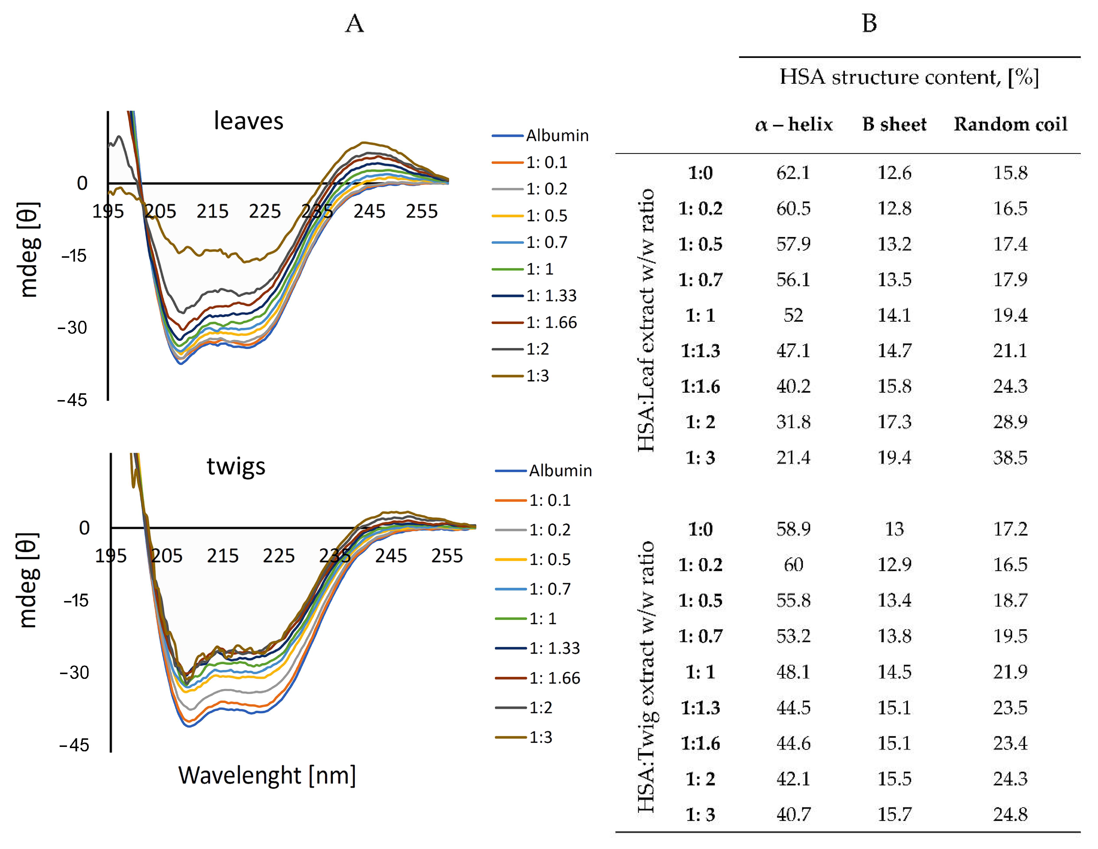
The HSA CD spectrum contained two characteristic negative bands in the far UV at 202 and 220 nm (Figure 2A). As increasing concentrations of the extracts were added, the amount of α-helix decreased, whereas the amounts of β-sheet and random coil increased. The leaf extract changed the albumin structure more markedly than did the twig extract. Percentage values calculated using CDNN software indicated a decrease in α-helical structure from 62.1% to 21.4% for the leaf extract and from 58.9% to 40.7% for the twig extract. The amount of β-sheet increased from 12.6% to 19.4%, and that of the random coil from 15.8% to 38.5% and from 17.2% to 24.8% in the leaf and twig extracts, respectively (Figure 2B).

3.5. Hematoxicity and Cytotoxicity

The R. canina leaf and twig extracts were subjected to a hemolysis test. Both proved non-toxic for human erythrocytes, and the hematoxicity did not exceed 5% even at the highest extracts concentrations (Figure 3A).
The cytotoxicity of the extracts was tested using human fibroblast BJ cell line (Figure 3B). The results indicate that the extracts were non-toxic toward these cells. There were no significant differences between the leaf and twig samples.

3.6. Markers of Oxidative Stress, Free Radical Scavenging and ROS Inhibition

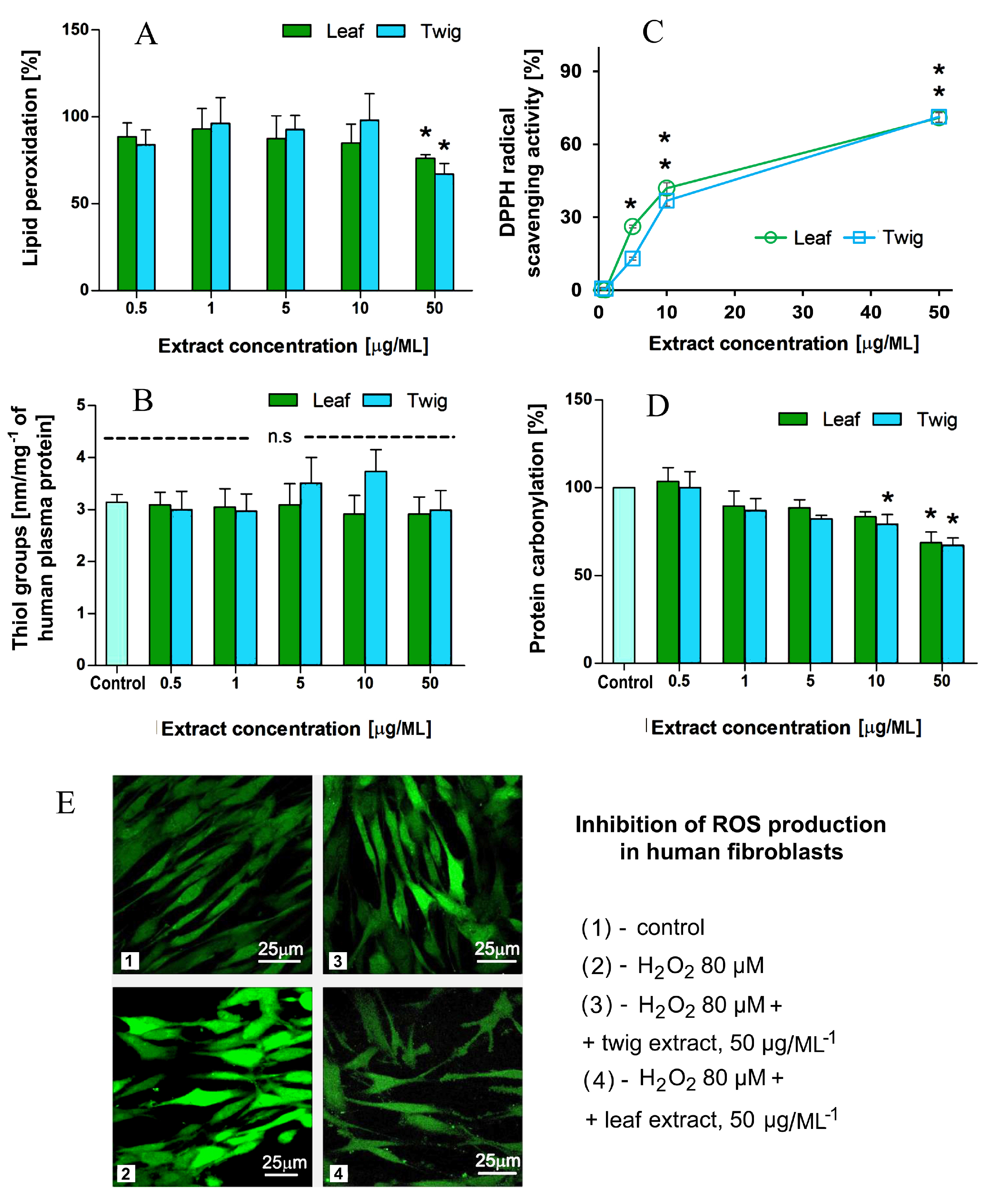
The tested extracts caused statistically significant change in plasma lipid peroxidation for the highest concentration used (50 μg mL−1) (Figure 4A), nor did they affect thiol groups in plasma proteins treated with H2O2/Fe (Figure 4B). The leaf extract at the highest concentration (50 μg mL−1) and the twig extract at 10 and 50 μg mL−1 inhibited H2O2/Fe-induced protein carbonylation (Figure 4D).
One of the aims of this study was to investigate the dose-dependent antiradical activity of the investigated extracts that was measured by the DPPH scavenging assay. Results show that both studied extracts from R. canina exhibited antiradical activity. The differences between the activities of the extracts were largest at concentrations of 5 and 10 µg/mL (Figure 4C); the leaf extracts caused 26% and 41% DPPH reduction while the twig extracts caused, respectively, 13% and 36%.
The ability of Rosa canina leaf and twig extracts to decrease the production of ROS in human fibroblasts was tested using a non-fluorescent H2DCFDA probe, which is converted to a highly fluorescent 2′,7′-dichlorofluorescein (DCF) upon oxidation by ROS. The decrease in fluorescence intensity was detected when the extracts were added to the cells. Both extracts protected human fibroblasts by inhibiting ROS production. The leaf extract had a stronger protective effect than the twig extract (Figure 4D).

4. Discussion

Many reports describe Rosa canina L. hips as an abundant source of antioxidants [1,2,3,4]. Here we demonstrated that other parts of this plant, the twigs and leaves, can be considered potential sources of compounds with beneficial properties for humans, including antioxidant activity. It is important to note, that in our in vitro study we tested the range of concentration of extracts which may be achieved in human plasma by oral administration [42,43].
HPLC analysis revealed over 40 different phenolic compounds in the R. canina leaves and twigs. The leaves used in these studies were rich in chlorogenic and neochlorogenic acid (460 mg/100 g DW and 5714.8 mg/100 g DW), cyanidin and procyanidin B2 (4744.8 mg/100 g DW and 2247.3 mg/100 g DW), ellagic acid (3588.1 mg/100 g DW), rutin (quercetin-3-O-rutinoside)—2666 mg/100 g DW, rosmarinic acid (440.6 mg/100 g DW), myricetin (392.8 mg/100 g DW), epicatechin (182.2 mg/100 g DW) and coumarin (128.5 mg/100 g DW), while the twigs had relatively high levels of ellagic acid (1444.8 mg/100 g DW), cyanidin (445.3 mg/100 g DW), myricetin (717.5 mg/100 g DW) and rutin (443.1 mg/100 g DW) and were rich in protocatechuic acid, catechin and rosmarinic acid (1391.1 mg/100 g DW, 1779.8 mg/100 g DW and 185.1 mg/100 g DW, respectively). The total phenolics in leaf and twig extracts were 22,131 mg/100 g DW and 22,267 mg/100 g DW, respectively. The total amount of flavanoles in our extracts were 1156.3 mg/100 g DW for leaves and 2000.8 mg/100 g DW for twigs. For the flavonols and flavones, the values were 2134.9 mg/100 g DW in leaves and 1253.9 mg/100 g DW in twigs.
Cunja et al. investigated the phenolic content in extracts from Rosa sp. [33]. The extract from Rosa canina was the richest in flavonols, flavanoles and phenolic acids and derivatives (900 mg/100 g FW, 450 mg/100 g FW and 200 mg/100 g FW, respectively). Among the flavonoles, quercetin-3-rhamnside was the most abundant (261.1 mg/100 g FW). We found quercetin-3-O-rutinoside (rutin) in Rosa canina leaves in high amounts, whereas Cunja et al. found this compound at 3.7 mg/100 g FW. In the case of flavanoles, our findings are consistent with values reported by Cunja et al. [33]. They found the significant amounts of procyanidine dimers (365.5 mg/100 g FW) and catechins (353.4 mg/100 g FW). In our study, the amounts of cyaniding and procyanidin B2 was also high. Interestingly, we found (–)epicatechins in leaf extracts (182.2 mg/100 g DW) that were not detected by Cunja’s group [33]. Among phenolic acids, we confirmed previous results that the most ubiquitous acids in Rosa canina leaf extracts were chlorogenic and neochlorogenic acids. We found 5714.8 mg/100 g DW of neochlorogenic acid, whereas 47.3 mg/100 g FW was detected by the Cunja group [33]. Another group found that Rosa canina extracts from leaves, flowers and fruits were rich in ellagic acids (5611.5–8469.6 µg GAE/g) and quercetin (4196–5832.7 µg GAE/g) [17]. They claimed that all parts of Rosa canina should be considered as a source of nutritional and functional components due to the occurrence of appreciable amounts of phenolic compounds and oils. In turn, Ouerghemmi et al. found low amounts of ellagic acid in Rosa canina leaf extracts [44]. They also found that the total phenolics constituted 197 µg GAE/mg of dry extract. They found that catechins, epicatechins and epicatechin gallate were abundant in all investigated Rosa extracts [44].
The same group investigated the twigs from different Rosa species [36]. They found that the total flavanole concentration in Rosa canina was 0.22 mg/100 g of dry extract. The total flavonol content was 1.04 mg/100 g of dry extract. Rutin was found at a concentration of 0.02 mg/100 g of dry weight and with quercetin-3-O-glucoside, and kaempherol–hexoside–deoxyhexoside constituted the most abundant compounds in Rosa canina extract. Interestingly, we found (–)epicatechin and epicatechin gallate at the concentrations 137.9 mg/100 g DW and 168 mg/100 g DW, respectively, whereas the Ouerghemmi group did not detect these compounds in their study [36].
The vitamin contents of R. canina leaves and twigs are still not well established. It is generally believed that the strongest contributor to the antioxidant properties of rose hips is the high vitamin C content. However, the hips from R. canina and R. rugosa are also very rich sources of tocopherols, which protect lipids against peroxidation [13]. Our study revealed tocopherol isoforms in both organs examined, though there was more in the leaves than the twigs. Additionally, only two isoforms were detected in hips, but all isoforms (α, β and γ tocopherols) were found in R. canina twigs and leaves in the present study.
The mean total tocopherol contents of hips were reported as 15.9 ± 1.7 μmol 100 g−1 in raw R. canina, 31.4 ± 3.2 μmol 100 g−1 in R. canina powder and 8.7 ± 1.1 μmol 100 g−1 in R. canina puree. In the fleshy parts of rose hips, only α- and γ-tocopherol were found, which indicates limited biosynthesis of δ-tocopherol and of tocotrienols during ripening. It is suggested that the γ- and δ-tocopherols are converted by the action of γ-tocopherol methyltransferase to α- and β-tocopherol, respectively [45]. Although there is no information about the total tocopherol content in Rosa canina leaves and twigs, the present study showed the presence of tocopherol isoforms, more in the leaves than in the twigs. The obtained amounts of tocopherols were 85 mg/100 g DW and 55 mg/100 g DW for leaves and twigs, respectively. Barros et al. tested Rosa canina hips and petals. They found 79.73 mg/100 g DW and 13.80 mg/100 g DW of tocopherols in ripened hips and petals, respectively [32].
B vitamins are not only very important in the human diet but are also considered as antioxidants in plants [46,47]. Some experiments indicate that they accumulate mainly in seeds, but they can also be found in other parts of plants including leaves and twigs. The tested extracts exhibited relatively low concentrations of B vitamins, B6 (pyridoxine) being the most abundant (570 mg/100 g DW in leaves and 620 mg/100 g DW in twigs). The lowest concentration was detected for B2 vitamin (48 mg/100 g DW in leaves and 51 mg/100 g DW in twigs). Igwenyi and Elekwa described Geranium roberatum as a good plant source of B vitamins. They found B1, B2 and B3 vitamins at concentrations of 288.17 mg/100 g DW, 818.21 mg/100 g DW and 319.13 mg/100 g DW, respectively [48].
Overall, since the leaves and twigs contain tocopherol isoforms and B vitamins, these parts of the plant can be considered as good sources of antioxidants [13,16,47]. Tocopherols and B vitamins might be considered as compounds responsible for antioxidant activity. We do not overestimate their role in antioxidant defense; however, they should be examined as some of many compounds improving antioxidant potential of leaves and twigs. Some amino acids also protect cells from oxidative stress, free radicals and heavy metals. Moreover, they mediate the synthesis of molecules such as glutathione, which is very important for the antioxidative response [49,50,51]. The total amino acid content of the extracts from R. canina leaves and twigs was measured in this study. The important information is that there were more amino acids in the leaf extracts than the twig extracts (6.1 vs. 1.6%, respectively). Both extracts contained essential amino acids such as lysine, phenylalanine, histidine, leucine, isoleucine, methionine, valine and threonine. Arginine was found only in the leaf extract, and tryptophan was detected in neither. The total amino acid content was 61.48 mg g−1 DW in the rose leaf extract and 16.86 mg g−1 DW in the twig extract, so the leaves seem a good source of protein. According to WHO standards, P. indica, P. hirta and E. thymifolia could serve as good sources of protein. The total amino acid contents were 58.80 mg g−1 DW in P. indica, 123.92 mg g−1 DW in E. thymifolia and 225.73 mg g−1 DW in P. hirta. The ratios of essential amino acids to total amino acids were 0.57, 0.47 and 0.65, respectively [52]. Although in our study the leaf extract was richer in amino acids, the ratio of essential to total amino acids was 0.37, whereas in the twig extract it was 0.55. Comparing the results obtained in our experiments with the literature, the extracts from leaves and twigs may serve as a source of amino acids, especially twigs, where the higher ratio of essential amino acids was detected.
We examined the conformational changes in human serum albumin (HSA) during titration with the extracts. Circular dichroism is useful for checking how compounds interact with protein and affect the secondary structures. The minima characteristic of α-helices were visible around 202 and 222 nm in HSA. With increasing concentrations of the extracts, the ellipticity increased.
The extracts changed the α-helix content and increased the amounts of other protein structures such as β helix and random coil. The results suggest that compounds in the extracts can interact with proteins and cause conformational changes. The leaf extract interacted more strongly with HAS albumin than did the twig extract. Almost twice as much α-helix was lost when the leaf extract was used (final content of α-helix was 21.4% for leaf and 40.7% for twig extract). Das at al. reported that extracts containing flavonoids (quercetin, myricetin, kaempferol) could interact with HSA. Slight changes in the albumin structure were observed when a 1:20 ratio was used [53]. The changes were more obvious when the maximum albumin:extract ratio was 1:3. HSA is most abundant protein in the bloodstream. It is important to investigate the albumin–molecules interaction, which may take place when they reach the circulation system. The changes in albumin structure may imply that this protein may be a carrier for compounds from plant extracts.
Extracts from R. canina leaves and twigs can be considered good sources of bioactive agents. Their antioxidant potential was demonstrated in vitro by several methods. While the antioxidant properties of rose hips are well documented, there are few reports about such properties in leaf and twig extracts [1,2,7,16]. We found that the extracts significantly decreased the lipid peroxidation level when the highest concentration of 50 µg/mL was used; the extracts did not affect thiol group oxidation. However, both extracts at higher concentrations reduced protein carbonylation significantly.
The DPPH and the ROS inhibition assays were used to confirm antiradical and antioxidant activities in the R. canina extracts. Both methods revealed that the leaf and twig extracts were highly effective. The free radical activity of DPPH was reduced when 5 µg mL−1 extracts were used. The data showed that the extracts almost immediately induced a free radical scavenging effect that was most pronounced at a 50 µg mL−1 concentration. Similar results were obtained when ROS inhibition was measured, but the leaf extract protected fibroblasts more effectively than did the twig extracts. The H2DCFDA assay allowed us to evaluate the level of reactive oxygen species generated in the cells. Upon cleavage of acetate groups by esterases and oxidation, H2DCFDA is converted to a highly fluorescent dichlorofluorescein (DCF) [54]. According to the literature, leaf extract from R. canina has one of the highest antioxidant potentials among the Rosaceae family, perhaps because R. canina has the highest phenolic content of all Rosaceae [35]. Moreover, although the results presented in this paper were obtained with ethanol extracts not methanol extracts as in other studies, all these results indicate that ethanol extracts from R. canina could have valuable antiradical properties.
Antioxidant activity has not been so extensively investigated in twig extracts. Nevertheless, Ouerghemmi et al. [36] tested twig extracts from R. canina, R. sempervirens and R. moschata and suggested that antioxidant activity could depend on the geographical origin of the plants. Moreover, the mechanism of extract activity could result from the deactivation of free radial species by hydrogen atom transfer (HAT). These authors also studied twig ethanol and methanol R. sempervirens and R. canina extracts and found that R. sempervirens ethanol extracts and R. canina methanol extracts were more effective than others in the DPPH assay [36].
Plant extracts must be non-toxic towards human cells. Since the extracts and their components might be intended for internal and topical application, fibroblasts and erythrocytes were chosen to determine their toxicity. In addition, they represent two types of cells: nuclear (human normal fibroblasts) and non-nuclear (human erythrocytes). Cell lines and erythrocytes are commonly used when the cytotoxicity must be evaluated [55,56,57]. Our studies revealed that the extracts from leaves and twigs of R. canina had no toxic effects on the human fibroblasts. Moreover, hemotoxicity was very low; it did not exceed 5% even when the highest concentration (50 μg mL−1) was used. Early analysis of cytotoxicity to human fibroblasts showed that an ethanolic R. beggeriana Schrenk extract was more toxic than the aqueous extract, but both extracts were less toxic to normal than cancer cells [38]. It is known that plant extracts have anticancer potential [58,59,60]. Importantly, they are harmless to normal cells and do not change their metabolism.
We found that the leaf and twig extracts from R. canina did not harm cell membranes, as confirmed by the hemotoxicity assay. Additionally, they were safe for human fibroblasts cells even in high concentrations. In order to evaluate more deeply the antioxidant properties of Rosa canina leaf and twig extracts, in vivo analysis must be performed. It is well know that under in vivo conditions, polyphenols can cause expression of genes of antioxidant enzymes, but this does not exclude their direct antiradical effect, especially in the spread of free radical oxidation of lipids, as for example vitamin E. The in vitro studies confirmed by in vivo experiments will provide more information about the positive impact of extracts on organisms. However, it should be accentuated that the extracts can be applied not only internally, but also topically as components of ointments, toothpaste, etc.
Rosa plant extracts are considered potent sources of natural antioxidants. It is important to investigate new properties of well-known plants. In our study we demonstrated antioxidant and antiradical profiles of extracts from Rosa canina L. twigs and leaves. Many reports describe rose hips as a valuable source of antioxidants [1,2,3,4]. However, numerous tests have revealed that other parts of the plant, such as twigs and leaves, can also be considered potential sources of beneficial compounds.

5. Conclusions

In our study, we investigated extracts from Rosa canina leaves and twigs. In general, twig extract was richer in catechins and leaf extract in neochlorogenic acid and ellagic acid. Similar levels of phenolic compounds were found in both. Five B vitamins and three tocopherol isoforms were also found. Additionally, essential and non-essential amino acids were detected. Circular dichroism revealed that leaf extract interacted more strongly with human serum albumin than twig extract. By checking oxidative stress markers, ROS inhibition and DPPH antiradical scavenging activity, the antioxidant properties of the leaf and twig extracts were revealed. The results suggest that twig extract performed better as an antioxidant. Both extracts were safe for a human fibroblast cell line and for isolated human erythrocytes. In view of these results it can be concluded that leaf and twig extracts from Rosa canina are promising sources of natural compounds, contain valuable nutrient components and show antiradical effects that should be further investigated in vivo.

Author Contributions

Conceptualization, M.K.M., and M.I.; Data curation, M.K. and M.I.; Formal analysis, M.K., M.Z. and M.S.; Funding acquisition, M.B.; Investigation, M.K., A.B.K., B.S., S.M. and M.W.; Methodology, M.K.M., A.N.A., M.S. and M.I.; Project administration, M.I.; Resources, A.B.K., A.N.A. and M.K.M.; Software, M.K., A.B.K. and S.M.; Supervision, M.I.; Validation, M.K.; Visualization, M.K., A.B.K., B.S., S.M., M.W. and M.K.M.; Writing—original draft, M.K.; Writing—review & editing, S.M., M.Z., M.S., M.B. and M.I. All authors have read and agreed to the published version of the manuscript.

Funding

No external funding was either sought or obtained for this work.

Acknowledgments

The authors would like to thank the UK English native speaker (BioMedES Co., UK) for the support in language correction.

Conflicts of Interest

The authors declare that they have no competing interest.

Ethics Approval

All experimental procedures were approved by the Ethics Committee of the University of Lodz, Poland (NR19/KBBN-UŁ/III/2019).

References

  1. Lesjak, M.M.; Šibul, F.S.; Anac, G.T.; Beara, I.N.; Mimica-dukic, N.M. Comparative study of biological activities and phytochemical composition of two rose hips and their preserves: Rosa canina L. and Rosa arvensis Huds. Food Chem. 2016, 192, 907–914. [Google Scholar]
  2. Williams, P. Consumer Understanding and Use of Health Claims for Foods. Nutr. Rev. 2005, 63, 256–264. [Google Scholar] [CrossRef] [PubMed]
  3. Abdallah, E.M. Plants: An alternative source for antimicrobials. J. Appl. Pharm. Sci. 2011, 1, 16–20. [Google Scholar]
  4. Oguntibeju, O.O. Medicinal plants with anti-inflammatory activities from selected countries and regions of Africa. J. Inflamm. Res. 2018, 11, 307–317. [Google Scholar] [CrossRef] [PubMed] [Green Version]
  5. Abu-darwish, M.S.; Efferth, T. Medicinal Plants from Near East for Cancer Therapy. Front. Pharm. 2018, 9, 56. [Google Scholar] [CrossRef] [Green Version]
  6. Perez, R.M. Antidiabetic effect of compounds isolated from plants. Phytomedicine 1998, 5, 55–75. [Google Scholar] [CrossRef]
  7. Surveswaran, S.; Cai, Y.; Corke, H.; Sun, M. Systematic evaluation of natural phenolic antioxidants from 133 Indian medicinal plants. Food Chem. 2007, 102, 938–953. [Google Scholar] [CrossRef]
  8. Fascella, G.; Angiolillo, F.D.; Massimo, M.; Amenta, M.; Romeo, F.V.; Rapisarda, P.; Ballistreri, G. Bioactive compounds and antioxidant activity of four rose hip species from spontaneous Sicilian flora. Food Chem. 2019, 289, 56–64. [Google Scholar] [CrossRef]
  9. Demir, N.; Yildiz, O.; Alpaslan, M.; Hayaloglu, A.A. Evaluation of volatiles, phenolic compounds and antioxidant activities of rose hip (Rosa L.) fruits in Turkey. LWT Food Sci. Technol. 2014, 57, 126–133. [Google Scholar] [CrossRef]
  10. Ogah, O.; Watkins, S.C.; Ubi, B.E.; Oraguzie, N. Phenolic compounds in Rosaceae fruit and nut crops—A review Department of Horticulture, Washington State University-Irrigated Agriculture and Extension. J. Agric. Food Chem. 2014, 62, 9369–9386. [Google Scholar] [CrossRef]
  11. Sardarodiyan, M.; Mohamadi, S.A. Natural antioxidants: Sources, extraction and application in food systems. Nutr. Food Sci. 2016, 46, 363–373. [Google Scholar] [CrossRef]
  12. Jiménez, S.; Jiménez-moreno, N.; Luquin, A.; Laguna, M. Chemical composition of rosehips from different Rosa species: An alternative source of antioxidants for food industry. Food Addit. Contam. Part A 2017, 34, 1121–1130. [Google Scholar] [CrossRef] [PubMed]
  13. Al-yafeai, A.; Malarski, A.; Böhm, V. Characterization of carotenoids and vitamin E in R. rugosa and R. canina: Comparative analysis. Food Chem. 2018, 242, 435–442. [Google Scholar] [CrossRef]
  14. Ercisli, S. Chemical composition of fruits in some rose (Rosa spp.) species. Food Chem. 2007, 104, 1379–1384. [Google Scholar] [CrossRef]
  15. Olsson, M.E.; Gustavsson, K.E.; Anderson, A.N.; Duan, R.D. Inhibition of Cancer Cell Proliferation in Vitro by Fruit and Berry Extracts and Correlations with Antioxidant Levels. J. Agric. Food Chem. 2004, 52, 7264–7271. [Google Scholar] [CrossRef] [PubMed]
  16. Genç, N.; Dölek, Ü.; Günes, M. Changes in flavonoid and phenolic acid contents in some Rosa species during ripening. Food Chem. 2017, 235, 154–159. [Google Scholar]
  17. Hosni, K.; Chrif, R.; Zahed, N.; Abid, I.; Medfei, W.; Sebei, H.; Ben Brahim, N. Fatty acid and phenolic constituents of leaves, flowers and fruits of tunisian dog rose (Rosa canina L.). Riv. Ital. Delle Sostanze Grasse 2010, 87, 117–123. [Google Scholar]
  18. Olsson, M.E.; Andersson, S.; Werlemark, G.; Uggla, M.; Gustavsson, K.E. Carotenoids and phenolics in rose hips. Acta Hortic. 2005, 690, 249–252. [Google Scholar] [CrossRef]
  19. Denev, P.; Kratchanova, M.; Ciz, M.; Lojek, A.; Vasicek, O.; Nedelcheva, P.; Blazheva, D.; Toshkova, R.; Gardeva, E.; Yossifova, L.; et al. Biological activities of selected polyphenol-rich fruits related to immunity and gastrointestinal health. Food Chem. 2014, 157, 37–44. [Google Scholar] [CrossRef]
  20. Koczka, N.; Stefanovits-Banyai, E.; Ombodi, A. Total Polyphenol Content and Antioxidant Capacity of Rosehips of Some Rosa Species. Medicines 2018, 5, 84. [Google Scholar] [CrossRef] [Green Version]
  21. Patel, S. Rose hips as complementary and alternative medicine: Overview of the present status and prospects. Med. J. Nutr. Metab. 2012, 6, 89–97. [Google Scholar] [CrossRef]
  22. Tumbas, V.T.; Canadanovic-Brunet, J.M.; Cetkovic-Simin, D.D.; Cetkovic, G.S.; Dilas, S.M.; Gille, L. Effect of rosehip (Rosa canina L.) phytochemicals on stable free radicals and human cancer cells. J. Sci. Food Agric. 2012, 92, 1273–1281. [Google Scholar] [CrossRef] [PubMed]
  23. Tomás-Barberán, F.A.; Espín, J.C. Phenolic compounds and related enzymes as determinants of quality in fruits and vegetables. J. Sci. Food Agric. 2001, 81, 853–876. [Google Scholar] [CrossRef]
  24. Chawla, R.; Arora, R.; Singh, S.; Sagar, R.K.; Sharma, R.K.; Kumar, R.; Sharma, A.; Gupta, M.L.; Singh, S.; Prasad, J.; et al. Radioprotective and antioxidant activity of fractionated extracts of berries of Hippophae rhamnoides. J. Med. Food 2007, 10, 101–109. [Google Scholar] [CrossRef] [PubMed]
  25. Nakamura, Y.; Watanabe, S.; Miyake, N.; Kohno, H.; Osawa, T. Dihydrochalcones: Evaluation as novel radical scavenging antioxidants. J. Agric. Food Chem. 2003, 51, 3309–3312. [Google Scholar] [CrossRef] [PubMed]
  26. Nogala-Kałucka, M.; Dwiecki, K.; Siger, A.; Górnaś, P.; Polewski, K.; Ciosek, S. Antioxidant synergism and antagonism between tocotrienols, quercetin and rutin in model system. Acta Aliment. 2013, 42, 360–370. [Google Scholar] [CrossRef]
  27. Ayati, Z.; Amiri, M.S.; Ramezani, M.; Delshad, E.; Sahebkar, A.; Emami, S.A. Phytochemistry, Traditional Uses and Pharmacological Profile of Rose Hip: A Review. Curr. Pharm. Des. 2018, 24, 4101–4124. [Google Scholar] [CrossRef]
  28. Wenzig, E.M.; Widowitz, U.; Kunert, O.; Chrubasik, S. Phytochemical composition and in vitro pharmacological activity of two rose hip (Rosa canina L.) preparations. Phytomedicine 2008, 15, 826–835. [Google Scholar] [CrossRef]
  29. Marino, T.; Galano, A.; Russo, N. Radical scavenging ability of gallic acid toward OH and OOH radicals-reaction mechanism and rate constants from the density functional theory. J. Phys. Chem. B 2014, 118, 10380–10389. [Google Scholar] [CrossRef]
  30. Kerasioti, E.; Apostolou, A.; Kafantaris, I.; Chronis, K.; Koulocheri, S.D.; Haroutounian, S.A.; Kouretas, D.; Stagos, D. Polyphenolic Composition of Rosa canina, Rosa sempervivens and Pyrocantha coccinea Extracts and Assessment of Their Antioxidant Activity in human endothelial cells. Antioxidants 2019, 8, 92. [Google Scholar] [CrossRef] [Green Version]
  31. Nađpal, J.D.; Lesjak, M.M.; Mrkonjić, Z.O.; Majkić, T.M.; Četojević-Simin, D.D.; Mimica-Dukić, N.M.; Beara, I.N. Phytochemical composition and in vitro functional properties of three wild rose hips and their traditional preserves. Food Chem. 2018, 241, 290–300. [Google Scholar] [CrossRef]
  32. Barros, L.; Carvalho, A.M.; Ferreira, I.C.F.R. Exotic fruits as a source of important phytochemicals: Improving the traditional use of Rosa canina fruits in Portugal. Food Res. Int. 2011, 44, 2233–2236. [Google Scholar] [CrossRef]
  33. Cunja, V.; Mikulic-Petkovsek, M.; Stampar, F.; Schmitzer, V. Compound Identification of Selected Rose Species and Cultivars: An Insight to Petal and Leaf Phenolic Profiles. J. Am. Soc. Hortic. Sci. 2014, 139, 157–166. [Google Scholar] [CrossRef] [Green Version]
  34. Sytar, O.; Hemmerich, I.; Zivcak, M.; Rauh, C.; Brestic, M. Comparative analysis of bioactive phenolic compounds composition from 26 medicinal plants. Saudi J. Biol. Sci. 2018, 25, 631–641. [Google Scholar] [CrossRef] [PubMed] [Green Version]
  35. Ieri, F.; Innocenti, M.; Possieri, L.; Gallori, S.; Mulinacci, N. Phenolic composition of “bud extracts” of Ribes nigrum L., Rosa canina L. and Tilia tomentosa M. J. Pharm. Biomed. Anal. 2015, 115, 1–9. [Google Scholar] [CrossRef] [PubMed]
  36. Ouerghemmi, S.; Saija, A.; Siracusa, L.; Ruberto, G.; Dhaouadi, K.; Cimino, F.; Cristani, M. LC-DAD-ESI-MS and HPLC-DAD phytochemical investigation and in vitro antioxidant assessment of Rosa sp. stem pruning products from different northern areas in Tunisia. Phytochem. Anal. 2020, 31, 98–111. [Google Scholar] [CrossRef]
  37. Riffault, L.; Destandau, E.; Pasquier, L.; André, P.; Elfakir, C. Phytochemical analysis of Rosa hybrida cv. ‘Jardin de Granville’ by HPTLC, HPLC-DAD and HPLC-ESI-HRMS: Polyphenolic fingerprints of six plant organs. Phytochemistry 2014, 99, 127–134. [Google Scholar] [CrossRef]
  38. Singleton, V.L.; Rossi, J.A. Colorimetry of Total Phenolics with Phosphomolybdic-Phosphotungstic Acid Reagents. Am. J. Enol. Vitic. 1965, 16, 144–158. [Google Scholar]
  39. Chang, C.C.; Yang, M.H.; Wen, H.M.; Chern, J.C. Estimation of Total Flavonoid Content in Propolis by Two Complementary Colorimetric Methods. J. Food Drug Anal. 2002, 10, 178–182. [Google Scholar]
  40. Bakkalbaşı, E.; Yemiş, O.; Aslanova, D.; Artık, N. Major flavan-3-ol composition and antioxidant activity of seeds from different grape cultivars grown in Turkey. Eur. Food Res. Technol. 2005, 221, 792–797. [Google Scholar] [CrossRef]
  41. Whitaker, J.R.; Granum, P.E. An absolute method for protein determination based on difference in absorbance at 235 and 280 nm. Anal. Biochem. 1980, 109, 156–159. [Google Scholar] [CrossRef]
  42. Manach, C.; Scalbert, A.; Morand, C.; Rémésy, C.; Jiménez, L. Polyphenols: Food sources and bioavailability. Am. J. Clin. Nutr. 2004, 79, 727–747. [Google Scholar] [CrossRef] [PubMed] [Green Version]
  43. Manach, C.; Williamson, G.; Morand, C.; Scalbert, A.; Rémésy, C. Bioavailability and bioefficacy of polyphenols in humans. I. Review of 97 bioavailability studies. Am. J. Clin. Nutr. 2005, 81, 230–242. [Google Scholar] [CrossRef] [Green Version]
  44. Ouerghemmi, S.; Sebei, H.; Siracusa, L.; Ruberto, G.; Saija, A.; Cimino, F.; Cristani, M. Comparative study of phenolic composition and antioxidant activity of leaf extracts from three wild Rosa species grown in different Tunisia regions: Rosa canina L., Rosa moschata Herrm. and Rosa sempervirens L. Ind. Crop. Prod. 2016, 94, 167–177. [Google Scholar] [CrossRef]
  45. Collakova, E.; DellaPenna, D. The Role of Homogentisate Phytyltransferase and Other Tocopherol Pathway Enzymes in the Regulation of Tocopherol Synthesis during Abiotic Stress. Plant Physiol. 2003, 133, 930–940. [Google Scholar] [CrossRef] [PubMed] [Green Version]
  46. Colinas, M.; Eisenhut, M.; Tohge, T.; Pesquera, M.; Fernie, A.R.; Weber, A.P.M.; Fitzpatrick, T.B. Balancing of B 6 vitamers is essential for plant development and metabolism in Arabidopsis. Plant Cell 2016, 28, 439–453. [Google Scholar] [CrossRef] [Green Version]
  47. Asensi-fabado, M.A.; Munne-Bosch, S. Vitamins in plants: Occurrence, biosynthesis and antioxidant function. Trends Plant Sci. 2010, 15, 582–592. [Google Scholar] [CrossRef] [PubMed]
  48. Igwenyi, I.O.; Elekwa, A.E. Phytochemical Analysis and Determination of Vitamin Contents of Geranium Robertianum. J. Dent. Med. Sci. 2014, 13, 44–47. [Google Scholar]
  49. Liang, M.; Wang, Z.; Li, H.; Cai, L.; Pan, J.; He, H.; Wu, Q.; Tang, Y.; Ma, J.; Yang, L. L-Arginine induces antioxidant response to prevent oxidative stress via stimulation of glutathione synthesis and activation of Nrf2 pathway. Food Chem. Toxicol. 2018, 115, 315–328. [Google Scholar] [CrossRef]
  50. Yang, M.; Vousden, K.H. Serine and one-carbon metabolism in cancer. Nat. Rev. Cancer 2016, 16, 650–662. [Google Scholar] [CrossRef]
  51. Kim, J.; Jang, H.; Cho, W.; Yeon, S.; Lee, C. In vitro antioxidant actions of sulfur-containing amino acids. Arab. J. Chem. 2020, 13, 1678–1684. [Google Scholar] [CrossRef]
  52. Prasad, K. HPLC Analysis of Amino Acid and Antioxidant Composition of Three Medicinal Plants of (Pithoragarh) Uttarakhand Himalayas. J. Anal. Pharm. Res. 2017, 6, 00816. [Google Scholar] [CrossRef]
  53. Das, P.; Chaudhari, S.K.; Das, A.; Kundu, S. Interaction of Flavonols with Human Serum Albumin: A biophysical study showing structure activity relationship and enhancement when coated on silver nanoparticles. J. Biomol. Struct. Dyn. 2019, 37, 1414–1426. [Google Scholar] [CrossRef] [PubMed]
  54. Wu, D.; Yotnda, P. Production and detection of reactive oxygen species (ROS) in cancers. J. Vis. Exp. 2011, 57, e3357. [Google Scholar] [CrossRef] [PubMed]
  55. Grauzdytė, D.; Pukalskas, A.; Viranaicken, W.; El Kalamouni, C.; Venskutonis, P.R. Protective effects of Phyllanthus phillyreifolius extracts against hydrogen peroxide induced oxidative stress in HEK293 cells. PLoS ONE 2018, 13, e0207672. [Google Scholar] [CrossRef] [PubMed] [Green Version]
  56. Ladokun, O.; Ojezele, M.; Arojojoye, O. Comparative study on the effects of aqueous extracts of viscum album (Mistletoe) from three host plants on hematological parameters in albino rats. Afr. Health Sci. 2015, 15, 606–612. [Google Scholar] [PubMed] [Green Version]
  57. De Oliveira, V.M.A.; Carneiro, A.L.B.; Cauper, G.S.D.B.; Pohlit, A.M. In vitro screening of amazonian plants for hemolytic activity and inhibition of platelet aggregation in human blood. Acta Amaz. 2009, 39, 973–980. [Google Scholar] [CrossRef] [Green Version]
  58. Nayebi, N.; Khalili, N.; Kamalinejad, M.; Emtiazy, M. A systematic review of the efficacy and safety of Rosa damascena Mill. with an overview on its phytopharmacological properties. Complement. Ther. Med. 2017, 34, 129–140. [Google Scholar] [CrossRef]
  59. Liu, C.M.; Kao, C.L.; Wu, H.M.; Li, W.J.; Huang, C.T.; Li, H.T.; Chen, C.Y. Antioxidant and anticancer aporphine alkaloids from the leaves of Nelumbo nucifera Gaertn. cv. Rosa-plena. Molecules 2014, 19, 17829–17838. [Google Scholar] [CrossRef]
  60. Zarei, O.; Yaghoobi, M.M. Cytotoxic and anti-proliferative effects of Rosa beggeriana Schrenk extracts on human liver and breast cancer cells. Avicenna J. Phytomed. 2019, 9, 386–395. [Google Scholar]
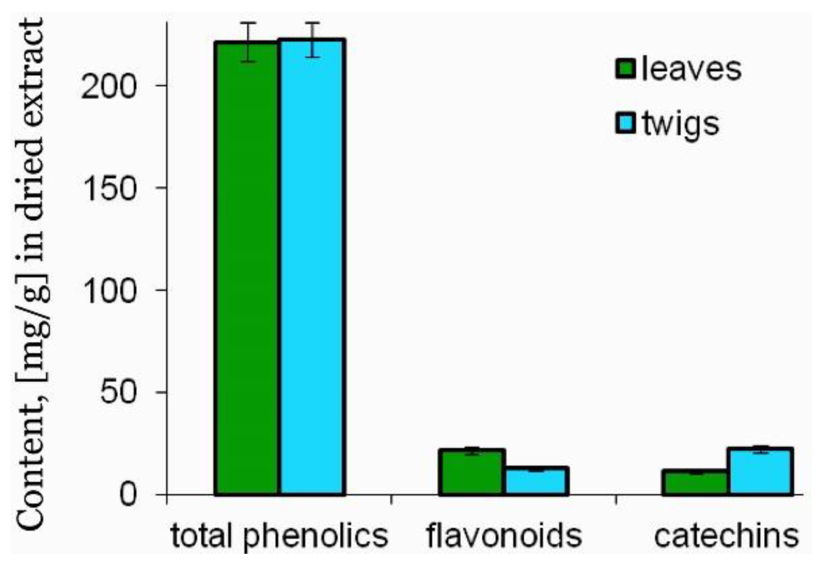
Figure 1. Total phenolic compound content in the leaf and twig extracts of R. canina. Values (chlorogenic acid, quercetin, (+)-catechin, respectively, mg equivalents per gram of dry extract) are means ± SD (n = 3).
Agronomy 10 01897 g001
Figure 2. Ellipticity changes (A) and changes in the secondary structure (B) of human serum albumin at the concentration of 1 µmol/L in the presence of varying ratios (1:0.1–1:3) of leaf and twig extracts of R. canina.
Agronomy 10 01897 g002
Figure 3. Hemolysis of human erythrocytes (A) and human fibroblasts viability (B) after 24 h incubation with the leaf and twig extracts of R. canina. Data are mean ± SD (n = 3). n.s.—not statistically significant, tested samples vs. non-treated control cells, p > 0.05.
Agronomy 10 01897 g003
Figure 4. Effect of the R. canina twig and leaf extracts (0.5–50 μg/mL−1) on H2O2/Fe-induced plasma lipid peroxidation (A); oxidation of thiol groups (B); percentages of DPPH free radical scavenging (C); protein carbonylation (D). Confocal microscopy images of BJ cells after 24 h incubation with the extracts. (E) 1—control; 2—H2O2 80 µmol/L; 3—H2O2 + twig extract 50 μg/mL−1; 4—H2O2 + leaf extract 50 μg/mL−1. Scale bar = 25 µm. The data are mean ± SEM (n = 3). * p < 0.05 vs. control. Ns—not statistically significant.
Agronomy 10 01897 g004
Table 1. Content of phenolic compounds detected using HPLC technique in dried extracts of R. canina leaves and twigs. RT—retention time. Values presented as mean ± SD from 3 measurements.
No.Phenolic CompoundsSynonymsQuantification Wavelengths:
A 235; 280; 325; 375 nm
Em 420 nm (Ex 270 nm)
RTContent (mg/g) in Dry Matter of Extract
LeafTwig
1Gallic acid3,4,5-Trihydroxybenzoic acid28069920.805 ± 0.0750.357 ± 0.033
2p-BenzoquinoneQuinone23511,6420.252 ± 0.0220.989 ± 0.087
3α-Resorcylic acid3,5-Dihydroxybenzoic acid42011,8410.138 ± 0.0130.117 ± 0.012
4Pyrocatechol1,2-Dihydroxybenzene; Catechol28012,2830.226 ± 0.0140.493 ± 0.049
5Protocatechuic acid3,4-Dihydroxybenzoic acid42012,4150.153 ± 0.01513.911 ± 1.303
6Neochlorogenic acidtrans-5-O-Caffeoylquinic acid32512,83357.148 ± 5.0310.258 ± 0.0262
7(−)-EpigallocatechinMonomeric flavan-3-ol23514,9920.207 ± 0.0221.680 ± 0.076
8(+)-CatechinFlavan-3-ol; monomeric flavan-3-ol23515,3082.804 ± 0.20617.798 ± 1.544
94-Hydroxybenzoic acid-23515,7001.182 ± 0.1110.323 ± 0.0361
10Gentisic acid2,5-Dihydroxybenzoic acid32516,2671.577 ± 0.1640.340 ± 0.033
11Procyanidin B2Polymeric flavan-3-ol; pentahydroxyflavane (cis,cis″-4,8″-Bi(3,3′,4′,5,7-pentahydroxyflavane)28016,50022.473 ± 2.0193.222 ± 0.317
124-Hydroxybenzaldehyde-28016,7331.111 ± 0.0950.263 ± 0.0277
13Chlorogenic acidtrans-3-O-Caffeoylquinic acid32517,4174.609 ± 0.4080.934 ± 0.0955
14Vanillic acid4-Hydroxy-3-methoxybenzoic acid42018,0620.102 ± 0.0140.379 ± 0.007
15Caffeic acidtrans-3,4-Dihydroxycinnamic acid32518,4920.035 ± 0.0030.203 ± 0.021
16β-Resorcylic acid2,4-Dihydroxybenzoic acid42018,6180.017 ± 0.0020.015 ± 0.001
17(−)-EpicatechinMonomeric flavan-3-ol ((−)-cis-3,3′,4′,5,7-pentahydroxyflavane)23518,9501.822 ± 0.1151.379 ± 0.125
18Syringic acid4-Hydroxy-3,5-dimethoxybenzoic acid42019,4200.613 ± 0.0570.134 ± 0.011
191.3-Dicaffeoylquinic acid1,5-Dicaffeoylquinic acid32519,6750.826 ± 0.0780.189 ± 0.019
20Cyanidin3,3′,4,5,7-Pentahydroxyflavone (3,3′,4,5,7-pentahydroxyflavylium chloride)28019,70847.448 ± 4.4614.453 ± 0.406
21Syringaldehyde4-Hydroxy-3,5-dimethoxybenzaldehyde28020,8250.402 ± 0.0070.262 ± 0.016
22p-Coumaric acidtrans-4-Hydroxycinnamic acid32522,5670.520 ± 0.0580.247 ±0.028
23Ferulic acid4-Hydroxy-3-methoxy-cinnamic acid42023,7370.439 ± 0.0410.081 ± 0.007
24Coumarin1,2-Benzopyrone28024,6001.285 ± 0.1160.170 ± 0.016
25Sinapic acid4-Hydroxy-3,5-dimethoxy-cinnamic acid42024,7310.214 ± 0.0110.140 ± 0.014
26trans-3-Hydroxycinnamic acidm-Coumaric acid28025,0670.241 ± 0.0220.119 ± 0.012
27Luteolin 7-O- β -D-glucosideGlucoluteolin; luteoloside32526,1331.614 ± 0.1711.417 ± 0.118
28Rutinquercetin-3-O-rutinoside37526,76526.66 ± 2.4814.431 ± 0.433
29Ellagic acid4,4′,5,5′,6,6′-Hexahydroxydiphenic acid 2,6,2′,6′-dilactone23526,78335.881 ± 3.34614.448 ± 1.451
30HesperidinHesperetin-7-rutinoside28027,0584.013 ± 0.3030.633 ± 0.006
31o-Coumaric acidtrans-2-Hydroxycinnamic acid42027,4830.711 ± 0.660.149 ± 0.015
32Rosmarinic acid3,4-Dihydroxycinnamic acid (R)-1-carboxy-2-(3,4-dihydroxyphenyl)ethyl ester42027,9704.406 ± 0.4111.851 ± 0.0182
33Salicylic acid2-Hydroxybenzoic acid42028,1580.457 ± 0.0520.474 ± 0.045
34Myricetin (flavonol)3,3′,4′,5,5′,7-Hexahydroxyflavone37528,3083.928 ± 0.2657.175 ± 0.711
35QuercetinFlavonol (3,3′,4′,5,7-pentahydroxyflavone)37532,1420.156 ± 0.0160.241 ± 0.025
36trans-Cinnamic acidCinnamic acid28032,5520.167 ± 0.0170.034 ± 0.003
37Naringenin4′,5,7-Trihydroxyflavanone28033,1420.461 ± 0.0370.924 ± 0.091
38Luteolin3′,4′,5,7-Tetrahydroxyflavone32533,4200.911 ± 0.0870.364 ± 0.036
39Kaempferol3,4′,5,7-Tetrahydroxyflavone37535,3750.148 ± 0.0150.167 ± 0.017
403-HydroxyflavoneFlavonol23546,9580.103 ± 0.0110.128 ± 0.006
Table 2. Total content of water soluble vitamins (A) and vitamin E isomers (B) in dry matter of the leaf and twig extracts from R. canina.
(A) Water Soluble Vitamins
Content (mg/g) in Dry Matter of the Extracts
LeafTwig
B1 (thiaminechloride) #1.12 ± 0.050.77 ± 0.05
B2 (riboflavin) #0.48 ± 0.070.51 ± 0.06
B3 (pantothenic acid) #2.10 ± 0.022.70 ± 0.12
B5 (nicotinicacid) #3.30 ± 0.183.10 ± 0.10
B6 (pyridoxine) #5.70 ± 0.106.20 ± 0.10
Bc (folic acid) #0.97 ± 0.110.86 ± 0.12
(B) Vitamin E Isomers
IsomersContent (mg/g) in Dry Matter of the Extracts
Leafα—tocopherol #0.54 ± 0.07
ß—tocopherol #0.13 ± 0.05
γ—tocopherol #0.18 ± 0.03
Twigα—tocopherol0.31 ± 0.05
ß—tocopherol0.15 ± 0.02
γ—tocopherol0.09 ± 0.02
#—statistically non-significant, p > 0.05 when leaf vs. twig was compared. n = 3, values presented as mean ± SD. Data presented as mg/g DW.
Table 3. The content of amino acids in dried extracts from twigs and leaves of R. canina.
Amino AcidsContent (mg/g) in Dry Matter of the Extracts
LeafTwig
Arginine #4.979 ± 0.009-
Lysine #3.378 ± 0.0081.426 ± 0.008
Tyrosine #3.023 ± 0.0020.713 ± 0.023
Phenylalanine #6.045 ± 0.0322.262 ± 0.003
Histidine #0.960 ± 0.0350.565 ± 0.005
Leucine + isoleucine#6.900 ± 0.0201.819 ± 0.020
Methionine #1.494 ± 0.0410.787 ± 0.063
Valine #0.460 ± 0.0100.713 ± 0.003
Proline #10.491 ± 0.0043.196 ± 0.040
Threonine #4.445 ± 0.0051.770 ± 0.010
Serine #4.801 ± 0.0202.040 ± 0.053
Alanine #5.334 ± 0.0191.573 ± 0.020
Glycine #4.801 ± 0.001-
Total content61.48716.864
#—statistically non-significant, p > 0.05 when leaf vs. twig was compared. n = 3, values presented as mean ± SD. Data presented as mg/g DW.
Publisher’s Note: MDPI stays neutral with regard to jurisdictional claims in published maps and institutional affiliations.