Piezoelectric Scaffolds as Smart Materials for Neural Tissue Engineering


Abstract
1. Introduction
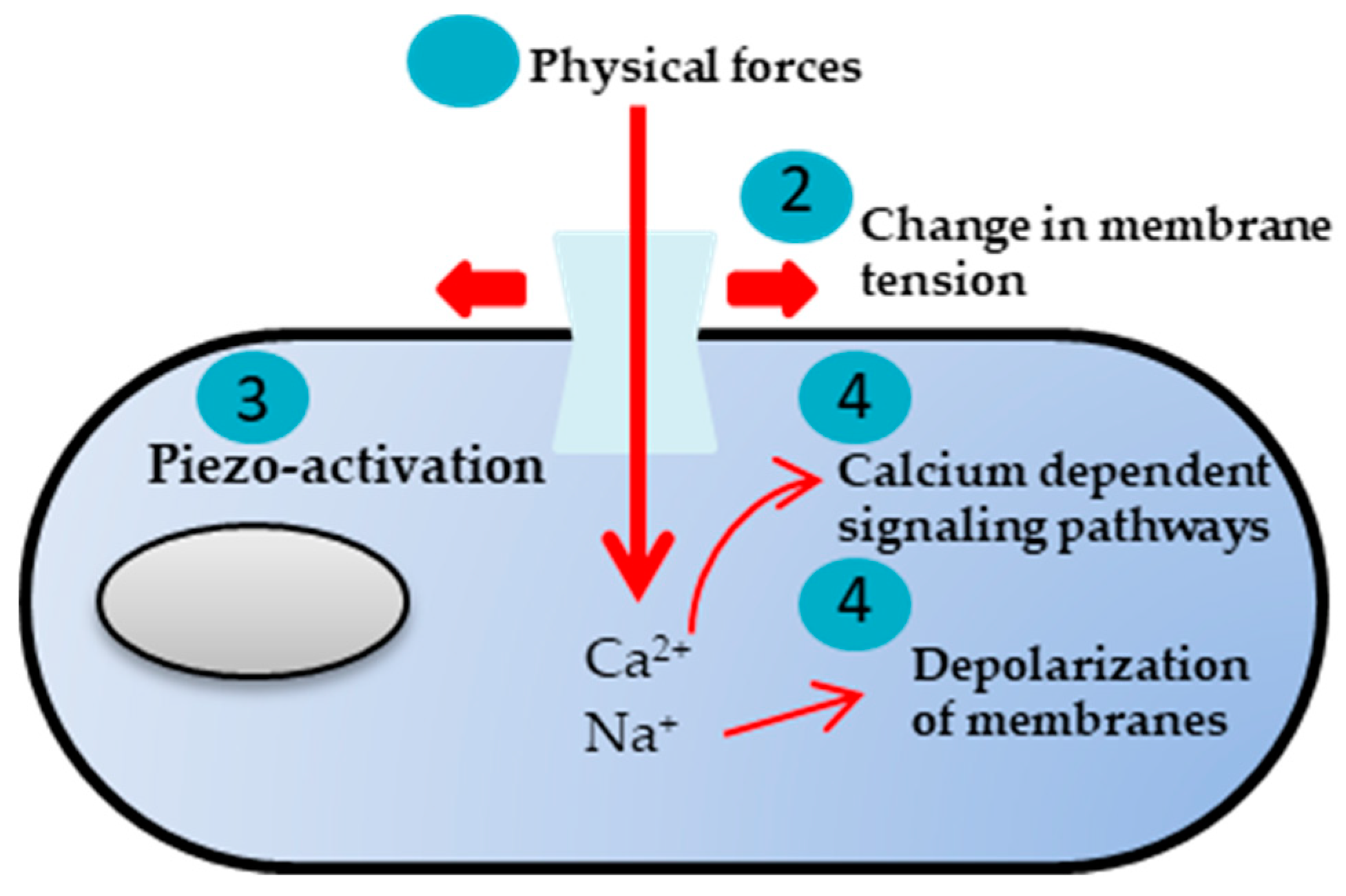
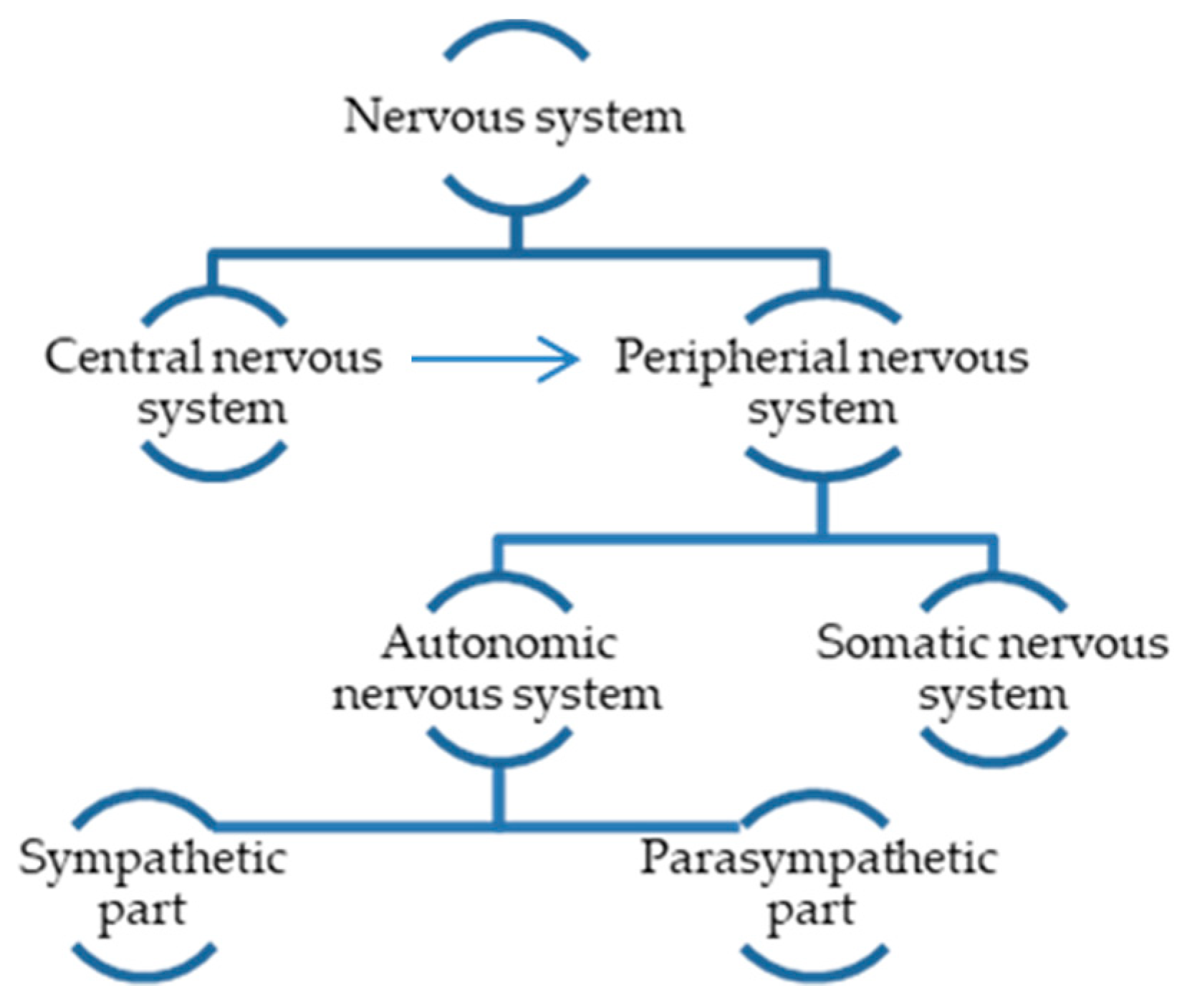
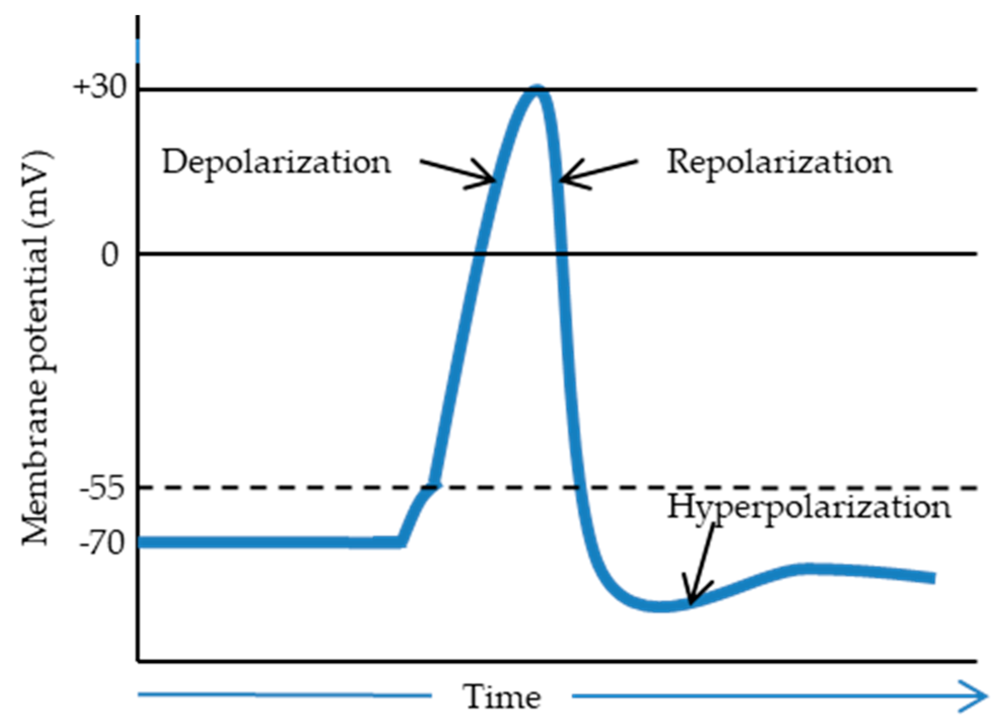
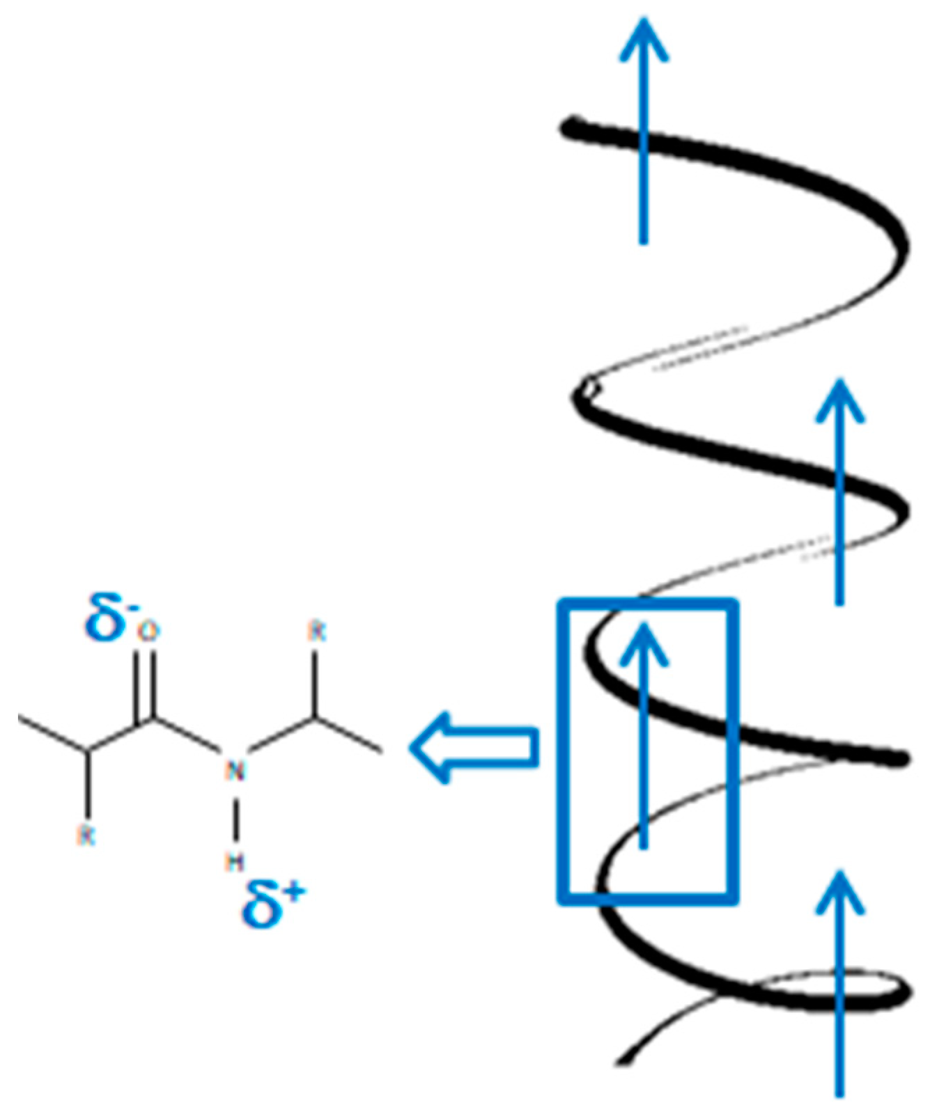
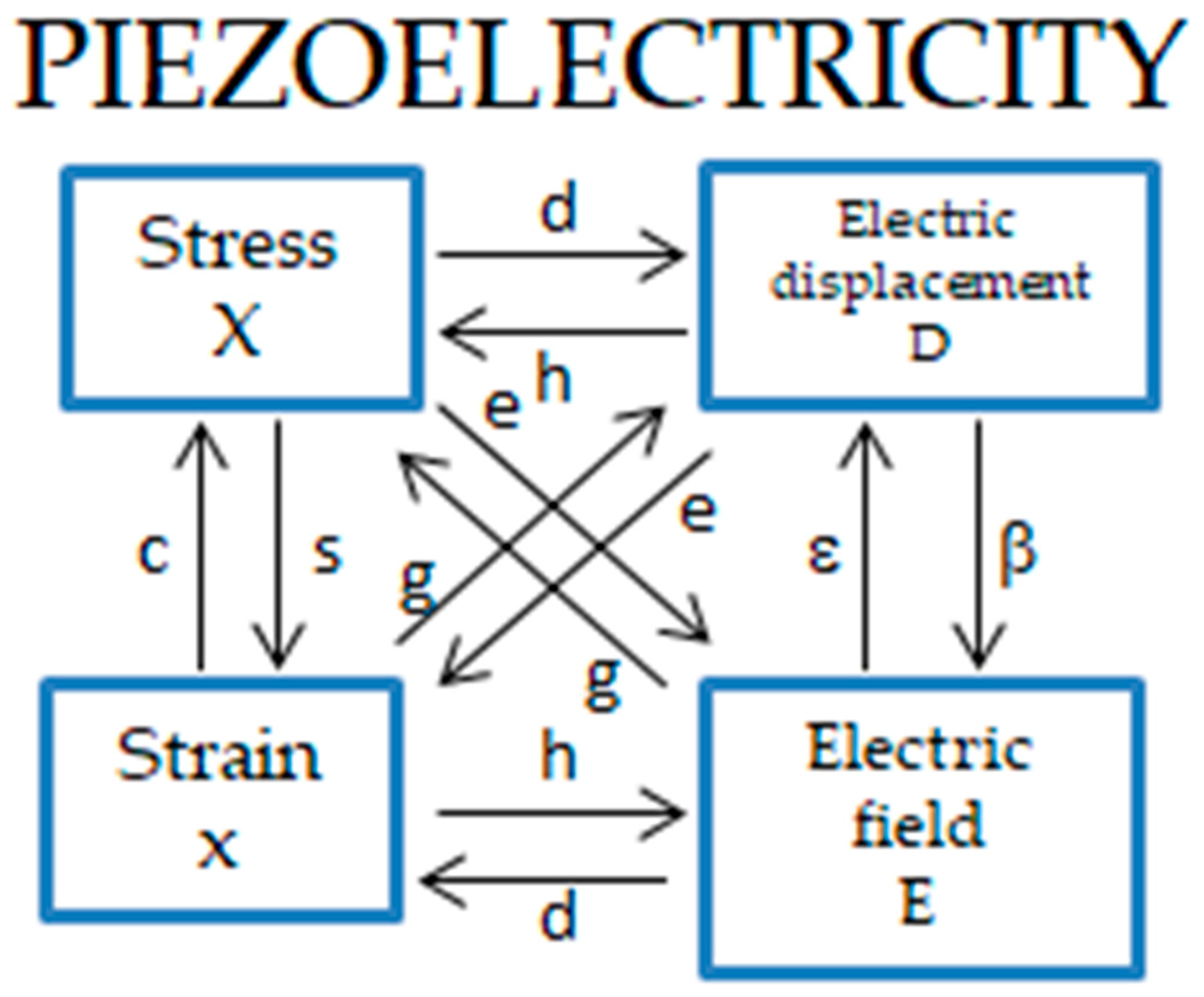
2. Mechanotransduction and Piezoelectricity in Living Organisms
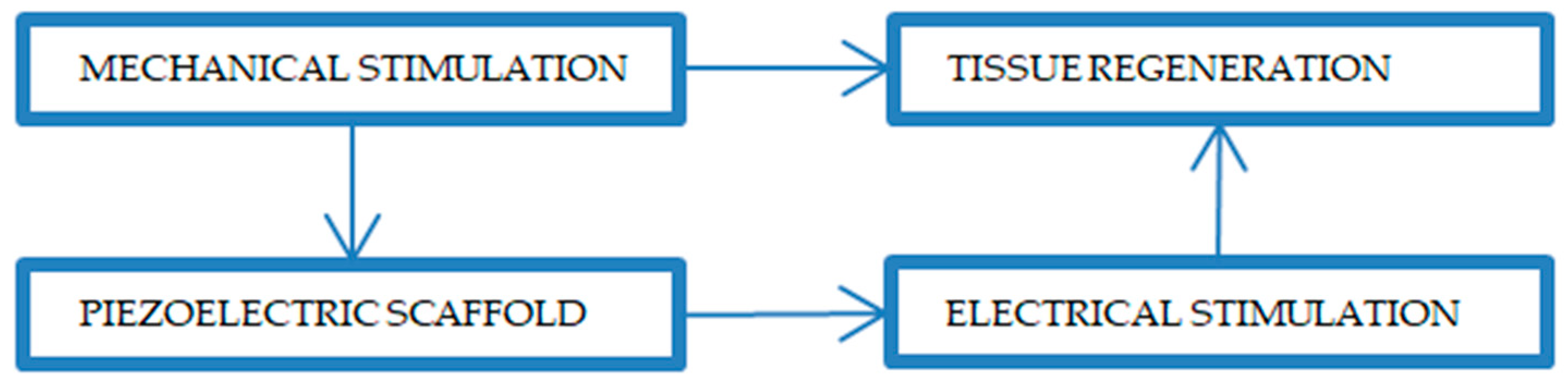
3. Scaffolds: Stimuli Responsive (Piezoelectric) vs. Passive
4. Application of Piezoelectric Biomaterials in Neural Tissue Engineering
4.1. Piezoceramics
4.1.1. Barium Titanate
4.1.2. Boron Nitride
4.1.3. Zinc Oxide
4.2. Piezopolymers
4.2.1. Synthetic Polymers
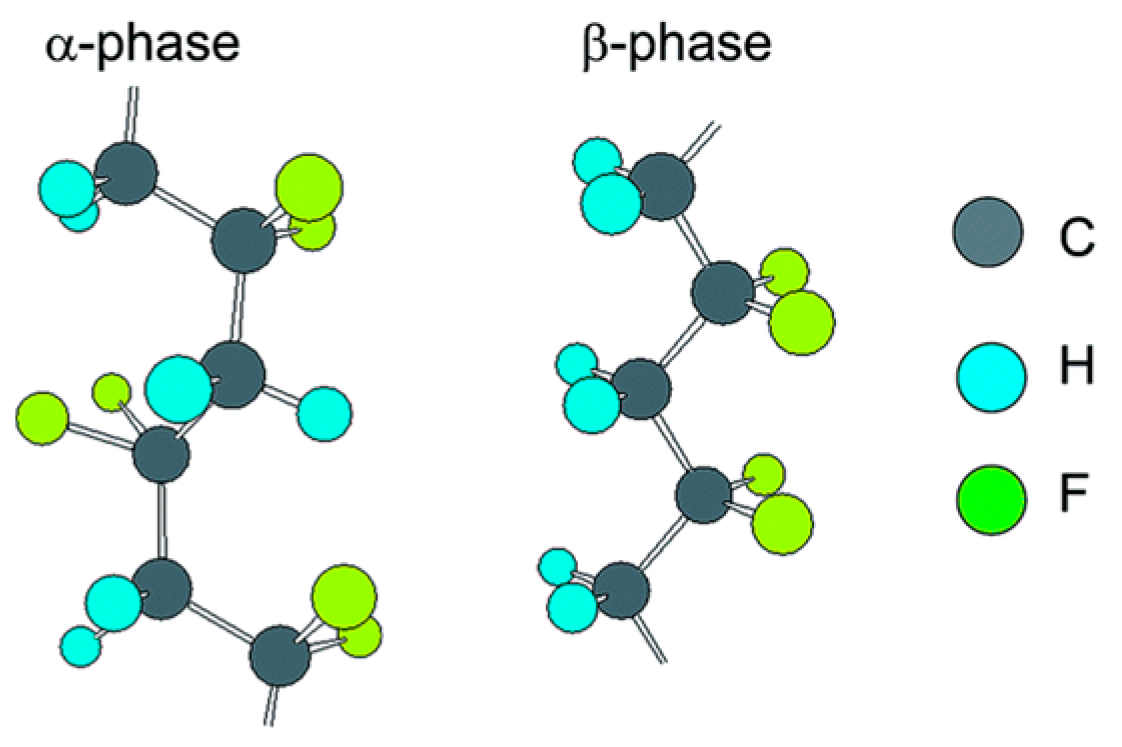
Polyvinylidene Fluoride
Poly-Vinylidene Fluoride-Trifluoroethylene
Poly-3-Hydroxybutyrate-3-Hydroxyvalerate
Poly-L-Lactic Acid
4.2.2. Natural Biopolymers
Cellulose
Chitin and Chitosan
Collagen
5. Conclusions and Future Perspectives
Author Contributions
Funding
Conflicts of Interest
References
- Wolfenson, H.; Yang, B.; Sheetz, M.P. Steps in mechanotransduction pathways that control cell morphology. Annu. Rev. Physiol. 2019, 81, 585–605. [Google Scholar] [CrossRef] [PubMed]
- Whalin, M.K.; Arora, S.S. Anatomy of the Brain and Spinal Cord. In Basic Sciences in Anesthesia; Farag, E., Argalious, M., Tetzlaff, J., Sharma, D., Eds.; Springer: New York, NY, USA, 2018; pp. 41–59. [Google Scholar]
- Lis, A.; Szarek, D.; Laska, J. The outlook for the use of polymeric scaffolds in the reconstruction and the regeneration stimulation of traumatic brain injuries. Polim. Med. 2018, 43, 302–312. [Google Scholar]
- Han, D.; Cheung, K.C. Biodegradable cell-seeded nanofiber scaffolds for neural repair. Polymers 2011, 3, 1684–1733. [Google Scholar] [CrossRef]
- Balint, R.; Cassidy, N.J.; Cartmell, S.H. Conductive polymers: Towards a smart biomaterial for tissue engineering. Acta Biomat. 2014, 10, 2341–2353. [Google Scholar] [CrossRef]
- Niemczyk, B.; Sajkiewicz, P.; Gradys, A. Injectable hydrogels as novel materials for central nervous system regeneration. J. Neural Eng. 2018, 15, 051002. [Google Scholar] [CrossRef]
- Lis, A.; Szarek, D.; Laska, J. Biomaterials engineering strategies for spinal cord regeneration: State of the art. Polim. Med. 2013, 43, 59–80. [Google Scholar]
- Taylor, C.A.; Bell, J.M.; Breiding, M.J.; Xu, L. Traumatic brain injury—Related emergency Department Visits, Hospitalizations, and Deaths—United States, 2007 and 2013. MMWR Surveill. Summ. 2017, 66, 1. [Google Scholar] [CrossRef]
- Mathieu, S.; Manneville, J.B. Intracellular mechanics: Connecting rheology and mechanotransduction. COCEBI 2019, 56, 34–44. [Google Scholar] [CrossRef]
- Delcroix, G.J.R.; Schiller, P.C.; Benoit, J.P.; MonteroMenei, C.N. Adult cell therapy for brain neuronal damages and the role of tissue engineering. Biomaterials 2010, 31, 2105–2120. [Google Scholar] [CrossRef]
- Wang, S.; Hou, J.; Bei, J.; Zhao, Y. Tissue engineering and peripheral nerve regeneration (III)—Sciatic nerve regeneration with PDLLA nerve guide. Sci. China 2001, 44, 419–426. [Google Scholar] [CrossRef]
- Adrian, H.; Mårten, K.; Salla, N.; Lasse, V. Biomarkers of Traumatic Brain Injury: Temporal Changes in Body Fluids. eNeuro 2016, 3, 0294-16. [Google Scholar] [CrossRef] [PubMed]
- Weidner, N.; Rudiger, R.; Tansey, K.E. Neurological Aspects of Spinal Cord Injury; Springer: Cham, Switzerland, 2017; Volume 1, pp. 3–17. [Google Scholar]
- Zhang, L.; Sirivisoot, S.; Balasundaram, G.; Webster, T.J. Advanced Biomaterials: Fundamentals, Processing and Applications; Basu, B., Katti, D., Kumar, A., Eds.; John Wiley & Sons, Inc.: Hoboken, NJ, USA, 2009. [Google Scholar]
- Langlois, J.A.; Rutland-Brown, W.; Thomas, K.E. Traumatic Brain Injury in the United States: Emergency Department Visits, Hospitalizations, and Deaths, Centers for Disease Control and Prevention; National Center for Injury Prevention and Control: Atlanta, GA, USA, 2004.
- Flax, J.D.; Aurora, S.; Yang, C. Engraftable human neural stem cells respond to developmental cues, replace neurons, and express foreign genes. Nature Biotechnol. 1998, 16, 1033–1438. [Google Scholar] [CrossRef] [PubMed]
- Kordower, J.H.; Tuszynski, M.H. CNS Regeneration: Basic Science and Clinical Advances; Kordower, J.H., Tuszynski, M.H., Eds.; Academic: Cambridge, MA, USA, 1999; pp. 159–182. [Google Scholar]
- Steel, E.M.; Sundararaghavan, H.G. Electrically Conductive Materials for Nerve Regeneration. In Neural Engineering; Springer: New York, NY, USA, 2016; pp. 145–179. [Google Scholar]
- Zhao, P.; Gu, H.; Mi, H.; Rao, C.; Fu, J.; Turng, L.S. Fabrication of scaffolds in tissue engineering: A review. Front. Mech. Eng. 2018, 13, 107–119. [Google Scholar] [CrossRef]
- Gao, S.; Tang, G.; Hua, D.; Xiong, R.; Han, J.; Jiang, S.; Huang, C. Stimuli-responsive bio-based polymeric systems and their applications. J. Mater. Chem. B 2019, 7, 709–729. [Google Scholar] [CrossRef]
- Jalili-Firoozinezhad, S.; Mirakhori, F.; Baharvand, H. Nanotissue Engineering of Neural Cells. Stem Cell Nanoeng. 2015, 265, 265–283. [Google Scholar]
- Nguyen, H.T.; Wei, C.; Chow, J.K.; Nguy, L.; Nguyen, H.K.; Schmidt, C.E. Electric field stimulation through a substrate influences Schwann cell and extracellular matrix structure. J. Neural Eng. 2013, 10, 046011. [Google Scholar] [CrossRef] [PubMed]
- Chew, S.Y.; Wen, Y.; Dzenis, Y.; Leong, K.W. The role of electrospinning in the emerging field of nanomedicine. Curr. Pharm. Des. 2006, 12, 4751–4770. [Google Scholar] [CrossRef]
- Murugan, R.; Ramakrishna, S. Nano-featured scaffolds for tissue engineering: A review of spinning methodologies. Tissue Eng. 2006, 12, 435–447. [Google Scholar] [CrossRef]
- Venugopal, J.; Low, S.; Choon, A.T.; Ramakrishna, S. Interaction of cells and nanofiber scaffolds in tissue engineering. J. Biomed. Mater. Res. B 2008, 84, 34–48. [Google Scholar] [CrossRef]
- Teo, W.E.; He, W.; Ramakrishna, S. Electrospun scaffold tailored for tissue-specific extracellular matrix. Biotechno. J. 2006, 1, 918–929. [Google Scholar] [CrossRef]
- Barnes, C.P.; Sell, S.A.; Boland, E.D.; Simpson, D.G.; Bowlin, G.L. Nanofiber technology: Designing the next generation of tissue engineering scaffolds. Adv. Drug Deliv. Rev. 2007, 59, 1413–1433. [Google Scholar] [CrossRef]
- Murugan, R.; Ramakrishna, S. Design strategies of tissue engineering scaffolds with controlled fiber orientation. Tissue Eng. 2007, 13, 1845–1866. [Google Scholar] [CrossRef] [PubMed]
- Heydarkhan-Hagvall, S.; Schenke-Layland, K.; Dhanasopon, A.P.; Rofail, F.; Smith, H.; Wu, B.M.; Shemin, R.; Beygui, R.E.; MacLellan, W.R. Three-dimensional electrospun ECM-based hybrid scaffolds for cardiovascular tissue engineering. Biomaterials 2008, 29, 2907–2914. [Google Scholar] [CrossRef] [PubMed]
- Li, W.J.; Mauck, J.A.; Cooper, X.; Yuan, R.S. Tuan Engineering controllable anisotropy in electrospun biodegradable nanofibrous scaffolds for musculoskeletal tissue engineering. J. Biomech. 2007, 40, 1686–1693. [Google Scholar] [CrossRef] [PubMed]
- Li, W.J.; Tuli, R.; Huang, X.; Laquerriere, P.; Tuan, R.S. Multilineage differentiation of human mesenchymal stem cells in a three-dimensional nanofibrous scaffold. Biomaterials 2005, 26, 5158–5166. [Google Scholar] [CrossRef] [PubMed]
- Yang, F.; Xu, C.Y.; Kotaki, M.; Wang, S.; Ramakrishna, S. Characterization of neural stem cells on electrospun poly(L-lactic acid) nanofibrous scaffold. J. Biomater. Sci. Polym. Ed. 2004, 15, 1483–1497. [Google Scholar] [CrossRef] [PubMed]
- Ting, Y.; Gunawan, H.; Sugondo, A.; Chiu, C. A New Approach of Polyvinylidene Fluoride (PVDF) Poling Method for Higher Electric Response. Ferroelectrics 2013, 446, 28–38. [Google Scholar] [CrossRef]
- Dang, Z.M.; Lin, Y.H.; Nan, C.W. Novel ferroelectric polymer composites with high dielectric constants. Adv. Mater. 2003, 15, 1625–1629. [Google Scholar] [CrossRef]
- Bera, B.; Sarkar, M. Piezoelectricity in PVDF and PVDF Based Piezoelectric Nanogenerator: A Concept. Int. J. Appl. Phys. 2017, 9, 95–99. [Google Scholar] [CrossRef]
- Damaraju, S.M.; Wu, S.; Jaffe, M.; Arinzeh, T.L. Structural changes in PVDF fibers due to electrospinning and its effect on biological function. Biomed. Mater. 2013, 8, 045007. [Google Scholar] [CrossRef]
- Defteralı, Ç.; Verdejo, R.; Majeed, S.; Boschetti-de-Fierro, A.; Méndez-Gómez, H.R.; Díaz-Guerra, E.; Vuluga, D. In vitro evaluation of biocompatibility of uncoated thermally reduced graphene and carbon nanotube-loaded PVDF membranes with adult neural stem cell-derived neurons and glia. Front. Bioeng. Biotechnol. 2016, 4, 94. [Google Scholar] [CrossRef] [PubMed]
- Young, T.H.; Lin, U.H.; Lin, D.J.; Chang, H.H.; Cheng, L.P. Immobilization of L-lysine on microporous PVDF membranes for neuron culture. J. Biomater. Sci. Polym. 2009, 20, 703–720. [Google Scholar] [CrossRef] [PubMed]
- Bar, H.N.; Bhat, M.R.; Murthy, C.R.L. Identification of failure modes in GFRP using PVDF sensors: ANN approach. Compos. Struct. 2004, 65, 231–237. [Google Scholar] [CrossRef]
- Fu, Y.S.; Shih, Y.T.; Cheng, Y.C.; Min, M.Y. Transformation of human umbilical mesenchymal cells into neurons in vitro. J. Biomed. Sci. 2004, 11, 652–660. [Google Scholar] [CrossRef]
- Ning, C.; Zhou, Z.; Tan, G.; Zhu, Y.; Mao, C. Electroactive polymers for tissue regeneration: Developments and perspectives. Prog. Polym. Sci. 2018, 81, 144–162. [Google Scholar] [CrossRef]
- Moran, H.; Cancel, L.M.; Mayer, M.A.; Qazi, H.; Munn, L.L.; Tarbell, J.M. The cancer cell glycocalyx proteoglycan glypican-1 mediates interstitial flow mechanotransduction to enhance cell migration and metastasis. Biorheology 2019, 56, 151–161. [Google Scholar] [CrossRef]
- Gargalionis, A.N.; Basdra, E.K.; Papavassiliou, A.G. Polycystins and Mechanotransduction in Human Disease. Int. J. Mol. Sci. 2019, 20, 2182. [Google Scholar] [CrossRef]
- Maurer, M.; Lammerding, J. The driving force: Nuclear mechanotransduction in cellular function, fate, and disease. Annu. Rev. Biomed. Eng. 2019, 21, 443–468. [Google Scholar] [CrossRef]
- Yamada, K.M.; Sixt, M. Mechanisms of 3D cell migration. Nat. Rev. Mol. 2019, 20, 738–752. [Google Scholar] [CrossRef]
- Chang, W.; Gu, J.G. Impairment of tactile responses and Piezo channel mechanotransduction in mice following chronic vincristine treatment. Neurosci. Lett. 2019, 705, 14–19. [Google Scholar] [CrossRef]
- Salvi, A.M.; DeMali, K.A. Mechanisms linking mechanotransduction and cell metabolism. COCEBI 2018, 54, 114–120. [Google Scholar] [CrossRef] [PubMed]
- Pariy, I.O.; Ivanova, A.A.; Shvartsman, V.V.; Lupascu, D.C.; Sukhorukov, G.B.; Ludwig, T.; Surmenev, R.A. Piezoelectric Response in Hybrid Micropillar Arrays of Poly (Vinylidene Fluoride) and Reduced Graphene Oxide. Polymers 2019, 11, 1065. [Google Scholar] [CrossRef] [PubMed]
- Ruan, L.; Yao, X.; Chang, Y.; Zhou, L.; Qin, G.; Zhang, X. Properties and Applications of the β Phase Poly (vinylidene fluoride). Polymers 2018, 10, 228. [Google Scholar] [CrossRef] [PubMed]
- Guilak, F.; Cohen, D.M.; Estes, B.T.; Gimble, J.M.; Liedtke, W.; Chen, C.S. Control of stem cell fate by physical interactions with the extracellular matrix. Cell Stem Cell 2009, 5, 17–26. [Google Scholar] [CrossRef] [PubMed]
- Lee, M.R.; Kwon, K.W.; Jung, H.; Kim, H.N.; Suh, K.Y.; Kim, K.; Kim, K.S. Direct differentiation of human embryonic stem cells into selective neurons on nanoscale ridge/groove pattern arrays. Biomaterials 2010, 31, 4360–4366. [Google Scholar] [CrossRef] [PubMed]
- McBeath, R.; Pirone, D.M.; Nelson, C.M.; Bhadriraju, K.; Chen, C.S. Cell shape, cytoskeletal tension, and RhoA regulate stem cell lineage commitment. Dev. Cell 2004, 6, 483–495. [Google Scholar] [CrossRef]
- Verkhratsky, A.; Ho, M.S.; Parpura, V. Evolution of Neuroglia. In Neuroglia in Neurodegenerative Diseases; Springer: New York, NY, USA, 2019; pp. 15–44. [Google Scholar]
- Georges, P.C.; Miller, W.J.; Meaney, D.F.; Sawyer, E.S.; Janmey, P.A. Matrices with compliance comparable to that of brain tissue select neuronal over glial growth in mixed cortical cultures. Biophys. J. 2006, 90, 3012–3018. [Google Scholar] [CrossRef]
- Doetsch, F. A niche for adult neural stem cells. Curr. Opin. Genet. Dev. 2003, 13, 543–550. [Google Scholar] [CrossRef]
- Alenghat, F.J.; Ingber, D.E. Mechanotransduction: All signals point to cytoskeleton, matrix, and integrins. Science’s STKE: Signal transduction knowledge environment. Sci. Signal. 2002, 119, pe6. [Google Scholar] [CrossRef]
- Kjellman, C.; Lidman, J.; Ljungström, K. Nilsson, Piezoelectric Sensor in a Living Organism for Fluid Pressure Measurement. U.S. Patent 6,886,411, 3 May 2005. [Google Scholar]
- Wada, Y.; Hayakawa, R. Piezoelectricity and pyroelectricity of polymers. Jpn. J. Appl. Phys. 1976, 15, 2041. [Google Scholar] [CrossRef]
- Parpaite, T.; Coste, B. Piezo channels. Curr. Biol. 2017, 27, R250–R252. [Google Scholar] [CrossRef] [PubMed]
- Liu, Y.; Gao, J.; Peng, M.; Meng, H.; Ma, H.; Cai, P.; Si, G. A review on central nervous system effects of gastrodin. Front. Pharmacol. 2018, 9, 24. [Google Scholar] [CrossRef] [PubMed]
- Piccoli, A.; Rossettini, G.; Cecchetto, S.; Viceconti, A.; Ristori, D.; Turolla, A.; Testa, M. Effect of attentional focus instructions on motor learning and performance of patients with central nervous system and musculoskeletal disorders: A systematic review. J. Funct. Morphol. Kinesiol. 2018, 3, 40. [Google Scholar] [CrossRef]
- Schulte, F.; Kunin-Batson, A.S.; Olson-Bullis, B.A.; Banerjee, P.; Hocking, M.C.; Janzen, L.; Krull, K.R. Social attainment in survivors of pediatric central nervous system tumors: A systematic review and meta-analysis from the Children’s Oncology Group. J. Cancer Surviv. 2019, 13, 921–931. [Google Scholar] [CrossRef]
- Saheb, N.; Mekid, S. Fiber-Embedded Metallic Materials: From Sensing towards Nervous Behavior. Materials 2015, 8, 7938–7961. [Google Scholar] [CrossRef]
- Stavoe, A.K.; Holzbaur, E.L. Autophagy in Neurons. Annu. Rev. Cell Dev. Biol. 2019, 35, 477–500. [Google Scholar] [CrossRef]
- Swenarchuk, L.E. Nerve, Muscle, and Synaptogenesis. Cells 2019, 8, 1448. [Google Scholar] [CrossRef]
- Martí, D.; Brunel, N.; Ostojic, S. Correlations between synapses in pairs of neurons slow down dynamics in randomly connected neural networks. Phys. Rev. E 2018, 97, 062314. [Google Scholar] [CrossRef]
- Van Driesche, S.J.; Martin, K.C. New frontiers in RNA transport and local translation in neurons. Dev. Neurobiol. 2018, 78, 331–339. [Google Scholar] [CrossRef]
- Zhao, W.; Cui, W.; Xu, S.; Cheong, L.Z.; Wang, D.; Shen, C. Direct study of the electrical properties of PC12 cells and hippocampal neurons by EFM and KPFM. Nanoscale Adv. 2019, 1, 537–545. [Google Scholar] [CrossRef]
- Averbeck, B.B.; Lee, D. Coding and transmission of information by neural ensembles. Trends Neurosci. 2004, 27, 225–230. [Google Scholar] [CrossRef]
- Patel, N. Orientation of neurite growth by extracellar electric fields. J. Neurosci. 1982, 2, 483–496. [Google Scholar] [CrossRef]
- Freeman, J.A.; Manis, P.B.; Snipes, G.J. Steady growth cone currents revealed by a novel circularly vibrating probe: A possible mechanism underlying neurite growth. J. Neurosci. 1985, 13, 257–283. [Google Scholar] [CrossRef] [PubMed]
- Sisken, B.F.; Kanje, M.; Lundborg, G.; Herbst, E.; Kurtz, W. Stimulation of rat sciatic nerve regeneration with pulsed electromagnetic fields. Brain Res. 1989, 485, 309–316. [Google Scholar] [CrossRef]
- Kimura, K.; Yanagida, Y.; Haruyama, T.; Kobatake, E.; Aizawa, M. Gene expression in the electrically stimulated differentiation of PC12 cells. J. Biotechnol. 1998, 63, 55–65. [Google Scholar] [CrossRef]
- Kotwal, A.; Schmid, C.E. Electrical stimulation alters protein adsorption and nerve cell interactions with electrically conducting biomaterials. Biomaterials 2001, 22, 1055–1064. [Google Scholar] [CrossRef]
- Ghasemi-Mobarakeh, L.; Prabhakaran, M.P.; Morshed, M.; Nasr-Esfahani, M.H.; Baharvand, H.; Kiani, S.; Ramakrishna, S. Application of conductive polymers, scaffolds and electrical stimulation for nerve tissue engineering. J. Tissue Eng. Regen. Med. 2011, 5, 17–35. [Google Scholar] [CrossRef]
- Martin, R.M. Piezoelectricity. Phys. Rev. B 1972, 5, 1607. [Google Scholar] [CrossRef]
- Shamos, M.H.; Lavine, L.S. Piezoelectricity as a fundamental property of biological tissues. Nature 1967, 213, 267–269. [Google Scholar] [CrossRef]
- Telega, J.J.; Wojnar, R. Piezoelectric effects in biological tissues. J. Theor. Appl. Mech. 2002, 40, 723–759. [Google Scholar]
- Fukada, E. Electrical phenomena in biorheology. Biorheology 1982, 19, 15–27. [Google Scholar] [CrossRef] [PubMed]
- Athenstaedt, H. Pyroelectric and piezoelectric behaviour of human dental hard tissues. Arch. Oral Biol. 1971, 16, 495–501. [Google Scholar] [CrossRef]
- De Rossi, D.; Domenici, D.; Pastacaldi, P. Piezoelectric Properties of Dry Human Skin. IEEE Trans. Electr. Insul. 1985, 21, 511–517. [Google Scholar] [CrossRef]
- Ingber, D.E. The architecture of life. Sci. Am. 1998, 278, 48–57. [Google Scholar] [CrossRef]
- Reyes-Gasga, J.; Galindo-Mentle, M.; Brès, E.; Vargas-Becerril, N.; Orozco, E.; Rodríguez-Gómez, A.; García-García, R. Detection of the piezoelectricity effect in nanocrystals from human teeth. J. Phys. Chem. Solids 2020, 136, 109140. [Google Scholar] [CrossRef]
- Udovč, L.; Spreitzer, M.; Vukomanović, M. Towards hydrophilic piezoelectric poly-L-lactide films: Optimal processing, post-heat treatment and alkaline etching. Polym. J. 2019, 1–13. [Google Scholar] [CrossRef]
- Hoop, M.; Chen, X.; Ferrari, A.; Fajer, M.; Gagik, G.; Theo, T.; Dimos, P.; Bradley, N.; Salvador, P. Ultrasound-mediated piezoelectric differentiation of neuron-like PC12 cells on PVDF membranes. Sci. Rep. 2017, 7, 4028. [Google Scholar] [CrossRef]
- Ahn, A.C.; Grodzinsky, A.J. Relevance of collagen piezoelectricity to “Wolff’s Law”: A critical review. Med. Eng. Phys. 2009, 31, 733–741. [Google Scholar] [CrossRef]
- Lang, S.B. Pyroelectric Effect in Bone and Tendon. Nature 1966, 212, 704–705. [Google Scholar] [CrossRef]
- Anderson, J.C.; Eriksson, C. Electrical properties of wet collagen. Nature 1968, 218, 166–168. [Google Scholar] [CrossRef]
- Anderson, J.C.; Eriksson, C. Piezoelectric properties of dry and wet bone. Nature 1970, 227, 491–492. [Google Scholar] [CrossRef] [PubMed]
- Fukada, E. Piezoelectricity in polymers and biological materials. Ultrasonics 1968, 6, 229–234. [Google Scholar] [CrossRef]
- Furukawa, T. Piezoelectricity and pyroelectricity in polymers. IEEE Tran. Electr. Insul. 1989, 24, 375–394. [Google Scholar] [CrossRef]
- Puppi, D.; Chiellini, F.; Piras, A.; Chiellini, E. Polymeric materials for bone and cartilage repair. Prog. Polym. Sci. 2010, 35, 403–440. [Google Scholar] [CrossRef]
- Ribeiro, C.; Sencadas, V.; Correia, D.M.; Lanceros-Méndez, S. Piezoelectric polymers as biomaterials for tissue engineering applications. Colloids Surf. B Biointerfaces 2015, 136, 46–55. [Google Scholar] [CrossRef]
- Fukada, E. History and recent progress in piezoelectric polymers. IEEE Trans. Ultrason. Ferroelectr. Freq. Control 2000, 47, 1277–1290. [Google Scholar] [CrossRef]
- Zhong, Y.; Bellamkonda, R. V Biomaterials for the central nervous system. J. R. Soc. Interface 2008, 5, 957–975. [Google Scholar] [CrossRef]
- Jeznach, O.; Kołbuk, D.; Sajkiewicz, P. Injectable hydrogels and nanocomposite hydrogels for cartilage regeneration. J. Biomed. Mater. Res. A 2018, 106, 2762–2776. [Google Scholar] [CrossRef]
- Brown, B.N.; Badylak, S.F. Extracellular matrix as an inductive scaffold for functional tissue reconstruction. Transl. Res. 2014, 163, 268–285. [Google Scholar] [CrossRef]
- Ravichandran, R.; Astrand, C.; Patra, H.K.; Turner, A.P.; Chotteau, V.; Phopase, J. Intelligent ECM mimetic injectable scaffolds based on functional collagen building blocks for tissue engineering and biomedical applications. RSC Adv. 2017, 7, 21068–21078. [Google Scholar] [CrossRef]
- Miguel, S.P.; Sequeira, R.S.; Moreira, A.F.; Cabral, C.C.; Mendonça, A.G.; Ferreira, P.; Correia, I.J. An overview of electrospun membranes loaded with bioactive molecules for improving the wound healing process. Eur. J. Pharm. Biopharm. 2019, 139, 1–22. [Google Scholar] [CrossRef] [PubMed]
- Okamoto, M. The role of scaffolds in tissue engineering. In Handbook of Tissue Engineering Scaffolds; Elsevier: Amsterdam, The Netherlands, 2019; Volume 1, pp. 23–49. [Google Scholar]
- Morgado, P.I.; Aguiar-Ricardo, A.; Correia, I.J. Asymmetric membranes as ideal wound dressings: An overview on production methods, structure, properties and performance relationship. J. Membr. Sci. 2015, 490, 139–151. [Google Scholar] [CrossRef]
- Subramanian, A.; Krishnan, U.M.; Sethuraman, S. Development of biomaterial scaffold for nerve tissue engineering: Biomaterial mediated neural regeneration. J. Biomed. Sci. 2009, 16, 108. [Google Scholar] [CrossRef] [PubMed]
- Shapiro, F. Overview of Deformities. In Pediatric Orthopedic Deformities; Springer: Cham, Switzerland, 2016; Volume 1, pp. 159–254. [Google Scholar]
- Royo-Gascon, N.; Wininger, M.; Scheinbeim, J.I.; Firestein, B.L.; Craelius, W. Piezoelectric substrates promote neurite growth in rat spinal cord neurons. Ann. Biomed. Eng. 2013, 41, 112–122. [Google Scholar] [CrossRef] [PubMed]
- Valentini, R.F.; Vargo, T.G.; Gardella, J.A., Jr.; Aebischer, P. Electrically charged polymeric substrates enhance nerve fibre outgrowth in vitro. Biomaterials 1992, 13, 183–190. [Google Scholar] [CrossRef]
- Aebischer, P.; Valentini, R.F.; Dario, P.; Domenici, C.; Galletti, P.M. Piezoelectric guidance channels enhance regeneration in the mouse sciatic nerve after axotomy. Brain Res. 1987, 436, 165–168. [Google Scholar] [CrossRef]
- Delaviz, H.; Faghihi, A.; Delshad, A.A.; Hadi Bahadori, M.; Mohamadi, J.; Roozbehi, A. Repair of peripheral nerve defects using a polyvinylidene fluoride channel containing nerve growth factor and collagen gel in adult rats. Cell J. 2011, 13, 137–142. [Google Scholar]
- Young, T.H.; Chang, H.H.; Lin, D.J.; Cheng, L.P. Surface modification of microporous PVDF membranes for neuron culture. J. Membr. Sci. 2010, 350, 32–41. [Google Scholar] [CrossRef]
- Ariga, K.; Jia, X.; Song, J.; Hsieh, C.T.; Hsu, S.H. Materials Nanoarchitectonics as Cell Regulators. ChemNanoMat 2019, 5, 692–702. [Google Scholar] [CrossRef]
- Ai, J.; Kiasat-Dolatabadi, A.; Ebrahimi-Barough, S.; Ai, A.; Lotfibakhshaiesh, N.; Norouzi-Javidan, A.; Aghayan, H.R. Polymeric scaffolds in neural tissue engineering: A review. Arch Neurosci. 2014, 1, 15–20. [Google Scholar] [CrossRef]
- Abzan, N.; Kharaziha, M.; Labbaf, S. Development of three-dimensional piezoelectric polyvinylidene fluoride-graphene oxide scaffold by non-solvent induced phase separation method for nerve tissue engineering. Mater. Design 2019, 167, 107636. [Google Scholar] [CrossRef]
- Khorshidi, S.; Ansari, S.; Naghizadeh, Z.; Akbari, N.; Karkhaneh, A.; Haghighipour, N. Concurrent effects of piezoelectricity and hydrostatic pressure on chondrogenic differentiation of stem cells. Mater. Lett. 2019, 246, 71–75. [Google Scholar] [CrossRef]
- Lee, Y.S.; Arinzeh, T.L. The influence of piezoelectric scaffolds on neural differentiation of human neural stem/progenitor cells. Tissue Eng. A 2012, 18, 2063–2072. [Google Scholar] [CrossRef] [PubMed]
- Lee, Y.S.; Collins, G.; Arinzeh, T.L. Neurite extension of primary neurons on electrospun piezoelectric scaffolds. Acta Biomater. 2011, 7, 3877–3886. [Google Scholar] [CrossRef]
- Genchi, G.G.; Sinibaldi, E.; Ceseracciu, L.; Labardi, M.; Marino, A.; Marras, S.; Ciofani, G. Ultrasound-activated piezoelectric P (VDF-TrFE)/boron nitride nanotube composite films promote differentiation of human SaOS-2 osteoblast-like cells. Nanomedicine 2018, 14, 2421–2432. [Google Scholar] [CrossRef]
- Fine, E.G.; Valentini, R.F.; Bellamkonda, R.; Aebischer, P. Improved nerve regeneration through piezoelectric vinylidenefluoride-trifluoroethylene copolymer guidance channels. Biomaterials 1991, 12, 775–780. [Google Scholar] [CrossRef]
- Wang, A.; Hu, M.; Zhou, L.; Qiang, X. Self-Powered Well-Aligned P (VDF-TrFE) Piezoelectric Nanofiber Nanogenerator for Modulating an Exact Electrical Stimulation and Enhancing the Proliferation of Preosteoblasts. Nanomaterials 2019, 9, 349. [Google Scholar] [CrossRef]
- Collazos-Castro, J.E.; Polo, J.L.; Hernández-Labrado, G.R.; Padial-Cañete, V.; García-Rama, C. Bioelectrochemical control of neural cell development on conducting polymers. Biomaterials 2010, 31, 9244–9255. [Google Scholar] [CrossRef]
- Ludwig, K.A.; Uram, J.D.; Yang, J.; Martin, D.C.; Kipke, D.R. Chronic neural recordings using silicon microelectrode arrays electrochemically deposited with a poly (3, 4-ethylenedioxythiophene)(PEDOT) film. J. Neural. Eng. 2006, 3, 59–70. [Google Scholar] [CrossRef]
- Pires, F.; Ferreira, Q.; Rodrigues, C.A.; Morgado, J.; Ferreira, F.C. Neural stem cell differentiation by electrical stimulation using a cross-linked PEDOT substrate: Expanding the use of biocompatible conjugated conductive polymers for neural tissue engineering. Biochim. Biophys. Acta 2015, 1850, 1158–1168. [Google Scholar] [CrossRef]
- Sebaa, M.; Nguyen, T.Y.; Dhillon, S.; Garcia, S.; Liu, H. The effects of poly (3, 4-ethylenedioxythiophene) coating on magnesium degradation and cytocompatibility with human embryonic stem cells for potential neural applications. J. Biomed. Mater. Res. A 2015, 103, 25–37. [Google Scholar] [CrossRef]
- Du, L.; Li, T.; Jin, F.; Wang, Y.; Li, R.; Zheng, J.; Feng, Z.Q. Design of high conductive and piezoelectric poly (3, 4-ethylenedioxythiophene)/chitosan nanofibers for enhancing cellular electrical stimulation. J. Colloid Interface Sci. 2020, 559, 65–75. [Google Scholar] [CrossRef] [PubMed]
- Evans, G.R.; Brandt, K.; Niederbichler, A.D.; Chauvin, P.; Hermann, S.; Bogle, M.; Patrick, C.W. Clinical long-term in vivo evaluation of poly (L-lactic acid) porous conduits for peripheral nerve regeneration. J. Biomater. Sci. Polym. 2000, 11, 869–878. [Google Scholar] [CrossRef] [PubMed]
- Jia, L.; Prabhakaran, M.P.; Qin, X.; Ramakrishna, S. Stem cell differentiation on electrospun nanofibrous substrates for vascular tissue engineering. Mater. Sci. Eng. C 2013, 33, 4640–4650. [Google Scholar] [CrossRef] [PubMed]
- Yang, F.; Murugan, R.; Ramakrishna, S.; Wang, X.; Ma, Y.X.; Wang, S. Fabrication of nano-structured porous PLLA scaffold intended for nerve tissue engineering. Biomaterials 2004, 25, 1891–1900. [Google Scholar] [CrossRef]
- Yang, F.; Murugan, R.; Wang, S.; Ramakrishna, S. Electrospinning of nano/micro scale poly (L-lactic acid) aligned fibers and their potential in neural tissue engineering. Biomaterials 2005, 26, 2603–2610. [Google Scholar] [CrossRef]
- Prabhakaran, M.P.; Venugopal, J.; Ramakrishna, S. Electrospun nanostructured scaffolds for bone tissue engineering. Acta Biomater. 2009, 5, 2884–2893. [Google Scholar] [CrossRef]
- Prabhakaran, M.P.; Ghasemi-Mobarakeh, L.; Jin, G.; Ramakrishna, S. Electrospun conducting polymer nanofibers and electrical stimulation of nerve stem cells. J. Biosci. Bioeng. 2011, 112, 501–507. [Google Scholar] [CrossRef]
- Jacob, J.; More, N.; Mounika, C.; Gondaliya, P.; Kalia, K.; Kapusetti, G. The Smart Piezoelectric Nanohybrid of Poly-(3-hydroxybutyrate-co-3-hydroxyvalerate) and Barium Titanate for Stimulated Cartilage Regeneration. ACS Appl. Bio Mater. 2019, 2, 4922–4931. [Google Scholar] [CrossRef]
- De Guzman, R.C.; Loeb, J.A.; VandeVord, P.J. Electrospinning of matrigel to deposit a basal lamina-like nanofiber surface. J. Biomater. Sci. Polym. Ed. 2010, 21, 1081–1101. [Google Scholar] [CrossRef]
- O’Shaughnessy, T.J.; Lin, H.J.; Ma, W. Functional synapse formation among rat cortical neurons grown on three-dimensional collagen gels. Neurosci. Lett. 2003, 340, 169–172. [Google Scholar] [CrossRef]
- Shuai, C.; Liu, G.; Yang, Y.; Yang, W.; He, C.; Wang, G.; Peng, S. Functionalized BaTiO3 enhances piezoelectric effect towards cell response of bone scaffold. Colloids Surf. B. 2020, 185, 110587. [Google Scholar] [CrossRef] [PubMed]
- Mercadelli, E.; Sanson, A.; Galassi, C. Porous Piezoelectric Ceramics. In Piezoelectric Ceramics; Suaste-Gomez, E., Ed.; InTech: Rijeka, Croatia, 2010; pp. 111–129. [Google Scholar]
- Wersing, W.; Lubitz, K.; Mohaupt, J. Dielectric, elastic and piezoelectric properties of porous PZT ceramics. Ferroelectrics 1986, 68, 77–97. [Google Scholar] [CrossRef]
- Ringgaard, E.; Lautzenhiser, F.; Bierregaard, L.; Zawada, T.; Molz, E. Development of porous piezoceramics for medical and sensor applications. Materials 2015, 8, 8877–8889. [Google Scholar] [CrossRef] [PubMed]
- Xue, C.; Hu, N.; Gu, Y.; Yang, Y.; Liu, Y.; Liu, J.; Ding, F.; Gu, X. Joint Use of a Chitosan/PLGA Scaffold and MSCs to Bridge an Extra Large Gap in Dog Sciatic Nerve. Neurorehabil. Neural Repair 2012, 26, 96–106. [Google Scholar] [CrossRef]
- Carville, N.C.; Collins, L.; Manzo, M.; Gallo, K.; Lukasz, B.I.; McKayed, K.K.; Rodriguez, B.J. Biocompatibility of ferroelectric lithium niobate and the influence of polarization charge on osteoblast proliferation and function. J. Biomed. Mater. Res. A 2015, 103, 2540–2548. [Google Scholar] [CrossRef]
- Furuya, K.; Morita, Y.; Tanaka, K.; Katayama, T.; Nakamachi, E. Acceleration of osteogenesis by using barium titanate piezoelectric ceramic as an implant material. In Proceedings of the International Society for Optics and Photonics, San Diego, CA, USA, 3–7 May 2011; Volume 7975, p. 79750U. [Google Scholar]
- Ball, J.P.; Mound, B.A.; Nino, J.C.; Allen, J.B. Biocompatible evaluation of barium titanate foamed ceramic structures for orthopedic applications. J. Biomed. Mater. Res. A 2014, 102, 2089–2095. [Google Scholar] [CrossRef]
- Lopes, H.B.; Santos, T.D.S.; De Oliveira, F.S.; Freitas, G.P.; De Almeida, A.L.; Gimenes, R.; Beloti, M.M. Poly (vinylidene-trifluoroethylene)/barium titanate composite for in vivo support of bone formation. J. Biomater. Appl. 2014, 29, 104–112. [Google Scholar] [CrossRef]
- Zhang, X.; Zhang, C.; Lin, Y.; Hu, P.; Shen, Y.; Wang, K.; Liu, Y. Nanocomposite membranes enhance bone regeneration through restoring physiological electric microenvironment. ACS Nano 2016, 10, 7279–7286. [Google Scholar] [CrossRef]
- Roberts, S. Dielectric and piezoelectric properties of barium titanate. Phys. Rev. 1947, 71, 890. [Google Scholar] [CrossRef]
- Baxter, F.R.; Bowen, C.R.; Turner, I.G.; Dent, A.C. Electrically active bioceramics: A review of interfacial responses. Ann. Biomed. Eng. 2010, 38, 2079–2092. [Google Scholar] [CrossRef] [PubMed]
- Ciofani, G.; Ricotti, L.; Canale, C.; D’Alessandro, D.; Berrettini, S.; Mazzolai, B.; Mattoli, V. Effects of barium titanate nanoparticles on proliferation and differentiation of rat mesenchymal stem cells. Colloids Surf. B Biointerfaces 2013, 102, 312–320. [Google Scholar] [CrossRef]
- Ciofani, G.; Ricotti, L.; Mattoli, V. Preparation, characterization and in vitro testing of poly (lactic-co-glycolic) acid/barium titanate nanoparticle composites for enhanced cellular proliferation. Biomed. Microdevices 2011, 13, 255–266. [Google Scholar] [CrossRef] [PubMed]
- Ivanova, O.; Williams, C.; Campbell, T. Additive manufacturing (AM) and nanotechnology: Promises and challenges. Rapid Prototyp. J. 2013, 19, 353–364. [Google Scholar] [CrossRef]
- Jacob, J.; More, N.; Kalia, K.; Kapusetti, G. Piezoelectric smart biomaterials for bone and cartilage tissue engineering. Inflamm. Regen. 2018, 38, 2. [Google Scholar] [CrossRef]
- Matassi, F.; Nistri, L.; Paez, D.C.; Innocenti, M. New biomaterials for bone regeneration. Clin. Cases Miner. Bone Metab. 2011, 8, 21. [Google Scholar]
- Ahmad, P.; Khandaker, M.U.; Khan, Z.R.; Amin, Y.M. Synthesis of boron nitride nanotubes via chemical vapour deposition: A comprehensive review. Mater. Sci. Eng. R. 2010, 70, 92–111. [Google Scholar] [CrossRef]
- Lahiri, D.; Rouzaud, F.; Richard, T.; Keshri, A.K.; Bakshi, S.R.; Kos, L.; Agarwal, A. Boron nitride nanotube reinforced polylactide–polycaprolactone copolymer composite: Mechanical properties and cytocompatibility with osteoblasts and macrophages in vitro. Acta Biomater. 2010, 6, 3524–3533. [Google Scholar] [CrossRef]
- Lahiri, D.; Singh, V.; Benaduce, A.P.; Seal, S.; Kos, L.; Agarwal, A. Boron nitride nanotube reinforced hydroxyapatite composite: Mechanical and tribological performance and in-vitro biocompatibility to osteoblasts. J. Mech. Behav. Biomed. 2011, 4, 44–56. [Google Scholar] [CrossRef]
- Li, X.; Zhi, C.; Hanagata, N.; Yamaguchi, M.; Bando, Y.; Golberg, D. Boron nitride nanotubes functionalized with mesoporous silica for intracellular delivery of chemotherapy drugs. Chem. Commun. 2013, 49, 7337–7339. [Google Scholar] [CrossRef]
- Ciofani, G.; Danti, S.; Genchi, G.G.; Mazzolai, B.; Mattoli, V. Boron nitride nanotubes: Biocompatibility and potential spill-over in nanomedicine. Small 2013, 9, 1672–1685. [Google Scholar] [CrossRef] [PubMed]
- Li, X.; Hanagata, N.; Wang, X.; Yamaguchi, M.; Yi, W.; Bando, Y.; Golberg, D. Multimodal luminescent-magnetic boron nitride nanotubes@ NaGdF 4: Eu structures for cancer therapy. Chem. Commun. 2014, 50, 4371–4374. [Google Scholar] [CrossRef]
- Weng, Q.; Wang, B.; Wang, X.; Hanagata, N.; Li, X.; Liu, D.; Golberg, D. Highly water-soluble, porous, and biocompatible boron nitrides for anticancer drug delivery. ACS Nano 2014, 8, 6123–6130. [Google Scholar] [CrossRef] [PubMed]
- Wang, J.; Lee, C.H.; Yap, Y.K. Recent advancements in boron nitride nanotubes. Nanoscale 2010, 2, 2028–2034. [Google Scholar] [CrossRef] [PubMed]
- Ciofani, G.; Raffa, V.; Menciassi, A.; Cuschieri, A. Boron nitride nanotubes: An innovative tool for nanomedicine. Nano Today 2009, 4, 8–10. [Google Scholar] [CrossRef]
- Rasmussen, J.W.; Martinez, E.; Louka, P.; Wingett, D.G. Zinc oxide nanoparticles for selective destruction of tumor cells and potential for drug delivery applications. Expert Opin. Drug Deliv. 2010, 7, 1063–1077. [Google Scholar] [CrossRef]
- Goel, S.; Kumar, B. A review on piezo-/ferro-electric properties of morphologically diverse ZnO nanostructures. J. Alloys Compd. 2019, 816, 152491. [Google Scholar] [CrossRef]
- Yin, Y.; Lin, Q.; Sun, H.; Chen, D.; Wu, Q.; Chen, X.; Li, S. Cytotoxic effects of ZnO hierarchical architectures on RSC96 Schwann cells. Res. Lett. 2012, 7, 439. [Google Scholar] [CrossRef]
- Safaei, M.; Sodano, H.A.; Anton, S.R. A review of energy harvesting using piezoelectric materials: State-of-the-art a decade later (2008–2018). Smart Mater. Struct. 2019, 28, 113001. [Google Scholar] [CrossRef]
- Ribeiro, C.; Correia, D.M.; Ribeiro, S.; Sencadas, V.; Botelho, G.; Lanceros-Méndez, S. Piezoelectric poly (vinylidene fluoride) microstructure and poling state in active tissue engineering. Eng. Life Sci. 2015, 15, 351–356. [Google Scholar] [CrossRef]
- Aguilar, M.R.; San Román, J. Introduction to smart polymers and their applications. In Smart Polymers and Their Applications; Woodhead Publishing: Sawston, UK, 2019; pp. 1–11. [Google Scholar]
- Piskin, E. Biodegradable polymers as biomaterials. J. Biomater. Sci. Polym. Ed. 1995, 6, 775–795. [Google Scholar] [CrossRef] [PubMed]
- Sajkiewicz, P. Crystallization behaviour of poly(vinylidene fluoride). Eur. Polym. J. 1999, 35, 1581–1590. [Google Scholar] [CrossRef]
- Gradys, A.; Sajkiewicz, P.; Adamovsky, S.; Minakov, A.A.; Schick, C. Crystallization of poly(vinylidene fluoride) during ultra-fast cooling. Thermochim. Acta 2007, 461, 153–157. [Google Scholar] [CrossRef]
- Esterly, D.M.; Love, B.J. Phase transformation to β-poly (vinylidene fluoride) by milling. J. Polym. Sci. B Polym. Phys. 2004, 42, 91–97. [Google Scholar] [CrossRef]
- Cozza, E.S.; Monticelli, O.; Marsano, E.; Cebe, S. On the Electrospinning of PVDF: Influence of the Experimental Conditions on the Nanofiber Properties. Polym. Int. 2013, 62, 41–48. [Google Scholar] [CrossRef]
- Yu, L.; Cebe, P. Crystal polymorphism in electrospun composite nanofibers of poly (vinylidene fluoride) with nanoclay. Polymer 2009, 50, 2133–2141. [Google Scholar] [CrossRef]
- El Mohajir, B.E.; Heymans, N. Changes in structural and mechanical behaviour of PVDF with processing and thermomechanical treatments. 1. Change in structure. Polymer 2001, 42, 5661–5667. [Google Scholar] [CrossRef]
- Imamura, R.; Silva, A.B.; Gregorio, R., Jr. γ→ β Phase transformation induced in poly (vinylidene fluoride) by stretching. J. Appl. Polym. Sci. 2008, 110, 3242–3246. [Google Scholar] [CrossRef]
- Wang, J.; Li, H.; Liu, J.; Duan, Y.; Jiang, S.; Yan, Y. On the α→β Transition of Carbon Coated Highly Oriented PVDF Ultrathin Film Induced by Melt Recrystallization. J. Am. Chem. Soc. 2003, 125, 1496–1497. [Google Scholar] [CrossRef]
- Kaura, T.; Nath, R.; Perlman, M.M. Simultaneous stretching and corona poling of PVDF films. J. Phys. D: Appl. Phys. 1991, 24, 1848. [Google Scholar] [CrossRef]
- Ramanathan, A.K.; Headings, L.M.; Dapino, M.J. Design optimization of flexible piezoelectric PVDF unimorphs for surface pressure transducer applications. In Smart Structures and NDE for Energy Systems and Industry 4.0; International Society for Optics and Photonics: Bellingham, WA, USA, 2019; Volume 10973, p. 1097307. [Google Scholar]
- Ellingford, C.; Smith, H.; Yan, X.; Bowen, C.; Figiel, Ł.; McNally, T.; Wan, C. Electrical dual-percolation in MWCNTs/SBS/PVDF based thermoplastic elastomer (TPE) composites and the effect of mechanical stretching. Eur. Polym. J. 2019, 112, 504–514. [Google Scholar] [CrossRef]
- Zhang, S.; Jia, Z.; Liu, T.; Wei, G.; Su, Z. Electrospinning Nanoparticles-Based Materials Interfaces for Sensor Applications. Sensors 2019, 19, 3977. [Google Scholar] [CrossRef] [PubMed]
- Li, Y.; Liao, C.; Tjong, S.C. Electrospun Polyvinylidene Fluoride-Based Fibrous Scaffolds with Piezoelectric Characteristics for Bone and Neural Tissue Engineering. Nanomaterials 2019, 9, 952. [Google Scholar] [CrossRef] [PubMed]
- Yu, L.; Zhou, P.; Wu, D.; Wang, L.; Lin, L.; Sun, D. Shoepad nanogenerator based on electrospun PVDF nanofibers. Microsyst. Technol. 2019, 25, 3151–3156. [Google Scholar] [CrossRef]
- Ribeiro, C.; Costa, C.M.; Correia, D.M.; Nunes-Pereira, J.; Oliveira, J.; Martins, P.; Lanceros-Méndez, S. Electroactive poly (vinylidene fluoride)-based structures for advanced applications. Nat. Protoc. 2018, 13, 681. [Google Scholar] [CrossRef] [PubMed]
- Fortunato, M.; Cavallini, D.; De Bellis, G.; Marra, F.; Tamburrano, A.; Sarto, F.; Sarto, M.S. Phase Inversion in PVDF Films with Enhanced Piezoresponse Through Spin-Coating and Quenching. Polymers 2019, 11, 1096. [Google Scholar] [CrossRef]
- Liu, Z.H.; Pan, C.T.; Lin, L.W.; Huang, J.C.; Ou, Z.Y. Direct-write PVDF nonwoven fiber fabric energy harvesters via the hollow cylindrical near-field electrospinning process. Smart Mater. Struct. 2013, 23, 025003. [Google Scholar] [CrossRef]
- Zaarour, B.; Zhu, L.; Jin, X. Controlling the surface structure, mechanical properties, crystallinity, and piezoelectric properties of electrospun PVDF nanofibers by maneuvering molecular weight. Soft Mater. 2019, 17, 181–189. [Google Scholar] [CrossRef]
- Singh, R.K.; Lye, S.W.; Miao, J. PVDF Nanofiber Sensor for Vibration Measurement in a String. Sensors 2019, 19, 3739. [Google Scholar] [CrossRef]
- Khalifa, M.; Janakiraman, S.; Ghosh, S.; Venimadhav, A.; Anandhan, S. PVDF/halloysite nanocomposite-based non-wovens as gel polymer electrolyte for high safety lithium ion battery. Polym. Compos. 2019, 40, 2320–2334. [Google Scholar] [CrossRef]
- Liang, S.; Kang, Y.; Tiraferri, A.; Giannelis, E.P.; Huang, X.; Elimelech, M. Highly hydrophilic polyvinylidene fluoride (PVDF) ultrafiltration membranes via postfabrication grafting of surface-tailored silica nanoparticles. ACS Appl. Mater. Interfaces 2013, 5, 6694–6703. [Google Scholar] [CrossRef] [PubMed]
- Mandal, D.; Henkel, K.; Schmeißer, D. The electroactive β-phase formation in poly (vinylidene fluoride) by gold nanoparticles doping. Materials Lett. 2012, 73, 123–125. [Google Scholar] [CrossRef]
- Li, J.H.; Shao, X.S.; Zhou, Q.; Li, M.Z.; Zhang, Q.Q. The double effects of silver nanoparticles on the PVDF membrane: Surface hydrophilicity and antifouling performance. App. Surf. Sci. 2013, 265, 663–670. [Google Scholar] [CrossRef]
- Liang, S.; Xiao, K.; Mo, Y.; Huang, X. A novel ZnO nanoparticle blended polyvinylidene fluoride membrane for anti-irreversible fouling. J. Membr. Sci. 2012, 394, 184–192. [Google Scholar] [CrossRef]
- Teow, Y.H.; Ahmad, A.L.; Lim, J.K.; Ooi, B.S. Preparation and characterization of PVDF/TiO2 mixed matrix membrane via in situ colloidal precipitation method. Desalination 2012, 295, 61–69. [Google Scholar] [CrossRef]
- Zhang, J.; Xu, Z.; Mai, W.; Min, C.; Zhou, B.; Shan, M.; Qian, X. Improved hydrophilicity, permeability, antifouling and mechanical performance of PVDF composite ultrafiltration membranes tailored by oxidized low-dimensional carbon nanomaterials. J. Mater. Chem. A 2013, 1, 3101–3111. [Google Scholar] [CrossRef]
- Song, H.; Shao, J.; He, Y.; Liu, B.; Zhong, X. Natural organic matter removal and flux decline with PEG–TiO2-doped PVDF membranes by integration of ultrafiltration with photocatalysis. J. Membr. Sci. 2012, 405, 48–56. [Google Scholar] [CrossRef]
- Li, N.; Xiao, C.; An, S.; Hu, X. Preparation and properties of PVDF/PVA hollow fiber membranes. Desalination 2010, 250, 530–537. [Google Scholar] [CrossRef]
- Gayen, A.L.; Mondal, D.; Roy, D.; Bandyopadhyay, P.; Manna, S.; Basu, R.; Nandy, P. Improvisation of electrical properties of PVDF-HFP: Use of novel metallic nanoparticles. J. Mater. Sci.: Mater. 2017, 28, 14798–14808. [Google Scholar] [CrossRef]
- Jaleh, B.; Sodagar, S.; Momeni, A.; Jabbari, A. Nanodiamond particles/PVDF nanocomposite flexible films: Thermal, mechanical and physical properties. Mater. Res. Express 2016, 3, 085028. [Google Scholar] [CrossRef]
- Fraczek-Szczypta, A. Carbon nanomaterials for nerve tissue stimulation and regeneration. Mater. Sci. Eng. C 2014, 34, 35–49. [Google Scholar] [CrossRef]
- Tsonos, C.; Pandis, C.; Soin, N.; Sakellari, D.; Myrovali, E.; Kripotou, S.; Siores, E. Multifunctional nanocomposites of poly (vinylidene fluoride) reinforced by carbon nanotubes and magnetite nanoparticles. Express Polym. Lett. 2015, 9. [Google Scholar] [CrossRef]
- Lorber, B.; Hsiao, W.K.; Hutchings, I.M.; Martin, K.R. Adult rat retinal ganglion cells and glia can be printed by piezoelectric inkjet printing. Biofabrication 2013, 6, 015001. [Google Scholar] [CrossRef] [PubMed]
- Inaoka, T.; Shintaku, H.; Nakagawa, T.; Kawano, S.; Ogita, H.; Sakamoto, T.; Ito, J. Piezoelectric materials mimic the function of the cochlear sensory epithelium. Proc. Natl. Acad. Sci. USA 2011, 108, 18390–18395. [Google Scholar] [CrossRef] [PubMed]
- Gao, Y.; Wang, Z.L. Electrostatic potential in a bent piezoelectric nanowire. The fundamental theory of nanogenerator and nanopiezotronics. Nano Lett. 2007, 7, 2499–2505. [Google Scholar] [CrossRef] [PubMed]
- Walsh, J.F.; Manwaring, M.E.; Tresco, P.A. Directional neurite outgrowth is enhanced by engineered meningeal cell-coated substrates. Tissue Eng. 2005, 11, 1085–1094. [Google Scholar] [CrossRef] [PubMed]
- Asano, T.; Kubo, T.; Nishikitani, Y. Electrochemical properties of dye-sensitized solar cells fabricated with PVDF-type polymeric solid electrolytes. J. Photochem. Photobiol. 2004, 164, 111–115. [Google Scholar] [CrossRef]
- Marino, A.; Arai, S.; Hou, Y.; Sinibaldi, E.; Pellegrino, M.; Chang, B.; Mazzolai, V.; Mattoli Suzuki, M.; Ciofani, G. Piezoelectric nanoparticle-assisted wireless neuronal stimulation. ACS Nano 2015, 9, 7678–7689. [Google Scholar] [CrossRef]
- Genchi, G.G.; Ceseracciu, L.; Marino, A.; Labardi, M.; Marras, S.; Pignatelli, F.; Bruschini, L.; Mattoli, V.; Ciofani, G. P(VDF-TrFE)/BaTiO3 nanoparticle composite films mediate piezoelectric stimulation and promote differentiation of SH-SY5Y neuroblastoma cells. Adv. Healthc. Mater. 2016, 5, 1808–1820. [Google Scholar] [CrossRef]
- Weber, N.; Lee, Y.S.; Shanmugasundaram, S.; Jaffe, M.; Arinzeh, T.L. Characterization and in vitro cytocompatibility of piezoelectric electrospun scaffolds. Acta Biomater. 2010, 6, 3550–3556. [Google Scholar] [CrossRef]
- De Ruiter, G.C.; Malessy, M.J.; Yaszemski, M.J.; Windebank, A.J.; Spinner, R.J. Designing ideal conduits for peripheral nerve repair. Neurosurg. Focus. 2009, 26, E5. [Google Scholar] [CrossRef] [PubMed]
- Ichihara, S.; Inada, Y.; Nakamura, T. Artificial nerve tubes and their application for repair of peripheral nerve injury: An update of current concepts. Injury 2008, 39, 29–39. [Google Scholar] [CrossRef] [PubMed]
- Ji, Y.; Jin, R.; Zhang, X.; Bouchilaoun, R.; Fan, J.; Zhao, R.; Yang, H. Electric polarizations in PVDF-TrFE nanorods under lateral nanoshaping. Int. J. Appl. 2019, 126, 174108. [Google Scholar] [CrossRef]
- Wan, C.; Bowen, C.R. Multiscale-structuring of polyvinylidene fluoride for energy harvesting: The impact of molecular-, micro- and macro-structure. J. Mater. Chem. A 2017, 5, 3091–3128. [Google Scholar] [CrossRef]
- Sun, F.C.; Dongare, A.M.; Asandei, A.D.; Alpay, S.P.; Nakhmanson, S. Temperature dependent structural, elastic, and polar properties of ferroelectric polyvinylidene fluoride (PVDF) and trifluoroethylene (TrFE). copolymers. J. Mater. Chem. C 2015, 3, 8389–8396. [Google Scholar] [CrossRef]
- Belkas, J.S.; Shoichet, M.S.; Midha, R. Peripheral nerve regeneration through guidance tubes. Neurol. Res. 2004, 26, 151–160. [Google Scholar] [CrossRef]
- Boni, R.; Ali, A.; Shavandi, A.; Clarkson, A.N. Current and novel polymeric biomaterials for neural tissue engineering. J Biomed. Sci. 2018, 25, 90. [Google Scholar] [CrossRef]
- Fine, E.G.; Valentini, R.F.; Bellamkonda, R.; Aebischer, P. Influence of surface texture of polymeric sheets throught piezoelectric vinylidenefluoride-trifluoroethylene copolymer guidance channels. Biomaterials 1991, 12, 259–263. [Google Scholar] [CrossRef]
- Martins, P.M.; Ribeiro, S.; Ribeiro, C.; Sencadas, V.; Gomes, A.C.; Gama, F.M.; Lanceros-Méndez, S. Effect of poling state and morphology of piezoelectric poly(vinylidene fluoride) membranes for skeletal muscle tissue engineering. RSC Adv. 2013, 3, 17938–17944. [Google Scholar] [CrossRef]
- Ke, S.; Huang, H.; Ren, L.; Wang, Y. Nearly constant dielectric loss behavior in poly (3-hydroxybutyrate-co-3-hydroxyvalerate) biodegradable polyester. J. Appl. Phys. 2009. [Google Scholar] [CrossRef]
- Numata, K.; Abe, H.; Doi, Y. Enzymatic processes for biodegradation of poly (hydroxyalkanoate) s crystals. Can. J. Chem. 2008, 86, 471–483. [Google Scholar] [CrossRef]
- Willerth, S.M.; Sakiyama-Elbert, S.E. Approaches to neural tissue engineering using scaffolds for drug delivery. Adv. Drug Deliv. Rev. 2007, 59, 325–338. [Google Scholar] [CrossRef] [PubMed]
- Wu, Q.; Wang, Y.; Chen, G.Q. Medical application of microbial biopolyesters polyhydroxyalkanoates. Artif. Cells Blood Substit. Immobil. Biotechnol. 2009, 37, 1–12. [Google Scholar] [CrossRef] [PubMed]
- Misra, S.K.; Valappil, S.P.; Roy, I.; Boccaccini, A.R. Polyhydroxyalkanoate (PHA)/inorganic phase composites for tissue engineering applications. Biomacromolecules 2006, 7, 2249–2258. [Google Scholar] [CrossRef]
- Prabhakaran, M.P.; Vatankhah, E.; Ramakrishna, S. Electrospun aligned PHBV/collagen nanofibers as substrates for nerve tissue engineering. Biotechnol Bioeng. 2013, 110, 2775–2784. [Google Scholar] [CrossRef]
- Rahman, M.S.; Tsuchiya, T. Enhancement of chondrogenic differentiation of human articular chondrocytes by biodegradable polymers. Tissue Eng. 2001, 7, 781–790. [Google Scholar] [CrossRef]
- Chen, W.; Tong, Y.W. PHBV microspheres as neural tissue engineering scaffold support neuronal cell growth and axon–dendrite polarization. Acta Biomater. 2012, 8, 540–548. [Google Scholar] [CrossRef]
- Rivera-Briso, A.L.; Serrano-Aroca, A. Poly(3-Hydroxybutyrate-co-3-Hydroxyvalerate): Enhancement Strategies for Advanced Applications. Polymers 2018, 10, 732. [Google Scholar] [CrossRef]
- Sencadas, V.; Ribeiro, C.; Heredia, A.; Bdikin, I.K.; Kholkin, A.L.; Lanceros-Méndez, S. Local piezoelectric activity of single poly (L-lactic acid)(PLLA) microfibers. Appl. Phys. A 2012, 109, 51–55. [Google Scholar] [CrossRef]
- Jin, L.; Feng, Z.Q.; Zhu, M.L.; Wang, T.; Leach, M.K.; Jiang, Q. A novel fluffy conductive polypyrrole nano-layer coated PLLA fibrous scaffold for nerve tissue engineering. J. Biomed. Nanotechnol. 2012, 8, 779–785. [Google Scholar] [CrossRef]
- Zhang, K.; Zheng, H.; Liang, S.; Gao, C. Aligned PLLA nanofibrous scaffolds coated with graphene oxide for promoting neural cell growth. Acta Biomater. 2016, 37, 131–142. [Google Scholar] [CrossRef] [PubMed]
- Zuidema, J.M.; Provenza, C.; Caliendo, T.; Dutz, S.; Gilbert, R.J. Magnetic NGF-releasing PLLA/iron oxide nanoparticles direct extending neurites and preferentially guide neurites along aligned electrospun microfibers. ACS Chem. Neurosci. 2015, 6, 1781–1788. [Google Scholar] [CrossRef] [PubMed]
- Venugopal, J.; Zhang, Y.Z.; Ramakrishna, S. Electrospun nanofibres: Biomedical applications. Proceedings of the institution of mechanical engineers. N J. Nanoeng. Nanosyst. 2004, 218, 35–45. [Google Scholar]
- Philipp, B.; Bock, W.; Schierbaum, F. Application of polysaccharides and their derivatives as supporting materials and auxiliary substances in medicine and nutrition. J. Polym. Sci. Polym. Symp. 1979, 66, 83–100. [Google Scholar] [CrossRef]
- Franz, G. Polysaccharides in pharmacy. Adv. Polym. Sci. 1986, 76, 1–30. [Google Scholar]
- Miyamoto, T.; Takahashi, S.I.; Ito, H.; Inagaki, H.; Noishiki, Y. Tissue biocompatibility of cellulose and its derivatives. J. Biomed. Mater. Res. 1989, 23, 125–133. [Google Scholar] [CrossRef]
- Ikada, Y. Biomedical applications of cellulose membranes. In Cellulose: Structural and Functional Aspects; Kennedy, J.F., Phillips, G.O., Williams, P.A., Eds.; Ellis Horwood: Chichester, UK, 1989; pp. 447–455. [Google Scholar]
- Barbié, C.; Chauveaux, D.; Barthe, X.; Baquey, C.; Poustis, J. Biological behaviour of cellulosic materials after bone implantation: Preliminary results. Clin. Mater. 1990, 5, 251–258. [Google Scholar] [CrossRef]
- Gross, U.; Muller-Mai, C.; Voigt, C. The tissue response on cellulose cylinders after implantation in the distal femur of rabbits. In Proceedings of the Fourth World Biomaterials Congress, Berlin, Germany, 19–24 May 1992; p. 192. [Google Scholar]
- Märtson, M.; Viljanto, J.; Hurme, T.; Saukko, P. Biocompatibility of cellulose sponge with bone. Eur. Surg. Res. 1998, 30, 426–432. [Google Scholar] [CrossRef]
- Bhatnagar, A.; Sain, M. Processing of cellulose nanofiber-reinforced composites. J. Reinf. Plast. Comp. 2005, 24, 1259–1268. [Google Scholar] [CrossRef]
- Fricain, J.C.; Granja, P.L.; Barbosa, M.A.; De Jéso, B.; Barthe, N.; Baquey, C. Cellulose phosphates as biomaterials. In vivo biocompatibility studies. Biomaterials 2002, 23, 971–980. [Google Scholar] [CrossRef]
- Svensson, A.; Nicklasson, E.; Harrah, T.; Panilaitis, B.; Kaplan, D.L.; Brittberg, M.; Gatenholm, P. Bacterial cellulose as a potential scaffold for tissue engineering of cartilage. Biomaterials 2005, 26, 419–431. [Google Scholar] [CrossRef] [PubMed]
- Märtson, M.; Viljanto, J.; Laippala, P.; Saukko, P. Connective tissue formation in subcutaneous cellulose sponge implants in the rat. Eur. Surg. Res. 1998, 30, 419–425. [Google Scholar] [CrossRef] [PubMed]
- Fundueanu, G.; Constantin, M.; Esposito, E.; Cortesi, R.; Nastruzzi, C.; Menegatti, E. Cellulose acetate butyrate microcapsules containing dextran ion-exchange resins as self-propelled drug release system. Biomaterials 2005, 26, 4337–4347. [Google Scholar] [CrossRef] [PubMed]
- Entcheva, E.; Bien, H.; Yin, L.; Chung, C.Y.; Farrell, M.; Kostov, Y. Functional cardiac cell constructs on cellulose-based scaffolding. Biomaterials 2004, 25, 5753–5762. [Google Scholar] [CrossRef]
- Tate, M.C.; Shear, D.A.; Hoffman, S.W.; Stein, D.G.; LaPlaca, M.C. Biocompatibility of methylcellulose-based constructs designed for intracerebral gelation following experimental traumatic brain injury. Biomaterials 2001, 22, 1113–1123. [Google Scholar] [CrossRef]
- Hoseini, S.M.; Khosravi-Darani, K.; Mozafari, M.R. Nutritional and medical applications of spirulina microalgae. Mini Rev. Med. Chem. 2013, 13, 1231–1237. [Google Scholar] [CrossRef]
- Granja, P.L.; Barbosa, M.A.; Pouységu, L.; De Jéso, B.; Rouais, F.; Baquey, C. Cellulose phosphates as biomaterials. Mineralization of chemically modified regenerated cellulose hydrogels. J. Mater. Sci. 2001, 36, 2163–2172. [Google Scholar] [CrossRef]
- Naseri-Nosar, M.; Salehi, M.; Hojjati-Emami, S. Cellulose acetate/poly lactic acid coaxial wet-electrospun scaffold containing citalopram-loaded gelatin nanocarriers for neural tissue engineering applications. Int. J. Biol. Macromol. 2017, 103, 701–708. [Google Scholar] [CrossRef]
- Wang, S.; Sun, C.; Guan, S.; Li, W.; Xu, J.; Ge, D.; Ma, X. Chitosan/gelatin porous scaffolds assembled with conductive poly (3, 4-ethylenedioxythiophene) nanoparticles for neural tissue engineering. J. Mat. Chem. B 2017, 5, 4774–4788. [Google Scholar] [CrossRef]
- Koide, S.S. Chitin-chitosan: Properties, benefits and risks. Nutr. Res. 1998, 18, 1091–1101. [Google Scholar] [CrossRef]
- Rinaudo, M. Chitin and chitosan: Properties and applications. Prog. Polym. Sci. 2006, 31, 603–632. [Google Scholar] [CrossRef]
- Madihally, S.V.; Matthew, H.W. Porous chitosan scaffolds for tissue engineering. Biomaterials 1999, 20, 1133–1142. [Google Scholar] [CrossRef]
- Ohkawa, K.; Cha, D.; Kim, H.; Nishida, A.; Yamamoto, H. Electrospinning of chitosan. Macromol. Rapid Commun. 2004, 25, 1600–1605. [Google Scholar] [CrossRef]
- Izzo, D.; Palazzo, B.; Scalera, F.; Gullotta, F.; Lapesa, V.; Scialla, S.; Gervaso, F. Chitosan scaffolds for cartilage regeneration: Influence of different ionic crosslinkers on biomaterial properties. Int. J. Polym. Mater. 2019, 68, 936–945. [Google Scholar] [CrossRef]
- Maged, A.; Abdelkhalek, A.A.; Mahmoud, A.A.; Salah, S.; Ammar, M.M.; Ghorab, M.M. Mesenchymal stem cells associated with chitosan scaffolds loaded with rosuvastatin to improve wound healing. Eur. J. Pharm. Sci. 2019, 127, 185–198. [Google Scholar] [CrossRef]
- Xue, Y.; Wu, M.; Liu, Z.; Song, J.; Luo, S.; Li, H.; Chen, F. In vitro and in vivo evaluation of chitosan scaffolds combined with simvastatin-loaded nanoparticles for guided bone regeneration. J. Mater. Sci. Mater. Med. 2019, 30, 47. [Google Scholar] [CrossRef]
- Skop, N.B.; Calderon, F.; Levison, S.W.; Gandhi, C.D.; Cho, C.H. Heparin crosslinked chitosan microspheres for the delivery of neural stem cells and growth factors for central nervous system repair. Acta Biomater. 2013, 9, 6834–6843. [Google Scholar] [CrossRef]
- Elnaggar, Y.S.R.; Etman, S.M.; Abdelmonsif, D.A. Intranasal Piperine-Loaded Chitosan Nanoparticles as Brain-Targeted Therapy in Alzheimer’s Disease: Optimization, Biological Efficacy, and Potential Toxicity. J. Pharm. Sci. 2015, 104, 3544–3556. [Google Scholar] [CrossRef]
- Raj, R.; Wairkar, S.; Sridhar, V.; Gaud, R. Pramipexole dihydrochloride loaded chitosan nanoparticles for nose to brain delivery: Development, characterization and in vivo anti-Parkinson activity. Int. J. Biol. Macromol. 2018, 109, 27–35. [Google Scholar] [CrossRef]
- Liu, S.H.; Ho, P.C. Intranasal administration of brain-targeted HP-β-CD/chitosan nanoparticles for delivery of scutellarin, a compound with protective effect in cerebral ischaemia. J. Pharm. Pharmacol. 2017, 69, 1495–1501. [Google Scholar] [CrossRef]
- Cooper, A.; Bhattarai, N.; Zhang, M. Fabrication and cellular compatibility of aligned chitosan–PCL fibers for nerve tissue regeneration. Carbohydr. Polym. 2011, 85, 149–156. [Google Scholar] [CrossRef]
- Kuo, Y.C.; Yeh, C.F.; Yang, J.T. Differentiation of bone marrow stromal cells in poly(lactide-co-glycolide)/chitosan scaffolds. Biomaterials 2009, 30, 6604–6613. [Google Scholar] [CrossRef] [PubMed]
- Marchesano, V.; Gennari, O.; Mecozzi, L.; Grilli, S.; Ferraro, P. Effects of lithium niobate polarization on cell adhesion and morphology. ACS Appl. Mater. Interfaces. 2015, 7, 18113–18119. [Google Scholar] [CrossRef] [PubMed]
- Tiffany, A.; Harley, B.A. Sequestration of biomolecules for controlled delivery in mineralized collagen scaffolds. In 42nd Society for Biomaterials Annual Meeting and Exposition 2019: The Pinnacle of Biomaterials Innovation and Excellence. Soc. Biomater. 2019, 40, 1526–7547. [Google Scholar]
- Sallent, I.; Capella-Monsonís, H.; Zeugolis, D.I. Production and Characterization of Chemically Cross-Linked Collagen Scaffolds. In Collagen; Humana Press: New York, NY, USA, 2019; pp. 23–38. [Google Scholar]
- Mohee, L.; Offeddu, G.S.; Husmann, A.; Oyen, M.L.; Cameron, R.E. Investigation of the intrinsic permeability of ice-templated collagen scaffolds as a function of their structural and mechanical properties. Acta Biomater. 2019, 83, 189–198. [Google Scholar] [CrossRef]
- Liu, Y.; Nelson, T.; Chakroff, J.; Cromeens, B.; Johnson, J.; Lannutti, J.; Besner, G.E. Comparison of polyglycolic acid, polycaprolactone, and collagen as scaffolds for the production of tissue engineered intestine. J. Biomed. Mater. Res. B 2019, 107, 750–760. [Google Scholar] [CrossRef]
- Lei, X.; Gao, J.; Xing, F.; Zhang, Y.; Ma, Y.; Zhang, G. Comparative evaluation of the physicochemical properties of nano-hydroxyapatite/collagen and natural bone ceramic/collagen scaffolds and their osteogenesis-promoting effect on MC3T3-E1 cells. Regen. Biomater. 2019, 6, 361–371. [Google Scholar] [CrossRef]
- Radhakrishnan, S.; Nagarajan, S.; Bechelany, M.; Kalkura, S.N. Collagen Based Biomaterials for Tissue Engineering Applications: A Review. In Processes and Phenomena on the Boundary between Biogenic and Abiogenic; Nature; Springer: Cham, Switzerland, 2020; pp. 3–22. [Google Scholar]
- Eltom, A.; Zhong, G.; Muhammad, A. Scaffold Techniques and Designs in Tissue Engineering Functions and Purposes: A Review. Adv. Mater. Sci. Eng. 2019. [Google Scholar] [CrossRef]
- Mohseni, M.; Castro, N.J.; Dang, H.P.; Nguyen, T.D.; Ho, H.M.; Tran, M.P.N.; Tran, P.A. Adipose tissue regeneration: Scaffold—Biomaterial strategies and translational perspectives. In Biomaterials in Translational Medicine; Academic Press: Cambridge, MA, USA, 2019; pp. 291–330. [Google Scholar]
- Ceballos, D.; Navarro, X.; Dubey, N.; Wendelschafer-Crabb, G.; Kennedy, W.R.; Tranquillo, R.T. Magnetically Aligned Collagen Gel Filling a Collagen Nerve Guide Improves Peripheral Nerve Regeneration. Exp. Neurol. 1999, 158, 290–300. [Google Scholar] [CrossRef]
- Dubey, N.; Letourneau, P.C.; Tranquillo, R.T. Guided Neurite Elongation and Schwann Cell Invasion into Magnetically Aligned Collagen in Simulated Peripheral Nerve Regeneration. Exp. Neurol. 1999, 158, 338–350. [Google Scholar] [CrossRef]
- Eguchi, Y.; Ogiue-Ikeda, M.; Ueno, S. Control of orientation of rat Schwann cells using an 8-T static magnetic field. Neurosci. Lett. 2003, 351, 130–132. [Google Scholar] [CrossRef]


| Natural Polymers | Piezoelectric Coefficient −d14 (pC/N) | Ref. | |
|---|---|---|---|
| Collagen | Skin | 0.2 | [92] |
| Bone | 0.7 | [92] | |
| Tendon | 2.0 | [93] | |
| Keratin | Horn | 1.8 | [94] |
| Wool | 0.1 | [94] | |
| Fibrin | Salmon DNA | 0.07 | [93] |
| Material Type | Scaffold Design | Cells Type Used | Ref. |
|---|---|---|---|
| Polyvinylidene Fluoride (PVDF) | Film * | Spinal cord neurons | [104] |
| Film * | Mouse neuroblastoma cells | [105] | |
| Channels | Mouse sciatic nerve model | [106] | |
| Tubes | Wistar rats | [107] | |
| Membranes | Neuronal cells | [108] | |
| Films | Stem cells | [109] | |
| Nanosheets * | Rat neuronal cell line | [110] | |
| Fibers * | Osteoblasts MG-63 cells | [111] | |
| Fibers | Mesenchymal stem cells | [112] | |
| Poly[(vinylidene fluoride-co-trifluoroethylene] (PVDF-TrFE) | Fibers | Poietics normal human neural progenitors | [113] |
| Dorsal root ganglion | [114] | ||
| Films | Poietics normal human neural progenitors | [113] | |
| Membranes * | Osteoblasts SaOS-2 cells | [115] | |
| Tubes | In vivo implementation: rat sciatic nerves | [116] | |
| Fibers * | Preosteoblasts | [117] | |
| Poly(3,4ethylenedioxythiophene) (PEDOT) | Films | Fibroblast growth factor (bFGF) | [118] |
| Films * | - | [119] | |
| Films * | Neural stem cells | [120] | |
| Films | Neural stem cells | [121] | |
| Nanofibers * | Brain neuroglioma cells | [122] | |
| Polylactic acid (PLLA) | Fibers | Sprague–Dawley rats | [123] |
| PLLA blends for vascular differentiation in vitro | [124] | ||
| Neural differentiation and growth in vitro | [125,126] | ||
| PLLA blends for bone formation in vitro | [127] | ||
| +PANi fibers * | Nerve stem cells | [128] | |
| Poly(3-hydroxybutyrate-co-3-hydroxyvalerate) (PHBV) | Fibers * | Human mesenchymal stem cell | [129] |
| Collagen | Fibers | Schwann cells | [130] |
| 3D gel matrices | Embryonic rat cerebral cortices | [131] | |
| BaTiO3 | +PVDF matrix | Osteoblasts | [132] |
© 2020 by the authors. Licensee MDPI, Basel, Switzerland. This article is an open access article distributed under the terms and conditions of the Creative Commons Attribution (CC BY) license (http://creativecommons.org/licenses/by/4.0/).
Share and Cite
Zaszczynska, A.; Sajkiewicz, P.; Gradys, A. Piezoelectric Scaffolds as Smart Materials for Neural Tissue Engineering. Polymers 2020, 12, 161. https://doi.org/10.3390/polym12010161
Zaszczynska A, Sajkiewicz P, Gradys A. Piezoelectric Scaffolds as Smart Materials for Neural Tissue Engineering. Polymers. 2020; 12(1):161. https://doi.org/10.3390/polym12010161
Chicago/Turabian StyleZaszczynska, Angelika, Paweł Sajkiewicz, and Arkadiusz Gradys. 2020. "Piezoelectric Scaffolds as Smart Materials for Neural Tissue Engineering" Polymers 12, no. 1: 161. https://doi.org/10.3390/polym12010161
APA StyleZaszczynska, A., Sajkiewicz, P., & Gradys, A. (2020). Piezoelectric Scaffolds as Smart Materials for Neural Tissue Engineering. Polymers, 12(1), 161. https://doi.org/10.3390/polym12010161