Article

Crystal Form Investigation and Morphology Control of Salbutamol Sulfate via Spherulitic Growth

1
School of Pharmaceutical Sciences, Shandong Analysis and Test Center, Qilu University of Technology (Shandong Academy of Sciences), Jinan 250014, China
2
Shouguang Fukang Pharmaceutical Co., Ltd., Shouguang 262700, China
3
Beyond Environmental Engineering Technology Co., Ltd., Hangzhou 310012, China
*
Authors to whom correspondence should be addressed.
Crystals 2025, 15(7), 651; https://doi.org/10.3390/cryst15070651
Submission received: 30 May 2025 / Revised: 9 July 2025 / Accepted: 15 July 2025 / Published: 16 July 2025
(This article belongs to the Special Issue Crystallization and Purification)

Abstract

Salbutamol sulfate is a selective β2-receptor agonist used to treat asthma and chronic obstructive pulmonary disease. The crystals of salbutamol sulfate usually appear as needles with a relatively large aspect ratio, showing poor powder properties. In this study, spherical particles of salbutamol sulfate were obtained via antisolvent crystallization. Four different antisolvents, including ethanol, n-propanol, n-butanol, and sec-butanol, were selected, and their effects on crystal form and morphology were compared. Notably, a new solvate of salbutamol sulfate with sec-butanol has been obtained. The novel crystal form was characterized by single-crystal X-ray diffraction, revealing a 1:1 stoichiometric ratio between solvent and salbutamol sulfate in the crystal lattice. In addition, the effects of crystallization temperature, solute concentration, ratio of antisolvent to solvent, feeding rate, and stirring rate on the morphology of spherical particles were investigated in different antisolvents. We have found that crystals grown from the n-butanol–water system at optimal conditions (25 °C, antisolvent/solvent ratio of 9:1, and drug concentration of 0.2 g·mL−1) could be developed into compact and uniform spherulites. The morphological evolution process was also monitored, and the results indicated a spherulitic growth pattern, in which sheaves of plate-like crystals gradually branched into a fully developed spherulite. This work paves a feasible way to develop new crystal forms and prepare spherical particles of pharmaceuticals.

1. Introduction

Crystallization is an essential stage in the manufacturing of numerous drugs and intermediates [1], representing an efficient approach to producing pure compounds. As a key separation and purification technology in the pharmaceutical industry [2], crystallization offers significant advantages, including effective impurity removal, low energy consumption, and formation of solid products with well-defined crystals. Consequently, the majority of active pharmaceutical ingredients (APIs) are manufactured by crystallization processes. Furthermore, crystallization plays a crucial role in controlling key product characteristics, such as polymorphic form, crystal habit, and physicochemical properties [3,4,5,6,7,8,9,10]. These parameters directly influence drug solubility and stability [11,12,13], thereby impacting downstream processing operations, including filtration, drying, and storage. Crystallization has become an indispensable tool in drug development and manufacturing due to its ability to precisely modulate drug properties.
Spherical crystallization technology is an efficient approach for fabricating spherical or near-spherical crystals [14,15]. This one-step methodology requires no specialized equipment while significantly enhancing the powder characteristics [16]. Furthermore, spherical crystals exhibit considerable potential in nanomaterial synthesis [17], novel dosage form development [18], and food processing [19], establishing a versatile platform for crystal engineering. The concept of spherical crystallization was first proposed and implemented in 1982 by Kawashima et al., who demonstrated that spherical agglomerate dimensions could be modulated by adjusting the crystallization conditions [20]. Since then, spherical crystals have been widely applied in a variety of pharmaceuticals and chemical products. Compared to needle-shaped crystals, spherical agglomerates possess superior flowability, compressibility, and bioavailability [21,22,23].
Current spherical crystallization techniques primarily consist of spherical agglomeration (SA), crystal co-agglomeration (CCA), and quasi-emulsion solvent diffusion (QESD), which achieve controlled preparation of spherical crystals by manipulating solvent diffusion, interfacial tension, and particle interaction mechanisms. SA uses binders to agglomerate microcrystals, which improves flowability and compressibility of poorly soluble drugs. QESD employs oil–water interfacial tension to form spherulites, suitable for pH-sensitive active pharmaceutical ingredients (APIs) with a narrow size distribution [24]. CCA achieves high drug loading (up to 80%) via API–excipient co-crystallization, preferred for multicomponent formulations. Jin et al. [25] prepared spherical KCl particles using cooling crystallization, demonstrating that agglomeration dominates the process. Water was identified as the optimal solvent through Lifshitz–van der Waals analysis. The optimized parameters substantially improved the caking resistance of the product. Wang et al. [26] have investigated the effects of bridging solvent ratio (BSR), initial concentration, and antisolvent addition rate on the morphology and particle size distribution of Arbidol hydrochloride spherical crystals. Through parameter optimization, they produced well-defined spherical agglomerates with uniform particle size distribution and improved powder properties. These spherical crystallization methods are primarily achieved through crystal agglomeration mechanisms. Spherulites could be formed through the radial growth of dendritic branches. This mode is predominantly observed in inorganic compounds and polymers, with limited applications in pharmaceutical molecules. Wang et al. [27] have examined the growth mechanisms and controlling factors of Li2CO3 spherulites. Their findings revealed that the introduction of sodium hexametaphosphate promoted surface branching, resulting in the formation of core–shell structured particles. Cui et al. [28] reported the preparation of spherical amoxicillin sodium crystals through a spherulitic growth strategy. Their work highlighted the critical role of agitation in inducing radial noncrystallographic branching. However, the growth mechanism of spherulites still remains unclear.
Salbutamol sulfate (C13H21NO3·½H2SO4, Figure 1) is a potent and selective β2-adrenergic receptor agonist first synthesized in 1966 by Sir David Jack [29]. As a bronchodilator, it exerts its therapeutic effects primarily through stimulation of β2-adrenergic receptors in bronchial smooth muscle, resulting in rapid airway relaxation [30]. Furthermore, β2-receptor activation stabilizes mast cells, thereby reducing histamine release and contributing to its anti-inflammatory properties [31]. It is commercially available in multiple dosage forms, including oral tablets, capsules, injectable solutions, and inhalable aerosol or powder formulations [32]. Despite its clinical effectiveness, crystallization of salbutamol sulfate presents significant challenges in pharmaceutical manufacturing. The compound typically crystallizes as needle-shaped particles with poor flowability, small particle size, and broad size distribution, which complicates downstream processing and formulation stability [33]. Current studies employed antisolvent recrystallization with parameter optimizations for particle size reduction. Nevertheless, the obtained products still exhibited needle-like morphology. Nocent et al. [34] prepared spherical crystals of salbutamol sulfate using the QESD method. They determined the types of solvent, antisolvent, and emulsifier, as well as emulsifier concentration, suitable for producing spherical particles in the size range of 80–500 μm. Kohei Tahara et al. [33] developed continuous spherical crystallization of salbutamol sulfate as an API model using a mixed-suspension, mixed-product removal (MSMPR) crystallizer. By employing the MSMPR crystallizer, steady-state continuous spherical crystallization of salbutamol sulfate was achieved via antisolvent crystallization, using water as the solvent and an ethyl acetate/emulsifier (Pluronic L-121) mixture as the antisolvent. There have been very few reports on polymorphs, and only one solvent-free crystal form of salbutamol sulfate has been reported so far [35]. Consequently, there is an urgent need to develop efficient crystallization strategies to optimize particle morphology without using bridging liquid and additives.
This work implemented a spherical crystallization strategy for the preparation of salbutamol sulfate spherical particles. The study has investigated the effects of antisolvent type and crystallization parameters (including agitation rate, temperature, solvent ratio, addition rate, and drug concentration) on crystal form and morphology. The obtained crystals were characterized by powder X-ray diffraction, thermal analysis, optical polarizing microscopy, and scanning electron microscopy. We have also prepared a sec-butanol solvate (S-SBA) and performed single-crystal X-ray diffraction to identify its crystal structure.

2. Materials and Methods

2.1. Materials

Salbutamol sulfate (purity > 99%) was supplied by Jiangxi Revere Biotech Co., Ltd. (Xiajiang, China). Ethanol, n-propanol, and n-butanol were analytical pure and purchased from Tianjin Fuyu Fine Chemical Co., Ltd. (Tianjin, China). Sec-Butanol was analytical pure and obtained from Tianjin Damao Chemical Reagent Factory Co., Ltd. (Tianjin, China). Deionized water was prepared in our laboratory (Arium Advance EDI, Sartorius AG, Göttingen, Germany).

2.2. Antisolvent Crystallization Experiments

The crystallization experiments were conducted using two double-jacketed crystallizers equipped with a temperature control system. In crystallizer 1, 2 g of salbutamol sulfate was completely dissolved in 10 mL of water to prepare a 0.2 g·mL−1 aqueous solution. In crystallizer 2, antisolvent was introduced, and its volume was 90 mL. The aqueous solution of salbutamol sulfate was then introduced into the antisolvent-containing crystallizer using a peristaltic pump at a controlled addition rate of 0.5 g·min−1. The solution was continuously stirred at 250 rpm, with temperature maintained at 25 °C using a circulating water bath (Ministat 230, Peter Huber Kaltemaschinenbau AG, Offenburg, Germany). After feeding, the suspension was stirred for another 1 h. Finally, the crystals were collected by filtration and dried at 40 °C.
A series of experiments was performed to investigate the effects of various parameters on crystal morphology. The type of antisolvent was first screened, including ethanol, n-propanol, n-butanol, sec-butanol, isopropanol, isoamyl alcohol, acetonitrile, acetone, ethyl acetate, and N,N-dimethylformamide (DMF). Then, the influences of crystallization temperature (10 °C, 25 °C, and 40 °C), antisolvent/solvent ratios (9:1, 12:1, and 15:1), and solute concentrations (0.1, 0.2, and 0.3 g·mL−1) were studied. Furthermore, the feeding rate (0.5 and 1 g·min−1) and stirring speed (250 and 350 rpm) were also varied. Detailed experimental conditions are summarized in Table S1.

2.3. Preparation of Salbutamol Sulfate Solvate

Solvated single crystals of salbutamol sulfate with sec-butanol (S-SBA) were grown by slow solvent evaporation. A saturated solution was prepared by dissolving salbutamol sulfate in sec-butanol at 50 °C under constant stirring (300 rpm) for 2 h. The solution was subsequently filtered through a 0.45 μm PTFE membrane to remove particulate impurities. Slow evaporation was performed at ambient temperature to facilitate gradual crystal growth. Transparent plate-like crystals suitable for single X-ray diffraction analysis typically form within 5–7 days.

2.4. Powder X-Ray Diffraction (PXRD)

To characterize the crystal form of salbutamol sulfate obtained in different solvent systems, powder X-ray diffraction analysis was performed. A MinFlex600 diffractometer (Rigaku Corporation, Tokyo, Japan) was used with a curved graphite crystal monochromator equipped with a Cu Kα1 radiation source (λ = 0.15405 nm). The instrument was operated at 40 kV and 30 mA. The PXRD data were collected over a 2θ range of 3–50° with a step size of 0.02° at room temperature, using a continuous scan mode at a rate of 8°·min.

2.5. Optical Polarizing Microscopy and Scanning Electron Microscopy (SEM)

The morphology of experimentally produced salbutamol sulfate crystals was characterized using an optical polarizing microscope (Olympus BX53, Olympus Corporation, Tokyo, Japan). A series of different magnifications was used in the observation process. The morphology of dried crystals was analyzed using a scanning electron microscope (TM4000, Hitachi, Tokyo, Japan). Before imaging, the samples were sputter-coated with a 5 nm gold layer (current: 9 mA; duration: 40 s) to enhance conductivity. The gold-coated samples were then transferred into the SEM chamber under high vacuum conditions. Imaging was performed at an accelerating voltage of 5–10 kV with a magnification range of 50× to 2000×.

2.6. Thermal Analysis

Thermal gravimetric analysis (TGA) was carried out to characterize the thermal stability of salbutamol sulfate using an integrated thermal analyzer (TGA/DSC 3+, Mettler Toledo Instrument Co., Ltd., Greifensee, Switzerland) under a nitrogen purge atmosphere. The sample mass was 5–10 mg. The flow rate of nitrogen was 50 mL·min−1, and the heating range was set at 25–500 °C with a heating rate of 5 °C·min−1. Additionally, differential scanning calorimetry (DSC) was employed on a Mettler Toledo DSC 3 instrument. The sample mass was 2–5 mg. The analysis was conducted with a nitrogen purge flow rate of 50 mL·min−1 at a heating range of 25–160 °C at a rate of 5 °C·min−1.

2.7. Single-Crystal X-Ray Diffraction (SCXRD)

The solvate was mounted on a Bruker SMART APEX II single-crystal X-ray diffractometer (Bruker Corporation, Karlsruhe, Germany) having graphite monochromatized (Mo-Kα = 0.71073 Å) radiation at a low temperature of 193 K. An X-ray generator was operated at 50 kV and 30 mA. The X-ray data acquisition was monitored by the APEX6 program suite (Bruker, 2024/9). The data were corrected for Lorentz–polarization and absorption effects using the SAINT and SADABS programs (Bruker, 2016/2), which are an integral part of the APEX2 package [36]. The crystal structure was solved by direct methods and refined by full-matrix least-squares using SHELXL (version: 2019/3) [37]. Crystal structures were refined using Olex21.5 software [38]. The result shows that in the crystal structure of solvate S-SBA, the stoichiometric ratio of salbutamol sulfate and sec-butanol is 1:1.

3. Results and Discussion

3.1. Powder X-Ray Diffraction Analysis

The crystal forms of salbutamol sulfate were obtained by antisolvent crystallization using four organic solvents as antisolvents and water as a good solvent. Figure 2 presents the PXRD patterns of salbutamol sulfate raw material and crystals grown via antisolvent crystallization (for experimental conditions, see Table S1, E1–E4). The solvent-free crystalline form of salbutamol sulfate (SS) (CCDC No. 1254453) [35] has been reported in the literature. The simulated XRD pattern was generated from the CIF file of SS using Mercury software (version: 2024.3.1). The experimental XRD of the raw material was compared to this simulated pattern to verify phase purity, showing consistent peak positions (Figure 2). The diffraction peaks of raw salbutamol sulfate exhibit consistency with those of crystals obtained from ethanol, n-propanol, and n-butanol systems. Solids obtained from antisolvent crystallization in water–sec-butanol exhibit significant variations, showing a distinctive diffraction peak at 2θ = 7.6 ± 0.2°. The crystal structure of the salbutamol sulfate single crystal grown from water–sec-butanol was identified by SCXRD. Salbutamol sulfate crystallized as a solvate with a sec-butanol molecule (S-SBA). The simulated PXRD pattern of S-SBA has also been shown in Figure 2. Comparative analysis indicates that antisolvent crystallization in water–sec-butanol produced mixed crystals of non-solvated and solvated forms.

3.2. TGA and DSC Analysis

The TGA and DSC curves of salbutamol sulfate samples were characterized by the integrated thermal analyzer are shown in Figure 3a. For raw material, the mass of salbutamol sulfate remains almost unchanged below 160 °C, followed by a significant decrease due to thermal decomposition at temperatures higher than 180 °C. In the DSC curve of raw material (Figure 3b), there are three main endothermic peaks at 200.58 °C, 261.58 °C, and 291.25 °C (peak temperature), respectively. It indicates a multi-step degradation process. The mass loss of samples at different degradation steps and the positions of endothermic peaks are also shown in Figure S1 and Table S2, respectively. Both the peaks and mass losses of raw material are similar to those of salbutamol sulfate reported in the literature [39,40,41].
The crystalline products obtained from antisolvent crystallization in different antisolvents have also been characterized by thermal analysis. Their experimental conditions are shown in Table S1, E1–E4. Samples crystallized in ethanol, n-propanol, and n-butanol show similar thermal behavior, with no weight loss before 160 °C. DSC analysis performed on the DSC 3 instrument also indicates that there is no endothermic or exothermic peak in this temperature range (Figure 3b). Compared with raw material, crystals obtained from antisolvent crystallization show similar weight losses at the main degradation steps, but the endothermic peaks occurred at relatively lower temperatures. A different phenomenon was also observed for the sample obtained in sec-butanol. It exhibits weight loss of about 10% at 100~150 °C, corresponding to an endothermic peak at 128.25 °C. According to the crystal structure of the solvate, the stoichiometric ratio between salbutamol sulfate and solvent molecules is 1:1. Hence, the theoretical mass fraction of the solvent should be 20.4%. The great difference between the theoretical value and experimental weight loss suggests that the sample is a mixture of the non-solvated form and the solvate. This is consistent with the PXRD results.

3.3. Crystal Structure of Salbutamol Sulfate Solvate

The identification of salbutamol–sec-butanol sulfate solvate (S-SBA) obtained in this work was confirmed by SCXRD analysis. Detailed crystallographic data are presented in Table 1. The crystal structure parameters of S-SBA are a = 28.0008 Å, b = 6.1365 Å, c = 11.4303 Å, β = 100.381, and Z = 2. It belongs to the monoclinic system with space group C2. Supplementary crystallographic data have been deposited in CCDC deposit CSD 2449793 and can be obtained free of charge from the Cambridge Crystallographic Data Centre via www.ccdc.cam.ac.uk/data_request/cif/ (accessed on 9 May 2025). The crystal structure of salbutamol sulfate (CCDC code: 1254453) has been previously reported in the literature [35]. The crystallographic information of the non-solvated form (SS) is summarized below. The space group is Cc, Cell: a = 28.069 (5) Å, b = 6.183 (1) Å, c = 16.914 (2) Å, α = 90°, β = 81.19 (1)°, γ = 90°, and Z = 8. Comparative analysis with SS and S-SBA shows some structural discrepancies. While the unit cell parameters a and b exhibit close similarity, other crystallographic features, including space group, c, β, unit cell volume, and Z value, demonstrate variations.
We have used Mercury to perform the crystallographic and intermolecular hydrogen bonding analysis. The molecular structure of S-SBA is presented in Figure S2a, showing the atom labeling scheme. The sulfate root (O4S2−) and the protonated salbutamol sulfate (C13H22NO3+) form an ion pair. In the asymmetric unit of S-SBA, the sulfate showed a disordered structure. All of the S1 and O4-O7 atoms are at two positions with occupation factors of 0.5 and 0.5, respectively. The presence of disordered atoms of O3 of the hydroxyl group on the side chain of the benzene ring of salbutamol sulfate, which may be present at positions such as O3A, is connected to O5 or O7 on the sulfate root by hydrogen bonding. Disordered atoms were also present in the hydroxyl groups of both salbutamol sulfate and sec-butanol. The atomic occupancy of all disordered atoms is listed in Table S3.
The unit cell of S-SBA contains four solvate molecules, including four sec-butanol molecules and four salbutamol sulfate molecules (Figure S2b). The intermolecular interactions and molecular packing of S-SBA and SS are depicted in Figure 4 and Figure S3. It is noted that only one position of the disordered component has been displayed for clarity, and the crystal packing diagram does not show all molecules of the unit cell. The crystal structure of S-SBA exhibits a network of intermolecular hydrogen bonds, involving both salbutamol sulfate and salbutamol–salbutamol interactions. Figure 4a shows hydrogen bonds of O3A-H3A···O5 (d = 2.713 Å, θ = 153.70°), O2-H2···O1 (d = 2.641 Å, θ = 163.49°), N1-H1A···O5 (d = 2.804 Å, θ = 176.03°), and N1-H1B···O4 (d = 2.734 Å, θ = 167.45°). These interactions collectively facilitate the formation of a stabilized cyclic architecture. Figure 4b illustrates that the intermolecular interactions were formed between salbutamol sulfate and sec-butanol through O1-H1···O8A (d = 2.684 Å, θ = 167.34°) and O8A-H8AA···O2 (d = 2.810 Å, θ = 133.68°) hydrogen bonds. These hydrogen bonds connect the two molecules into an extended supramolecular assembly. The crystal packing of S-SBA along the b-axis is shown in Figure 4c. The salbutamol molecules interact with sulfate and sec-butanol by hydrogen bonding, forming a chain composed of five molecules. The chains align along the c-axis, leading to the formation of a 2D layered structure. The supramolecular synthons depicted in Figure 4a,b are connected and extended along the b-axis and form a 3D hydrogen bond network (Figure 4d). Compared with S-SBA, the solvent-free crystal form SS has similar hydrogen bonds between salbutamol and sulfate (Figure S3a). In the absence of sec-butanol, two salbutamol molecules form a dimer via two hydrogen bonds of O-H···O (Figure S3b). The dimers are connected via hydrogen bonds with dimers as well as sulfate ions, contributing to a chain-like structure along the b-axis (Figure S3c,d). Therefore, both S-SBA and SS exhibit the layered packing structure, but their hydrogen-bonding networks demonstrate distinct connectivity patterns. The solvent molecules play an important role in the molecular assembly of crystals. This phenomenon has also been discovered in the crystal structure of other substances [42,43].

3.4. Spherical Crystallization

We found that salbutamol sulfate could be crystallized into spherical crystals via antisolvent crystallization in some specific solvents. Optimization of the key parameters enables directional regulation of the crystallization process [26,27]. In this work, we have investigated the effects of various crystallization conditions to optimize the morphology of particles. For morphological characterization of the crystalline products, slurry samples were collected upon completion of antisolvent crystallization for optical microscopy analysis, and the dried products were characterized by SEM.

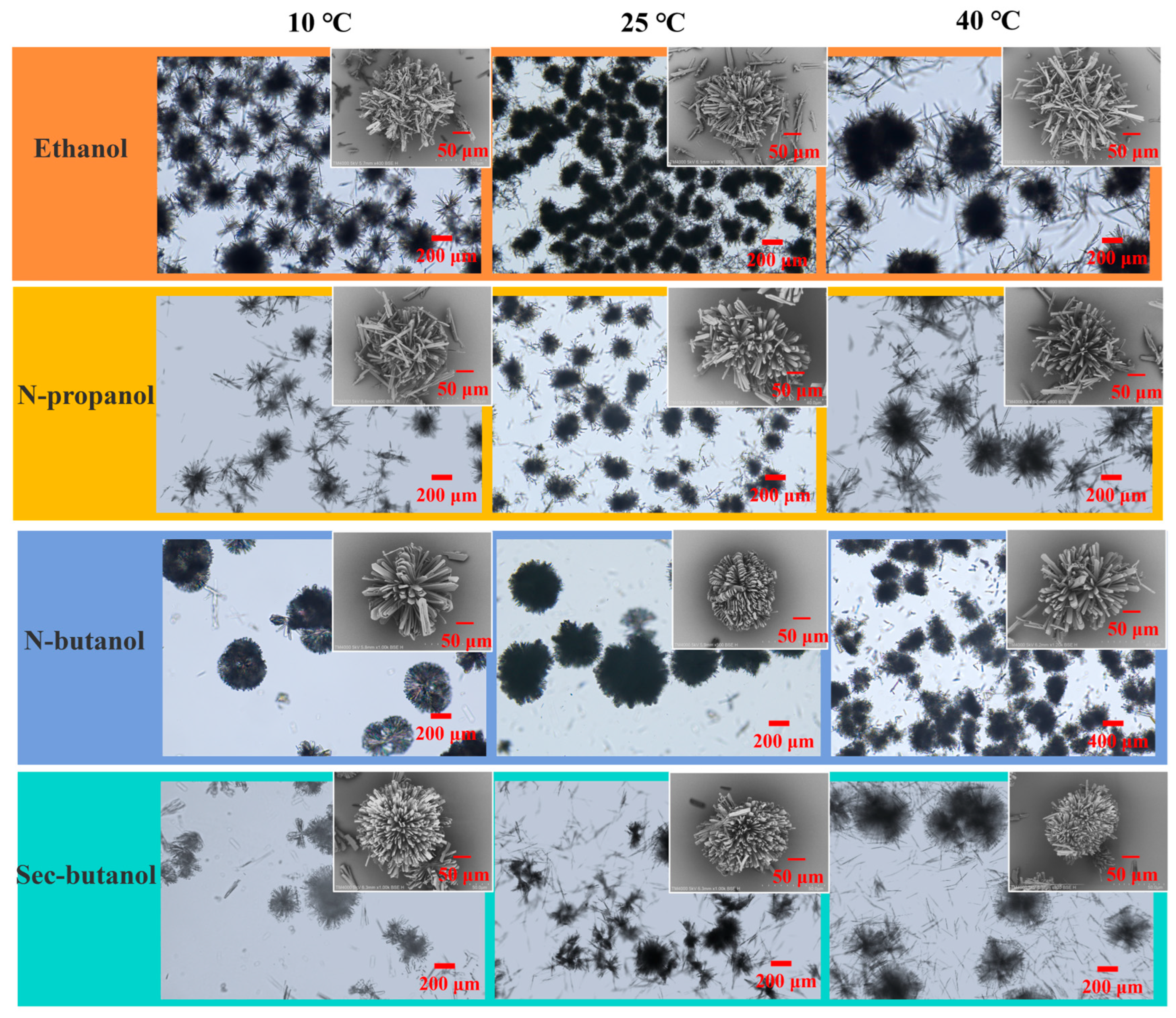
3.4.1. Effect of Antisolvent Type and Temperature

Different organic solvents can significantly affect crystal morphology [44]. In preliminary experiments, we investigated the effects of antisolvent type. It was found that needle-like crystals were generated in acetonitrile, acetone, DMF, isopropanol, and isoamyl alcohol, while irregular aggregates appeared in ethyl acetate (Table S1, P1–P6 and Figure S4). Interestingly, approximately spherical particles can be obtained when the antisolvent is ethanol, n-propanol, n-butanol, or sec-butanol. Three temperatures (10 °C, 25 °C, and 40 °C) were selected to investigate their effects on particle morphology in these four antisolvents (Table S1, E1–E12). As shown in Figure 5, the polycrystalline particles obtained from antisolvent crystallization in all four mixtures were spherulites, in which needle-like or plate-like subunits of the aggregates grew radially. In a water–ethanol system, many spherulites can be formed at 10 °C, exhibiting evident opening angles. As the temperature increased to 25 °C, the space was filled by more individual crystals, and spherulites became more compact. At 40 °C, larger spherulites appeared, indicating that higher temperature promotes heterogeneous nucleation and crystal growth. This occurs because elevated temperatures shift the crystallization process from nucleation-dominant to growth-dominant, resulting in reduced microcrystal population and increased particle size [45]. When abundant spherulites with diminished size are observed, this indicates the promoted nucleation in the bulk solution. It was also observed that there were more needle-like fine crystals at increased temperature. This suggests that higher temperature also favors nucleation from the bulk solution. Temperature exerted a similar influence on the crystallization process in the other three mixed solvents. It seems that the antisolvent type has a greater impact on particle morphology. In the water–n-propanol system, the spherulites were a bit spiky, while the spherulites grown from water–n-butanol were the most massive and uniform. In the water–sec-butanol system, the SEM images show that some spherulites were fully developed, but from the microscopic images, we can see that needles and irregular aggregates predominated.

3.4.2. Effect of Volume Ratio of Antisolvent to Solvent

The amount of antisolvent can affect the degree of supersaturation, which directly affects the nucleation and growth behavior. We maintained the constant experimental conditions at 25 °C while varying the antisolvent-to-water ratio (Table S1, E1–E4 and E13–E20). As shown in Figure 6, in the water–ethanol system, as the antisolvent/solvent ratio increased from 9 to 15, larger polycrystalline aggregates were formed, and meanwhile, there still existed many needle-like crystals. Interestingly, the majority of crystals grown from water–n-propanol evolved into a double-leaf morphology at larger antisolvent/solvent ratios. It seems that higher supersaturation was created, and it facilitated noncrystallographic branching of a large number of nuclei. As the supersaturation was significantly consumed, it could not afford to enhance additional branching. Meanwhile, the solution was diluted by more antisolvents, which also prevented further spherulitic growth. Similar phenomena took place in water–n-butanol and water–sec-butanol systems, where dumbbell-shaped and star-like morphologies appeared.

3.4.3. Effect of Solute Concentration

To investigate the effect of solute concentration in aqueous solution on crystal morphology, the solute concentration from 0.1 to 0.3 g·mL−1 was used at 298.15 K (Table S1, E1–E4 and E21–E28). The crystal morphology of salbutamol sulfate obtained at different solute concentrations is presented in Figure 7. It is clear that a lower concentration could not facilitate the formation of spherical particles. When the solute concentration was reduced to 0.1 g·mL−1, all four solvent mixtures produced irregular aggregates and incomplete spherulites. More spherulites were formed as the initial solute concentration increased to 0.2 g·mL−1. It created higher supersaturation and provided sufficient materials, which was in favor of spherulitic growth. But greater solute concentration also led to extensive nucleation in the solution [46]. Needles and spherulites developed at different stages coexisted in water–ethanol, water–n-propanol, and water–sec-butanol systems. In particular, larger and complete spherulites can be obtained in water–n-butanol mixtures even at higher solute concentration. This system provides a more appropriate environment for preparing spherical particles of salbutamol sulfate.

3.4.4. Effect of Feeding Rate and Stirring Speed

The influence of feeding rates and agitation speeds on crystal morphology was also investigated while maintaining all other experimental conditions constant (Table S1, E29–E40). As illustrated in Figure 8, the crystal morphology of salbutamol sulfate exhibited distinct variations under different feeding and stirring rates across four antisolvent systems. In the water–ethanol system, a faster feeding rate decreased the amount of spherical particles. Meanwhile, increased stirring rates caused the breakage of particles. It suggests that the subunits of the aggregates formed in water–ethanol were loosely contacted. The water–n-propanol system demonstrated remarkable stability in the formation of spherulites regardless of stirring rates. Increasing the feeding rate from 0.5 to 1 g·min−1 enhanced the formation of spherulites. Optimal crystallization in the water–n-butanol system was achieved at 350 rpm, where increased stirring rates effectively minimized needle-like crystals and promoted the formation of complete spherulites, particularly at a feeding rate of 0.5 g·min−1. The spherulites obtained in this system were significantly larger and exhibited more densely packed subunits compared to those produced in other systems. SEM characterization also confirmed that 350 rpm yielded regular spherical crystals. Similarly, the water–sec-butanol system produced crystals with superior morphology at 350 rpm, whereas lower stirring rates (<250 rpm) resulted in irregular aggregates due to insufficient particle collisions. Notably, the feeding rate showed minimal influence on crystal morphology, while the stirring rate emerged as a dominant factor controlling the formation of spherical particles. Optimization of the stirring conditions and feeding rate is critical for preparing spherical particles.

3.5. Spherulitic Growth Pattern of Salbutamol Sulfate

The morphological evolution process of spherical crystals of salbutamol sulfate was further investigated by SEM. The growth evolution of spherulites at different time intervals after crystallization initiation is presented in Figure 9. The crystallization process was conducted in a water–n-butanol system at 25 °C with an antisolvent/solvent ratio of 9:1 and a drug concentration of 0.3 g·mL−1. The solution was added at a rate of 0.5 g·min−1 under constant stirring at 350 rpm, with crystal growth monitored via periodic sampling. At the early stage, heterogeneous nucleation occurred on the existing crystals, and sheaves of plate-like crystals appeared (Figure 9a,b). Then, a double-leaf structure evolved via noncrystallographic branching (Figure 9c). The subunits spread out laterally, generating the round morphology with two hollow cores (Figure 9d). Heterogeneous nucleation and successive small-angle branching led to the formation of a hemispherical shape (Figure 9e). A massive spherulite developed when more branches filled the voids (Figure 9f). As discussed above, the morphology of spherulites and the density of subunits are strongly related to antisolvent type and the operating parameters. As the temperature increased from 10 to 45 °C, the particle size of spherulites progressively enlarged. However, excessive addition of antisolvent diluted the solution and substantially depleted supersaturation, thereby inhibiting further spherulite growth. The experimental results demonstrate that well-defined spherical crystals were more readily obtained at higher solute concentrations. Notably, both feeding rate and stirring speed exhibited negligible effects on crystal morphology, allowing flexible parameter adjustment based on experimental requirements. The optimized conditions establish an ideal environment for the controlled preparation of salbutamol sulfate spherical particles.

4. Conclusions

In this work, spherical crystals of salbutamol sulphate were successfully prepared in the four types of solvent mixtures using antisolvent crystallization, eliminating the need for toxic bridging liquids and demonstrating potential for environmentally friendly pharmaceutical manufacturing. The crystal forms of samples obtained from different solvents were identified. A novel solvate (S-SBA) was discovered in the water–sec-butanol system, and the single crystals were obtained by slow solvent evaporation. The crystal structure and molecular packing of S-SBA have been analyzed. Multiple intermolecular hydrogen bonds were formed between salbutamol–salbutamol, salbutamol sulfate, and salbutamol–sec-butanol molecules. During the spherical crystallization process, the effects of antisolvent type, temperature, ratio of antisolvent to water, drug concentration, feeding rate, and agitation speed were investigated. The results showed that under the optimized conditions (25 °C, antisolvent/solvent ratio of 9:1, drug concentration of 0.2 g·mL−1, feeding rate of 0.5 g·min−1, and stirring speed of 350 rpm), the n-butanol–water system yielded dense and uniform spherulites. At lower solute concentrations and stirring rates, sparse spherulites were obtained. Increasing solute concentration promoted branching and yielded massive spherulites. Notably, the influence of stirring rate exhibited system-dependent behavior; in the water–ethanol system, elevated stirring rates led to increased particle fragmentation, whereas in the other three solvent systems, higher stirring rates demonstrated the beneficial effects by promoting particle dispersion and facilitating spherulite growth. The morphology evolution of salbutamol sulfate spherulites was also explored. It indicated a spherulitic growth pattern, in which the spherulite developed from sheaves and a double-leaf structure via noncrystallographic branching. This work should help provide an improved strategy for engineering the crystal form and morphology of salbutamol sulfate.

Supplementary Materials

The following supporting information can be downloaded at: https://www.mdpi.com/article/10.3390/cryst15070651/s1, Figure S1: DSC and TGA data detected up to 500 °C of (a) raw material of salbutamol sulfate and crystals obtained by antisolvent crystallization in different antisolvents: (b) ethanol, (c) n-propanol, (d) n-butanol, and (e) sec-butanol; Figure S2: (a) The molecular structure of salbutamol sulfate solvate (S-SBA), showing the atom labelling scheme. (b) The unit cell of S-SBA; Figure S3: Intermolecular hydrogen bonding interactions (a, b) and molecular packing (c, d) of SS; Figure S4: Microscopic images of crystals obtained via anti-solvent crystallization in different antisolvent systems: (a) acetonitrile, (b) acetone, (c) ethyl acetate, (d) N,N-dimethylformamide (DMF), (e) isopropanol and (f) isoamyl alcohol; Table S1: The operating conditions for antisolvent crystallization; Table S2: Endothermic peaks of salbutamol sulfate samples during the process of thermal decomposition; Table S3: Atomic occupancy for S-SBA.

Author Contributions

Conceptualization, X.Q.; methodology, X.C.; formal analysis, X.Q. and Y.D.; investigation, S.D.; resources, H.L.; writing—original draft preparation, X.Q.; writing—review and editing, Y.W. and S.D.; supervision, Y.W., S.D., and F.X.; funding acquisition, Y.W., S.D., and F.X. All authors have read and agreed to the published version of the manuscript.

Funding

This research was funded by the National Natural Science Foundation of China (22378216), Young Talent of Lifting engineering for Science and Technology in Shandong (SDAST2024QTA005), Jinan Introducing Innovation Team Project (202228033), and Youth Innovation Team of Universities in Shandong Province (2023KJ141).

Data Availability Statement

The original contributions presented in the study are included in the article/Supplementary Materials; further inquiries can be directed to the corresponding authors.

Conflicts of Interest

Authors Hongcheng Li and Yanni Du were employed by the company Shouguang Fukang Pharmaceutical Co., Ltd., and Beyond Environmental Engineering Technology Co., Ltd., respectively. The remaining authors declare that the research was conducted in the absence of any commercial or financial relationships that could be construed as potential conflicts of interest.

References

  1. ter Horst, J.H.; Sefcik, J.; Simone, E. The Future of Industrial Crystallization. Cryst. Growth Des. 2024, 24, 8667–8668. [Google Scholar] [CrossRef]
  2. Li, B. Structure transition in Cu crystallization. Phys. Scr. 2023, 98, 045916. [Google Scholar] [CrossRef]
  3. Bhargava, N.; Mor, R.S.; Kumar, K.; Sharanagat, V.S. Advances in application of ultrasound in food processing: A review. Ultrason. Sonochemistry 2021, 70, 105293. [Google Scholar] [CrossRef] [PubMed]
  4. Thapa, P.; Tripathi, J.; Jeong, S.H. Recent trends and future perspective of pharmaceutical wet granulation for better process understanding and product development. Powder Technol. 2019, 344, 864–882. [Google Scholar] [CrossRef]
  5. Ma, Y.; Sun, M.; Liu, Y.; Chen, M.; Wu, S.; Wang, M.; Wang, L.; Gao, Z.; Han, D.; Liu, L.; et al. Design of Spherical Crystallization of Active Pharmaceutical Ingredients via a Highly Efficient Strategy: From Screening to Preparation. ACS Sustain. Chem. Eng. 2021, 9, 9018–9032. [Google Scholar] [CrossRef]
  6. Wang, J.; Cao, W.; Wang, J.; Zhu, L. A Novel Spherical Crystallization Method Using Pickering Emulsions. J. Pharm. Sci. 2022, 111, 1625–1632. [Google Scholar] [CrossRef] [PubMed]
  7. Shtukenberg, A.G.; Tan, M.; Vogt-Maranto, L.; Chan, E.J.; Xu, W.; Yang, J.; Tuckerman, M.E.; Hu, C.T.; Kahr, B. Melt Crystallization for Paracetamol Polymorphism. Cryst. Growth Des. 2019, 19, 4070–4080. [Google Scholar] [CrossRef]
  8. Trueman, M.; Akporiaye, D.; Anderson, M.W. Simulating intergrowth formation in zeolite crystals: Impact on habit and functionality. Faraday Discuss. 2022, 235, 343–361. [Google Scholar] [CrossRef] [PubMed]
  9. Qu, H.; Li, Z.; Zhang, G.; Zhou, Z.; Wu, S. Tuning the physicochemical properties of axitinib by crystallization: Preparation, calculation and Structure-property relationship. J. Ind. Eng. Chem. 2023, 124, 570–578. [Google Scholar] [CrossRef]
  10. Li, L.; Ji, X.; Cheng, X.; Li, D.; Wang, T.; Huang, X.; Wang, N.; Yin, Q.; Hao, H. Effect of the solvent on the morphology of sulfamerazine crystals and its molecular mechanism. CrystEngComm 2022, 24, 5497–5506. [Google Scholar] [CrossRef]
  11. Dutt, B.; Choudhary, M.; Budhwar, V. Enhancement of stability profile of aspirin through cocrystallization technique. Res. J. Pharm. Technol. 2022, 15, 768–772. [Google Scholar] [CrossRef]
  12. Liu, Y.; Wang, S.; Li, J.; Guo, S.; Yan, H.; Li, K.; Tong, L.; Gao, Y.; Li, T.; Chen, M.; et al. Preparation of ethyl vanillin spherical particles with functions of sustained release and anti-caking by an organic solvent-free process. Food Chem. 2023, 402, 134518. [Google Scholar] [CrossRef] [PubMed]
  13. Zhou, Q.; Tan, Z.; Yang, D.; Tu, J.; Wang, Y.; Zhang, Y.; Liu, Y.; Gan, G. Improving the Solubility of Aripiprazole by Multicomponent Crystallization. Crystals 2021, 11, 343. [Google Scholar] [CrossRef]
  14. Sun, M.; Bi, J.; Zhao, Y.; Gong, J. Particle Design of Drugs via Spherical Crystallization: A Review from Fundamental Aspects to Technology Development. Cryst. Growth Des. 2024, 24, 2266–2287. [Google Scholar] [CrossRef]
  15. Chen, K.; Hou, B.; Wu, H.; Huang, X.; Li, F.; Xiao, Y.; Li, J.; Bao, Y.; Hao, H. Hollow and Solid Spherical Azithromycin Particles Prepared by Different Spherical Crystallization Technologies for Direct Tableting. Processes 2019, 7, 276. [Google Scholar] [CrossRef]
  16. Gupta, M.; Chatterjee, A.; Srivastava, B. Spherical crystallization: A technique use to reform solubility and flow property of active pharmaceutical ingredients. Int. J. Pharm. Investig. 2017, 7, 4–9. [Google Scholar] [CrossRef] [PubMed]
  17. Xue, Z.-H.; Xu, R.; Qin, J.; Wang, Z.; Liu, Y.; Yan, Q.-L. Desensitization of spherical CL-20 composites by embedding insensitive nanosized energetic crystals. CrystEngComm 2024, 26, 5617–5631. [Google Scholar] [CrossRef]
  18. Xing, X.; Ouyang, J.; Guo, S.; Chen, M.; Gao, Z.; He, F.; Zhou, L.; Xie, Z. Spherical particles design of vanillin via crystallization method: Preparation, characterization and mechanism. Sep. Purif. Technol. 2023, 314, 123622. [Google Scholar] [CrossRef]
  19. Yan, H.; Liu, Y.; Peng, H.; Li, K.; Li, C.; Jiang, S.; Chen, M.; Han, D.; Gong, J. Improving calcium citrate food functions through spherulitic growth in reactive crystallization and a mechanism study. Food Chem. 2023, 404, 134550. [Google Scholar] [CrossRef] [PubMed]
  20. Kawashima, Y.; Okumura, M.; Takenaka, H. Spherical Crystallization: Direct Spherical Agglomeration of Salicylic Acid Crystals During Crystallization. Science 1982, 216, 1127–1128. [Google Scholar] [CrossRef] [PubMed]
  21. Guo, S.; Yu, C.; Feng, S.; Wei, J.; Tong, L.; Li, K.; Gao, Y.; Zhao, P.; Li, T.; Chen, M.; et al. Enabling the drug combination of celecoxib through a spherical co-agglomeration strategy with controllable and stable drug content and good powder properties. Int. J. Pharm. 2022, 626, 122180. [Google Scholar] [CrossRef] [PubMed]
  22. Guo, S.; Feng, S.; Yu, C.; Liu, Y.; Chen, M.; Gong, J. Sustainable preparation of spherical amphoteric organics:Isoelectric point-spherical agglomeration technology. Powder Technol. 2022, 407, 117645. [Google Scholar] [CrossRef]
  23. Guo, S.; Feng, S.; Yu, C.; Liu, Y.; Li, K.; Chen, M.; Han, D.; Gong, J. Design of spherical agglomerates via crystallization with a non-toxic bridging liquid: From mechanism to application. Powder Technol. 2022, 408, 117725. [Google Scholar] [CrossRef]
  24. Chen, H.; Wang, C.; Kang, H.; Zhi, B.; Haynes, C.L.; Aburub, A.; Sun, C.C. Microstructures and pharmaceutical properties of ferulic acid agglomerates prepared by different spherical crystallization methods. Int. J. Pharm. 2020, 574, 118914. [Google Scholar] [CrossRef] [PubMed]
  25. Jin, S.; Chen, M.; Li, Z.; Wu, S.; Du, S.; Xu, S.; Rohani, S.; Gong, J. Design and mechanism of the formation of spherical KCl particles using cooling crystallization without additives. Powder Technol. 2018, 329, 455–462. [Google Scholar] [CrossRef]
  26. Wang, X.; Li, Z.; Zhang, C.; Wen, T.; Zhou, Y.; Ouyang, J. Designing Spherical Particles of Arbidol Hydrochloride via Spherical Crystallization: Preparation and Characterization. Ind. Eng. Chem. Res. 2024, 63, 5249–5260. [Google Scholar] [CrossRef]
  27. Wang, Y.; Du, S.; Wang, X.; Sun, M.; Yang, Y.; Gong, J. Spherulitic growth and morphology control of lithium carbonate: The stepwise evolution of core-shell structures. Powder Technol. 2019, 355, 617–628. [Google Scholar] [CrossRef]
  28. Cui, P.; Yang, W.; Jia, L.; Zhou, L.; Zhang, M.; Bao, Y.; Xie, C.; Hou, B.; Yin, Q. Spherulitic Growth Strategy for Agitation-Induced Formation of Spherical Amoxicillin Sodium Products. Ind. Eng. Chem. Res. 2022, 61, 9821–9832. [Google Scholar] [CrossRef]
  29. Cullum, V.A.; Farmer, J.; Jack, D.; Levy, G. Salbutamol: A new, selective β-adrenoceptive receptor stimulant. Br. J. Pharmacol. 1969, 35, 141–151. [Google Scholar] [CrossRef] [PubMed]
  30. Sawale, A.V.; Muneshwar, S.D.; Honale, V.S. Formulation and Evaluation of Oral Soft Jelly Containing Salbutamol Sulphate for the Treatment of Asthma. J. Drug Deliv. Ther. 2023, 13, 118–124. [Google Scholar] [CrossRef]
  31. Montuschi, P.; Ciabattoni, G. Bronchodilating Drugs for Chronic Obstructive Pulmonary Disease: Current Status and Future Trends. J. Med. Chem. 2015, 58, 4131–4164. [Google Scholar] [CrossRef] [PubMed]
  32. Vorobei, A.M.; Pokrovskiy, O.I.; Ustinovich, K.B.; Parenago, O.O.; Lunin, V.V.; Miroshnichenko, A.G. Micronization of Salbutamol Sulfate by Supercritical Antisolvent Precipitation: The Effect of Process Parameters on the Size and Morphology of Particles. Russ. J. Phys. Chem. B 2018, 12, 1240–1248. [Google Scholar] [CrossRef]
  33. Tahara, K.; O’Mahony, M.; Myerson, A.S. Continuous Spherical Crystallization of Albuterol Sulfate with Solvent Recycle System. Cryst. Growth Des. 2015, 15, 5149–5156. [Google Scholar] [CrossRef]
  34. Nocent, M.; Bertocchi, L.; Espitalier, F.; Baron, M.; Couarraze, G. Definition of a solvent system for spherical crystallization of salbutamol sulfate by quasi-emulsion solvent diffusion (QESD) method. J. Pharm. Sci. 2001, 90, 1620–1627. [Google Scholar] [CrossRef] [PubMed]
  35. Leger, J.M.; Goursolle, M.; Gadret, M.; Carpy, A. Structure cristalline du sulfate de salbutamol [tert-butylamino-2 (hydroxy-4 hydroxymethyl-3 phenyl)-1 ethanol.0.5H2SO4]. Acta Crystallogr. Sect. B 1978, 34, 1203–1208. [Google Scholar] [CrossRef]
  36. Shaikh, T.R.; Shelke, N.; Tothadi, S. Multicomponent Solvate Crystals of 3, 5-Dinitrobenzoic Acid and Acetamide and CSD Analysis of Solvates. ACS Omega 2023, 8, 24644–24653. [Google Scholar] [CrossRef] [PubMed]
  37. Sheldrick, G.M. Crystal structure refinement with SHELXL. Cryst. Struct. Commun. 2015, 71, 3–8. [Google Scholar] [CrossRef] [PubMed]
  38. Dolomanov, O.V.; Bourhis, L.J.; Gildea, R.J.; Howard, J.A.K.; Puschmann, H. OLEX2: A complete structure solution, refinement and analysis program. J. Appl. Crystallogr. 2009, 42, 339–341. [Google Scholar] [CrossRef]
  39. Varun, N.; Ghoroi, C. Engineered inhalable micro-balloon shaped drug particles for carrier-free dry powder inhalation (DPI) application. Powder Technol. 2022, 408, 117705. [Google Scholar] [CrossRef]
  40. Sonvico, F.; Coleman, V.; Traini, D.; Young, P.M. Evolved gas analysis during thermal degradation of salbutamol sulphate. J. Therm. Anal. Calorim. 2015, 120, 789–794. [Google Scholar] [CrossRef]
  41. Felix, F.S.; Cides da Silva, L.C.; Angnes, L.; Matos, J.R. Thermal behavior study and decomposition kinetics of salbutamol under isothermal and non-isothermal conditions. J. Therm. Anal. Calorim. 2009, 95, 877–880. [Google Scholar] [CrossRef]
  42. Widauer, A.; Petrick, T.L.; Braun, D.E. Comprehensive Insights into Sulfaguanidine in the Solid State: An Experimental and Computational Study. Cryst. Growth Des. 2024, 24, 1438–1457. [Google Scholar] [CrossRef] [PubMed]
  43. Trimdale-Deksne, A.; Grante, I.; Mishnev, A.; Orola, L.; Bērziṇš, A. Similarity and Differences of Dasatinib Solvates: A Crystallographic Perspective. Cryst. Growth Des. 2024, 24, 5014–5026. [Google Scholar] [CrossRef]
  44. Li, J.; Yang, R.; Zeng, T.; Hu, J.; Tang, W.; Liu, Z.; Gong, L. Preparation and growth mechanism of micro spherical ammonium dinitramide crystal based on ultrasound-assisted solvent-antisolvent method. Ultrason. Sonochemistry 2021, 78, 105716. [Google Scholar] [CrossRef] [PubMed]
  45. Zhang, Y.; Yan, Q.; Liu, Y.; Hu, C. Study on the regulation mechanism of effective glass transition temperature on the crystallization of crystalline solid dispersion. Drug Deliv. Transl. Res. 2023, 13, 2677–2689. [Google Scholar] [CrossRef] [PubMed]
  46. Singh, M.R.; Ramkrishna, D. Dispersions in crystal nucleation and growth rates: Implications of fluctuation in supersaturation. Chem. Eng. Sci. 2014, 107, 102–113. [Google Scholar] [CrossRef]
Figure 1. Chemical structure of salbutamol sulfate.
Crystals 15 00651 g001
Figure 2. The calculated PXRD patterns of salbutamol sulfate and experimental patterns of raw material and crystals obtained in different antisolvent systems.
Crystals 15 00651 g002
Figure 3. (a) TG curves and (b) DSC curves of the raw material of salbutamol sulfate and crystals obtained by antisolvent crystallization in different antisolvents.
Crystals 15 00651 g003
Figure 4. Intermolecular interactions (a,b) and crystal packing of S-SBA (c,d). Color coding is used for atomic visualization: H (white), O (red), S (yellow), and N (blue).
Crystals 15 00651 g004
Figure 5. Optical micrographs and SEM images of salbutamol sulfate spherical crystals prepared in four different antisolvent systems at varying temperatures.
Crystals 15 00651 g005
Figure 6. Optical micrographs and SEM images of salbutamol sulfate spherical crystals prepared in different antisolvent/solvent ratios.
Crystals 15 00651 g006
Figure 7. Optical micrographs and SEM images of salbutamol sulfate spherical crystals prepared with different solute concentrations.
Crystals 15 00651 g007
Figure 8. Optical micrographs and SEM images of salbutamol sulfate spherical crystals prepared with different feeding rates and stirring speeds.
Crystals 15 00651 g008
Figure 9. SEM images of morphology evolution of salbutamol sulfate spherulites: (a) 1 min; (b) 2 min; (c) 3 min; (d) 5 min; (e) 7 min; (f) 10 min.
Crystals 15 00651 g009
Table 1. Crystallographic data of salbutamol sulfate.
SS [35]S-SBA
formulaC13H23NO7S2(C13H22NO3)·2(C4H10O)·O4S
formula wt337.39724.93
cryst systemmonoclinicmonoclinic
space groupCcC2
a (Å)28.069 (5)28.0008 (14)
b (Å)6.183 (1)6.1365 (3)
c (Å)16.914 (2)11.4303 (6)
α (°)9090
β (°)81.19 (1)100.381 (3)
γ (°)9090
V3)2900.81931.88 (17)
Z82
Temp (K)295193
R10.06700.0413 (3934)
wR20.06700.1090 (3999)
CCDC no.12544532449793
Disclaimer/Publisher’s Note: The statements, opinions and data contained in all publications are solely those of the individual author(s) and contributor(s) and not of MDPI and/or the editor(s). MDPI and/or the editor(s) disclaim responsibility for any injury to people or property resulting from any ideas, methods, instructions or products referred to in the content.

