Abstract
The present work aimed to synthesize 2-methylthio-triazoloquinazoline derivatives and study their X-ray, NMR, DFT and Hirshfeld characteristics. The cyclocondensation of dimethyl-N-cyanodithiocarbonate with 2-hydrazinobenzoic acid hydrochloride resulted in an intermediate, 2-methylthio-[1,2,4]triazolo[1,5-a]quinazolin-5-one (A), which upon treatment with phosphorus pentasulfide, transformed into the 2-methylthio-[1,2,4]triazolo[1,5-a]quinazolin-5-thione (B). Reaction of 2-methylthio-triazoloquinazolines (A&B) with alkyl halides (allyl bromide and ethyl iodide) in basic medium afforded 4-allyl-2-methylthio-[1,2,4]triazolo[1,5-a]quinazolin-5-one (1; N-alkylated) and 5-ethylthio-2-methylthio-[1,2,4]triazolo[1,5-a]quinazoline (2; S-alkylated), respectively. Their molecular and supramolecular structures were presented. Unambiguously, the molecular structures of 1 and 2 were confirmed via NMR and single-crystal X-ray diffraction. The resulting findings confirmed the structures of 1 and 2 and determined their crystalized system (monoclinic system; P21/n space group). Hirshfeld analysis of 1 revealed the importance of the significantly short O···H (6.7%), S···S (1.2%) and C···C (2.8%); however, the short H···H (42.6%), S···H (16.3%) and C···C (4.3%) were showed in 2 by intermolecular interactions in the molecular packing. The 1,2,4-triazoloquinzolines (1&2) were anticipated to be relatively polar compounds with net dipole moments of 2.9284 and 4.2127 Debye, respectively. The molecular electrostatic potential, atomic charge distribution maps and reactivity descriptors for 1 and 2 were also determined. The calculated nuclear magnetic resonance spectra of the targets 1 and 2 were well correlated with the experimental data.
Keywords:
triazoloquinazoline; cyclocondensation; thionation; alkylation; X-ray; DFT; Hirshfeld analysis 1. Introduction
In pharmaceutical chemistry, the synthesis of new bioactive agents has recently achieved notable prominence as a result of the increasing spread of various diseases [1,2]. Nitrogen heterocycles, which are found in various natural and synthetic products, represent the most important class of biologically active substances and pharmaceutically relevant molecules. Among them, quinazolines and triazoles have attracted considerable attention in synthetic medicinal chemistry and the materials science area. Due to the significance of these pharmacophore classes as well as their roles in the construction of pharmacologically active molecules, including antimicrobial, antimalarial, anti-inflammatory, antihypertensive, anticonvulsant, antidiabetic and anticancer agents as well as cholinesterase inhibitors, their synthesis has been a hot topic in organic synthesis [3,4]. Thus, the combination of both quinazoline and triazole moieties results in the synthesis of a triazoloquinazoline scaffold, which exhibits varied pharmacological activities [3]. Indeed, many triazoloquinazolines have been reported to exhibit promising antihistamine activity [5,6]. Some 1,2,4-triazoloquinazolines have been investigated as potent adrenoblockers and were found to display good hypotensive activity [7,8]. A series of 2-alkoxy-triazoloquinazolines were found to act as potent adenosine receptor antagonists [9,10]. The presence of lactam and thiolactam in triazoloquinazolines skeleton was valuable to appear in more pharmacophoric moieties associated with a diversity in biological properties. For example, S-alkylated triazoloquinazolines showed significant antimicrobial effects [11,12]; however, their N-alkylated compounds demonstrated strong antiviral activity [13,14]. Furthermore, potential antidiabetic activity was reported for triazoloquinazoline, which manifested itself via its ability to inhibit α-glucosidase activity [15]. Additionally, some 1,2,4-triazoloquinazolines have been observed to possess potential antioxidant properties and promising cytotoxicity [16,17,18,19,20,21]. Hence, we focus on triazoloquinazoline chemistry, particularly studying the crystal structure of 1,2,4-triazolo[1,5-a]quinazoline derivatives [22,23,24,25,26,27]; herein, we report X-crystal structures, NMR, MS, IR, DFT and Hirshfeld studies of the synthesized triazoloquinazolines 1 and 2.
2. Materials and Methods
2.1. General Information
The IR spectrum was determined using a Perkin Elmer FT-IR Spectrum BX system. The NMR spectra were measured with a Bruker AMX 500-MHz instrument, and the data were described as ppm values relative to tetramethylsilane at 500 and 125 MHz for 1H and 13C NMR, respectively, and DMSO-d6 used as solvent. Employing a Micromass Quattro microTM triple-quadruple tandem mass spectrometer to measure ESI-MS spectra.
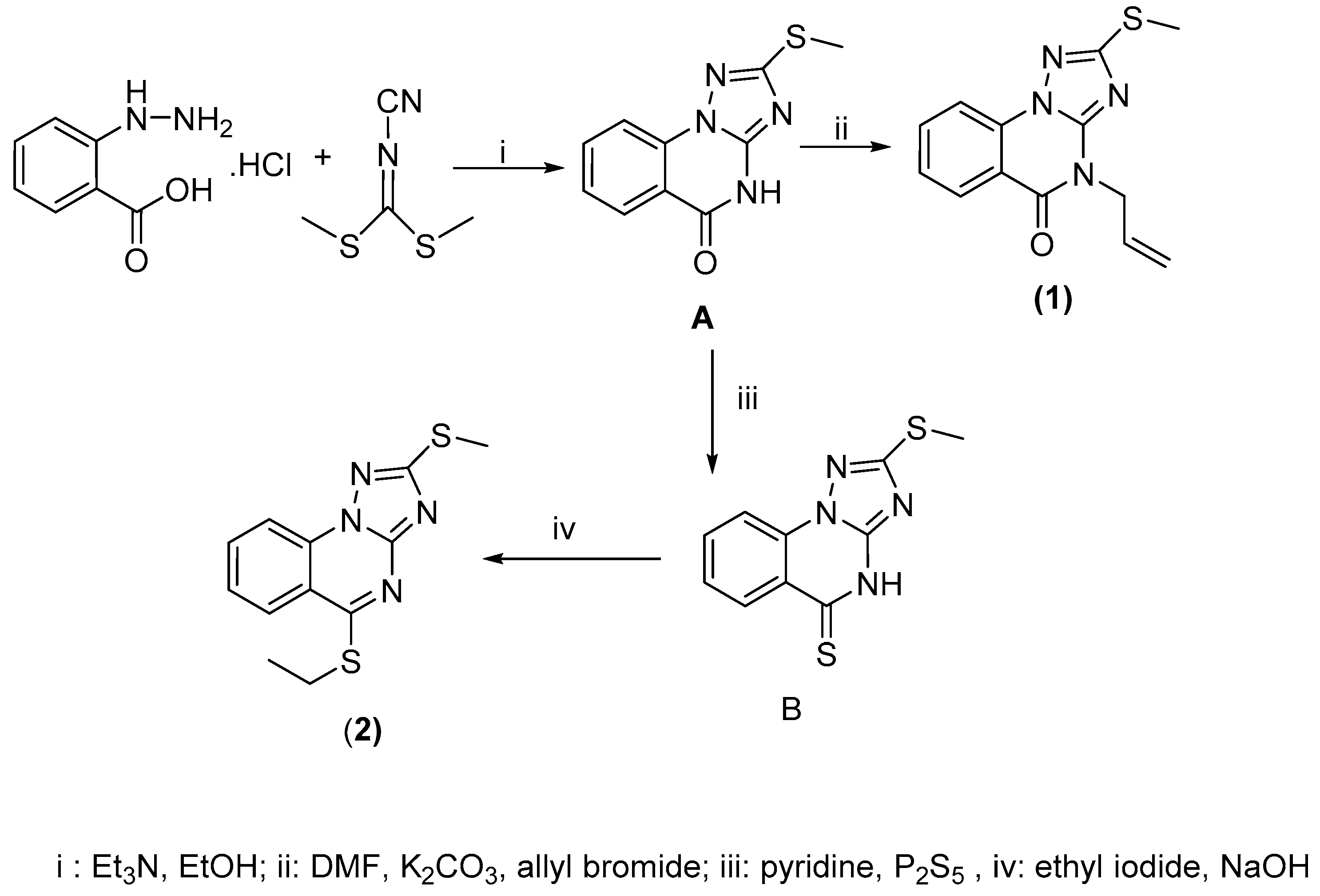
2.2. Synthesis of the Triazoloquinazolines 1 and 2
According to the reported detailed methodologies in our previous papers [28,29], the compounds 1 and 2 were prepared.
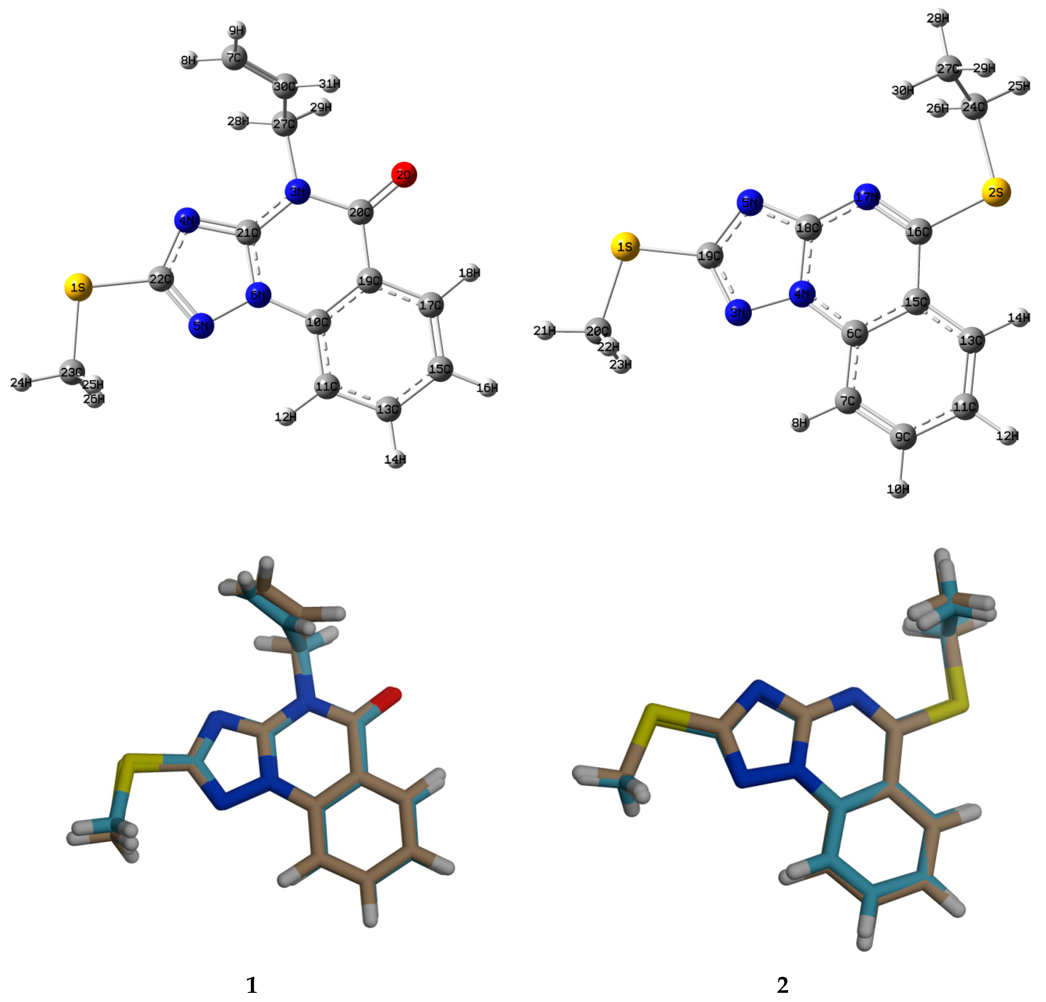
2.2.1. 4-Allyl-2-methylthio-[1,2,4]triazolo[1,5-a]quinazolin-5-one (1)
1H NMR (DMSO-d6): δ ppm 8.26 (br d, J = 8.0 Hz, 1H), 7.77 (br d, J = 8.0 Hz, 1H), 7.75 (br t, J = 7.5 Hz, 1H), 7.51 (br t, J = 7.50 Hz, 1H), 6.00 (m, 1H), 5.31 (br d, J = 17.50 Hz, 1H), 5.21 (br d, J = 10.50 Hz,1H), 4.80 (d, J = 5.00 Hz, 2H), 2.65 (s, 3H, SCH3); 13C NMR (DMSO-d6): δ ppm 167.32, 154.43, 145.72, 138.14, 133.17, 131.33, 129.21, 127.60, 117.23, 114.90, 112.25, 45.50, 13.90 ppm; ESI-MS (m/z): 271.1 [M−H]− (negative mode) for MW = 272.
2.2.2. 5-Ethylthio-2-methylthio-[1,2,4]triazolo[1,5-a]quinazoline (2)
1H NMR (DMSO-d6): δ ppm 8.22 (br d, J = 8.2 Hz, 2H), 8.08 (br d, J = 8.05 Hz, 1H), 7.69 (br t, J = 7.68 Hz, 1H), 4.80 (q, J = 7.20 Hz, 2H, SCH2CH3), 2.69 (s, 3H, SCH3), 1.42 (t, J = 7.00 Hz, 3H, SCH2CH3); 13C NMR (DMSO-d6): δ (ppm) 167.69, 164.61, 152.73, 135.95, 133.73, 126.78, 126.01, 117.50, 115.55, 24.48, 14.44, 13.97; ESI-MS (m/z): 275.3 [M−H]− (negative mode) for MW = 276.
2.3. X-ray Crystallography Analysis
Crystals of 2-Methylthiotriazoloquinazolines (1&2) were formed from saturated ethanolic solution by evaporation over 2 days at room temperature. Data collection was conducted on a Bruker APEX-II D8 Venture area diffractometer equipped with graphite monochromatic Mo Kα radiation at 100 (2) K. By employing Bruker SAINT SHELXT software, the cell refinement and reduction data were performed [30,31], and the solved structures were obtained by using SHELXT software. The final refinement was carried out using a full-matrix least-squares techniques with anisotropic thermal data for non-hydrogen atoms on F. Triazoloquinazolines 1 and 2 comprise the crystallographic data supplementary and can be obtained free of charge from the Cambridge Crystallographic Data Center via www.ccdc.cam.ac.uk/data_request/cif (2 September 2021).
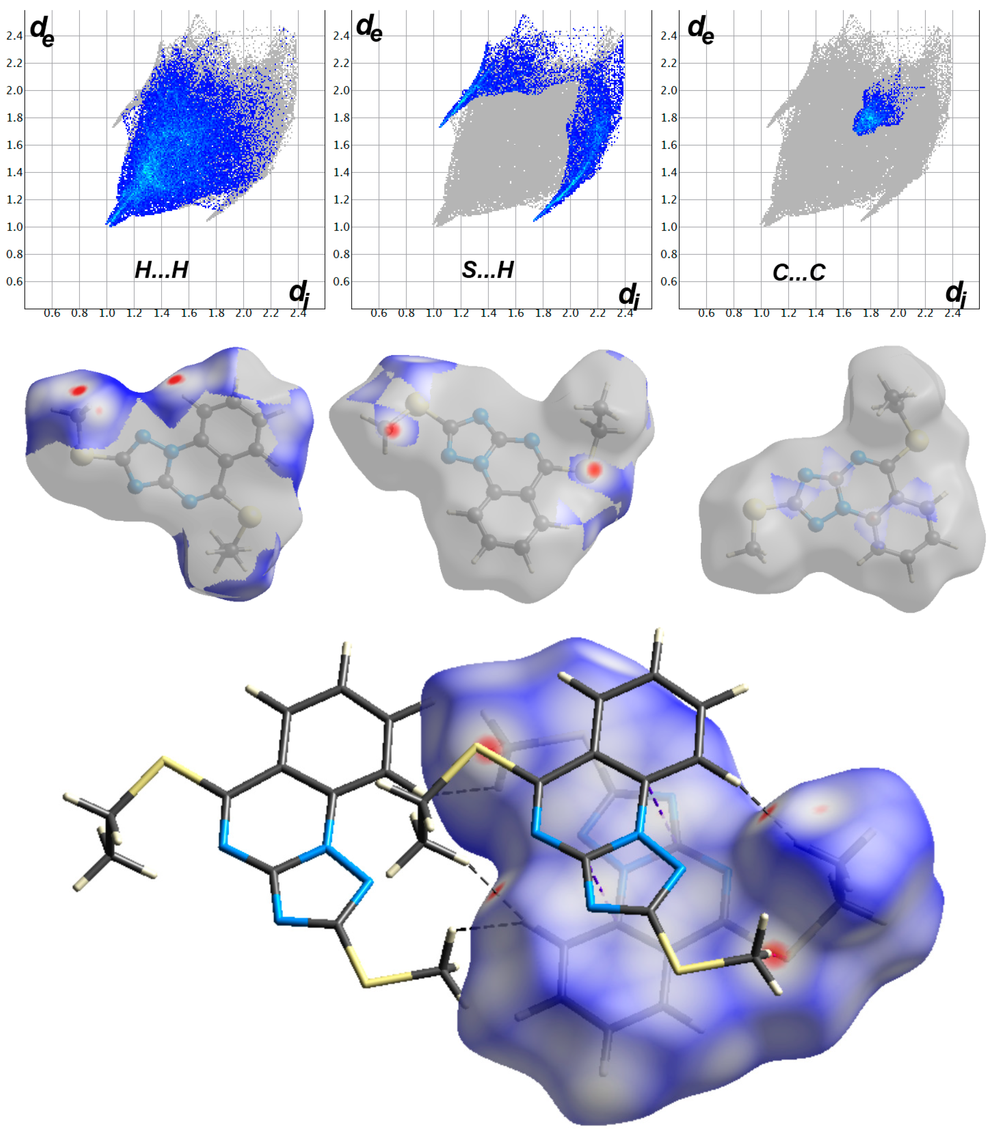
2.4. Hirshfeld Surface Analysis
Using Crystal Explorer 17.5 program, the topology analyses were carried out [32].
Computational Methods
Employing gaussian 09 software package, B3LYP/6-31G(d,p) method and NBO 3.1 program, all DFT calculations were carried out, the optimized obtaining structures revealed no imaginary frequencies and natural population analysis was completed, respectively [33,34,35,36,37]. The chemical shifts of 1H and 13C NMR spectrometry were computed applying the GIAO method in DMSO-d6 [38].
3. Results and Discussion
3.1. Chemistry
The synthetic routes of the target compounds, namely 4-allyl-triazoloquinazolin-5-one (1) and 5-ethyl-triazoloquinazoline (2), are outlined in Scheme 1. Reaction of 2-hydrazinobenzoic HCl (1.1 mmol) with dimethyl-N-cyanoimidodithiocarbonate (1 mmol) in ethanolic solution (15 mL) and triethyl amine (3 mmol) yielded an intermediate (A). Treatment of A (1 mmol) with allyl bromide (1.5 mmol) in the presence of K2CO3 (0.5 mmol) using DMF (10 mL) as the solvent resulted in the synthesis of the target compound 1 [28]. Thionation of A (1 mmol) with P2S5 (1 mmol) in pyridine (10 mL) under reflux conditions gave 1,2,4-triazoloquinazolin-5-thione (B, 1 mmol), which was transformed into the target compound 5-thioethyl-triazoloquinazoline (2) upon treatment with ethyl iodide (1.5 mmol) in a basic aqueous solution (8 mL) [29]. The final products 1 and 2 were obtained as plate-like crystals after crystallization.
Scheme 1.
Route for synthesis of the targets triazoloquinazolines (1&2).
3.2. Spectroscopic Data
Confirmation of triazoloquinazolines structures 1 and 2 was achieved by IR, NMR and MS-spectrometry. Although the chemical structures of the intermediates A and B have been previously determined [28,29] and the physiochemical properties of 1 have also been reported, we reiterated the relevant NMR data of 1 for additional explanation in this study. The IR spectrum of 1 revealed a strong absorption band due to carbonyl group at 1679 cm−1. The 1H NMR spectra of 1 and 2 are characterized by the singlet signal of the S-methyl protons at δ 2.65 and 2.69 ppm, respectively, and their 13C NMR resonances were recorded at δ 13.90 and 13.97 ppm, respectively. Notably, N-alkylation of substrate A with an allyl moiety (Scheme 1) was proven by its four 1H characteristic resonances at about 6.0 (m), 5.31 (br d, J = 17.5 Hz), 5.21 (br d, J = 10.5 Hz) and 4.80 (d, J = 5 Hz), which are assignable to propene moiety (CH2CHCH2) proton types. However, the S-alkylated product 2 confirmed by the presence of two characteristic 1H resonances with intrinsic splitting patterns at approximately 4.80 ppm (q, J = 7.2 Hz) and 1.41 ppm (t, J = 7.0 Hz), which ascribed to −SCH2CH3 and −SCH2CH3 protons, respectively (Figure 1). Three 13C resonances at δ ppm 131.33, 112.25 and 45.50 were attributed to the C-12, C-13 and C-11 carbons of the allyl group in 1; whereas, two signals were detected at δ 24.48 and 14.44 ppm attributable to C11 and C12 of the ethyl group in 2 (Figure 2). In triazoloquinazolines 1 and 2, the protons of the benzofused moiety were inferred to give rise to the resonance peaks with two broad doublets and two broad triplets with Jortho values ranging between 7.5 and 8.2 Hz. Additionally, further confirmation of 1,2,4-triazoloquinazoline structures (1&2) was achieved by single X-ray analysis and electrospray ionization mass spectrometry (ESI-MS). For the molecular weight of 272 (compound 1), the ESI-MS spectrum revealed an ion band at m/z 271.1 [M−H]− in the negative detection mode and showed an ion band at m/z 275.3 [M−H]− for the target 2 (MW = 276). In addition to MS data, the single-crystal X-ray analysis provided an obvious confirmation of the expected structures of 1 and 2.
Figure 1.
ORTEP of triazoloquinazolines 1 and 2. At the 40% probability level for non-H atoms, the displacement ellipsoids are plotted.
Figure 2.
Three-dimensional networks viewed approximately in molecular packing of 1 and 2 (H-bonds are drawn as dashed lines).
Triazoloquinazoline 1 has shown promising antihistaminic effect against histamine induced bronchospasm (IC50 = 0.6528 mM) in comparison with the theophylline (IC50 = 0.01996 mM) [6]. Furthermore, 1 was evaluated as an α-glucosidase inhibitor in vitro assay and showed the best inhibition effect (IC50 = 72.28 μM) in regards to acarbose (IC50 = 143.54 μM) [14]. Moreover, the triazoloquinazoline 1 was also reported as an antiviral, antimicrobial and antioxidant active agent (see Introduction).
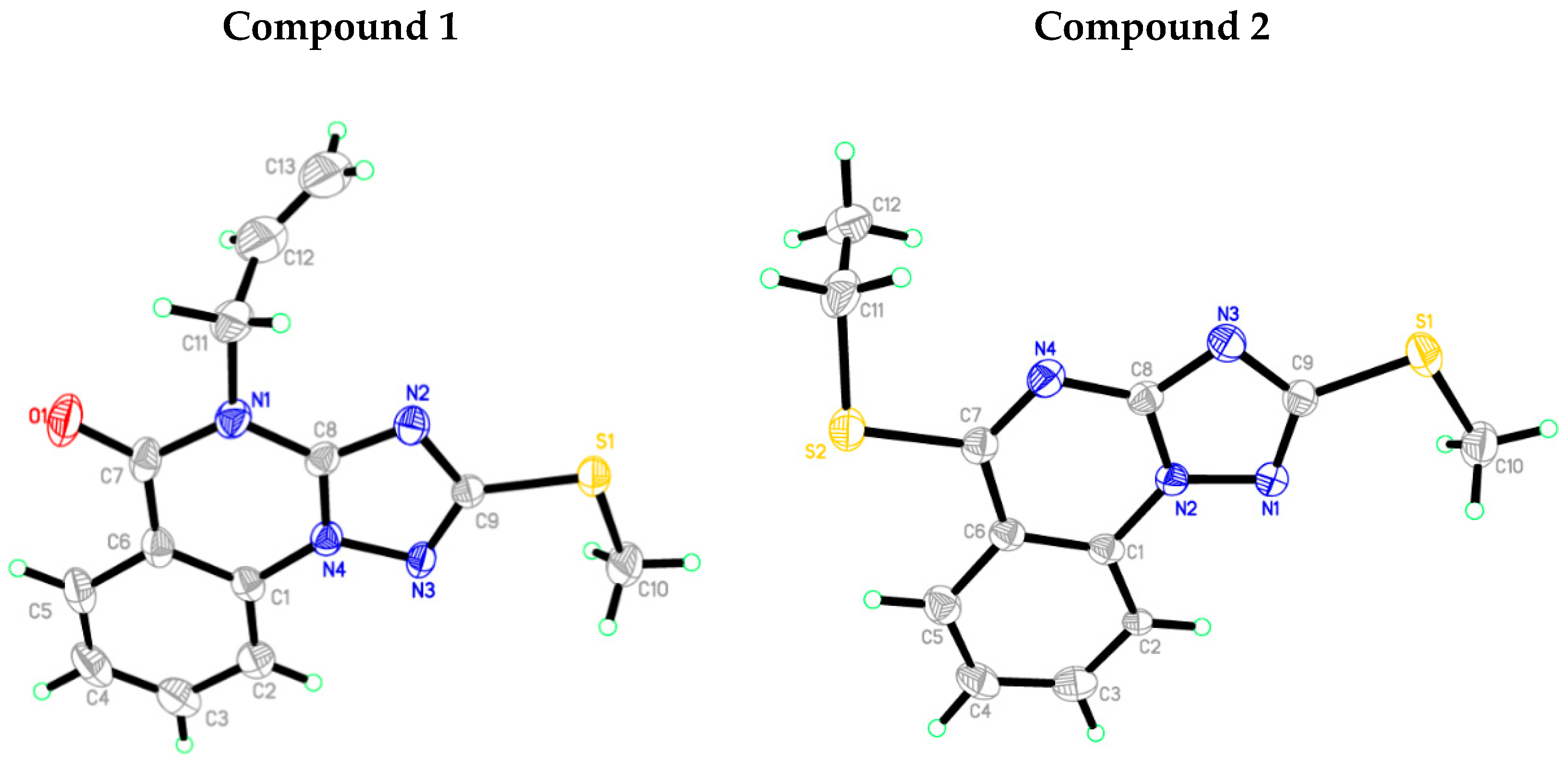
3.3. Crystallographic Data
The refinement information and crystallographic data of triazoloquinazolines 1 and 2 (C13H12N4OS and C12H12N4S2) structures are presented in Table 1. As it can be evinced from Figure 1, the asymmetric units of 1 and 2 contain only one molecule. The length of all bonds and the angles demonstrated values in the ordinary ranges [39]. In the crystal packing (Figure 2), the molecules of compounds 1 and 2 are connected to another one via a single non-classical intermolecular hydrogen bonding interaction (Table 2).
Table 1.
Experimental details of X-ray crystallography of triazoloquinazolines 1 and 2.
Table 2.
For triazoloquinazolines 1 and 2.
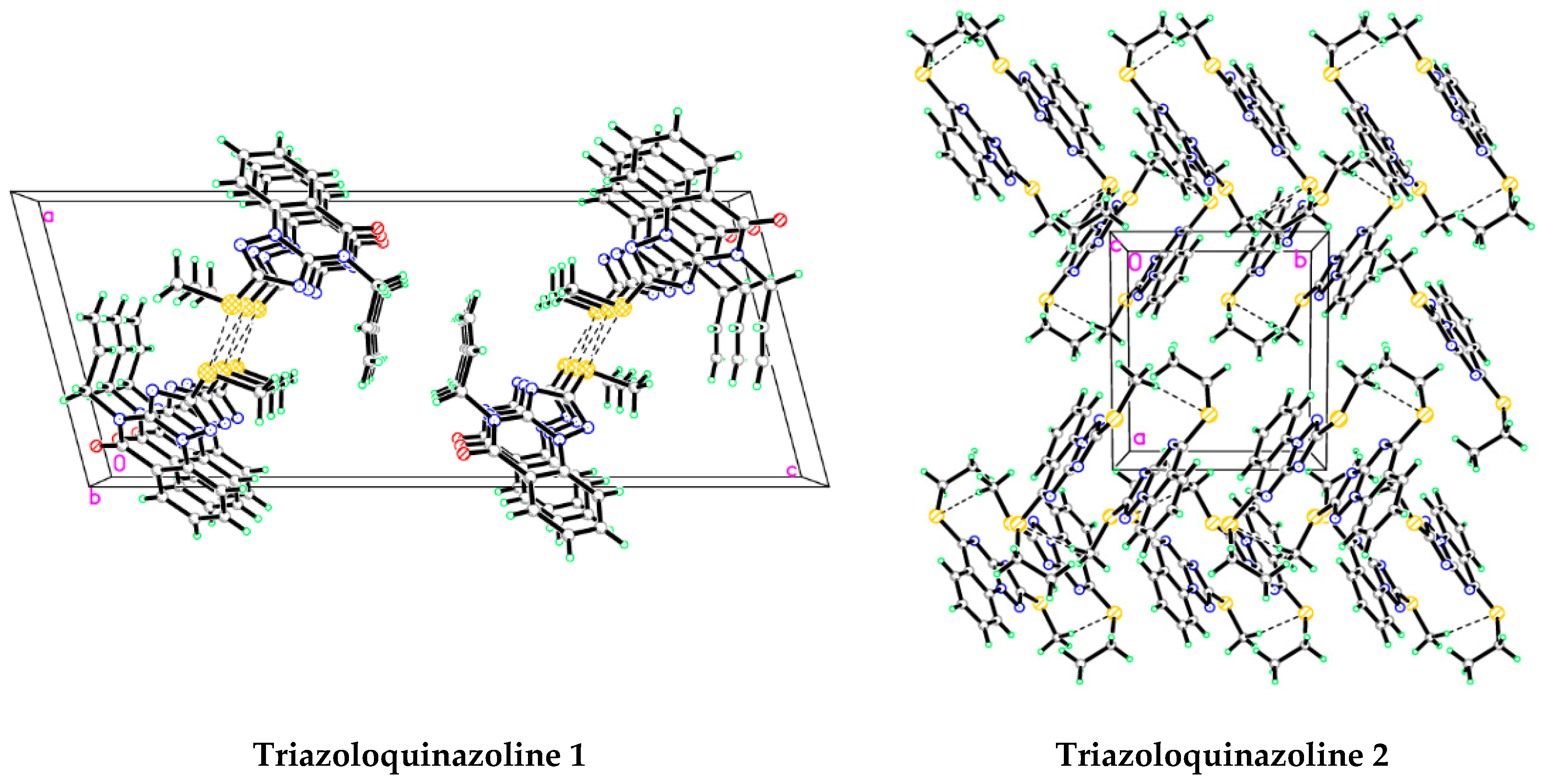
3.4. Analysis of Molecular Packing
The intermolecular interactions play an essential role for determining the stability of the studied crystals. In this regard, various intermolecular interactions in the crystal structures of both systems are analyzed quantitatively, performing Hirshfeld calculations (Figures S1 and S2). Their percentages and detected contacts are presented in Figure 3. The molecular packing of 1 is controlled by significantly short O···H (6.7%), S···S (1.2%) and C···C (2.8%) (Figure 3). In 2, the packing is controlled by short H···H (42.6%), S···H (16.3%) and C···C (4.3%) interactions. All such interactions have the characteristic features of short interactions and contribute significantly to the stability of 1 and 2 crystal structures. Notably, although the H···H (47.0%), H···C (12.97%), N···H (9.7%) and S···H (5.8%) contacts make a significant contribution to the overall molecular packing interactions in 1 (Figure 4), these contacts exhibited the characteristic features of intermolecular interactions between atoms found at longer mutual distances than the sums of the said atoms Van der Waals radii. Concerning 2, the contact distances of the most significant H···H, S···H and C···C interactions are summarized in Table 3, and other key contacts that contributed to the molecular packing included the N···H (15.1%) and H···C (9.8%) interactions. Indeed, values for the contact distances of the most important H···H, S···H and C···C interactions were significant, which are highlighted by the corresponding red spots in the dnorm maps. Moreover, characteristics of short interactions were observed in the fingerprint plots (Figure 5). These interactions in 2 exhibited considerably longer interaction distances than the sum of the Van der Waals radii of the interacting atoms as well.
Figure 3.
Intermolecular interactions summary and their % contributions to the overall molecular packing interactions of 1 and 2 crystal structures.
Figure 4.
Results of the Hirshfeld analysis conducted on 1. Red, white and blue colors indicated shorter, equal and longer contact distances than the vdWs radii sum of the interacting atoms, respectively.
Table 3.
Values for the short intermolecular contacts in triazoloquinazolines 1 and 2.
Figure 5.
Results of the Hirshfeld analysis conducted on 2. Red, white and blue colors indicated shorter, equal and longer contacts distances than the vdWs radii sum of the interacting atoms, respectively.
4. DFT Analysis
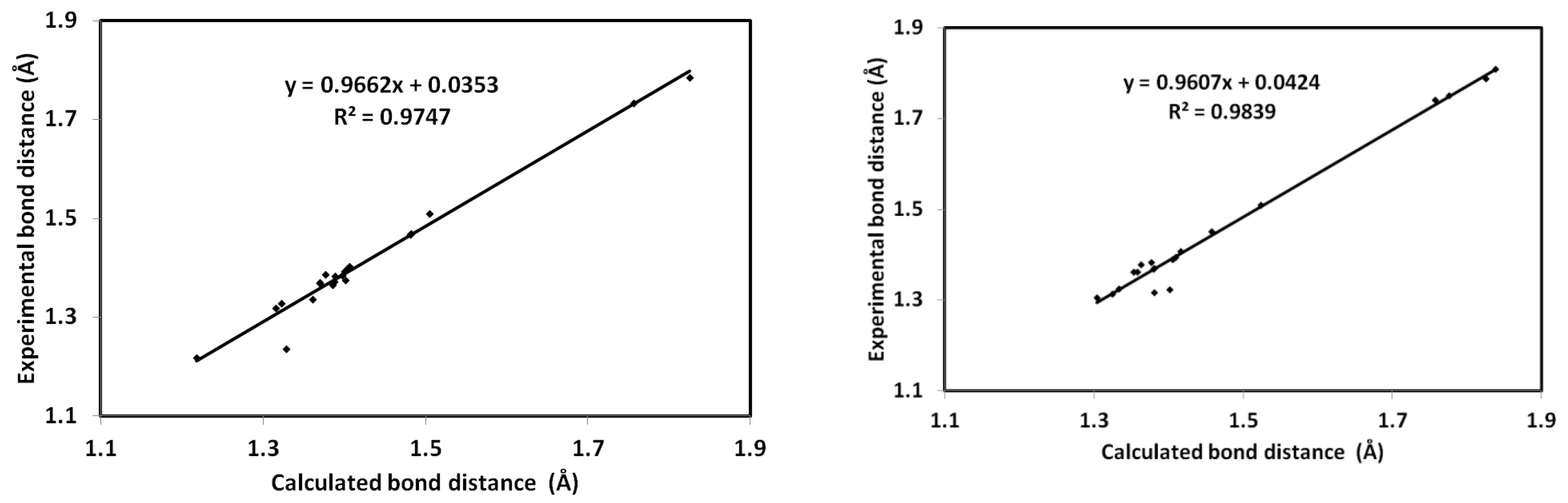
4.1. Geometric Parameters
The compounds 1 and 2 optimized geometries are presented in Figure 6. It can be evinced from this figure that the calculated geometric features of both structures match very well their counter parts experimentally determined, using X-ray diffraction data. The calculated and optimized bond distances are in good agreement with each other (Table S1). Good correlations were also determined to exist between the experimental and the calculated bond distances for the 1 and 2 structures (Figure 7). Indeed, the correlation coefficients values are very close to unity, and the maximum absolute errors do not exceed 0.04 Å and 3.8° for bond distances and bond angles, respectively.
Figure 6.
Optimized structures (upper) and matching between the calculated and experimental structures (lower) of 1 and 2.

Figure 7.
Correlation between the calculated and experimental bond distances of 1 and 2.
The calculated natural atomic charges are presented (Table S2). For triazoloquinazoline 1, the O and N atoms as well as the majority C atoms are negatively charged. The atomic site with the largest negative charge is the carbonyl oxygen (O2: −0.6098 e). Hydrogen and carbon atoms attached to N, O or S atoms are all positively charged. The atom with the largest positive charge is the carbonyl carbon (C20:0.6855 e). In the case of 2, it was found that N5 (−0.5299 e) and C18 (0.5649 e) were the most negative and positive sites, respectively. The molecular electrostatic potential maps reported in Figure 8 demonstrated these results very well. Triazoloquinazolines 1 and 2 are polar compounds characterized by values for the net dipole moments of 2.9284 and 4.2127 Debye.
Figure 8.
Molecular electrostatic potential maps of 1 and 2. Blue, turquoise, yellow and red indexed colors provide visual information on the electron density—turquoise indicates the lowest electron density, whereas red indicates the highest electron density.
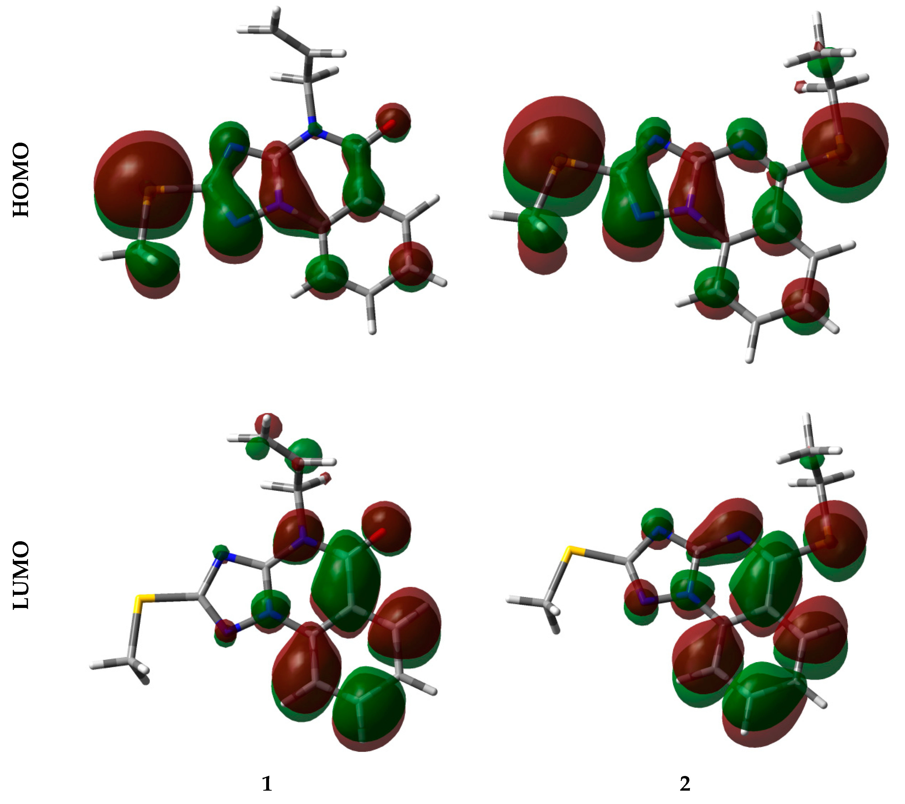
4.2. Analysis of Reactivity
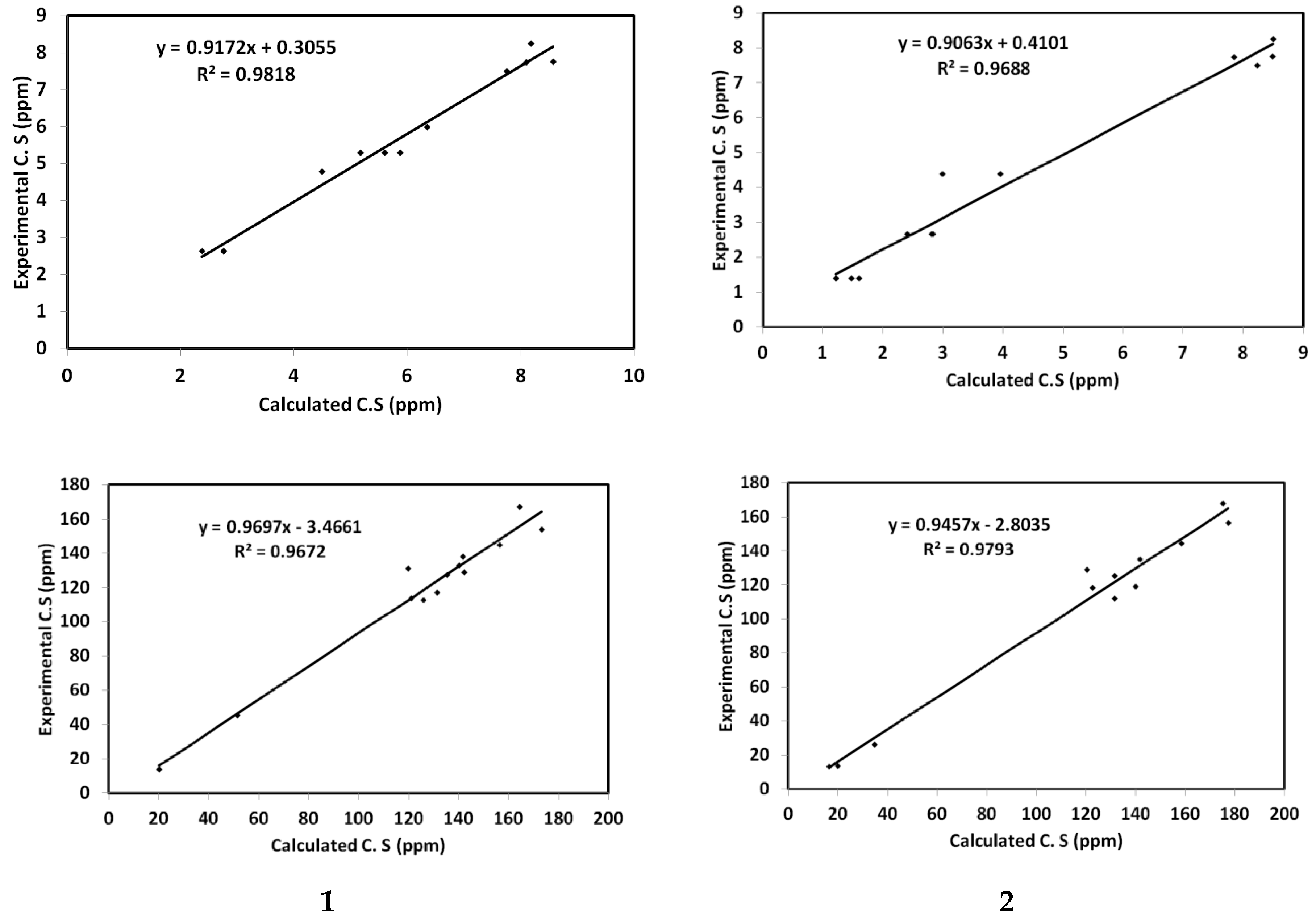
Investigation of the highest occupied and lowest unoccupied molecular orbitals (HOMO and LUMO) of 1 and 2 indicated beyond doubt the distribution of these molecular orbitals over the π-system of the studied molecules (Figure 9). The energies of these orbitals and the corresponding reactivity indices [40,41,42,43,44,45], such as ionization potential, electron affinity, chemical potential, hardness and electrophilicity (I, A, μ, η and ω) were calculated (Table 4). Both HOMO levels have almost the same energy in the two compounds. However, the energy of LUMO in 1 is higher than 2. From the results, both compounds have similar ionization potentials but different electron affinities. In the case of 1, the η value (4.3250 eV) is higher than that of 2 (3.8053 eV). In addition, the electrophilicity of 1 is lower (1.7852 eV) than that of 2 (2.2087 eV). In Table 4, all the calculated values for I, A, ω, and the HOMO–LUMO gap for both crystals are listed.
Figure 9.
Visual representations of HOMO and LUMO of 1 and 2.
Table 4.
The calculated reactivity indices of 1 and 2.
5. NMR Spectra
The chemical shifts of the resonance peaks in the 1H and 13C NMR spectra of triazoloquinazolines 1 and 2 were computed, and the findings are listed in Tables S3 and S4, alongside the corresponding experimental data. As it can be evinced from Figure 10, a good relation exists between the calculated and the experimentally determined chemical shifts. In particular, the correlation coefficients are 0.97 and 0.98 for both 1H and 13C NMR spectral data.
Figure 10.
Correlations between the calculated and experimentally determined 1HNMR (left) and 13C (right) chemical shift (C.S) values for both crystals.
6. Conclusions
Triazoloquinazolines 1 and 2 were synthesized and confirmed by NMR and X-ray diffractometry. The crystal structures of 1 and 2, as determined by X-ray diffractometry, were characterized by bond lengths and angles that were in the normal ranges; moreover, molecules of 1 and 2 were observed to be included in a non-classical intermolecular hydrogen bonding interaction. The molecules 1 and 2 were anticipated to be polar, with net dipole moments of 2.9284 and 4.2127 Debye. The practical data were correlated with theoretical DFT calculations (employing the B3LYP/6-311++G(d,p) basis set). The supramolecular structures of the investigated compounds were studied performing Hirshfeld calculations. The calculated geometric features of the studied compounds were appeared to be in good agreement with those experimentally identified by X-ray. Moreover, good agreement was detected between the calculated and the experimental NMR data. Different electronic and reactivity descriptors of 1 and 2 were calculated and discussed.
Supplementary Materials
The following are available online at www.mdpi.com/2073-4352/11/10/1195/s1, Figure S1: Hirshfeld analysis of 2, Figure S2: Hirshfeld analysis of 1, Table S1: The calculated bond distances of 1 and 2, Table S2: Natural charge populations at the different atomic sites of the studied compounds, Table S3: The calculated and experimental chemical shifts (ppm) for 1 (according to Figure 6), Table S4: The calculated and experimental chemical shifts (ppm) for 2 (according to Figure 6).
Author Contributions
H.A.A.: conceptualization, writing—original draft, investigation, methodology and data curation; S.M.S.: conceptualization, writing—original draft, software, data curation and validation; H.A.G.: conceptualization, software and validation; M.M.: writing—reviewing; M.M.A.: writing—reviewing; R.A.-S.: conceptualization, writing—original draft, investigation, methodology, data curation and supervision. All authors have read and agreed to the published version of the manuscript.
Funding
The authors extend their appreciation to the Researchers Supporting Project, King Saud University, Riyadh, Saudi Arabia for funding this work through grant no. RSP-2021/353.
Institutional Review Board Statement
Not applicable.
Informed Consent Statement
Not applicable.
Acknowledgments
The authors extend their appreciation to the Researchers Supporting Project, King Saud University, Riyadh, Saudi Arabia for funding this work through grant no. RSP-2021/353.
Conflicts of Interest
The authors declare that they have no known competing financial interests or personal relationships that could have appeared to influence the work reported in this paper.
References
- Al-Salahi, R.; Gamal-Eldeen, A.M.; Alanazi, A.; Al-Omar, M.; Marzouk, M.; Fouda, M.G. Cytotoxic and anti-inflammatory active methylsulfanyltriazoloq uinazolines. J. Pure Appl. Microbiol. 2013, 7, 189–198. [Google Scholar]
- Abuelizz, H.A.; Al-Salahi, R.; Al-Asri, J.; Mortier, J.; Marzouk, M.; Ezzeldin, E.; Ali, A.A.; Khalil, M.G.; Wolber, G.; Ghabbour, H.A. Synthesis, crystallographic characterization, molecular docking and biological activity of isoquinoline derivatives. Chem. Cent. J. 2017, 11, 103. [Google Scholar] [CrossRef] [Green Version]
- Abuelizz, H.A.; Al-Salahi, R. An overview of triazoloquinazolines: Pharmacological significance and recent developments. Bioorg. Chem. 2021, 115, 105263. [Google Scholar] [CrossRef]
- Al-Salahi, R.; Marzouk, M. Some 2-aminobenzo[de]isoquinolin1,3-diones as antimicrobial agents. Asian J. Chem. 2014, 26, 8163–8165. [Google Scholar] [CrossRef]
- Alagarsamy, V.; Rupeshkumar, M.; Kavitha, K.; Meena, S.; Shankar, D.; Siddiqui, A.A.; Rajesh, R. Synthesis and pharmacological investigation of novel 4-(2-methylphenyl)-1-substituted-4H-[1,2,4]triazolo[4,3-a]quinazolin-5-ones as new class of H(1)-antihistaminic agents. Eur. J. Med. Chem. 2008, 43, 2331–2337. [Google Scholar] [CrossRef]
- Al-Salahi, R.; Marzouk, M.; El-Eraky, W.I.; Saleh, O.S. Antihistamine Activity of a new set 1,2,4-triazolo[1,5-a]quinazolines. Asian J. Chem. 2014, 26, 8625–8627. [Google Scholar]
- Alagarsamy, V.; Pathak, U.S. Synthesis and antihypertensive activity of novel 3-benzyl-2-substituted-3H-[1,2,4]triazolo[5,1-b]quinazolin-9-ones. Bioorg. Med. Chem. 2007, 15, 3457–3462. [Google Scholar] [CrossRef]
- Al-Salahi, R.; Tahir, K.E.; Alswaidan, I.; Lolak, N.; Hamidaddin, M.; Marzouk, M. Biological effects of a new set 1,2,4-triazolo[1,5-a]quinazolines on heart rate and blood pressure. Chem. Cent. J. 2014, 8, 3. [Google Scholar] [CrossRef] [Green Version]
- Francis, J.E.; Cash, W.D.; Psychoyos, S.; Ghai, G.; Wenk, P.; Friedmann, R.C.; Atkins, C.; Warren, V.; Furness, P.; Hyun, J.L.; et al. Structure–activity profile of a series of novel triazoloquinazoline adenosine antagonists. J. Med. Chem. 1988, 31, 1014–1020. [Google Scholar] [CrossRef]
- Al-Salahi, R.; Geffken, D.; Koellner, M. A new series of 2-Alkoxy(aralkoxy)-[1,2,4]triazolo[1,5-a]quinazolin-5-ones as Adenosine receptor antagonists. Chem. Pharm. Bull. 2011, 59, 730–733. [Google Scholar] [CrossRef] [PubMed] [Green Version]
- Al-Salahi, R.; Marzouk, M.; Awad, G.; Al-Omar, M.; Ezzeldin, E. Antimicrobial activity of newly synthesized methylsulfanyl-triazoloquinazoline derivatives. J. Pharm. Pharmacol. 2013, 65, 790–797. [Google Scholar] [CrossRef]
- Al-Salahi, R.; Alswiadan, I.; Alomer, M.; Marzouk, M. Synthesis and antimicrobial of new 2-Phenoxy-4H-[1,2,4]triazolo[1,5-a]quinazoline derivatives. Life Sci. J. 2013, 10, 2018–2028. [Google Scholar]
- Al-Salahi, R.; Alswiadan, I.; Alomer, M.; Marzouk, M. Antiviral activity of 2-Phenoxy-4H-[1,2,4]triazolo[1,5-a]quinazoline derivatives. Life Sci. J. 2013, 10, 2164–2169. [Google Scholar]
- Al-Salahi, R.; Alswiadan, I.; Marzouk, M.; Alsenousy, W.; Abd Elgalil, A. Antiviral activities of some methylsulfanyltriazoloquinazolines. Res. Chem. Intermed. 2015, 41, 151–161. [Google Scholar] [CrossRef]
- Abuelizz, H.A.; Anouar, E.H.; Ahmad, R.; Azman, N.I.I.N.; Marzouk, M.; Al-Salahi, R. Triazoloquinazolines as a new class of potent α-glucosidase inhibitors: In vitro evaluation and docking study. PLoS ONE 2019, 14, e0220379. [Google Scholar] [CrossRef] [Green Version]
- Al-Salahi, R.; Anouar, E.H.; Marzouk, M.; Taie, H.A.; Abuelizz, H.A. Screening and evaluation of antioxidant activity of some 1,2,4-triazolo[1,5-a]quinazoline derivatives. Future Med. Chem. 2018, 10, 379–390. [Google Scholar] [CrossRef] [PubMed]
- Sompalle, R.; Roopan, S.M.; Al-Dhabi, N.A.; Suthindhiran, K.; Sarkar, G.; Arasu, M.V. 1,2,4-triazoloquinazoline-thiones: Non-conventional synthetic approach, study of solvatochromism and antioxidant assessment. J. Photochem. Photobiol. B Biol. 2016, 162, 232–239. [Google Scholar] [CrossRef] [PubMed]
- Al-Salahi, R.; Marzouk, M.; Ashour, A.E.; Alswaidan, I. Synthesis and antitumor activity of 1,2,4-triazolo[1,5-a]quinazolines. Asian J. Chem. 2014, 26, 2173–2176. [Google Scholar] [CrossRef]
- Al-Salahi, R.; Ashour, A.E.; Marzouk, M.; Ashok, K. In Vitro Cytotoxicity Evaluation of a New Series of Benzo[g][1,2,4]triazolo[1,5-α]quinazolines. Lat. Am. J. Pharm. 2015, 34, 1926–1930. [Google Scholar]
- Al-Salahi, R.; Elsayed, E.A.; El Dib, R.A.; Wadaan, M.; Ezzeldin, E.; Marzouk, M. Synthesis, Characterization and Cytotoxicity Evaluation of 5-Hydrazono-[1,2,4]triazolo[1,5-a]quinazolines (Part I). Lat. Amer. J. Pharm. 2016, 35, 58–65. [Google Scholar]
- Almehizia, A.A.; Abuelizz, H.A.; Taie, H.A.A.; Anouar, E.A.; Marzouk, M.; Al-Salahi, R. Investigation the antioxidant activity of benzo[g]triazoloquinazolines correlated with a DFT study. Saudi Pharm. J. 2019, 27, 133–137. [Google Scholar] [CrossRef] [PubMed]
- Al-Salahi, R.; Marzouk, M.; Abbas, M.; Ng, S.W. 2-Methylsulfonyl-1,2,4-triazolo[1,5-a]quinazolin-5 (4H)-one. Acta Crystallogr. 2012, E68, o1806. [Google Scholar] [CrossRef] [PubMed] [Green Version]
- Al-Salahi, R.; Marzouk, M.; Al-Omar, M.; Amr, A.E.; Ng, S.W.; Tiekink, E.R.T. 2-Methylsulfanyl-1,2,4-triazolo[1,5-a]quinazoline-5(4H)-thione. Acta Crystallogr. 2013, E69, o434. [Google Scholar] [CrossRef] [PubMed] [Green Version]
- Al-Salahi, R.; Nabih, L.; Al-Omar, M.; Ng, S.W. 2-Phenoxy-1,2,4-triazolo[1,5-a] quinazolin-5(4H)-one. Acta Crystallogr. 2012, E68, o1808. [Google Scholar] [CrossRef] [PubMed] [Green Version]
- Al-Salahi, R.; Al-Omar, M.; Marzouk, M.; Ng, S.W.; Tiekink, E.R.T. 5-chloro-2-methylsulfonyl-1,2,4-triazolo[1,5-a]quinazoline. Acta Crystallogr. 2012, E68, o1809. [Google Scholar] [CrossRef] [Green Version]
- Al-Salahi, R.; Geffken, D.; Bari, A. 2-benzyloxy-1,2,4-triazolo[1,5-a] quinazolin-5(4H)-one. Acta Crystallogr. 2011, E67, o1861. [Google Scholar] [CrossRef] [Green Version]
- Al-Salahi, R.; Geffken, D. Synthesis of Novel 2-methylsulfanyl-4H-[1,2,4]triazolo[1,5-a]quinazolin-5-one and derivatives. Synth. Commun. 2011, 41, 3512–3523. [Google Scholar] [CrossRef]
- Al-Salahi, R.; Geffken, D. Synthesis and reactivity of [1,2,4]triazolo-annelated quinazolines. Molecules 2010, 15, 7016–7034. [Google Scholar] [CrossRef] [Green Version]
- Sheldrick, G.M. SHELXT-integrated space-group and crystal-structure determination. Acta Crystallogr. A Found. Adv. 2015, 71, 3–8. [Google Scholar] [CrossRef] [Green Version]
- Allen, F.H.; Kennard, O.; Watson, D.G.; Brammer, L.; Orpen, A.G.; Taylor, R. Tables of bond Lengths determined by X-ray and neutron diffraction. Part 1. Bond lengths in organic compounds. J. Chem. Soc. Perkin Trans. 1987, II, S1–S19. [Google Scholar] [CrossRef]
- Turner, M.J.; McKinnon, J.J.; Wolff, S.K.; Grimwood, D.J.; Spackman, P.R.; Jayatilaka, D.; Spackman, M.A. Crystal Explorer. University of Western Australia. 2017. Available online: http://hirshfeldsurface.net (accessed on 2 September 2021).
- Frisch, M.J.; Trucks, G.W.; Schlegel, H.B.; Scuseria, G.E.; Robb, M.A.; Cheeseman, J.R.; Scalmani, G.; Barone, V.; Mennucci, B.; Petersson, G.A.; et al. Fox, GAUSSIAN 09. Revision A02; Gaussian Inc.: Wallingford, CT, USA, 2009. [Google Scholar]
- Dennington, R., II; Keith, T.; Millam, J. GaussView; Version 4.1; Semichem Inc.: Shawnee Mission, KS, USA, 2007. [Google Scholar]
- Reed, A.E.; Curtiss, L.A.F.; Weinhold, F. Intermolecular interactions from a natural bond orbital, donor-acceptor viewpoint. Chem. Rev. 1988, 88, 899–926. [Google Scholar] [CrossRef]
- Marten, B.; Kim, K.; Cortis, C.; Friesner, R.A.; Murphy, R.B.; Ringnalda, M.N.; Sitkoff, D.; Honig, B. New model for calculation of solvation free energies: correction of self-consistent reaction field continuum dielectric theory for short-range hydrogen-bonding effects. J. Phys. Chem. 1996, 100, 11775–11788. [Google Scholar] [CrossRef]
- Tannor, D.J.; Marten, B.; Murphy, R.; Friesner, R.A.; Sitkoff, D.; Nicholls, A.; Ringnalda, M.; Goddard, W.A.; Honig, B. Accurate first principles calculation of molecular charge distributions and solvation energies from ab initio quantum mechanics and continuum dielectric theory. J. Am. Chem. Soc. 1994, 116, 11875–11882. [Google Scholar] [CrossRef]
- Cheeseman, J.R.; Trucks, G.W.; Keith, T.A.; Frisch, M.J. A Comparison of Models for Calculating Nuclear Magnetic Resonance Shielding Tensors. J. Chem. Phys. 1996, 104, 5497–5509. [Google Scholar] [CrossRef]
- Sheldrick, G.M. A short history of SHELX. Acta Crystallogr. 2008, A64, 112–122. [Google Scholar] [CrossRef] [PubMed] [Green Version]
- Foresman, J.B.; Frisch, A.E. Exploring Chemistry with Electronic Structure Methods, 2nd ed.; Gaussian: Pittsburgh, PA, USA, 1996. [Google Scholar]
- Chang, R. Chemistry, 7th ed.; McGraw-Hill: New York, NY, USA, 2001. [Google Scholar]
- Kosar, B.; Albayrak, C. Spectroscopic investigations and quantum chemical computational study of (E)-4-methoxy-2-[(p-tolylimino)methyl]phenol. Spectrochim. Acta 2011, 78, 160–167. [Google Scholar] [CrossRef] [PubMed]
- Koopmans, T. About the assignment of wave functions and eigenvalues to the individual electrons of an atom. Physica 1934, 1, 104–113. [Google Scholar] [CrossRef]
- Parr, R.G.; Yang, W. Density-Functional Theory of Atoms and Molecules; Oxford University Press: New York, NY, USA, 1989. [Google Scholar]
- Parr, G.; Von Szentpály, L.; Liu, S.B. Electrophilicity Index. J. Am. Chem. Soc. 1999, 121, 1922–1924. [Google Scholar] [CrossRef]
- Singh, R.N.; Kumar, A.; Tiwari, R.K.; Rawat, P.; Gupta, V.P. A combined experimental and quantum chemical (DFT and AIM) study on molecular structure, spectroscopic properties, NBO and multiple interaction analysis in a novel ethyl 4-[2-(carbamoyl)hydrazinylidene]-3,5-dimethyl-1H-pyrrole-2-carboxylate and its dimer. J. Mol. Struct. 2013, 1035, 427–440. [Google Scholar] [CrossRef]
Publisher’s Note: MDPI stays neutral with regard to jurisdictional claims in published maps and institutional affiliations. |
© 2021 by the authors. Licensee MDPI, Basel, Switzerland. This article is an open access article distributed under the terms and conditions of the Creative Commons Attribution (CC BY) license (https://creativecommons.org/licenses/by/4.0/).