Article

Solar-Driven Photocatalytic Degradation of Clothianidin Using Green NiO-GO Composite

1
Department of Chemistry, Government College University Faisalabad, Faisalabad 38000, Pakistan
2
Physics Department, College of Science, Imam Mohammad Ibn Saud Islamic University (IMSIU), Riyadh 11623, Saudi Arabia
3
Department of Civil Engineering, Government Polytechnic Mankeda, Agra 283102, Uttar Pradesh, India
*
Authors to whom correspondence should be addressed.
Catalysts 2025, 15(11), 1078; https://doi.org/10.3390/catal15111078 (registering DOI)
Submission received: 15 October 2025 / Revised: 5 November 2025 / Accepted: 11 November 2025 / Published: 13 November 2025
(This article belongs to the Special Issue Advanced Catalysis for Energy and a Sustainable Environment)

Abstract

The extensive use of clothianidin pesticide poses significant risks to non-target organisms and water resources. In this study, NiO-GO is reported as an effective photocatalyst for the degradation of clothianidin in aqueous medium. Nickel oxide (NiO) nanoparticles were synthesized by a green method using Pisum sativum (pea) peel extract, which serves as a natural reducing and stabilizing agent, and subsequently integrated with graphene oxide (GO) through ultrasonication to form a NiO-GO composite in a 1:1 ratio. The materials were characterized by various techniques. Photocatalytic degradation of clothianidin under natural sunlight was systematically investigated, assessing the effects of pH, catalyst dosage, initial pollutant concentration, and agitation speed. The NiO-GO composite exhibited superior photocatalytic performance (96% degradation at pH 3 within 60 min) compared to pristine NiO and GO, with a rate constant 4.4 and 3.3 times higher, respectively. The as-prepared NiO-GO photocatalyst exhibited nearly consistent degradation efficiency over two successive cycles, demonstrating its excellent structural stability and reusability. The enhanced performance is attributed to improved charge separation afforded by GO support. This low-cost, green, and efficient NiO-GO photocatalyst demonstrates promising potential for sustainable pesticide remediation in aqueous environments.

1. Introduction

The rapid expansion of industrialization and the swift growth of the human population are two main factors that have significantly exacerbated environmental degradation. Although the industrial sector is under pressure to pursue sustainability goals through innovative technologies, eco-friendly practices, and effective governance mechanisms to address environmental challenges, it still releases waste directly into the environment [1]. The rapid growth of the global population has not only increased the challenges of resource depletion and environmental degradation [2,3] but has also contributed significantly to the escalating global energy crisis [4,5,6,7]. The increase in the rate of population and urbanization encourages the utilization of pesticides in agricultural fields. However, the excessive use of pesticides and their accumulation in soil and water pose serious threats to both human health and the environment. Among the four major types of pollution—air, water, noise, and electromagnetic—water pollution has attracted considerable attention from researchers [8,9,10,11]. Clothianidin belongs to the 2nd generation neonicotinoid insecticides, having the IUPAC name of (E)-1-(2-Chloro-1,3-thiazol-5-ylmethyl)-3-methyl-2-nitroguanidine, and it was developed in 2001 by Takeda Chemical Industries and Bayer AG [12]. Clothianidin has been frequently utilized to eliminate and kill different insects, such as diptera, hemipteran, lepidoptera, thysanoptera, and coleopteran. Clothianidin is an agonist of nicotinic acetylcholine receptors (nAChRs), which disturbs the central nervous system of insects but is said to be harmless for human beings and other animals [13,14]. Nevertheless, it has been reported recently that clothianidin may also be harmful to the nervous systems of non-targeted species, such as honeybees, birds, and humans. Therefore, the European Food Safety Authority limited the use of clothianidin in 2013 in order to re-evaluate the possible environmental concerns [15,16]. The molecular size of clothianidin is comparatively small, and its solubility in water is very high. Additionally, it is less volatile and its half-life in oil varies from 148 to 6931 days. It has also been reported that clothianidin is resistant to hydrolysis at ambient pH and temperature [17,18]. The persistence of clothianidin in the environment and its chemical properties indicate that residues of clothianidin leach and transport into water, exerting an adverse impact on aquatic biota, which makes it a serious concern. It has also been confirmed that residues of clothianidin were found in ground and surface water, which were incorporated from the soil [19,20].
In the modern era, the interlinked challenges of environmental pollution have driven scientific research toward the pursuit of sustainable solutions [21,22]. In the last several years, diverse efforts have been taken to manage the hazardous effects of pesticides, but advanced oxidation processes (AOPs) have gained more attention due to their high degradation ability and complete mineralization of pollutants. During AOPs, highly reactive species like OH and O2●− are generated, which may efficiently attack the target pollutants, resulting in their conversion into water and carbon dioxide or less toxic compounds [23]. Heterogeneous photocatalysis is one of the most important types of AOPs, which is governed by the interaction of near UV or solar visible radiation as a technology for wastewater treatment [24,25,26,27,28]. In the last couple of decades, numerous materials have been employed as photocatalysts, like metal oxides, polymers, glasses, ceramics, MOF, and composite materials, for various industrial purposes [29,30,31]. However, metal oxides, for instance, titanium dioxide, vanadium oxide, tungsten trioxide, zirconium oxide, and vanadium oxide, are widely used among these materials for wastewater treatment [32,33,34,35,36].
Nickel oxide (NiO) has gained remarkable attention owing to its inexpensive, chemically stable, broad band gap (3.6–4 eV), and superior magnetic and optical properties. NiO belongs to a p-type semiconductor, which easily generates electron–hole pairs when irradiated with sunlight [37]. However, NiO commonly has limitations of inadequate quantum efficiency and the ability to agglomerate easily. The aforementioned reasons constrain the scientists from improving the photocatalytic capability of NiO through modifications in NiO by doping metals or non-metals, incorporating cavities, plasma induction, formation of semiconductor composites, etc. Particularly, developing and designing hybrid materials, synthesized from the coupling of carbonaceous materials and metal oxides, is the most attractive choice for enhancing the photocatalytic activities of metal oxides when irradiated with UV/visible radiation [22,38,39,40,41].
Graphene oxide (GO) is well-founded as a typical carrier of nanoparticles. The presence of oxygen in large quantities as a functional group on the surface of GO may build a powerful relationship between GO and nanoparticles, and thus be favorable for the formation of defect sites on the surface of composite materials [42]. In contrast to other catalyst carriers such as carbon nanotubes, activated carbon, titanium oxide, silicon dioxide, etc., the GO has numerous benefits, including high mechanical strength, low resistivity, and good thermal conductivity. Synchronously, GO may increase the degree of movement of electrons, increase the adsorption of pollutants on the surface of catalysts, and prolong the spectral range of the catalysts [43,44].
Based on the above reason, nickel oxide was first prepared via green synthesis using peels of green peas (Pisum sativum), and graphene oxide was constructed by amending Hummer’s method. At the same time, a nickel oxide–graphene oxide (NiO-GO) composite was fabricated using the ultrasonic method. After characterization, the synthesized materials were used as photocatalysts for the degradation of clothianidin pesticide in aqueous medium.
The green-synthesized NiO-GO composite in this study offers a unique combination of sustainability and superior performance as compared to previously reported photocatalysts for pesticide degradation. Conventional NiO systems, often synthesized through hydrothermal or chemical precipitation routes using hazardous reagents, typically achieve 60–85% degradation of target pollutants under UV or solar irradiation, with significantly lower rate constants due to rapid recombination of electrons and holes. On the other hand, the present NiO-GO composite synthesized through a green route achieved 96% degradation of the highly persistent clothianidin pesticide within 60 min under natural sunlight, with a rate constant 4.4 and 3.3 times higher than pristine NiO and GO, respectively. The enhanced photocatalytic performance may be attributed to the synergistic effect of GO, which likely prevents NiO agglomeration, improves light harvesting, and facilitates efficient charge separation, as suggested by morphological and structural evidence and previously reported studies. Furthermore, the use of agricultural waste as a precursor aligns this work with green chemistry and circular economy principles, offering both environmental and economic advantages over previously reported systems.

2. Results

2.1. Characterization

The as-prepared NiO, GO, and NiO-GO were characterized with SEM, XPS, FTIR, and XRD.
The SEM images of NiO, GO, and NiO-GO composite before and after degradation of clothianidin are illustrated in Figure 1. It seems from Figure 1a that NiO consists of irregular-shaped particles and looks like an agglomeration of particles. It is hypothesized that the ultrasonication-assisted synthesis likely facilitated a reasonably uniform dispersion of NiO over GO, which is consistent with similar reports in the literature [45]. The SEM image of GO displays clumped and stacked platelets, which are closely attached, as depicted in Figure 1b. It may be illustrated from Figure 1c that NiO particles are positioned over GO sheets after the construction of NiO-GO composite [46]. It is also apparent from Figure 1d that the SEM image of the NiO-GO nanocomposite becomes a more aggregated structure as compared with the NiO-GO composite before degradation, suggesting the degradation of clothianidin. Hence, the SEM analyses confirm the successful fabrication of NiO-GO by providing sufficient morphological information to distinguish between the structural features of NiO, GO, and NiO-GO.
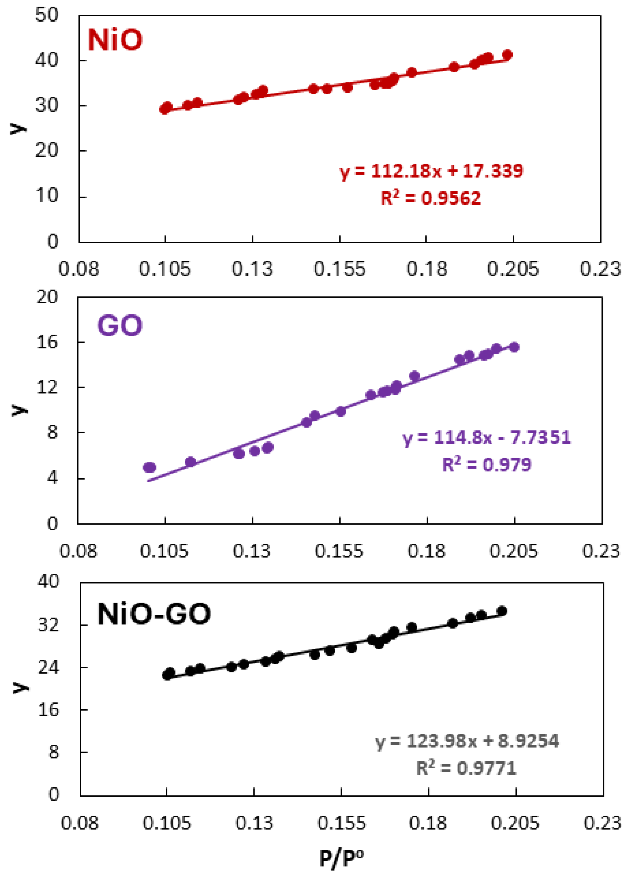
The surface area of a photocatalyst is an important parameter that significantly influences the adsorption capacity, light absorption, and overall photocatalytic efficiency. BET analysis was performed using nitrogen adsorption isotherms to evaluate the specific surface areas of NiO, GO, and NiO-GO composites. The adsorption data within the relative vapor pressure range of 0.1 to 0.2 were analyzed according to the Brunauer–Emmett–Teller (BET) equation (Equation (1)) for surface area measurement. Figure 2 displays the BET plots for NiO, GO, and BNiO-GO. The monolayer volume (Xm) was determined from the slope and intercept of the BET plots. Subsequently, the specific surface area was calculated using Equation (2), incorporating Avogadro’s number (NA), the cross-sectional area of a nitrogen molecule (16.2 Å2), the molar volume of nitrogen, and the mass of the adsorbent. The calculated surface areas for the samples are presented in Table 1. The data given in Table 1 shows that pristine NiO exhibited a specific surface area of 74.8 m2/g, while GO showed a significantly higher value of 164.3 m2/g, which can be attributed to its highly exfoliated layered structure. Upon formation of the NiO-GO composite, the specific surface area decreased to 146.4 m2/g, which, although lower than that of pure GO, remained considerably higher than that of bare NiO. This reduction in surface area compared to GO alone can be ascribed to the partial coverage of GO sheets by NiO nanoparticles. The strong interfacial interaction between NiO and GO likely led to a more compact structure, restricting nitrogen accessibility to certain adsorption sites. Due to its relatively high surface area, GO effectively prevents the agglomeration of NiO nanoparticles, maintaining a high degree of dispersion. Such an improvement in surface morphology is beneficial for photocatalytic applications, as it increases the number of active sites for pollutant adsorption and enhances light–matter interactions, ultimately improving the degradation performance:
1 W [ P o P 1 ] = 1 X m C + C 1 X m C ( P P o )
(W is the amount of N2 adsorbed, Xm is the monolayer capacity, and C is a constant).
Surface   area = X m N A S V m
(NA, S, V, and m represent the Avogadro’s number, the sectional area of the nitrogen molecule (16.2 Å2), the molar volume of nitrogen, and the mass of the adsorbent, respectively).
Figure 2. BET adsorption isotherms ( y =   1 W [ P o P 1 ] ) .
Catalysts 15 01078 g002
Table 1. Surface area measurement.
AdsorbentMass (g)Surface Area (m2/g)
NiO0.36274.8
GO0.198164.3
NiO-GO0.179146.4
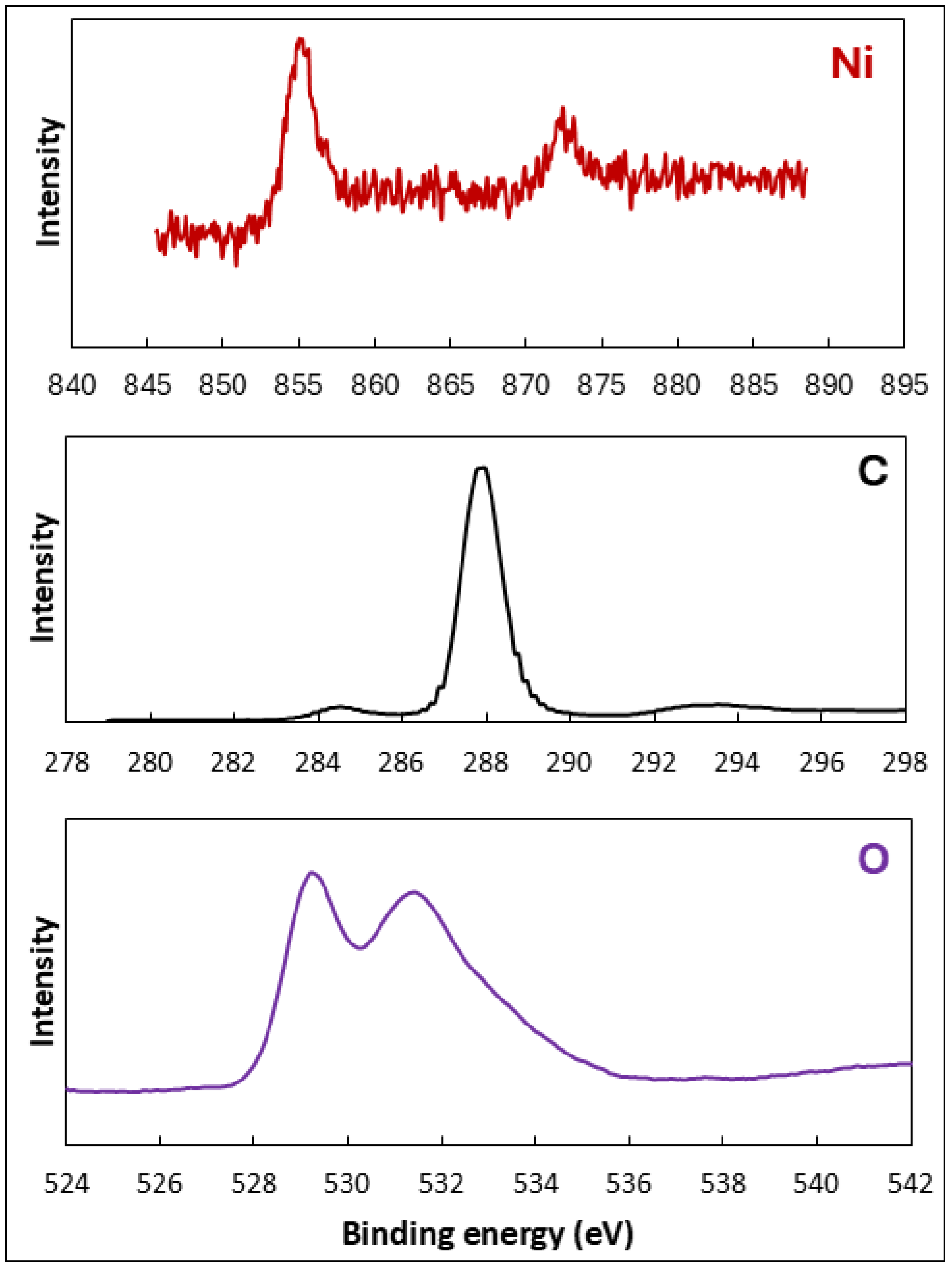
X-ray photoelectron spectroscopy was employed for the determination of the elemental composition and oxidation state of the fabricated NiO-GO. Figure 3 illustrates the XPS analysis of NiO-GO. The XPS analysis confirms the presence of Ni, O, and C elements in fabricated NiO-GO, indicating the successful formation of NiO on the graphene oxide surface without any impurity peaks. The high-resolution Ni 2p spectrum exhibits two main peaks at around 854.2 eV and 871.4 eV, corresponding to Ni 2p3/2 and Ni 2p1/2, respectively, along with their associated satellite peaks. These features are characteristic of Ni2+ species in NiO, confirming the oxidation state of nickel in the composite. The C 1s spectrum shows peaks around 284.6 eV and 288.1 eV, attributed to sp2 carbon components and C-O single bond components of hydroxyl groups, respectively. Shoulder peaks at 286.9 and 289.6 eV are associated with the C=O bond and the C atom, respectively. Pu et al. [47] have assigned the peaks at 284 eV and 289 eV to C=C and O–C=O, respectively. These features confirm that the oxygen-containing groups of GO remain partially intact after composite formation, providing anchoring sites for NiO nanoparticles. The O 1s spectrum can be deconvoluted into multiple components, typically centered at about 529.3 eV and 531.2 eV, representing lattice oxygen (O2− in Ni–O bonds) and surface-adsorbed oxygen or hydroxyl groups, respectively. The relatively higher intensity of surface oxygen peaks suggests the presence of oxygen-containing functional groups originating from GO, which could enhance interfacial interaction and charge transfer between NiO and GO [48,49,50,51].
Overall, the XPS results confirm the successful fabrication of NiO-GO heterostructures with the Ni2+ oxidation state and intimate interfacial contact between NiO and GO. Such interfacial bonding is expected to facilitate charge separation and transfer during catalytic processes, thereby enhancing the overall functional performance of the material.
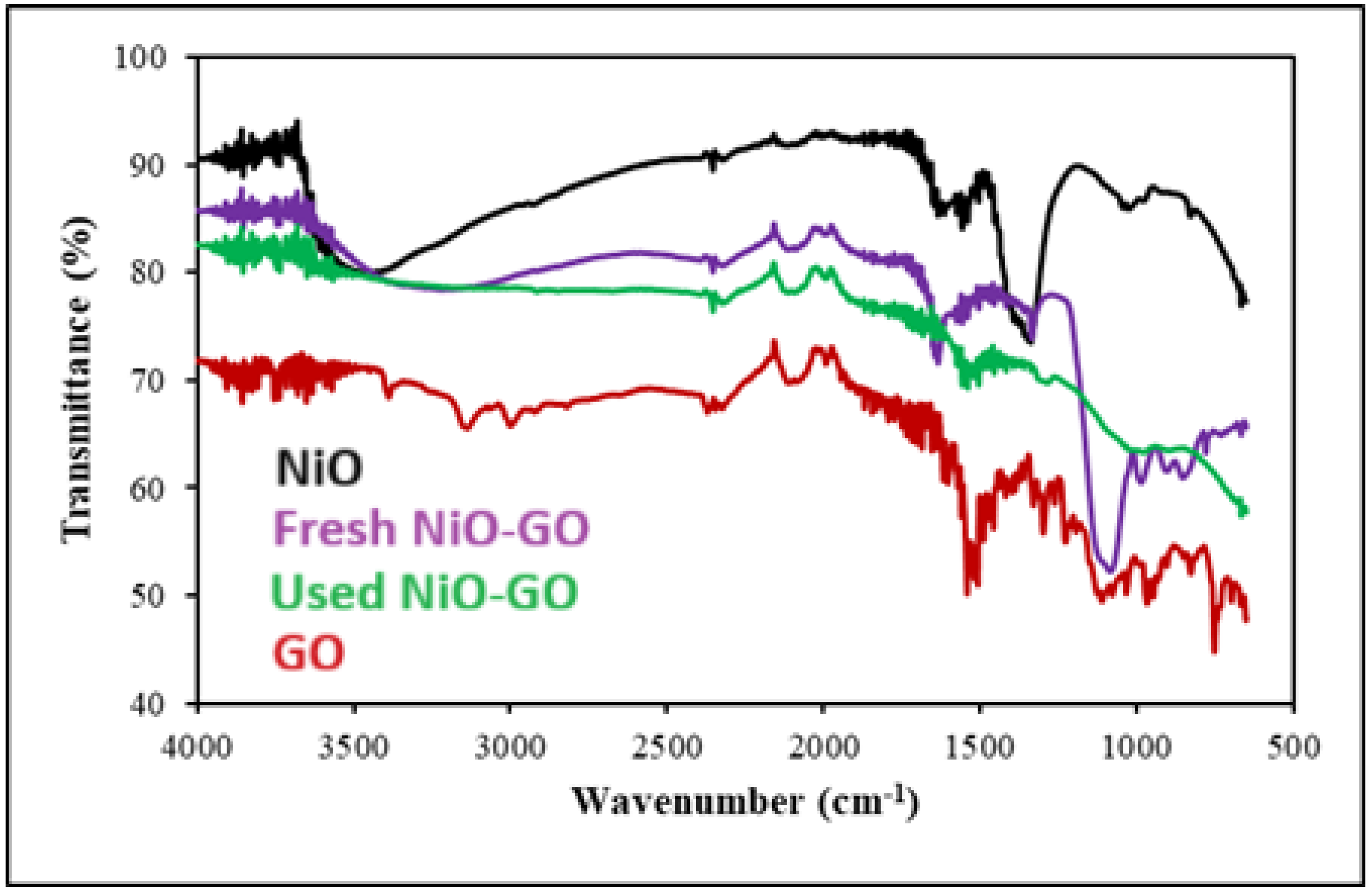
The FTIR analysis was executed to explore the functional groups of NiO, GO, and NiO-GO composite and their variations before and after degradation of clothianidin. Figure 4 illustrates the FTIR spectra of NiO, GO, and NiO-GO composite. The broad peaks at 3485.06, 1349.29, and 1023.15 cm−1 are due to the bending and stretching vibration mode of the O–H functional group adsorbed on the surface of NiO nanoparticles from the atmosphere during the time of performance of FTIR analysis. Gao and co-workers [52] have reported the absorption bands at 3115 and 1612 cm−1 for stretching and bending vibrations of water molecules, respectively. Moreover, the peak at 732.42 cm−1 is designated to the Ni–O stretching vibration mode of the NiO [48,53]. The FTIR analysis of GO is consistent with data on GO reported in the literature. The peak shown at 3388.15 cm−1 represents the O–H stretching vibration of H2O present on the surface of graphene oxide. Some studies [54,55] have reported the absorption band at 3432 and 1078 cm−1 for stretching and bending vibrations of the hydroxyl group. The peaks at 2974.41 and 1541.25 cm−1 confirmed the occurrence of C–H and C=O stretching vibrations, respectively [56]. Whereas an intense peak was seen at about 1107.01 cm−1 and was credited to the C–O stretching vibrations [57]. It may be illustrated from the Figure that most of the peaks that appeared in the GO spectrum disappeared, and some were shifted toward lower wave numbers, which indicates the formation of NiO-GO composite. The SEM of the sample used in the degradation experiments of clothianidin shows a shift of 1630.71 and 1082.79 cm−1 toward 1539.39 and 991.47 cm−1, suggesting the interaction between NiO-GO composite and clothianidin. Moreover, the peak at 1334.38 cm−1 disappeared, which implies the interaction between the composite and the pesticide [58].
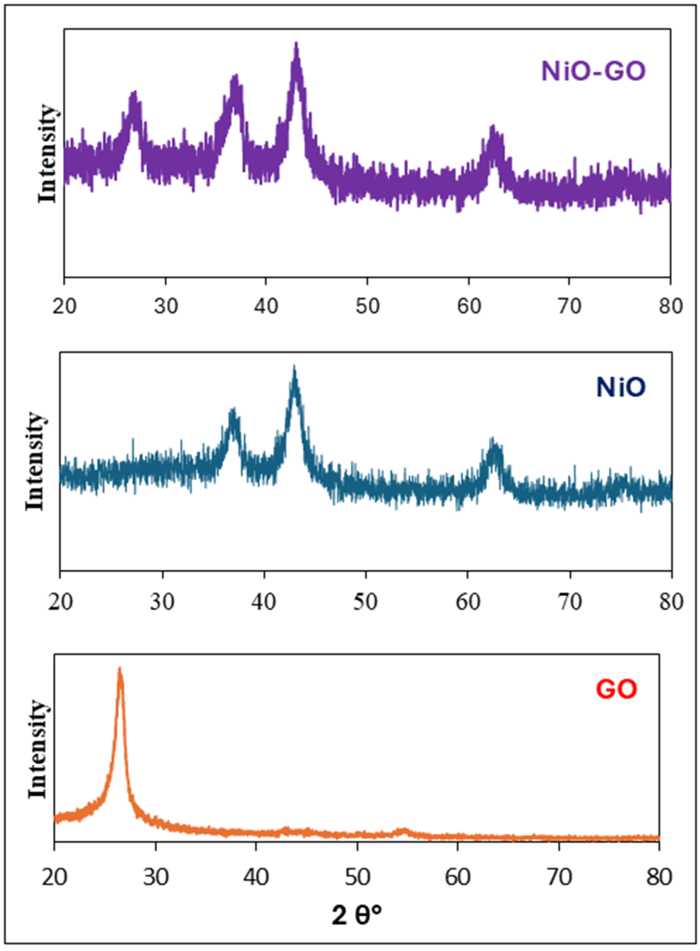
The purity and crystallinity of the NiO, GO, and NiO-GO composite were determined by XRD analysis. The XRD of GO shows a peak at about 26°, which is a representative peak of GO as given in Figure 5. This peak represents the 002 plane of GO [26,59]. The diffraction patterns of NiO exhibited reflection peaks located at diffraction angles (2θ) of approximately 37°, 42°, and 62°, as illustrated in Figure 3. These peaks represent the crystal planes 111, 200, and 220 of NiO, respectively. The diffraction peaks with high intensity represent the face-centered cubic unit of nickel oxide nanoparticles of strong crystallinity. The crystal shape of the NiO nanoparticles synthesized in the present study appeared to be accurately similar and comparable with the reported chart (JCPDS card no. 47–1049). The XRD of NiO-GO indicates peaks for both GO and NiO with a slight shift in peak positions. Hence, the XRD confirms the successful synthesis of NiO-GO [51,60]. Online Sherrer’s equation was used for the calculation of crystallite size. The crystallite size for GO, NiO, and NiO-GO was calculated as 4.1, 46.5, and 4.3 nm, respectively.

2.2. Evaluation of Photocatalytic Activity

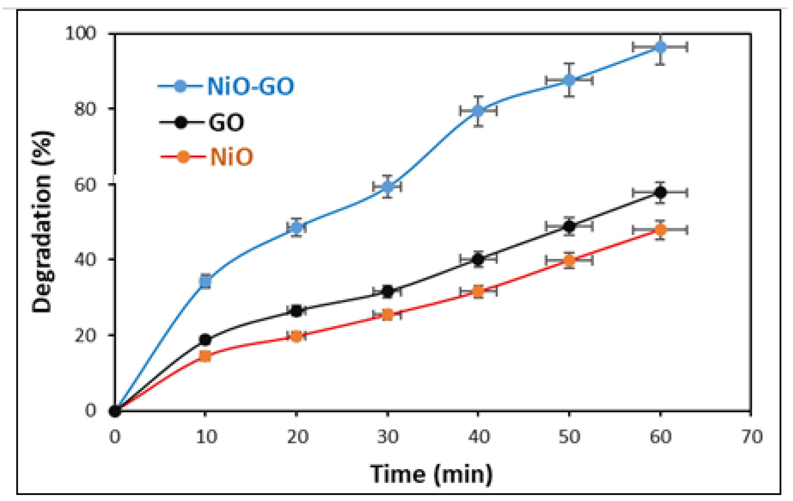
For comparison of photocatalytic activities of NiO, GO, and NiO-GO, separate degradation experiments of clothianidin were conducted in the presence of NiO, GO, and NiO-GO. The concentration of clothianidin was kept at 20 ppm, while the initial pH was kept at 3. The dose of each catalyst was 0.05 g. Before the addition of the catalyst, clothianidin solution was stirred under visible light for 30 min to determine the removal of clothianidin by photolysis. The analysis of the reaction mixture confirmed that there was no change in the concentration of clothianidin during stirring in visible light. This result shows that there is no removal or degradation of clothianidin due to photolysis. Afterwards, the removal of clothianidin by adsorption was also verified. It was performed by stirring the catalyst containing clothianidin solution for 30 min under dark conditions. About a 13% decrease in the concentration of clothianidin was observed by sorption at GO. Similarly, about a 20% decrease in concentration was noticed with each of NiO and NiO-GO. After confirmation of photolysis and adsorption, the photocatalytic degradation of clothianidin was initiated by stirring the reaction mixture under visible light irradiation. Samples were taken every 10 min and analyzed. The results obtained are given in Figure 6. The Figure exhibits that the degradation efficiency of NiO-GO composite is significantly higher than pristine NiO and GO under similar experimental conditions. The enhanced catalytic activity of NiO-GO is due to efficient separation of the photogenerated charge carriers. The error bars in Figure 6 represent the standard deviation, ensuring the reproducibility and reliability of the catalytic activity data. The relatively small size of the error bars indicates good consistency in the degradation experiments, suggesting minimal experimental fluctuations.
The degradation of clothianidin was confirmed by chemical oxygen demand (COD) measurement as well. For this purpose, a fresh solution of clothianidin (20 ppm, 30 mL) and a solution treated with NiO-GO for 60 min were used. About a 73% reduction in COD was observed. The decrease in COD confirms that NiO-GO acts as a photocatalyst in the degradation of clothianidin.
The Langmuir–Hinshelwood mechanism is generally used to describe the kinetics of heterogeneous catalytic reactions. The kinetics of the present study are also described by the same mechanism. Accordingly, the NiO-GO-catalyzed degradation of clothianidin proceeds in the following steps.
Adsorption of clothianidin on NiO-GO.
Harvesting of sunlight.
Degradation of clothianidin.
The rate of reaction is given by the following:
Rate = C clothianidin dt     Irradiation   C clothianidin
The continuous irradiation makes the reaction dependent on the concentration of clothianidin only. Hence, the following:
Rate = d C clothianidin dt   = k   C clothianidin
Rate = dC clothianidin C clothianidin = k   dt
On integration
ln [ C clothianidin ] o [ C clothianidin ] t = k   t
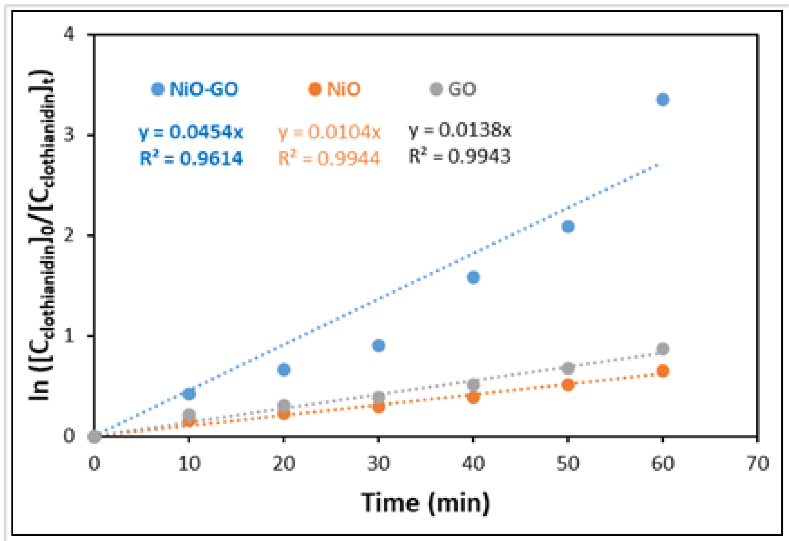
The degradation data was analyzed according to the kinetics Equation (6). Figure 7 shows the results of the kinetics analysis. The rate constants were determined as 0.0454, 0.0104, and 0.0138 min−1 for NiO-GO, NiO, and GO, respectively. The rate constant for the NiO-GO-catalyzed degradation of clothianidin was 4.4 and 3.3 times higher than the rate constant over NiO and GO, respectively.
The higher photocatalytic performance of NiO-GO is due to the enhanced separation of the charge carriers generated by absorption of sunlight. The absorption of sunlight generates charge carriers (positive holes and electrons) in the valence band and conduction band of NiO. The electrons quickly flow to the GO surface due to high electronegativity, favorable work function alignment, and high electron mobility of GO. The flow of electrons to the GO causes a separation of charges and prevents the recombination. The oxygen-containing functional groups on GO support the separation of charges by providing anchoring sites for NiO. The separated electrons react with dissolved oxygen and generate the superoxide anion radicals. Similarly, the positive holes generate hydroxyl radicals through reaction with water. The generated reactive species attacked the clothianidin molecules adsorbed on the NiO-GO surface, leading to mineralization into simple inorganic molecules. This whole process can be summarized as follows:
NiO GO + hv e CB NiO + h VB ( NiO ) +
e CB NiO e GO
e GO + O 2 O 2 ˙
h VB ( NiO ) + + H 2 O OH ˙ + H +
Clothianidin + ( O 2 , h + , OH ˙ ) Degradation   products ˙
The production of charge carriers was confirmed by scavenging tests. The scavenging tests were conducted using 2-propanol (֗OH scavenger), EDTA (h+ scavenger), and benzoquinone ( O 2 ˙ scavenger). About 24, 36, and 48% degradation of clothianidin was observed using NiO-GO as a photocatalyst in the presence of 2-propanol, EDTA, and benzoquinone, respectively. These observations suggest that electrons, holes, and hydroxyl radicals are the main photoactive species in the degradation of clothianidin [61].
The specific surface area plays a decisive role in determining the photocatalytic efficiency of a material because it directly influences the density of surface-active sites available for pollutant adsorption and reaction. In the present study, NiO-GO facilitates greater adsorption of clothianidin molecules before photodegradation due to its large surface area. A larger surface area also enhances the contact between the catalyst and incident photons, increasing light harvesting and the generation of electron–-hole pairs. Moreover, the GO sheets act as a high-surface-area support that prevents NiO nanoparticle agglomeration and ensures uniform dispersion, thereby exposing more active sites for redox reactions. Therefore, the superior catalytic performance of the NiO-GO composite can be attributed not only to the effective charge separation at the NiO-GO interface but also to its higher surface area, improved pollutant adsorption, and optimized light–matter interaction.
The photocatalytic activity of NiO-GO for the degradation of clothianidin was compared with other catalysts and adsorbents reported for the removal of clothianidin in the literature. Table 2 shows the comparison of the catalytic activity of the catalyst reported in this study with already reported catalysts and adsorbents. The comparative analysis shows that the NiO-GO composite developed in this study exhibits excellent photocatalytic efficiency toward clothianidin degradation under natural sunlight. The catalyst achieved 96% degradation of a 20 mg/L solution within 60 min, demonstrating performance comparable to or better than several reported photocatalysts operating under artificial light sources.

2.3. Effect of pH

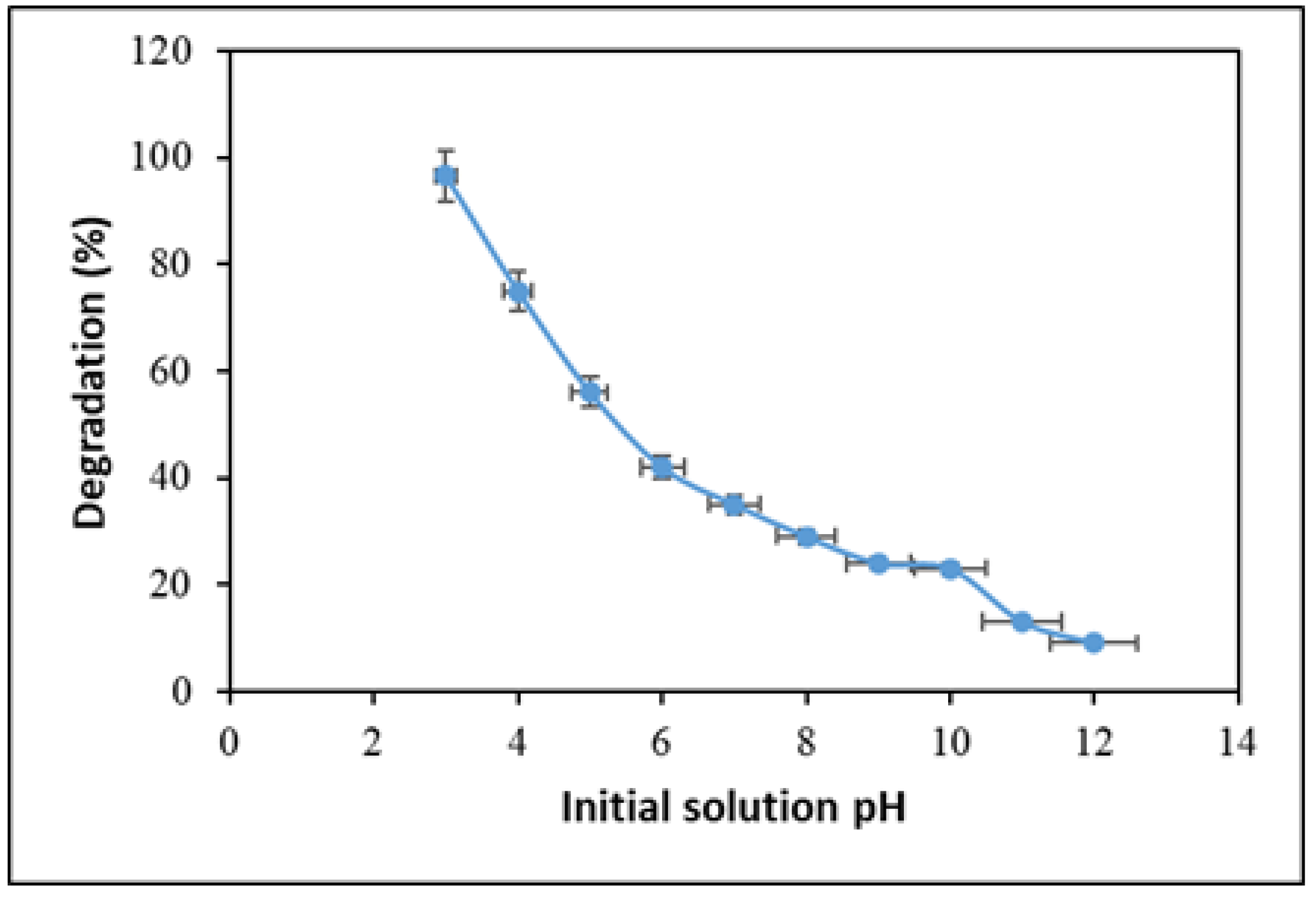
Solution pH is a vital agent for the photocatalytic degradation of pollutants in water, as industrial effluents have a complex nature. Hence, the impact of pH on the photocatalytic degradation of clothianidin by the NiO-GO composite was investigated by varying the pH in the range of 3–12 using a Britton–Robinson buffer. The concentration of clothianidin was maintained at 20 ppm, and the dose of NiO-GO composite was 0.05 g. The outcome is presented in Figure 8. The Figure exhibits that the degradation efficiency of NiO-GO decreases with an increase in pH of the solution, and maximum photocatalytic degradation (96%) was achieved at pH 3 within 60 min of reaction. The result implies that the acidic medium is more favorable for maximum degradation, which may be described by surface charges on the photocatalyst. The solution pH competes with the degradation of pesticides because it alters the surface charges on photocatalysts. Hence, charges on the surface are closely associated with the adsorption of pesticides before their photocatalytic oxidation. Based on the results obtained, it is suggested that the acidic medium favors higher degradation efficiency. Although zeta potential measurements were not performed in this study, it is hypothesized that at low pH, the surface of the NiO-GO composite may acquire a more positive charge, which could enhance the electrostatic interaction with the anionic clothianidin molecules (pKa = 11). This assumption aligns with previously reported studies on similar photocatalysts [70,71]. Hence, further photodegradation experiments of clothianidin were performed in an acidic medium (at pH 3).

2.4. Effect of Photocatalyst Loading

The catalyst dosage plays a critical role in evaluating the performance of photocatalytic degradation, as it directly affects the number of active sites available for the adsorption and subsequent degradation of pollutant molecules. In this study, the photocatalytic performance of the NiO-GO composite was examined using different catalyst doses from 0.1 to 1.0 g, while keeping the clothianidin concentration constant at 20 ppm and the reaction time at 60 min. The results obtained are given in Figure 9. The results confirm a distinct trend: the degradation performance increased with increasing catalyst dose up to 0.5 g, with a maximum of 96% degradation, beyond which a further increase in dosage resulted in a decrease in photocatalytic activity. The initial increase in degradation is due to the greater number of active sites and enhanced light absorption. Both of these factors facilitate the generation of electron–hole pairs and reactive oxygen species (ROS), which are essential for pollutant degradation. However, the observed decline in performance at higher doses may be due to several factors. First, excessive catalyst loading can cause scattering and shielding effects of light, which reduce the penetration of photons into the suspension and limit the activation of deeper catalyst layers. Second, agglomeration of catalyst particles may take place at higher doses and causes a decrease in available surface area, thereby lowering the number of accessible active sites. Third, the dense suspension is due to the high concentration of the catalyst, and the mass transfer of clothianidin molecules to the catalyst surface is hindered, further reducing degradation efficiency [72,73]. Similar trends have been reported in previous studies; therefore, it is of the utmost importance to optimize the catalyst dose to balance the trade-off between the availability of active sites and minimizing light attenuation effects. This finding highlights the importance of optimizing catalyst dosage for large-scale applications to ensure cost-effectiveness while maintaining high photocatalytic efficiency.

2.5. Effect of Initial Concentration

It is necessary to inspect the impact of the initial substrate concentration on photocatalytic degradation because the initial concentration of the substrate is an essential parameter and has a prominent effect on photocatalysis [74]. To find out the effect of initial clothianidin concentration on photodegradation, the initial concentration of clothianidin varied in the range of 20 to 100 ppm. The amount of NiO-GO composite (0.05 g) and pH (3) were kept constant. The results obtained are given in Figure 10. The data given in Figure 10 illustrates that there is an inverse relationship between the photodegradation efficiency of NiO-GO composite and the initial concentration of clothianidin. The degradation efficiency of the NiO-GO nanocomposite decreased from 96% to 62% with an increase in the initial concentration of clothianidin from 20 to 100 ppm. This outcome may be justified by using Beer–Lambert’s law, which states that as a concentration increases, the path length of incoming photons into the reaction mixture reduces, and a few photons might arrive at the surface of the photocatalyst. This implies that the efficiency of the NiO-GO composite declines by increasing the initial concentration of clothianidin [75,76,77].

2.6. Effect of Agitation Speed

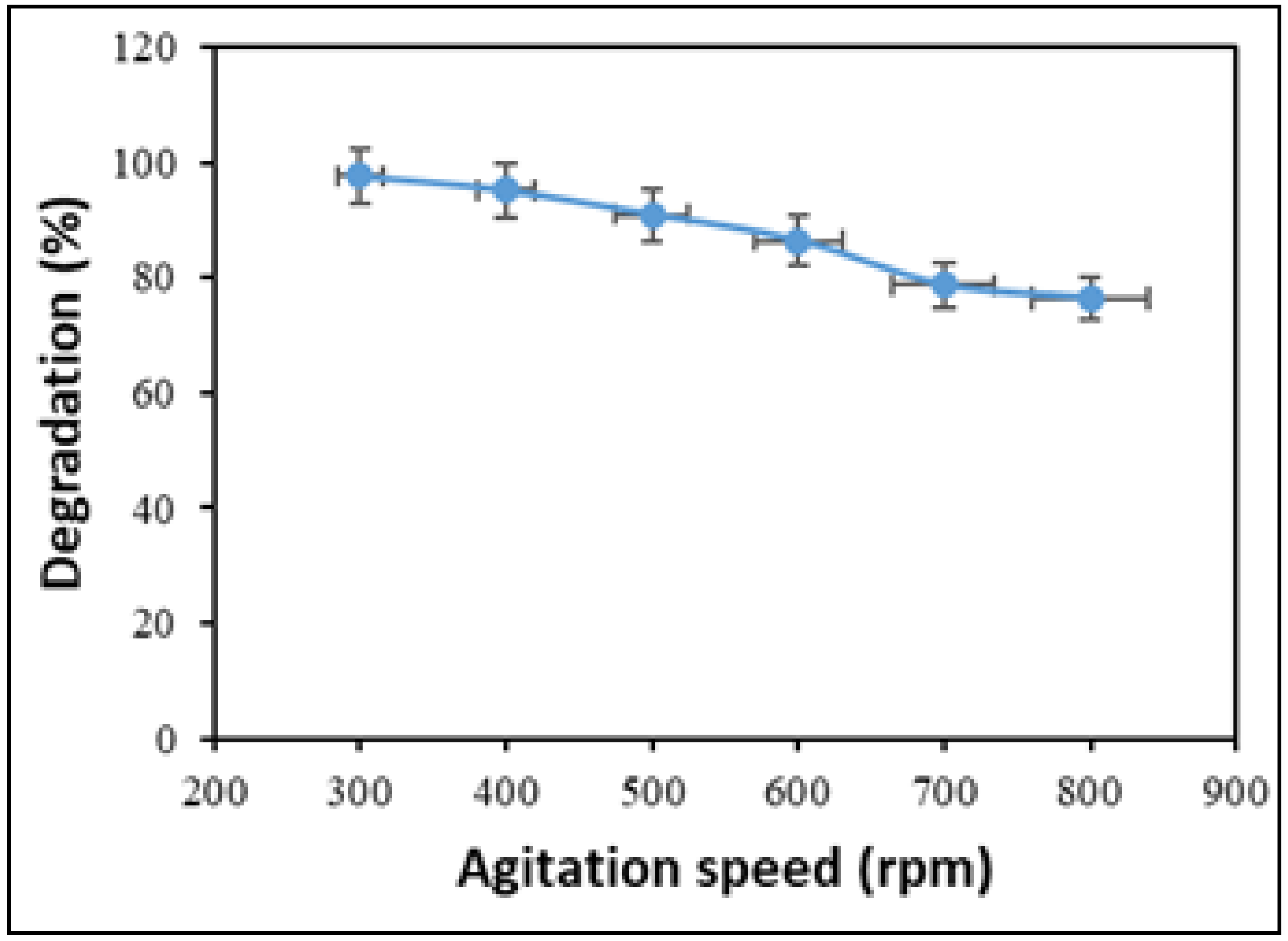
The agitation speed is a key variable in photocatalytic degradation because it plays a crucial role in promoting better interaction between pollutants and photocatalysts. Generally, increasing the agitation speed improves the efficiency of photocatalytic degradation, but in some cases, it can negatively affect the process. Therefore, the effect of agitation speed was studied to determine the optimal conditions by varying the stirring speed from 300 to 800 rpm, while keeping the amount of NiO-GO composite (0.05 g), pH (3), and contact time (60 min) constant. As shown in Figure 11, degradation efficiency steadily decreases as agitation speed increases. The decline in efficiency at higher agitation speeds may be due to increased turbulence in the solution, which could reduce the interaction between pollutant molecules and photocatalysts. Consequently, further photocatalytic degradation experiments on clothianidin were conducted at an agitation speed of 300 rpm [78,79].

3. Materials and Methods

3.1. Chemicals and Reagents

In this work chemicals and reagents utilized were of high purity. Sodium carbonate, hydrochloric acid, hydrogen peroxide, ethanol, methanol, potassium permanganate, phosphoric acid, and nickel nitrate were used without any further purification.

3.2. Instruments

The measurement of solution pH was performed by a pH meter (Tecpel Co., Ltd., New Taipei, Taiwan), while stirring of the mixtures was executed with the help of a hot plate magnetic stirrer (Sci Finetech Co., Seoul, Republic of Korea). The oven (Memmert + Co. KG, Schwabach, Germany) and centrifuge machine (Kaida manufacturer, Dongyang, China) were used for heating and centrifugation of the materials, respectively. The concentration of clothianidin was measured by a UV-Vis spectrophotometer (Hitachi Hi-tech Group, Tokyo, Japan), and ultrasonication was carried out by an ultrasonicator (Qsonica, Newtown, CT, USA, Q125). The morphological study of the constructed materials was executed with a scanning electron microscope (Capovani Brothers Inc., Scotia, NY, USA), while functional groups analysis of the materials was accomplished with Fourier Transform Infrared (Bruker Co., Billerica, MA, USA). X-ray diffraction (Bruker Co., Billerica, MA, USA) analysis was executed for examination of the phase structure and crystallography of the materials.

3.3. Collection and Preparation of Pea Peels

Fresh green peas were purchased from a local market near the Government College University Faisalabad, Pakistan. First, the seeds of the green peas were separated from the peels, and the peels were washed with tap water. Then, the materials were washed with distilled water numerous times and dried in daylight. The dried peels were crushed in an electric grinder, and the larger particles were powdered with a mortar and pestle. To obtain an aqueous extract of pea peels, about 10 g of the ground pea peels were taken in a beaker, and 100 mL of distilled water was added to it, and it was boiled for 30 min [80,81]. The pea peel extract was kept cool and filtered, and finally, it was stored for further study.

3.4. Preparation of NiO Nanoparticles

To prepare nanoparticles of NiO through green synthesis, a 180 mL solution of 0.1 M Ni(NO3)2.6H2O was transferred to an Erlenmeyer flask and agitated on a hot plate and magnetic stirrer for 30 min. A total of 20 mL of pea peel extract was then added drop by drop, and the mixture was agitated for 30 min at 100 °C with an agitation frequency of 4000 rpm. The pH of the flask’s content was maintained at 10 with 5.0 M NaOH drop-wise, and the resulting mixture was agitated for two hours at ambient temperature. As a result, the precipitate was obtained and washed repeatedly with distilled water and ethanol. The resulting product was transferred to a crucible and dried at 80 °C for two hours in an oven. In the end, the precipitate was calcined in a muffle furnace at 300 °C for 3–4 h, and the resultant product was kept in a sealed bottle for further study [82].
The pea peel extract performs a dual role in the synthesis of NiO, both as a reducing agent and a stabilizing (capping) agent, which makes the process an eco-friendly alternative to conventional chemical synthesis routes.
Pea peels contain a wide range of bioactive compounds, like polyphenols, flavonoids, proteins, and sugars. These compounds act as reducing agents for the reduction of Ni ions to Ni atoms. Flavonoids are a large group of polyphenolic compounds that can actively chelate metal ions and reduce them to form nanoparticles. Metal ions are captured and immobilized by biological elements and subsequently undergo reduction, sintering, and smelting processes, leading to the formation of nanoparticles. The formation of nanoparticles is primarily driven by the abundance of reactive functional groups, particularly carbonyl and multiple hydroxyl moieties. Consequently, the elevated levels of flavonoids and phenolic compounds in the aqueous extract facilitated the efficient reduction of Ni ions to Ni atoms. These Ni atoms form NiO on calcination. The bioactive compounds of pea peels replace hazardous chemical reducing agents, making the process safer and more sustainable [83].

3.5. Preparation of Graphene Oxide (GO)

The Hummer’s method was followed with some modifications to construct graphene oxide. A mixture of graphite (5.0 g), NaNO3 (2.5 g), and H2SO4 (300 mL) was taken in a beaker and agitated in an ice bath for 30 min. To this mixture, KMnO4 (30 g) was added gradually, and as a result, a purple-green mixture was obtained. The suspension was agitated magnetically for 2 h while the temperature was maintained up to 40 °C. As a result, a dark brown paste was obtained, and the mixture was diluted with the gradual addition of 200 mL of distilled water. The suspension was stirred for 30 min at 98 °C. A golden brown sol was obtained after the slow addition of 30 mL of hydrogen peroxide (30%) to the mixture. To the above mixture, 50 mL of distilled water was added, and the resulting product was washed with distilled water numerous times until the pH reached about 6. Lastly, the resultant product was dried at 80 °C in an oven for one day and stored in an airtight bottle for further study.

3.6. Preparation of NiO-GO Nanocomposite

Firstly, a dispersion of GO was prepared by taking an accurately weighed quantity of GO (1.0 g) in 100 mL of distilled water, and it was ultrasonicated for one hour. Then, the prepared NiO nanoparticles (1.0 g) were added slowly to the GO dispersion and ultrasonicated for a further two hours to ensure complete mixing. A grayish-black material was obtained and filtered through an ordinary filter paper. The resulting material was first washed with ethanol and, finally, rinsed thoroughly with distilled water many times to rid it of impurities and moisture content. The final product was dried in an oven at 50 °C for 12 h and stored in a sealed container for further study [30].

3.7. Photocatalytic Degradation Experiments

Photocatalytic analyses were performed for the degradation of clothianidin under daylight in the absence of any artificial light. In a typical run, 50 mL of clothianidin solution and a predetermined amount of catalyst were taken in a flask. The Britton–Robinson buffer solution was used for the adjustment of pH. The mixture was magnetically agitated under visible light. The degradation experiments in this study were conducted under natural sunlight during June–July in Faisalabad, Pakistan, where the average solar irradiance ranges from approximately 850–1000 W/m2, and the ambient temperature varied between 40 and 45 °C. The degradation experiments were typically performed between 10:00 a.m. and 3:00 p.m. on clear, sunny days to ensure consistent light intensity. We did not employ an artificial visible light lamp for this study; instead, the term “visible light” referred to the natural sunlight spectrum available during these months. Samples were taken at regular intervals of 10 min and analyzed using a UV-Visible spectrophotometer. The percentage degradation of clothianidin was determined using the following equation:
D e g r a d a t i o n % = C i C e C i × 100
Here, Ci and Ce represent the concentration (ppm) of clothianidin before and after the degradation study, respectively.
For the measurement of chemical oxygen demand (COD), a sample solution of 10 mL and 1 mL mercury sulfate solution was taken in a reflux flask. The mercury sulfate solution was prepared by adding 0.1 g HgSO4 to 5 mL of concentrated sulfuric acid. Then, potassium dichromate solution (6.13 g/L, 5 mL) and silver sulfate–sulfuric acid solution (10 g/L sulfuric acid, 15 mL) were added to the reflux flask, followed by refluxing for 2 h. Then, the reaction mixture was titrated with ferrous ammonium sulfate solution using ferroin indicator. The ferrous ammonium sulfate solution was prepared by taking 9.8 g of it in a solvent comprising 100 mL of distilled water and 20 mL of sulfuric acid. COD was calculated using the following equation:
C O D = 8 × 1000 × M ( V b V s ) V S a m p l e ( m L )
M: Molarity of ferrous ammonium sulfate solution.
Vb: Volume of ferrous ammonium sulfate solution consumed in the blank experiment.
Vs: Volume of ferrous ammonium sulfate solution consumed in a sample experiment.

4. Conclusions

In this study, NiO-GO composite was successfully synthesized, and its photocatalytic efficiency for the degradation of the persistent neonicotinoid pesticide clothianidin was systematically evaluated under natural sunlight. The synthesized NiO-GO demonstrated significantly enhanced catalytic activity compared to pristine NiO and GO, achieving 96% degradation within 60 min at pH 3 with a rate constant 4.4 and 3.3 times higher than NiO and GO, respectively. The superior performance of NiO-GO can be attributed to the synergistic role of graphene oxide in suppressing NiO agglomeration, enhancing light absorption, and facilitating charge carrier separation. The photocatalytic degradation followed pseudo-first-order kinetics. Reaction parameters such as solution pH, catalyst loading, initial pesticide concentration, and agitation speed were found to critically influence photocatalytic efficiency, with acidic pH and optimized catalyst dosage favoring maximum degradation. Overall, this work highlights the potential of NiO-GO composite as a sustainable, efficient, and cost-effective photocatalyst for the remediation of hazardous pesticides in aqueous environments. The use of agricultural waste (pea peels) in catalyst synthesis not only aligns with green chemistry principles but also adds value to biomass resources within a circular economy framework. Although intermediate identification was not performed, future studies will focus on the identification of degradation intermediates and toxicity assessment to ensure the environmental safety of the process. Future studies may focus on real wastewater applications and scale-up feasibility to further validate its practical applicability in environmental remediation.

Author Contributions

Conceptualization, A.u.H. and M.S.; methodology, A.u.H. and M.S.; validation, A.u.H. and M.S.; formal analysis, H.M.A.; investigation, H.M.A.; resources, R.K.H.; data curation, M.S.; writing—original draft preparation, A.u.H. and M.S.; writing—review and editing, S.P. and S.A.A.; supervision, A.u.H.; project administration, A.u.H. and M.S.; funding acquisition, R.K.H. All authors have read and agreed to the published version of the manuscript.

Funding

This work was funded by the Deanship of Scientific Research at Imam Mohammad Ibn Saud Islamic University (IMSIU) (grant number IMSIU-DDRSP2503).

Data Availability Statement

The original contributions presented in this study are included in the article.

Conflicts of Interest

The authors declare no conflicts of interest.

References

  1. Li, B.; Wang, X.; Khurshid, A.; Saleem, S.F. Environmental Governance, Green Finance, and Mitigation Technologies: Pathways to Carbon Neutrality in European Industrial Economies. Int. J. Environ. Sci. Technol. 2025, 22, 14899–14912. [Google Scholar] [CrossRef]
  2. Wang, X.; Su, H.; Liu, X. The Impact of Green Technological Innovation on Industrial Structural Optimization Under Dual-Carbon Targets: The Role of the Moderating Effect of Carbon Emission Efficiency. Sustainability 2025, 17, 6313. [Google Scholar] [CrossRef]
  3. Fang, Q.; Sun, Q.; Ge, J.; Wang, H.; Qi, J. Multidimensional Engineering of Nanoconfined Catalysis: Frontiers in Carbon-Based Energy Conversion and Utilization. Catalysts 2025, 15, 477. [Google Scholar] [CrossRef]
  4. Lu, S.; Shen, C.; Liu, M.; Zhang, J.; Fang, M.; Wang, F.; Li, Q.; Li, C.; Zhang, J. Research and Application Analysis on the Pollution Control of Tail Gas Emissions from CO2 Capture Absorber. Results Eng. 2025, 25, 103677. [Google Scholar] [CrossRef]
  5. Lu, S.; Zhang, Y.-J.; Cheng, Y.-J.; Qin, Z.-H.; Wang, G.-D.; Bai, Y.; Lin, Y.; Wang, H.; Sui, Y.; Hou, L.; et al. Capture of CO2 from N2 and CH4 over a Wide Temperature Range on a Robust MOF with Brønsted Acidic and Lewis Basic Dual Functional Sites. J. Mater. Chem. A Mater. 2025, 13, 10581–10589. [Google Scholar] [CrossRef]
  6. Su, H.; Zio, E.; Zhang, J.; Li, Z.; Wang, H.; Zhang, F.; Chi, L.; Fan, L.; Wang, W. A Systematic Method for the Analysis of Energy Supply Reliability in Complex Integrated Energy Systems Considering Uncertainties of Renewable Energies, Demands and Operations. J. Clean. Prod. 2020, 267, 122117. [Google Scholar] [CrossRef]
  7. Su, H.; Zio, E.; Zhang, J. A Method for the Risk Analysis of Energy Supply in Integrated Energy Systems. In Proceedings of the 30th European Safety and Reliability Conference and 15th Probabilistic Safety Assessment and Management Conference, Venice, Italy, 1–5 November 2020; Research Publishing Services: Singapore, 2020; pp. 1829–1835. [Google Scholar]
  8. Sha, X.; Zhu, Y.; Sha, X.; Guan, Z.; Wang, S. ZHPO-LightXBoost an Integrated Prediction Model Based on Small Samples for Pesticide Residues in Crops. Environ. Model. Softw. 2025, 188, 106440. [Google Scholar] [CrossRef]
  9. Das, R.; Vecitis, C.D.; Schulze, A.; Cao, B.; Ismail, A.F.; Lu, X.; Chen, J.; Ramakrishna, S. Recent Advances in Nanomaterials for Water Protection and Monitoring. Chem. Soc. Rev. 2017, 46, 6946–7020. [Google Scholar] [CrossRef]
  10. Xu, C.; Chen, L.; You, L.; Xu, Z.; Ren, L.-F.; Yew-Hoong Gin, K.; He, Y.; Kai, W. Occurrence, Impact Variables and Potential Risk of PPCPs and Pesticides in a Drinking Water Reservoir and Related Drinking Water Treatment Plants in the Yangtze Estuary. Environ. Sci. Process Impacts 2018, 20, 1030–1045. [Google Scholar] [CrossRef]
  11. Zhang, Y.; Xu, L.; Wang, J.; Pan, H.; Dou, M.; Teng, Y.; Fu, X.; Liu, Z.; Huang, X.; Wang, M. Bagasse-Based Porous Flower-like MoS2/Carbon Composites for Efficient Microwave Absorption. Carbon Lett. 2025, 35, 145–160. [Google Scholar] [CrossRef]
  12. Jeschke, P.; Nauen, R.; Schindler, M.; Elbert, A. Overview of the Status and Global Strategy for Neonicotinoids. J. Agric. Food Chem. 2011, 59, 2897–2908. [Google Scholar] [CrossRef] [PubMed]
  13. Uneme, H. Chemistry of Clothianidin and Related Compounds. J. Agric. Food Chem. 2011, 59, 2932–2937. [Google Scholar] [CrossRef] [PubMed]
  14. Jeschke, P.; Nauen, R. Neonicotinoids—From Zero to Hero in Insecticide Chemistry. Pest. Manag. Sci. 2008, 64, 1084–1098. [Google Scholar] [CrossRef] [PubMed]
  15. Hirano, T.; Yanai, S.; Takada, T.; Yoneda, N.; Omotehara, T.; Kubota, N.; Minami, K.; Yamamoto, A.; Mantani, Y.; Yokoyama, T.; et al. NOAEL-Dose of a Neonicotinoid Pesticide, Clothianidin, Acutely Induce Anxiety-Related Behavior with Human-Audible Vocalizations in Male Mice in a Novel Environment. Toxicol. Lett. 2018, 282, 57–63. [Google Scholar] [CrossRef]
  16. Mori, T.; Wang, J.; Tanaka, Y.; Nagai, K.; Kawagishi, H.; Hirai, H. Bioremediation of the Neonicotinoid Insecticide Clothianidin by the White-Rot Fungus Phanerochaete sordida. J. Hazard. Mater. 2017, 321, 586–590. [Google Scholar] [CrossRef]
  17. Goulson, D. REVIEW: An Overview of the Environmental Risks Posed by Neonicotinoid Insecticides. J. Appl. Ecol. 2013, 50, 977–987. [Google Scholar] [CrossRef]
  18. Kim, B.M.; Park, J.-S.; Choi, J.-H.; Abd El-Aty, A.M.; Na, T.W.; Shim, J.-H. Residual Determination of Clothianidin and Its Metabolites in Three Minor Crops via Tandem Mass Spectrometry. Food Chem. 2012, 131, 1546–1551. [Google Scholar] [CrossRef]
  19. Sánchez-Bayo, F.; Hyne, R.V. Detection and Analysis of Neonicotinoids in River Waters—Development of a Passive Sampler for Three Commonly Used Insecticides. Chemosphere 2014, 99, 143–151. [Google Scholar] [CrossRef]
  20. de Perre, C.; Murphy, T.M.; Lydy, M.J. Fate and Effects of Clothianidin in Fields Using Conservation Practices. Environ. Toxicol. Chem. 2014, 34, 258–265. [Google Scholar] [CrossRef]
  21. Yang, K.; Li, C.; Zhu, Q.; Wang, H.; Qi, J. Rich Oxygen Vacancies in Bimetallic MnCo2O4.5 Spheres for Enhancing Lean Methane Catalytic Oxidation. Nanomaterials 2025, 15, 524. [Google Scholar] [CrossRef]
  22. Hou, B.; Zhao, Y.; Sun, W.; Lu, S.; Chen, S. Glycine Based Modification of Activated Carbons for VOCs Adsorption. Chem. Eng. J. Adv. 2021, 7, 100126. [Google Scholar] [CrossRef]
  23. Du, H.; Zhang, A.; Zhang, Q.; Sun, Y.; Zhu, H.; Wang, H.; Tan, Z.; Zhang, X.; Chen, G. Fabrication of Recoverable Bi2O2S/Bi5O7I/ZA Hydrogel Beads for Enhanced Photocatalytic Hg0 Removal in the Presence of H2O2. Sep. Purif. Technol. 2025, 359, 130597. [Google Scholar] [CrossRef]
  24. Jia, M.; Yang, Z.; Xu, H.; Song, P.; Xiong, W.; Cao, J.; Zhang, Y.; Xiang, Y.; Hu, J.; Zhou, C.; et al. Integrating N and F Co-Doped TiO2 Nanotubes with ZIF-8 as Photoelectrode for Enhanced Photo-Electrocatalytic Degradation of Sulfamethazine. Chem. Eng. J. 2020, 388, 124388. [Google Scholar] [CrossRef]
  25. Siddique, M.; Khan, N.M.; Saeed, M.; Ali, S.; Shah, Z. Green Synthesis of Cobalt Oxide Nanoparticles Using Citrus Medica Leaves Extract: Characterization and Photo-Catalytic Activity. Z. Für Phys. Chem. 2021, 235, 663–681. [Google Scholar] [CrossRef]
  26. Pervaiz, S.; Saeed, M.; Habila, M.A.; Jamal, M.A.; Haq, A.U.; Khan, I.; Javed, M. Synthesis and Photocatalytic Performance of G-C3N4-CuO-ZnO for Efficient Degradation of Crystal Violet (CV) and Methyl Orange (MO) Dyes under Sunlight Irradiation. Prog. React. Kinet. Mech. 2025, 50, e010. [Google Scholar] [CrossRef]
  27. Saeed, M.; Asghar, H.; Khan, I.; Akram, N.; Usman, M. Synthesis of TiO2-g-C3N4 for Efficient Photocatalytic Degradation of Congo Red Dye. Catal. Today 2025, 447, 115154. [Google Scholar] [CrossRef]
  28. You, Z.; Lu, D.; Kondamareddy, K.K.; Gu, W.; Su, Y.; Pan, J.; Yang, J.; Cheng, P.; Ho, W. Organic-Inorganic PVDF/Zn0.5Cd0.5S/Ti-NT Photocatalytic Membrane with Synergistic Effect of Heterojunction and Its Derived Multi-Element Excitation NO Purification. Sep. Purif. Technol. 2025, 361, 131293. [Google Scholar] [CrossRef]
  29. Zeng, X.; Zhang, A.; Liu, C.; Cheng, J.; Hu, M. Helical Polyether-Immobilized Chiral Aza-Bis(Oxazolines): Synthesis and Synergistic Effect on the Enantioselectivity of Zn-Catalyzed Henry Reaction. Eur. Polym. J. 2023, 194, 112160. [Google Scholar] [CrossRef]
  30. Guo, L.; Chen, W.; Wang, C.; Dong, B. Application of Electrochemically Assisted Synthesis of MOFs-Derived Phosphides as Catalyst for CH4-CO2 Reforming. Int. J. Electrochem. Sci. 2023, 18, 26–32. [Google Scholar] [CrossRef]
  31. Wang, J.; Zhang, J.; Cheng, G.; Zhang, K.; Liu, X. Performance and Mechanism of Tetracycline Removal by Peroxymonosulfate-Assisted Double Z-Scheme LaFeO3/g-C3N4/ZnO Heterojunction under Visible Light Drive. Environ. Technol. Innov. 2025, 39, 104302. [Google Scholar] [CrossRef]
  32. Hussain, S.; Khan, A.J.; Arshad, M.; Javed, M.S.; Ahmad, A.; Shah, S.S.A.; Khan, M.R.; Akram, S.; Zulfiqar; Ali, S.; et al. Charge Storage in Binder-Free 2D-Hexagonal CoMoO4 Nanosheets as a Redox Active Material for Pseudocapacitors. Ceram. Int. 2021, 47, 8659–8667. [Google Scholar] [CrossRef]
  33. Hussain, S.; Yang, X.; Aslam, M.K.; Shaheen, A.; Javed, M.S.; Aslam, N.; Aslam, B.; Liu, G.; Qiao, G. Robust TiN Nanoparticles Polysulfide Anchor for Li–S Storage and Diffusion Pathways Using First Principle Calculations. Chem. Eng. J. 2020, 391, 123595. [Google Scholar] [CrossRef]
  34. DeVree, B.T.; Steiner, L.M.; Głazowska, S.; Ruhnow, F.; Herburger, K.; Persson, S.; Mravec, J. Current and Future Advances in Fluorescence-Based Visualization of Plant Cell Wall Components and Cell Wall Biosynthetic Machineries. Biotechnol. Biofuels 2021, 14, 78. [Google Scholar] [CrossRef]
  35. Eray, E.; Candelario, V.M.; Boffa, V.; Safafar, H.; Østedgaard-Munck, D.N.; Zahrtmann, N.; Kadrispahic, H.; Jørgensen, M.K. A Roadmap for the Development and Applications of Silicon Carbide Membranes for Liquid Filtration: Recent Advancements, Challenges, and Perspectives. Chem. Eng. J. 2021, 414, 128826. [Google Scholar] [CrossRef]
  36. Feng, G.; Jiang, F.; Hu, Z.; Jiang, W.; Liu, J.; Zhang, Q.; Wu, Q.; Hu, Q.; Miao, L.; Cheng, S. A Novel Porous Egg-White (EW)/Titania Composite Photocatalytic Material for Efficient Photodegradation Applications. RSC Adv. 2020, 10, 8525–8529. [Google Scholar] [CrossRef]
  37. Pérez-Molina, Á.; Morales-Torres, S.; Maldonado-Hódar, F.; Pastrana-Martínez, L. Functionalized Graphene Derivatives and TiO2 for High Visible Light Photodegradation of Azo Dyes. Nanomaterials 2020, 10, 1106. [Google Scholar] [CrossRef]
  38. Liu, W.M.; Li, J.; Zhang, H.Y. Reduced Graphene Oxide Modified Zinc Oxide Composites Synergistic Photocatalytic Activity under Visible Light Irradiation. Optik 2020, 207, 163778. [Google Scholar] [CrossRef]
  39. Ashfaq, M.; Ali, A.; Abbood, N.K.; Panchal, S.; Akram, N.; Saeed, M.; Doshi, O.P.; Ali, F.; Muhammad, S.; Sameeh, M.Y.; et al. Enhanced Photocatalytic Activity of the Bi2O3-NiO Heterojunction for the Degradation of Methyl Orange under Irradiation of Sunlight. Water 2023, 15, 3182. [Google Scholar] [CrossRef]
  40. Saeed, M.; Albalawi, K.; Khan, I.; Akram, N.; Abd El-Rahim, I.H.A.; Alhag, S.K.; Ezzat Ahmed, A. Faiza Synthesis of P-n NiO-ZnO Heterojunction for Photodegradation of Crystal Violet Dye. Alex. Eng. J. 2023, 65, 561–574. [Google Scholar] [CrossRef]
  41. Zhao, W.; Yan, A.; Su, Z.; Huang, F.; Wang, Q.; Li, S.; Lu, S.; Wang, C.; Zhang, T.; Zhang, J.; et al. Multiobjective-Optimization MoS2/Cd-ZnIn2S4 /CdS Composites Prepared by In Situ Structure-Tailored Technique for High-Efficiency Hydrogen Generation. Small Struct. 2024, 5, 2300569. [Google Scholar] [CrossRef]
  42. Wang, X.; Zhao, B.; Liang, J.; Liu, G.; Guo, Z.; Hao, H.; Li, W.; Shen, W. Defect-Engineered RGO−CoNi2S4 with Enhanced Electrochemical Performance for Asymmetric Supercapacitor. Trans. Nonferrous Met. Soc. China 2025, 35, 563–578. [Google Scholar] [CrossRef]
  43. Pham, C.V.; Repp, S.; Thomann, R.; Krueger, M.; Weber, S.; Erdem, E. Charge Transfer and Surface Defect Healing within ZnO Nanoparticle Decorated Graphene Hybrid Materials. Nanoscale 2016, 8, 9682–9687. [Google Scholar] [CrossRef]
  44. Sampaio, M.J.; Bacsa, R.R.; Benyounes, A.; Axet, R.; Serp, P.; Silva, C.G.; Silva, A.M.T.; Faria, J.L. Synergistic Effect between Carbon Nanomaterials and ZnO for Photocatalytic Water Decontamination. J. Catal. 2015, 331, 172–180. [Google Scholar] [CrossRef]
  45. Sadhukhan, S.; Bhattacharyya, A.; Rana, D.; Ghosh, T.K.; Orasugh, J.T.; Khatua, S.; Acharya, K.; Chattopadhyay, D. Synthesis of RGO/NiO Nanocomposites Adopting a Green Approach and Its Photocatalytic and Antibacterial Properties. Mater. Chem. Phys. 2020, 247, 122906. [Google Scholar] [CrossRef]
  46. Al-Rawashdeh, N.A.F.; Allabadi, O.; Aljarrah, M.T. Photocatalytic Activity of Graphene Oxide/Zinc Oxide Nanocomposites with Embedded Metal Nanoparticles for the Degradation of Organic Dyes. ACS Omega 2020, 5, 28046–28055. [Google Scholar] [CrossRef] [PubMed]
  47. Pu, Q.; Zou, J.; Wang, J.; Lu, S.; Ning, P.; Huang, L.; Wang, Q. Systematic Study of Dynamic CO2 Adsorption on Activated Carbons Derived from Different Biomass. J. Alloys Compd. 2021, 887, 161406. [Google Scholar] [CrossRef]
  48. Ramu, A.G.; Umar, A.; Ibrahim, A.A.; Algadi, H.; Ibrahim, Y.S.A.; Wang, Y.; Hanafiah, M.M.; Shanmugam, P.; Choi, D. Synthesis of Porous 2D Layered Nickel Oxide-Reduced Graphene Oxide (NiO-RGO) Hybrid Composite for the Efficient Electrochemical Detection of Epinephrine in Biological Fluid. Environ. Res. 2021, 200, 111366. [Google Scholar] [CrossRef]
  49. Lamba, P.; Singh, P.; Singh, P.; Kumar, A.; Singh, P.; Bharti; Kumar, Y.; Gupta, M. Simple and Rapid Eco-Friendly Synthesis of NiO/RGO Nanocomposites Using Guava Leaf Extract and Their Physicochemical Characterization. Mater. Today Proc. 2022, 68, 2705–2714. [Google Scholar] [CrossRef]
  50. Rabchinskii, M.K.; Dideikin, A.T.; Kirilenko, D.A.; Baidakova, M.V.; Shnitov, V.V.; Roth, F.; Konyakhin, S.V.; Besedina, N.A.; Pavlov, S.I.; Kuricyn, R.A.; et al. Facile Reduction of Graphene Oxide Suspensions and Films Using Glass Wafers. Sci. Rep. 2018, 8, 14154. [Google Scholar] [CrossRef]
  51. Song, R.; Zhang, H.; Lv, J. NiO Nanoparticle-Decorated Graphene Oxide Nanosheets Modified Glassy Carbon Electrode for Sensitive Electrochemical Detection of Pethidine. Int. J. Electrochem. Sci. 2023, 18, 100221. [Google Scholar] [CrossRef]
  52. Gao, X.; Dai, Y.; Zhang, C.; Zhang, Y.; Zong, W.; Zhang, W.; Chen, R.; Zhu, J.; Hu, X.; Wang, M.; et al. When It’s Heavier: Interfacial and Solvation Chemistry of Isotopes in Aqueous Electrolytes for Zn-ion Batteries. Angew. Chem. Int. Ed. 2023, 62, e202300608. [Google Scholar] [CrossRef]
  53. Khairnar, S.D.; Shrivastava, V.S. Facile Synthesis of Nickel Oxide Nanoparticles for the Degradation of Methylene Blue and Rhodamine B Dye: A Comparative Study. J. Taibah Univ. Sci. 2019, 13, 1108–1118. [Google Scholar] [CrossRef]
  54. Zhou, H.; Guo, J.; Zhu, G.; Xie, F.; Tang, X.; Luo, X. Highly Efficient Preparation of Crystalline Yttrium Carbonate in Sodium Carbonate System: Formation and Growth Mechanism. J. Rare Earths 2025, 43, 1492–1501. [Google Scholar] [CrossRef]
  55. Zhou, H.; Guo, J.; Zhu, G.; Xu, H.; Tang, X.; Luo, X. Flotation Behavior and Mechanism of Smithsonite under the System of Bidentate Ligand Sulfide Sodium Thiocyanate. Sep. Purif. Technol. 2024, 334, 126086. [Google Scholar] [CrossRef]
  56. Emran, M.Y.; Khalifa, H.; Gomaa, H.; Shenashen, M.A.; Akhtar, N.; Mekawy, M.; Faheem, A.; El-Safty, S.A. Hierarchical C-N Doped NiO with Dual-Head Echinop Flowers for Ultrasensitive Monitoring of Epinephrine in Human Blood Serum. Microchim. Acta 2017, 184, 4553–4562. [Google Scholar] [CrossRef]
  57. Ngo, Y.-L.T.; Hur, S.H. Low-Temperature NO2 Gas Sensor Fabricated with NiO and Reduced Graphene Oxide Hybrid Structure. Mater. Res. Bull. 2016, 84, 168–176. [Google Scholar] [CrossRef]
  58. Anirudhan, T.S.; Manjusha, V.; Shainy, F. Magnetically Retrievable Cysteine Modified Graphene Oxide@nickelferrite@titanium Dioxide Photocatalyst for the Effective Degradation of Chlorpyrifos from Aqueous Solutions. Environ. Technol. Innov. 2021, 23, 101633. [Google Scholar] [CrossRef]
  59. Anjum, F.; Asiri, A.M.; Khan, M.A.; Khan, M.I.; Khan, S.B.; Akhtar, K.; Bakhsh, E.M.; Alamry, K.A.; Alfifi, S.Y.; Chakraborty, S. Photo-Degradation, Thermodynamic and Kinetic Study of Carcinogenic Dyes via Zinc Oxide/Graphene Oxide Nanocomposites. J. Mater. Res. Technol. 2021, 15, 3171–3191. [Google Scholar] [CrossRef]
  60. Maruthupandy, M.; Muneeswaran, T.; Vennila, T.; Vaishali, C.V.; Anand, M.; Cho, W.-S.; Quero, F. Photocatalytic Efficiency of Graphene/Nickel Oxide Nanocomposites towards the Degradation of Anionic and Cationic Dye Molecules under Visible Light. J. Photochem. Photobiol. A Chem. 2022, 427, 113819. [Google Scholar] [CrossRef]
  61. Alsalmah, H.A.; Aadil, M.; Zulfiqar, S. Hydrothermal–Ultrasonication-Assisted Fabrication of Ce-Doped ZnO/g-C3N4 Heterojunctions for Enhanced Visible-Light Degradation of Dye and Drug Pollutants. Ceram. Int. 2025, 51, 46808–46818. [Google Scholar] [CrossRef]
  62. Mishra, S.R.; Gadore, V.; Singh, K.R.B.; Pandey, S.S.; Ahmaruzzaman, M. Developing In2S3 upon Modified MgTiO3 Anchored on Nitrogen-Doped CNT for Sustainable Sensing and Removal of Toxic Insecticide Clothianidin. Environ. Res. 2024, 259, 119435. [Google Scholar] [CrossRef]
  63. Duan, P.; Qi, Y.; Feng, S.; Peng, X.; Wang, W.; Yue, Y.; Shang, Y.; Li, Y.; Gao, B.; Xu, X. Enhanced Degradation of Clothianidin in Peroxymonosulfate/Catalyst System via Core-Shell FeMn @ N-C and Phosphate Surrounding. Appl. Catal. B 2020, 267, 118717. [Google Scholar] [CrossRef]
  64. de la Flor, M.P.; Camarillo, R.; Martínez, F.; Jiménez, C.; Quiles, R.; Rincón, J. Synthesis and Characterization of Bimetallic TiO2/CNT/Pd-Cu for Efficient Remediation of Endocrine Disruptors under Solar Light. J. Environ. Chem. Eng. 2022, 10, 107245. [Google Scholar] [CrossRef]
  65. Mohanta, D.; Ahmaruzzaman, M. A Novel Au-SnO2-RGO Ternary Nanoheterojunction Catalyst for UV-LED Induced Photocatalytic Degradation of Clothianidin: Identification of Reactive Intermediates, Degradation Pathway and in-Depth Mechanistic Insight. J. Hazard. Mater. 2020, 397, 122685. [Google Scholar] [CrossRef] [PubMed]
  66. de la Flor, M.P.; Camarillo, R.; Martínez, F.; Jiménez, C.; Quiles, R.; Rincón, J. Synthesis and Characterization of TiO2/CNT/Pd: An Effective Sunlight Photocatalyst for Neonicotinoids Degradation. J. Environ. Chem. Eng. 2021, 9, 106278. [Google Scholar] [CrossRef]
  67. Žabar, R.; Komel, T.; Fabjan, J.; Kralj, M.B.; Trebše, P. Photocatalytic Degradation with Immobilised TiO2 of Three Selected Neonicotinoid Insecticides: Imidacloprid, Thiamethoxam and Clothianidin. Chemosphere 2012, 89, 293–301. [Google Scholar] [CrossRef]
  68. Radwan, I.M.; Wang, C.; Kim, J.-H.; Wei, H.; Wang, D. Sorptive Removal of Neonicotinoid Pesticides by Nanobiochars: Efficiency, Kinetics, and Reusability. J. Hazard. Mater. 2025, 493, 138354. [Google Scholar] [CrossRef]
  69. Satheeshkumar, A.; Duraimurugan, R.; Velu, P.; Muthukumar, B.; Santhosh, S.; Devanesan, S.; AlSalhi, M.S.; Sathishkumar, K.; Rahman, P.K.; Rajasekar, A. Biodegradation Pathway and Mechanism of New Emerging Pollutants Neonicotinoids. J. Environ. Sci. 2026, 160, 197–205. [Google Scholar] [CrossRef]
  70. Rahimi, B.; Jafari, N.; Abdolahnejad, A.; Farrokhzadeh, H.; Ebrahimi, A. Application of Efficient Photocatalytic Process Using a Novel BiVO/TiO2-NaY Zeolite Composite for Removal of Acid Orange 10 Dye in Aqueous Solutions: Modeling by Response Surface Methodology (RSM). J. Environ. Chem. Eng. 2019, 7, 103253. [Google Scholar] [CrossRef]
  71. Bruckmann, F.S.; Schnorr, C.; Oviedo, L.R.; Knani, S.; Silva, L.F.O.; Silva, W.L.; Dotto, G.L.; Bohn Rhoden, C.R. Adsorption and Photocatalytic Degradation of Pesticides into Nanocomposites: A Review. Molecules 2022, 27, 6261. [Google Scholar] [CrossRef]
  72. Xiao, Q.; Zhang, J.; Xiao, C.; Si, Z.; Tan, X. Solar Photocatalytic Degradation of Methylene Blue in Carbon-Doped TiO2 Nanoparticles Suspension. Sol. Energy 2008, 82, 706–713. [Google Scholar] [CrossRef]
  73. Pardeshi, S.K.; Patil, A.B. A Simple Route for Photocatalytic Degradation of Phenol in Aqueous Zinc Oxide Suspension Using Solar Energy. Sol. Energy 2008, 82, 700–705. [Google Scholar] [CrossRef]
  74. Hanafi, M.F.; Sapawe, N. Effect of Initial Concentration on the Photocatalytic Degradation of Remazol Brilliant Blue Dye Using Nickel Catalyst. Mater. Today Proc. 2020, 31, 318–320. [Google Scholar] [CrossRef]
  75. Ajiboye, T.O.; Oyewo, O.A.; Ben Smida, Y.; Onwudiwe, D.C. Visible Light-Driven Photocatalytic Reduction of Monovalent Silver Using a Composite of Ni3Bi2S2 and O-Doped GC3N4. Results Eng. 2022, 15, 100540. [Google Scholar] [CrossRef]
  76. Onwudiwe, D.C.; Gobile, N.; Oyewo, O.A.; Makgato, S.S. Photocatalytic Reduction of Hexavalent Chromium Using Zn2SnO4–ZnO Modified g-C3N4 Composite. Results Eng. 2023, 20, 101521. [Google Scholar] [CrossRef]
  77. Alwared, A.I.; Sulaiman, F.A.; Raad, H.; Al-Musawi, T.J.; Mohammed, N.A. Ability of FeNi3/SiO2/TiO2 Nanocomposite to Degrade Amoxicillin in Wastewater Samples in Solar Light-Driven Processes. S. Afr. J. Bot. 2023, 153, 195–202. [Google Scholar] [CrossRef]
  78. Ghasemi, A.H.; Zoqi, M.J.; Zanganeh Ranjbar, P. Enhanced Photocatalytic Degradation of Methylene Blue Using a Novel Counter-Rotating Disc Reactor. Front. Chem. 2024, 12, 1335180. [Google Scholar] [CrossRef]
  79. Haleem, A.; Shafiq, A.; Chen, S.-Q.; Nazar, M. A Comprehensive Review on Adsorption, Photocatalytic and Chemical Degradation of Dyes and Nitro-Compounds over Different Kinds of Porous and Composite Materials. Molecules 2023, 28, 1081. [Google Scholar] [CrossRef]
  80. Ezhilarasi, A.A.; Vijaya, J.J.; Kaviyarasu, K.; Zhang, X.; Kennedy, L.J. Green Synthesis of Nickel Oxide Nanoparticles Using Solanum Trilobatum Extract for Cytotoxicity, Antibacterial and Photocatalytic Studies. Surf. Interfaces 2020, 20, 100553. [Google Scholar] [CrossRef]
  81. Arun, L.; Karthikeyan, C.; Philip, D.; Unni, C. Optical, Magnetic, Electrical, and Chemo-Catalytic Properties of Bio-Synthesized CuO/NiO Nanocomposites. J. Phys. Chem. Solids 2020, 136, 109155. [Google Scholar] [CrossRef]
  82. Firisa, S.G.; Muleta, G.G.; Yimer, A.A. Synthesis of Nickel Oxide Nanoparticles and Copper-Doped Nickel Oxide Nanocomposites Using Phytolacca dodecandra L’Herit Leaf Extract and Evaluation of Its Antioxidant and Photocatalytic Activities. ACS Omega 2022, 7, 44720–44732. [Google Scholar] [CrossRef]
  83. El-Seedi, H.R.; El-Shabasy, R.M.; Khalifa, S.A.M.; Saeed, A.; Shah, A.; Shah, R.; Iftikhar, F.J.; Abdel-Daim, M.M.; Omri, A.; Hajrahand, N.H.; et al. Metal Nanoparticles Fabricated by Green Chemistry Using Natural Extracts: Biosynthesis, Mechanisms, and Applications. RSC Adv. 2019, 9, 24539–24559. [Google Scholar] [CrossRef]
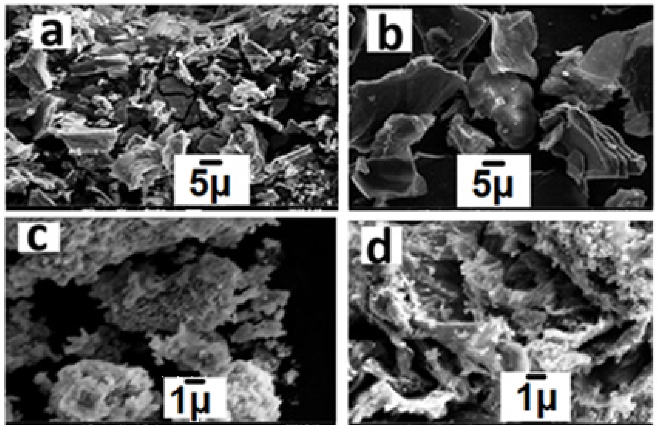
Figure 1. SEM image of NiO (a), GO (b), and NiO-GO nanocomposite before degradation (c) after degradation (d).
Catalysts 15 01078 g001
Figure 3. XPS analysis.
Catalysts 15 01078 g003
Figure 4. FTIR spectrum of NiO, GO, and NiO-GO before and after degradation.
Catalysts 15 01078 g004
Figure 5. XRD spectrum of NiO, GO, and NiO-GO.
Catalysts 15 01078 g005
Figure 6. Comparison of catalytic activities of NiO, GO, and NiO-GO for the degradation of clothianidin.
Catalysts 15 01078 g006
Figure 7. Kinetics analysis of NiO, GO, and NiO-GO catalyzed degradation of clothianidin.
Catalysts 15 01078 g007
Figure 8. Effect of pH on photocatalytic degradation of clothianidin during 60 min of reaction.
Catalysts 15 01078 g008
Figure 9. Effect of catalytic dose on photocatalytic degradation of clothianidin.
Catalysts 15 01078 g009
Figure 10. Effect of initial concentration on photocatalytic degradation of clothianidin.
Catalysts 15 01078 g010
Figure 11. Effect of agitation speed on photocatalytic degradation of clothianidin.
Catalysts 15 01078 g011
Table 2. Comparison of activities of various systems in the removal of clothianidin reported in the literature.
NoCatalyst/AdsorbentConditionsEfficiencyReference
1In2S3/MgTiO3/TiO2@N-CNTConc. 10 mg/L, Catalyst 0.3 g, 23 W LED/H2O298% in 18 min[62]
2FeMn@N–CConc. 5 mg/L, Catalyst 0.1 g, Visible light/PMS99% in 90 min[63]
3TiO2/CNT/Pd–CuConc. 3 mg/L, Catalyst 0.1 g, 450 W Xe arc lamp/H2O2100% in 180 min[64]
4Au–SnO2-rGOConc. 1 mg/L, Catalyst 0.003 g, 30 W UV-LED lamp97% in 120 min[65]
5TiO2/CNT/PdConc. 5 mg/L, Catalyst 0.1 g, 450 W Xe arc lamp/H2O285% in 180 min[66]
6TiO2 on glassConce. 100 mg/L, Catalyst 10 mg, UVA (315–400 nm)14% in 120 min[67]
7BiocharsConc. 100 ng/L, Sorbent 0.5 g/L100% [68]
8Stutzerimonas sp. SA1 and Pseudomonas sp. SA3Conc. 100 mg/L, Bacteria culture 3.2 × 10287%[69]
9NiO-GOConc. 20 mg/L, Catalyst 0.05 g, Natural sunlight96% in 60 minThis study
Disclaimer/Publisher’s Note: The statements, opinions and data contained in all publications are solely those of the individual author(s) and contributor(s) and not of MDPI and/or the editor(s). MDPI and/or the editor(s) disclaim responsibility for any injury to people or property resulting from any ideas, methods, instructions or products referred to in the content.