Article

Novel and Green Synthesis of Nitrogen-Doped Carbon Cohered Fe3O4 Nanoparticles with Rich Oxygen Vacancies and Its Application

1
Laboratory for Development & Application of Cold Plasma Technology, College of Chemistry and Chemical Engineering, Luoyang Normal University, Luoyang 471022, China
2
College of Civil Engineering, Luoyang Institute of Science and Technology, Luoyang 471023, China
3
Department of Chemistry, Texas A&M University-Commerce, Commerce, TX 75429-3011, USA
4
Department of Chemistry, University of California, 900 University Ave., Riverside, CA 92521, USA
*
Authors to whom correspondence should be addressed.
Catalysts 2022, 12(6), 621; https://doi.org/10.3390/catal12060621
Submission received: 5 April 2022 / Revised: 16 May 2022 / Accepted: 3 June 2022 / Published: 6 June 2022
(This article belongs to the Special Issue Functional Materials for Application in Adsorption & Catalysis)

Abstract

:
A one-pot and green synthesis methodology was successfully designed to prepare nitrogen-doped carbon (NC) cohered Fe3O4 nanoparticles with rich oxygen vacancies (Fe3O4-OVs/NC). The preparation was achieved via cold-atmospheric-pressure air plasma using Fe2O3 nanoparticles as the only precursor, and pyridine as the carbon and nitrogen source. Systematic characterization results of the as-prepared Fe3O4-OVs/NC confirmed the transition from Fe2O3 to Fe3O4, along with the generation of oxygen vacancies, while preserving the original needle-like morphology of Fe2O3. Moreover, the results indicated the formation of the NC attaching to the surface of the formed Fe3O4 nanoparticles with a weight percent of ~13.6%. The synthesized nanocomposite was further employed as a heterogeneous Fenton catalyst to remove phenol from an aqueous solution. The material has shown excellent catalytic activity and stability, demonstrating a promising application for wastewater treatment.

1. Introduction

Ferro ferric oxide nanoparticles (referred to as Fe3O4 NPs) have attracted much attention recently due to their low cost, unique magnetism and other excellent physicochemical properties [1], such as biocompatibility [2], environmental friendliness [3], high theoretical discharge specific capacity [4], corrosion resistance [5] and peroxidase-like activity [6]. Concomitantly, it has been explored that Fe3O4 NPs had a great potential in heterogeneous catalysis [7], medicines [8], batteries and supercapacitors [1,9,10], microwave absorbers [11], wastewater treatments [12], and so on. With the development of mechanistic research, it has been found that surface oxygen vacancies (OVs) dramatically influenced the physical and chemical properties of metal oxides, and thus played a significant role in the energetics, kinetics and catalytic activity [13,14,15]. Therefore, some investigation has been pursued to increase oxygen vacancies of Fe3O4 NPs to improve the photocatalytic and electrocatalytic properties [16,17], Fenton activities [18], microwave absorption [19], etc. However, the oxygen vacancy of Fe3O4 NPs is usually generated under conditions at high temperature and high pressure or via complicated procedures [16,17,18,19], which are energy-intensive or have safety concerns and, thus, are not environmentally friendly.
On the other hand, Fe3O4 NPs tend to aggregate into large particles due to the nano-size effects. Aggregation can result in the reduction of specific surface areas, and then inevitably cause a decrease in dispersion, therefore, a decrease in catalytic activity and stability of Fe3O4 NPs. This is a great challenge for the application of Fe3O4 NPs [20,21,22]. Loading Fe3O4 NPs onto suitable substrates to form nanocomposites is a viable and effective method for inhibiting aggregation. Therefore, it has attracted considerable research attention. Various carbon materials [4,5,9,17,19,23,24], organic polymers [25,26,27], and other materials, such as ZnO, CeO2, TiO2, and metal-organic frameworks [12,18,22,28] have been widely studied as supports for Fe3O4 NPs. For the carbon material supports, nitrogen doping has been proven to be an efficient means to greatly enhance their physio-chemical properties. Compared with pure carbon materials, the electron transport characteristics, chemical reactivity and stability of nitrogen-doped carbon (NC) can be significantly improved due to N doping [29,30,31,32,33]. However, as far as we know, previous preparation conditions of the NC materials used are either harsh, complex, or both, such as high temperatures (typically 500–1000 °C), high pressure, inert gas conditions, or using complex repolymerization precursors [30,33].
In the current study, a novel and green methodology for the preparation of nitrogen-doped carbon (NC) cohered Fe3O4 NPs with surface oxygen vacancies (OVs), by a one-pot process via the assistance of cold plasma at normal pressure in air atmosphere, was developed. The resulted catalyst was used for the degradation of phenol in a Fenton-like reaction system. The effects of the initial solution pH, initial H2O2 concentration and catalyst dosage on phenol degradation were investigated. The relationship between the morphologic structure and the catalytic performance for the degradation of phenol was discussed.

2. Results and Discussion

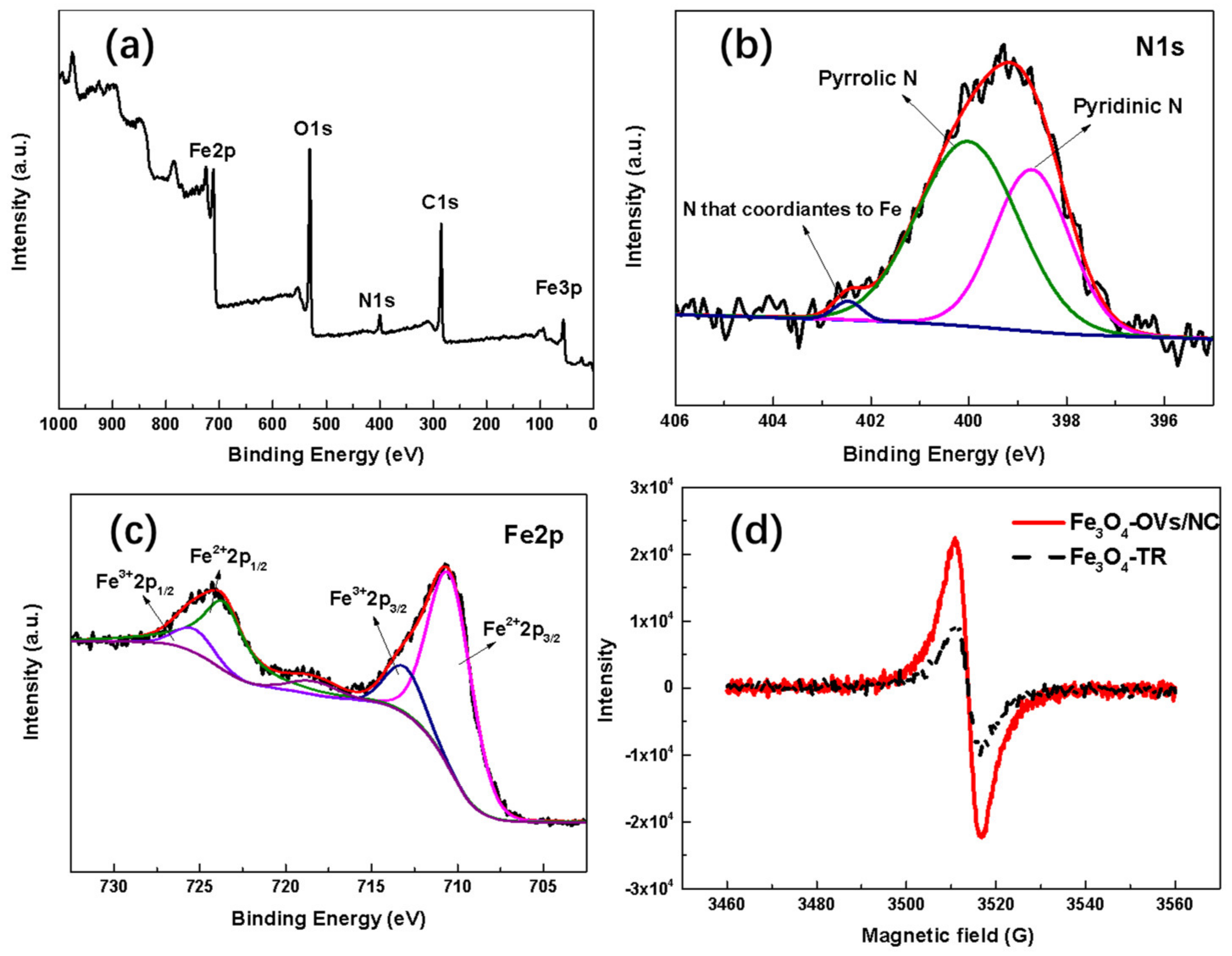
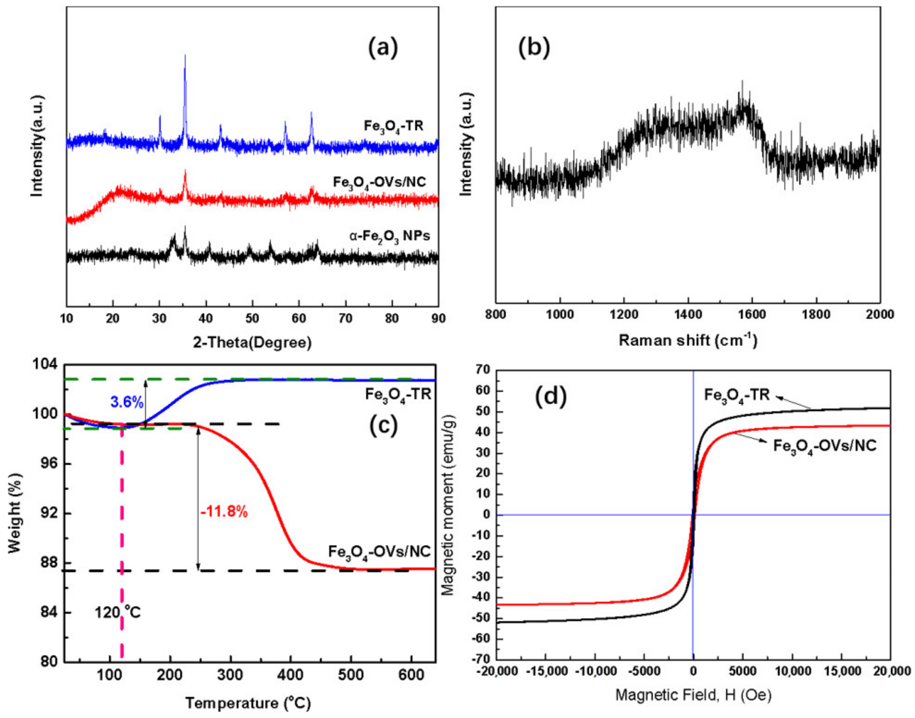
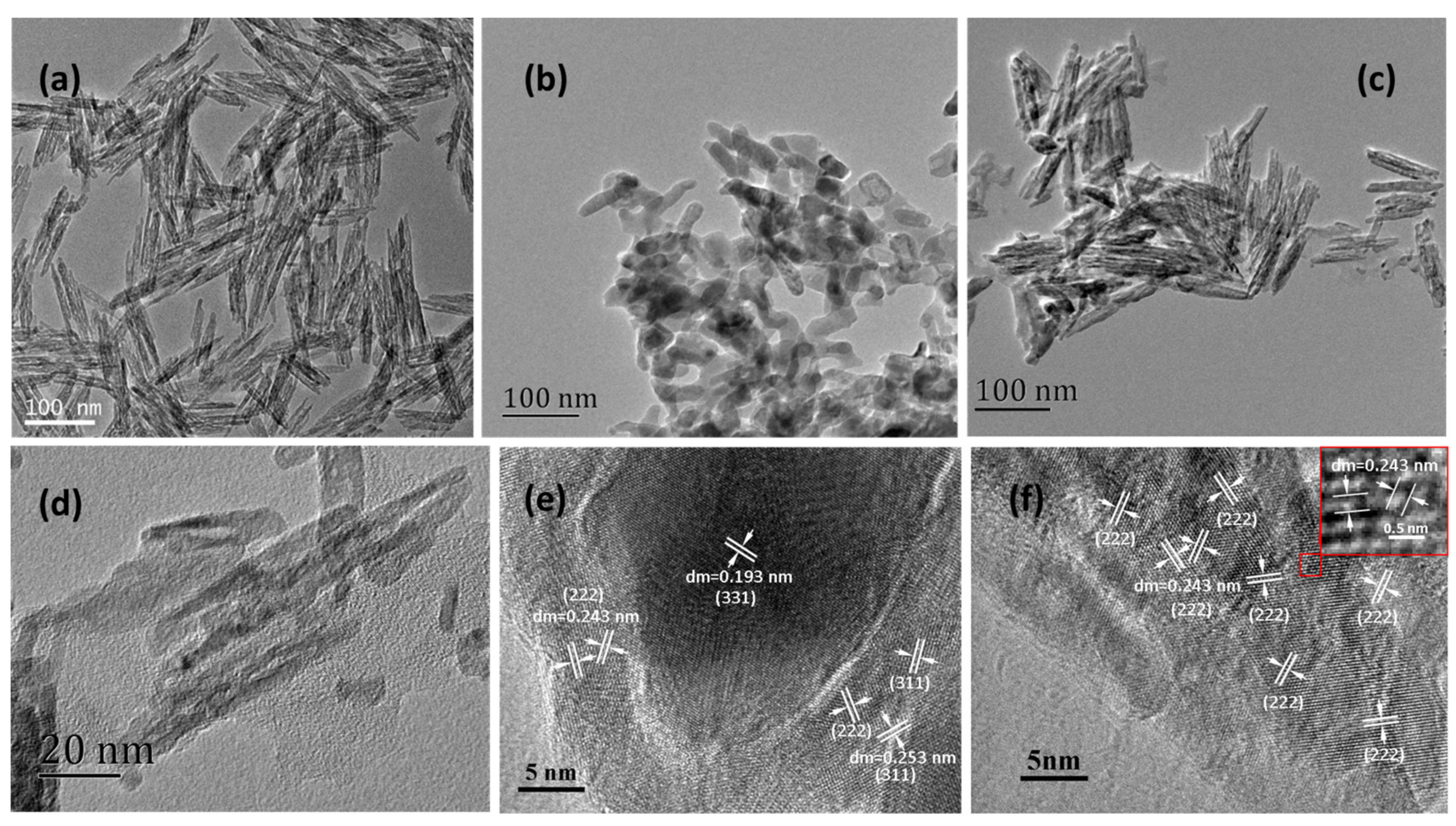
2.1. Microstructural, Morphological and Physio-Chemical Characterization

The XRD patterns of Fe3O4-OVs/NC, Fe3O4-TR and the precursor α-Fe2O3 NPs are illustrated in Figure 1a. The diffraction peaks of Fe2O3 NPs are in line with pure α-hematite Fe2O3 (JCPDS card no. 89–0597). The diffraction peaks, located at 2θ = 24.13°, 33.12°, 35.60°, 40.82°, 49.41°, 54.00°, 57.51°, 62.38° and 63.95°, are corresponding to the lattice planes of (012), (104), (110), (113), (024), (116), (018), (214) and (300) of pure α-Fe2O3, respectively [34,35]. In more detail, however, for the α-Fe2O3 NPs, the intensity ratios of the (104) to (104) and (110) to (104) peaks are both bigger than those in previous reports [34]. This should be attributed to the oriented growth of the needle-shaped α-Fe2O3 NPs used in this study. Jian and co-workers [35] have also observed similar phenomena for their α-Fe2O3 samples with different shapes. In the cases of Fe3O4-OVs/NC and Fe3O4-TR, five distinctly characteristic peaks belonging to the face-centered-cubic (fcc) Fe3O4 (JCPDS card no. 19-0629) are successfully observed. They are located at 2θ = 30.10°, 35.42°, 43.05°, 56.94°, and 62.52°, and could be indexed to the crystal faces of (220), (311), (400), (511) and (440), respectively [5,26]. Notably, no peaks at 2θ =33.12° and 40.82° are detected for Fe3O4-OVs/NC and Fe3O4-TR. This means that the two peaks which are attributed to the (104) and (113) crystal faces of α-Fe2O3 disappeared completely after the DBD plasma or thermal reduction treatment. That is, the Fe3O4 nanoparticles in Fe3O4-OVs/NC and Fe3O4-TR should be pure Fe3O4. Additionally, there is a broad diffraction peak centered at around 2θ = 22° for Fe3O4-OVs/NC, indicating the presence of amorphous carbon [28]. Moreover, compared with Fe3O4-TR, the intensity of all diffraction peaks assigned to Fe3O4 are much weaker for Fe3O4-OVs/NC. This could be due to the formation of the carbon cladding on the surface of the formed Fe3O4 NPs. In addition, from the full width at half maximum of the (110) diffraction peak of α-Fe2O3, and the (311) diffraction peaks of Fe3O4-TR and Fe3O4-OVs/NC, the average sizes of the crystallites were estimated to be 12 nm (α-Fe2O3), 18 nm (Fe3O4-TR) and 14 nm (Fe3O4-OVs/NC) using Scherrer’s equation, respectively. The crystal size of Fe3O4 obtained by conventional thermal reduction became obviously larger than its precursor, α-Fe2O3.
To further confirm the formation of carbon by the method that we proposed in this study, the Raman spectra were collected and presented in Figure 1b. Two distinct characteristic D (about 1320 cm−1) and G (about 1570 cm−1) bands of carbon can be observed. The D band is ascribed to the edges, defects and structurally disordered carbon, while the G band originates from in-plane vibrations of sp2-hybridized carbon atoms. It also can be seen that the two bands are both broad, indicating that the formed carbon exists in amorphous forms [5,30]. The TG curves (Figure 1c) display the thermal decomposition of Fe3O4-TR and Fe3O4-OVs/NC in an air atmosphere. The weight loss before 120 °C is attributed to the release of physically adsorbed water [36]. However, for Fe3O4-TR, in the range between 120 °C and 650 °C, with the temperature increase, its weight increases obviously until the temperature reaches 300 °C, and then remains constant. The final weight gain is 3.6%. This is in line well with the theoretical weight gain (3.5%) of Fe2O3 during the transformation from Fe3O4. On the contrary, the temperature-programmed treatment for Fe3O4-OVs/NC, in a range from 120 °C to 650 °C, is a weight loss process, and the final weight loss reached 11.8%. This results from the synergistic effect of the transformation from Fe3O4 to Fe2O3 and the combustion of carbon. Based on the above data, the formed nitrogen-doped carbon in Fe3O4-OVs/NC is calculated to be ~14.8 wt.%. Moreover, the analysis of the C and N elements for Fe3O4-OVs/NC was also performed in this study. The results show that the contents of C and N are 12.2 wt.% and 1.4 wt.%, which agree fairly well with that derived from the TG analysis. Note, however, that the carbon combustion mainly occurs between 250 °C and 450 °C in this case, which is well consistent with the previous report by Liu, et al. [37]; however, the temperature range is much lower than that of the combustion of pure carbon [31]. This results from the iron oxides acting as catalysts, playing an important role in promoting carbon combustion. The magnetic properties of Fe3O4-TR and Fe3O4-OVs/NC at room temperature were characterized by VSM. As presented in Figure 1d, the saturation magnetization (Ms) values of Fe3O4-TR and Fe3O4-OVs/NC were 52 emu·g−1 and 45 emu·g1, respectively. The smaller Ms value of Fe3O4-OVs/NC compared with Fe3O4-TR should be attributed to the existence of nitrogen-doped carbon (NC) reducing the content of Fe3O4 in Fe3O4-OVs/NC. Nonetheless, the magnetic response is still enough for Fe3O4-OVs/NC to meet the need for magnetic separation.
To evaluate the morphology and microstructure of the samples, TEM images were collected and shown in Figure 2. For the precursor α-Fe2O3 NPs, the TEM image clearly exhibits that the morphology is a needle-shaped nanostructure, and the average length and diameter are ~117 nm and ~14 nm, respectively (Figure 2a). After thermal reduction under H2 atmosphere, the formed Fe3O4 nanoparticles present an irregularly short worm-like shape with severe aggregation and adhesion (Figure 2b). This indicates that the needle-shaped structure of α-Fe2O3 NPs was totally damaged during the transformation process from Fe2O3 to Fe3O4. However, as shown in Figure 2c, the morphology of Fe3O4, which was obtained from the α-Fe2O3 NPs via the treatment of DBD plasma with pyridine, is well retained without obvious structural damages. In addition, Figure 2d clearly shows that the Fe3O4 NPs are adhered by membranous substances, which are identified to be NC according to Figure 1a,b and Figure 3a below. From both the high-resolution TEM images of Fe3O4-TR (Figure 2d) and Fe3O4-OVs/NC (Figure 2e), a lattice fringe spacing of 0.243 nm is observed, which is assigned to the (222) plane of fcc Fe3O4 [38]. Moreover, lattice fringes with d = 0.193 nm and 0.253 nm are also detected in Figure 2d, which match well with the (331) and (311) lattice planes of cubic Fe3O4 [39]. Noteworthily, in the case of Fe3O4-OVs/NC, all the lattice spacings are 0.243 nm in Figure 3e, and given this, it is speculated that the main exposed lattice planes in Fe3O4-OVs/NC should be (222) faces.
XPS spectra for Fe3O4-OVs/NC were collected to investigate the surface chemical composition. Obvious peaks at about 285, 399, 530 and 711 eV can be observed in the survey spectrum of Fe3O4-OVs/NC (Figure 3a), which confirms the existence of C, N, O, and Fe. Furthermore, the survey spectrum also demonstrates that the nitrogen content is up to 4.0 wt.%. The high-resolution N1s spectrum of Fe3O4-OVs/NC (Figure 3b) contained three deconvolution peaks whose binding energies are 398.5, 400.2, and 402.4 eV, corresponding to pyridinic N, pyrrolic N and N, which coordinates to Fe, respectively [40]. The Fe2p spectrum in Figure 3c shows, as expected, peaks of Fe 2p1/2 (~724.1 eV), Fe 2p 3/2 (~710.3 eV), and a satellite peak (~718.5 eV). The Fe 2p1/2 and Fe 2p3/2 peaks can further be deconvoluted into four contributory peaks at 710.7, 713.6, 722.9 and 725.2 eV. The peaks at 725.2 eV and 722.9 eV correspond to the binding energies of 2p1/2 of Fe(III) and Fe(II), respectively, meanwhile, the peaks at 713.6 eV and 710.7 eV can be assigned to the 2p 3/2 of Fe(II) ion and Fe(III) ion, respectively. The satellite peak also indicates the coexistence of the Fe(II )and Fe(III) ions [41]. These conclusions are consistent with the previous report and further verify the existence of Fe3O4 [40,41], which is agreeable with the XRD results. Furthermore, it is known that the EPR measurement can provide strong evidence to investigate the presence of oxygen vacancies. As shown in Figure 3d, the EPR signals at a g-value of 2.003 are clearly observed for both samples, Fe3O4-TR and Fe3O4-OVs/NC, which demonstrates the surface electron trapping sites caused by the O vacancy [16,17]. However, compared with Fe3O4-TR, Fe3O4-OVs/NC shows a much stronger EPR response, indicating the existence of much more O vacancies.

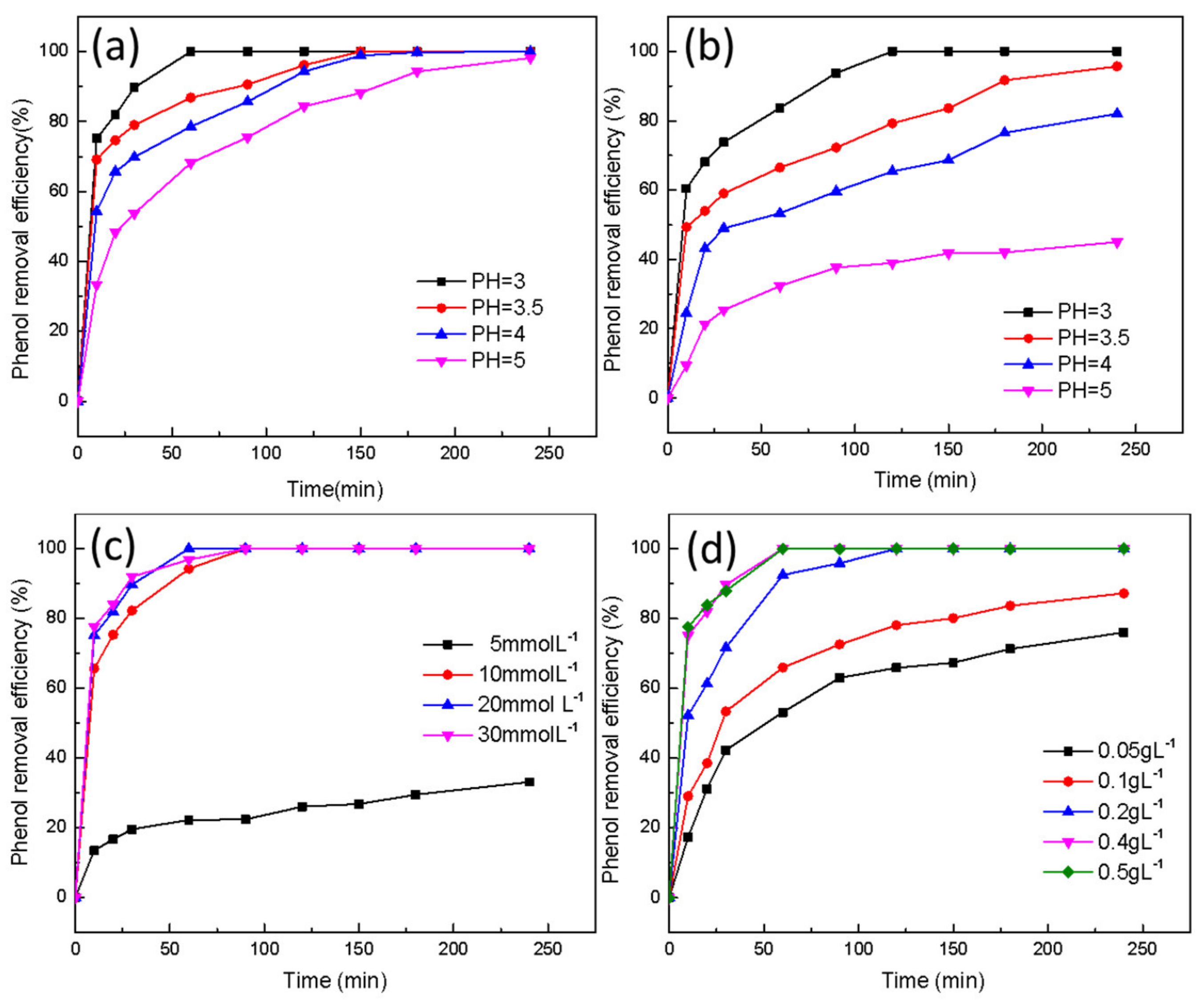
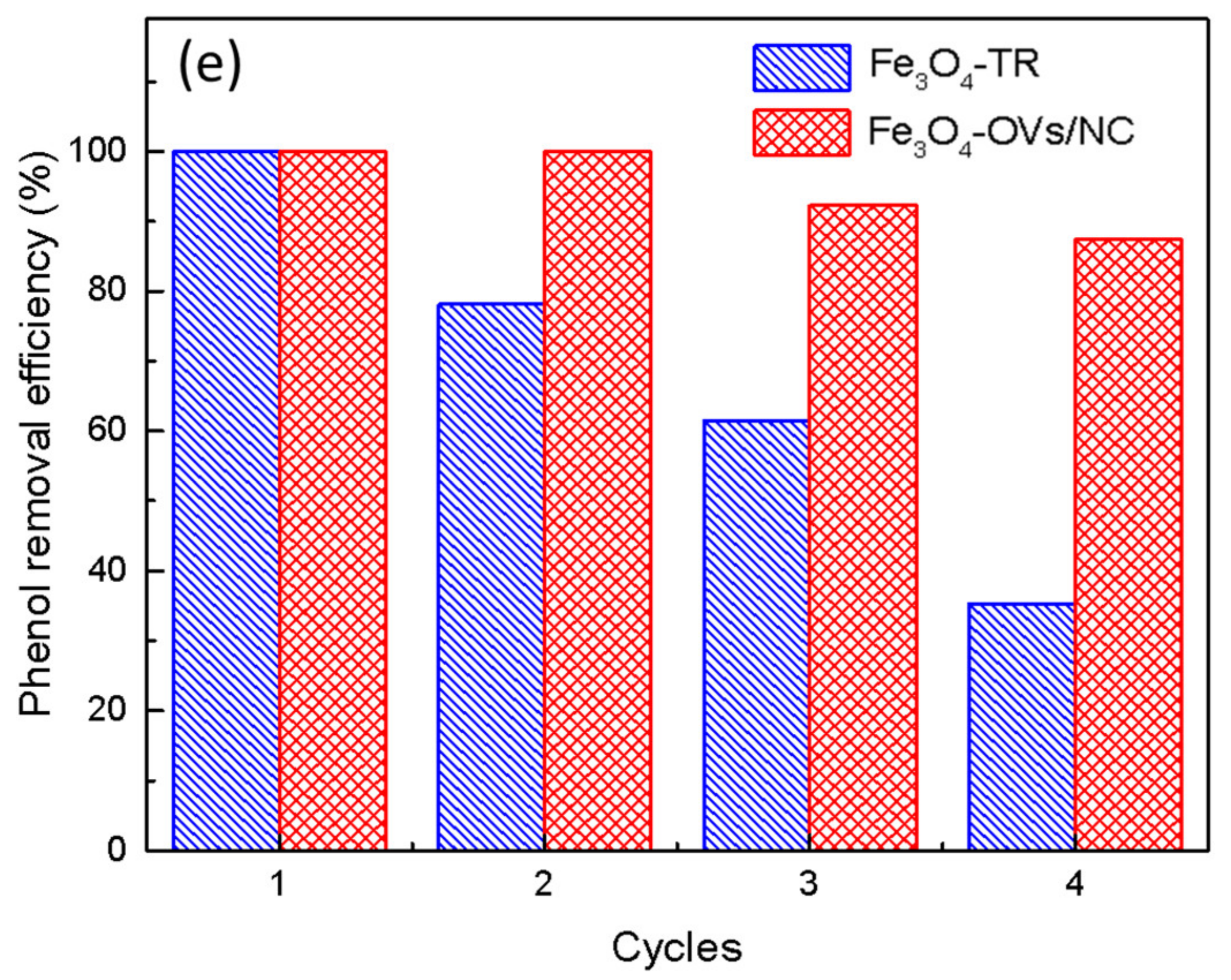
2.2. Catalytic Performance of Fe3O4-TR and Fe3O4-OVs/NC

In order to evaluate the performance of Fe3O4-TR and Fe3O4-OVs/NC for catalysis in a Fenton-like process, both catalysts were tested under different conditions. It is shown in Figure 4a,b, that it is obvious the removal of phenol is highly pH-dependent for both samples. With the increase in pH, the efficiency of phenol degradation decreases correspondingly, and at a pH of around 3.0, the two samples show optimal performance for the catalytic phenol degradation. These are in agreement with previous study results for the degradation of organic contaminants with Fe3O4-based catalysts [24,27]. This is because, at high pH (>3.0), inert oxide film might form on the surface of the Fe3O4 nanoparticles, which would prevent the active Fe sites from catalytically decomposing H2O2, generating sufficient reactive radicals [27]. Further, from Figure 4a, it can be seen that 0.2g L−1 phenol could be completely removed only in 60 min under the initial H2O2 concentration of 20 mmol L−1 at pH 3.0 using a 0.4 g L−1 dosage of Fe3O4-OVs/NC. In contrast, it needs as long as 120 min to reach complete phenol removal under the same reaction condition. Moreover, leaving the other parameters unchanged, while increasing the pH value to 5.0, the maximum removal of phenol is only 47% with Fe3O4-TR. On the other hand, however, the removal efficiency is still up to 98% for Fe3O4-OVs/NC. This indicates that the catalytic performance of the as-prepared nanocomposite, Fe3O4-OVs/NC is remarkable in a wide range of pH values. The great improvement in the catalytic performance of Fe3O4-OVs/NC compared with Fe3O4-TR should be attributed to its small Fe3O4 nanoparticles and abundant oxygen vacancies [42]. Moreover, the NC formed during the cold plasma treatment should also play a significant role in the catalytic performance improvement of Fe3O4-OVs/NC [43,44]. In addition, the optimum reaction conditions for the catalytic removal of phenol with Fe3O4-OVs/NC have also been discussed, by changing one parameter and leaving the others unchanged, and the results are shown in Figure 4c,d. The H2O2 concentration dramatically affects the removal efficiency of phenol (Figure 4c). The phenol degradation efficiency is rapidly increased from 40% to 100% as the concentration of H2O2 increases from 5 to 10 mmol L−1, and the time to the equilibrium state is also significantly reduced from about 250 min to 100 min. However, with the further increase in the H2O2 concentration, the degradation efficiency no longer changes notably. Therefore, the optimal concentration of H2O2 is confirmed to be 20 mmol L−1. The effect of the Fe3O4-OVs/NC dosage on the degradation of phenol, with the 20 mmol L1 initial H2O2 at pH 3.0, is shown in Figure 4d. The degradation of phenol was significantly enhanced by the addition of Fe3O4-OVs/NC, even when the amount of catalyst is as low as 0.05 g L−1, which demonstrates that Fe3O4-OVs/NC has an outstanding catalytic ability to activate the decomposition of H2O2. However, when the dosage of Fe3O4-OVs/NC is further increased from 0.4 to 0.5 g L−1, the increase in phenol degradation efficiency diminishes. Given the above, the optimal dosage of Fe3O4-OVs/NC in the selected reaction condition is 0.4 g L−1.
The reusability of Fe3O4-OVs/NC (Figure 4e) was examined under the optimum conditions of a Fe3O4-OVs/NC dosage of 0.4 g L−1, initial H2O2 dosage of 20 mmol L−1, and pH of 3.0. After four Fenton reaction recycles, the catalytic degradation efficiency of phenol for Fe3O4-TR sharply decreases from 100% to 33%. For Fe3O4-OVs/NC, however, as the number of cycles increases, phenol degradation efficiency decreases much slower in comparison with Fe3O4-TR. After four recycles, phenol degradation efficiency still reaches nearly 87.5%, suggesting that the reused Fe3O4-OVs/NC still remains highly active and Fe3O4-OVs/NC possesses much better stability than Fe3O4-TR.

3. Materials and Methods

3.1. Material Preparation

For the preparation of Fe3O4-OVs/NC nanocomposite, the reagents applied, including needle-shaped α-Fe2O3 NPs (≥99 wt.%) and pyridine (99.5 mol.%) were both supplied by Tianjin Zhiyuan Chemical Reagent Co. (Tianjin, China), and used without further purification. Distilled water was used in all the experiments. The cold plasma used in this study was generated by a dielectric barrier discharge (DBD) plasma generator (CTP-2000 K), which was purchased from Corona Laboratory, Nanjing, China. The structure of the generator and the operation of the system have been described in detail elsewhere [45]. Briefly, a chamber was constructed by two pieces of quartz plates with a diameter and thickness of 90 mm and 2 mm, respectively, and a quartz ring with a height, internal diameter and external diameter of 8 mm, 50 mm and 54 mm, respectively. The two quartz plates served as the upper and lower surfaces of the chamber, which sandwiched the quartz ring as the chamber’s wall. The chamber was fixed by two columnar steel electrodes with a height and diameter of 50 mm and 54 mm, respectively, by squeezing the two quartz plates during the plasma treatment. The preparation procedures of the Fe3O4-VOs/NC nanocomposites are as follows. Firstly, the mixture of α-Fe2O3 NPs and pyridine was obtained via the incipient wet impregnation method. Further, 0.3 g of α-Fe2O3 NPs were introduced into the quartz chamber of the DBD plasma generator and then mixed with 200 μL of pyridine. Secondly, the chamber was fixed with two steel plate electrodes, and then the mixture in the chamber was treated with DBD plasma for 3 min. The average voltage applied was 100 V and the average power was 250 W. Static air was directly used as the medium gas for the DBD plasma generation. Thirdly, the processed mixture was grounded after the plasma treatment, then mixed with another 200μL of pyridine, followed by another treatment of DBD plasma for 3 min again. This process was repeated 5 times. Finally, the resulted sample was washed with an ultrasonic washer, and further dried in a vacuum oven at 50 °C. The obtained black nanocomposite was designated as Fe3O4-OVs/NC. For purposes of comparison, the α-Fe2O3 NPs were conventionally thermally reduced at 598 K under a hydrogen atmosphere for 1 h by following the reported methods with slight changes in the holding time [46]. The as-prepared black sample was denoted as Fe3O4-TR.

3.2. Material Characterization

The compositions’ phases of the as-prepared materials were identified by a Bruker D8 (Bruker Co., Billerica, MA, USA) advance X-ray diffractometer (XRD) equipped with Cu Kα radiation (λ = 1.5418 Å) and operated at 30 kV and 40 mA with a 4°∙min−1 scan rate. To demonstrate the presence of the carbon in Fe3O4-OVs/NC, Raman spectra were collected by a JY-HR800 laser confocal micro-Raman spectrometer (Horiba Jobin Yvon, Paris, France) with a 532 nm wavelength laser. Thermal-gravimetric analyses (TGA) for the two samples were carried out on a Shimadzu TGA-50 (Shimadzu Co., Kyoto, Japan) at a heating rate of 10 °C·min−1 from an ambient temperature to 700 °C in air with a flow rate of 25 cm3∙min−1. Elemental analysis was carried out by using a Vario MACRO cube CHNS analyzer (Elementar Analysensysteme GmbH, Hanau, Germany) to analyze C and N in Fe3O4-OVs/NC. The magnetic properties of the two samples were measured by a 7400-S vibrating sample magnetometer (Lakeshore Inc., Columbus, OH, USA) at room temperature. The morphological information of the two samples was surveyed using a JEM- 2100F (JEOL Ltd., Tokyo, Japan)high-resolution transmission electron microscopy (HTEM) working at 200 kV. The surface chemical compositions and chemical states were examined by X-ray photoelectron spectroscopy (XPS), performed on a Thermo ESCALAB250Xi X-ray Photoelectron spectrometer (Thermo Fisher Scientific, Waltham, MA, USA) equipped with a monochromatic Al Kα (hv = 1486.6 eV) radiation source. The carbonaceous C1s line at 284.6 eV was taken as the reference for the calibration of binding energies (BEs). Electron paramagnetic resonance (EPR) spectra were conducted on a Bruker EMX EPR spectrometer (Bruker Co., Billerica, MA, USA) to monitor the oxygen vacancies of samples.

3.3. Catalytic Degradation Experiments

The potential of Fe3O4-OVs/NC used as a catalyst for the removal of phenol in a Fenton-like process was examined. For comparison, the corresponding performance tests of Fe3O4-TR, prepared by conventional thermal reduction, were also carried out. The entire experiments in this study were performed in the dark to eliminate the effect of photo-decomposition of H2O2 and in a water-bath thermostatic oscillator with continuous shaking at 40 ± 2 °C. The initial concentration of phenol and the volume of the reaction solution were 200 mg L−1 and 100 mL, respectively. The solution pH was initially adjusted by using 0.1 M HCl or 0.1 M NaOH. The reactions were initiated by adding the desired dosage of H2O2 to a pH-adjusted reaction solution containing the target substance and a specific amount of catalyst. Samples were withdrawn from the reaction mixture at given time intervals and filtered immediately through a filter membrane (0.22 μm) for subsequent analysis.
The concentration of phenol was measured by a high-performance liquid chromatography (HPLC) method using the Agilent 1200 Series equipment (Agilent Technologies Inc., Santa Clara, CA, USA) coupled with a Zorbax Eclipse XDB-C18 column (3.5 µm, 2.1 mm × 100 mm) and a UV diode array detector. The column temperature was maintained at 30 °C, and the UV detector wavelength was set at 270 nm. A mixture of acetonitrile and water with 70:30 v/v was used as the mobile phase at a flow rate of 1.0 mL∙min−1.

4. Conclusions

Cold-atmospheric-pressure air plasma was successfully introduced in this study for the preparation of nitrogen-doped carbon (NC) cohered Fe3O4 nanoparticles with rich oxygen vacancies. The plasma-treatment time was only 15 min, the process temperature was less than 150 °C [34], and the Fe2O3 nanoparticles and pyridine were the only two precursors used. Overall, the synthesis methodology has shown its simple, green, fast and ‘one-pot’ properties. XRD, Raman and XPS characterizations confirmed the formation of Fe3O4 and NC. The EPR result indicated that the as-prepared sample possessed much more O vacancies than the thermally reduced Fe3O4. Moreover, the TEM images showed that the generated Fe3O4 nanoparticles were cohered by NC and basically keep the original needle-like morphology of the precursor, Fe2O3. This provides the possibility of achieving the flexible shape control of the Fe3O4 NPs just by selecting the suitable morphologic structure of the precursor, Fe2O3 NPs. The synthesized nanocomposite was further used as a heterogeneous catalyst for the degradation of phenol in a Fenton-like reaction system. The experimental results demonstrated that 0.2 g L−1 of phenol could be completely removed only in 60 min with Fe3O4-OVs/NC under optimal conditions, such as a catalyst dosage of 0.4 g L−1, initial H2O2 concentration of 20 mmol L−1, and pH of 3.0. Even when pH was up to 5.0, about 98% of the phenol can still be eliminated by 0.4 g L−1 of the as-prepared nanocomposite within 240 min. In addition, under optimal conditions, the nanocomposite showed favorable reusability with 87.5% of phenol removal efficiency after four cycles. Compared with the Fe3O4 that was prepared by traditional thermal reduction, the synergistic effect of smaller particle size, more regular structure, much more O vacancies, and NC agglutination should be the reasons for the as-prepared nanocomposite possessing outstanding catalytic performance for phenol removal.

Author Contributions

Conceptualization, X.C. and B.W.-L.J.; methodology, H.Z.; validation, S.T., B.W.-L.J. and H.Z.; formal analysis, A.M.; investigation, C.Y., L.J., J.Z. and J.Q.; resources, S.T.; data curation, L.Z.; writing—original draft preparation, X.C.; writing—review and editing, B.W.-L.J., A.M. and H.Z.; supervision, B.W.-L.J. and H.Z.; project administration, X.C.; funding acquisition, B.W.-L.J. and X.C. All authors have read and agreed to the published version of the manuscript.

Funding

This research was funded by the American Chemical Society Petroleum Research Fund, ACS PRF (#57596-UR5) and the Key R&D and Promotion Special Project (Science and Technology) of Henan Province, China (No. 212102210214).

Conflicts of Interest

The authors declare no conflict of interest.

References

  1. Zhou, W.; Guo, L. Iron triad (Fe, Co, Ni) nanomaterials: Structural design, functionalization and their applications. Chem. Soc. Rev. 2015, 44, 6697–6707. [Google Scholar] [CrossRef]
  2. Wu, S.; Gu, L.; Qin, J.; Zhang, L.; Sun, F.; Liu, Z.; Wang, Y.; Shi, D. Rapid label-free isolation of circulating tumor cells from patients’ peripheral blood using electrically charged Fe3O4 nanoparticles. ACS Appl. Mater. Interfaces 2020, 12, 4193–4203. [Google Scholar] [CrossRef]
  3. Zhao, Y.; Wang, J.; Ma, C.; Cao, L.; Shao, Z. A self-adhesive graphene nanoscroll/nanosheet paper with confined Fe1-xS/Fe3O4 hetero-nanoparticles for high-performance anode material of flexible Li-ion batteries. Chem. Eng. J. 2019, 370, 536–546. [Google Scholar] [CrossRef]
  4. Li, L.; Wang, H.; Xie, Z.; An, C.; Jiang, G.; Wang, Y. 3D graphene-encapsulated nearly monodisperse Fe3O4 nanoparticles as high-performance lithium-ion battery anodes. J. Alloys Compd. 2020, 815, 152337. [Google Scholar] [CrossRef]
  5. Wang, X.; Pan, F.; Xiang, Z.; Zeng, Q.; Pei, K.; Che, R.; Lu, W. Magnetic vortex core-shell Fe3O4@C nanorings with enhanced microwave absorption performance. Carbon 2020, 157, 130–139. [Google Scholar] [CrossRef]
  6. Gao, L.; Zhuang, J.; Nie, L.; Zhang, J.; Zhang, Y.; Gu, N.; Wang, T.; Feng, J.; Yang, D.; Perrett, S.; et al. Intrinsic peroxidase-like activity of ferromagnetic nanoparticles. Nat. Nanotechnol. 2007, 2, 577–583. [Google Scholar] [CrossRef]
  7. Veisi, H.; Ebrahimi, Z.; Karmakar, B.; Tamoradi, T. A convenient green protocol for oxidative esterification of arylaldehydes over Pd NPs decorated polyplex encapsulated Fe3O4 microspheres. Int. J. Biol. Macromol. 2022, 200, 132–138. [Google Scholar] [CrossRef]
  8. Cheng, Q.; Yue, L.; Li, J.; Gao, C.; Ding, Y.; Sun, C.; Xu, M.; Yuan, Z.; Wang, R. Supramolecular tropism driven aggregation of nanoparticles in situ for tumor-specific bioimaging and photothermal therapy. Small 2021, 17, 2101332. [Google Scholar] [CrossRef]
  9. He, D.; Sun, M.; Cao, D.; He, G.; Chen, H. Rational design of nano-Fe3O4 encapsulated in 3D honeycomb biochar for enhanced lithium storage performance. Nanotechnology 2022, 33, 035401. [Google Scholar] [CrossRef]
  10. Xue, S.; Wu, G.; Li, M.; Liu, Z.; Deng, Y.; Han, W.; Lv, X.; Wan, S.; Xi, X.; Yang, D.; et al. Generalized assembly of sandwich-like 0D/2D/0D heterostructures with highly exposed surfaces toward superior electrochemical performances. Nano Res. 2022, 15, 255–263. [Google Scholar] [CrossRef]
  11. Ma, W.; He, P.; Wang, T.; Xu, J.; Liu, X.; Zhuang, Q.; Cui, Z.; Lin, S. Microwave absorption of carbonization temperature-dependent uniform yolk-shell H-Fe3O4@C microspheres. Chem. Eng. J. 2021, 420, 129875. [Google Scholar] [CrossRef]
  12. Foroutan, R.; Mohammadi, R.; Ahmadi, A.; Bikhabar, G.; Ramavandi, B. Impact of ZnO and Fe3O4 magnetic nanoscale on the methyl violet 2B removal efficiency of the activated carbon oak wood. Chemosphere 2022, 286, 131632. [Google Scholar] [CrossRef]
  13. Setvín, M.; Wagner, M.; Schmid, M.; Parkinson, G.S.; Diebold, U. Surface point defects on bulk oxides: Atomically-resolved scanning probe microscopy. Chem. Soc. Rev. 2017, 46, 1772–1784. [Google Scholar] [CrossRef] [PubMed] [Green Version]
  14. Zhai, T.; Xie, S.; Yu, M.; Fang, P.; Liang, C.; Lu, X.; Tong, Y. Oxygen vacancies enhancing capacitive properties of MnO2 nanorods for wearable asymmetric supercapacitors. Nano Energy 2014, 8, 255–263. [Google Scholar] [CrossRef]
  15. Jian, W.; Wang, S.-P.; Zhang, H.-X.; Bai, F.-Q. Disentangling the role of oxygen vacancies on the surface of Fe3O4 and γ-Fe2O3. Inorg. Chem. Front. 2019, 6, 2660–2666. [Google Scholar] [CrossRef]
  16. Bui, H.T.; Im, S.M.; Kim, K.; Kima, W.; Lee, H. Photocatalytic degradation of phenolic compounds of defect engineered Fe3O4: An alternative approach to solar activation via ligand-to-metal charge transfer. Appl. Surf. Sci. 2020, 509, 144853. [Google Scholar] [CrossRef]
  17. Deng, Y.; Tian, X.; Shen, G.; Gao, Y.; Lin, C.; Ling, L.; Cheng, F.; Zhang, S.; Liao, S. Coupling hollow Fe3O4 nanoparticles with oxygen vacancy on mesoporous carbon as a high-efficiency ORR electrocatalyst for Zn-air battery. J. Colloid Interface Sci. 2020, 567, 410–418. [Google Scholar] [CrossRef] [PubMed]
  18. SujayShekar, G.C.; Khaled, A.; Abdo, H.; Ali, A.; Nabil, A.Z.; Lokanath, N.K. Enhanced photo-Fenton activity over a sunlight-driven ignition synthesized α-Fe2O3-Fe3O4/CeO2 heterojunction catalyst enriched with oxygen vacancies. J. Mol. Liq. 2021, 335, 116186. [Google Scholar] [CrossRef]
  19. Su, Z.; Zhang, W.; Lu, J.; Tian, L.; Yi, S.; Zhang, Y.; Zhou, S.; Niu, B.; Long, D. Oxygen-vacancy-rich Fe3O4/carbon nanosheets enabling high-attenuation and broadband microwave absorption through the integration of interfacial polarization and charge-separation polarization. J. Mater. Chem. A 2022, 10, 8479–8490. [Google Scholar] [CrossRef]
  20. Zhang, H.; Zhao, Y.; Zhang, Y.; Zhang, M.; Cheng, M.; Yu, J.; Liu, H.; Ji, M.; Zhu, C.; Xu, J. Fe3O4 encapsulated in porous carbon nanobowls as efficient oxygen reduction reaction catalyst for Zn-air batteries. Chem. Eng. J. 2019, 375, 122058. [Google Scholar] [CrossRef]
  21. Wang, M.; Yin, Y. Magnetically responsive nanostructures with tunable optical properties. J. Am. Chem. Soc. 2016, 138, 6315–6323. [Google Scholar] [CrossRef]
  22. Mishra, P.; Patnaik, S.; Parida, K. An overview of recent progress on noble metal modified magnetic Fe3O4 for photocatalytic pollutant degradation and H2 evolution. Catal. Sci. Technol. 2019, 9, 916–941. [Google Scholar] [CrossRef]
  23. Dong, P.; Liu, W.; Wang, S.; Wang, H.; Wang, Y.; Zhao, C. In suit synthesis of Fe3O4 on carbon fiber paper@polyaniline substrate as novel self-supported electrode for heterogeneous electro-Fenton oxidation. Electrochim. Acta 2019, 308, 54–63. [Google Scholar] [CrossRef]
  24. Tolba, A.; Alalm, M.G.; Elsamadony, M.; Mostafa, A.; Afify, H.; Dionysiou, D.D. Modeling and optimization of heterogeneous Fenton-like and photo-Fenton processes using reusable Fe3O4-MWCNTs. Process Saf. Environ. 2019, 128, 273–283. [Google Scholar] [CrossRef]
  25. Jiang, D.; Murugadoss, V.; Wang, Y.; Lin, J.; Ding, T.; Wang, Z.; Shao, Q.; Wang, C.; Liu, H.; Lu, N.; et al. Electromagnetic interference shielding polymers and nanocomposites—A review. Poly. Rev. 2019, 59, 280–337. [Google Scholar] [CrossRef]
  26. Pujol, A.A.; León, I.; Cárdenas, J.; Sepúlveda-Guzmán, S.; Manríquez, J.; Sirés, I.; Bustos, E. Degradation of phenols by heterogeneous electro-Fenton with a Fe3O4-chitosan composite and a boron-doped diamond anode. Electrochim. Acta 2020, 337, 135784. [Google Scholar] [CrossRef]
  27. Shen, J.; Zhou, Y.; Li, S.; Gu, P.; Xue, G. Hydrogel-coated Fe3O4 nanoparticles as an efficient heterogeneous Fenton catalyst for degradation of phenol. J. Mater. Sci. 2019, 54, 10684–10694. [Google Scholar] [CrossRef]
  28. Silvestre, M.E.; Franzreb, M.; Weidler, P.G.; Shekhah, O.; Wöll, C. Magnetic cores with porous coatings: Growth of metal-organic frameworks on particles using liquid phase epitaxy. Adv. Funct. Mater. 2013, 23, 1210–1213. [Google Scholar] [CrossRef]
  29. Guo, D.; Shibuya, R.; Akiba, C.; Saji, S.; Kondo, T.; Nakamura, J. Active sites of nitrogen-doped carbon materials for oxygen reduction reaction clarified using model catalysts. Science 2016, 351, 361–365. [Google Scholar] [CrossRef] [PubMed]
  30. Inagaki, M.; Toyoda, M.; Soneda, Y.; Morishita, T. Nitrogen-doped carbon materials. Carbon 2018, 132, 104–140. [Google Scholar] [CrossRef]
  31. Yang, Z.; Ren, J.; Zhang, Z.; Chen, X.; Guan, G.; Qiu, L.; Zhang, Y.; Peng, H. Recent advancement of nanostructured carbon for energy applications. Chem. Rev. 2015, 115, 5159–5223. [Google Scholar] [CrossRef] [PubMed]
  32. Zhou, Y.; Luo, L.; Yan, W.; Li, Z.; Fan, M.; Du, G.; Zhao, W. Controlled preparation of nitrogen-doped hierarchical carbon cryogels derived from Phenolic-Based resin and their CO2 adsorption properties. Energy 2022, 246, 123367. [Google Scholar] [CrossRef]
  33. Xu, H.; Lv, X.; Wang, H.; Ye, J.; Yuan, J.; Wang, Y.; Zhou, Z.; Sun, S. Impact of pore structure on two-electron oxygen reduction reaction in nitrogen-doped carbon materials: Rotating ring-disk electrode vs. flow cell. ChemSusChem 2022, 15, e202102587. [Google Scholar] [CrossRef]
  34. Wang, L.; Wei, G.; Dong, X.; Zhao, Y.; Xing, Z.; Hong, H.; Ju, Z. Hollow α-Fe2O3 Nanotubes embedded in graphene aerogel as high-performance anode material for lithium-ion batteries. ChemistrySelect 2019, 4, 11370–11377. [Google Scholar] [CrossRef]
  35. Jian, Y.; Yu, T.; Jiang, Z.; Yu, Y.; Douthwaite, M.; Liu, J.; Albilali, R.; He, C. In-depth understanding of the morphology effect of α-Fe2O3 on catalytic ethane destruction. ACS Appl. Mater. Interfaces 2019, 11, 11369–11383. [Google Scholar] [CrossRef] [PubMed] [Green Version]
  36. Cao, X.; Lyu, T.; Xie, W.; Mirjalili, A.; Bradicich, A.; Huitema, R.; Jang, B.W.-L.; Keum, J.K.; More, K.; Liu, C.; et al. Preparation and investigation of Pd doped Cu catalysts forselective hydrogenation of acetylene. Front. Chem. Sci. Eng. 2020, 14, 522–533. [Google Scholar] [CrossRef]
  37. Liu, Y.; Fu, Y.; Liu, L.; Li, W.; Guan, J.; Tong, G. Low-cost carbothermal reduction preparation of monodisperse Fe3O4/C core−shell nanosheets for improved microwave absorption. ACS Appl. Mater. Interfaces 2018, 10, 16511–16520. [Google Scholar] [CrossRef]
  38. Hong, W.; Hsu, I.; Huang, S.; Lee, C.; Ko, H.; Tsai, P.; Shieh, D.; Huang, C. Assembled growth of 3D Fe3O4@Au nanoparticles for efficient photothermal ablation and SERS detection of microorganisms. J. Mater. Chem. B 2018, 6, 5689–5697. [Google Scholar] [CrossRef] [PubMed]
  39. Zhong, Y.; Yu, L.; Chen, Z.; He, H.; Ye, F.; Cheng, G.; Zhang, Q. Microwave-assisted synthesis of Fe3O4 nanocrystals with predominantly exposed facets and their heterogeneous UVA/Fenton catalytic activity. ACS Appl. Mater. Interfaces 2017, 9, 29203–29212. [Google Scholar] [CrossRef]
  40. Huang, D.; Luo, Y.; Li, S.; Wang, M.; Shen, Y. Hybrid of Fe@Fe3O4 core-shell nanoparticle and iron-nitrogen-doped carbon material as an efficient electrocatalyst for oxygen reduction reaction. Electrochim. Acta 2015, 174, 933–993. [Google Scholar] [CrossRef]
  41. Wang, H.; Wang, W.; Xu, Y.; Dong, S.; Xiao, J.; Wang, F.; Liu, H.; Xia, B.Y. Hollow nitrogen-doped carbon spheres with Fe3O4 nanoparticles encapsulated as a highly active oxygen-reduction catalyst. ACS Appl. Mater. Interfaces 2017, 9, 10610–10617. [Google Scholar] [CrossRef] [PubMed]
  42. Xia, Q.; Yao, Z.; Zhang, D.; Li, D.; Zhang, Z.; Jiang, Z. Rational synthesis of micronano dendritic ZVI@Fe3O4 modified with carbon quantum dots and oxygen vacancies for accelerating Fenton-like oxidation. Sci. Total Environ. 2019, 671, 1056–1065. [Google Scholar] [CrossRef]
  43. Yang, W.; Hong, P.; Yang, D.; Yang, Y.; Wu, Z.; Xie, C.; He, J.; Zhang, K.; Kong, L.; Liu, J. Enhanced Fenton-like degradation of sulfadiazine by single atom iron materials fixed on nitrogen-doped porous carbon. J. Colloid Interface Sci. 2021, 597, 56–65. [Google Scholar] [CrossRef]
  44. Beker, S.A.; Khudur, L.S.; Cole, I.; Ball, A.S. Catalytic degradation of methylene blue using iron and nitrogen-containing carbon dots as Fenton-like catalysts. New J. Chem. 2022, 46, 263–275. [Google Scholar] [CrossRef]
  45. Cao, X.; Zhou, R.; Rui, N.; Wang, Z.; Wang, J.; Zhou, X.; Liu, C. Co3O4/HZSM-5 catalysts for methane combustion: The effect of preparation methodologies. Catal. Today 2017, 297, 219–227. [Google Scholar] [CrossRef]
  46. Salamun, N.; Ni, H.X.; Triwahyono, S.; Jalil, A.A.; Karim, A.H. Synthesis and characterization of Fe3O4 nanoparticles by electrodeposition and reduction methods. Mal. J. Fund. Appl. Sci. 2011, 7, 89–92. [Google Scholar] [CrossRef] [Green Version]
Figure 1. (a) XRD patterns of Fe3O4-OVs/NC, Fe3O4-TR and α-Fe2O3 NPs (precursor). (b) Raman spectra of Fe3O4-OVs/NC. (c) Thermogravimetric curves of Fe3O4-TR and Fe3O4-OVs/NC. (d) Magnetization curve of Fe3O4-OVs/NC.
Catalysts 12 00621 g001
Figure 2. TEM images of (a) α-Fe2O3 NPs, (b) Fe3O4-TR, and (c,d) Fe3O4-OVs/NC. High-resolution TEM images of (e) Fe3O4-TR and (f) Fe3O4-OVs/NC.
Catalysts 12 00621 g002
Figure 3. (a) XPS survey spectrum of Fe3O4-OVs/NC. High-resolution XPS spectra of (b) N 1s and (c) Fe 2p. (d) EPR spectra of Fe3O4-TR and Fe3O4-OVs/NC.
Catalysts 12 00621 g003
Figure 4. Effect of solution pH on the degradation of phenol catalyzed by (a) Fe3O4-OVs/NC and (b) Fe3O4-TR (0.4 g L−1 catalyst dosage, 20 mmol L−1 H2O2 dosage). Effect of (c) H2O2 concentration (0.4 g L−1 catalyst dosage, pH = 3) and (d) catalyst dosage (20 mmol L−1 H2O2 dosage, pH = 3). (e) Stability and reusability of Fe3O4-OVs/NC and Fe3O4-TR after each Fenton cycle.
Catalysts 12 00621 g004aCatalysts 12 00621 g004b
Publisher’s Note: MDPI stays neutral with regard to jurisdictional claims in published maps and institutional affiliations.

