Article

Reactive Deep Eutectic Solvents (RDESs): A New Tool for Phospholipase D-Catalyzed Preparation of Phospholipids

by
Chiara Allegretti
1,
Francesco G. Gatti
1,
Stefano Marzorati
2,
Letizia Anna Maria Rossato
1,
Stefano Serra
2,
Alberto Strini
3 and
Paola D’Arrigo
1,2,*
1
Department of Chemistry, Materials and Chemical Engineering “Giulio Natta”, Politecnico di Milano, p.zza L. da Vinci 32, 20133 Milano, Italy
2
Istituto di Scienze e Tecnologie Chimiche “Giulio Natta”, Consiglio Nazionale delle Ricerche (SCITEC-CNR), via Luigi Mancinelli 7, 20131 Milano, Italy
3
Istituto per le Tecnologie della Costruzione, Consiglio Nazionale delle Ricerche (ITC-CNR), via Lombardia 49, 20098 San Giuliano Milanese, Italy
*
Author to whom correspondence should be addressed.
Catalysts 2021, 11(6), 655; https://doi.org/10.3390/catal11060655
Submission received: 23 April 2021 / Revised: 17 May 2021 / Accepted: 21 May 2021 / Published: 22 May 2021
(This article belongs to the Special Issue Biocatalysis in Lipids Modification)

Abstract

:
The use of Reactive Deep Eutectic Solvents (RDESs) in the preparation of polar head modified phospholipids (PLs) with phospholipase D (PLD)-catalyzed biotransformations has been investigated. Natural phosphatidylcholine (PC) has been submitted to PLD-catalyzed transphosphatidylations using a new reaction medium composed by a mixture of RDES/buffer. Instead of exploiting deep eutectic solvents conventionally, just as the reaction media, these solvents have been designed here in order to contribute actively to the synthetic processes by participating as reagents. RDESs were prepared using choline chloride or trimethyl glycine as hydrogen-bond acceptors and glycerol or ethylene glycol, as hydrogen-bond donors as well as nucleophiles for choline substitution. Specifically designed RDES/buffer reaction media allowed the obtainment of PLs with optimized yields in the perspective of a sustainable process implementation.

Graphical Abstract

1. Introduction

In recent years, the demand for new synthetic approaches with reduced environmental impact is highly present in the political agenda. In this context, the use of new protocols involving biocatalysis as well as the investigation of greener suitable solvents in the chemical modification of organic materials fits perfectly the research focused on the achievement of the Sustainable Development Goals [1,2,3,4].
Deep Eutectic Solvents (DESs) are a relatively new class of solvents [5,6] prepared for the first time by Abbott et al. in 2003 [7] and, since then, deeply studied in many fields including, among the others, materials science [8], analytical and preparative separation [9,10], biomass processing [11,12], organic synthesis [13,14], and biocatalysis [15,16,17,18,19]. DESs are eutectic mixtures composed by a hydrogen–bond acceptor (HBA) and a hydrogen–bond donor (HBD) that can be easily prepared by mixing and heating the components generating clear liquid phases, which present a significantly lower melting point compared to their individual components. Clearly, these solvents are a promising alternative to ionic liquids (ILs), since they share many important features [20] such as low volatility, conductivity, high thermal stability, negligible flammability, and a broad liquid range. DESs can be prepared from a broad range of components including biomass-derivatives and renewable organic compounds. DESs have therefore a key advantage in the development of reaction media for environment-friendly and sustainable chemical processes [21]. Moreover, a distinctive feature of DESs is the extensive hydrogen bonding that, besides ensuring very good and modulable solvating properties, can actively participate in the process as catalyst or co-catalyst [13]. Because of the very large combinatorial space of the main components [6] and the possibility to add co-solvents or anti-solvents such as water [22], DES-based process media benefit from an exceptional versatility, which allows the design of highly optimized mixtures with special characteristics such as specific catalytic activities and adjusted solvation properties. All of these qualities lead the way to the development of tailored biocatalytic processes [17,23,24], particularly with the possible addition of a water component in order to ensure the establishment of an enzyme-friendly environment [24,25]. On top of that, a recent and very interesting development is the promotion of the DESs role from solvent/catalyst to actual reactive component. Reactive Deep Eutectic Solvents (RDESs) can exploit the versatility of DESs in such a way to provide a reaction medium mixture containing one or more process reagents, resulting in several advantages that include a substantial mass effect due to the very high concentration. Moreover, the highly adaptable solvating properties allow for the designing of specifically tuned mixtures for the reactants dissolution as well as for the efficient separation of products by precipitation, enhancing the overall process yield. This can be particularly important in the optimization of enzymatic processes based on equilibrium reactions, a common case in bio-based sustainable green chemistry. RDESs are known to have been successfully employed in cellulose derivatization [26,27], free-radical polymerizations [28], heterocyclic synthesis [29,30], and in biocatalytic processes [24] such as the enzymatic synthesis of 1,3-diacylglycerols [31], glycolipids [32], and menthol fatty acid esters [33].
Phospholipids (PLs) represent the main constituents of all biological membranes and they are the topic of many areas of biomedical research due to their natural involvement in many cellular functions such as cellular regeneration processes, differentiation of neurological pathways, and promotion of molecules transportation through cell membranes [34,35]. Furthermore, they are able to promote the biological activity of various membrane linked proteins and receptors, and they have been successfully exploited as therapeutic agents, drug-delivery systems, and diagnostic markers for different diseases [36,37,38,39]. Moreover, some neurological pathologies such as stress-related disorders, schizophrenia, dementia, and Parkinson’s disease have showed to be associated with a disequilibrium in PLs metabolism, signaling, and transport [40,41]. Dietary PLs are essential for preventing a broad range of human diseases such as inflammation, cancer, and coronary heart issues [42,43]. Other interesting applications involve their use as natural emulsifiers, surfactants, food stabilizers, and detergents [44,45]. From a structural point of view, as reported in Figure 1, PLs molecules present a glycerol backbone, esterified in sn-1 and sn-2 positions with saturated or unsaturated long chain fatty acids (lipophilic moieties) and in sn-3 with a phosphate diester (polar head group).
The amphiphilic nature of these molecules, due their peculiar structure, is the key factor responsible for their spontaneous aggregation (in bilayers and micelles) in aqueous environments, a very appealing property for the applications within the drug delivery and cosmetic sectors [46]. In the last year, after the beginning of the Covid-19 pandemic, the design of new vaccines has been the major studied worldwide topic. In particular, the design considerations about the new formulations have focused on liposome preparations, which are employed in modern vaccine technologies because their design is mandatory to ensure proper immune responses [47,48]. Liposomes serve in fact as adjuvants with the role of potentiating the immune response of the vaccines by improving their efficacy [49].
The biological activity of PLs is strictly dependent on the chemical identity of the polar head and the acyl chains. The commercial values of these compounds are highly related to the source and the purity of the products [50]. Natural PLs can be obtained from vegetable sources such as soybeans, sunflower, corn, peanut, flaxseed, rape (canola) seed wheatgerm, and animal sources like egg yolk, meat, milk and krill [51,52,53,54]. The challenging chemical preparation of the different PLs can be achieved by semi- or total synthesis starting from appropriate chiral precursors. These procedures usually require, in addition to the introduction of the desired functional groups, a complex sequence of protection and deprotection steps. For these reasons, the set-up of simpler biocatalytic approaches for the preparation of these specific PLs from natural sources is a suitable alternative. Furthermore, these greener protocols based on the use of harmless, natural, and safer reagents are especially preferred when the product’s destination is for the food or pharmaceutical sectors [53,55,56].
The most abundant PL in nature is phosphatidylcholine (PC, reported in Figure 1 as 1-palmitoyl-2-linoleyl-PC). This compound can be modified in its polar head moiety by means of phospholipase D (PLD, E.C.3.1.4.4), an enzyme able to hydrolyze PC into phosphatidic acid (PA). In particular, different bacterial PLDs from Streptomyces sp. were studied in our research group [57,58,59]. Although they are extracellular hydrolytic enzymes with a broad substrate specificity, they are also able to catalyze a transphosphatidylation reaction in the presence of an alcohol (X-OH) allowing the formation of PX by the polar head modification even in the presence of water as a cosolvent [60,61,62]. This property is quite unique among hydrolytic enzymes, and it is essential in practical applications for the preparation of non-natural PLs or less natural abundant ones, starting from crude or purified PC (see Figure 2).
These reactions are usually carried out in aqueous/organic solvents biphasic systems because, due to their amphiphilic features, PLs are only soluble in organic solvents with low polarity such as chloroform, diethylether, hexane, and toluene. However, in these reaction media, the formation of PA as a by-product is quite unavoidable, but the extent of the competing hydrolytic reaction depends on the nature of the nucleophile and on the reaction conditions (see Figure 2). Hence, the competitive production of PA lowers the transphosphatidylation yield, and the need of further purification procedures becomes mandatory. The purification of the final products PX is not straightforward, and it requires various chromatographic and selective precipitation steps. For this reason, the modification and the improvement of the reaction conditions in the synthesis of polar head modified PLs have been the object of many studies exploiting new biocatalytic strategies [63]. In particular, the screening of a great number of novel eco-friendly solvents such as γ-valerolactone, limonene, and p-cymene were used for the synthesis of phosphatidylserine (PS), a PL with a high commercial interest because of its nutritional and medical functions [64,65,66,67]. On this topic, the authors investigated the possibility to perform the preparation of PS, exploiting a new solvent composed by the ionic liquid (IL) 1-butyl-3-methylimidazolium hexafluorophosphate [BMIm][PF6] that allowed the preparation of the desired product with 91.4% yield with a quite complete suppression of unwanted hydrolytic side reaction [68].
The use of DESs as a solvent for the PLD-catalyzed transformations of PC was only reported by Yang et al. [69] for the preparation of PS using a commercial enzyme from Streptomyces chromofuscus. In this work, the authors reported the screening of different DESs and identified ChCl/Glycerol (Gly) and ChCl/ethylene glycol (EG) including 5% of water as the most suitable solvents for PS synthesis.
In the present work, the use of RDES-based protocols for the biocatalytic conversion of PC in two different PLs, the natural phosphatidylglycerol (PG), and the non-natural phosphatidylethyleneglycol (P-EG) has been investigated. All conversions were catalyzed by PLD from Streptomyces netropsis. The glycerol and ethylene glycol needed for the transphosphatidylation reactions were supplied as the reactant part of RDESs. Tailored RDES/buffer mixtures have been developed in order to optimize the effects of the medium composition in a perspective of a more sustainable process definition. Reaction yields have been optimized both by the high reactant concentration due to the use of RDES and by tuning the solvation capability of the RDES/buffer medium in order to obtain the product separation by precipitation. Moreover, using RDES/buffer medium, unwanted PC hydrolysis appears nearly completely inhibited leading to a yield enhancement and a higher product purity.

2. Results and Discussion

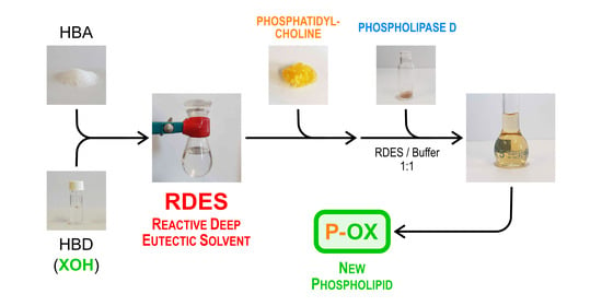
2.1. Preliminary Method Development

The first tests were carried out to screen the solubility of PC in order to select the potential DES candidates for the biocatalytic transformations aiming to produce PS, using PLD from Streptomyces netropsis. In particular, ChCl/Gly 1/2 (mol/mol), ChCl/acetic acid 1/2, choline p-toluensulphonate/acetic acid 1/2, ChCl/urea/acetic acid 1/1/1, and ChCl/levulinic acid 1/2 were tested. The starting PC was soluble in all these solvents, but only the first DES of the list was selected for the first enzymatic trials. The low pH (around 3) associated with all other DESs was in fact unsuitable for the enzymatic activity. However, using ChCl/Gly, besides the desired PS, the presence of PG (derived from the parasitic reaction with DES) was detected as well as a high quantity of unreacted PC. This leads to the decision of developing a specific RDES reaction medium, where the reacting nucleophile was provided as part of the process medium. The nature of the HBA component of RDES and the water content of the reaction medium (needed to preserve the enzymatic activity) were studied in order to increase the solubility of PC and decrease those of the products. The limitation of the product concentration in the reaction medium by precipitation allowed the enhancement of the yields. The RDES design process yielded to the formulations of four RDESs (Section 2.2) and, of the final reaction medium, composed by a RDES/water buffer mixture (1:1, v/v).

2.2. Preparation of RDES Media

In this work, four different RDESs consisting of mixture of HBA/HBD in a 1:2 molar ratio have been prepared. The quaternary ammonium salts choline chloride (ChCl) and trimethylglycine (also called betaine glycine; BetG) have been used as HBAs, whereas glycerol (Gly) and ethylene glycol (EG) constituted HBDs and reactants (see Table 1).
The RDESs have been prepared by using a defined protocol which consisted of mixing the components in a defined molar ratio and heating them at 90 °C for 2–5 h, under constant stirring in a sealed vial until a stable homogeneous colorless one phase liquid was formed (see Supplementary Materials Figure S1). They have been dried at reduced pressure for 24 h, preserved under argon, and then characterized by 1H NMR spectra (see Supplementary Materials Figure S2). The selected HBAs, ChCl, and BetG possess an evident biocompatibility and an availability from natural products which constitute one of their major advantages. In addition, the HBDs Gly and EG are highly available as bulk commodity chemicals. Furthermore, Gly is produced as a by-product of different hydrolysis and transesterification processes of oils and fats most notably in the soap and biodiesel manufacturing industries.
These RDES were suitable for PL modifications because their density was in the range of 1.06 to 1.16 g/cm3, which allowed a good mass transfer and interaction between the reaction components (see Table 2). For that reason, many other RDESs, showing higher density values, have been excluded from the screening. According to the literature, DES’s densities are usually higher than water’s value and are dependent on the working temperatures. As reported in Table 2, the densities of Gly-containing RDESs (RDES A1 and B1) were higher than the densities of EG-containing ones (RDES A2 and B2).

2.3. Transphosphatidylation Reactions

The substrate used for all experiments was a commercial PC from soya beans which was preliminarily purified by precipitation in cold acetone. The acyl chains pattern of PC was identified with the method by ESI/MS and GC/MS previously reported [70]. The fatty acid chains composition is reported in Figure 3 and Table 3.

2.3.1. Preparation of Phosphatidylglycerol

Phosphatidylglycerol (PG) constitutes one of the most abundant PLs at the basis of natural membranes. PG possesses an excellent liposome forming ability and unique surfactant/lubricant properties, especially when mixed with appropriate proteins, and, for these reasons, it has been studied for a long time [71], especially as an emulsifier for drug delivery systems [72,73]. Moreover, in mammalian lungs, PG is highly present compared to other mammalian membranes and therefore the research has been focused on the development of new surfactant preparations based on PG derivatives for therapeutic treatment of diseases such as a neonatal respiratory distress syndrome [74]. Moreover, it has been used for the preparation of plasmid-DNA lipid and polymeric nanovaccines showing that anionic PLs could increase antigen delivery, transport, and stability [75]. Unfortunately, PG could be recovered only in small amounts from natural sources and, even if it has been prepared by different groups [76,77,78], the possibility to set-up new strategies of synthesis is mandatory especially for the biomedical investigations.
Up to now, PG, when enzymatically synthesized, was usually prepared from natural PC by a PLD-mediated process in an organic solvent/buffer biphasic system containing glycerol as the nucleophile. In this work, a new protocol was investigated aiming to avoid the presence of the organic solvent in the reaction medium by using a RDES (see Figure 4). In the first attempts, PLD had been suspended in pure RDES, but the total absence of water completely inactivated the enzyme. As already shown for ILs, a minimal concentration of at least 5% of water was mandatory for the enzyme activity [29]. The process medium composition was optimized by water addition setting the RDES/aqueous phase ratio to the value 1:1 (v/v), resulting also in a modulation of the solvating properties leading to the product precipitation. In particular, the aqueous phase was a 0.1 M NaOAc buffer solution at pH 5.6 containing 0.1 M CaCl2. PC was fully solubilized in RDES and then a buffer solution containing PLD was added. After 40 h at 45 °C, the products that precipitated in the reaction mixture were recovered and analyzed. Remarkably, the extraction of the remaining reaction mixture demonstrated that most of the products were effectively in the precipitate and just a minor amount (≈ 5%) remained dissolved in the RDES/buffer medium. This proved the effectiveness of the RDES/buffer medium design in order to minimize the product concentration in the liquid phase and thus pushing the equilibrium towards the reaction completeness.
When the reaction was performed with RDES A1 (ChCl/Gly 1:2) unreacted PC (~14 %, value obtained by HPLC analysis) was revealed, whereas, in RDES B1 (BetG/Gly 1:2), PC was completely converted in PG. In the first case, the presence of choline as HBA probably prevented the reaction to be completely shifted versus products because of its mass effect opposed to the HBD component (pushing the equilibrium point towards the substrate). In fact, as it resulted in the 1H NMR spectrum (see Figure 5, green spectrum), PGA1 showed a residual peak around 3.2 ppm, which demonstrated the presence of residual unreacted PC. This peak disappeared completely in PGB1, because of the total conversion of PC to the final desired product.
Moreover, the presence of the RDES medium seems to inhibit nearly completely the detrimental hydrolysis side-reaction driving the conversion toward transphosphatidylation in comparison to what happened when the reaction was conducted in organic solvent/water as ESI/MS analysis confirmed (see Supplementary Materials).

2.3.2. Preparation of Phosphatidylethyleneglycol (P-EG).

Polyethyleneglycol (PEG)-grafted PLs are widely studied in the medical field, as they are one of the main components of liposomes used as carriers for mRNA, siRNA [79], and for anticancer active compounds such as paclixatel [80]. The relationship between PEG chain length and concentration in PEG-grafted PLs based vesicles has been deeply investigated [81], aiming to obtain optimized properties, in terms of water permeability and membrane strength, which resulted in a higher liposome lifetime in the blood circulation, and a better drug delivery. In this context, the authors decided to synthesize a non-natural phosphatidylethyleneglycol (P-EG), which could be an interesting building block for the production of new P-EG containing PLs. The free OH on the polar head could be then available to undergo esterification reactions, allowing the production of novel tailor-made products. In order to obtain a reference P-EG, the product has been firstly prepared with the conventional method in toluene/buffer solution. Subsequently, PC was fully solubilized in RDESs A2 (ChCl/EG 1:2) and B2 (BetG/EG 1:2) containing both ethylene glycol which constituted HBD as well as the nucleophile in the PLD catalyzed transphosphatidylation (see Figure 6). A buffer solution containing PLD was added to the solution to start the reaction. After 40 h at 45 °C, the products, precipitated in the reaction medium, were recovered and analyzed.
Also in this case, as described in Section 2.3.1, most of the products precipitated and just a minor part (≈10%) was recovered by solvent extraction from the RDES/buffer medium. This confirmed again the efficacy of the process medium optimization in the modulation of the reagent and products solubility. 1H NMR spectra of the final products are reported in Figure 7 (P-EGA2, green line, and P-EGB2 blue line). As already observed in the PGs preparation in 2.2.1, when the reaction was performed in RDES containing ChCl, namely RDES A2, a small quantity of PC (~8%, value obtained by HPLC analysis) remained unreacted, whereas, in RDES B2 (BetG/EG 1:2), PC was completely converted to P-EG.

3. Materials and Methods

All chemicals were purchased from Merck (Merck Life Science S.r.l., Milan, Italy) and used without further purification. The employed solvents were of analytical or HPLC grade when necessary. Phosphatidylcholine was supplied from Lucas Meyer (Epikuron 200, soya lecithin, Hamburg, Germany) and used after precipitation in cold acetone (10 g Ep200 dissolved in 15 mL of CH2Cl2, added to 300 mL of cold acetone). 1H NMR and 31P NMR spectra were recorded on a 400 MHz Bruker Avance spectrometer (Milano, Italy). Acquisition and data treatment were performed with Bruker TopSpin 3.2 software. Chemical shifts were reported in δ units (ppm), relative to tetramethylsilane (TMS) as the internal standard and all spectra were recorded in D2O or CDCl3/CD3OD. Deuterated solvents were purchased from Eurisotop (Saint-Aubin, France).

3.1. 31P NMR Sample Preparation

In addition, 50 mg of the sample was dissolved with 5 mg of triphenyl phosphate in 0.6 mL of a CDCl3/CH3OH 2:1 solution mixed with 0.4 mL of a CsEDTA solution (prepared. by titration of 0.2 M EDTA in D2O with CsCO3 until pH 7.6 and then diluted 1:4 (v/v) with methanol). Low-soluble samples were vigorously stirring to allow the dissolution. After the complete solubilization, 0.2 mL of water was added to the mixture and, after stirring, the solution was transferred into an NMR tube and left to rest in order to achieve the correct separation of the two liquid phases. The 31P uncoupled experiments were performed with the following parameters: D1: 10 s, SW: 40 ppm, O1: 10 ppm, FID size: 32768, ns: 32, T: 320 K.

3.2. Thin Layer Chromatography (TLC)

TLC plates Silica gel 60 SIL G-25 UV254 glass 250 µm (Macherey Nagel, Düren, Germany) were used for analytical TLC. The employed eluents were constituted by or CHCl3/CH3OH/CH3COCH3/CH3COOH/H2O: 50/10/20/10/5 and CHCl3/CH3OH/NH3 (33% aq.): 65/30/2.5 (ratios reported in volume). Detection was performed by staining with a cerium molibdate reagent.

3.3. Gas Chromatography

To determine the fatty acid composition in PC from soya, fatty acid methyl esters (FAMEs) were obtained by hydrolysis of PC with methanol/ 2 M NaOH under reflux for 4 h. The reaction mixture was then cooled, evaporated to remove methanol, acidified, and extracted carefully three times with ethylacetate. The organic solutions were collected, anhydrified on sodium sulphate and evaporated at p.r. The residue has been dissolved in diethyl ether and treated with an ethereal solution of diazomethane 30 min at r.t. After evaporation, the residue was dissolved in CH2Cl2 and analyzed using a gas chromatograph system HP-6890 equipped with a HP-5973 mass detector and a HP-5MS column (30 m length × 250 µm internal diameter, 0.25 µm film thickness, Hewlett Packard, Palo Alto, CA, USA). Helium was the carrier gas with a flux of 1 mL min−1; the splitting ratio was 1:30. The injector and detector temperatures were set at 250 °C. The used temperature program is reported here: 120 °C (3 min)–12 °C/min–195 °C (10 min)–12 °C/min–300 °C (10 min). The FAMEs were identified by comparison with known standards and by means of NIST 2008 mass spectral library search.

3.4. High Performance Liquid Chromatography (HPLC)

HPLC analyses were performed on a Merck Hitachi L4000 apparatus (Japan) fitted with a Sepachrom Adamas®Silica (Rho, Milano, Italy) 5µ-2 column, length/internal diameter 250/4.6, and a UV detector set to λ = 206 nm. The column temperature was maintained at r.t. PL mixtures were analyzed using an isocratic mobile phase consisting of CH3CN/CH3OH/H3PO4 952/36/10.9 at a flow rate of 0.5 mL/min. The samples were dissolved in a mixture of hexane/isopropanol (1:1, v/v) with a final concentration of 1 mg/mL and filtered through a 0.45 µm filter before injection

3.5. Electrospray Ionization Mass Spectrometry

Mass spectra were recorded on a ESI/MS Bruker Esquire 3000 PLUS (ESI Ion Trap LC/MSn System, Bruker, Milano, Italy), by direct infusion of a dichloromethane solution of compounds with an infusion rate of 4 µL/min. Samples were analyzed using both positive and negative ionization modes to allow the detection of all relevant compounds.

3.6. PLD Preparation

3.6.1. Microorganism Fermentation

PLD was obtained from the fermentation broth of Streptomyces netropsis (DSM 40093). The medium for strain growth and maintenance was composed by glucose 4 g/L; yeast extract 5 g/L; malt extract 10 g/L.
Medium for PLD preparation: glucose 5 g/L; yeast extract 5 g/L; malt extract 10 g/L; peptone from soyabean 3 g/L; peptone from casein 1 g/L; MgSO4 0.2 g/L; NaCl 0.2 g/L.
A single colony of active Streptomyces netropsis culture was picked up from a Petri dish, was suspended in 1 mL of sterile water, and was then inoculated in a 500 mL conical Pyrex flask containing 150 mL of the growth medium. The flask was shaken for 5 days at 30 °C and 140 rpm. After this period, the cells were centrifuged for 15 min (4 °C, 4000× g) and collected, removing the medium. The cells were suspended in 10 mL of fresh medium and were added to a sterilized fermenter vessel of a 5 L bioreactor (Biostat A BB-8822000, Sartorius Stedim, Göttingen, Germany) containing 4 L of medium for PLD preparation. The temperature, the stirring speed, and the pH were set to 30 °C, 250 rpm and 7.0, respectively. The value of pH was controlled by addition of sterilized aqueous solutions (10% w/w in water) of either acetic acid or ammonia. The fermentation was performed aerobically, setting a minimum air flow of 2 L/min (0.5 v/v/min). After 48 h, the fermentation broth was filtered through a celite pad, and the clear liquid was stored at 4 °C in dark bottles.

3.6.2. Precipitation and Dialysis

To 1 L of the filtered broth, 430 g of ammonium sulfate was added over 1 h at 4 °C under stirring. Once the ammonium sulfate was completely dissolved, the broth was left overnight at 4 °C to allow the precipitation. After 24 h, the precipitated pellet was separated from the broth by centrifugation at 8000 rpm for 15 min at 4 °C. The solid was then dissolved in a small amount of supernatant broth and dialyzed with a seamless cellulose tube (D0655 Sigma-Aldrich, Milano, Italy) against 10 mM sodium acetate buffer at pH 5. The dialyzed broth (80 mL), which had an activity of 0.525 U/mL, was finally lyophilized (240 mg, 0.175 U/mg) and used as solid powder for bioconversions.

3.7. Protein Determination

Protein concentration was determined in the dialyzed broth by the Bradford method using bovine serum albumin (BSA) as standard [82] and was 0.94 mg/mL (analysis performed in triplicate).

3.8. PLD Activity Determination

In addition, 360 mg of phosphatidyl-p-nitrophenol (PpNP), prepared as described in our previous work [83], was dissolved in diethylether (1 mL) and added with 15 mL of 0.1 M Tris buffer and 15 mL of a solution of 10% Triton X-100 in the same buffer, giving a final concentration of 16 mM. The mixture was heated gently to 60 °C until it became clear, the organic solvent was removed under reduced pressure, and the solution was stored in dark vials at −18 °C. PLD activity was determined spectrophotometrically using a UV-Vis Double Beam spectrophotometer V-730 Jasco (Jasco Europe, Cremella, Italy) in 0.05 M Tris Buffer, pH 8, by monitoring the hydrolysis of PpNP to phosphatidic acid and p-nitrophenol at 405 nm using a molar extinction coefficient of 18.450 mmol−1 L cm−1. One unit (U) is defined as the amount of enzyme able to hydrolyze one µmol of PpNP in 1 min.

3.9. RDES Preparation

HBA (choline chloride or glycine betaine) and HBD (glycerol or ethylene glycol) were previously dried for 10 h under vacuum. They were then mixed in a 1:2 molar ratio (see Table 2 for detailed composition) and heated under magnetic stirring for 2–4 h at 70–90 °C until a homogeneous and clear liquid phase was obtained. The synthesized RDESs were left to cool slowly to r.t., and they were stored in a sealed vial under argon until used. They were analyzed by 1H NMR (spectra are reported in Supplementary Materials Figure S2).

3.10. RDES Density Measurement

The density of the RDESs prepared in 3.9 has been measured at atmospheric pressure and at 18 °C with the following procedure. In addition, 2 mL volumetric flasks have been weighed on an analytical balance with a resolution of 0.1 mg. Then, they have been filled with the opportune RDES and thermostated at 18 °C for 1 h. Then, the volume was adjusted to the right value and the flasks were weighted. Each measure had been performed in duplicate. The density has been calculated for each RDES as the ratio between the mass of RDES (mass of filled flask–mass of empty flask) and the volume of the flask. The final value reported in Table 2 is the average of the two measurements.

3.11. RDES Preparation of Phosphatidylglycerols PGA1 and PGB1 in RDES A1 and B1

Furthermore, 150 mg of PC was dissolved at 45 °C in 10 mL of RDES (A1 or B1). Once full solubilization of the substrate in RDES was achieved, 10 mL of buffer solution containing 0.1 M of sodium acetate, 0.1 M of calcium chloride, and 15 mg (2.6 U) of solid PLD adjusted to pH 5.6 were added to the solution. This mixture was left stirring at 45 °C for 40 h. The product precipitated in the reaction mixture and was isolated by filtration on a Buchner under vacuum. The liquid solution was extracted with toluene, in order to check the presence of residual PLs. The precipitated products for RDES B1 accounted for 60% (w/w, 90 mg) of the reaction yield, while the products recovered from the medium extraction were instead 3% (w/w, 5 mg) of the reaction yield. The product was then purified from residual RDES: it was dissolved in 1 mL of toluene, and the solution was left stirring with 5 mL of water for 30 min at r.t. The organic phase was then separated and evaporated under reduced pressure. The PLs mixture recoveries were 130 mg (86% w/w) for PGA1 preparation and 95 mg (63% w/w) for PGB1. The final mixtures were analyzed by ESI/MS and NMR.
PGA1 ESI/MS negative ion spectrum m/z values: 16:0/18:2-PG [745.6], 18:2/18:2-PG [769.5]. Residual PC was seen in the positive ion spectrum with m/z: 16:0/18.2-PC [780.7 + Na]+, 18:2/18:2-PC [804.6 + Na]+. 1H NMR: δH 0.84−0.85 (m, 6H, CH3), 1.23−1.27 (m, 34 H, CH2 acyl chains), 1.50−1.60 (m, 4H, CH2CH2CO), 1.99−2.04 (m, 7H, CH2−CHCH), 2.23−2.30 (m, 4H, CH2CO), 2.72−2.77 (m, 3H, CHCH−CH−CHCH), 3.58−3.64 (m, 3H, CHOHCH2OH), 3.93−4.12 (m, 5H, O−CH2−CHOH−CH2O and CH2−OPO3−), 4.34−4.37 (m, 1H, CH2−OPO3−), 5.18 (m, 1H, CH−OH), 5.29−5.35 (m, 6H, CHCH). 31P NMR δ: 0.59 ppm.
PGB1 ESI/MS negative ion spectrum—the desired product was identified with the following m/z values: 16:0/18:2-PG [745.5], 18:2/18:2-PG [769.5]. No residual PC was observed in the positive ion spectra. 1H NMR: δH 0.82−0.86 (m, 6H, CH3), 1.21−1.27 (m, 34 H, CH2 acyl chains), 1.50−1.60 (m, 4H, CH2CH2CO), 1.98−2.03 (m, 7 H, CH2−CHCH), 2.22-2.29 (m, 4H, CH2CO), 2.71-2.76 (m, 3H, CHCH−CH−CHCH), 3.59−3.64 (m, 3H, CHOHCH2OH), 3.85−4.13 (m, 5H, O−CH2−CHOH−CH2O and CH2−OPO3−), 4.33−4.36 (m, 1H, CH2−OPO3−), 5.17 (m, 1H, CH−OH), 5.27−5.35 (m, 6H, CHCH). 31P NMR δ: 0.59 ppm.

3.12. RDES Preparation of Phosphatidylethyleneglycols P-EGA2 and P-EGB2 in RDES A2 and B2

In addition, 150 mg of PC was dissolved at 45 °C in 10 mL of RDES (A2 or B2). Once full solubilization of the substrate in RDES was achieved, 10 mL of buffer solution containing 0.1 M of sodium acetate, 0.1 M of calcium chloride, and 15 mg (2.6 U) of PLD powder adjusted to pH 5.6 were added to the solution. This mixture was left stirring at 45 °C for 40 h. The liquid solution was then separated by filtration from the solid product. The solution was extracted with toluene, whereas the solid was dissolved in toluene. The precipitated products for RDES B2 accounted for 63.3% (w/w, 95 mg) of the reaction yield, while the products recovered from the medium extraction were instead 6.6% (w/w, 10 mg) of the reaction yield. All the organic phases were collected and evaporated under reduced pressure. The product containing RDES residues was dissolved in 1 mL of toluene, and the solution was left stirring with 5 mL of water for 30 min at r.t. The organic phase was separated and evaporated under reduced pressure. The PL mixture recoveries were 140 mg (93% w/w) for P-EGA2 and 105 mg (70% w/w) for P-EGB2. The final mixtures were analyzed by ESI/MS and NMR.
P-EGA2: ESI/MS negative ion spectrum—the desired product was identified with the following m/z values: 16:0/18:2-P-EG [715.6]-, 18:2/18:2-P-EG [739.6]. Some PC was recorded in the positive ion spectrum with m/z: 16:0/18.2-PC [780+Na]+, 18:2/18:2-PC [804.7+Na]+. 1H NMR: δH 0.84−0.88 (m, 6H, CH3), 1.23−1.27 (m, 34 H, CH2 acyl chains), 1.56 (m, 4H, CH2CH2CO), 2.00−2.05 (m, 7 H, CH2−CHCH), 2.24−2.29 (m, 4H, CH2CO), 2.72−2.76 (m, 3H, CHCH−CH−CHCH), 3.38 (m, 1H, CH2CH2OH), 3.64−4.24 (m, 6H, O−CH2−CHO−CH2O and CH2−OPO3−), 4.35−4.38 (m, 1H, CH2−OPO3), 5.19 (m, 1H, CH−OH), 5.31−5.37 (m, 6H, CHCH). 31P NMR δ: 0.35 ppm.
P-EGB2: ESI/MS negative ion spectrum—the desired product P-EG was identified with the following m/z values: 16:0/18:2-P-EG [715.8], 18:2/18:2-P-EG [739.6]. No PC was recorded in the positive ion spectrum. 1H NMR: δH 0.86−0.89 (m, 6H, CH3), 1.25−1.29 (m, 34 H, CH2 acyl chains), 1.58 (m, 4H, CH2CH2CO), 2.01−2.06 (m, 7 H, CH2−CHCH), 2.24−2.30 (m, 4H, CH2CO), 2.74−2.77 (m, 3H, CHCH−CH−CHCH), 3.30 (m, 1H, CH2CH2OH), 3.71−4.25 (m, 6H, O−CH2−CHO−CH2O and CH2−OPO3−), 4.36−4.40 (m, 1H, CH2−OPO3), 5.20 (m, 1H, CH−OH), 5.30−5.38 (m, 6H, CHCH). 31P NMR δ: 0.35 ppm.

4. Conclusions

The present work constitutes an innovative approach for the biocatalytic preparation of polar head modified PLs based on the use of RDESs/buffer mixtures, which allows the conversion of natural PC in high yields. RDESs act both as solvents and reactants because they contain the alcoholic nucleophiles that will substitute the choline moiety in the transphosphatidylation reaction. The best results have been obtained using two RDESs constituted by glycine betaine as HBA and glycerol or ethyleneglycol as HBD with a total conversion of starting PC due to the absence of choline in the composition of RDES. The implementation of specifically designed reaction media based on RDESs and buffer resulted in several distinctive advantages, including the yield enhancement by the synergic combination of a substantial mass effect (due to the high concentration of nucleophile reactant in the RDES) and the product precipitation (which maintains at a low value its concentration in the medium). Moreover, the implementation of RDES/buffer reaction medium demonstrates the inhibition of the parasitic competitive hydrolysis of PC to PA, improving the reaction yields and easing the product recovery and purification. Detailed studies about the interaction of RDESs with PLD catalysis are under way. In fact, the effect of RDESs (and DESs) on the phospholipase’s active site as well as on PLs aggregates is hardly predictable, and further studies are needed to fully exploit this promising approach to biocatalysis.

Supplementary Materials

The following are available online at https://www.mdpi.com/article/10.3390/catal11060655/s1, Figure S1: RDES preparation example (left image: initial mixture of ChCl (HBD) and ethylene glycol (HBA), right image: RDES A2). Figure S2: 1H NMR spectra of the four RDES A1, A2, B1 and B2 in D2O. Figure S3: 1H NMR Spectrum of PC. Figure S4: Mass spectra of PGA1 obtained in RDES A1. Figure S5: Mass spectra of PGB1 obtained in RDES B1. Figure S6: Mass spectra of P-EGA2 obtained in RDES A2. Figure S7: Mass spectra of P-EGB2 obtained in RDES B2.

Author Contributions

Conceptualization, P.D.; methodology, C.A., F.G.G., S.S., A.S., P.D.; investigation, C.A., F.G.G., S.M., L.A.M.R., S.S.; resources, P.D. and S.S; writing—original draft preparation, C.A., A.S., P.D.; writing—review and editing, C.A., F.G.G., L.A.M.R., S.S., A.S., P.D.; funding acquisition, S.S., P.D. All authors have read and agreed to the published version of the manuscript.

Funding

This work has been in part supported by Fondazione Cariplo and Innovhub-SSI, Grant No. 2017-1015 (SOAVE-Bando congiunto Ricerca Integrata sulle Biotecnologie industriali e sulla Bioeconomia ed. 2017). L.R. is a PhD student of the Research Doctorate Program in Chemical Engineering and Industrial Chemistry at Politecnico di Milano.

Data Availability Statement

Data is contained within the article or supplementary material.

Acknowledgments

The authors wish to thank Walter Panzeri (SCITEC-CNR) for his technical support on ESI/MS analysis.

Conflicts of Interest

The authors declare no conflict of interest.

References

  1. Sheldon, R.A.; Woodley, J.M. Role of Biocatalysis in Sustainable Chemistry. Chem. Rev. 2018, 118, 801–838. [Google Scholar] [CrossRef]
  2. Expanding biocatalysis for a sustainable future. Nat. Catal. 2020, 3, 179–180. [CrossRef]
  3. Akretche, H.; Pierre, G.; Moussaoui, R.; Michaud, P.; Delattre, C. Valorization of olive mill wastewater for the development of biobased polymer films with antioxidant properties using eco-friendly processes. Green Chem. 2019, 21, 3065–3073. [Google Scholar] [CrossRef]
  4. Cicco, L.; Dilauro, G.; Perna, F.M.; Vitale, P.; Capriati, V. Advances in deep eutectic solvents and water: Applications in metal- and biocatalyzed processes, in the synthesis of APIs, and other biologically active compounds. Org. Biomol. Chem. 2021, 19, 2558–2577. [Google Scholar] [CrossRef]
  5. Smith, E.L.; Abbott, A.P.; Ryder, K.S. Deep Eutectic Solvents (DESs) and Their Applications. Chem. Rev. 2014, 114, 11060–11082. [Google Scholar] [CrossRef] [Green Version]
  6. Hansen, B.B.; Spittle, S.; Chen, B.; Poe, D.; Zhang, Y.; Klein, J.M.; Horton, A.; Adhikari, L.; Zelovich, T.; Doherty, B.W.; et al. Deep Eutectic Solvents: A Review of Fundamentals and Applications. Chem. Rev. 2021, 121, 1232–1285. [Google Scholar] [CrossRef]
  7. Abbott, A.P.; Capper, G.; Davies, D.L.; Rasheed, R.K.; Tambyrajah, V. Novel solvent properties of choline chloride/urea mixtures. Chem. Commun. 2003, 70–71. [Google Scholar] [CrossRef] [PubMed] [Green Version]
  8. Nahar, Y.; Thickett, S.C. Greener, Faster, Stronger: The Benefits of Deep Eutectic Solvents in Polymer and Materials Science. Polymers 2021, 13, 447. [Google Scholar] [CrossRef] [PubMed]
  9. Santana-Mayor, Á.; Rodríguez-Ramos, R.; Herrera-Herrera, A.V.; Socas-Rodríguez, B.; Rodríguez-Delgado, M.Á. Deep eutectic solvents. The new generation of green solvents in analytical chemistry. TrAC Trends Anal. Chem. 2021, 134. [Google Scholar] [CrossRef]
  10. Şahin, S. Tailor-designed deep eutectic liquids as a sustainable extraction media: An alternative to ionic liquids. J. Pharm. Biomed. Anal. 2019, 174, 324–329. [Google Scholar] [CrossRef] [PubMed]
  11. Grudniewska, A.; de Melo, E.M.; Chan, A.; Gniłka, R.; Boratyński, F.; Matharu, A.S. Enhanced Protein Extraction from Oilseed Cakes Using Glycerol–Choline Chloride Deep Eutectic Solvents: A Biorefinery Approach. ACS Sustain. Chem. Eng. 2018, 6, 15791–15800. [Google Scholar] [CrossRef]
  12. Kalhor, P.; Ghandi, K. Deep Eutectic Solvents as Catalysts for Upgrading Biomass. Catalysts 2021, 11, 178. [Google Scholar] [CrossRef]
  13. Hooshmand, S.E.; Afshari, R.; Ramón, D.J.; Varma, R.S. Deep eutectic solvents: Cutting-edge applications in cross-coupling reactions. Green Chem. 2020, 22, 3668–3692. [Google Scholar] [CrossRef]
  14. Di Carmine, G.; Abbott, A.P.; D’Agostino, C. Deep eutectic solvents: Alternative reaction media for organic oxidation reactions. React. Chem. Eng. 2021, 6, 582–598. [Google Scholar] [CrossRef]
  15. Durand, E.; Lecomte, J.; Baréa, B.; Piombo, G.; Dubreucq, E.; Villeneuve, P. Evaluation of deep eutectic solvents as new media for Candida antarctica B lipase catalyzed reactions. Process Biochem. 2012, 47, 2081–2089. [Google Scholar] [CrossRef]
  16. Xu, P.; Zheng, G.-W.; Zong, M.-H.; Li, N.; Lou, W.-Y. Recent progress on deep eutectic solvents in biocatalysis. Bioresour. Bioprocess. 2017, 4, 34. [Google Scholar] [CrossRef] [PubMed]
  17. Panić, M.; Cvjetko Bubalo, M.; Radojčić Redovniković, I. Designing a biocatalytic process involving deep eutectic solvents. J. Chem. Technol. Biotechnol. 2021, 96, 14–30. [Google Scholar] [CrossRef]
  18. Cicco, L.; Ríos-Lombardía, N.; Rodríguez-Álvarez, M.J.; Morís, F.; Perna, F.M.; Capriati, V.; García-Álvarez, J.; González-Sabín, J. Programming cascade reactions interfacing biocatalysis with transition-metal catalysis in Deep Eutectic Solvents as biorenewable reaction media. Green Chem. 2018, 20, 3468–3475. [Google Scholar] [CrossRef]
  19. Vitale, P.; Perna, F.M.; Agrimi, G.; Pisano, I.; Mirizzi, F.; Capobianco, R.V.; Capriati, V. Whole-Cell Biocatalyst for Chemoenzymatic Total Synthesis of Rivastigmine. Catalysts 2018, 8, 55. [Google Scholar] [CrossRef] [Green Version]
  20. Płotka-Wasylka, J.; de la Guardia, M.; Andruch, V.; Vilková, M. Deep eutectic solvents vs. ionic liquids: Similarities and differences. Microchem. J. 2020, 159. [Google Scholar] [CrossRef]
  21. Perna, F.M.; Vitale, P.; Capriati, V. Deep eutectic solvents and their applications as green solvents. Curr. Opin. Green Sustain. Chem. 2020, 21, 27–33. [Google Scholar] [CrossRef]
  22. Vilková, M.; Płotka-Wasylka, J.; Andruch, V. The role of water in deep eutectic solvent-base extraction. J. Mol. Liq. 2020, 304. [Google Scholar] [CrossRef]
  23. Tan, J.-N.; Dou, Y. Deep eutectic solvents for biocatalytic transformations: Focused lipase-catalyzed organic reactions. Appl. Microbiol. Biotechnol. 2020, 104, 1481–1496. [Google Scholar] [CrossRef]
  24. Pätzold, M.; Siebenhaller, S.; Kara, S.; Liese, A.; Syldatk, C.; Holtmann, D. Deep Eutectic Solvents as Efficient Solvents in Biocatalysis. Trends Biotechnol. 2019, 37, 943–959. [Google Scholar] [CrossRef] [PubMed]
  25. Guajardo, N.; Domínguez de María, P.; Ahumada, K.; Schrebler, R.A.; Ramírez-Tagle, R.; Crespo, F.A.; Carlesi, C. Water as Cosolvent: Nonviscous Deep Eutectic Solvents for Efficient Lipase-Catalyzed Esterifications. ChemCatChem 2017, 9, 1393–1396. [Google Scholar] [CrossRef]
  26. Sirviö, J.A.; Visanko, M. Lignin-rich sulfated wood nanofibers as high-performing adsorbents for the removal of lead and copper from water. J. Hazard. Mater. 2020, 383. [Google Scholar] [CrossRef] [PubMed]
  27. Sirviö, J.A.; Heiskanen, J.P. Synthesis of Alkaline-Soluble Cellulose Methyl Carbamate Using a Reactive Deep Eutectic Solvent. ChemSusChem 2017, 10, 455–460. [Google Scholar] [CrossRef] [PubMed] [Green Version]
  28. Mota-Morales, J.D.; Sánchez-Leija, R.J.; Carranza, A.; Pojman, J.A.; del Monte, F.; Luna-Bárcenas, G. Free-radical polymerizations of and in deep eutectic solvents: Green synthesis of functional materials. Prog. Polym. Sci. 2018, 78, 139–153. [Google Scholar] [CrossRef]
  29. Gore, S.; Baskaran, S.; Koenig, B. Efficient synthesis of 3,4-dihydropyrimidin-2-ones in low melting tartaric acid–urea mixtures. Green Chem. 2011, 13. [Google Scholar] [CrossRef] [Green Version]
  30. Singh, B.S.; Lobo, H.R.; Pinjari, D.V.; Jarag, K.J.; Pandit, A.B.; Shankarling, G.S. Comparative material study and synthesis of 4-(4-nitrophenyl)oxazol-2-amine via sonochemical and thermal method. Ultrason. Sonochemistry 2013, 20, 633–639. [Google Scholar] [CrossRef]
  31. Zeng, C.-X.; Qi, S.-J.; Xin, R.-P.; Yang, B.; Wang, Y.-H. Enzymatic selective synthesis of 1,3-DAG based on deep eutectic solvent acting as substrate and solvent. Bioprocess Biosyst. Eng. 2015, 38, 2053–2061. [Google Scholar] [CrossRef] [PubMed]
  32. Siebenhaller, S.; Muhle-Goll, C.; Luy, B.; Kirschhöfer, F.; Brenner-Weiss, G.; Hiller, E.; Günther, M.; Rupp, S.; Zibek, S.; Syldatk, C. Sustainable enzymatic synthesis of glycolipids in a deep eutectic solvent system. J. Mol. Catal. B Enzym. 2016, 133, S281–S287. [Google Scholar] [CrossRef]
  33. Hümmer, M.; Kara, S.; Liese, A.; Huth, I.; Schrader, J.; Holtmann, D. Synthesis of (-)-menthol fatty acid esters in and from (-)-menthol and fatty acids—Novel concept for lipase catalyzed esterification based on eutectic solvents. Mol. Catal. 2018, 458, 67–72. [Google Scholar] [CrossRef]
  34. Lamari, F.; Mochel, F.; Sedel, F.; Saudubray, J.M. Disorders of phospholipids, sphingolipids and fatty acids biosynthesis: Toward a new category of inherited metabolic diseases. J. Inherit. Metab. Dis. 2013, 36, 411–425. [Google Scholar] [CrossRef]
  35. Calvano, C.D.; Losito, I.; Cataldi, T. Editorial to the Special Issue “Lipidomics and Neurodegenerative Diseases”. Int. J. Mol. Sci. 2021, 22, 1270. [Google Scholar] [CrossRef] [PubMed]
  36. D’Arrigo, P.; Scotti, M. Lysophospholipids: Synthesis and Biological Aspects. Curr. Org. Chem. 2013, 17, 812–830. [Google Scholar] [CrossRef]
  37. Li, J.; Wang, X.; Zhang, T.; Wang, C.; Huang, Z.; Luo, X.; Deng, Y. A review on phospholipids and their main applications in drug delivery systems. Asian J. Pharm. Sci. 2015, 10, 81–98. [Google Scholar] [CrossRef]
  38. Gliszczyńska, A.; Niezgoda, N.; Gładkowski, W.; Czarnecka, M.; Świtalska, M.; Wietrzyk, J. Synthesis and Biological Evaluation of Novel Phosphatidylcholine Analogues Containing Monoterpene Acids as Potent Antiproliferative Agents. PloS ONE 2016, 11, e0157278. [Google Scholar] [CrossRef]
  39. Falconi, M.; Ciccone, S.; D’Arrigo, P.; Viani, F.; Sorge, R.; Novelli, G.; Patrizi, P.; Desideri, A.; Biocca, S. Design of a novel LOX-1 receptor antagonist mimicking the natural substrate. Biochem. Biophys. Res. Commun. 2013, 438, 340–345. [Google Scholar] [CrossRef] [Green Version]
  40. Kosicek, M.; Hecimovic, S. Phospholipids and Alzheimer’s Disease: Alterations, Mechanisms and Potential Biomarkers. Int. J. Mol. Sci. 2013, 14, 1310–1322. [Google Scholar] [CrossRef]
  41. Shamim, A.; Mahmood, T.; Ahsan, F.; Kumar, A.; Bagga, P. Lipids: An insight into the neurodegenerative disorders. Clin. Nutr. Exp. 2018, 20, 1–19. [Google Scholar] [CrossRef] [Green Version]
  42. Küllenberg, D.; Taylor, L.A.; Schneider, M.; Massing, U. Health effects of dietary phospholipids. Lipids Health Dis. 2012, 11, 3. [Google Scholar] [CrossRef] [PubMed] [Green Version]
  43. Schverer, M.; O’Mahony, S.M.; O’Riordan, K.J.; Donoso, F.; Roy, B.L.; Stanton, C.; Dinan, T.G.; Schellekens, H.; Cryan, J.F. Dietary phospholipids: Role in cognitive processes across the lifespan. Neurosci. Biobehav. Rev. 2020, 111, 183–193. [Google Scholar] [CrossRef] [PubMed]
  44. Cui, L.; Decker, E.A. Phospholipids in foods: Prooxidants or antioxidants? J. Sci. Food Agric. 2016, 96, 18–31. [Google Scholar] [CrossRef]
  45. Sun, N.; Chen, J.; Wang, D.; Lin, S. Advance in food-derived phospholipids: Sources, molecular species and structure as well as their biological activities. Trends Food Sci. Technol. 2018, 80, 199–211. [Google Scholar] [CrossRef]
  46. Pucek, A.; Niezgoda, N.; Kulbacka, J.; Wawrzeńczyk, C.; Wilk, K.A. Phosphatidylcholine with conjugated linoleic acid in fabrication of novel lipid nanocarriers. Colloids Surf. APhysicochem. Eng. Asp. 2017, 532, 377–388. [Google Scholar] [CrossRef]
  47. Aldosari, B.N.; Alfagih, I.M.; Almurshedi, A.S. Lipid Nanoparticles as Delivery Systems for RNA-Based Vaccines. Pharmaceutics 2021, 13, 206. [Google Scholar] [CrossRef]
  48. Samaridou, E.; Heyes, J.; Lutwyche, P. Lipid nanoparticles for nucleic acid delivery: Current perspectives. Adv. Drug Deliv. Rev. 2020, 154–155, 37–63. [Google Scholar] [CrossRef]
  49. De Serrano, L.O.; Burkhart, D.J. Liposomal vaccine formulations as prophylactic agents: Design considerations for modern vaccines. J. Nanobiotechnol. 2017, 15, 83. [Google Scholar] [CrossRef] [Green Version]
  50. D’Arrigo, P.; Giordano, C.; Macchi, P.; Malpezzi, L.; Pedrocchi-Fantoni, G.; Servi, S. Synthesis, Platelet Adhesion and Cytotoxicity Studies of New Glycerophosphoryl-Containing Polyurethanes. Int. J. Artif. Organs 2007, 30, 133–143. [Google Scholar] [CrossRef]
  51. Zhiguang, H.; Hui, Z.; Wenqiang, G.; Jianfu, L.; Charles, B.; Don, K.; Maneesha, S.M. Vesicle properties and health benefits of milk phospholipids: A review. J. Food Bioact. 2019, 5. [Google Scholar] [CrossRef] [Green Version]
  52. Lordan, R.; Tsoupras, A.; Zabetakis, I. Phospholipids of Animal and Marine Origin: Structure, Function, and Anti-Inflammatory Properties. Molecules 2017, 22, 1964. [Google Scholar] [CrossRef] [Green Version]
  53. Chojnacka, A.; Gladkowski, W.; Kielbowicz, G.; Wawrzenczyk, C. Isolation of egg-yolk phospholipids and enzymatic modification of their acyl chains. Lipid Technol. 2012, 24, 33–35. [Google Scholar] [CrossRef]
  54. Niezgoda, N.; Gliszczyńska, A.; Gładkowski, W.; Chojnacka, A.; Kiełbowicz, G.; Wawrzeńczyk, C. Production of concentrates of CLA obtained from sunflower and safflower and their application to the lipase-catalyzed acidolysis of egg yolk phosphatidylcholine. Eur. J. Lipid Sci. Technol. 2016, 118, 1566–1578. [Google Scholar] [CrossRef]
  55. D’Arrigo, P.; Servi, S. Using phospholipases for phospholipid modification. Trends Biotechnol. 1997, 15, 90–96. [Google Scholar] [CrossRef]
  56. Baldassarre, F.; Allegretti, C.; Tessaro, D.; Carata, E.; Citti, C.; Vergaro, V.; Nobile, C.; Cannazza, G.; D’Arrigo, P.; Mele, A.; et al. Biocatalytic Synthesis of Phospholipids and Their Application as Coating Agents for CaCO3 Nano–crystals: Characterization and Intracellular Localization Analysis. ChemistrySelect 2016, 1, 6507–6514. [Google Scholar] [CrossRef]
  57. Leiros, I.; Hough, E.; D’Arrigo, P.; Carrea, G.; Pedrocchi-Fantoni, G.; Secundo, F.; Servi, S. Crystallization and preliminary X-ray diffraction studies of phospholipase D from Streptomyces sp. Acta Crystallogr. Sect. D Biol. Crystallogr. 2000, 56, 466–468. [Google Scholar] [CrossRef]
  58. Carrea, G.; D’Arrigo, P.; Secundo, F.; Servi, S. Purification and applications of a phospholipase D from a new strain of Streptomyces. Biotechnol. Lett. 1997, 19, 1083–1085. [Google Scholar] [CrossRef]
  59. Carrea, G.; D’Arrigo, P.; Piergianni, V.; Roncaglio, S.; Secundo, F.; Servi, S. Purification and properties of two phospholipases D from Streptomyces sp. Biochim. Biophys. Acta Lipids Lipid Metab. 1995, 1255, 273–279. [Google Scholar] [CrossRef]
  60. D’Arrigo, P.; de Ferra, L.; Piergianni, V.; Ricci, A.; Scarcelli, D.; Servi, S. Phospholipase D from Streptomyces catalyses the transfer of secondary alcohols. J. Chem. Soc. Chem. Commun. 1994, 1709–1710. [Google Scholar] [CrossRef]
  61. D’Arrigo, P.; de Ferra, L.; Piergianni, V.; Selva, A.; Servi, S.; Strini, A. Preparative transformation of natural phospholipids catalysed by phospholipase D from Streptomyces. J. Chem. Soc. Perkin Trans. 1 1996, 2651–2656. [Google Scholar] [CrossRef]
  62. Bossi, L.; D’Arrigo, P.; Pedrocchi-Fantoni, G.; Mele, A.; Servi, S.; Leiros, I. The substrate requirements of phospholipase D. J. Mol. Catal. B Enzym. 2001, 11, 433–438. [Google Scholar] [CrossRef]
  63. Allegretti, C.; Denuccio, F.; Rossato, L.; D’Arrigo, P. Polar Head Modified Phospholipids by Phospholipase D-Catalyzed Transformations of Natural Phosphatidylcholine for Targeted Applications: An Overview. Catalysts 2020, 10, 997. [Google Scholar] [CrossRef]
  64. Kim, H.-Y.; Huang, B.X.; Spector, A.A. Phosphatidylserine in the brain: Metabolism and function. Prog. Lipid Res. 2014, 56, 1–18. [Google Scholar] [CrossRef] [Green Version]
  65. Mozzi, R.; Buratta, S.; Goracci, G. Metabolism and Functions of Phosphatidylserine in Mammalian Brain. Neurochem. Res. 2003, 28, 195–214. [Google Scholar] [CrossRef] [PubMed]
  66. Duan, Z.-Q.; Hu, F. Highly efficient synthesis of phosphatidylserine in the eco-friendly solvent γ-valerolactone. Green Chem. 2012, 14, 1581–1583. [Google Scholar] [CrossRef]
  67. Duan, Z.-Q.; Hu, F. Efficient synthesis of phosphatidylserine in 2-methyltetrahydrofuran. J. Biotechnol. 2013, 163, 45–49. [Google Scholar] [CrossRef] [PubMed]
  68. D’Arrigo, P.; Cerioli, L.; Chiappe, C.; Panzeri, W.; Tessaro, D.; Mele, A. Improvements in the enzymatic synthesis of phosphatidylserine employing ionic liquids. J. Mol. Catal. B Enzym. 2012, 84, 132–135. [Google Scholar] [CrossRef]
  69. Yang, S.-L.; Duan, Z.-Q. Insight into enzymatic synthesis of phosphatidylserine in deep eutectic solvents. Catal. Commun. 2016, 82, 16–19. [Google Scholar] [CrossRef]
  70. Allegretti, C.; Bono, A.; D’Arrigo, P.; Denuccio, F.; De Simeis, D.; Di Lecce, G.; Serra, S.; Tessaro, D.; Viola, M. Valorization of Corn Seed Oil Acid Degumming Waste for Phospholipids Preparation by Phospholipase D-Mediated Processes. Catalysts 2020, 10, 809. [Google Scholar] [CrossRef]
  71. Gregoriadis, G. Liposomes for drugs and vaccines. Trends Biotechnol. 1985, 3, 235–241. [Google Scholar] [CrossRef]
  72. Sakdiset, P.; Okada, A.; Todo, H.; Sugibayashi, K. Selection of phospholipids to design liposome preparations with high skin penetration-enhancing effects. J. Drug Deliv. Sci. Technol. 2018, 44, 58–64. [Google Scholar] [CrossRef] [Green Version]
  73. Hossann, M.; Wiggenhorn, M.; Schwerdt, A.; Wachholz, K.; Teichert, N.; Eibl, H.; Issels, R.D.; Lindner, L.H. In vitro stability and content release properties of phosphatidylglyceroglycerol containing thermosensitive liposomes. Biochim. Biophys. Acta Biomembr. 2007, 1768, 2491–2499. [Google Scholar] [CrossRef] [PubMed] [Green Version]
  74. Mazela, J.; Merritt, T.A.; Gadzinowski, J.; Sinha, S. Evolution of pulmonary surfactants for the treatment of neonatal respiratory distress syndrome and paediatric lung diseases. Acta Paediatr. 2006, 95, 1036–1048. [Google Scholar] [CrossRef] [PubMed]
  75. Tejeda-Mansir, A.; García-Rendón, A.; Guerrero-Germán, P. Plasmid-DNA lipid and polymeric nanovaccines: A new strategic in vaccines development. Biotechnol. Genet. Eng. Rev. 2019, 35, 46–68. [Google Scholar] [CrossRef] [PubMed]
  76. Juneja, L.R.; Hibi, N.; Inagaki, N.; Yamane, T.; Shimizu, S. Comparative study on conversion of phosphatidylcholine to phosphatidylglycerol by cabbage phospholipase D in micelle and emulsion systems. Enzym. Microb. Technol. 1987, 9, 350–354. [Google Scholar] [CrossRef]
  77. Chen, L.; Beppu, F.; Miyashita, K.; Hosokawa, M. Preparation of n-3 Polyunsaturated Phosphatidylglycerol from Salmon Roe Lipids by Phospholipase D and In Vitro Digestion. Eur. J. Lipid Sci. Technol. 2020, 122. [Google Scholar] [CrossRef]
  78. D’Arrigo, P.; de Ferra, L.; Pedrocchi-Fantoni, G.; Scarcelli, D.; Servi, S.; Strini, A. Enzyme-mediated synthesis of two diastereoisomeric forms of phosphatidylglycerol and of diphosphatidylglycerol (cardiolipin). J. Chem. Soc. Perkin Trans. 1 1996, 2657–2660. [Google Scholar] [CrossRef]
  79. He, X.; Li, L.; Su, H.; Zhou, D.; Song, H.; Wang, L.; Jiang, X. Poly(ethylene glycol)-block-poly(ε-caprolactone)-and phospholipid-based stealth nanoparticles with enhanced therapeutic efficacy on murine breast cancer by improved intracellular drug delivery. Int. J. Nanomed. 2015, 10, 1791–1804. [Google Scholar] [CrossRef] [Green Version]
  80. Yamamoto, T.; Teramura, Y.; Itagaki, T.; Arima, Y.; Iwata, H. Interaction of poly(ethylene glycol)-conjugated phospholipids with supported lipid membranes and their influence on protein adsorption. Sci. Technol. Adv. Mater. 2016, 17, 677–684. [Google Scholar] [CrossRef] [Green Version]
  81. Mahendra, A.; James, H.P.; Jadhav, S. PEG-grafted phospholipids in vesicles: Effect of PEG chain length and concentration on mechanical properties. Chem. Phys. Lipids 2019, 218, 47–56. [Google Scholar] [CrossRef] [PubMed]
  82. Bradford, M.M. A rapid and sensitive method for the quantitation of microgram quantities of protein utilizing the principle of protein-dye binding. Anal. Biochem. 1976, 72, 248–254. [Google Scholar] [CrossRef]
  83. Cardillo, R.; D’Arrigo, P.; De Ferra, L.; Piergianni, V.; Scarcelli, D.; Servi, S. A simple assay for the quantitative evaluation of Phospholipase D activity. Biotechnol. Tech. 1993, 7, 795–798. [Google Scholar] [CrossRef]
Figure 1. General structure describing the PC (here 1-palmitoyl-2-linoleyl-PC) structure composed by two acyl chains (in red), a glycerol backbone (in blue) and a polar head (in green). The attack position cleaved by PLD is also indicated with the orange arrow.
Catalysts 11 00655 g001
Figure 2. Phospholipase D-catalysed transformations of PC.
Catalysts 11 00655 g002
Figure 3. GC/MS analysis of acyl chains in starting PC with the indication of the chemical formula of each acid (Retention time reported in min).
Catalysts 11 00655 g003
Figure 4. Transphosphatidylation of PC to PG.
Catalysts 11 00655 g004
Figure 5. 1H NMR spectra of starting PC (red line), PGA1 (green line) and PGB1 (blue line) in CDCl3/CD3OD.
Catalysts 11 00655 g005
Figure 6. Scheme of transphosphatidylation reaction of PC to P-EG.
Catalysts 11 00655 g006
Figure 7. 1H NMR spectra of starting PC (red line), P-EGA2 (green line) and P-EGB2 (blue line) in D2O.
Catalysts 11 00655 g007
Table 1. Structure, name, and symbols of the HBAs and HBDs used here as components in the preparation of RDESs.
Hydrogen–Bond Acceptors (HBAs)Hydrogen–Bond Donors (HBDs)
Catalysts 11 00655 i001choline chloride
(ChCl)
Catalysts 11 00655 i002Glycerol
(Gly)
Catalysts 11 00655 i003trimethyl glycine
(BetG)
Catalysts 11 00655 i004ethylene glycol
(EG)
Table 2. List, composition, and density of RDESs (measured at 18 °C) and reaction mixtures (RDES/buffer 1/1) which have been prepared and used in this study.
RDES
Symbol
HBAHBDMolar Ratio
(HBA/HBD)
Density
of Pure RDES
(g/cm3)
Density of
RDES/Buffer Solutions
(g/cm3)
RDES A1choline chlorideglycerol1:21.161.09
RDES A2choline chlorideethylene glycol1:21.061.03
RDES B1trimethyl glycineglycerol1:21.161.08
RDES B2trimethyl glycineethylene glycol1:21.111.05
Table 3. Fatty acid composition of PC (results obtained by GC analysis reported in Figure 3).
AcidChaintR% in PC
palmitic acidC16:015.711.2
linoleic acidC18:221.369.3
oleic acidC18:121.415.8
stearic acidC18:022.03.6
Publisher’s Note: MDPI stays neutral with regard to jurisdictional claims in published maps and institutional affiliations.

