Differentiating Borderline from Malignant Ovarian-Adnexal Tumours: A Multimodal Predictive Approach Joining Clinical, Analytic, and MRI Parameters
 , , , and
Simple Summary
Abstract
1. Introduction
2. Materials and Methods
2.1. Patients
2.2. MRI Acquisition and Analysis
2.3. Clinicopathological Data
2.4. Statistical Analysis
3. Results
3.1. Patients and Clinicopathological Data
3.2. O-RADS MRI System Diagnostic Performance
3.3. Variable Identification and Selection
3.4. Decision-Tree Model Development and BOTs Classification
4. Discussion
5. Conclusions
Author Contributions
Funding
Institutional Review Board Statement
Informed Consent Statement
Data Availability Statement
Conflicts of Interest
Abbreviations
| ACR | American College of Radiology |
| ADC | Apparent diffusion coefficient |
| AUC | Area Under the Curve |
| BOT | Borderline ovarian-adnexal tumour |
| CA125 | Cancer Antigen 125 |
| CART | Classification and regression tree |
| CI | Confidence interval |
| DCE | Dynamic contrast-enhanced |
| DWI | Diffusion-weighted imaging |
| FSS | Fertility-Sparing Surgery |
| HE4 | Human Epididymis Protein 4 |
| ICC | Intraclass Correlation Coefficient |
| IOTA | International Ovarian Tumour Analysis |
| MRI | Magnetic Resonance Imaging |
| NPV | Negative predictive value |
| O-RADS | Ovarian-Adnexal Reporting and Data System |
| OR | Odds Ratio |
| PPV | Positive predictive value |
| ROI | Region of Interest |
| SD | Standard deviation |
| SIR | Signal Intensity Ratio |
| STUMP | Smooth Muscle Tumour of Uncertain Malignant Potential |
| TIC | Time-intensity curve |
| US | Ultrasound |
References
- Sahu, S.A.; Shrivastava, D. A comprehensive review of screening methods for ovarian masses: Towards earlier detection. Cureus 2023, 15, e48113. [Google Scholar] [CrossRef] [Scilit] [PubMed]
- Avesani, G.; Panico, C.; Nougaret, S.; Woitek, R.; Gui, B.; Sala, E. ESR essentials: Characterisation and staging of adnexal masses with MRI and CT—Practice recommendations by ESUR. Eur. Radiol. 2024, 34, 7673–7689. [Google Scholar] [CrossRef] [Scilit] [PubMed]
- Wu, M.; Cai, S.; Zhu, L.; Yang, D.; Huang, S.; Huang, X.; Zhang, Y.; Lin, C.; Guan, Y.; Rao, S.; et al. Diagnostic performance of a modified O-RADS classification system for adnexal lesions incorporating clinical features. Abdom. Radiol. 2024, 49, 953–965. [Google Scholar] [CrossRef] [Scilit] [PubMed]
- Chen, H.; Liu, Y.; Shen, L.F.; Jiang, M.J.; Yang, Z.F.; Fang, G.P. Ovarian thecoma–fibroma groups: Clinical and sonographic features with pathological comparison. J. Ovarian Res. 2016, 9, 81. [Google Scholar] [CrossRef] [Scilit]
- Strachowski, L.M.; Jha, P.; Phillips, C.H.; Blanchette Porter, M.M.; Froyman, W.; Glanc, P.; Andreotti, R.F.; Reinhold, C. O-RADS US v2022: An update from the American College of Radiology’s Ovarian-Adnexal Reporting and Data System US Committee. Radiology 2023, 308, e222019. [Google Scholar] [CrossRef] [Scilit]
- Arora, A.; Shadbolt, C.; Lam, K.; Bahure, S.; Kitzing, Y.X. Pathology and risk stratification-based evaluation of ovarian masses on MRI. J. Med. Imaging Radiat. Oncol. 2024, 68, 123–131. [Google Scholar] [CrossRef] [Scilit]
- Sadowski, E.A.; Rockall, A.; Thomassin-Naggara, I.; Barroilhet, L.M.; Wallace, S.K.; Jha, P.; Gupta, A.; Shinagare, A.B.; Guo, Y.; Reinhold, C. Adnexal lesion imaging: Past, present, and future. Radiology 2023, 307, e223281. [Google Scholar] [CrossRef] [Scilit]
- Dabi, Y.; Rockall, A.; Sadowski, E.A.; Touboul, C.; Razakamanantsoa, L.; Thomassin-Naggara, I. O-RADS MRI to classify adnexal tumors: From clinical problem to daily use. Insights Imaging 2024, 15, 11. [Google Scholar] [CrossRef] [Scilit]
- Rizzo, S.; Avesani, G.; Panico, C.; Manganaro, L.; Gui, B.; Lakhman, Y.; Andrieu, P.C.; Bharwani, N.; Rockall, A.; Thomassin-Naggara, I.; et al. Ovarian cancer staging and follow-up: Updated guidelines from the European Society of Urogenital Radiology female pelvic imaging working group. Eur. Radiol. 2025, 35, 4029–4039. [Google Scholar] [CrossRef] [Scilit]
- Thomassin-Naggara, I.; Poncelet, E.; Jalaguier-Coudray, A.; Guerra, A.; Fournier, L.S.; Stojanovic, S.; Millet, I.; Bharwani, N.; Juhan, V.; Cunha, T.M.; et al. Ovarian-Adnexal Reporting Data System MRI score for risk stratification of sonographically indeterminate adnexal masses. JAMA Netw. Open 2020, 3, e1919896. [Google Scholar] [CrossRef] [Scilit]
- Sadowski, E.A.; Thomassin-Naggara, I.; Rockall, A.; Maturen, K.E.; Forstner, R.; Jha, P.; Andreotti, R.F.; Reinhold, C.; Siegelman, E.S.; Reinhold, C. O-RADS MRI risk stratification system: Guide for assessing adnexal lesions from the ACR O-RADS Committee. Radiology 2022, 303, 35–47. [Google Scholar] [CrossRef] [Scilit] [PubMed]
- Wu, M.; Tang, Q.; Cai, S.; Zhu, L.; Lin, C.; Guan, Y.; Rao, S.; Zhou, J. Accuracy and reproducibility of the O-RADS MRI risk stratification system based on enhanced non-DCE MRI in the assessment of adnexal masses. Eur. J. Radiol. 2023, 159, 110670. [Google Scholar] [CrossRef] [Scilit] [PubMed]
- Wong, B.Z.Y.; Causa Andrieu, P.I.; Sonoda, Y.; Chi, D.S.; Aviki, E.M.; Vargas, H.A.; Woo, S. Improving risk stratification of indeterminate adnexal masses on MRI: What imaging features help predict malignancy in O-RADS MRI 4 lesions? Eur. J. Radiol. 2023, 168, 110959. [Google Scholar] [CrossRef] [Scilit] [PubMed]
- Bourourou, R.; Nougaret, S.; Rockall, A.; Bazot, M.; Razakamanantsoa, L.; Thomassin-Naggara, I. Apparent diffusion coefficient analysis of solid tissue helps distinguish borderline from invasive malignant adnexal masses rated O-RADS MRI 4. Diagn. Interv. Imaging 2024, 105, 557–565. [Google Scholar] [CrossRef] [Scilit]
- Jamdade, K.; Hashi, A.; Deo, N. Clinical characteristics, surgical management, and outcomes of borderline ovarian tumours: A retrospective observational study from North East London. J. Clin. Med. 2025, 14, 2187. [Google Scholar] [CrossRef] [Scilit]
- Fournier, L. The role of MR imaging in ovarian tumor risk stratification. Diagn. Interv. Imaging 2024, 105, 353–354. [Google Scholar] [CrossRef] [Scilit]
- Ju, U.C.; Kang, W.D.; Kim, S.M. Enhancing the accuracy of preoperative and intraoperative evaluation of malignant ovarian germ cell tumors with a focus on fertility preservation in young women. Int. J. Gynecol. Obstet. 2025, 168, 1238–1243. [Google Scholar] [CrossRef] [Scilit]
- Dochez, V.; Caillon, H.; Vaucel, E.; Dimet, J.; Winer, N.; Ducarme, G. Biomarkers and algorithms for diagnosis of ovarian cancer: CA125, HE4, RMI and ROMA, a review. J. Ovarian Res. 2019, 12, 28. [Google Scholar] [CrossRef] [Scilit]
- du Bois, A.; Trillsch, F.; Mahner, S.; Heitz, F.; Harter, P. Management of borderline ovarian tumors. Ann. Oncol. 2016, 27, i20–i22. [Google Scholar] [CrossRef] [Scilit]
- Xu, S.; Gong, W.; Chen, X.; Wang, J.; Zhu, Y.; Zhang, T.; Gu, Y.; Zheng, J.; Xu, J. Tumor biomarkers contribute to the diagnosis and clinical management of the O-RADS MRI risk stratification system for epithelial ovarian tumors. World J. Surg. Oncol. 2025, 23, 52. [Google Scholar] [CrossRef] [Scilit]
- Dabi, Y.; Rockall, A.; Razakamanantsoa, L.; Guerra, A.; Fournier, L.S.; Fotopoulou, C.; Touboul, C.; Thomassin-Naggara, I.; Eurad Study Group. O-RADS MRI scoring system has the potential to reduce the frequency of avoidable adnexal surgery. Eur. J. Obstet. Gynecol. Reprod. Biol. 2024, 294, 135–142. [Google Scholar] [CrossRef] [Scilit] [PubMed]
- Thomassin-Naggara, I.; Dabi, Y.; Florin, M.; Saltel-Fulero, A.; Manganaro, L.; Bazot, M.; Razakamanantsoa, L. O-RADS MRI score: An essential first-step tool for the characterization of adnexal masses. J. Magn. Reson. Imaging 2024, 59, 720–736. [Google Scholar] [CrossRef] [Scilit] [PubMed]
- Nougaret, S.; Lakhman, Y.; Bahadir, S.; Sadowski, E.A.; Thomassin-Naggara, I.; Reinhold, C. Ovarian-Adnexal Reporting and Data System for Magnetic Resonance Imaging (O-RADS MRI): Genesis and future directions. Can. Assoc. Radiol. J. 2023, 74, 370–381. [Google Scholar] [CrossRef] [Scilit] [PubMed]
- Cabedo, L.; Sebastià, C.; Munmany, M.; Fusté, P.; Gaba, L.; Saco, A.; Nicolau, C.; Rodriguez, A.; Paño, B.; Nicolau, C. O-RADS MRI scoring system: Key points for correct application in inexperienced hands. Insights Imaging 2024, 15, 180. [Google Scholar] [CrossRef] [Scilit]
- Lamghare, P.; Paidlewar, S.; Arkar, R.; Rangankar, V.; Sharma, O.; Julakanti, S.; Pandey, A. MRI evaluation and characterization of ovarian lesions based on the Ovarian-Adnexal Reporting and Data System MRI. Cureus 2024, 16, e67784. [Google Scholar] [CrossRef] [Scilit]
- Hottat, N.A.; Badr, D.A.; Van Pachterbeke, C.; Vanden Houte, K.; Denolin, V.; Jani, J.C.; Cannie, M.M. Added value of quantitative analysis of diffusion-weighted imaging in Ovarian-Adnexal Reporting and Data System Magnetic Resonance Imaging. J. Magn. Reson. Imaging 2022, 56, 158–170. [Google Scholar] [CrossRef] [Scilit]
- Association, W.M. World Medical Association Declaration of Helsinki: Ethical principles for medical research involving human subjects. JAMA 2013, 310, 2191–2194. [Google Scholar] [CrossRef] [Scilit]
- Rockall, A.G.; Jalaguier-Coudray, A.; Thomassin-Naggara, I. MR imaging of the adnexa: Technique and imaging acquisition. Magn. Reson. Imaging Clin. N. Am. 2023, 31, 149–161. [Google Scholar] [CrossRef] [Scilit]
- Reinhold, C.; Rockall, A.; Sadowski, E.A.; Siegelman, E.S.; Maturen, K.E.; Vargas, H.A.; Forstner, R.; Glanc, P.; Andreotti, R.F.; Thomassin-Naggara, I. Ovarian-Adnexal Reporting Lexicon for MRI: A white paper of the ACR Ovarian-Adnexal Reporting and Data Systems MRI Committee. J. Am. Coll. Radiol. 2021, 18, 713–729. [Google Scholar] [CrossRef] [Scilit]
- Van Calster, B.; Van Hoorde, K.; Valentin, L.; Testa, A.C.; Fischerova, D.; Van Holsbeke, C.; Savelli, L.; Franchi, D.; Epstein, E.; Kaijser, J.; et al. Evaluating the risk of ovarian cancer before surgery using the ADNEX model: Prospective multicentre diagnostic study. BMJ 2014, 349, g5920. [Google Scholar] [CrossRef] [Scilit]
- Rizzo, S.; Cozzi, A.; Dolciami, M.; Del Grande, F.; Scarano, A.L.; Papadia, A.; Gui, B.; Gandolfo, N.; Catalano, C.; Manganaro, L. O-RADS MRI: A systematic review and meta-analysis of diagnostic performance and category-wise malignancy rates. Radiology 2023, 307, e220795. [Google Scholar] [CrossRef] [Scilit]
- Kılıçkap, G. Diagnostic performance of the O-RADS MRI system for discriminating benign and malignant adnexal lesions: A systematic review, meta-analysis, and meta-regression. Diagn. Interv. Radiol. 2024, 30, 171. [Google Scholar] [CrossRef] [Scilit] [PubMed]
- Timmerman, D.; Testa, A.C.; Bourne, T.; Ameye, L.; Jurkovic, D.; Van Holsbeke, C.; Paladini, D.; Van Calster, B.; Vergote, I.; Van Huffel, S.; et al. Simple ultrasound-based rules for the diagnosis of ovarian cancer. Ultrasound Obstet. Gynecol. 2008, 31, 681–690. [Google Scholar] [CrossRef] [Scilit] [PubMed]
- Zhang, Q.; Dai, X.; Li, W. Systematic review and meta-analysis of O-RADS ultrasound and O-RADS MRI for risk assessment of ovarian and adnexal lesions. AJR Am. J. Roentgenol. 2023, 221, 21–33. [Google Scholar] [CrossRef] [Scilit] [PubMed]
- Breiman, L.; Friedman, J.H.; Olshen, R.A.; Stone, C.J. Classification and Regression Trees; Routledge: New York, NY, USA, 2017. [Google Scholar]
- Therneau, T.M.; Atkinson, B. rpart: Recursive Partitioning and Regression Trees; R Foundation for Statistical Computing: Vienna, Austria, 1999. [Google Scholar]
- Sebastià, C.; Cabedo, L.; Fusté, P.; Munmany, M.; Nicolau, C. The O-RADS MRI score for the characterization of indeterminate ovarian masses: From theory to practice. Radiología 2022, 64, 97–109. [Google Scholar] [CrossRef]
- Ye, R.; Zheng, Y.; Pan, F.; Wang, H.; Yan, C.; Li, Y. Differentiating struma ovarii from FIGO stage I malignant ovarian tumors in O-RADS MRI 5 lesions: A targeted cohort study. Abdom. Radiol. 2024, 49, 1426–1434. [Google Scholar] [CrossRef] [Scilit]
- Thomassin-Naggara, I.; Razakamanantsoa, L.; Rockall, A. O-RADS MRI: Where are we and where are we going? Eur. Radiol. 2023, 33, 8155–8156. [Google Scholar] [CrossRef] [Scilit]
- Chen, T.; Qian, X.; Zhu, Z.; Zhang, Y.; Wei, C.; Shen, X. Assessment of the O-RADS scoring system for the differentiation of different types of ovarian neoplasms: A modified approach with non-DCE MRI. Clin. Imaging 2024, 110, 110285. [Google Scholar] [CrossRef] [Scilit]
- Campos, A.; Villermain-Lécolier, C.; Sadowski, E.A.; Bazot, M.; Touboul, C.; Razakamanantsoa, L.; Thomassin-Naggara, I. O-RADS scoring system for adnexal lesions: Diagnostic performance on TVUS performed by an expert sonographer and MRI. Eur. J. Radiol. 2023, 169, 111172. [Google Scholar] [CrossRef] [Scilit]
- Saida, T.; Yoshida, M.; Ishiguro, T.; Hoshiai, S.; Sakai, M.; Amano, T.; Shibuki, S.; Satoh, T.; Nakajima, T. Comparison of benign, borderline, and malignant ovarian seromucinous neoplasms on MR imaging. Magn. Reson. Med. Sci. 2024, 23, mp-2024. [Google Scholar] [CrossRef] [Scilit]
- Ozyilmaz, S.; Kulali, F.; Topal, C.S.; Yalcinkaya, C. Salient magnetic resonance imaging findings in the differential diagnosis of benign, borderline, and malignant ovarian mucinous tumors. Abdom. Radiol. 2024, 49, 1009–1017. [Google Scholar] [CrossRef] [Scilit]
- Isozaki, T.; Matsuki, M.; Yamamoto, A.; Takahashi, S.; Mori, H. A case of solid-appearing struma ovarii: Pitfall in the assessment using Ovarian-Adnexal Reporting and Data System Magnetic Resonance Imaging score. Cureus 2024, 16, e58133. [Google Scholar] [CrossRef] [Scilit]
- Wang, Q.; Dong, H.; Zhou, P. Application value of combined detection of DCE-MRI and serum tumor markers HE4, Ki67, and HK10 in the diagnosis of ovarian cancer. Contrast Media Mol. Imaging 2022, 2022, 6840123. [Google Scholar] [CrossRef] [Scilit]
- Assouline, V.; Dabi, Y.; Jalaguier-Coudray, A.; Stojanovic, S.; Millet, I.; Reinhold, C.; Bazot, M.; Thomassin-Naggara, I.; EURAD Study Group. How to improve O-RADS MRI score for rating adnexal masses with cystic component? Eur. Radiol. 2022, 32, 5943–5953. [Google Scholar] [CrossRef] [Scilit]
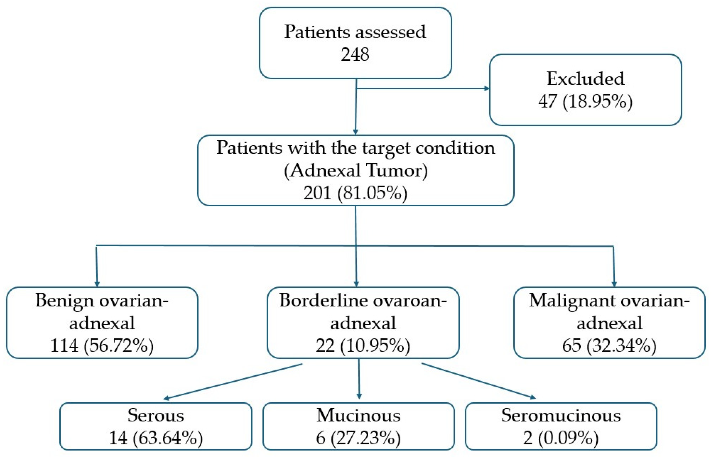



| Target Population | Full Cohort | |
|---|---|---|
| N = 201 * | N = 248 * | |
| Number of ovarian-adnexal masses | ||
| 1 | 162 (81%) | 162 (65%) |
| 2 | 36 (18%) | 36 (14.5%) |
| 3 | 0 (0%) | 0 (0%) |
| ≥4 | 3 (1.5%) | 3 (0.1%) |
| Absence of ovarian-adnexal mass | 0 (0%) | 47 (19%) |
| Age | 52 (39, 63) | 52 (39.62) |
| Menopause | 101 (50%) | 117 (47%) |
| Oncologic history | 27 (14%) | 27 (11%) |
| Symptoms | ||
| Asymptomatic | 172 (86%) | 210 (85%) |
| Abdominal pain | 3 (1.5%) | 6 (2.4%) |
| Fever | 0 (0%) | 1 (0.4%) |
| Other | 26 (13%) | 31 (13%) |
| Elevated tumour markers | 65 (36%) | 69 (33%) |
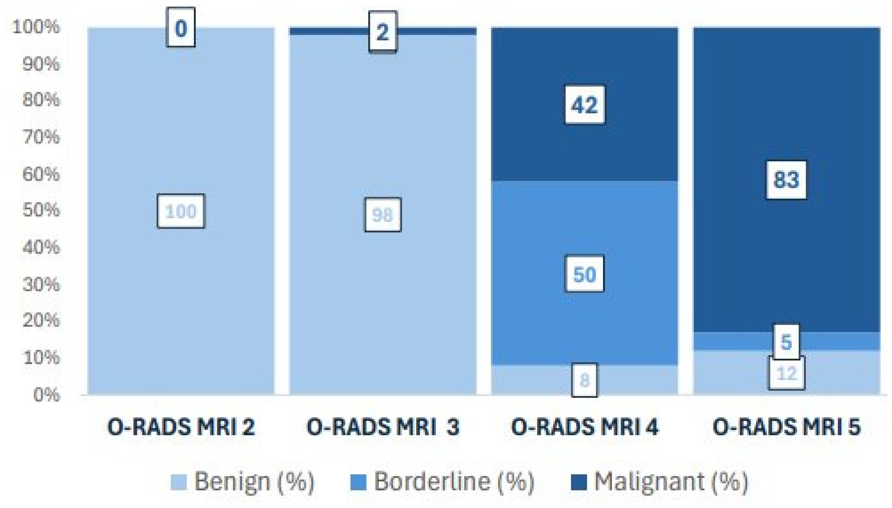
| O-RADS MRI Category | Benign Ovarian-Adnexal n = 114 * | Borderline Ovarian-Adnexal n = 22 * | Malignant Ovarian-Adnexal n = 65 * |
|---|---|---|---|
| Benign–Low-risk (Score 2–3) | 104 (99%) | 1 (1.0%) | 0 (0%) |
| Indeterminate-risk (Score 4) | 3 (8.3%) | 18 (50%) | 15 (42%) |
| High-risk (Score 5) | 7 (12%) | 3 (5.0%) | 50 (83%) |
| Diagnostic Performance | Sensitivity | Specificity | PPV | NPV | |
|---|---|---|---|---|---|
| O-RADS MRI | 0.856 (0.799, 0.901) | ||||
| - Benign | 0.912 (0.845, 0.957) | 0.989 (0.938, 1) | 0.99 (0.9481, 1) | 0.896 (0.817, 0.949) | |
| - Borderline | 0.818 (0.597, 0.948) | 0.899 (0.846, 0.939) | 0.5 (0.329, 0.671) | 0.976 (0.939, 0.993) | |
| - Malignant | 0.769 (0.648, 0.865) | 0.926 (0.869, 0.964) | 0.833 (0.715, 0.917) | 0.894 (0.831, 0.939) | |
| Full Model | 0.955 (0.917, 0.979) | ||||
| - Benign | 0.982 (0.938, 0.998) | 0.954 (0.886, 0.987) | 0.966 (0.9141, 0.991) | 0.976 (0.918, 0.997) | |
| - Borderline | 0.818 (0.597, 0.948) | 0.989 (0.96, 0.999) | 0.9 (0.683, 0.988) | 0.978 (0.944, 0.994) | |
| - Malignant | 0.954 (0.871, 0.99) | 0.978 (0.937, 0.995) | 0.954 (0.871, 0.99) | 0.978 (0.937, 0.995) | |
| Simplified Model | 0.905 (0.856, 0.942) | ||||
| - Benign | 0.912 (0.845, 0.957) | 0.989 (0.938, 1) | 0.99 (0.9481, 1) | 0.896 (0.817, 0.949) | |
| - Borderline | 0.727 (0.498, 0.893) | 0.978 (0.944, 0.994) | 0.8 (0.563,0.943) | 0.967 (0.929, 0.988) | |
| - Malignant | 0.954 (0.871, 0.99) | 0.897 (0.833, 0.943) | 0.816 (0.71, 0.895) | 0.976 (0.931, 0.995) |
| O-RADS MRI | Prevalence * | Decision Criteria | Predicted Risk Category | Observed Prevalence | Diagnostic Accuracy |
|---|---|---|---|---|---|
| 2–3 (99% Benign) | 52% [105] | - | BENIGN | 52% [105] | 99% |
| 4 (50% Borderline, 42% Malignant) | 18% [36] | Solid ADC ≥ 993, age < 67, asymptomatic | BORDERLINE | 9% [18] | 89% |
| Rest | MALIGNANT | 9% [18] | 77.78% | ||
| 5 (83% Malignant) | 30% [60] | Solid ADC ≥ 993, he4 ≤ 33, enhancement ratio ** < 66 | BENIGN | 1% [3] | 66.67% |
| Solid ADC ≥ 993, he4 < 19 | BENIGN | 2% [4] | 75% | ||
| Solid ADC < 993, T1w signal intensity < 67 | BENIGN | 2% [4] | 75% | ||
| Solid ADC ≥ 993, he4 in [19,33] | BORDERLINE | 1% [1] | 100% | ||
| Rest | MALIGNANT | 24% [48] | 97.92% |
| O-RADS MRI | Prevalence * | Decision Criteria | Predicted Risk Category | Observed Prevalence | Diagnostic Accuracy |
|---|---|---|---|---|---|
| 2–3 (99% Benign) | 52% [105] | - | BENIGN | 52% [105] | 99% |
| 4 (50% Borderline, 42% Malignant) | 18% [36] | Solid ADC ≥ 993, age < 67 | BORDERLINE | 10% [20] | 80% |
| Rest | MALIGNANT | 8% [16] | 68.75% | ||
| 5 (83% Malignant) | 30% [60] | - | MALIGNANT | 30% [60] | 83% |
Disclaimer/Publisher’s Note: The statements, opinions and data contained in all publications are solely those of the individual author(s) and contributor(s) and not of MDPI and/or the editor(s). MDPI and/or the editor(s) disclaim responsibility for any injury to people or property resulting from any ideas, methods, instructions or products referred to in the content. |
© 2026 by the authors. Licensee MDPI, Basel, Switzerland. This article is an open access article distributed under the terms and conditions of the Creative Commons Attribution (CC BY) license.
Share and Cite
Cabedo, L.; Sebastià, C.; Munmany, M.; Saco, A.; Gallardo, E.; Sáenz de Argandoña, O.; Peón, G.; Carrasco, J.L.; Nicolau, C. Differentiating Borderline from Malignant Ovarian-Adnexal Tumours: A Multimodal Predictive Approach Joining Clinical, Analytic, and MRI Parameters. Cancers 2026, 18, 516. https://doi.org/10.3390/cancers18030516
Cabedo L, Sebastià C, Munmany M, Saco A, Gallardo E, Sáenz de Argandoña O, Peón G, Carrasco JL, Nicolau C. Differentiating Borderline from Malignant Ovarian-Adnexal Tumours: A Multimodal Predictive Approach Joining Clinical, Analytic, and MRI Parameters. Cancers. 2026; 18(3):516. https://doi.org/10.3390/cancers18030516
Chicago/Turabian StyleCabedo, Lledó, Carmen Sebastià, Meritxell Munmany, Adela Saco, Eduardo Gallardo, Olatz Sáenz de Argandoña, Gonzalo Peón, Josep Lluís Carrasco, and Carlos Nicolau. 2026. "Differentiating Borderline from Malignant Ovarian-Adnexal Tumours: A Multimodal Predictive Approach Joining Clinical, Analytic, and MRI Parameters" Cancers 18, no. 3: 516. https://doi.org/10.3390/cancers18030516
APA StyleCabedo, L., Sebastià, C., Munmany, M., Saco, A., Gallardo, E., Sáenz de Argandoña, O., Peón, G., Carrasco, J. L., & Nicolau, C. (2026). Differentiating Borderline from Malignant Ovarian-Adnexal Tumours: A Multimodal Predictive Approach Joining Clinical, Analytic, and MRI Parameters. Cancers, 18(3), 516. https://doi.org/10.3390/cancers18030516