Tumor Microenvironment: Insights from Multiparametric MRI in Pancreatic Ductal Adenocarcinoma
by
 , , , , ,
Ramesh Paudyal
1, 
James Russell
1,
H. Carl Lekaye
1,
Joseph O. Deasy
1,
John L. Humm
1,
Muhammad Awais
1,
Saad Nadeem
1,
Richard K. G. Do
2,
Eileen M. O’Reilly
3 ,
,
Lawrence H. Schwartz
2 and
Amita Shukla-Dave
1,2,* 1
Department of Medical Physics, Memorial Sloan Kettering Cancer Center, New York, NY 10065, USA
2
Department of Radiology, Memorial Sloan Kettering Cancer Center, New York, NY 10065, USA
3
Department of Medicine, Memorial Sloan Kettering Cancer Center, New York, NY 10065, USA
*
Author to whom correspondence should be addressed.
Cancers 2026, 18(2), 273; https://doi.org/10.3390/cancers18020273
Submission received: 16 December 2025
/
Revised: 6 January 2026
/
Accepted: 8 January 2026
/
Published: 15 January 2026
(This article belongs to the Special Issue Image-Assisted High-Precision Radiation Oncology)
Simple Summary
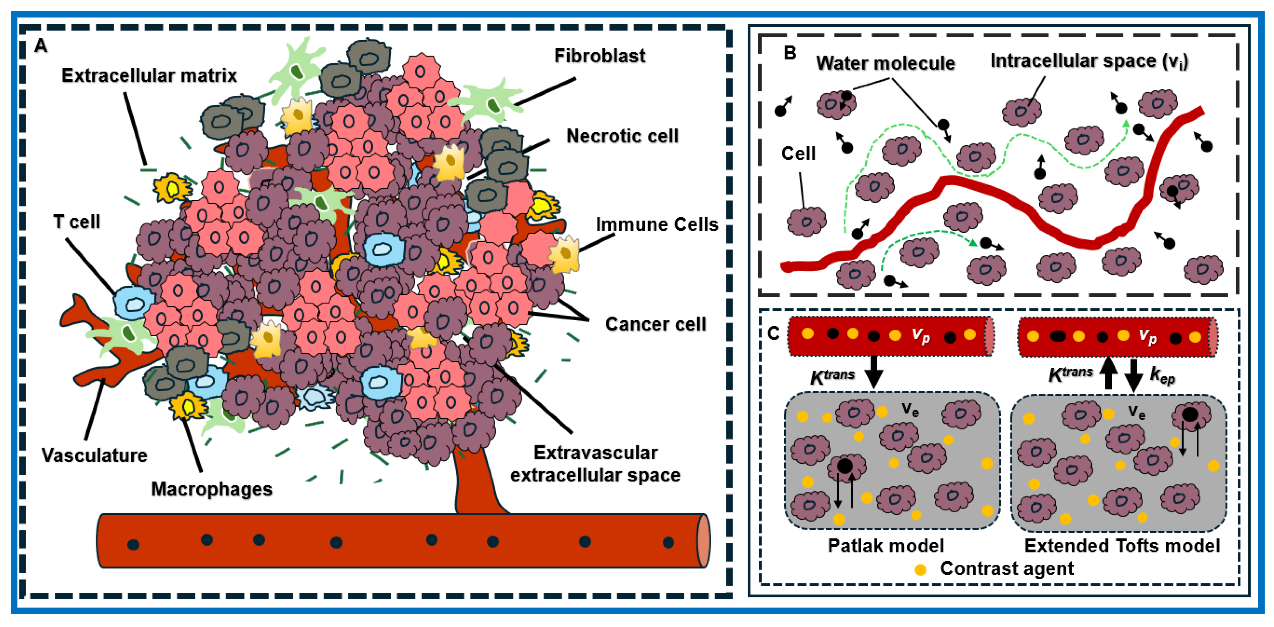
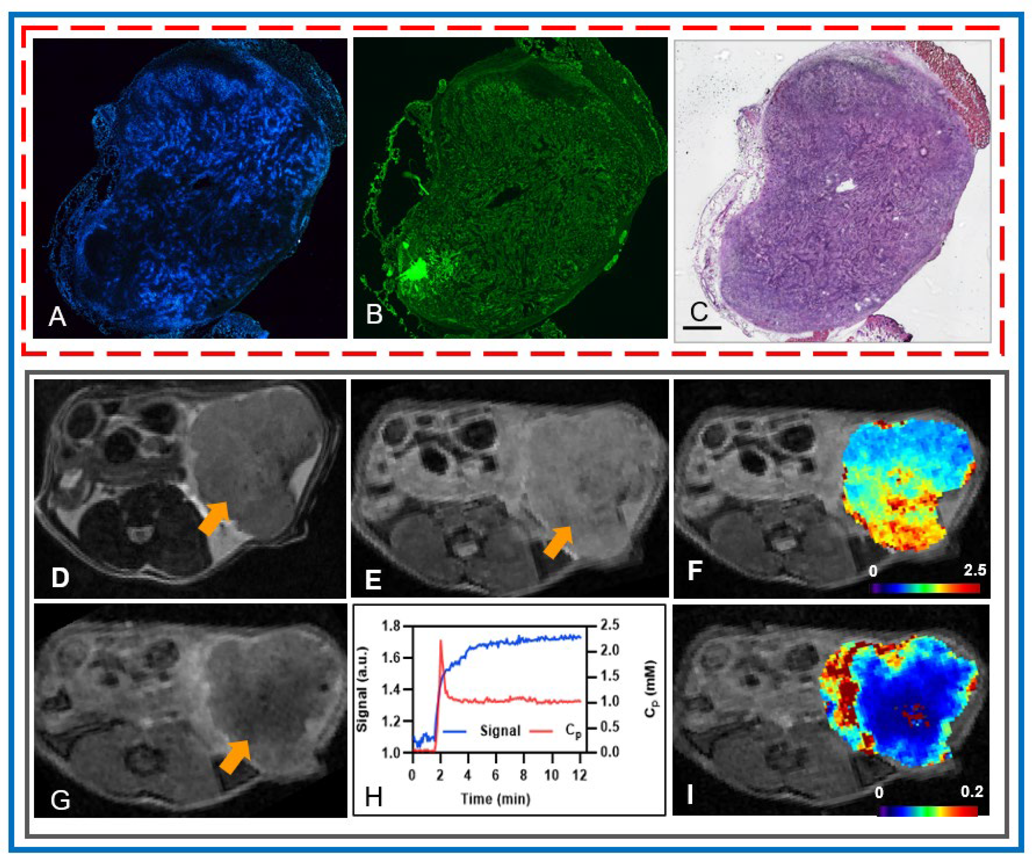
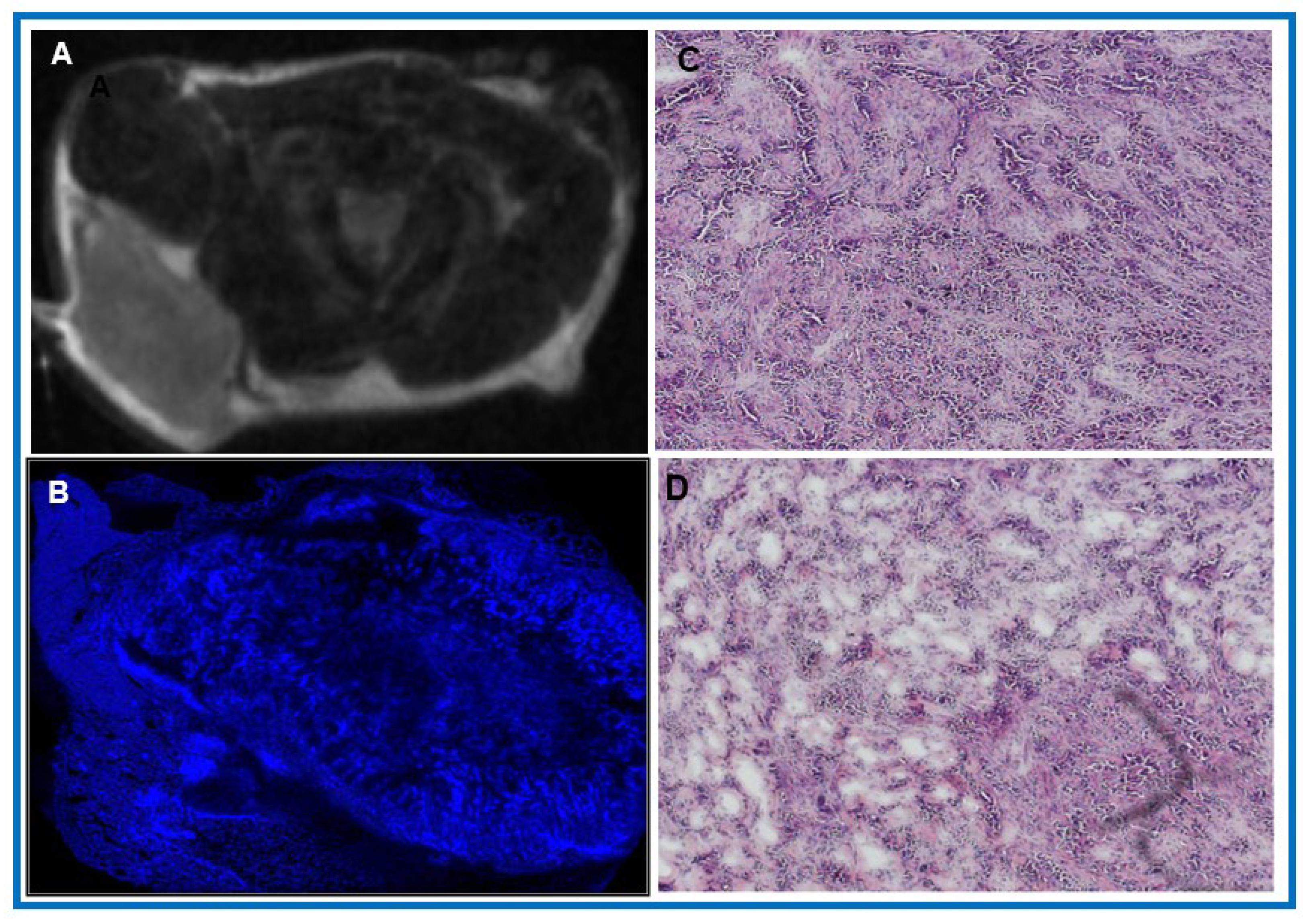
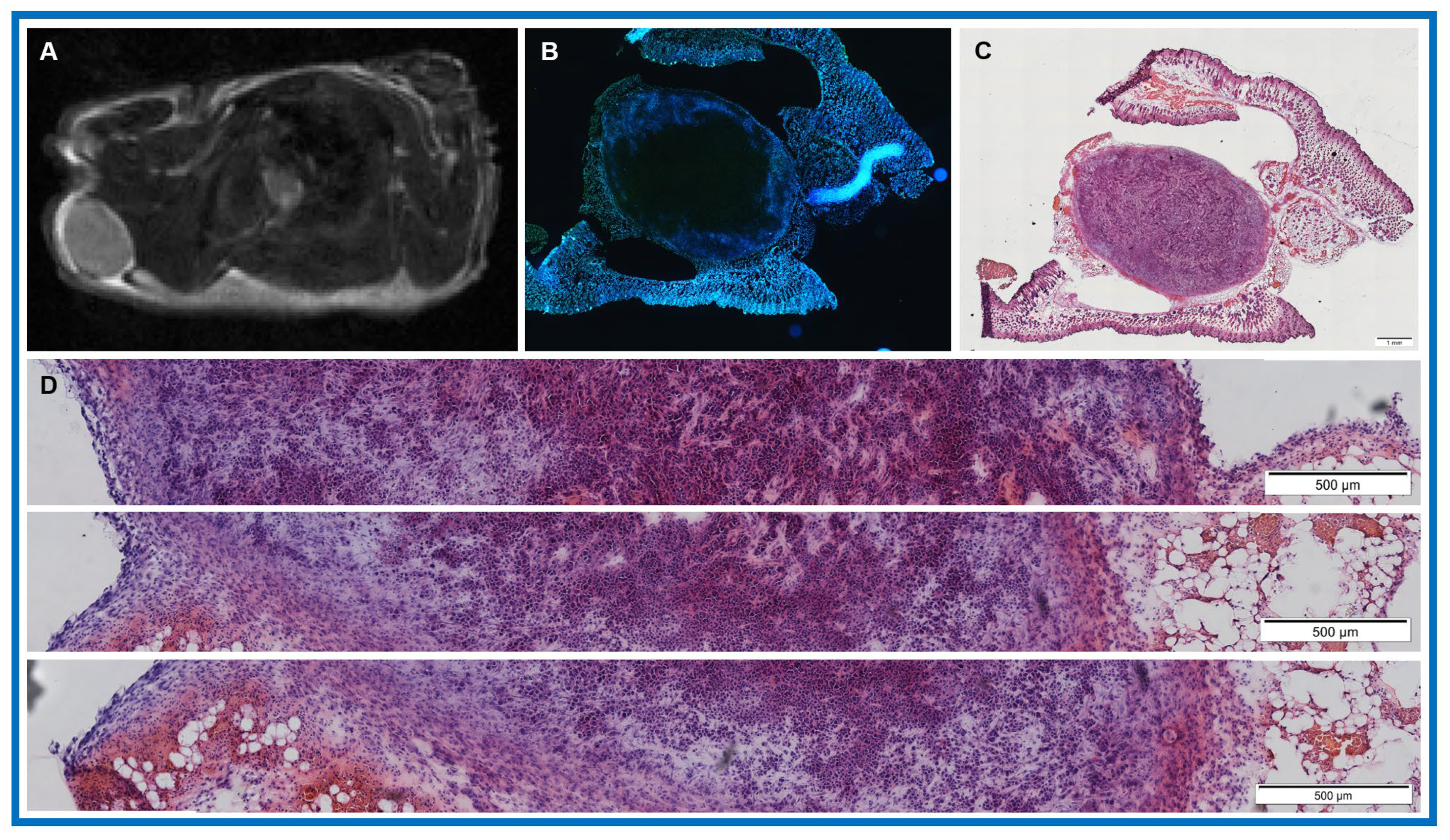
Pancreatic ductal adenocarcinoma (PDAC) is characterized by a highly heterogeneous tumor microenvironment (TME), enriched with stromal components such as cancer-associated fibroblasts and dense extracellular matrix, which contribute to therapeutic resistance. Multiparametric magnetic resonance imaging (mpMRI) can yield valuable quantitative imaging biomarkers (QIBs) derived from diffusion-weighted (DW) and dynamic contrast–enhanced (DCE) MRI that can be used to assess characteristics of the TME such as cellularity and vascular permeability. Meanwhile, histological staining (Hoechst, hematoxylin and eosin [H&E]) provides insights into the TME spatial organization. Harnessing mpMRI and histology together in PDAC is vital for combating therapeutic resistance and enhancing treatment efficacy. This study establishes a foundation for future co-clinical research to evaluate emerging drug combinations that target both tumor and stroma, thus advancing our understanding of the TME in PDAC.
Abstract
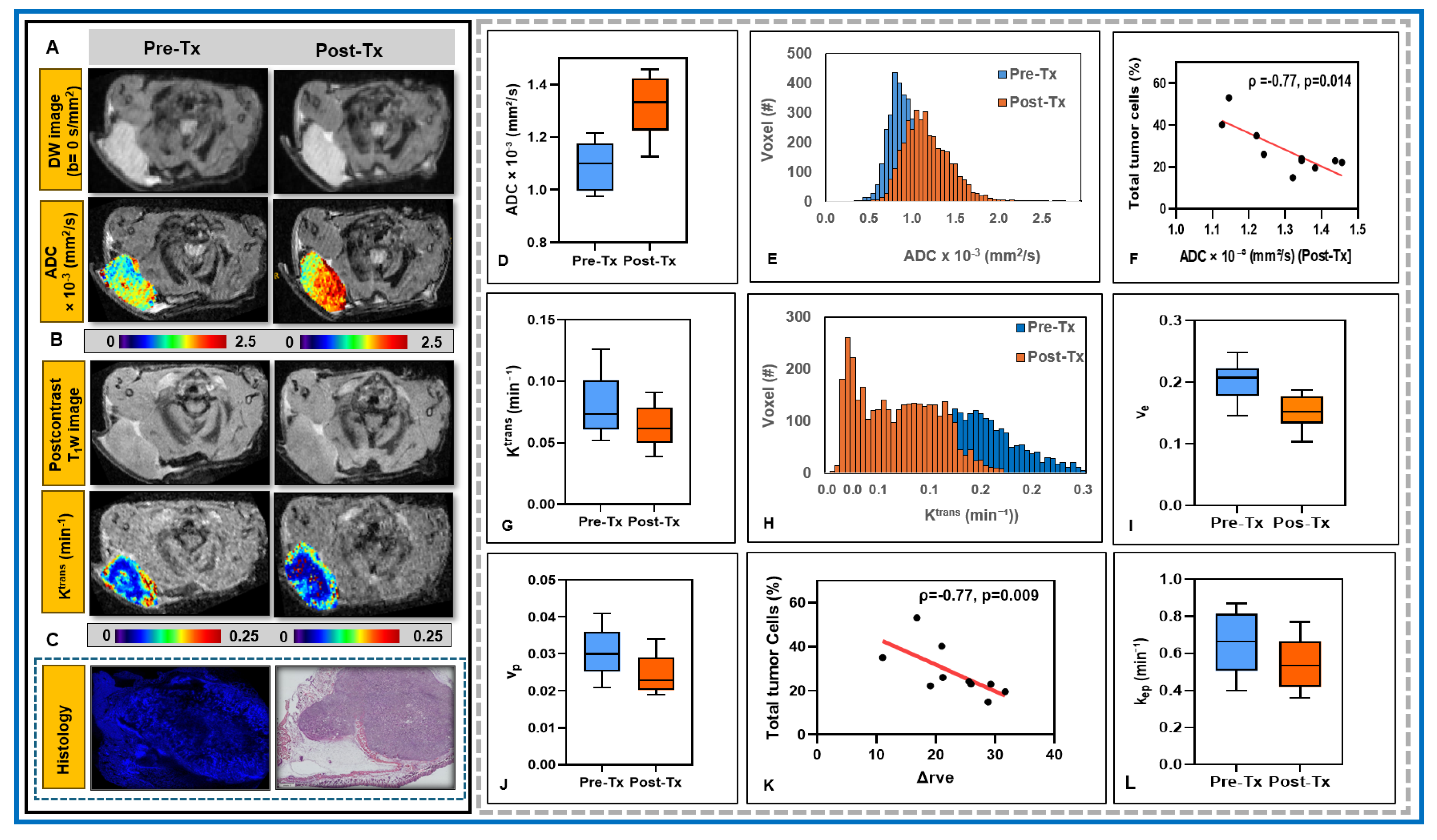
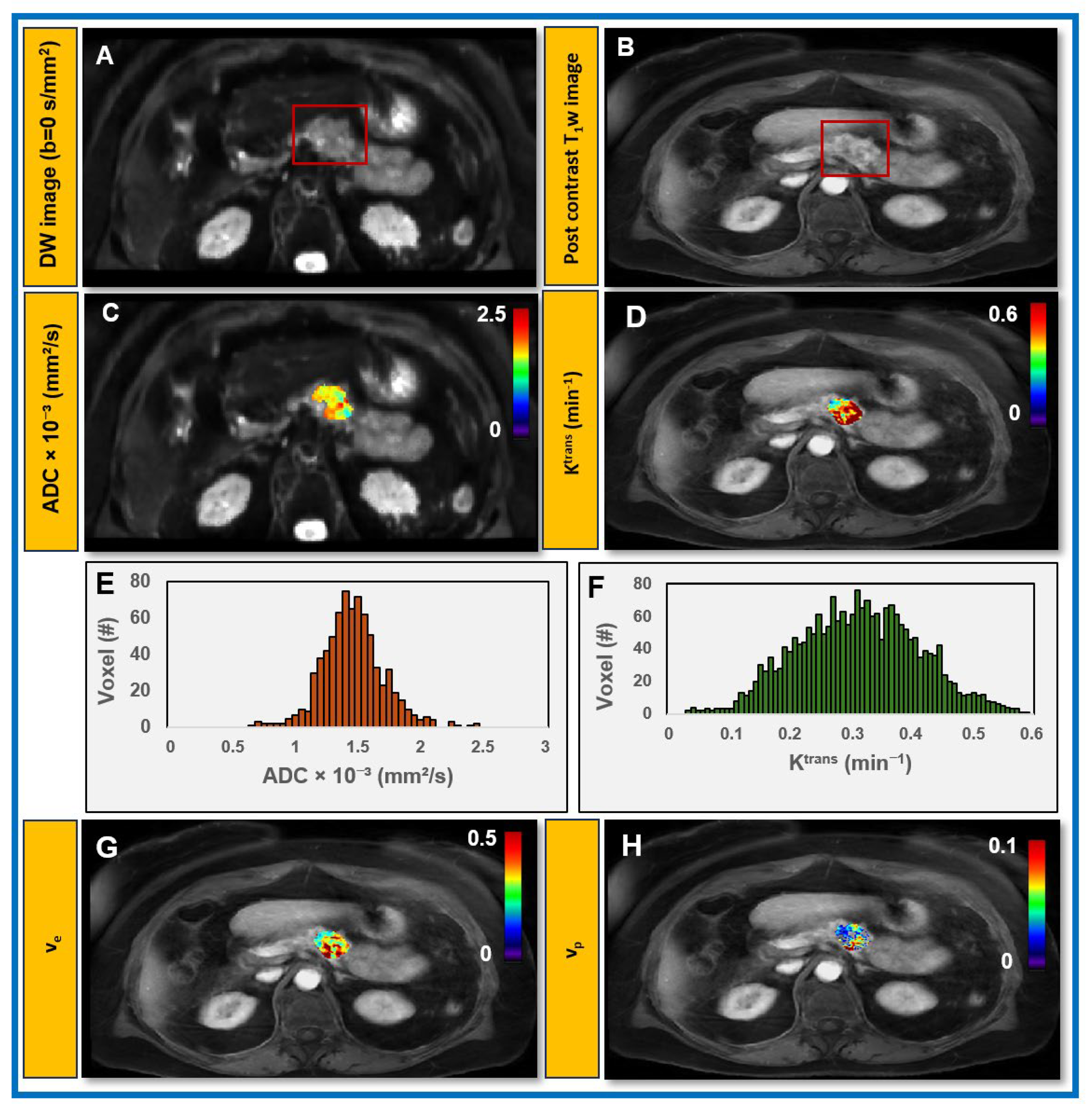
Background/Objectives: The tumor microenvironment (TME) of pancreatic ductal adenocarcinoma (PDAC) is characterized by an enriched stroma, hampering the effectiveness of therapy. This co-clinical study aimed to (1) provide insight into early post-treatment changes in the TME using multiparametric magnetic resonance imaging (mpMRI)-derived quantitative imaging biomarkers (QIBs) in a preclinical PDAC model treated with radiotherapy and correlate these QIBs with histology; (2) evaluate the feasibility of obtaining these QIBs in patients with PDAC using clinically approved mpMRI data acquisitions. Methods: Athymic mice (n = 12) at pre- and post-treatment as well as patients with PDAC (n = 11) at pre-treatment underwent mpMRI including diffusion-weighted (DW) and dynamic contrast-enhanced (DCE) data acquisition sequences. DW and DCE data were analyzed using monoexponential and extended Tofts models, respectively. DeepLIIF quantified the total percentage (%) of tumor cells in hematoxylin and eosin (H&E)-stained tissues from athymic mice. Spearman correlation and Wilcoxon signed rank tests were performed for statistical analysis. Results: In the preclinical PDAC model, mean pre- and post-treatment ADC and Ktrans values differed significantly (p < 0.01), changing by 20.50% and 20.41%, respectively, and the median total tumor cells quantified by DeepLIIF was 24% (range: 15–53%). Post-treatment ADC values and relative change in ve (rΔve) showed a significant negative correlation with total tumor cells (ρ = −0.77, p < 0.014 for ADC and ρ = −0.77, p = 0.009 for rΔve). In patients with PDAC, pre-treatment mean ADC and Ktrans values were 1.76 × 10⁻³ (mm2/s) and 0.24 (min−1), respectively. Conclusions: QIBs in both preclinical and clinical settings underscore their potential for future co-clinical research to evaluate emerging drug combinations targeting both tumor and stroma.