Presence of Trace Metals in the Biological Samples of Prostate Cancer Patients: A Systematic Review of Case-Control Studies


Simple Summary
Abstract
1. Introduction
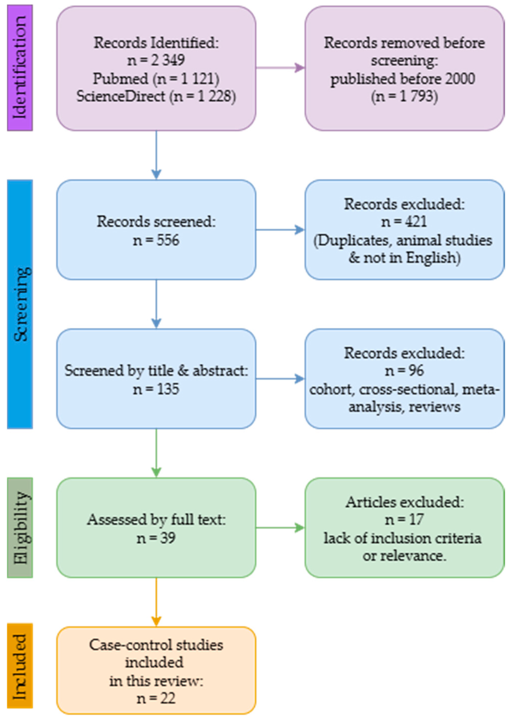
2. Search Strategy
2.1. Study Selection and Data Collection
2.2. Data Extraction and Analysis
3. Results

| Study | Country | Trace Metals | Biological Sample | Study Group [n] | Control Group [n] | Analytical Method |
|---|---|---|---|---|---|---|
| [Bede-Ojimadu et al., 2023] [16] | Nigeria | Zn, Cd | Blood, urine | 82 | 98 | ICP-MS |
| [Lim et al., 2019] [17] | Singapore | Mn, Zn, As, Se, Cd, Pb, Ni | Serum | 141 | 114 | ICP-MS |
| [Saleh et al., 2017] [18] | Saudi Arabia | Zn, Mn, Se | Hair | 58 | 52 | ICP-MS |
| [Saleh et al., 2020] [19] | Saudi Arabia | Mn, Zn, Se | Serum | 40 | 30 | ICP-MS |
| [Adedapo et al., 2012] [23] | Nigeria | Se, Zn, Mn | Serum | 40 | 40 | AAS |
| [Amadi & Aleme, 2019] [24] | Nigeria | Zn | Serum | 220 | 220 | AAS |
| [Olooto et al., 2021] [25] | Nigeria | Se, Zn | Serum | 55 | 25 | AAS |
| [Onyema-iloh et al., 2014] [26] | Nigeria | Zn, Se | Serum | 100 | 50 | AAS |
| [Karimi et al., 2012] [34] | Malaysia | Se, Zn, Mn | Hair, nails | 50 | 50 | ICP-MS |
| [Aydin et al., 2006] [27] | Turkey | Zn | Serum | 25 | 24 | AAS |
| [Eken et al., 2016] [28] | Turkey | Zn, Mn, Se | Serum | 42 | 40 | AAS |
| [Kaba et al., 2014] [29] | Turkey | Mn, Zn, Pb, Cd | Serum | 30 | 32 | AAS |
| [Ozmen et al., 2006] [30] | Turkey | Ni, Zn | Blood | 20 | 21 | AAS |
| [Saleh et al., 2019] [31] | Saudi Arabia | Zn, Se, Cd | Serum | 58 | 30 | ICP-MS |
| [Guntupalli et al., 2007] [32] | India | Mn, Ni, Zn, Se | Tissue | 27 | 27 | PIXE |
| [Vinceti et al., 2007] [33] | Italy | Cd | Toenails | 40 | 58 | AAS |
| [Steinbrecher et al., 2010] [35] | Germany | Se | Serum | 248 | 492 | DRC-ICP-MS |
| [Drozdz-Afelt et al., 2024] [36] | Poland | Ni, As, Cd, Pb, Hg, Zn | Blood | 66 | 64 | ICP-MS |
| [Dhillon et al., 2022] [37] | Australia | Se, Zn | Plasma | 116 | 132 | ICP-AES |
| [Chang et al., 2018] [38] | Taiwan | Cd, Ni, Hg, Pb, Zn, As | Serum | 20 | 23 | ICP-MS |
| [Zaichick & Zaichick, 2019] [39] | Russia | Zn | Prostatic fluid | 24 | 38 | XRF |
| [Qayyum & Shah, 2014] [40] | Pakistan | Cd, Mn, Ni, Pb, Zn | Blood, Hair, Nails | B-74 H-67 N-60 | 66 67 60 | AAS |
| Trace Metal | p-Value | Study Group—Concentration Mean ± Standard Dev (Min–Max) | Control Group—Concentration Mean ± Standard Dev (Min–Max) | Age (Years) Mean ± SD/Range Case (Control) | Country/Region | References |
|---|---|---|---|---|---|---|
| Blood/Serum/Plasma | ||||||
| Zinc | 0.666 | 4118.57 (3460.31–4827.80) | 4071.10 (3554.53–4639.60) | 64–76 (62–75) | Nigeria | [16] |
| 1.72 × 10⁻¹⁵ | 857.4 ± 162.1 (441.1–1557.8) | 690.7 ± 149.3 (373.9–1129.7) | ≥50 | Singapore | [17] | |
| <0.05 | 0.51 ± 0.09 a | 0.87 ± 0.29 a | 68.2 ± 5.2 (65.8 ± 6.8) | Saudi Arabia | [19] | |
| 0.107 | 119,800.0 ± 25,200.0 | 114,400.0 ± 25,200.0 | 68.5 ± 9.6 (65.2 ± 6.7) | Nigeria | [23] | |
| <0.001 | 1064.46 ± 341.62 (384.61–2013.54) a | 1722.82 ± 505.65 (463.79–2748.82) a | 69.7 ± 7.7 (68.97 ± 7.3) | Nigeria | [24] | |
| 0.000 | 43.3 ± 3.3 | 139.9 ± 7.7 | NS * | Nigeria | [25] | |
| <0.05 | 1687.80 ± 598.00 | 1477.50 ± 420.50 | NS * | Nigeria | [26] | |
| <0.01 | 5760.0 ± 102.0 | 7110.0 ± 164.0 | 67.5 ± 8.8 (65.0 ± 6.0) | Turkey | [27] | |
| <0.05 | 0.86 ± 0.22 a | 0.91 ± 0.18 a | 46–85 | Turkey | [28] | |
| 0.001 | 712.72 ± 339.29 (67.09–1173.0) | 2945.33 ± 498.18 (2125.0–4021.0) | 65.4 ± 4.2 (62.8 ± 5.8) | Turkey | [29] | |
| <0.001 | 4375.0 ± 620.0 | 5843.0 ± 747.0 | 72.5 ± 7.8 (66.3 ± 8.3) | Turkey | [30] | |
| 0.001 | 0.73 ± 0.41 | 2.64 ± 0.56 | 71.1 ± 5.4 | Saudi Arabia | [31] | |
| 0.003 | 4735.6 ± 1124.3 (3005.3–12,017.9) | 5211.3 ± 1463.0 (2692.3–12,539.8) | ≥48 (>50) | Poland | [36] | |
| 0.23 | 0.77 ± 0.1 (0.52–1.24) | 0.78 ± 0.01 (0.52–1.04) | 71.2 ± 7.18(69.1 ± 7.99) | Australia | [37] | |
| 0.001 | 421.9 ± 132.2 | 600.26 ± 189.01 | 73.4 ± 6.7 (71.3 ± 8.1) | Taiwan | [38] | |
| <0.05 | 2.483 ± 0.210 * (0.059–8.536) | 6.571 ± 0.656 * (0.940–32.74) | 32–75 (27–68) | Pakistan | [40] | |
| Cadmium | 0.000 | 1.30 (0.003–1.88) | 0.003 (0.003–0.003) | 64–76 (62–75) | Nigeria | [16] |
| 0.82 | 0.22 ± 0.041 (0.22–0.58) | 0.25 ± 0.32 (0.22–3.56) | ≥50 | Singapore | [17] | |
| 0.630 | 1.32 ± 0.15 (2.01–9.54) | 1.11 ± 0.11 (1.01–1.35) | 65.4 ± 4.2 (62.8 ± 5.8) | Turkey | [29] | |
| 0.63 | 0.0015 ± 0.001 | 0.0011 ± 0.001 | 71.1 ± 5.4 | Saudi Arabia | [31] | |
| 0.002 | 0.46 ± 0.45 | 0.9 ± 0.35 | 73.4 ± 6.7 (71.3 ± 8.1) | Taiwan | [38] | |
| <0.05 | 1.084 ± 0.107 * (0.059–3.832) | 0.774 ± 0.096 * (0.017–3.946) | 32–75 (27–68) | Pakistan | [40] | |
| Selenium | 4.91 × 10⁻¹⁰ | 131.4 ± 30.2 (86.1–296.9) | 108.9 ± 27.0 (55.7–220.0) | ≥50 | Singapore | [17] |
| <0.005 | 0.07 ± 0.01 a | 0.14 ± 0.03 a | 68.2 ± 5.2 (65.8 ± 6.8) | Saudi Arabia | [19] | |
| 0.741 | 586.0 ± 128.0 | 597.0 ± 123.0 | 68.5 ± 9.6 (65.2 ± 6.7) | Nigeria | [23] | |
| 0.000 | 615.0 ± 34.4 | 956.2 ± 13.9 | NS * | Nigeria | [25] | |
| <0.05 | 43.00 ± 14.52 | 82.092 ± 7.26 | NS * | Nigeria | [26] | |
| <0.05 | 68.52 ± 6.93 a | 83.49 ± 5.69 a | 46–85 | Turkey | [28] | |
| 0.001 | 53.00 ± 25.00 | 193.00 ± 32.00 | 71.1 ± 5.4 | Saudi Arabia | [31] | |
| NS * | 86.2 ± 14.2 | 87.7 ± 13.4 | 58.1 ± 4.8 (58.1 ± 4.8) | Germany | [35] | |
| 0.002 | 116.11.59 (71.83–157.6) | 125.6 ± 2.56 (79.17–238.1) | 71.2 ± 7.18(69.1 ± 7.99) | Australia | [37] | |
| Manganese | 6.05 × 10⁻⁷ | 5.84 ± 12.6 (0.50–131.2) | 2.24 ± 3.69 (0.50–29.0) | ≥50 | Singapore | [17] |
| <0.005 | 0.01 ± 0.001 a | 0.0026 ± 0.001 a | 68.2 ± 5.2 (65.8 ± 6.8) | Saudi Arabia | [19] | |
| 0.175 | 36,800.0 ± 5800.0 | 37,100.0 ± 7900.0 | 68.5 ± 9.6 (65.2 ± 6.7) | Nigeria | [23] | |
| <0.05 | 0.62 ± 0.19 a | 0.22 ± 0.08 a | 46–85 | Turkey | [28] | |
| 0.001 | 97.53 ± 75.35 (1.10–437.80) | 746.06 ± 219.83 (111.14–989.91) | 65.4 ± 4.2 (62.8 ± 5.8) | Turkey | [29] | |
| <0.05 | 1.524 ± 0.140 * (0.93–5.761) | 0.895 ± 0.068 * (0.032–2.066) | 32–75 (27–68) | Pakistan | [40] | |
| Arsenic | 1.89 × 10⁻⁹ | 4.78 ± 4.46 (0.50–26.4) | 2.66 ± 3.67 (0.50–25.0) | ≥50 | Singapore | [17] |
| <0.001 | 0.9 ± 1.2 (0.0–6.4) | 0.3 ± 0.5 (0.0–2.7) | ≥48 (>50) | Poland | [36] | |
| 0.078 | 6.8 ± 7.2 | 13.14 ± 8.66 | 73.4 ± 6.7 (71.3 ± 8.1) | Taiwan | [38] | |
| Lead | 0.00024 | 5.26 ± 42.6 (0.60–505.6) | 1.31 ± 6.82 (0.60–73.4) | ≥50 | Singapore | [17] |
| 0.001 | 57.96 ± 30.62 (27.41–169.60) | 1.21 ± 0.13 (1.0–1.5) | 65.4 ± 4.2 (62.8 ± 5.8) | Turkey | [29] | |
| 0.001 | 17.4 ± 61.9 (0.0–508.9) | 31.9 ± 152.4 (0.0–1231.1) | ≥48 (>50) | Poland | [36] | |
| 0.015 | 18.5 ± 17 | 54.6 ± 62.04 | 73.4 ± 6.7 (71.3 ± 8.1) | Taiwan | [38] | |
| <0.05 | 3.658 ± 0.358 * (0.023–12.67) | 2.248 ± 0.216 * (0.019–7.474) | 32–75 (27–68) | Pakistan | [40] | |
| Nickel | <2.2 × 10⁻¹⁶ | 17.8 ± 41.6 (1.46–438.0) | 2.10 ± 2.27 (1.46–17.3) | ≥50 | Singapore | [17] |
| <0001 | 426.00 ± 84.00 | 238.00 ± 101.00 | 72.5 ± 7.8 (66.3 ± 8.3) | Turkey | [30] | |
| 0.295 | 30.9 ± 48.7 (0.0–234.7) | 21.1 ± 38.7 (0.0–239.0) | ≥48 (>50) | Poland | [36] | |
| <0.001 | 57.1 ± 22.9 | 54.71 ± 35.43 | 73.4 ± 6.7 (71.3 ± 8.1) | Taiwan | [38] | |
| <0.05 | 4.335 ± 0.384 * (0.030–10.35) | 2.687 ± 0.241 * (0.012–8.836) | 32–75 (27–68) | Pakistan | [40] | |
| Mercury | 0.808 | 1.15 ± 1.25 (0.07–7.54) | 1.05 ± 0.77 (0.12–3.62) | ≥48 (>50) | Poland | [36] |
| <0.001 | 6.9 ± 9 | 1.79 ± 1.7 | 73.4 ± 6.7 (71.3 ± 8.1) | Taiwan | [38] | |
| Urine | ||||||
| Zinc | 0.234 | 638.87 (527.18–774.57) | 696.95 (591.96–820.36) | 64–76 (62–75) | Nigeria | [16] |
| Cadmium | 0.221 | 1.65 (1.33–2.042) | 1.95 (1.64–2.33) | 64–76 (62–75) | Nigeria | [16] |
| Tissue | ||||||
| Zinc | 8.0 × 10⁻⁶ | 50.7 ± 3.26 | 366.7 ± 5.59 | 52.5 ± 9.2 | India | [32] |
| Manganese | 0.059 | 11 ± 1.82 | 3 ± 1.45 | 52.5 ± 9.2 | India | [32] |
| Selenium | 0.0059 | 1.1 ± 0.34 | 3.2 ± 0.41 | 52.5 ± 9.2 | India | [32] |
| Nickel | 0.0006 | 24.32 ± 2.95 | 2.4 ± 0.72 | 52.5 ± 9.2 | India | [32] |
| Prostatic fluid [mg/L] | ||||||
| Zinc | 0.64 | 62.0 ± 98.3 | 598 ± 207 | 65 ± 10 (59 ± 11) | Russia | [39] |
| Trace Metal | p-Value | Study Group—Concentration Mean ± Standard Dev (Min–Max) | Control Group—Concentration Mean ± Standard Dev (Min–Max) | Age (Years) Mean ± SD/Range Case (Control) | Country/Region | References |
|---|---|---|---|---|---|---|
| Hair | ||||||
| Zinc | <0.005 | 3.1 ± 0.7 | 4.4 ± 0.4 | 71.1 ± 5.4 (66.8 ± 7.8) | Saudi Arabia | [18] |
| 0.018 | 3.29 ± 2.22 (1.48–3.99) | 4.29 ± 2.53 (0.05–0.08) | 72.4 ± 6.7 (71.7 ± 7.9) | Malaysia | [34] | |
| <0.05 | 160.7 ± 9.07 * (59.75–591.0) | 582.7 ± 25.73 * (6.85–1207) | 38–72 (31–68) | Pakistan | [40] | |
| Cadmium | <0.05 | 1.629 ± 0.159 * (0.05–4.70) | 1.021 ± 0.090 * (0.05–2.80) | 38–72 (31–68) | Pakistan | [40] |
| Selenium | <0.005 | 7.3 ± 1.4 | 11.5 ± 2.1 | 71.1 ± 5.4 (66.8 ± 7.8) | Saudi Arabia | [18] |
| 0.001 | 7.15 ± 3.5 (4.48–9.74) | 10.4 ± 4.52 (6.88–14.00) | 72.4 ± 6.7 (71.7 ± 7.9) | Malaysia | [34] | |
| Manganese | <0.005 | 0.083 ± 0.02 | 0.058 ± 0.03 | 71.1 ± 5.4 (66.8 ± 7.8) | Saudi Arabia | [18] |
| 0.001 | 0.07 ± 0.04 (0.05–0.09) | 0.055 ± 0.05 (0.01–0.10) | 72.4 ± 6.7 (71.7 ± 7.9) | Malaysia | [34] | |
| <0.05 | 4.65 ± 0.37 * (0.40–12.70) | 2.85 ± 0.24 * (0.15–7.50) | 38–72 (31–68) | Pakistan | [40] | |
| Nickel | <0.05 | 37.21 ± 2.82 * (1.05–85.95) | 19.35 ± 1.84 * (0.05–57.25) | 38–72 (31–68) | Pakistan | [40] |
| Lead | <0.05 | 35.87 ± 2.49 * (0.75–82.00) | 13.03 ± 1.41 * (0.2549.35) | 38–72 (31–68) | Pakistan | [40] |
| Nails/Toenails | ||||||
| Zinc | 0.01 | 2.70 ± 1.49 (1.31–4.07) | 3.97 ± 4.06 (2.73–4.36) | 72.4 ± 6.7 (71.7 ± 7.9) | Malaysia | [34] |
| NS | 114.2 ± 4.53 * (22.5–182.1) | 122.7 ± 8.54 * (45.00–383.2) | 32–75 (27–68) | Pakistan | [40] | |
| Selenium | 0.001 | 7.23 ± 3.11 (5.26–8.98) | 9.03 ± 3.69 (5.90–11.69) | 72.4 ± 6.7 (71.7 ± 7.9) | Malaysia | [34] |
| Manganese | 0.001 | 0.10 ± 0.06 (0.06–0.13) | 0.05 ± 0.04 (0.01–0.08) | 72.4 ± 6.7 (71.7 ± 7.9) | Malaysia | [34] |
| <0.05 | 11.98 ± 1.05 * (0.25–35.42) | 8.63 ± 0.75 * (1.25–24.58) | 32–75 (27–68) | Pakistan | [40] | |
| Cadmium | <0.05 | 5.66 ± 0.50 * (0.33–14.75) | 3.06 ± 0.29 * (0.33–8.29) | 32–75 (27–68) | Pakistan | [40] |
| 0.004 | 4.7 (1.3–17.5) | 0.088 (0.018–0.210) | 43–83 | Italy | [33] | |
| Lead | NS | 24.49 ± 2.17 * (2.00–67.00) | 23.83 ± 2.53 * (1.30–67.92) | 32–75 (27–68) | Pakistan | [40] |
| Nickel | <0.05 | 101.1 ± 10.19 * (0.85–267.1) | 56.65 ± 5.36 * (1.88–153.2) | 32–75 (27–68) | Pakistan | [40] |
| Trace Metal | Cases vs. Controls | Analysis |
|---|---|---|
| Zn | ↓ decreased in cases | Linked to impaired antioxidant and DNA damage |
| Se | ↓ decreased in cases | Linked to oxidative damage |
| Mn | ↑ slightly increased in cases | Variable by exposure source |
| Cd | ↑ increased in cases | Suggests toxic burden |
| Pb | ↑ slightly increased in cases | Possible environmental contribution |
| Ni | ↑ increased in cases | Possible environmental contribution |
| As | ↑ slightly increased in cases | Possible environmental contribution |
| Hg | ↑ slightly increased in cases | Possible environmental contribution |
4. Discussion
5. Limitations
6. Conclusions
Supplementary Materials
Author Contributions
Funding
Institutional Review Board Statement
Informed Consent Statement
Data Availability Statement
Conflicts of Interest
References
- Sung, H.; Ferlay, J.; Siegel, R.L.; Laversanne, M.; Soerjomataram, I.; Jemal, A.; Bray, F. Global Cancer Statistics 2020: GLOBOCAN Estimates of Incidence and Mortality Worldwide for 36 Cancers in 185 Countries. CA Cancer J. Clin. 2021, 71, 209–249. [Google Scholar] [CrossRef] [Scilit]
- Zhang, W.; Cao, G.; Wu, F.; Wang, Y.; Liu, Z.; Hu, H.; Xu, K. Global Burden of Prostate Cancer and Association with Socioeconomic Status, 1990–2019: A Systematic Analysis from the Global Burden of Disease Study. J. Epidemiol. Glob. Health 2023, 13, 407–421. [Google Scholar] [CrossRef] [Scilit] [PubMed]
- Al-Ghazawi, M.; Salameh, H.; Amo-Afful, S.; Khasawneh, S.; Ghanem, R. An In-Depth Look Into the Epidemiological and Etio-logical Aspects of Prostate Cancer: A Literature Review. Cureus 2024, 15, e48252. [Google Scholar]
- Bensussen, A.; Díaz, J. Dynamics of p53 and Cancer|IntechOpen. 2015. Available online: https://www.intechopen.com/chapters/48635 (accessed on 11 August 2024).
- Hjelmborg, J.B.; Scheike, T.; Holst, K.; Skytthe, A.; Penney, K.L.; Graff, R.E.; Pukkala, E.; Christensen, K.; Adami, H.-O.; Holm, N.V.; et al. The Heritability of Prostate Cancer in the Nordic Twin Study of Cancer. Cancer Epidemiol. Biomark. Prev. 2014, 23, 2303–2310. [Google Scholar] [CrossRef] [Scilit]
- Krstev, S.; Knutsson, A. Occupational Risk Factors for Prostate Cancer: A Meta-analysis. J. Cancer Prev. 2019, 24, 91–111. [Google Scholar] [CrossRef] [Scilit]
- Sauvé, J.F.; Lavoué, J.; Parent, M.É. Occupation, industry, and the risk of prostate cancer: A case-control study in Montréal, Canada. Environ. Health 2016, 15, 100. [Google Scholar] [CrossRef] [Scilit]
- Baj, J.; Flieger, W.; Barbachowska, A.; Kowalska, B.; Flieger, M.; Forma, A.; Teresiński, G.; Portincasa, P.; Buszewicz, G.; Radzikowska-Büchner, E.; et al. Consequences of Disturbing Manganese Home-ostasis. Int. J. Mol. Sci. 2023, 24, 14959. [Google Scholar] [CrossRef] [Scilit]
- Briffa, J.; Sinagra, E.; Blundell, R. Heavy metal pollution in the environment and their toxicological effects on humans. Heliyon 2020, 6, e04691. [Google Scholar] [CrossRef] [Scilit]
- Jaishankar, M.; Tseten, T.; Anbalagan, N.; Mathew, B.B.; Beeregowda, K.N. Toxicity, mechanism and health effects of some heavy metals. Interdiscip. Toxicol. 2014, 7, 60–72. [Google Scholar] [CrossRef] [Scilit]
- Balali-Mood, M.; Naseri, K.; Tahergorabi, Z.; Khazdair, M.R.; Sadeghi, M. Toxic Mechanisms of Five Heavy Metals: Mercury, Lead, Chromium, Cadmium, and Arsenic. Front. Pharmacol. 2021, 12, 643972. [Google Scholar] [CrossRef] [Scilit]
- Mitraa, S.; Chakrabortya, A.J.; Tareqb, A.M.; Emranc, T.B.; Nainud, F.; Khusroe, A.; Idris, A.M.; Khandakerh, M.U.; Osmani, H.; Alhumaydhi, F.A.; et al. Impact of heavy metals on the environment and human health: Novel therapeutic insights to counter the toxicity. J. King Saud Univ.-Sci. 2022, 34, 101865. [Google Scholar] [CrossRef] [Scilit]
- Górska, A.; Markiewicz-Gospodarek, A.; Trubalski, M.; Żerebiec, M.; Poleszak, J.; Markiewicz, R. Assessment of the Impact of Trace Essential Metals on Cancer Development. Int. J. Mol. Sci. 2024, 25, 6842. [Google Scholar] [CrossRef] [Scilit] [PubMed]
- Pal, S.; Firdous, S.M. Unraveling the role of heavy metals xenobiotics in cancer: A critical review. Discov. Oncol. 2024, 15, 615. [Google Scholar] [CrossRef] [Scilit] [PubMed]
- Tshoni, U.A.; Mbonane, T.P.; Rathebe, P.C. The Role of Trace Metals in the Development and Progression of Prostate Cancer. Int. J. Mol. Sci. 2024, 25, 10725. [Google Scholar] [CrossRef] [Scilit]
- Bede-Ojimadu, O.; Nnamah, N.; Onuegbu, J.; Grant-Weaver, I.; Barraza, F.; Orakwe, J.; Abiahu, J.; Orisakwe, O.E.; Nriagu, J. Cadmium exposure and the risk of prostate cancer among Nigerian men: Effect modification by zinc status. J. Trace Elements Med. Biol. 2023, 78, 127168. [Google Scholar] [CrossRef] [Scilit]
- Lim, J.T.; Tan, Y.Q.; Valeri, L.; Lee, J.; Geok, P.P.; Chia, S.E.; Ong, C.N.; Seow, W.J. Association between serum heavy metals and prostate cancer risk—A multiple metal analysis. Environ. Int. 2019, 132, 105109. [Google Scholar] [CrossRef] [Scilit]
- Saleh, S.A.K.; Adly, H.M.; Nassir, A.M. Altered Trace Elements Levels in Hair of Prostate Cancer Patients. 2017. Available online: https://www.researchgate.net/publication/315500394_Altered_Trace_Elements_Levels_in_Hair_of_Prostate_Cancer_Patients (accessed on 13 August 2024).
- Saleh, S.A.; Adly, H.M.; Abdelkhaliq, A.A.; Nassir, A.M. Serum Levels of Selenium, Zinc, Copper, Manganese, and Iron in Prostate Cancer Patients. Curr. Urol. 2020, 14, 44–49. [Google Scholar] [CrossRef] [Scilit]
- Page, M.J.; McKenzie, J.E.; Bossuyt, P.M.; Boutron, I.; Hoffmann, T.C.; Mulrow, C.D.; Shamseer, L.; Tetzlaff, J.M.; Akl, E.A.; Brennan, S.E.; et al. The PRISMA 2020 statement: An updated guideline for reporting systematic reviews. Syst. Rev. 2021, 10, 89. [Google Scholar] [CrossRef] [Scilit]
- Morgan, R.L.; Whaley, P.; Thayer, K.A.; Schünemann, H.J. Identifying the PECO: A framework for formulating good questions to explore the association of environmental and other exposures with health outcomes. Environ. Int. 2018, 121, 1027–1031. [Google Scholar] [CrossRef] [Scilit]
- Wells, G.A.; Shea, B.; O’Connell, D.; Pereson, J.; Welch, V.; Losos, M.; Tugwell, P. The Newcastle–Ottawa Scale (NOS) for Assessing the Quality of Nonrandomised Studies in Meta-Analyses. Available online: http://www.ohri.ca/programs/clinical_epidemiology/oxford.asp (accessed on 23 January 2025).
- Adedapo, K.; Arinola, O.; Shittu, O.; Kareem, O.; Okolo, C.; Nwobi, L. Diagnostic value of lipids, total antioxidants, and trace metals in benign prostate hyperplasia and prostate cancer. Niger. J. Clin. Pract. 2012, 15, 293–297. [Google Scholar] [CrossRef] [Scilit]
- Amadi, C.; Aleme, B.M. The Prevalence of Zinc Deficiency among Men with and without Prostate Cancer in Port Harcourt, Nigeria. Nutr. Cancer 2019, 72, 1018–1025. [Google Scholar] [CrossRef] [Scilit] [PubMed]
- Olooto, W.E.; Oyelekan, A.A.; Adewole, O.O.; Fajobi, A.O.; Adedo, A.A.; Olasimbo, O. Serum gonadotropins, cortisol, PSA, and micronutrient levels among men with prostate carcinoma. Afr. J. Urol. 2021, 27, 109. [Google Scholar] [CrossRef] [Scilit]
- Onyema-iloh, B.O.; Meludu, S.C.; Iloh, E.; Nnodim, J.; Onyegbule, O.; Mykembata, B. Biochemical changes in some trace elements, antioxidant vitamins and their therapeutic importance in prostate cancer patients. Asian J. Med. Sci. 2014, 6, 95–97. [Google Scholar] [CrossRef] [Scilit]
- Aydin, A.; Arsova-Sarafinovska, Z.; Sayal, A.; Eken, A.; Erdem, O.; Erten, K.; Özgök, Y.; Dimovski, A. Oxidative stress and antioxidant status in non-metastatic prostate cancer and benign prostatic hyperplasia. Clin. Biochem. 2006, 39, 176–179. [Google Scholar] [CrossRef] [Scilit]
- Eken, A.; Kaya, E.; Nluendirlik, B.; Erdem, O.; Akay, C.; Ozgok, Y. Evaluation of trace element levels in patients with prostate cancer, benign prostatic hyperplasia and chronic prostatitis. Gulhane Med. J. 2016, 58, 27. [Google Scholar] [CrossRef] [Scilit]
- Kaba, M.; Pirincci, N.; Yuksel, M.B.; Gecit, I.; Gunes, M.; Ozveren, H.; Eren, H.; Demir, H. Serum Levels of Trace Elements in Patients with Prostate Cancer. Asian Pac. J. Cancer Prev. 2014, 15, 2625–2629. [Google Scholar] [CrossRef] [Scilit]
- Ozmen, H.; Erulas, F.A.; Karatas, F.; Cukurovali, A.; Yalcin, O. Comparison of the concentration of trace metals (Ni, Zn, Co, Cu and Se), Fe, vitamins A, C and E, and lipid peroxidation in patients with prostate cancer. Clin. Chem. Lab. Med. 2006, 44, 175–179. [Google Scholar] [CrossRef] [Scilit]
- Saleh, S.A.K.; Adly, H.M.A.; Abdulkhaliq, A.M.; Nassir, A. Correlation of Some Trace Elements Serum Levels with Prostate Cancer Progression in Saudi Patients. 2019. Available online: https://openpublichealthjournal.com/VOLUME/12/PAGE/212/ (accessed on 16 August 2025).
- Guntupalli, J.N.R.; Padala, S.; Gummuluri, A.R.M.; Muktineni, R.K.; Byreddy, S.R.; Sreerama, L.; Kedarisetti, P.C.; Angalakuduru, D.P.; Satti, B.R.; Venkatathri, V.; et al. Trace elemental analysis of normal, benign hypertrophic and cancerous tissues of the prostate gland using the particle-induced X-ray emission technique. Eur. J. Cancer Prev. 2007, 16, 108–115. [Google Scholar] [CrossRef] [Scilit]
- Vinceti, M.; Venturelli, M.; Sighinolfi, C.; Trerotoli, P.; Bonvicini, F.; Ferrari, A.; Bianchi, G.; Serio, G.; Bergomi, M.; Vivoli, G. Case-control study of toenail cadmium and prostate cancer risk in Italy. Sci. Total Environ. 2007, 373, 77–81. [Google Scholar] [CrossRef] [Scilit]
- Karimi, G.; Shahar, S.; Homayouni, N.; Rajikan, R.; Abu Bakar, N.F.; Othman, M.S. Association between Trace Element and Heavy Metal Levels in Hair and Nail with Prostate Cancer. Asian Pac. J. Cancer Prev. 2012, 13, 4249–4253. [Google Scholar] [CrossRef] [Scilit]
- Steinbrecher, A.; Méplan, C.; Hesketh, J.; Schomburg, L.; Endermann, T.; Jansen, E.; Åkesson, B.; Rohrmann, S.; Linseisen, J. Effects of Selenium Status and Polymorphisms in Selenoprotein Genes on Prostate Cancer Risk in a Prospective Study of European Men. Cancer Epidemiol. Biomark. Prev. 2010, 19, 2958–2968. [Google Scholar] [CrossRef] [Scilit]
- Drozdz-Afelt, J.M.; Koim-Puchowska, B.; Kaminski, P. Concentration of trace elements in blood of Polish patients with prostate cancer. Environ. Toxicol. Pharmacol. 2024, 107, 104425. [Google Scholar] [CrossRef] [Scilit] [PubMed]
- Dhillon, V.S.; Deo, P.; Fenech, M. Plasma Micronutrient Profile of Prostate Cancer Cases Is Altered Relative to Healthy Controls—Results of a Pilot Study in South Australia. Cancers 2022, 15, 77. [Google Scholar] [CrossRef] [Scilit] [PubMed]
- Chang, W.-H.; Lee, C.-C.; Yen, Y.-H.; Chen, H.-L. Oxidative damage in patients with benign prostatic hyperplasia and prostate cancer co-exposed to phthalates and to trace elements. Environ. Int. 2018, 121, 1179–1184. [Google Scholar] [CrossRef] [Scilit] [PubMed]
- Zaichick, V.; Zaichick, S. Using Prostatic Fluid Levels of Zinc to Iron Concentration Ratio in Non-Invasive and Highly Accurate Screening for Prostate Cancer. Int. J. Med. Sci. 2019, 6, 24–31. [Google Scholar] [CrossRef] [Scilit]
- Qayyum, M.A.; Shah, M.H. Comparative Study of Trace Elements in Blood, Scalp Hair and Nails of Prostate Cancer Patients in Relation to Healthy Donors. Biol. Trace Element Res. 2014, 162, 46–57. [Google Scholar] [CrossRef] [Scilit]
- Bimonte, V.M.; Catanzaro, G.; Po, A.; Trocchianesi, S.; Besharat, Z.M.; Spinello, Z.; Curreli, M.; Fabi, A.; Bei, R.; Milella, M.; et al. The endocrine disruptor cadmium modulates the androgen–estrogen receptors ratio and induces inflammatory cytokines in luminal (A) cell models of breast cancer. Endocrine 2023, 83, 798–809. [Google Scholar] [CrossRef] [Scilit]
- Neslund-Dudas, C.; Kandegedara, A.; Kryvenko, O.N.; Gupta, N.; Rogers, C.; Rybicki, B.A.; Dou, Q.P.; Mitra, B. Prostate Tissue Metal Levels and Prostate Cancer Recurrence in Smokers. Biol. Trace Element Res. 2014, 157, 107–112. [Google Scholar] [CrossRef] [Scilit]
- Hussein, M.A.; Kamalakkannan, A.; Valinezhad, K.; Kannan, J.; Paleati, N.; Saad, R.; Kajdacsy-Balla, A.; Munirathinam, G. The dynamic face of cadmium-induced Carcinogenesis: Mechanisms, emerging trends, and future directions. Curr. Res. Toxicol. 2024, 6, 100166. [Google Scholar] [CrossRef] [Scilit]
- Das, S.C.; Al-Naemi, H.A. Cadmium Toxicity: Oxidative Stress, Inflammation and Tissue Injury. Occup. Dis. Environ. Med. 2019, 7, 144–163. [Google Scholar] [CrossRef]
- Genchi, G.; Sinicropi, M.S.; Lauria, G.; Carocci, A.; Catalano, A. The Effects of Cadmium Toxicity. Int. J. Environ. Res. Public Health 2020, 17, 3782. [Google Scholar] [CrossRef] [Scilit] [PubMed]
- Pizent, A.; Anđelković, M.; Lovaković, B.T.; Semren, T.Ž.; Djordjevic, A.B.; Gamulin, M.; Bonderović, V.; Aćimović, M.; Bulat, Z. Environmental Exposure to Metals, Parameters of Oxidative Stress in Blood and Prostate Cancer: Results from Two Cohorts. Antioxidants 2022, 11, 2044. [Google Scholar] [CrossRef] [Scilit] [PubMed]
- Chen, B.; Yu, P.; Chan, W.N.; Xie, F.; Zhang, Y.; Liang, L.; Leung, K.T.; Lo, K.W.; Yu, J.; Tse, G.M.K.; et al. Cellular zinc metabolism and zinc signaling: From biological functions to diseases and therapeutic targets. Signal Transduct. Target. Ther. 2024, 9, 6. [Google Scholar] [CrossRef] [Scilit] [PubMed]
- King, J.C.; Brown, K.H.; Gibson, R.S.; Krebs, N.F.; Lowe, N.M.; Siekmann, J.H.; Raiten, D.J. Biomarkers of Nutrition for Development (BOND)—Zinc Review. J. Nutr. 2016, 146, 858S–885S. [Google Scholar] [CrossRef] [Scilit]
- Li, D.; Stovall, D.B.; Wang, W.; Sui, G. Advances of Zinc Signaling Studies in Prostate Cancer. Int. J. Mol. Sci. 2020, 21, 667. [Google Scholar] [CrossRef] [Scilit]
- Jena, A.B.; Samal, R.R.; Bhol, N.K.; Duttaroy, A.K. Cellular Red-Ox system in health and disease: The latest update. Biomed. Pharmacother. 2023, 162, 114606. [Google Scholar] [CrossRef] [Scilit]
- Lee, J.-G.; Jang, J.-Y.; Baik, S.-M. Selenium as an Antioxidant: Roles and Clinical Applications in Critically Ill and Trauma Patients: A Narrative Review. Antioxidants 2025, 14, 294. [Google Scholar] [CrossRef] [Scilit]
- Shahidin Wang, Y.; Wu, Y.; Chen, T.; Wu, X.; Yuan, W.; Zhu, Q.; Wang, X.; Zi, C. Selenium and Selenoproteins: Mechanisms, Health Functions, and Emerging Applications. Molecules 2025, 30, 437. [Google Scholar] [CrossRef] [Scilit]
- Liu, D.; Shi, Q.; Liu, C.; Sun, Q.; Zeng, X. Effects of Endocrine-Disrupting Heavy Metals on Human Health. Toxics 2023, 11, 322. [Google Scholar] [CrossRef] [Scilit]
- Ungureanu, E.L.; Mustatea, G.; Ungureanu, E.L.; Mustatea, G. Toxicity of Heavy Metals. In Environmental Impact and Remediation of Heavy Metals; IntechOpen: London, UK, 2022; Available online: https://www.intechopen.com/chapters/80161 (accessed on 12 August 2025).
- Ali, H.; Khan, E.; Ilahi, I. Environmental Chemistry and Ecotoxicology of Hazardous Heavy Metals: Environmental Persistence, Toxicity, and Bioaccumulation. J. Chem. 2019, 2019, 6730305. [Google Scholar] [CrossRef] [Scilit]
- Drago, G.; Perrino, C.; Canepari, S.; Ruggieri, S.; Longo, V.; Colombo, P.; Frasca, D.; Balzan, M.; Cuttitta, G.; Scaccianoce, G.; et al. Relationship between domestic smoking and metals and rare earth elements concentration in indoor PM2.5. Environ. Res. 2018, 165, 71–80. [Google Scholar] [CrossRef] [Scilit]
- Rai, P.K.; Lee, S.S.; Zhang, M.; Tsang, Y.F.; Kim, K.-H. Heavy metals in food crops: Health risks, fate, mechanisms, and management. Environ. Int. 2019, 125, 365–385. [Google Scholar] [CrossRef] [Scilit]
- Sarker, A.; Kim, J.-E.; Islam, A.R.M.T.; Bilal, M.; Rakib, R.J.; Nandi, R.; Rahman, M.M.; Islam, T. Heavy metals contamination and associated health risks in food webs—A review focuses on food safety and environmental sustainability in Bangladesh. Environ. Sci. Pollut. Res. 2021, 29, 3230–3245. [Google Scholar] [CrossRef] [Scilit]
- Tchounwou, P.B.; Yedjou, C.G.; Patlolla, A.K.; Sutton, D.J. Heavy Metals Toxicity and the Environment. EXS 2012, 101, 133–164. [Google Scholar]

| Study | Selection (Max 4 Stars) | Comparability (Max 2 Stars) | Outcome (Max 3 Stars) | Total Score (9 Stars) | |||||
|---|---|---|---|---|---|---|---|---|---|
| Adequate Case Definition? | Representativeness of Cases | Selection of Controls | Definition of Controls | Design or Analysis | Ascertainment of Exposure | Same Method of Ascertainment for Cases and Controls | Non-Response Rate | ||
| [Bede-Ojimadu et al., 2023] [16] | * | * | * | * | ** | * | * | * | 9 |
| [Lim et al., 2019] [17] | * | * | * | * | ** | * | * | * | 9 |
| [Saleh et al., 2017] [18] | * | * | * | * | ** | * | * | * | 9 |
| [Saleh et al., 2020] [19] | * | * | * | * | ** | 0 | * | * | 8 |
| [Adedapo et al., 2012] [23] | * | * | * | * | ** | * | * | * | 9 |
| [Amadi & Aleme, 2019] [24] | * | * | * | * | ** | 0 | * | * | 8 |
| [Olooto et al., 2021] [25] | * | * | * | * | * | * | * | * | 8 |
| [Onyema-iloh et al., 2014] [26] | * | * | * | * | 0 | 0 | * | * | 6 |
| [Aydin et al., 2006] [27] | * | * | * | * | * | * | * | * | 8 |
| [Eken et al., 2016] [28] | * | * | * | * | ** | * | * | * | 9 |
| [Kaba et al., 2014] [29] | * | * | * | * | * | * | * | * | 8 |
| [Ozmen et al., 2006] [30] | * | * | * | * | * | * | * | * | 8 |
| [Saleh et al., 2019] [31] | * | * | * | * | ** | * | * | * | 9 |
| [Guntupalli et al., 2007] [32] | * | * | * | * | * | * | * | * | 8 |
| [Vinceti et al., 2007] [33] | * | * | * | * | * | * | * | * | 8 |
| [Karimi et al., 2012] [34] | * | * | * | * | ** | * | * | * | 9 |
| [Steinbrecher et al., 2010] [35] | * | * | * | * | ** | * | * | * | 9 |
| [Drozdz-Afelt et al., 2024] [36] | * | * | * | * | ** | * | * | * | 9 |
| [Dhillon et al., 2022] [37] | * | * | * | * | * | * | * | * | 9 |
| [Chang et al., 2018] [38] | * | * | * | * | ** | * | * | * | 9 |
| [Zaichick & Zaichick, 2019] [39] | * | * | * | * | ** | 0 | * | * | 8 |
| [Qayyum & Shah, 2014] [40] | * | * | * | * | ** | * | * | * | 9 |
Disclaimer/Publisher’s Note: The statements, opinions and data contained in all publications are solely those of the individual author(s) and contributor(s) and not of MDPI and/or the editor(s). MDPI and/or the editor(s) disclaim responsibility for any injury to people or property resulting from any ideas, methods, instructions or products referred to in the content. |
© 2026 by the authors. Licensee MDPI, Basel, Switzerland. This article is an open access article distributed under the terms and conditions of the Creative Commons Attribution (CC BY) license.
Share and Cite
Tshoni, U.A.; Mbonane, T.P.; Rathebe, P.C. Presence of Trace Metals in the Biological Samples of Prostate Cancer Patients: A Systematic Review of Case-Control Studies. Cancers 2026, 18, 236. https://doi.org/10.3390/cancers18020236
Tshoni UA, Mbonane TP, Rathebe PC. Presence of Trace Metals in the Biological Samples of Prostate Cancer Patients: A Systematic Review of Case-Control Studies. Cancers. 2026; 18(2):236. https://doi.org/10.3390/cancers18020236
Chicago/Turabian StyleTshoni, Unathi A., Thokozani P. Mbonane, and Phoka C. Rathebe. 2026. "Presence of Trace Metals in the Biological Samples of Prostate Cancer Patients: A Systematic Review of Case-Control Studies" Cancers 18, no. 2: 236. https://doi.org/10.3390/cancers18020236
APA StyleTshoni, U. A., Mbonane, T. P., & Rathebe, P. C. (2026). Presence of Trace Metals in the Biological Samples of Prostate Cancer Patients: A Systematic Review of Case-Control Studies. Cancers, 18(2), 236. https://doi.org/10.3390/cancers18020236