Review

Reprogramming Tumorigenesis and the Tumor Microenvironment with Flavokawains

1
Cancer Biology Laboratory, Department of Biosciences and Bioengineering, Indian Institute of Technology Guwahati (IITG), Guwahati 781039, Assam, India
2
AIST-INDIA DAILAB, National Institute of Advanced Industrial Science and Technology (AIST), Tsukuba 305-8565, Japan
*
Authors to whom correspondence should be addressed.
Cancers 2026, 18(14), 2211; https://doi.org/10.3390/cancers18142211
Submission received: 14 May 2026 / Revised: 21 June 2026 / Accepted: 29 June 2026 / Published: 9 July 2026

Simple Summary

Cancer continues to be one of the leading causes of death worldwide, and despite advances in treatment, many patients face limited options, significant side effects, and high costs. There is an urgent need for safer and more effective therapies. Flavokawains, a class of naturally derived plant compounds, have attracted growing attention for their potential medicinal properties. Growing laboratory evidence suggests that these compounds can kill cancer cells, inhibit their growth, and prevent their spread, while exhibiting relatively low toxicity toward normal cells. This review critically evaluates available preclinical research on the anticancer properties of flavokawains across various cancer types. By summarizing how these compounds work at the molecular level and assessing their safety profiles, this review aims to highlight their potential as future cancer treatments or as supplements to existing therapies, thereby guiding researchers and clinicians toward promising new directions in cancer drug development.

Abstract

Cancer remains one of the most frightening global health challenges, contributing substantially to morbidity and mortality across diverse populations. In recent years, naturally derived compounds have attracted considerable attention due to their potential therapeutic efficacy and fewer adverse effects. Among these, the flavokawain subclass of chalcones, comprising Flavokawains A, B, and C, obtained from various plant sources, has emerged as a promising group of bioactive phytochemicals exhibiting a broad spectrum of pharmacological activities, with notable anticancer potential. This review critically compiles and evaluates the existing preclinical evidence regarding the anticancer mechanisms of flavokawains across various cancer models. It was found that these compounds have significant potential to inhibit cancer cell proliferation, induce apoptosis, disrupt cell-cycle progression, and modulate multiple molecular pathways implicated in tumorigenesis, including phosphoinositide 3 kinase/Akt/mammalian target of rapamycin (PI3K/Akt/mTOR), nuclear factor kappa-light-chain-enhancer of activated B cells (NF-κB), extracellular-signal regulated kinase/c-Jun N-terminal kinase/mitogen-activated protein kinase (ERK/JNK/MAPK) and so on. Importantly, flavokawains exert significant modulatory effects within the tumor microenvironment by suppressing angiogenesis through downregulation of vascular endothelial growth factor (VEGF) and Angiopoietin-1 (Ang-1), attenuating epithelial-mesenchymal transition via restoration of E-cadherin and suppression of vimentin and Snail1, inhibiting matrix metalloproteinase (MMP)-mediated extracellular matrix remodeling, and disrupting cancer stem cell (CSC)-supportive niches. Preclinical toxicity profiles suggest a favorable safety margin, though further investigation is required to fully elucidate their therapeutic index. Due to their multifaceted mechanisms of action and selective cytotoxicity toward cancer cells, flavokawains are considered promising preclinical candidates for development as adjuncts or alternatives to conventional chemotherapeutic agents.

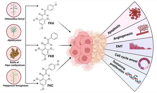
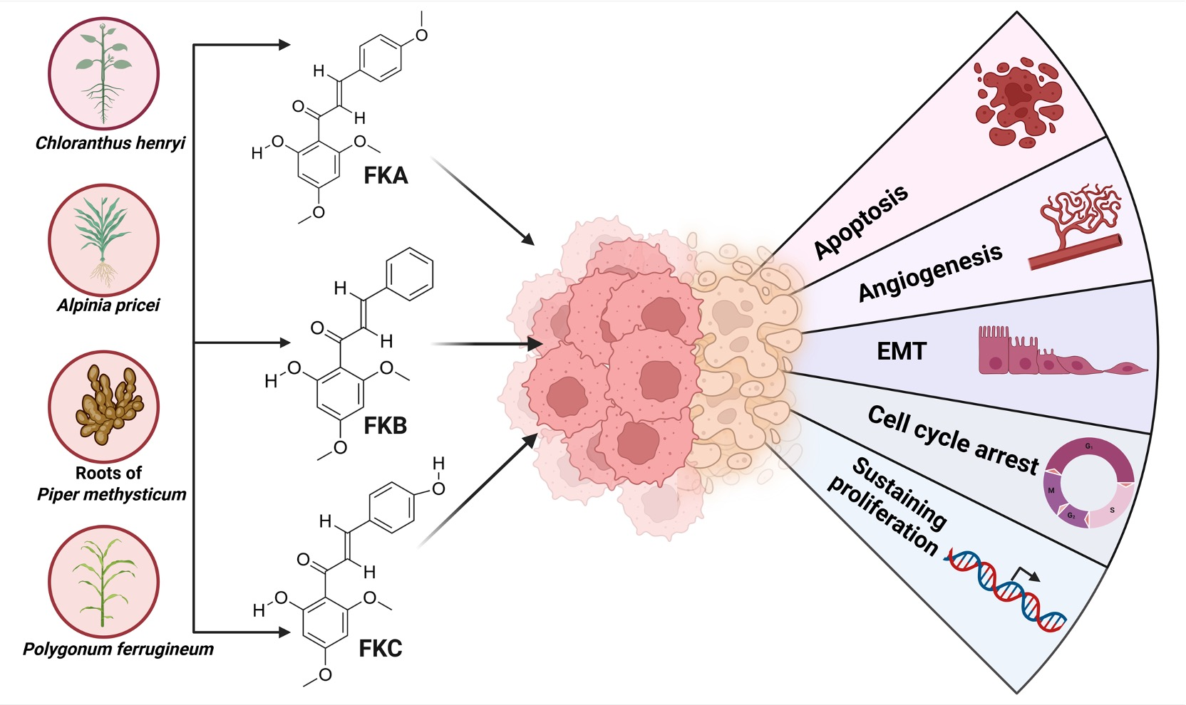
Graphical Abstract

1. Introduction

Cancer remains a major global health burden, exerting profound physical, emotional, economic, and social impacts on patients, and currently stands as the second leading contributor to mortality worldwide [1,2,3,4]. The GLOBOCAN 2022 reported an estimated global burden of nearly 20 million new cancer diagnoses alongside 9.7 million fatalities [5]. In addition, global cancer cases are projected to exceed 35.3 million by 2050, reflecting a 76.6% increase in incidence rates [6]. While conventional interventions, including surgery, radiotherapy, and chemotherapy, are widely utilized, and newer modalities like immunotherapy and gene therapy have emerged, these treatment modalities are often constrained by substantial limitations and considerable financial burden, collectively diminishing patients’ quality of life [7,8,9,10,11,12]. Therefore, it is imperative to develop innovative, highly efficacious and safer anticancer therapeutics that can address the current limitations of conventional treatments [13].
Natural compounds have garnered substantial recognition in modern pharmacology owing to their remarkable chemical diversity, relatively low toxicity, multi-target activity, and widespread availability, making them an indispensable reservoir for drug discovery and the development of efficacious therapeutics [14,15,16]. Approximately 41% of the anticancer drugs have been derived from natural compounds [17]. Furthermore, the use of phytochemicals for cancer prevention is a promising and effective strategy to reduce cancer risk [18,19]. This review critically evaluates the anticancer potential of flavokawains, their biological properties, toxicity profiles, impacts on key tumor microenvironment (TME)-associated processes and explores future perspectives based on a range of preclinical studies.

2. Selection Criteria

A literature search was performed in PubMed, using “flavokawain” as the primary keyword. This initial search identified 140 records. Titles and abstracts were screened for relevance, followed by full-text evaluation against predefined inclusion and exclusion criteria. Publications such as book chapters, errata, editorials, narrative reviews, and non-English articles were excluded. After this selection process, a total of 53 peer-reviewed studies published up to 31 January 2026 met the inclusion criteria and were included in this review.

3. Chemical Nature of Flavokawains

Flavokawains exist in three distinct forms, namely Flavokawain A (FKA), Flavokawain B (FKB), and Flavokawain C (FKC). FKA, chemically identified as 2′-hydroxy-4,4′,6′-trimethoxychalcone, is a yellow crystalline chalcone derivative (molecular formula: C18H18O5, molecular weight: 314.3 g/mol, and melting point: ~113 °C) [PubChem CID: 5355469] [20]. FKB, known as 4′,6′-dimethoxy-2′-hydroxychalcone, is a related trans-chalcone, a yellow crystalline derivative bearing two methoxy groups at the 4′ and 6′ positions and a hydroxy group at the 2′ position. It has a molecular formula of C17H16O4, a molecular weight of 284.31 g/mol, and a lower melting point of 91 °C [PubChem CID: 5356121]. FKC or 2′,4-dihydroxy-4′,6′-dimethoxychalcone is also a yellow crystalline compound like other flavokawains (molecular formula: C17H16O5, molecular weight: 300.30 g/mol, and melting point of 194–195 °C) [PubChem CID: 6293081] [20]. Flavokawains exhibit poor aqueous solubility but dissolve readily in organic solvents such as dimethyl sulfoxide (DMSO) [21,22,23,24] (Table 1).
The three flavokawains share identical A-ring substitution patterns; however, their structural diversity lies in the B-ring substitution, which fundamentally influences their physicochemical characteristics and biological potencies. FKB, possessing an unsubstituted B-ring, displays the highest electrophilicity, conferring superior cytotoxic capabilities. FKA, with a 4-methoxy substitution on the B-ring, exhibits high selectivity toward cancer cells coupled with moderate potency and favorable safety attributes. FKC, bearing a 4-hydroxy B-ring group, demonstrates stronger hydrogen-bonding capacity, resulting in improved bioavailability while maintaining high selectivity and reduced hepatotoxic potential (Table 1). Consequently, these structural variations among the three flavokawains result in differential ADME (absorption, distribution, metabolism, excretion) profiles and binding affinities, which collectively determine their distinct anticancer mechanisms and therapeutic applicability. While all three compounds induce apoptosis and trigger G2/M cell-cycle arrest across various cancer models, their varying potencies and selectivity profiles suggest complementary roles in cancer therapeutics, with each compound offering unique advantages for different clinical applications and treatment strategies.
The chemical structures, physicochemical properties, and bioactivities of flavokawains are illustrated in Figure 1.

4. Sources of Flavokawain

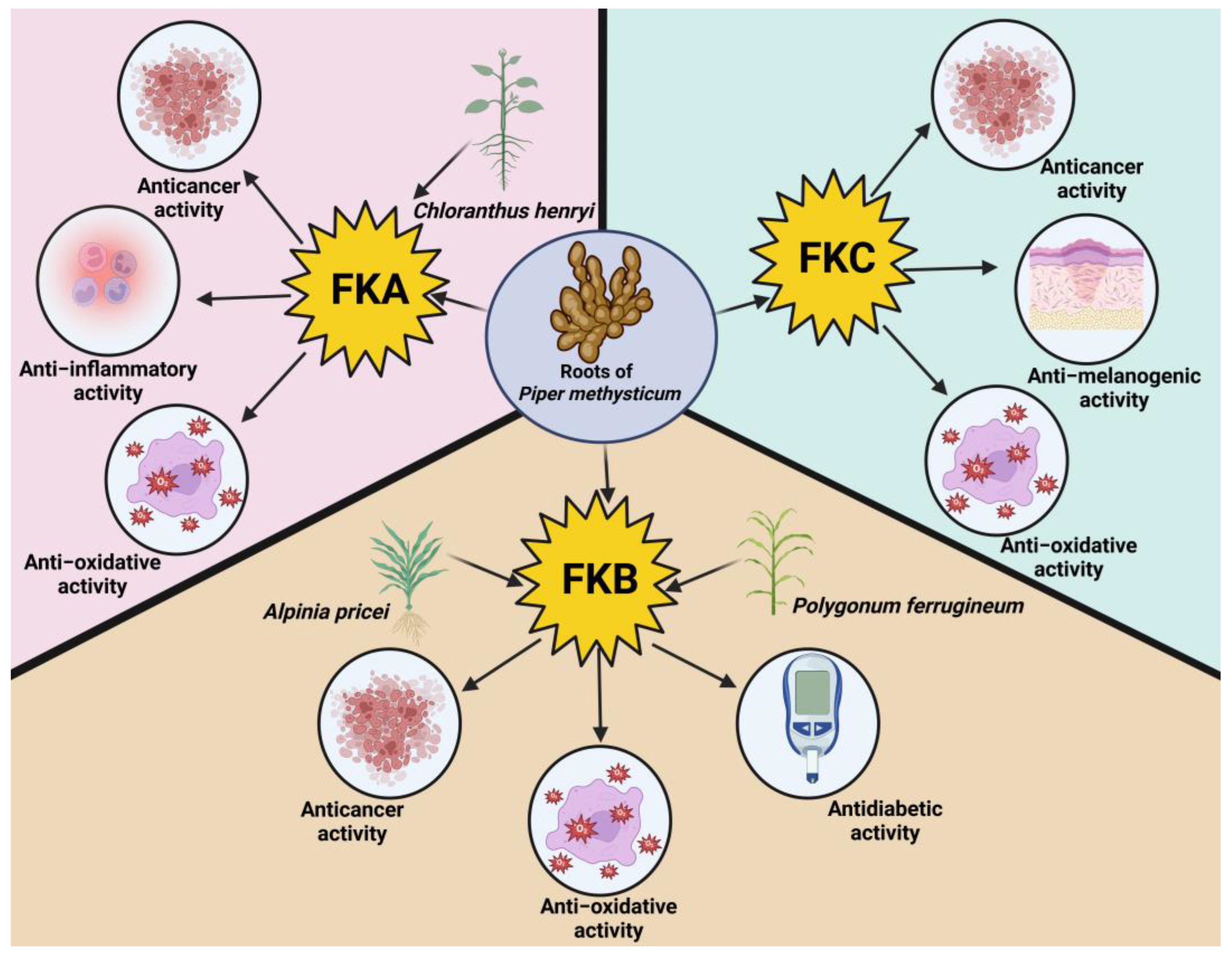
Flavokawains represent a subclass of naturally occurring chalcones predominantly identified in the kava plant (Piper methysticum), a perennial species of the Piperaceae (pepper) family. This plant is indigenous to the South Pacific, including Melanesia, Micronesia, and Polynesia, where it has been traditionally used for its medicinal and ceremonial properties [24,25,26]. FKA, FKB, and FKC are primarily isolated from kava plant roots [23,27]. Among the identified chalcones, FKA is the most abundant, comprising approximately 0.46% of the kava extract, whereas FKB and FKC are present at lower concentrations, at approximately 0.015% and 0.012%, respectively [25]. Another source of FKA, Chloranthus henryi, is a herbaceous perennial distributed across the southern regions of China. Traditionally, this herb has been used in Chinese folk medicine to manage several conditions, including pain and bone fractures [28,29]. In addition to P. methysticum, FKB has been identified in a rhizomatous herb, Alpinia pricei Hayata, indigenous to Taiwan. Traditionally, this plant has been used in herbal medicine to alleviate abdominal bloating and enhance gastric secretion. Furthermore, the leaves of A. pricei are commonly employed in the preparation of zongzi, a traditional Taiwanese glutinous rice dumpling [30,31]. Notably, Polygonum ferrugineum Wedd., commonly referred to as caatay guazú, is a plant native to South America that accumulates the chalcone FKB. In Argentine ethnomedical practices, preparations derived from this plant are employed as natural remedies for their antibacterial, antifungal, wound-healing, and antiseptic effects [32,33]. The plant sources and diverse biological activities of flavokawains are summarized in Figure 2.

5. Biological Properties of Flavokawains

Nature offers a wide variety of plant-based resources, which exhibit numerous inherent therapeutic properties that have been widely used in traditional medicine for the management of numerous diseases [34,35]. Similarly, flavokawains have shown substantial promise for treating a range of diseases, attributed to their diverse pharmacological effect and multi-targeted mechanism of action. Some of these diseases include osteoarthritis, oral mucositis, inflammatory bowel disease, idiopathic pulmonary fibrosis, etc. [36,37,38,39]. For example, preclinical evidence suggests that FKA may ameliorate endometriosis by reducing inflammation and angiogenesis while promoting apoptotic cell death [26]. In fibrosis-related contexts, FKA has been shown to interfere with transforming growth factor beta 1 (TGF-β1) signaling in vascular smooth muscle cells by modulating the mothers against decapentaplegic homolog 3 (Smad3) pathway and suppressing reactive oxygen species (ROS) [40]. FKA’s anti-inflammatory activity has been further attributed to inhibition of nuclear factor kappa-light-chain-enhancer of activated B cells (NF-κB) and activator protein 1 (AP-1) activation, along with suppression of c-Jun N-terminal kinase (JNK) and p38 mitogen-activated protein kinase (MAPK) phosphorylation, and downregulation of lipopolysaccharide (LPS)-induced inducible nitric oxide synthase (iNOS) and cyclooxygenase-2 (COX-2) expression in murine macrophages [41]. FKB has been shown to exert significant hypoglycemic activity upon binding to peroxisome proliferator-activated receptor gamma (PPARγ), supporting its potential role in managing type 2 diabetes mellitus. In vitro cytotoxicity assessment in HepG2 and 3T3-L1 cells revealed minimal toxicity at a pharmacologically relevant concentration (5 μM). In vivo evaluation in HFD/STZ-induced and db/db T2DM mouse models demonstrated that FKB did not induce significant weight gain or hepatotoxicity, as indicated by alanine aminotransferase/aspartate aminotransferase (ALT/AST) levels, in contrast to rosiglitazone, thereby supporting its potential as a safer selective PPARγ modulator [42]. FKB was shown to exert a concentration-dependent inhibitory effect on carrageenan-induced mechanical hyperalgesia, likely mediated through the modulation of the nitric oxide (NO)/cyclic guanosine monophosphate (cGMP)/potassium (K+) channel signaling pathway [43]. Interestingly, FKB demonstrated significant trypanocidal activity, suggesting its potential activity in the management of trypanosomiasis [33]. In a recent study, FKC was shown to exhibit anti-melanogenic activity when incorporated into nanofibers via electrospinning, a process that significantly enhanced its aqueous solubility and transdermal permeability. The improved formulation led to suppression of melanin synthesis in skin cells by reducing melanin production-associated proteins [23]. The collective findings from the aforementioned studies indicate that flavokawains have enormous potential as therapeutic candidates for managing a variety of diseases. This review delves into the anticancer potential of flavokawains, emphasizing their mechanisms of action and therapeutic efficacy.
A pharmacokinetic evaluation conducted in Sprague Dawley rats following oral administration of FKB at 10 mg/kg demonstrated rapid systemic absorption. The compound achieved a maximum plasma concentration (Cmax) of 265.2 ± 117.6 ng/mL at a time to maximum concentration (Tmax) of approximately 1.00 ± 0.55 h. The systemic exposure, as reflected by the area under the plasma concentration–time curves (AUC0–24 and AUC0–∞), was 1009.8 ± 601.3 ng h/mL and 1024.2 ± 618.0 ng h/mL, respectively. FKB exhibited a moderate rate of elimination, with an observed terminal half-life (T1/2) of 2.76 ± 1.19 h. These results suggest that FKB is efficiently absorbed after oral administration and has a moderate duration of systemic exposure [44].

6. Anticancer Potential of Flavokawains

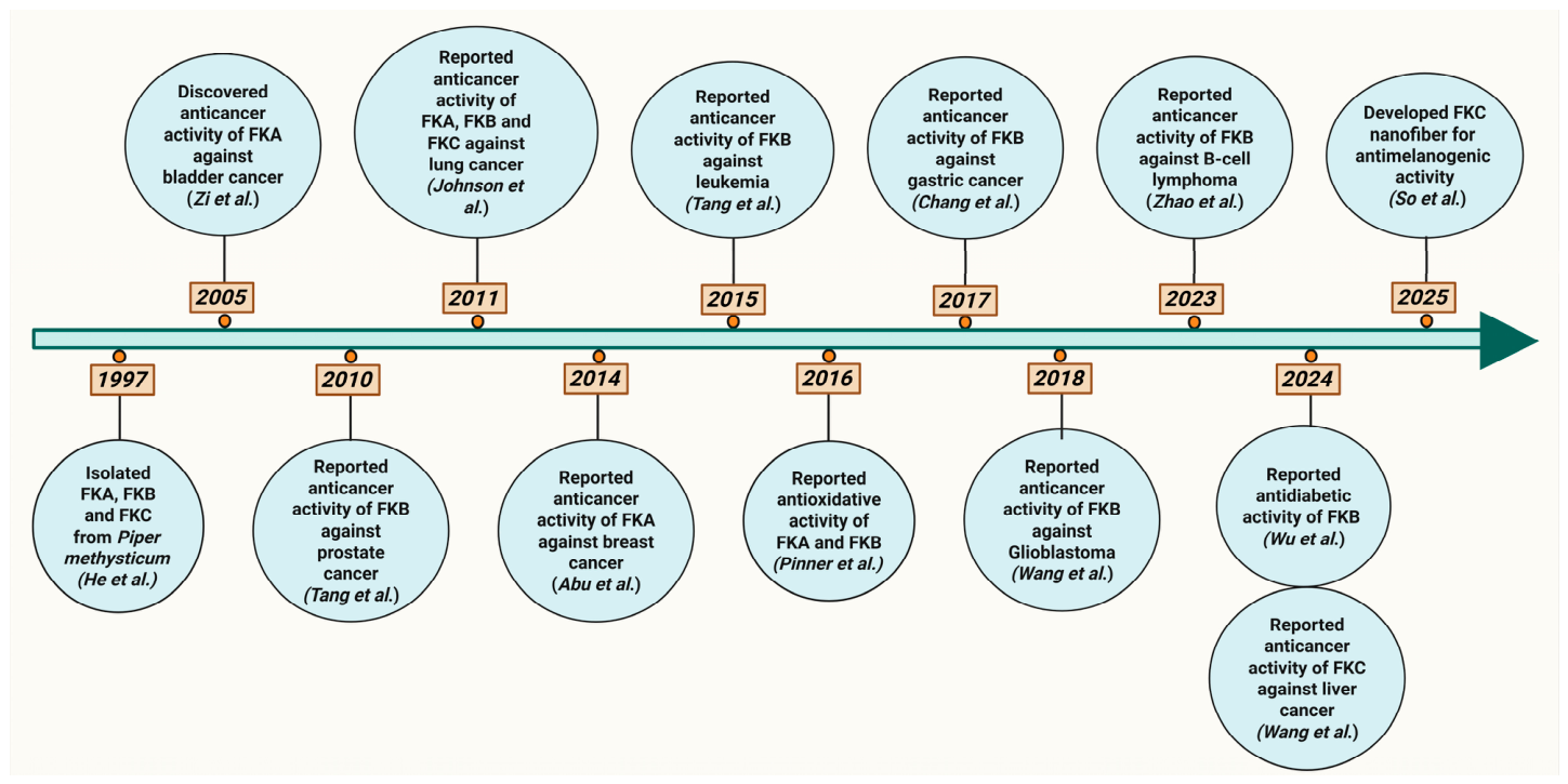
The anticancer activities of flavokawains have been extensively investigated in in vitro cellular systems and in vivo tumor models. The present review systematically evaluates the accumulating preclinical evidence, highlighting their multi-targeted anti-tumor effects through modulation of oncogenic signaling pathways, induction of apoptosis, suppression of metastasis, and remodeling of the TME. A timeline highlighting key discoveries and pharmacological advancements of flavokawains is presented in Figure 3. A comprehensive summary of the therapeutic effects of flavokawains across various cancer types is provided in Table 2.

6.1. B-Cell Lymphoma

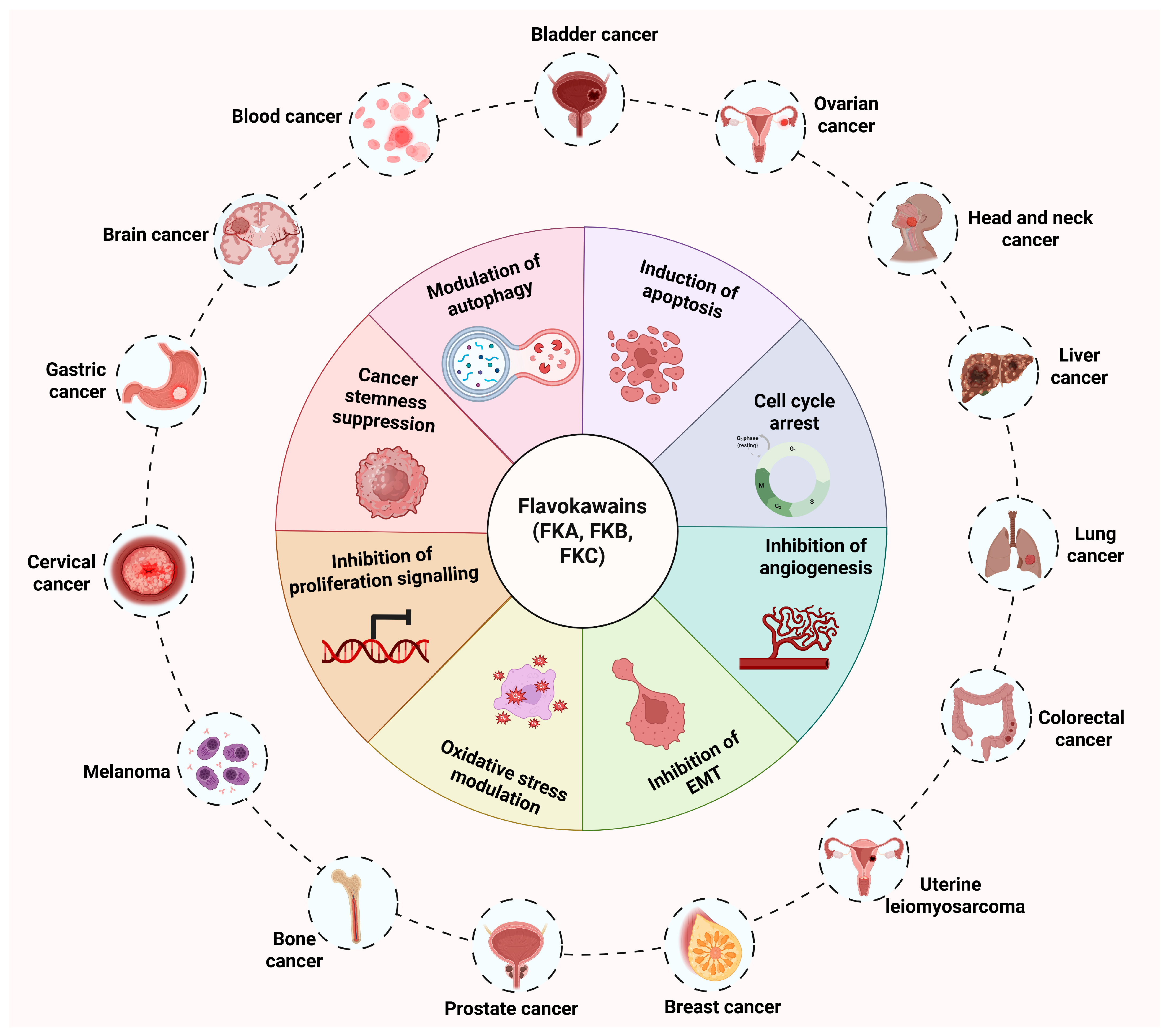
Lymphomas are a heterogeneous group of lymphoproliferative cancers that arise from B cells, T cells, or natural killer cells, and are primarily classified as Hodgkin’s lymphoma (HL) or non-Hodgkin’s lymphoma (NHL) [96,97]. FKB has been reported to exert prominent anti-tumor activity against B-cell lymphoma by interfering with the phosphoinositide 3-kinase (PI3K)/protein kinase B (Akt) signaling pathway [46]. In vitro studies revealed that this compound reduced Akt phosphorylation at Ser473, inhibited cell viability in a dose-dependent manner, and triggered apoptosis via downregulation of B-cell lymphoma-extra large (Bcl-xL), activation of caspase-3, and cleavage of poly(ADP-ribose) polymerase (PARP). Also, ABT-199, a selective B-cell lymphoma 2 inhibitor, exhibits synergistic anticancer activity in combination with FKB. The strongest synergy was observed in SUDHL-4 cells, with a combination index below 0.55, compared with Raji and Jeko-1 cells. This combination led to a concentration-dependent reduction in cell viability in B-lymphoma cells. Further, in vivo efficacy was confirmed in an SUDHL-4-derived xenograft model in nude mice, where FKB treatment markedly reduced tumor weight and decreased the number of Ki-67-positive cells [46]. The broad spectrum of anticancer activities of flavokawains across different cancer types is depicted in Figure 4.

6.2. Bladder Cancer

Bladder cancer is a highly aggressive neoplasm arising from the urothelium covering the inner lining of the bladder and ranks as the ninth most commonly diagnosed cancer worldwide [5,98]. FKA has attracted interest as a candidate inhibitor of protein arginine methyltransferase 5 (PRMT5), an epigenetic regulatory enzyme linked to poor clinical prognosis in bladder cancer [55]. Liu et al. demonstrated that FKA selectively suppressed the proliferation of PRMT5-expressing bladder cancer cells and promoted apoptosis, accompanied by a marked decrease in the methylation of histone residues H2AR3 and H4R3. In UMUC3 cells derived xenograft model, treatment with FKA led to a significant reduction in tumor size and decreased methylation levels at H2AR3 and H4R3. Importantly, when compared to clinically investigated PRMT5 inhibitors such as EPZ015666 and GSK3326595, FKA exhibited superior anti-tumor activity. Combinatorial treatment with FKA and standard chemotherapeutic agents, including cetuximab, gemcitabine, and cisplatin, resulted in enhanced therapeutic efficacy, suggesting a potential synergistic interaction [55]. A separate study has reported that, in a UPII-SV40T transgenic mouse model of bladder cancer, dietary administration of FKA at a dose of 6 g/kg incorporated into the American Institute of Nutrition 93 maintenance diet (AIN-93M) was associated with prolonged overall survival of mice [56]. Additionally, FKA administration reduced tumor-associated bladder weight. Histopathological analysis revealed that this compound attenuated the progression from carcinoma in situ to more aggressive high-grade papillary and muscle-invasive urothelial carcinoma, suggesting a delay in malignant transformation. Immunohistochemical analysis revealed decreased Ki-67 expression, elevated p27 levels, and increased terminal deoxynucleotidyl transferase dUTP nick end labeling (TUNEL)-positive cells. This pro-apoptotic effect was accompanied by downregulation of anti-apoptotic proteins, including Bcl-2, X-linked inhibitor of apoptosis protein (XIAP), and survivin, and upregulation of the death receptor (DR5) [56]. Another study by Tang Y et al. showed that FKA exhibited differential cell-cycle regulatory effects in bladder cancer cells depending on p53 status [57]. In RT4 cells expressing wild-type p53, FKA triggered G1 phase arrest through upregulation of the cyclin-dependent kinase (CDK) inhibitors p21/WAF1 and p27/Kip1, alongside suppression of S-phase Kinase-associated Protein 2 (SKP2) and CDK2 activity. In T24 cells harboring mutant p53, FKA instead induced G2/M phase arrest accompanied by a time-dependent increase in CDK1 activity, along with a decrease in phosphorylation of Cdc25C at Ser216. Moreover, the G2/M checkpoint regulators Myt1 and Wee1 were also downregulated. Oral administration of FKA at 50 mg/kg body weight significantly suppressed tumor growth in RT4-induced xenograft in nude mice, leading to an approximately 64% reduction in tumor volume [57]. Furthermore, FKA reduced cell proliferation in the human bladder cancer cell lines RT4, T24, and EJ by 90–95% [45]. This growth inhibition was accompanied by the induction of apoptosis via activation of caspase-3 and caspase-9 and PARP cleavage, indicating involvement of the intrinsic mitochondrial apoptotic pathway. The anti-apoptotic proteins XIAP and survivin were also downregulated following FKA treatment. Oral administration of FKA at a dosage of 50 mg/kg body weight in nude mice bearing EJ cell-derived xenografts significantly suppressed tumor growth, resulting in a 57% reduction in tumor volume [45]. Interestingly, the combination of FKA with yangonin exhibited a synergistic inhibitory effect on bladder cancer cell proliferation, resulting in a 36–52% greater reduction in cell growth than treatment with either agent alone [58].
Taken together, FKA exhibits significant anti-bladder cancer activity through multiple mechanisms, including inhibition of PRMT5-mediated epigenetic regulation, induction of apoptosis, and modulation of cell-cycle progression. These effects translate into reduced tumor growth, delayed disease progression, and prolonged survival in both xenograft and transgenic bladder cancer models. Moreover, its synergistic effects with conventional chemotherapeutic agents highlight its potential as a promising therapeutic candidate for bladder cancer. However, further safety evaluations and clinical studies are required to validate its efficacy and translational applicability.

6.3. Bone Cancer

Bone cancer malignancies are severe, life-threatening conditions that particularly affect children and adolescents worldwide [99]. Flavokawains have gained significant attention for their distinct anticancer properties in the management of various bone-related malignancies. In a pivotal study investigating the therapeutic effect of FKA on osteosarcoma, treatment with FKA resulted in a marked inhibition of cell proliferation [59]. Moreover, FKA significantly suppressed the invasive capabilities of these cells in a concentration-dependent manner. Molecular analyses revealed that FKA downregulated S-phase kinase-associated protein 2 (SKP2) at both transcript and protein levels, along with an increased accumulation of p21 and cleaved caspase-3. FKA also induced G2/M phase arrest and promoted PARP cleavage. In an in vivo model, oral administration of FKA led to a notable reduction in SKP2 expression in lung tissues and a concomitant decrease in p27 expression in metastatic lung nodules, suggesting attenuation of metastatic progression. Additionally, there was a notable increase in TUNEL-positive cells, indicating enhanced apoptosis in metastatic sites [59]. Wang et al. conducted a study using a synovial sarcoma cell model; FKA produced concentration-dependent inhibition of both cellular proliferation and invasive capacity, induced G2/M phase arrest, and triggered apoptosis, as evidenced by the cleavage of PARP and caspase-7. Mechanistic investigations revealed that FKA downregulated SKP2 expression in a dose-dependent manner, suggesting a role in modulating cell-cycle progression and apoptotic sensitivity. Oral administration of FKA at 600 mg/kg, in severe combined immunodeficiency (SCID) mice bearing HSSY-II-induced synovial sarcoma xenografts, significantly reduced tumor volume. Notably, when combined with doxorubicin, FKA exhibited a strong synergistic effect, markedly enhancing cytotoxicity and further reducing synovial sarcoma cell viability beyond the effects observed with either agent alone [60]. Further, FKA dose-dependently inhibited cell proliferation and colony formation in synovial sarcoma cell lines, with induction of apoptosis confirmed by elevated enzymatic activities of caspases-3, -7, -8, and -9, indicating activation of both extrinsic and intrinsic apoptotic pathways. Pro-apoptotic regulators such as DR5, Bim, and Puma were elevated, while survivin expression was reduced. A shift in the balance of Bcl-2 family proteins was also observed, with an upregulation of BCL2-associated X protein (Bax) and a downregulation of Bcl-2, further contributing to the pro-apoptotic effect of FKA [61].
Collectively, these findings demonstrate that FKA possesses significant anti-tumor activity against osteosarcoma and synovial sarcoma by inhibiting cell proliferation and invasion, inducing G2/M cell-cycle arrest, promoting apoptosis, and suppressing SKP2-mediated oncogenic signaling. The observed reduction in tumor growth and metastasis, along with its synergistic effects with doxorubicin, highlights the potential of FKA as a promising therapeutic candidate for bone-related malignancies. However, further clinical studies are required to establish its safety and efficacy in humans.

6.4. Breast Cancer

Breast cancer is a highly aggressive malignancy and ranks as the second most frequently diagnosed cancer worldwide, with strong propensity to metastasize to distant organs including bone, liver, lung and brain [5,100,101]. Advancements in synthetic derivatives of FKB have unveiled their potency in breast cancer treatments. For instance, a series of twenty-three FKB derivatives (compounds 123) was assessed for anticancer activity in breast cancer cell lines. Among these, compounds FKB-13, FKB-15, and FKB-16 demonstrated markedly enhanced cytotoxic effects relative to the other compounds in the series, warranting further development [62]. In human epidermal growth factor receptor 2 (HER2)-overexpressing breast cancer, FKA has demonstrated an inhibitory effect on cell proliferation, significantly suppressing clonogenic capacity by up to 80% and inducing G2/M phase arrest [63]. Mechanistically, FKA activated Cdc2 kinase activity while concurrently decreasing its phosphorylation at Tyr15, which correlated with reduced expression of its upstream inhibitory regulators, Wee1 and Myt1. FKA also promoted apoptosis in these cells by upregulating Bim and Bax and suppressing survivin, XIAP, Bcl-2, and Bcl-xL. Notably, co-treatment with Herceptin yielded a synergistic reduction in cell viability, highlighting the potential of FKA as an effective adjunct in targeting HER2-driven breast cancer [63]. Further supporting these findings, FLS, a synthetic derivative of flavokawain, exhibited significant antiproliferative effects in breast cancer cells by triggering apoptosis and reducing cell viability, and was associated with G2/M cell-cycle arrest and a progressive increase in the expression of p53 and Bax, alongside decreased Bcl-2 [64]. Additionally, FLS treatment led to elevated caspase-9 activity and enhanced time-dependent cytosolic release of cytochrome c, implicating activation of the intrinsic mitochondrial apoptotic pathway in its mechanism of action [64]. Similarly, a synthesized form of FKB also suppressed breast cancer cell proliferation in a dose-dependent manner, accompanied by induction of apoptosis and G2/M phase arrest of the cell cycle. Under ex vivo conditions, FKB significantly reduced both the invasive and migratory capabilities of breast cancer cells in a concentration-dependent manner and impaired angiogenic activity, further supporting its potential as a multi-targeted therapeutic agent against breast cancer progression [65]. Another study revealed that FKA demonstrated a dose-dependent inhibition of breast cancer cell proliferation, achieving approximately 50% growth suppression at concentrations below 50 µM [21]. The compound exhibited greater potency in MDA-MB-231 cells (IC50 = 17.49 µM) than in MCF-7 cells (IC50 = 25.13 µM). Mechanistic investigations revealed that FKA induced G2/M arrest, promoted apoptosis through activation of caspase-8 and -9, upregulation of pro-apoptotic markers such as Bax and cytochrome c, and downregulation of the mitotic regulators Polo-like kinase 1 (PLK1) and Forkhead box protein M1 (FOXM1). In functional assays, FKA reduced the invasive and migratory behavior of cancer cells in a concentration-dependent manner. Additionally, its anti-angiogenic properties were confirmed using the rat aortic ring model, in which FKA effectively inhibited vessel outgrowth from aortic fragments, supporting its potential as a multi-targeted agent in breast cancer therapy [21].
Flavokawains have demonstrated broad-spectrum preclinical anti-tumor activity against breast cancer through diverse mechanisms, including inhibition of cell proliferation, induction of apoptosis via intrinsic and extrinsic pathways, G2/M cell-cycle arrest, suppression of invasion and migration, and inhibition of angiogenesis. The synergistic efficacy of FKA with Herceptin in HER2-overexpressing breast cancer, along with the enhanced cytotoxic potential of synthetic derivatives such as FKB-13, FKB-15, FKB-16, and FLS, further supports the translational promise of this class of compounds. Nonetheless, advancement toward clinical application necessitates comprehensive in vivo safety and efficacy validation, as well as well-structured clinical trials to firmly establish the therapeutic utility of flavokawains in breast cancer management.

6.5. Cervical Cancer

Cervical cancer is a leading female malignancy that contributes significantly to the global health burden, ranking as the fourth most commonly diagnosed cancer among females worldwide [5,102]. An investigation into the effects of FKB on HeLa cells revealed cytotoxic activity, with an IC50 of 17.5 µM. FKB treatment also resulted in G2/M phase arrest and induced apoptosis, indicating its potential to disrupt cellular proliferation and promote programmed cell death [66]. Furthermore, a notable upregulation of intracellular antioxidant markers, including superoxide dismutase (SOD) and glutathione (GSH), was observed in FKB-treated cells, suggesting enhanced antioxidant defense. When combined with hydrogen peroxide (H2O2), FKB markedly attenuated reactive oxygen species (ROS) levels, implying its role in modulating oxidative stress within the cellular environment [66]. FKB has exhibited promising preclinical anticancer activity in cervical cancer, as evidenced by its cytotoxic effects, induction of G2/M arrest and apoptosis, and modulation of the cellular oxidative stress environment through upregulation of the antioxidant markers SOD and GSH.
These findings show the multifaceted mechanistic potential of FKB as a therapeutic candidate in cervical cancer management. However, the current evidence remains limited to a single in vitro study, and extensive preclinical investigations encompassing in vivo models, followed by rigorous clinical evaluation, are imperative to comprehensively establish its therapeutic efficacy in this malignancy.

6.6. Cholangiocarcinoma

Cholangiocarcinoma is a highly aggressive malignancy arising from the epithelial cells of the bile ducts or liver [103,104]. To improve therapeutic efficacy, combinatorial approaches are being evaluated. For instance, a combination treatment of FKB and cisplatin produced a synergistic reduction in cell viability and enhanced apoptosis in cholangiocarcinoma cells, with effects increasing in a dose-dependent manner relative to monotherapy with either agent [22]. Mechanistically, this enhanced apoptotic response was associated with suppression of the Akt signaling pathway. Additionally, co-treatment led to a marked elevation in cleaved PARP expression, indicating increased apoptotic activity compared to single-agent treatments. In vivo co-treatment of FKB with cisplatin or gemcitabine markedly inhibited tumor growth in SNU-478 xenograft-bearing nude mice, further supporting the therapeutic potential of FKB as a sensitizing agent to cisplatin or gemcitabine in cholangiocarcinoma [22]. Despite these promising preclinical findings, further investigations are required to comprehensively evaluate the therapeutic efficacy of all three flavokawains and to determine their potential integration into standard treatment strategies for cholangiocarcinoma.

6.7. Colorectal Cancer

Globally, colorectal cancer is diagnosed in approximately two million individuals each year, ranking as the third most commonly diagnosed malignancy and second most common cause of death worldwide, with an estimated 60% rise worldwide by 2030 [105,106]. In a recent study, FKA exhibited concentration-dependent cytotoxicity in human colorectal cancer cells, with a marked reduction in cell viability at elevated concentrations [67]. Supporting these findings, another study [71] demonstrated that treatment with FKC significantly reduced cell viability and induced G2/M phase arrest through upregulation of p21 and p27. Apoptotic induction was confirmed by activation of caspases-3, -8, and -9, indicating involvement of both intrinsic and extrinsic pathways. Additionally, FKC suppressed the expression of IAPs, including XIAP, c-IAP1, and c-IAP2. A dose-dependent increase in intracellular ROS levels was accompanied by a decrease in SOD activity, suggesting that oxidative stress may contribute to FKC-induced cell death [71]. Proteomic profiling following FKC treatment revealed significant modulation of specific proteins. Notably, 17 proteins were upregulated, including molecular chaperones and stress-response proteins such as Hspa8, Hsp70-Hom, Hsp27, Hsp70-1/2, and Hsp86, as well as cytoskeletal and metabolic regulators such as TCP-1-eta, CK-18, Tubulin beta-2 chain, and Gamma actin. Additional upregulated proteins included P4HB, heme oxygenase-1 gene (HMOX1), FKBP4, HR23B, SFPQ, PARK7, GLOD4, and GLRX3, suggesting an activation of stress adaptation and redox homeostasis pathways. Conversely, 18 proteins were downregulated in response to FKC, including proteins involved in cytoskeletal function (MLC-3), metabolism (GAMT, PGAM1), detoxification (GSTO1, COMT), and protein synthesis or degradation pathways (DCK, eIF-5A1, EF-2, eIF-3I, Skp1, CBX3, TCEB1, RanBP1, hTom22, and ATP5H) [70]. Moreover, in a xenograft model using HCT-116 colorectal cancer cells in nude mice, administration of FKC at doses of 1 mg/kg and 3 mg/kg produced tumor growth inhibition of approximately 18.7–24.0% and 23.4–52.2%, respectively [68]. Importantly, FKC treatment was well tolerated, without notable changes in body weight or systemic toxicity indicators. Histological analysis of tumor tissues revealed an increase in TUNEL-positive cells and elevated levels of cleaved caspase-3, indicative of apoptosis induction. Concurrently, a reduction in Ki-67-positive cells was observed, reflecting suppression of cell proliferation [68]. In another study, FKB was assessed for its anticancer potential in human colorectal cancer cells, where it demonstrated a dose-dependent inhibition of cell growth. This antiproliferative effect was accompanied by a notable reduction in proliferating cell nuclear antigen (PCNA) expression. Mechanistic analysis revealed activation of caspase-3, indicating the induction of apoptosis. Additionally, FKB treatment led to cell-cycle arrest at the G2/M phase, suggesting that disruption of cell-cycle progression contributes to its cytotoxic activity [69]. Furthermore, FKB was shown to exert dose-dependent antiproliferative activity in vitro, significantly suppressing clonogenic capacity and inducing G2/M phase arrest [72]. Apoptosis was triggered via a mitochondria-dependent intrinsic pathway, leading to caspase activation, as indicated by cytochrome c release. Additionally, FKB treatment upregulated growth arrest and DNA damage-inducible protein 153 (GADD153), elevated intracellular ROS levels, and promoted autophagy, suggesting its multifaceted role in disrupting cancer cell survival [72]. In a recent study, researchers investigated the anti-CRC effects of FKA using an AOM/DSS-induced colorectal cancer model in mice [73]. FKA treatment produced a significant, dose-dependent reduction in the number of colorectal tumors and polyp size, as confirmed by hematoxylin and eosin (H&E) histopathology. Employing integrated 16S rRNA sequencing and untargeted metabolomics, the study further elucidated the underlying mechanisms. FKA restored gut microbiota diversity, evidenced by improved α-diversity indices, including Chao1, Shannon, and Faith’s PD. Further, PICRUSt2 functional prediction revealed that FKA significantly downregulated the LPS biosynthesis superpathway, a key pro-inflammatory endotoxin route, thereby attenuating the inflammatory-to-carcinogenic transition in the colon. Untargeted metabolomics of colonic tissue further demonstrated that FKA inversely modulated lipid and arachidonic acid metabolic pathways, thereby suppressing the generation of pro-inflammatory eicosanoids, including prostaglandins and leukotrienes [73].
Collectively, the available evidence demonstrates that flavokawains, particularly FKA, FKB, and FKC, exhibit potent anticancer activity against colorectal cancer through multiple mechanisms, including inhibition of cell proliferation, induction of G2/M cell-cycle arrest, promotion of apoptosis, generation of oxidative stress, and modulation of key survival pathways. In addition, FKA has shown chemopreventive potential by suppressing colorectal tumor formation, restoring gut microbiota homeostasis, and attenuating inflammation-associated carcinogenic pathways. These findings highlight the promise of flavokawains as potential therapeutic and preventive agents for colorectal cancer; however, further clinical studies are required to validate their safety and efficacy in humans.

6.8. Gastric Cancer

The incidence of gastric cancer increases markedly with age, particularly between 55 and 80 years, and is approximately threefold higher in men compared to women [107]. FKB has been evaluated in multiple gastric cancer cell lines, where it consistently inhibited cell viability and proliferation and induced apoptosis and autophagy [49,74,75]. Treatment with FKB promoted cytochrome-c release and activated caspase-9, implicating the mitochondrial apoptotic pathway. Concurrently, increased expression of Fas and FasL, along with activation of caspase-8, indicated engagement of the extrinsic apoptotic pathway [74]. FKB also induced G2/M phase arrest by reducing Cyclin A, Cyclin B1, CDK1, Cdc25C, and Cdc2 [49,75]. Interestingly, treatment reduced the levels of both Bcl-2 and Bax [49]. In addition, FKB modulated oncogenic signaling cascades, including PI3K/Akt/mammalian target of rapamycin (mTOR), ROS-JNK, and TGF-β [49,74,75]. In vivo, administration of FKB significantly reduced tumor volume and significantly upregulated TSPAN12, TGF-β1, and SMAD4 proteins in AGS- and SGC-7901-derived xenograft models [49,74,75]. Furthermore, the combination of FKB with doxorubicin enhanced cytotoxicity and autophagy in gastric cancer cells in vitro and produced marked inhibition of tumor growth in vivo. Notably, the synergistic effect between FKB and doxorubicin was further confirmed by a combination index < 1 [74].
These findings warrant further investigation to elucidate the effects of FKB on key processes, including cell invasion, migration, and metastasis, in gastric cancer cells. In addition, evaluating its efficacy across a broader panel of gastric cancer cell lines is necessary to validate its clinical potential. Moreover, the anticancer effects of related flavokawains, including FKA and FKC, should also be explored in gastric cancer models.

6.9. Head and Neck Cancers

Oral squamous cell carcinoma (OSCC) is the predominant form of oral cancer and is mostly diagnosed at an advanced stage [108,109]. At relatively low concentrations of 10 μg/mL for FKA and 2.5 μg/mL for FKB, both compounds significantly suppressed cell growth in OSCC cells. In addition to their antiproliferative effects, both flavokawains markedly diminished the migratory and invasive potential of OSCC cells [76]. In addition, FKB inhibited the growth of oral carcinoma cells by inducing apoptosis and causing G2/M cell-cycle arrest [77]. This was associated with reduced expression of cyclin A, cyclin B, Cdc2, and Cdc25C. FKB also promoted the proteolytic activation of procaspase-9 and -3, decreased Bcl-2 levels, and increased Bax expression. Additionally, FKB suppressed PI3K/Akt phosphorylation and p38 MAPK signaling, while inducing a time-dependent upregulation of JNK1/2 and extracellular signal-regulated kinase (ERK1/2), indicating its involvement in both apoptotic and stress-related signaling pathways [77]. Further, under in vitro conditions, FKB inhibited cell proliferation by approximately 85% in oral adenoid cystic carcinoma cells, primarily by inducing G2/M arrest. Additionally, FKB triggered apoptosis and dose-dependently upregulated mRNA expression of the pro-apoptotic markers Bim, Bak, and Bax, as well as Bcl-2, suggesting involvement of mitochondrial apoptotic pathways [78].
Over recent decades, the incidence of nasopharyngeal carcinoma, a highly aggressive malignancy of the head and neck, has shown a marked increase [110]. Treatment with FKC notably inhibited cell proliferation and triggered apoptosis in nasopharyngeal carcinoma cells, primarily by targeting heat shock protein 90 beta family member 1 (HSP90B1) and reducing the expression of the angiogenic factors angiopoietin-1 (Ang-1) and vascular endothelial growth factor (VEGF) [79]. Additionally, FKC suppressed the phosphorylation of key components within the epidermal growth factor receptor EGFR/PI3K/Akt/mTOR signaling cascade, contributing to its anti-tumor effects. In vivo, FKC administration led to a substantial decrease in both tumor volume and weight. This was associated with downregulation of HSP90B1, glucose transporter 1 (GLUT1), hexokinase 2 (HK2), Ang-1, and VEGF, further supporting the inhibition of the EGFR/PI3K/Akt/mTOR axis and related metabolic and angiogenic processes [79].
Collectively, these studies demonstrate that flavokawains exert potent anti-tumor effects against head and neck malignancies, including oral squamous cell carcinoma, oral adenoid cystic carcinoma, and nasopharyngeal carcinoma. Their anticancer activity is mediated through the suppression of cell proliferation, migration, invasion, angiogenesis, and tumor growth, alongside the induction of apoptosis and cell-cycle arrest. Mechanistically, flavokawains modulate multiple oncogenic signaling pathways, including PI3K/Akt, MAPK, EGFR/PI3K/Akt/mTOR, and mitochondrial apoptotic pathways, highlighting their potential as promising multi-target therapeutic agents for the management of head and neck cancers.

6.10. Liver Cancer

Liver cancer accounted for approximately 905,677 new cases and 830,180 fatalities globally, with both incidence and mortality rates significantly higher in males compared to females [111]. The effect of flavokawains was investigated in liver cancer. For example, FKA treatment suppressed the viability, invasive capacity, migration, and vasculogenic mimicry of hepatocellular carcinoma (HCC) cells, accompanied by reduced expression of VE-cadherin, vimentin, and Snail1, and increased E-cadherin levels, indicative of epithelial–mesenchymal transition (EMT) inhibition [29]. This effect was associated with decreased phosphorylation of PI3K and Akt, and with downregulation of key EMT-associated transcription factors, including HIF-1α, NF-κB, and Twist1. In a HepG2 xenograft model, FKA administration suppressed tumor growth and metastasis, elevated E-cadherin expression, and suppressed Twist1, VE-cadherin, vimentin, and p-Akt levels, suggesting that FKA hinders EMT progression by targeting the PI3K/Akt/Twist1 signaling axis [29]. Another study compared the cytotoxicity of FKA and FKB in HepG2 cells and found that FKA exhibited less cytotoxicity than FKB [50]. Both compounds induced a concentration-dependent increase in nuclear factor erythroid 2-related factor 2 (Nrf2) and HSF1 expression, as well as elevated mRNA levels of antioxidant and stress-response genes, including HMOX1 and GCLC. Additionally, dose-dependent upregulation of HSPA1A and DNAJA4 was observed. Protein levels of HO-1 and Hsp70-1 were also increased following treatment with FKA and FKB. However, both compounds were shown to enhance intracellular GSH levels and confer protection against H2O2-induced oxidative cell death, suggesting a role in cellular defense through activation of antioxidant and heat shock responses in these cells [50]. Therefore, more studies are required to validate these findings as both compounds induce resistance in cancer cells, which is not good for cancer treatment. Expanding the investigation to FKC, a recent study reported that treatment of liver cancer cells with FKC resulted in inhibition of cell proliferation and clonogenic capacity, along with induction of apoptosis, as evidenced by decreased Bcl-2 and elevated Bax protein levels [48]. FKC also promoted DNA damage, indicated by increased γ-H2AX expression. Additionally, it impaired cell adhesion and significantly suppressed migration, which correlated with reduced phosphorylation of focal adhesion kinase (FAK), PI3K, and Akt, suggesting disruption of key signaling pathways involved in cell motility and survival. In vivo, FKC administration led to reduced tumor growth, lower Ki-67 expression, and elevated γ-H2AX levels, indicating reduced proliferation and increased DNA damage in a Huh-7 xenograft mouse model [48].
Collectively, these findings suggest the promising anticancer potential of flavokawains in liver cancer; however, further studies are warranted to fully elucidate their mechanisms of action, address concerns about chemoresistance, and validate their therapeutic efficacy in clinical settings.

6.11. Lung Cancer

Lung cancer is the most commonly diagnosed malignancy globally, with an estimated 2.5 million new cases annually [112]. FKA induced a concentration-dependent inhibition of cell viability in lung cancer cells by inducing apoptosis as confirmed by PARP cleavage, while exhibiting no toxicity toward normal human hepatic epithelial THLE-3 cells. Additionally, FKA markedly suppressed P-glycoprotein, Akt, and p-Akt (Ser473), suggesting its potential role in overcoming drug resistance through modulation of the PI3K/Akt signaling pathway [80]. Additionally, in a murine model of tumorigenesis induced by 4-(Methylnitrosamino)-1-(3-pyridyl)-1-butanone (NNK) and benzo[a]pyrene (BaP), treatment with FKA, FKB, and FKC each reduced tumor multiplicity by 27%, 34%, and 23%, respectively, suggesting a differential yet significant chemopreventive effect of each flavokawain [51]. Furthermore, FKB markedly reduced cell viability and clonogenic potential in lung cancer cells, while promoting apoptosis, as indicated by increased TUNEL-positive cells, activation of caspase-3 and -9, and subsequent PARP cleavage. Additionally, FKB induced ROS generation, contributing to oxidative stress-mediated cell death. An increase in microtubule-associated protein 1A/1B-light chain 3-II (LC3-II) expression was observed, suggesting the induction of autophagy. Concurrently, FKB treatment led to reductions in Bcl-2 and Bax protein levels, implicating disruption of mitochondrial integrity [82]. In another study, Chalcone-24, a synthetic flavokawain, was found to suppress both cell viability and NF-κB activation in lung cancer cells. At lower concentrations, treatment with Chalcone-24 elevated ERK1/2 and JNK phosphorylation, accompanied by enhanced caspase activity [81].
Collectively, these findings indicate that flavokawains possess significant anti-lung cancer activity by inhibiting tumor cell growth, inducing apoptosis and autophagy, suppressing oncogenic signaling pathways, and reducing tumor burden. However, further pharmacokinetic, toxicity, and clinical studies are required to validate their therapeutic potential.

6.12. Leukemia

An increase in white blood cell count in the blood and bone marrow is characteristic of this hematological malignancy [113]. Leukemia represents a notable global health burden, accounting for about 2.5% and 3.1% of all global cancer cases and mortality, respectively [114]. In a recent study, FKA was shown to induce a concentration-dependent reduction in the viability of acute myeloid leukemia (AML) cells and trigger G1 phase arrest through the downregulation of CCND1, CCNE1, CCNE2, CDK2, CDK4, CDK6, and CDT1, along with upregulation of the CDK inhibitor CDKN1B [83]. In ex vivo experiments using primary leukemic cells isolated from AML patients, FKA similarly reduced cell viability in a dose-dependent manner, with IC50 values ranging from 3.86 to 9.45 μg/mL, reinforcing its therapeutic potential in AML [83]. Another study showed that FKB markedly inhibited proliferation and induced apoptosis in acute lymphoblastic leukemia (ALL) cells in vitro, in a dose-dependent manner, by enhancing caspase-3 activation and PARP cleavage. FKB treatment also led to a concentration-dependent upregulation of key pro-apoptotic regulators, including p53, Bax, and Puma. In vivo, FKB administration resulted in a notable reduction in leukocytes, white blood cell counts, and a decrease in splenomegaly, indicating systemic anti-leukemic activity. Ex vivo analysis of patient-derived B-ALL and T-ALL confirmed these findings, showing marked suppression of cell proliferation and elevated levels of p53, Bax, and Puma [52]. Further investigation demonstrated that FKB enhanced daunorubicin-induced reduction in cell viability, demonstrating an additive cytotoxic effect. Interestingly, rather than suppressing NF-κB activity, the combination treatment activated NF-κB, suggesting a complex interaction between FKB and daunorubicin [84].
Taken together, flavokawains exhibit potent anti-leukemic effects through the induction of cell-cycle arrest, activation of apoptotic pathways, and enhancement of chemotherapeutic efficacy, supporting their potential as novel therapeutic agents against leukemia. Nevertheless, further pharmacokinetic, toxicological, and clinical investigations are needed to facilitate their translation into leukemia therapy.

6.13. Melanoma

Melanoma, particularly in its advanced stages, presents a significant clinical challenge and is often associated with a poor prognosis [115]. In addition, synthetic flavokawains A (FLA) and B (FLB) demonstrated potent cytotoxic effects, reducing cell viability by approximately 85% and 80%, respectively. Both compounds induced a dose-dependent decrease in intracellular melatonin levels, indicating suppression of melatonin biosynthesis. This was further supported by reduced expression of key melanogenic markers, including tyrosinase (Tyr), tyrosinase-related proteins 1 and 2 (Trp-1, Trp-2), and microphthalmia-associated transcription factor (Mitf). In a zebrafish toxicity model, both FLA and FLB exhibited high survival rates, indicating low systemic toxicity while still effectively reducing melatonin content [85].

6.14. Neuro-Oncological Malignancies

Glioblastoma (GBM) is a highly aggressive and uniformly fatal primary brain tumor, classified as a grade IV astrocytoma, and accounts for over 60% of all brain tumor cases [116,117]. To examine newer treatment modalities, FKB was studied in vitro in glioblastoma cells, where it produced a concentration-dependent reduction in cell viability [53]. This compound further induced G2/M arrest and promoted cellular senescence, as evidenced by the accumulation of senescence-associated β-galactosidase (SA-β-gal)-positive cells. Markers of autophagy, including increased MAP1LC3B-II and decreased SQSTM1 expression, indicated activation of the autophagic pathway. This autophagy appeared to be dependent on endoplasmic reticulum (ER) stress, as evidenced by the upregulation of ER stress-associated proteins, including HSPA5, phosphorylated EIF2AK3 and EIF2A, ATF4, and DDIT3. Additionally, FKB suppressed the phosphorylation of Akt, mTOR, and RPS6KB1, thereby inhibiting the Akt-mTOR-RPS6KB1 signaling axis and further implicating this pathway in autophagy induction by this compound. Moreover, in vivo administration of FKB at 50 mg/kg significantly reduced tumor growth in a U251 glioblastoma xenograft model in nude mice, indicating its potential anti-tumor efficacy [53]. However, effective glioblastoma treatment requires penetration of the blood–brain barrier (BBB), and there is currently no compelling evidence that FKB can adequately traverse it. Therefore, further investigations are required to evaluate its BBB permeability and elucidate potential strategies or delivery mechanisms to facilitate its transport across the BBB, thereby enhancing its suitability as a therapeutic candidate for glioblastoma treatment.
Despite the development of various therapeutic approaches, neuroblastoma remains associated with a high risk of relapse and disease-related mortality [118]. FKA exerted a dose-dependent suppression of cell proliferation and clonogenic potential in neuroblastoma cells by inducing G1-phase cell-cycle arrest and promoting apoptosis. In addition to its antiproliferative effects, FKA significantly impaired the cells’ invasive and migratory capabilities and downregulated EMT markers, including N-cadherin and Snail. Furthermore, FKA reduced VE-cadherin expression in a dose-dependent manner, indicating its capacity to inhibit angiogenesis and suppress tumor progression [86].
Collectively, these findings demonstrate that flavokawains exert promising anti-tumor effects against neuro-oncological malignancies by suppressing cell proliferation, inducing cell-cycle arrest, apoptosis, autophagy, and senescence, and inhibiting invasion, migration, angiogenesis, and EMT. Mechanistically, these effects are mediated through modulation of ER stress-associated pathways, inhibition of Akt/mTOR signaling, and regulation of EMT-related markers. However, further studies are required to elucidate their pharmacokinetic properties, validate their efficacy in clinically relevant preclinical models, and, particularly for glioblastoma, determine their ability to penetrate the BBB and achieve therapeutically effective concentrations within the central nervous system.

6.15. Ovarian Cancer

Ovarian cancer is a heterogeneous malignancy that frequently affects women under the age of 40 and ranks as the eighth most common cancer among females worldwide [119]. The combinatorial administration of FKA-A and PTX-A nanoparticles markedly reduced proliferation and clonogenic capacity in ovarian cancer cell lines. This treatment also significantly impaired cellular migratory ability, as evidenced by elevated E-cadherin and reduced vimentin expression, indicating suppression of EMT. Furthermore, in an A2780 xenograft mouse model, the combined therapy substantially inhibited tumor growth [87]. Another study reported that FKC led to a notable reduction in ovarian cancer cell viability [88]. However, further preclinical and clinical investigations are warranted to confirm the therapeutic potential of flavokawains in ovarian cancer and facilitate their clinical translation.

6.16. Prostate Cancer

Prostate cancer is a major malignancy that poses a significant health concern for the male population [120]. FKA treatment produced a significant reduction in both the size and number of prostaspheres derived from DU145 and 22Rv1 cancer stem cells (CSCs), accompanied by downregulation of stemness-associated markers Oct4, Sox2, and Nanog. In vitro, FKA dose-dependently suppressed c-Myc expression in prostaspheres from both cell lines. In an in vivo model, dietary administration of FKA resulted in a 48% reduction in tumor volume, a 64% decrease in Ki-67-positive proliferating cells, and a marked decline in CD44-positive CSCs. Additionally, FKA inhibited Ubc12 neddylation and reduced c-Myc levels in xenograft tumor tissues [89]. In addition, FKA was shown to suppress proliferation in prostate cancer cells by inducing apoptosis and causing cell-cycle arrest at the G2/M phase, an effect associated with decreased survivin expression. Additionally, FKA treatment led to a dose-dependent reduction in intracellular GSH levels and glutathione synthetase (GSS) activity, resulting in a marked increase in ROS accumulation [90]. Also, FKB inhibited the neddylation of Cullin1 and Ubc12 by directly binding to the NEDD8-activating enzyme (NAE) regulatory subunit APP-BP1, thereby impairing SCFSKP2 complex function. This results in enhanced SKP2 ubiquitination and proteasomal degradation without affecting proteasome activity. This promoted SKP2 ubiquitination and proteasomal degradation without affecting proteasome activity, leading to reduced SKP2 expression and increased p27/Kip1 levels in prostate cancer cells [91]. Furthermore, FKB induced a ~90% reduction in prostate cancer cell viability at 17.6 µM, primarily via activation of caspases-3, -8, and -9, indicating apoptosis via intrinsic and extrinsic pathways. Upregulation of DR5 enhanced TRAIL-mediated apoptosis, while increased Bim and Puma and decreased XIAP and survivin further supported pro-apoptotic effects. In DU145 xenograft mice, FKB (50 mg/kg/day) reduced tumor volume by ~67% and increased Bim expression in tumor tissues [54]. Moreover, FKA significantly inhibited prostate cancer cell growth by approximately 93% in Rb-deficient DU145 cells and reduced the viability of Rb-deficient MEFs by 92%. The increased sensitivity of SKP2-overexpressing cells to FKA suggests that SKP2 may be a potential target of FKA in pRb-deficient prostate cancers. FKA treatment also decreased NEDDylation of Cullin1 and Ubc12, indicating disruption of protein degradation pathways. In a transgenic adenocarcinoma of mouse prostate (TRAMP) mouse model, dietary FKA suppressed high-grade prostatic intraepithelial neoplasia (HG-PIN) lesions by 69% and prostate adenocarcinomas by 43%, resulting in an overall 73% reduction in tumors. This was accompanied by decreased proliferation, enhanced apoptosis, and absence of metastasis [92]. In another study, FKB treatment led to a marked reduction in androgen receptor (AR) expression and prostate-specific antigen (PSA) protein levels. When combined with other kavalactones, FKB suppressed prostate cancer cell growth by over 70%. In patient-derived xenografts in SCID mice, FKB inhibited tumor growth by approximately 77.3% and significantly reduced serum PSA levels by the end of treatment [93].
Collectively, these studies demonstrate that flavokawains exert potent anti-prostate cancer activity by suppressing tumor cell proliferation, cancer stemness, androgen receptor signaling, and tumor progression while promoting cell-cycle arrest, oxidative stress, and apoptosis. Mechanistically, flavokawains target multiple oncogenic pathways, including NEDDylation, Skp2-mediated signaling, c-Myc regulation, and AR/PSA signaling, and exhibit efficacy in both castration-sensitive and castration-resistant prostate cancer models. Furthermore, their significant tumor-suppressive effects in xenograft and transgenic mouse models underscore their therapeutic potential.

6.17. Squamous Carcinoma

In line with findings observed in other cancer types, flavokawain compounds have also been investigated in squamous carcinoma models, demonstrating significant anticancer activity. FKB treatment reduced cell viability and induced G2/M phase arrest in KB and HGF cells, accompanied by increased ROS production and mitochondrial dysfunction [94]. This was associated with cytochrome c release, activation of caspase-3 and -9, and upregulation of Fas and FasL, indicating activation of both intrinsic and extrinsic apoptotic pathways. FKB also downregulated Bcl-2 and upregulated Bax. Cell-cycle regulators, including cyclin A, cyclin B1, Cdc2, and Cdc25C, were suppressed, while p21/WAF1, Wee1, and p53 were upregulated. Additionally, FKB reduced the expression of the metastasis-associated proteins matrix metalloproteinase (MMP-9) and urokinase-type plasminogen activator (u-PA) and increased the expression of their inhibitors, tissue inhibitor of metalloproteinase-1 (TIMP-1) and plasminogen activator inhibitor-1 (PAI-1), in a dose-dependent manner. In vivo, FKB administration (0.75 mg/kg) significantly reduced tumor volume, suppressed angiogenesis, and increased TUNEL-positive cells, indicating enhanced apoptosis and reduced tumor cell proliferation in KB xenograft mice model [94].

6.18. Uterine Leiomyosarcoma

Uterine leiomyosarcoma is a highly aggressive malignancy characterized by frequent recurrence, metastatic potential, and poor clinical outcomes [121]. FKB markedly inhibited uterine leiomyosarcoma cell growth by approximately 80%, induced G2/M cell-cycle arrest, and triggered apoptosis. This was associated with upregulation of pro-apoptotic proteins Bim, Puma, and DR5, along with downregulation of IAP and survivin. Notably, FKB exhibited a synergistic effect when combined with gemcitabine or docetaxel, resulting in enhanced suppression of cell proliferation [95].
The major molecular signaling pathways modulated by flavokawains across various cancer types are illustrated in Figure 5.

7. Flavokawains and the Tumor Microenvironment (TME)

Flavokawains exert multifaceted regulatory effects within the TME, targeting key processes including angiogenesis and EMT. Therapeutic strategies aimed at reprogramming or disrupting the TME have garnered significant attention in oncological research because the TME plays a pivotal role in tumor initiation, progression, metastasis, and therapeutic resistance. Current therapeutic approaches targeting the TME include anti-angiogenic therapies, like bevacizumab (VEGF inhibitor), which aim to disrupt pathological neovascularization that sustains tumor growth [122]. Immune checkpoint inhibitors, including PD-1/PD-L1 and CTLA-4 antibodies, release inhibitory brakes on cytotoxic T cells, restoring anti-tumor immunity [123]. Evidence from the aforementioned studies indicates that FKA suppresses vasculogenic mimicry and metastatic behavior in HCC by downregulating VE-cadherin, vimentin, Snail1, CXCR4, HIF-1α, NF-κB, and phosphorylated PI3K/Akt, while restoring E-cadherin expression [29]. Similarly, FKC inhibits migration in liver cancer cells by attenuating p-FAK, p-PI3K, and p-Akt signaling and enhances γ-H2AX expression in vivo, indicating impaired survival signaling and increased DNA damage within the tumor niche [48].
Angiogenesis, a key hallmark of TME, is markedly suppressed by flavokawains. For example, synthetic FKA and FKB inhibited angiogenesis in breast cancer models [21,65]. Moreover, FKC treatment significantly downregulated the expression of key pro-angiogenic mediators, including VEGF and Ang-1, in nasopharyngeal carcinoma [79]. Further, FKA modulates the neuroblastoma TME by inactivating ERK signaling, leading to suppression of VEGF-driven angiogenesis, inhibition of MMP2, MMP9, MMP14, and inhibition of EMT-associated migration and invasion, collectively impairing pro-metastatic niche formation [86].
FKB modulates the TME in squamous carcinoma by targeting metastasis- and invasion-associated proteolytic systems. Specifically, treatment with FKB suppressed MMP-9 and u-PA expression and activity, while upregulating their endogenous inhibitors TIMP-1 and PAI-1, thereby limiting extracellular matrix degradation and suppressing invasive niche formation. In vivo xenograft analyses further demonstrated reduced angiogenesis in FKB-treated tumors [94]. FKA significantly downregulated Nanog expression in prostate CSC-derived prostaspheres and xenograft tumors, thereby disrupting the CSC-supportive TME [89]. FKA-A NPs in ovarian cancer downregulated SKP2 expression, thereby impairing SKP2-mediated YAP activation. This led to decreased migratory capacity and suppression of EMT, characterized by upregulation of E-cadherin and downregulation of vimentin, collectively restricting invasion and metastatic progression [87]. In OSCC, FKA and FKB significantly reduced tumor cell migration and invasion without affecting adhesion to extracellular matrix components [76].
Beyond their direct effects on tumor cells, flavokawains exhibit notable immunomodulatory properties. In a murine in vivo study, oral administration of FKA and FKB (50 mg/kg/day for 28 days) increased the populations of CD4+/CD3+ T helper cells and CD8+/CD3+ cytotoxic T lymphocytes in splenocytes, with FKB producing a more pronounced effect. This was accompanied by a dose-dependent upregulation of IL-2, a cytokine essential for T-cell proliferation and immune homeostasis [27]. Both compounds suppressed TNF-α levels, reduced NO production, and significantly decreased M2-polarized macrophage populations, with FKB reducing M2 phenotype from 11.6% to 2.2% [27].
Given that M2 tumor-associated macrophages are well-established drivers of tumor progression and immune evasion, the ability of flavokawains to suppress M2 polarization and restore T cell effector populations suggests a potential capacity to modulate immune-related components of the TME. Although direct investigations of flavokawain-mediated immune regulation within the TME remain limited, evidence from a murine model suggests that these compounds possess immunomodulatory properties that may have implications for TME regulation.
Taken together, the available evidence indicates that flavokawains influence multiple interconnected components of the TME rather than acting solely through direct cytotoxic effects on tumor cells. By suppressing angiogenesis, limiting extracellular matrix remodeling, reversing EMT-associated changes, targeting cancer stemness-related pathways, and exhibiting preliminary immunomodulatory activity, flavokawains may alter the biological conditions that support tumor growth, invasion, and metastasis. Although current evidence is derived predominantly from preclinical studies, these collective findings provide a stronger rationale for considering flavokawains as potential modulators of TME reprogramming.

8. Toxicity Concerns and Safety Issues

In comparative cytotoxicity analyses conducted in HepG2 cells, the three chalcones were evaluated; among them, FKB exhibited the greatest cytotoxic activity, with an LD50 of approximately 15.3 µM, whereas FKA and FKC showed comparatively lower potency, with LD50 values of 75 µM and 70 µM, respectively [124]. FKB also induced significant cell death in non-cancerous L-02 hepatocytes. Morphological assessment of L-02 cells following FKB treatment revealed features characteristic of apoptosis, including cell rounding, loss of microvilli, and membrane blebbing. Treatment with FKB at 30 µM in HepG2 cells resulted in caspase-3 activation, further confirming the induction of apoptosis. Additionally, FKB markedly inhibited NF-κB transcriptional activity in HepG2 cells in a dose-dependent manner, as demonstrated by suppression of TNF-α-induced IκB transcription and reporter gene assays. Pre-treatment of HepG2 cells with FKB (30 µM), followed by TNF-α stimulation, led to a significant increase in the phosphorylation of stress-related MAPKs, including JNK, p38, and ERK, suggesting activation of stress response pathways. In L-02 cells, FKB treatment also reduced intracellular GSH levels, indicating oxidative stress. Under in vivo conditions, oral administration of FKB at 25 mg/kg in mice resulted in hepatic injury, evidenced by elevated serum aspartate aminotransferase (AST) and alkaline phosphatase (AKP) levels. Moreover, FKB significantly suppressed LPS-induced NF-κB activation in mouse liver tissue, supporting its immunomodulatory effect [124]. In contrast, a study using TRAMP mice with an FVB/N genetic background found that dietary administration of FKA at 0.6% (w/w), equivalent to 6 g/kg in the AIN-76A diet, was well tolerated and produced no observable adverse effects on food intake or body weight. Histopathological evaluation demonstrated that FKA exposure did not influence the relative organ weights or morphology of key organs, including the heart, liver, lungs, spleen, kidneys, thymus, colon, and testes. Tissue analysis showed no evidence of pathological changes, including inflammation, fibrosis, atrophy, parenchymal disruption, or epithelial abnormalities. Furthermore, the serum biochemical markers ALT, AST, alkaline phosphatase, creatinine, glucose, and cholesterol remained within physiological limits, indicating preserved hepatic, renal, and systemic function. Additionally, assessment of bone marrow cell colony formation revealed no significant alterations in clonogenic potential following FKA treatment, further supporting its favorable safety profile [125]. The hepatotoxic potential of FKA and FKB was investigated in C57BL/6 mice to assess their individual and combined effects on liver function. When administered separately, FKB (11.5 mg/kg) does not produce any signs of liver toxicity, as evidenced by serum ALT and AST levels remaining within physiological ranges. Moreover, FKA exhibited no toxicity toward normal human hepatic epithelial THLE-3 cells, highlighting its selective anticancer activity and favorable safety profile. However, FKB, when combined with acetaminophen (APAP), resulted in a marked potentiation of APAP-induced liver injury, demonstrated by a significant increase in ALT and AST levels, suggesting a synergistic interaction that enhances hepatocellular damage. Combinatorial treatment of FKA and FKB with APAP resulted in higher ALT and AST levels, resulting in hepatotoxicity [126]. The toxicity profile of flavokawains in different experimental models is summarized in Table 3.
Collectively, the available evidence indicates that the safety profile of flavokawains is both compound- and context-dependent. While FKB has demonstrated hepatotoxic potential under specific experimental conditions and acetaminophen-induced liver injury models, flavokawains administered alone have generally exhibited acceptable tolerability in several in vivo models. Thus, the favorable safety outcomes reported for certain flavokawains should be interpreted within the context of the specific compounds, doses, and experimental models evaluated. Therefore, future pharmacokinetic, dose-escalation, and long-term safety studies are required to more clearly define their therapeutic index and overall safety profile.

9. Limitations and Future Perspectives

While this review provides a comprehensive overview of the anticancer potential of flavokawains, certain limitations should be acknowledged. The literature search was conducted primarily using the PubMed database, which, despite its extensive coverage of biomedical research, may not encompass all relevant studies indexed in other databases. Future reviews incorporating multiple databases and broader search strategies would provide a more exhaustive assessment of the available evidence.
Flavokawains have demonstrated significant anticancer potential across various in vitro and in vivo cancer models, primarily through modulation of key molecular pathways involved in proliferation, apoptosis, invasion, migration, angiogenesis, and EMT. However, several limitations hinder their translational application. First, although flavokawains show promising cytotoxic effects, comprehensive toxicity profiling in normal, non-cancerous cells and animal models remains limited. Dose-dependent toxicological studies are essential to evaluate their safety margins and potential off-target effects. Given the growing clinical success of immune checkpoint blockades, future studies should investigate whether flavokawains modulate immune checkpoint pathways, including PD-1/PD-L1, CTLA-4, TIGIT, and LAG-3, and evaluate their potential as adjuvants to immune checkpoint inhibitors.
The clinical utility of flavokawains also requires deeper investigation into their potential roles as chemosensitizing and radiosensitizing agents. Such studies could reveal synergistic effects with conventional therapies and help overcome resistance mechanisms. Furthermore, although a limited number of studies have used flavokawain-based nanoparticles, liposomes, or other delivery systems to improve pharmacokinetics, more extensive, standardized nanocarrier-based studies are needed to optimize targeted delivery and minimize systemic toxicity. Furthermore, the potential for herb–drug interactions involving flavokawains requires comprehensive evaluation in future studies.
Beyond oncology, the therapeutic potential of flavokawain in chronic diseases such as neurological, cardiovascular, and gastrointestinal disorders, like gastritis, which may predispose individuals to cancer, remains underexplored. Investigating their multi-targeted effects in these contexts could broaden their clinical relevance. Future research should include well-designed in vivo studies with dose–response evaluation and long-term safety assessment. Finally, robust clinical trials involving larger and more diverse populations are imperative to establish their pharmacological efficacy, safety, and potential integration into standard treatment regimens.

10. Conclusions

Flavokawains have emerged as bioactive chalcone derivatives with significant antineoplastic activity across a range of cancer models in preclinical investigations. Accumulating evidence indicates that these compounds can potentiate the efficacy of established chemotherapeutic agents through synergistic interactions, thereby improving overall therapeutic outcomes. Importantly, flavokawains demonstrate a degree of tumor selectivity, exerting cytotoxic effects on malignant cells while sparing normal tissues to a considerable extent. This differential activity positions them as favorable candidates for drug development. Collectively, these findings support continued exploration of flavokawains as novel phytochemical-based therapeutics, either as standalone agents or in combination regimens, with the potential to achieve enhanced efficacy and reduced systemic toxicity compared to conventional treatments.

Author Contributions

N.P.: Writing—original draft, visualization, investigation, formal analysis. B.S.A.: Writing—original draft, visualization, investigation, formal analysis. B.B.: Writing—original draft, visualization, investigation, formal analysis. S.L.: Writing—review and editing. M.H.: Writing—review and editing. S.C.K.: Writing—review and editing. K.H.: Writing—review and editing. R.W.: Writing—review and editing, supervision. A.B.K.: Writing—review and editing, supervision, funding acquisition, conceptualization. All authors have read and agreed to the published version of the manuscript.

Funding

This work was supported by Ajaikumar B. Kunnumakkara’s Professional Development Fund (PDF), BSBE/ABK/01 from IIT Guwahati.

Data Availability Statement

No new data were created or analyzed in this study. Data sharing is not applicable to this article.

Acknowledgments

Pampita Nath acknowledges the Ministry of Education (MoE), Government of India, for providing her with the fellowship. Babu Santha Aswani acknowledges University Grants Commission (UGC), Government of India, New Delhi, for the fellowship. Bandari BharathwajChetty (PMRF ID: 1903287) acknowledges the Prime Minister’s Research Fellowship (PMRF) program, Ministry of Education (MoE), the Government of India, for providing him with the fellowship. Sameena Lone acknowledges the Institute Postdoctoral Fellowship (Grant No. IITG/AR/IPDF/2025-26/040) from the Cancer Biology Laboratory, Department of Biosciences and Bioengineering, Indian Institute of Technology Guwahati, Assam, India. Mangala Hegde acknowledges Project Research Scientist-II position from the Extramural Grants (IIRPSG-2025-01-02508), Indian Council of Medical Research (ICMR), Government of India. The figures in this paper were created using Biorender software (https://www.biorender.com). The authors acknowledge the use of artificial intelligence for improving language and readability.

Conflicts of Interest

The authors declare no conflicts of interest.

Abbreviations

AKPAlkaline phosphatase
AktProtein kinase B
ALTAlanine aminotransferase
Ang-1Angiopoietin-1
AP-1Activator protein 1
APAPAcetaminophen
ARAndrogen receptor
ASTAspartate aminotransferase
BaPBenzo[a]pyrene
BaxBCL2-associated X protein
BBBBlood–brain barrier
Bcl-2B-cell lymphoma 2 protein
Bcl-xLB-cell lymphoma-extra large
CDKCyclin-dependent kinase
COX-2Cyclooxygenase-2
CSCCancer stem cell
DR5Death receptor 5
EGFREpidermal growth factor receptor
EMTEpithelial–mesenchymal transition
EREndoplasmic reticulum
ERKExtracellular signal-regulated kinase
FAKFocal adhesion kinase
FKAFlavokawain A
FKBFlavokawain B
FKCFlavokawain C
FOXM1Forkhead box protein M1
GADD153Growth arrest and DNA damage-inducible protein 153
GBMGlioblastoma
GLUT1Glucose transporter 1
GSH
H&E
Glutathione
Hematoxylin and Eosin
HCCHepatocellular carcinoma
HER2Human epidermal growth factor receptor 2
HIF-1αHypoxia-inducible factor 1-alpha
HK2Hexokinase 2
HMOX1Heme oxygenase-1 gene
HSP90B1Heat shock protein 90 beta family member 1
iNOSInducible nitric oxide synthase
JNKc-Jun N-terminal kinase
LC3-II
LD50
Microtubule-associated protein 1A/1B-light chain 3-II (autophagy marker)
Median lethal dose
LPSLipopolysaccharide
MAPKMitogen-activated protein kinase
MMPMatrix metalloproteinase
mTORMammalian target of rapamycin
NF-κBNuclear factor kappa-light-chain-enhancer of activated B cells
NNK4-(Methylnitrosamino)-1-(3-pyridyl)-1-butanone (tobacco carcinogen)
NONitric oxide
Nrf2Nuclear factor erythroid 2-related factor 2
OSCCOral squamous cell carcinoma
PAI-1Plasminogen activator inhibitor-1
PARPPoly (ADP-ribose) polymerase
PCNAProliferating cell nuclear antigen
PI3KPhosphoinositide 3-kinase
PLK1Polo-like kinase 1
PPARγPeroxisome proliferator-activated receptor gamma
PRMT5Protein arginine methyltransferase 5
PSAProstate-specific antigen
ROS
SA-β-gal
Reactive oxygen species
Senescence-associated β-galactosidase
SCIDSevere combined immunodeficiency
Skp2S-phase kinase-associated protein 2
SODSuperoxide dismutase
TGF-β1Transforming growth factor beta 1
TIMP-1Tissue inhibitor of metalloproteinase-1
TMETumor microenvironment
TRAMPTransgenic adenocarcinoma of mouse prostate
TUNELTerminal deoxynucleotidyl transferase dUTP nick end labeling
u-PAUrokinase-type plasminogen activator
VEGFVascular endothelial growth factor
XIAPX-linked inhibitor of apoptosis protein

References

  1. Hassanpour, S.H.; Dehghani, M. Review of cancer from perspective of molecular. J. Cancer Res. Pract. 2017, 4, 127–129. [Google Scholar] [CrossRef]
  2. Rodriguez-Gonzalez, A.; Carmona-Bayonas, A.; Hernandez San Gil, R.; Cruz-Castellanos, P.; Antonanzas-Basa, M.; Lorente-Estelles, D.; Corral, M.J.; Gonzalez-Moya, M.; Castillo-Trujillo, O.A.; Esteban, E. Impact of systemic cancer treatment on quality of life and mental well-being: A comparative analysis of patients with localized and advanced cancer. Clin. Transl. Oncol. 2023, 25, 3492–3500. [Google Scholar] [CrossRef] [PubMed]
  3. Wu, Z.; Xia, F.; Lin, R. Global burden of cancer and associated risk factors in 204 countries and territories, 1980–2021: A systematic analysis for the GBD 2021. J. Hematol. Oncol. 2024, 17, 119. [Google Scholar] [CrossRef] [PubMed]
  4. Shabnam, B.; Padmavathi, G.; Banik, K.; Girisa, S.; Monisha, J.; Sethi, G.; Fan, L.; Wang, L.; Mao, X.; Kunnumakkara, A.B. Sorcin a potential molecular target for cancer therapy. Transl. Oncol. 2018, 11, 1379–1389. [Google Scholar] [CrossRef] [PubMed]
  5. Bray, F.; Laversanne, M.; Sung, H.; Ferlay, J.; Siegel, R.L.; Soerjomataram, I.; Jemal, A. Global cancer statistics 2022: GLOBOCAN estimates of incidence and mortality worldwide for 36 cancers in 185 countries. CA Cancer J. Clin. 2024, 74, 229–263. [Google Scholar] [CrossRef] [PubMed]
  6. Bizuayehu, H.M.; Ahmed, K.Y.; Kibret, G.D.; Dadi, A.F.; Belachew, S.A.; Bagade, T.; Tegegne, T.K.; Venchiarutti, R.L.; Kibret, K.T.; Hailegebireal, A.H. Global disparities of cancer and its projected burden in 2050. JAMA Netw. Open 2024, 7, e2443198. [Google Scholar] [CrossRef] [PubMed]
  7. Ramos, A.; Sadeghi, S.; Tabatabaeian, H. Battling chemoresistance in cancer: Root causes and strategies to uproot them. Int. J. Mol. Sci. 2021, 22, 9451. [Google Scholar] [CrossRef] [PubMed]
  8. Mali, S.B. Cancer treatment: Role of natural products. Time to have a serious rethink. Oral Oncol. Rep. 2023, 6, 100040. [Google Scholar] [CrossRef]
  9. Wu, Y.; Song, Y.; Wang, R.; Wang, T. Molecular mechanisms of tumor resistance to radiotherapy. Mol. Cancer 2023, 22, 96. [Google Scholar] [CrossRef] [PubMed]
  10. Okwundu, N.; Grossman, D.; Hu-Lieskovan, S.; Grossmann, K.F.; Swami, U. The dark side of immunotherapy. Ann. Transl. Med. 2021, 9, 1041. [Google Scholar] [CrossRef] [PubMed]
  11. Zafar, A.; Khatoon, S.; Khan, M.J.; Abu, J.; Naeem, A. Advancements and limitations in traditional anti-cancer therapies: A comprehensive review of surgery, chemotherapy, radiation therapy, and hormonal therapy. Discov. Oncol. 2025, 16, 607. [Google Scholar] [CrossRef] [PubMed]
  12. Milliron, B.J.; Packel, L.; Dychtwald, D.; Klobodu, C.; Pontiggia, L.; Ogbogu, O.; Barksdale, B.; Deutsch, J. When Eating Becomes Torturous: Understanding Nutrition-Related Cancer Treatment Side Effects among Individuals with Cancer and Their Caregivers. Nutrients 2022, 14, 356. [Google Scholar] [CrossRef] [PubMed]
  13. Joshi, D.C.; Sharma, A.; Prasad, S.; Singh, K.; Kumar, M.; Sherawat, K.; Tuli, H.S.; Gupta, M. Novel therapeutic agents in clinical trials: Emerging approaches in cancer therapy. Discov. Oncol. 2024, 15, 342. [Google Scholar] [CrossRef] [PubMed]
  14. Aswathy, M.; Vijayan, A.; Daimary, U.D.; Girisa, S.; Radhakrishnan, K.V.; Kunnumakkara, A.B. Betulinic acid: A natural promising anticancer drug, current situation, and future perspectives. J. Biochem. Mol. Toxicol. 2022, 36, e23206. [Google Scholar] [CrossRef] [PubMed]
  15. Naeem, A.; Hu, P.; Yang, M.; Zhang, J.; Liu, Y.; Zhu, W.; Zheng, Q. Natural products as anticancer agents: Current status and future perspectives. Molecules 2022, 27, 8367. [Google Scholar] [CrossRef] [PubMed]
  16. Albahri, G.; Badran, A.; Abdel Baki, Z.; Alame, M.; Hijazi, A.; Daou, A.; Baydoun, E. Potential anti-tumorigenic properties of diverse medicinal plants against the majority of common types of cancer. Pharmaceuticals 2024, 17, 574. [Google Scholar] [CrossRef] [PubMed]
  17. Newman, D.J.; Cragg, G.M. Natural products as sources of new drugs over the nearly four decades from 01/1981 to 09/2019. J. Nat. Prod. 2020, 83, 770–803. [Google Scholar] [CrossRef] [PubMed]
  18. Chunarkar-Patil, P.; Kaleem, M.; Mishra, R.; Ray, S.; Ahmad, A.; Verma, D.; Bhayye, S.; Dubey, R.; Singh, H.N.; Kumar, S. Anticancer drug discovery based on natural products: From computational approaches to clinical studies. Biomedicines 2024, 12, 201. [Google Scholar] [CrossRef] [PubMed]
  19. Nan, Y.; Su, H.; Zhou, B.; Liu, S. The function of natural compounds in important anticancer mechanisms. Front. Oncol. 2023, 12, 1049888. [Google Scholar] [CrossRef] [PubMed]
  20. Dharmaratne, H.R.W.; Nanayakkara, N.D.; Khan, I.A. Kavalactones from Piper methysticum, and their 13C NMR spectroscopic analyses. Phytochemistry 2002, 59, 429–433. [Google Scholar] [CrossRef] [PubMed]
  21. Abu, N.; Akhtar, M.N.; Yeap, S.K.; Lim, K.L.; Ho, W.Y.; Zulfadli, A.J.; Omar, A.R.; Sulaiman, M.R.; Abdullah, M.P.; Alitheen, N.B. Flavokawain A induces apoptosis in MCF-7 and MDA-MB231 and inhibits the metastatic process in vitro. PLoS ONE 2014, 9, e105244. [Google Scholar] [CrossRef] [PubMed]
  22. Son, J.H.; Choi, Y.H.; Lee, S.H.; Paik, W.H.; Ryu, J.K.; Kim, Y.-T. Flavokawain B inhibits growth of cholangiocarcinoma cells by suppressing the Akt pathway. In Vivo 2023, 37, 1077–1084. [Google Scholar] [CrossRef]
  23. So, P.B.; Wang, Y.-C.; Huang, P.-H.; Wu, T.-H.; Yen, F.-L. Physicochemical Characterization, Skin Penetration, Anti-Melanogenesis and Safety Assessment of Flavokawain C Nanofibers. Int. J. Mol. Sci. 2025, 26, 2966. [Google Scholar] [CrossRef] [PubMed]
  24. Chlipała, P.; Tronina, T.; Dymarska, M.; Urbaniak, M.; Kozłowska, E.; Stępień, Ł.; Kostrzewa-Susłow, E.; Janeczko, T. Multienzymatic biotransformation of flavokawain B by entomopathogenic filamentous fungi: Structural modifications and pharmacological predictions. Microb. Cell Fact. 2024, 23, 65. [Google Scholar] [CrossRef] [PubMed]
  25. Abu, N.; Ho, W.Y.; Yeap, S.K.; Akhtar, M.N.; Abdullah, M.P.; Omar, A.R.; Alitheen, N.B. The flavokawains: Uprising medicinal chalcones. Cancer Cell Int. 2013, 13, 102. [Google Scholar] [CrossRef] [PubMed]
  26. Wei, Z.; Gu, X.; Zhang, J.; Chen, Y.; Jiang, T.; Hu, D.; Miao, M.; Zhou, H.; Cheng, R.; Teichmann, A.T. Beneficial biological effects of Flavokawain A, a chalcone constituent from kava, on surgically induced endometriosis rat model. J. Ethnopharmacol. 2024, 318, 116896. [Google Scholar] [PubMed]
  27. Abu, N.; Mohameda, N.E.; Tangarajoo, N.; Yeap, S.K.; Akhtar, M.N.; Abdullah, M.P.; Omar, A.R.; Alitheen, N.B. In vitro toxicity and in vivo immunomodulatory effects of flavokawain A and flavokawain B in Balb/C mice. Nat. Prod. Commun. 2015, 10, 1199–1202. [Google Scholar] [CrossRef] [PubMed]
  28. Luo, Y.M.; Luo, Y.D.; Chen, F.Y.; Liu, H.; Fu, J.J. Studies on the Chemical Constituents of Chloranthus henryi. Adv. Mater. Res. 2014, 1033, 279–282. [Google Scholar] [CrossRef]
  29. Xiao, T.; Bao, J.; Tian, J.; Lin, R.; Zhang, Z.; Zhu, Y.; He, Y.; Gao, D.; Sun, R.; Zhang, F. Flavokawain A suppresses the vasculogenic mimicry of HCC by inhibiting CXCL12 mediated EMT. Phytomedicine 2023, 112, 154687. [Google Scholar] [CrossRef] [PubMed]
  30. Lin, C.-T.; Senthil Kumar, K.J.; Tseng, Y.-H.; Wang, Z.-J.; Pan, M.-Y.; Xiao, J.-H.; Chien, S.-C.; Wang, S.-Y. Anti-inflammatory activity of Flavokawain B from Alpinia pricei Hayata. J. Agric. Food Chem. 2009, 57, 6060–6065. [Google Scholar] [CrossRef] [PubMed]
  31. Chang, N.; Wu, C.; Wang, S.; Pei, R.; Lin, C. Alpinia pricei Hayata rhizome extracts have suppressive and preventive potencies against hypercholesterolemia. Food Chem. Toxicol. 2010, 48, 2350–2356. [Google Scholar] [CrossRef] [PubMed]
  32. López, S.; Furlan, R.; Zacchino, S. Detection of antifungal compounds in Polygonum ferrugineum Wedd. extracts by bioassay-guided fractionation. Some evidences of their mode of action. J. Ethnopharmacol. 2011, 138, 633–636. [Google Scholar] [CrossRef] [PubMed]
  33. Rodrigues, D.F.; Maniscalco, D.A.; Silva, F.A.; Chiari, B.G.; Castelli, M.V.; Isaac, V.L.; Cicarelli, R.M.; Lopez, S.N. Trypanocidal activity of flavokawin B, a component of Polygonum ferrugineum Wedd. Planta Med. 2017, 83, 239–244. [Google Scholar] [PubMed]
  34. Khwairakpam, A.D.; Damayenti, Y.D.; Deka, A.; Monisha, J.; Roy, N.K.; Padmavathi, G.; Kunnumakkara, A.B. Acorus calamus: A bio-reserve of medicinal values. J. Basic Clin. Physiol. Pharmacol. 2018, 29, 107–122. [Google Scholar] [PubMed]
  35. Nasim, N.; Sandeep, I.S.; Mohanty, S. Plant-derived natural products for drug discovery: Current approaches and prospects. Nucleus 2022, 65, 399–411. [Google Scholar] [CrossRef]
  36. Chen, Y.; Jin, T.; Zhang, M.; Hong, B.; Jin, B.; Hu, C.; Wang, J.; Chen, Y.; Zhang, L.; Wang, Y. Flavokawain B inhibits NF-κB inflammatory signaling pathway activation in inflammatory bowel disease by targeting TLR2. Toxicol. Appl. Pharmacol. 2024, 486, 116922. [Google Scholar] [PubMed]
  37. Jing, S.; Wan, J.; Wang, T.; He, Z.; Ding, Q.; Sheng, G.; Wang, S.; Zhao, H.; Zhu, Z.; Wu, H. Flavokawain A alleviates the progression of mouse osteoarthritis: An in vitro and in vivo study. Front. Bioeng. Biotechnol. 2022, 10, 1071776. [Google Scholar] [CrossRef] [PubMed]
  38. Yiannis, C.; Huang, K.; Tran, A.N.; Zeng, C.; Dao, E.; Baselyous, O.; Mithwani, M.A.; Paolini, R.; Cirillo, N.; Yap, T. Protective effect of kava constituents in an in vitro model of oral mucositis. J. Cancer Res. Clin. Oncol. 2020, 146, 1801–1811. [Google Scholar] [CrossRef] [PubMed]
  39. Xiao, T.; Gao, D.; Gu, X.; Zhang, Y.; Zhu, Y.; Zhang, Z.; He, Y.; Wei, L.; Li, H.; Zhou, H. Flavokawain A ameliorates pulmonary fibrosis by inhibiting the TGF-β signaling pathway and CXCL12/CXCR4 axis. Eur. J. Pharmacol. 2023, 958, 175981. [Google Scholar] [PubMed]
  40. Hseu, Y.C.; Yang, T.Y.; Li, M.L.; Rajendran, P.; Mathew, D.C.; Tsai, C.H.; Lin, R.W.; Lee, C.C.; Yang, H.L. Chalcone flavokawain A attenuates TGF-β1-induced fibrotic pathology via inhibition of ROS/Smad3 signaling pathways and induction of Nrf2/ARE-mediated antioxidant genes in vascular smooth muscle cells. J. Cell. Mol. Med. 2019, 23, 775–788. [Google Scholar] [PubMed]
  41. Kwon, D.-J.; Ju, S.M.; Youn, G.S.; Choi, S.Y.; Park, J. Suppression of iNOS and COX-2 expression by flavokawain A via blockade of NF-κB and AP-1 activation in RAW 264.7 macrophages. Food Chem. Toxicol. 2013, 58, 479–486. [Google Scholar] [PubMed]
  42. Wu, Q.; Jiao, Y.; Li, J.; Ma, Y.; Wang, J.; Luo, M.; Wang, Y.; Fan, X.; Liu, C. Flavokawain B is an effective natural peroxisome proliferator-activated receptor γ-selective agonist with a strong glucose-lowering effect. Biochem. Pharmacol. 2024, 229, 116548. [Google Scholar] [PubMed]
  43. Kamaldin, M.N.; Akhtar, M.N.; Mohamad, A.S.; Lajis, N.; Perimal, E.K.; Akira, A.; Ming-Tatt, L.; Israf, D.A.; Sulaiman, M.R. Peripheral antinociception of a chalcone, flavokawin B and possible involvement of the nitric oxide/cyclic guanosine monophosphate/potassium channels pathway. Molecules 2013, 18, 4209–4220. [Google Scholar] [PubMed]
  44. Yang, X.; Zan, T.; Yan, H.; Liu, B. UPLC–MS/MS determination of flavokawain B, a novel anti-tumor chemotherapeutic agent in rat plasma and its application to a pharmacokinetic study in rats. Biomed. Chromatogr. Biomed. Chromatogr. 2019, 33, e4391. [Google Scholar] [PubMed]
  45. Zi, X.; Simoneau, A.R. Flavokawain A, a novel chalcone from kava extract, induces apoptosis in bladder cancer cells by involvement of Bax protein-dependent and mitochondria-dependent apoptotic pathway and suppresses tumor growth in mice. Cancer Res. 2005, 65, 3479–3486. [Google Scholar] [CrossRef] [PubMed]
  46. Zhao, M.; Jiang, X.; Fang, J.; Lin, Y.; Li, Y.; Pei, R.; Ye, P.; Lu, Y.; Jiang, L. The kava chalcone flavokawain B exerts inhibitory activity and synergizes with BCL-2 inhibition in malignant B-cell lymphoma. Phytomedicine 2023, 120, 155074. [Google Scholar] [CrossRef] [PubMed]
  47. He, X.G.; Lin, L.Z.; Lian, L.Z. Electrospray high performance liquid chromatography-mass spectrometry in phytochemical analysis of kava (Piper methysticum) extract. Planta Med. 1997, 63, 70–74. [Google Scholar] [CrossRef] [PubMed]
  48. Wang, R.; Li, R.; Yang, H.; Chen, X.; Wu, L.; Zheng, X.; Jin, Y. Flavokawain C inhibits proliferation and migration of liver cancer cells through FAK/PI3K/AKT signaling pathway. J. Cancer Res. Clin. Oncol. 2024, 150, 117. [Google Scholar] [PubMed]
  49. Chang, C.-T.; Hseu, Y.-C.; Thiyagarajan, V.; Lin, K.-Y.; Way, T.-D.; Korivi, M.; Liao, J.-W.; Yang, H.-L. Chalcone flavokawain B induces autophagic-cell death via reactive oxygen species-mediated signaling pathways in human gastric carcinoma and suppresses tumor growth in nude mice. Arch. Toxicol. 2017, 91, 3341–3364. [Google Scholar] [PubMed]
  50. Pinner, K.D.; Wales, C.T.; Gristock, R.A.; Vo, H.T.; So, N.; Jacobs, A.T. Flavokawains A and B from kava (Piper methysticum) activate heat shock and antioxidant responses and protect against hydrogen peroxide-induced cell death in HepG2 hepatocytes. Pharm. Biol. 2016, 54, 1503–1512. [Google Scholar] [PubMed]
  51. Johnson, T.E.; Hermanson, D.; Wang, L.; Kassie, F.; Upadhyaya, P.; O’Sullivan, M.G.; Hecht, S.S.; Lu, J.; Xing, C. Lung tumorigenesis suppressing effects of a commercial kava extract and its selected compounds in A/J mice. Am. J. Chin. Med. 2011, 39, 727–742. [Google Scholar] [CrossRef] [PubMed]
  52. Tang, Y.-L.; Huang, L.-B.; Tian, Y.; Wang, L.-N.; Zhang, X.-L.; Ke, Z.-Y.; Deng, W.-G.; Luo, X.-Q. Flavokawain B inhibits the growth of acute lymphoblastic leukemia cells via p53 and caspase-dependent mechanisms. Leuk. Lymphoma 2015, 56, 2398–2407. [Google Scholar] [CrossRef] [PubMed]
  53. Wang, J.; Qi, Q.; Zhou, W.; Feng, Z.; Huang, B.; Chen, A.; Zhang, D.; Li, W.; Zhang, Q.; Jiang, Z. Inhibition of glioma growth by flavokawain B is mediated through endoplasmic reticulum stress induced autophagy. Autophagy 2018, 14, 2007–2022. [Google Scholar] [CrossRef] [PubMed]
  54. Tang, Y.; Li, X.; Liu, Z.; Simoneau, A.R.; Xie, J.; Zi, X. Flavokawain B, a kava chalcone, exhibits robust apoptotic mechanisms on androgen receptor-negative, hormone-refractory prostate cancer cell lines and reduces tumor growth in a preclinical model. Int. J. Cancer 2010, 127, 1758. [Google Scholar] [PubMed]
  55. Liu, S.; Liu, Z.; Piao, C.; Zhang, Z.; Kong, C.; Yin, L.; Liu, X. Flavokawain A is a natural inhibitor of PRMT5 in bladder cancer. J. Exp. Clin. Cancer Res. 2022, 41, 293. [Google Scholar] [CrossRef] [PubMed]
  56. Liu, Z.; Xu, X.; Li, X.; Liu, S.; Simoneau, A.R.; He, F.; Wu, X.R.; Zi, X. Kava chalcone, flavokawain A, inhibits urothelial tumorigenesis in the UPII-SV40T transgenic mouse model. Cancer Prev. Res. 2013, 6, 1365–1375. [Google Scholar] [CrossRef] [PubMed]
  57. Tang, Y.; Simoneau, A.R.; Xie, J.; Shahandeh, B.; Zi, X. Effects of the kava chalcone flavokawain A differ in bladder cancer cells with wild-type versus mutant p53. Cancer Prev. Res. 2008, 1, 439–451. [Google Scholar] [CrossRef] [PubMed]
  58. Liu, Z.; Ha, U.S.; Yu, K.; Wu, C.; Yokoyama, N.; Zi, X. Kavalactone yangonin induces autophagy and sensitizes bladder cancer cells to flavokawain A and docetaxel via inhibition of the mTOR pathway. J. Biomed. Res. 2017, 31, 408–418. [Google Scholar] [CrossRef] [PubMed]
  59. Zhang, Y.; Zvi, Y.S.; Batko, B.; Zaphiros, N.; O’Donnell, E.F.; Wang, J.; Sato, K.; Yang, R.; Geller, D.S.; Koirala, P. Down-regulation of Skp2 expression inhibits invasion and lung metastasis in osteosarcoma. Pharmaceutics 2018, 8, 14294. [Google Scholar] [CrossRef]
  60. Wang, J.; Sato, K.; O’donnell, E.; Singla, A.; Yaguare, S.; Aldahamsheh, O.; Batko, B.; Borjihan, H.; Tingling, J.; Zhang, J. Skp2 depletion reduces tumor-initiating properties and promotes apoptosis in synovial sarcoma. Transl. Oncol. 2020, 13, 100809. [Google Scholar] [CrossRef] [PubMed]
  61. Sakai, T.; Eskander, R.N.; Guo, Y.; Kim, K.J.; Mefford, J.; Hopkins, J.; Bhatia, N.N.; Zi, X.; Hoang, B.H. Flavokawain B, a kava chalcone, induces apoptosis in synovial sarcoma cell lines. J. Orthop. Res. 2012, 30, 1045–1050. [Google Scholar] [PubMed]
  62. Abu Bakar, A.; Akhtar, M.N.; Mohd Ali, N.; Yeap, S.K.; Quah, C.K.; Loh, W.-S.; Alitheen, N.B.; Zareen, S.; Ul-Haq, Z.; Shah, S.A.A. Design, synthesis and docking studies of flavokawain B type chalcones and their cytotoxic effects on MCF-7 and MDA-MB-231 cell lines. Molecules 2018, 23, 616. [Google Scholar] [CrossRef] [PubMed]
  63. Jandial, D.D.; Krill, L.S.; Chen, L.; Wu, C.; Ke, Y.; Xie, J.; Hoang, B.H.; Zi, X. Induction of G2M arrest by flavokawain A, a kava chalcone, increases the responsiveness of HER2-overexpressing breast cancer cells to herceptin. Molecules 2017, 22, 462. [Google Scholar] [PubMed]
  64. Ali, N.M.; Akhtar, M.N.; Ky, H.; Lim, K.L.; Abu, N.; Zareen, S.; Ho, W.Y.; Alan-Ong, H.K.; Tan, S.W.; Alitheen, N.B. Flavokawain derivative FLS induced G2/M arrest and apoptosis on breast cancer MCF-7 cell line. Drug Des. Dev. Ther. 2016, 10, 1897–1907. [Google Scholar] [CrossRef]
  65. Abu, N.; Akhtar, M.N.; Yeap, S.K.; Lim, K.L.; Ho, W.Y.; Abdullah, M.P.; Ho, C.L.; Omar, A.R.; Ismail, J.; Alitheen, N.B. Flavokawain B induced cytotoxicity in two breast cancer cell lines, MCF-7 and MDA-MB231 and inhibited the metastatic potential of MDA-MB231 via the regulation of several tyrosine kinases In vitro. BMC Complement. Altern. Med. 2016, 16, 86. [Google Scholar] [PubMed]
  66. Yeap, S.K.; Abu, N.; Akthar, N.; Ho, W.Y.; Ky, H.; Tan, S.W.; Alitheen, N.B.; Kamarul, T. Gene expression analysis reveals the concurrent activation of proapoptotic and antioxidant-defensive mechanisms in flavokawain B–treated cervical cancer HeLa cells. Integr. Cancer Ther. 2017, 16, 373–384. [Google Scholar] [PubMed]
  67. Zhang, M.; Xue, J.; Chen, X.; Elsaid, F.G.; Salem, E.T.; Ghanem, R.A.; El-kott, A.F.; Xu, Z. Bioactivity of hamamelitannin, flavokawain A, and triacetyl resveratrol as natural compounds: Molecular docking study, anticolon cancer, and anti-Alzheimer potentials. Biotechnol. Appl. Biochem. 2023, 70, 730–745. [Google Scholar] [PubMed]
  68. Phang, C.-W.; Abd Malek, S.N.; Karsani, S.A. Flavokawain C exhibits anti-tumor effects on in vivo HCT 116 xenograft and identification of its apoptosis-linked serum biomarkers via proteomic analysis. Biomed. Pharmacother. 2021, 137, 110846. [Google Scholar] [PubMed]
  69. Palko-Łabuz, A.; Kostrzewa-Susłow, E.; Janeczko, T.; Środa-Pomianek, K.; Poła, A.; Uryga, A.; Michalak, K. Cyclization of flavokawain B reduces its activity against human colon cancer cells. Hum. Exp. Toxicol. 2020, 39, 262–275. [Google Scholar] [PubMed]
  70. Phang, C.-W.; Gandah, N.A.; Abd Malek, S.N.; Karsani, S.A. Proteomic analysis of flavokawain C-induced cell death in HCT 116 colon carcinoma cell line. Hum. Exp. Toxicol. 2019, 853, 388–399. [Google Scholar] [CrossRef]
  71. Phang, C.-W.; Karsani, S.A.; Abd Malek, S.N. Induction of apoptosis and cell cycle arrest by flavokawain C on HT-29 human colon adenocarcinoma via enhancement of reactive oxygen species generation, upregulation of p21, p27, and Gadd153, and inactivation of inhibitor of apoptosis proteins. Pharmacogn. Mag. 2017, 13, S321. [Google Scholar] [CrossRef] [PubMed]
  72. Kuo, Y.-F.; Su, Y.-Z.; Tseng, Y.-H.; Wang, S.-Y.; Wang, H.-M.; Chueh, P.J. Flavokawain B, a novel chalcone from Alpinia pricei Hayata with potent apoptotic activity: Involvement of ROS and GADD153 upstream of mitochondria-dependent apoptosis in HCT116 cells. Free Radic. Biol. Med. 2010, 49, 214–226. [Google Scholar] [CrossRef] [PubMed]
  73. Zhang, X.; Wang, D.; Wang, Y.; Wang, M.; Wang, J.; Sun, Y.; Chen, S.; Qu, X.; Xia, A.; Liu, H. Integrated 16S rRNA Sequencing and Metabolomics Analysis Reveal the Protective Effects of (E)-Flavokawain A on AOM/DSS-Induced Colorectal Cancer in Mice. Nutrients 2026, 18, 310. [Google Scholar] [PubMed]
  74. Hseu, Y.C.; Lin, R.W.; Shen, Y.C.; Lin, K.Y.; Liao, J.W.; Thiyagarajan, V.; Yang, H.L. Flavokawain B and Doxorubicin Work Synergistically to Impede the Propagation of Gastric Cancer Cells via ROS-Mediated Apoptosis and Autophagy Pathways. Cancers 2020, 12, 2475. [Google Scholar] [CrossRef] [PubMed]
  75. Zhu, Y.; Fan, W.; Wang, Y.; Ding, H.; Yang, S.; He, F. Flavokawain B weakens gastric cancer progression via the TGF-β1/SMAD4 pathway and attenuates M2 macrophage polarization. J. Immunol. Res. 2022, 2022, 4903333. [Google Scholar] [PubMed]
  76. Celentano, A.; Yiannis, C.; Paolini, R.; Zhang, P.; Farah, C.S.; Cirillo, N.; Yap, T.; McCullough, M. Kava constituents exert selective anticancer effects in oral squamous cell carcinoma cells in vitro. Sci. Rep. 2020, 10, 15904. [Google Scholar] [CrossRef] [PubMed]
  77. Hseu, Y.-C.; Lee, M.-S.; Wu, C.-R.; Cho, H.-J.; Lin, K.-Y.; Lai, G.-H.; Wang, S.-Y.; Kuo, Y.-H.; Senthil Kumar, K.; Yang, H.-L. The chalcone flavokawain B induces G2/M cell-cycle arrest and apoptosis in human oral carcinoma HSC-3 cells through the intracellular ROS generation and downregulation of the Akt/p38 MAPK signaling pathway. J. Agric. Food Chem. 2012, 60, 2385–2397. [Google Scholar] [PubMed]
  78. Zhao, X.; Chao, Y.-L.; Wan, Q.-B.; Chen, X.-M.; Su, P.; Sun, J.; Tang, Y. Flavokawain B induces apoptosis of human oral adenoid cystic cancer ACC-2 cells via up-regulation of Bim and down-regulation of Bcl-2 expression. Can. J. Physiol. Pharmacol. 2011, 89, 875–883. [Google Scholar] [PubMed]
  79. Hu, Y.; Yu, C.; Cheng, L.; Zhong, C.; An, J.; Zou, M.; Liu, B.; Gao, X. Flavokawain C inhibits glucose metabolism and tumor angiogenesis in nasopharyngeal carcinoma by targeting the HSP90B1/STAT3/HK2 signaling axis. Cancer Cell Int. 2024, 24, 158. [Google Scholar] [PubMed]
  80. Li, J.; Zheng, L.; Yan, M.; Wu, J.; Liu, Y.; Tian, X.; Jiang, W.; Zhang, L.; Wang, R. Activity and mechanism of flavokawain A in inhibiting P-glycoprotein expression in paclitaxel resistance of lung cancer. Oncol. Lett. 2020, 19, 379–387. [Google Scholar] [PubMed]
  81. Warmka, J.K.; Solberg, E.L.; Zeliadt, N.A.; Srinivasan, B.; Charlson, A.T.; Xing, C.; Wattenberg, E.V. Inhibition of mitogen activated protein kinases increases the sensitivity of A549 lung cancer cells to the cytotoxicity induced by a kava chalcone analog. Biochem. Biophys. Res. Commun. 2012, 424, 488–492. [Google Scholar] [CrossRef] [PubMed]
  82. Hseu, Y.C.; Huang, Y.C.; Thiyagarajan, V.; Mathew, D.C.; Lin, K.Y.; Chen, S.C.; Liu, J.Y.; Hsu, L.S.; Li, M.L.; Yang, H.L. Anticancer activities of chalcone flavokawain B from Alpinia pricei Hayata in human lung adenocarcinoma (A549) cells via induction of reactive oxygen species-mediated apoptotic and autophagic cell death. J. Cell. Physiol. 2019, 234, 17514–17526. [Google Scholar] [PubMed]
  83. Jin, L.; Jiang, X.; Li, Y.; Xiang, S.; Pei, R.; Lu, Y.; Jiang, L. Flavokawain a induces cell cycle arrest through CDT1-dependent p27 regulation and synergizes with venetoclax in acute myeloid leukemia. Toxicol. Appl. Pharmacol. 2025, 498, 117305. [Google Scholar] [CrossRef] [PubMed]
  84. Lee, J.J.; Koh, K.-N.; Park, C.-J.; Jang, S.; Im, H.J.; Kim, N. The combination of flavokawain B and daunorubicin induces apoptosis in human myeloid leukemic cells by modifying NF-κB. Anticancer Res. 2018, 38, 2771–2778. [Google Scholar] [PubMed]
  85. Mohd Sakeh, N.; Md Razip, N.N.; Mohd Ma’in, F.I.; Abdul Bahari, M.N.; Latif, N.; Akhtar, M.N.; Balia Yusof, Z.N.; Ahmad, S. Melanogenic inhibition and toxicity assessment of flavokawain A and B on B16/F10 melanoma cells and zebrafish (Danio rerio). Molecules 2020, 25, 3403. [Google Scholar] [CrossRef] [PubMed]
  86. Dong, M.; Li, G.; Geng, G.; Ming, M.; Xu, Y. Flavokawain A suppresses the malignant progression of neuroblastoma in vitro depending on inactivation of ERK/VEGF/MMPs signaling pathway. Discov. Oncol. 2024, 15, 677. [Google Scholar] [PubMed]
  87. Li, J.; Wang, R.; Liu, Y.; Wu, Y.; Han, L.; Zheng, L.; Bao, Z. FKA-A NPs enhances PTX-A NPs efficacy to suppress ovarian cancer via regulating Skp2/YAP pathway. Fundam. Clin. Pharmacol. 2023, 37, 125–136. [Google Scholar] [PubMed]
  88. Fu, M.; Ji, C.; Yang, T.; Mao, F.; Shati, A.A.; El-Kott, A.F.; El-Maksoud, M.M.A.; Negm, S.; Ji, Y. Anti-ovarian cancer potential, in silico studies, and anti-Alzheimer’s disease effects of some natural compounds as cholinesterase inhibitors. Biotechnol. Appl. Biochem. 2023, 70, 1085–1099. [Google Scholar] [PubMed]
  89. Song, L.; Mino, M.; Yamak, J.; Nguyen, V.; Lopez, D.; Pham, V.; Fazelpour, A.; Le, V.; Fu, D.; Tippin, M. Flavokawain A reduces tumor-initiating properties and stemness of prostate cancer. Front. Oncol. 2022, 12, 943846. [Google Scholar] [CrossRef] [PubMed]
  90. Wang, K.; Zhang, W.; Wang, Z.; Gao, M.; Wang, X.; Han, W.; Zhang, N.; Xu, X. Flavokawain A inhibits prostate cancer cells by inducing cell cycle arrest and cell apoptosis and regulating the glutamine metabolism pathway. J. Pharm. Biomed. Anal. 2020, 186, 113288. [Google Scholar] [CrossRef] [PubMed]
  91. Li, X.; Pham, V.; Tippin, M.; Fu, D.; Rendon, R.; Song, L.; Uchio, E.; Hoang, B.H.; Zi, X. Flavokawain B targets protein neddylation for enhancing the anti-prostate cancer effect of Bortezomib via Skp2 degradation. Cell Commun. Signal. 2019, 17, 25. [Google Scholar] [CrossRef] [PubMed]
  92. Li, X.; Yokoyama, N.N.; Zhang, S.; Ding, L.; Liu, H.-m.; Lilly, M.B.; Mercola, D.; Zi, X. Flavokawain A induces deNEDDylation and Skp2 degradation leading to inhibition of tumorigenesis and cancer progression in the TRAMP transgenic mouse model. Oncotarget 2015, 6, 41809. [Google Scholar] [CrossRef] [PubMed]
  93. Li, X.; Liu, Z.; Xu, X.; Blair, C.A.; Sun, Z.; Xie, J.; Lilly, M.B.; Zi, X. Kava components down-regulate expression of AR and AR splice variants and reduce growth in patient-derived prostate cancer xenografts in mice. PLoS ONE 2012, 7, e31213. [Google Scholar] [PubMed]
  94. Lin, E.; Lin, W.-H.; Wang, S.-Y.; Chen, C.-S.; Liao, J.-W.; Chang, H.-W.; Chen, S.-C.; Lin, K.-Y.; Wang, L.; Yang, H.-L. Flavokawain B inhibits growth of human squamous carcinoma cells: Involvement of apoptosis and cell cycle dysregulation in vitro and in vivo. J. Nutr. Biochem 2012, 23, 368–378. [Google Scholar] [CrossRef] [PubMed]
  95. Eskander, R.N.; Randall, L.M.; Sakai, T.; Guo, Y.; Hoang, B.; Zi, X. Flavokawain B, a novel, naturally occurring chalcone, exhibits robust apoptotic effects and induces G2/M arrest of a uterine leiomyosarcoma cell line. J. Obstet. Gynaecol. Res. 2012, 38, 1086–1094. [Google Scholar] [CrossRef] [PubMed]
  96. Wang, F.-F.; Zhang, X.-N. Utility of Multimodal Magnetic Resonance Imaging in Orbital Lymphoma Diagnosis. J. Craniofac. Surg. 2025, 36, e661–e665. [Google Scholar] [CrossRef] [PubMed]
  97. Doval, D.; Bhurani, D.C.; Nair, R.; Gujral, S.; Malhotra, P.; Ramanan, G.; Mohan, R.; Biswas, G.; Dattatreya, S.; Agarwal, S. Indian Council of Medical Research Consensus document for the management of non-Hodgkin’s lymphoma (high grade). Indian J. Med. Paediatr. Oncol. 2017, 38, 51–58. [Google Scholar] [PubMed][Green Version]
  98. Sanli, O.; Dobruch, J.; Knowles, M.A.; Burger, M.; Alemozaffar, M.; Nielsen, M.E.; Lotan, Y. Bladder cancer. Nat. Rev. Dis. Primers 2017, 3, 17022. [Google Scholar] [CrossRef] [PubMed]
  99. Liu, P.; Lv, H.; Li, Y.; Liu, Z.; Yang, X.; Hu, H. Global bone cancer incidence and death rate analysis at 40 years. Discov. Oncol. 2025, 16, 1087. [Google Scholar] [CrossRef] [PubMed]
  100. Sun, Y.-S.; Zhao, Z.; Yang, Z.-N.; Xu, F.; Lu, H.-J.; Zhu, Z.-Y.; Shi, W.; Jiang, J.; Yao, P.-P.; Zhu, H.-P. Risk factors and preventions of breast cancer. Int. J. Biol. Sci. 2017, 13, 1387. [Google Scholar] [CrossRef] [PubMed]
  101. Shao, H.; Varamini, P. Breast cancer bone metastasis: A narrative review of emerging targeted drug delivery systems. Cells 2022, 11, 388. [Google Scholar] [CrossRef] [PubMed]
  102. Cohen, P.A.; Jhingran, A.; Oaknin, A.; Denny, L. Cervical cancer. Lancet 2019, 393, 169–182. [Google Scholar] [CrossRef] [PubMed]
  103. Brindley, P.J.; Bachini, M.; Ilyas, S.I.; Khan, S.A.; Loukas, A.; Sirica, A.E.; Teh, B.T.; Wongkham, S.; Gores, G.J. Cholangiocarcinoma. Nat. Rev. Dis. Prim. 2021, 7, 65. [Google Scholar] [CrossRef] [PubMed]
  104. Li, Y.; Yu, J.; Zhang, Y.; Peng, C.; Song, Y.; Liu, S. Advances in targeted therapy of cholangiocarcinoma. Ann. Med. 2024, 56, 2310196. [Google Scholar] [CrossRef] [PubMed]
  105. Marcellinaro, R.; Spoletini, D.; Grieco, M.; Avella, P.; Cappuccio, M.; Troiano, R.; Lisi, G.; Garbarino, G.M.; Carlini, M. Colorectal cancer: Current updates and future perspectives. J. Clin. Med. 2023, 13, 40. [Google Scholar] [CrossRef] [PubMed]
  106. Roshandel, G.; Ghasemi-Kebria, F.; Malekzadeh, R. Colorectal cancer: Epidemiology, risk factors, and prevention. Cancers 2024, 16, 1530. [Google Scholar] [CrossRef] [PubMed]
  107. Thrift, A.P.; El-Serag, H.B. Burden of gastric cancer. Clin. Gastroenterol. Hepatol. 2020, 18, 534–542. [Google Scholar] [PubMed]
  108. Harsha, C.; Banik, K.; Ang, H.L.; Girisa, S.; Vikkurthi, R.; Parama, D.; Rana, V.; Shabnam, B.; Khatoon, E.; Kumar, A.P. Targeting AKT/mTOR in oral cancer: Mechanisms and advances in clinical trials. Int. J. Mol. Sci. 2020, 21, 3285. [Google Scholar] [PubMed]
  109. Silva, L.C.; Faustino, I.S.P.; Ramos, J.C.; Colafemina, A.C.E.; Di Pauli-Paglioni, M.; Leite, A.A.; Santos-Silva, A.R.; Lopes, M.A.; Vargas, P.A. The importance of early treatment of oral squamous cell carcinoma: Case report. Oral Oncol. 2023, 144, 106442. [Google Scholar] [CrossRef] [PubMed]
  110. Su, Z.Y.; Siak, P.Y.; Lwin, Y.Y.; Cheah, S.-C. Epidemiology of nasopharyngeal carcinoma: Current insights and future outlook. Cancer Metastasis Rev. 2024, 43, 919–939. [Google Scholar] [CrossRef] [PubMed]
  111. Guo, Q.; Zhu, X.; Beeraka, N.M.; Zhao, R.; Li, S.; Li, F.; Mahesh, P.A.; Nikolenko, V.N.; Fan, R.; Liu, J. Projected epidemiological trends and burden of liver cancer by 2040 based on GBD, CI5plus, and WHO data. Sci. Rep. 2024, 14, 28131. [Google Scholar] [CrossRef] [PubMed]
  112. Shong, L.Y.; Lam, D.C. Emerging Trends in Global Lung Cancer Burden. Semin. Respir. Crit. Care Med. 2025, 46, 409–418. [Google Scholar] [CrossRef] [PubMed]
  113. Li, F.; Wang, H.; Ye, T.; Guo, P.; Lin, X.; Hu, Y.; Wei, W.; Wang, S.; Ma, G. Recent advances in material technology for leukemia treatments. Adv. Mater. 2024, 36, 2313955. [Google Scholar] [CrossRef]
  114. Huang, J.; Chan, S.C.; Ngai, C.H.; Lok, V.; Zhang, L.; Lucero-Prisno, D.E., III; Xu, W.; Zheng, Z.-J.; Elcarte, E.; Withers, M. Disease burden, risk factors, and trends of leukaemia: A global analysis. Front. Oncol. 2022, 12, 904292. [Google Scholar] [CrossRef] [PubMed]
  115. Boutros, A.; Croce, E.; Ferrari, M.; Gili, R.; Massaro, G.; Marconcini, R.; Arecco, L.; Tanda, E.T.; Spagnolo, F. The treatment of advanced melanoma: Current approaches and new challenges. Crit. Rev. Oncol. Hematol. 2024, 196, 104276. [Google Scholar] [CrossRef] [PubMed]
  116. White, J.; White, M.P.; Wickremesekera, A.; Peng, L.; Gray, C. The tumour microenvironment, treatment resistance and recurrence in glioblastoma. J. Transl. Med. 2024, 22, 540. [Google Scholar] [CrossRef] [PubMed]
  117. Obrador, E.; Moreno-Murciano, P.; Oriol-Caballo, M.; Lopez-Blanch, R.; Pineda, B.; Gutierrez-Arroyo, J.L.; Loras, A.; Gonzalez-Bonet, L.G.; Martinez-Cadenas, C.; Estrela, J.M. Glioblastoma therapy: Past, present and future. Int. J. Mol. Sci. 2024, 25, 2529. [Google Scholar] [CrossRef] [PubMed]
  118. Bonine, N.; Zanzani, V.; Van Hemelryk, A.; Vanneste, B.; Zwicker, C.; Thoné, T.; Roelandt, S.; Bekaert, S.-L.; Koster, J.; Janoueix-Lerosey, I. NBAtlas: A harmonized single-cell transcriptomic reference atlas of human neuroblastoma tumors. Cell Rep. 2024, 43, 114804. [Google Scholar] [CrossRef] [PubMed]
  119. Webb, P.M.; Jordan, S.J. Global epidemiology of epithelial ovarian cancer. Nat. Rev. Clin. Oncol. 2024, 21, 389–400. [Google Scholar] [CrossRef] [PubMed]
  120. Liu, D.; Wang, L.; Guo, Y. Advances in and prospects of immunotherapy for prostate cancer. Cancer Lett. 2024, 601, 217155. [Google Scholar] [CrossRef] [PubMed]
  121. Kourti, A.; Kalogiannidis, I.; Makedou, K.; Georgiou, E. MicroRNAs in Uterine Leiomyosarcoma: From Molecular Mechanisms to Clinical Applications. Int. J. Mol. Sci. 2025, 26, 10952. [Google Scholar] [CrossRef] [PubMed]
  122. Garcia, J.; Hurwitz, H.I.; Sandler, A.B.; Miles, D.; Coleman, R.L.; Deurloo, R.; Chinot, O.L. Bevacizumab (Avastin®) in cancer treatment: A review of 15 years of clinical experience and future outlook. Cancer Treat. Rev. 2020, 86, 102017. [Google Scholar] [CrossRef] [PubMed]
  123. Wojtukiewicz, M.Z.; Rek, M.M.; Karpowicz, K.; Górska, M.; Polityńska, B.; Wojtukiewicz, A.M.; Moniuszko, M.; Radziwon, P.; Tucker, S.C.; Honn, K.V. Inhibitors of immune checkpoints—PD-1, PD-L1, CTLA-4—New opportunities for cancer patients and a new challenge for internists and general practitioners. Cancer Metastasis Rev. 2021, 40, 949–982. [Google Scholar] [PubMed]
  124. Zhou, P.; Gross, S.; Liu, J.-H.; Yu, B.-Y.; Feng, L.-L.; Nolta, J.; Sharma, V.; Piwnica-Worms, D.; Qiu, S.X. Flavokawain B, the hepatotoxic constituent from kava root, induces GSH-sensitive oxidative stress through modulation of IKK/NF-κB and MAPK signaling pathways. FASEB J. 2010, 24, 4722. [Google Scholar] [PubMed]
  125. Li, X.; Xu, X.; Ji, T.; Liu, Z.; Gu, M.; Hoang, B.H.; Zi, X. Dietary feeding of Flavokawain A, a Kava chalcone, exhibits a satisfactory safety profile and its association with enhancement of phase II enzymes in mice. Toxicol. Rep. 2014, 1, 2–11. [Google Scholar] [CrossRef] [PubMed]
  126. Narayanapillai, S.C.; Leitzman, P.; O’Sullivan, M.G.; Xing, C. Flavokawains a and B in kava, not dihydromethysticin, potentiate acetaminophen-induced hepatotoxicity in C57BL/6 mice. Chem. Res. Toxicol. 2014, 27, 1871–1876. [Google Scholar] [PubMed]
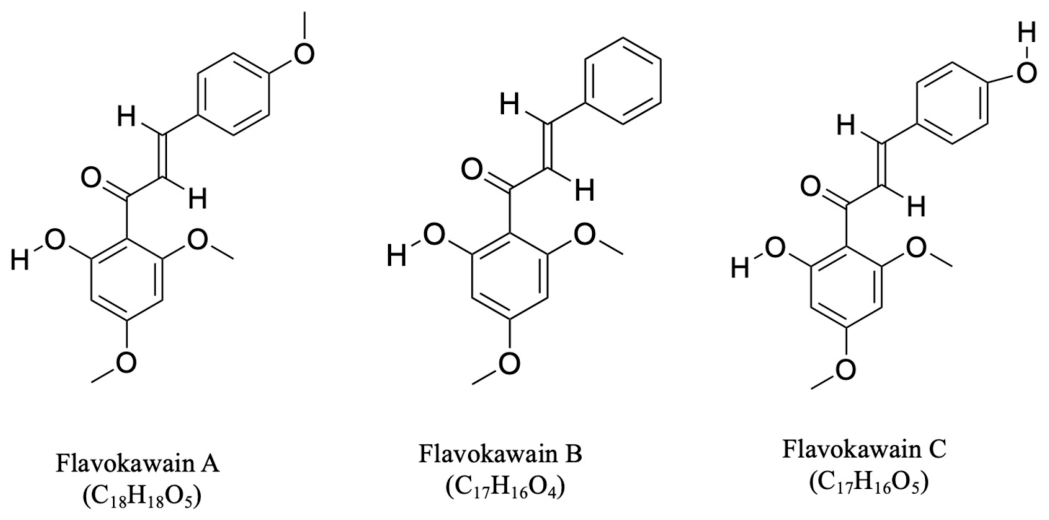
Figure 1. The chemical structures of the principal flavokawains-Flavokawain A (FKA), Flavokawain B (FKB), and Flavokawain C (FKC). These compounds belong to the chalcone class of flavonoids. Structural differences among FKA, FKB, and FKC arise from variations in the number and positioning of methoxy and hydroxyl functional groups, which influence their physicochemical characteristics and biological activity.
Cancers 18 02211 g001
Figure 2. The different plant sources and biological activities of flavokawains. These compounds are predominantly isolated from the roots of Piper methysticum (kava plant), with additional sources including Alpinia pricei, Polygonum ferrugineum, and Chloranthus henryi. The figure integrates phytochemical origin with biological function, highlighting the broad spectrum of pharmacological activities exhibited by flavokawains. Abbreviations: FKA (Flavokawain A), FKB (Flavokawain B), FKC (Flavokawain C).
Cancers 18 02211 g002
Figure 3. Timeline of major discoveries and pharmacological advancements in flavokawain research. The figure summarizes key experimental discoveries and pharmacological advancements reported between 1997 and 2025, beginning with the initial isolation of flavokawains and subsequently highlighting their progressive evaluation across a wide range of therapeutic applications. Abbreviations: FKA (Flavokawain A), FKB (Flavokawain B), FKC (Flavokawain C) [21,23,42,45,46,47,48,49,50,51,52,53,54].
Cancers 18 02211 g003
Figure 4. Flavokawains target multiple hallmarks of cancer across diverse malignancies. The figure illustrates the broad-spectrum anticancer activities of flavokawains across a wide range of cancer types, including bladder, ovarian, head and neck, liver, lung, colorectal, uterine leiomyosarcoma, breast, prostate, melanoma, bone, cervical, gastric, brain, and hematological cancers. The central panel highlights the key anticancer mechanisms influenced by flavokawains, including apoptotic activation, cell-cycle blockade, angiogenic restraint, modulation of oxidative stress, attenuation of EMT, disruption of proliferative signaling networks, reduction in cancer stemness, and autophagic remodeling. Abbreviations: FKA (Flavokawain A), FKB (Flavokawain B), FKC (Flavokawain C), EMT (epithelial-mesenchymal transition).
Cancers 18 02211 g004
Figure 5. Molecular signaling pathways targeted by flavokawains in cancer. This figure illustrates the multi-targeted anticancer mechanisms of flavokawains. Flavokawains inhibit key oncogenic signaling pathways, including PI3K/Akt/mTOR, NF-κB, MAPK, and Wnt/β-catenin, thereby suppressing proliferation and tumor-initiating gene expression. It reduces metastasis by inhibiting MMPs. Flavokawains induce oxidative stress and endoplasmic reticulum stress, leading to mitochondrial dysfunction, cytochrome c release, caspase activation, and apoptosis, as well as downregulation of XIAP and survivin. It also promotes DNA damage (γ-H2AX), activates p53, and causes cell-cycle arrest via modulation of cyclins, CDKs, p21, and p27. Additionally, FKB triggers autophagy by regulating ULK1 and LC3. The yellow sphere represents flavokawains, red coloured inhibitory sign indicates suppression/downregulation and green coloured arrow indicates induction/upregulation. Abbreviations: Akt (Protein kinase B), β-catenin (Beta-catenin), CDK (Cyclin-dependent kinase), ERK1/2 (Extracellular signal-regulated kinase 1/2), GSK3β (Glycogen synthase kinase 3 beta), KLF4 (Krüppel-like factor 4), MMP (Matrix metalloproteinase), mTORC2 (Mammalian target of rapamycin complex 2), NF-κB (Nuclear factor kappa-light-chain-enhancer of activated B cells), OCT4 (Octamer-binding transcription factor 4), PI3K (Phosphoinositide 3-kinase), SOX2 (SRY-box transcription factor 2), XIAP (X-linked inhibitor of apoptosis protein).
Cancers 18 02211 g005
Table 1. The chemical structures, physicochemical properties, and bioactivities of flavokawains.
FeaturesFlavokawain AFlavokawain BFlavokawain C
Natural sourcePiper methysticum, Chloranthus henryiPiper methysticum, Alpinia pricei Hayata, Polygonum ferrugineum Wedd.Piper methysticum
Molecular formulaC18H18O5C17H16O4C17H16O5
Molecular weight314.3 g/mol284.31 g/mol300.30 g/mol
Melting point113 °C91 °C194–195 °C
Appearance Yellow crystallineYellow crystallineYellow crystalline
A-Ring substituents2′-Hydroxy, 4′6′-Methoxy2′-Hydroxy, 4′6′-Methoxy2′-Hydroxy, 4′6′-Methoxy
B-Ring substituents4-Methoxy-4-Hydroxy
Core α, β-unsaturated ketoneα, β-unsaturated ketoneα, β-unsaturated ketone
Hydroxyl content1 OH group1 OH group2 OH group
BindingWeak H-bondNo extra H-bondStrong H-bond
ElectrophilicityModerateHighLow
Antioxidant activityLowLowLow
Apoptosis inductionHighHighModerate
G2/M arrestYesYesYes
CytotoxicityModerate HighModerate
SelectivityHighModerateHigh
Table 2. Therapeutic effects of flavokawains on different cancer types.
Cancer TypesFlavokawainsIn Vitro/In Vivo/Ex VivoConc./DosageModelMechanism of Action or OutcomeReferences
B-cell lymphomaFKBIn vitro1.25, 2.5, 5, 10, 20 µg/mLSUDHL-4, Raji, Jeko↓ Cell viability, p-Akt, p-mTOR, BcL-xL, p-GSK3β; ↑ cleaved PARP, caspase-3[46]
In vivo0.75 mg/kg.b.wt
(every 4 days)
SUDHL-4-derived xenograft in nude mice↓ Tumor weight, Ki-67
Bladder CancerFKAIn vitro10, 20, 40, 60, 80 μMUMUC3, T24 cells↓ PRMT5, cell viability, H2AR3, H4R3 methylation; ↑ apoptosis[55]
In vivo30 mg/kg.b.wt
(every 3 days)
UMUC3-derived xenograft in nude mice↓ Tumor size, H2A, H4 R3 methylation
FKA + GCIn vitro10–80 μMUMUC3, T24 cells↓ Cell viability
FKAIn vivo6 g/kg in AIN-93M dietUPII-SV40T transgenic mice↓ Tumor weight, Ki-67, Bcl-2, XIAP, survivin; ↑ survival of mice, TUNEL-positive cells, DR-5, p27[56]
FKAIn vitro4–40 µMRT4 cells↓ Cell growth, CDK2, SKP2; ↑ G1, G2/M phase arrest, p21, p27[57]
In vivo50 mg/kg.b.wt/dayRT4-derived xenograft in nude mice↓ Tumor growth
FKAIn vitro0.05, 0.5, 5, 12.5, 25 µg/mLT24 cells↓ Cell proliferation, survivin, XIAP, Bcl-xL; ↑ caspase-3, -9, Bax[45]
In vivo50 mg/kg.b.wt/dayEJ-derived xenograft in nude mice↓ Tumor growth
FKA + yangoninIn vitroFKA: 50 µg/mL; Yangonin: 5, 12.5 µg/mLUMUC3 cells↓ Cell viability[58]
Bone Cancer
Osteosarcoma
FKAIn vitro2.5, 5, 10, 25, 50 μg/mL143B, SaOS-2 cells↓ Cell viability, invasion, SKP2, RhoA, MMP-9; ↑ G2/M arrest, caspase-3, cleaved PARP, p21[59]
In vivo200, 600 mg/kg.b.wt/day143B-derived xenograft in SCID mice↓ Lung metastasis, p27, SKP2; ↑ apoptosis
Synovial sarcomaFKAIn vitro2.5, 5, 10, 25 μg/mLHSSY-II, SYO-I, Yamato, Aska cells↓ Cell viability, invasion, SKP2, ETV4, ECT2, vimentin, Twist, ZO-1, sarcosphere; ↑ apoptosis, G2/M arrest, E-cadherin, cleaved PARP, p27[60]
In vivo600 mg/kg.b.wt/dayHSSY-II-derived xenograft in SCID mice↓ Tumor growth
FKBIn vitro2.5, 5, 7.5 μg/mLSYO-I, HSSY-II cells↓ Cell growth, colony formation, IAP, survivin, Bcl-2; ↑ caspase-3, -7, -8, -9, DR5, Bim, Puma, Bax[61]
Breast Cancer13, 15, 16 (FKB derivatives)In vitro0.47–30 µg/mLMCF-7, MDA-MB-231 cells↑ Cytotoxicity[62]
FKAIn vitro2, 4, 8, 16 μMSKBR3, MCF-7, MDA-MB-468 cells↓ Cell growth, colony formation, HER2, p-Akt, Bcl-2, survivin, XIAP; ↑ G2/M arrest, Cdc2, apoptosis, Bim, Bax, cleaved PARP [63]
FLS (FK derivative)In vitro3–180 μMMCF-7, MDA-MB-231 cells↓ Cell viability, Bcl-2, Cdc2; ↑ G2/M arrest, apoptosis, caspase-9, cytochrome c, Bax, p53, c-Jun, WEE-1[64]
FKB (synthetic)In vitro6–40.5 μMMDA-MB-231 cells↓ Cell proliferation, migration, invasion, angiogenesis; NF-κB, COX-2 ↑ G2/M arrest, apoptosis[65]
Ex vivo6, 12.3, 24.6 μMMale Sprague Dawley rats↓ Angiogenesis
FKA
(synthetic)
In vitro6.5–70 μMMCF-7, MDA-MB-231 cells↓ Cell proliferation, PLK1, FOXM1; ↑ apoptosis, G2/M arrest, caspase-8, -9, Bax, cytochrome c[21]
Ex vivo6.5, 17.5, 65 μMMale Sprague Dawley rats↓ Angiogenesis
Cervical CancerFKBIn vitro1.56–100 µMHeLa cells↑ Cytotoxicity, G2/M arrest, apoptosis, SOD2, GSH, cytochrome C, p-p38α, p-HSP27, HSP70[66]
CholangiocarcinomaFKB; cisplatin;
FKB + cisplatin
In vitro0–100 μMSNU-478↓ Cell viability, p-Akt; ↑ apoptosis, cleaved PARP[22]
FKB; FKB + cisplatin/gemcitabineIn vivoCisplatin: 5 mg/kg, gemcitabine: 100 mg/kg and FKB: 25 mg/kg (twice a week)SNU-478-derived xenograft in nude mice↓ Tumor growth
Colorectal CancerFKAIn vitro25,100, 200, 500 μg/mLSW620, DLD-1, HT-29, HCT-8, HCT-116 cells↓ Cell viability[67]
FKCIn vivo1, 3 mg/kg.b.wt
(thrice weekly)
HCT-116-derived xenograft in nude mice↓ Tumor growth, Ki-67; ↑ apoptosis, cleaved caspase-3[68]
FKB In vitro10, 20 μMLoVo, LoVo/Dx cells↓ Cell growth, PCNA; ↑ apoptosis, G2/M arrest[69]
FKCIn vitro60 μMHCT-116 cells↓ MLC-3, GAMT, DCK, PGAM1, GSTO1, COMT, BPNT1, CNPY2, TCEB1, CRABP2, eIF-5A, EEF-2, eIF-3I, CBX3, SKP1, ATP5H, RanBP1, hTom22; ↑ Hspa8, Hsp70-Hom, Hsp27, Hsp70-1/Hsp70-2, Hsp86, TCP-1-eta, CK-18, Tubulin beta-2 chain, Gamma actin, P4HB, HMOX1, FKBP4, HR23B, SFPQ, PARK7, GLOD4, GLRX3[70]
FKCIn vitro40, 60, 80 µMHCT-116 cells, HT-29↓ Cell viability, SOD; ↑ apoptosis, caspase-3-, -8-, -9-positive cells, cleaved PARP-1, ROS, p21, p27, G2/M arrest[71]
FKBIn vitro0.1–50 μMHCT-116 cells↓ Cell viability, proliferation, colony formation, Bcl-2; ↑ apoptosis, cleaved PARP, GADD153, Bim L, Bim S, p-38 MAPK, ROS, G2/M arrest, autophagy[72]
FKAIn vivo20, 80 mg/kg/2 daysAOM/DSS-induced CRC in mice↓ Tumor count, polyp size, LPS biosynthesis pathway[73]
Gastric CancerFKB + doxorubicinIn vitroFKB: 1.25, 2.5, 5 µg/mL; Dox: 0.5 µg/mL AGS, SCM-1, MKN-45↓ ATG4B; ↑ autophagy, apoptosis, caspase-3, -8, -9, Fas-FasL, LC3-II, Beclin-1, ROS[74]
In vivoFKB: 0.75 mg/kg.b.wt; Dox: 1.5 mg/kg.b.wt
(every 2 days)
AGS-derived xenograft in nude mice↓ Tumor growth
FKBIn vitro2.5–20 μg/mLAGS, NCI-N87, Hs738, Kato-III, TSGH-9201 cells↓ Cell survival, colony formation, ATG4B, HER2, PI3K, Akt, mTOR; ↑ LC3-II, autophagy, ROS, apoptosis, JNK, ERK, G2/M arrest, cyclin A, cyclin B1, CDK1, Cdc25C[49]
In vivo1.5, 7.5 mg/kg.b.wt
(every 2 days)
AGS-derived xenograft in nude mice↓ Tumor growth
FKBIn vitro10 μg/mLSGC-7901↓ Cell growth, proliferation, Cdc2, Cdc25C, cyclin A, cyclin B1, CCR2, macrophage migration; ↑ G2/M arrest, apoptosis, caspase-3, -7, -8, -9, TSPAN12, TGF-β1, SMAD4[75]
In vivo1.5 mg/kg.b.wt
(every 2 days)
SGC-7901-derived xenograft in nude mice↓ Tumor weight; ↑ SMAD4, TGF-β1, TSPAN12, survival time
Head and Neck Cancers
Oral squamous cell carcinoma
FKA, FKBIn vitro10 μg/mL, 2.5 μg/mLH400, BICR56, OKF6 cells↓ Cell growth, migration, invasion[76]
Oral carcinomaFKBIn vitro1.25–10 μg/mL; 4.4–35.2 μMHSC-3, Cal-27 cells↓ Cell viability, cyclin A, cyclin B, Cdc2, Cdc25C, Bcl-2, PI3K/Akt, p38 MAPK; ↑ G2/M arrest, apoptosis, cytochrome c, Bax[77]
Oral adenoid cystic carcinomaFKB (synthetic) In vitro1.1, 2.2, 4.4, 8.8, 17.6, 44, 87.9 µmol/LACC-2 cells↓ Cell growth, Bcl-2; ↑ apoptosis, G2/M arrest, Bim, Bak, Bax [78]
Nasopharyngeal carcinomaFKCIn vitro0.5, 1, 2, 4 μMHNE1, HNE2, CNE1, CNE2, HONE1 cells↓ HSP90B1, Ang-1, VEGF, EGFR/PI3K/Akt/mTOR[79]
In vivo3 mg/kg.b.wtHNE1-derived xenograft in nude mice ↓ Tumor volume, tumor weight, HSP90B1, GLUT1, HK2, Ang-1, VEGF, EGFR/PI3K/Akt/mTOR
Liver CancerFKAIn vitro10, 20, 40 μMSMMC-7721, huh7, PANC-1, HepG2, HeLa, Hep3B, A549 cells↓ Cell viability, migration, invasion, CXCR4, proliferation, VM formation, VE-cadherin, vimentin, Snail1, EMT, p-PI3K, p-Akt, HIF-1α, NF-κB, Twist1; ↑ apoptosis, E-cadherin[29]
In vivo30, 60, 120 mg/kg.b.wt/dayHepG2-derived xenograft in nude mice↓ Tumor growth, EMT, metastasis, VE-cadherin, vimentin, Twist1, p-Akt; ↑ E-cadherin
FKA In vitro2–100 μMHepG2 cells↓ Cell viability; ↑ toxicity, Nrf2, HSF1, HMOX1, GCLC, HSPA1A, DNAJA4, GSH[50]
FKBIn vitro2–100 μMHepG2 cells↓ Cell viability; ↑ toxicity, Nrf2, HSF1, HMOX1, GCLC, HSPA1A, DNAJA4, GSH
FKC In vitro4, 8, 16 μM Huh-7, Hep3B, HepG2 cells↓ Cell proliferation, migration, Bax, Bcl-2, p-FAK, p-PI3K, p-Akt; ↑ apoptosis[48]
In vivo16 mg/kg.b.wt/dayHuh-7-derived xenograft in nude mice ↓ Tumor growth, Ki-67; ↑ γ-H2AX
Lung CancerFKAIn vitro5–30 µMA549, PTX-resistant A549 (A549/T), THLE-3 cells↓ Cell viability, cell growth, P-gp, Akt, p-Akt; ↑ apoptosis, PARP[80]
FKA, FKB, FKCIn vivoFKA, FKB: 5 mg/g, FKC: 2.5 mg/g.b.wtNNK, BaP-induced tumor in mice↓ Tumor multiplicity[51]
Chalcone-24 (FK synthesized)In vitro0.3, 1, 3 μMA549 cells↓ Cell viability, NF-κB; ↑ ERK1/2, JNK, caspase activity[81]
FKBIn vitro5–15 μg/mLA549, H1299 cells↓ Cell viability, colony formation, Bcl-2; ↑ caspase-3, -9, Bax, LC3[82]
Leukemia
Acute Myeloid Leukemia (AML)
FKAIn vitro2.5, 5, 10, 20 μg/mLMV4-11, THP-1, MOLM-13, U937 cells↓ Cell viability, CDT1, CCND1, CCNE1, CCNE2, CDK2, CDK4, CDK6; ↑ p27, G1 arrest, CDKN1B[83]
Ex vivo2.5, 5, 10, 20 μg/mLPrimary AML blasts from patients↓ Cell viability
Acute Lymphoblastic Leukemia (ALL)FKBIn vitro0.1–120 μMCCRF-CEM, CEM-C1, Jurkat, RS4-11↓ Cell viability; ↑ apoptosis, caspase-3, cleaved PARP, p53, Bax, Puma[52]
In vivo0.75 mg/kg.b.wt/dayCCRF-CEM-inoculated Balb/c mice↓ Leukocytes, WBC, splenomegaly
Ex vivo25–100 μMPrimary B-ALL blasts, T-ALL blasts↓ Cell proliferation; ↑ p53, Bax, Puma
Myeloid LeukemiaFKBIn vitro0.5–24 µMHL-60, K562, MOLT4, CEM1 cells↓ Cell viability; ↑ apoptosis[84]
FKB + Daunorubicin (DNR)In vitroFKB: 10, 12, 2, 3 µM; DNR: 0.15, 0.05, 0.005, 0.01 µMHL-60, K562, MOLT4, CEM1 cells↓ Cell viability; ↑ NF-ĸB
MelanomaFKA, FKB (synthetic)In vitro1.56, 6.25, 25 µMB16/F10 cells↓ Cell viability, cellular melanin, Tyr, Trp-1, Trp-2, Mitf[85]
In vivoFKA: 0.78–25 µM, 25 µM; FKB: 0.78–12.5 µM, 6.25 µMZebrafish ↓ Melanin production; ↑ survival rate
Neuro-oncological malignancies
Glioblastoma
FKBIn vitro1–5 μg/mLU251, U87, T98 cells↓ Cell viability, proliferation, SQSTM1, p-mTOR, p-Akt, p-RPS6KB1; ↑ G2/M arrest, senescence, autophagy, MAP1LC3B-II, HSPA5, p-EIF2AK3, p-EIF2A, ATF4, DDIT3, p-γH2AX[53]
In vivo50 mg/kg.b.wt/dayU251-derived xenograft in nude mice↓ Tumor growth
NeuroblastomaFKA In vitro12.5, 25, 50 μMSK-N-SH, HUVEC cells↓ Cell viability, colony formation, migration, invasion, angiogenesis, N-cadherin, Snail, VE-cadherin; ↑ apoptosis, E-cadherin, G1 arrest[86]
Ovarian CancerFKA-A NPs+
PTX-A NPs
In vitroFKA-A NPs: 2.5–60 μM); PTX-A NPs: 2.5–60 μMA2780, SKOV-3 cells↓ Cell proliferation, colony formation, migration, vimentin, SKP2, nuclear translocation of YAP; ↑ E-cadherin[87]
In vivoPTX-A
NPs + FKA-A NPs: 2.5 + 2.5 mg/kg.b.wt
(every 3 days)
A2780-derived xenograft in nude mice↓ Tumor growth
FKCIn vitro1–100 µMSKOV-3, SW-626,
OVCAR3, CaOV-3, IOSE80
↓ Cell viability[88]
Prostate CancerFKAIn vitro5, 12.5, 25 µM22Rv1, DU145, CD44+/CD133 ↓ Tumor spheroid size, number, Oct4, Sox2, Nanog, c-Myc, CK8, Ubc12 neddylation[89]
In vivo6 g/kg FKA in diet CD44+/CD133+ 22Rv1-derived xenograft in NOD/SCID mice↓ Tumor growth, Ki-67, c-Myc, Ubc12 neddylation, Nanog
FKAIn vitro1.56, 3.12, 6.25, 12.5, 25, 50, 100 µMPC3 cell line↓ Cell proliferation, survivin, GSH, GSS; ↑ apoptosis, G2/M arrest, GSTP1, ROS[90]
FKB In vitro8.8 μMC4-2B, PC3↓ SKP2; ↑ p27/Kip1[91]
FKBIn vitro1.1, 2.2, 4.4, 8.8, 17.6 µMLNCaP, LAPC4, DU145, PC3↓ Cell viability, XIAP, survivin; ↑ apoptosis, caspase-3, -8, -9, DR5, Bim, Puma[54]
In vivo50 mg/kg.b.wt/dayDU145-derived xenograft in nude mice ↓ Tumor growth; ↑ Bim
FKAIn vitro4–80 µMDU145 (Rb−/−), PC3 (Rb+/+), 22Rv1 (Rb+/+), MEF (Rb+/+, Rb−/−), MPEC (Rb+/+, Rb−/−) cells↓ Cell growth, SKP2, Cullin-1, Ubc12 neddylation[92]
In vivo3, 6 g/kg FKA in dietTRAMP mice↓ Cell proliferation, HG-PIN, prostate adenocarcinoma, tumor burden, metastasis, Ki-67, SKP2, NEDD8; ↑ apoptosis, p27
FKBIn vitro0.63–25 μg/mLLNCaP, LAPC4, 22Rv1, PC3, DU145, WPMY-1 cells↓ Cell growth, AR, PSA, TMPRSS2[93]
In vivo200 mg/kg.b.wt/dayPatient-derived xenograft in SCID mice↓ Tumor growth, PSA, AR
Squamous carcinomaFKBIn vitro5, 10, 20 μg/mLKB cells↓ Cell viability, Bcl-2, cyclin A, cyclin B1, Cdc2, Cdc25C, MMP-9, uPA; ↑ apoptosis, G2/M arrest, caspase-3, -9, Bax, p21/WAF1, Wee1, p53, TIMP-1, PAI-1[94]
In vivo0.75 mg/kg.b.wt
(every 2 days)
KB-induced xenograft in nude mice↓ Tumor volume, angiogenesis; ↑ TUNEL-positive cells
Uterine leiomyosarcomaFKBIn vitro1.1, 2.2, 4.4, 8.8 μMSK-LMS-1, ECC-1, T-HESC cells↓ Cell growth, IAP, survivin; ↑ G2/M arrest, DR5, Bim, Puma[95]
FKB + Docetaxel + GemcitabineIn vitroFKB: 2.2, 4.4 μM; Docetaxel + Gemcitabine: IC50SK-LMS-1 cells↓ Cell proliferation
Symbols and Abbreviations: ↓ (Downregulated/Decreased), ↑ (Upregulated/Increased); Akt (Protein kinase B), AML (acute myeloid leukemia), Ang-1 (Angiopoietin-1), AR (androgen receptor), ATF4 (Activating Transcription Factor 4), ATG4B (Autophagy-Related 4B Cysteine Peptidase), AOM (Azoxymethane), BaP (Benzo[a]pyrene), Bax (Bcl-2-associated X protein), Bcl-2 (B-cell lymphoma 2), Bcl-xL (B-cell lymphoma-extra large), Bim (Bcl-2-interacting mediator of cell death), BPNT1 (3′(2′),5′-Bisphosphate Nucleotidase 1), CBX3 (Chromobox Protein Homolog 3), CCR2 (C-C Motif Chemokine Receptor 2), CDK (Cyclin-Dependent Kinase), CDT1 (Chromatin Licensing and DNA Replication Factor 1), CK8 (Cytokeratin 8), COMT (Catechol-O-Methyltransferase), CNPY2 (Canopy FGF Signaling Regulator 2), COX-2 (Cyclooxygenase-2), CRABP2 (Cellular Retinoic Acid-Binding Protein 2), CXCR4 (C-X-C Motif Chemokine Receptor 4), DCK (Deoxycytidine Kinase), DDIT3 (DNA Damage-Inducible Transcript 3), DNR (Daunorubicin), DR5 (Death Receptor 5), DSS (Dextran Sulfate Sodium), EF-2 (Elongation Factor 2), EGFR (Epidermal Growth Factor Receptor), EIF2AK3 (Eukaryotic Translation Initiation Factor 2 Alpha Kinase 3/PERK), EIF-3I (Eukaryotic Translation Initiation Factor 3 Subunit I), EIF-5A1 (Eukaryotic Translation Initiation Factor 5A1), EMT (epithelial-mesenchymal transition), ERK1/2 (Extracellular Signal-Regulated Kinase 1/2), ETV4 (ETS Variant Transcription Factor 4), ECT2 (Epithelial Cell Transforming Sequence 2), FAK (Focal Adhesion Kinase), FasL (Fas Ligand), FKA (Flavokawain A), FKB (Flavokawain B), FKC (Flavokawain C), FKA-A NPs (Flavokawain A-loaded nanoparticles), FLS (Flavokawain-like Synthetic Derivative), FOXM1 (Forkhead Box M1), GADD153 (Growth Arrest and DNA Damage-Inducible Protein 153/CHOP), GAMT (Guanidinoacetate N-Methyltransferase), GCLC (Glutamate-Cysteine Ligase Catalytic Subunit), GLUT1 (Glucose Transporter 1), GLOD4 (Glyoxalase Domain Containing 4), GLRX3 (Glutaredoxin 3), GSH (Glutathione), GSSG (Oxidized Glutathione), GSTO1 (Glutathione S-Transferase Omega 1), GSTP1 (Glutathione S-Transferase Pi 1), HER2 (Human Epidermal Growth Factor Receptor 2), HIF-1α (Hypoxia-Inducible Factor 1 Alpha), HK2 (Hexokinase 2), HMOX1 (Heme Oxygenase 1), HSP27 (Heat Shock Protein 27), HSP70 (Heat Shock Protein 70), HSP86 (Heat Shock Protein 86), HSP90B1 (Heat Shock Protein 90 Beta Family Member 1), HSF1 (Heat Shock Factor 1), HR23B (UV Excision Repair Protein RAD23 Homolog B), IAP (Inhibitor of Apoptosis Protein), JNK (c-Jun N-terminal Kinase), LC3-II (Microtubule-associated Protein 1 Light Chain 3-II), LPS (lipopolysaccharide), MAPK (Mitogen-Activated Protein Kinase), MAP1LC3B-II (Microtubule-Associated Protein 1 Light Chain 3 Beta-II), MLC-3 (Myosin Light Chain 3), MMP-9 (Matrix Metalloproteinase-9), mTOR (Mammalian Target of Rapamycin), NF-κB (Nuclear Factor Kappa B), NEDD8 (Neural Precursor Cell-Expressed Developmentally Downregulated Protein 8), Nrf2 (Nuclear Factor Erythroid 2-Related Factor 2), Oct4 (Octamer-Binding Transcription Factor 4), PARP (Poly(ADP-ribose) Polymerase), PAI-1 (Plasminogen Activator Inhibitor-1), PARK7 (Parkinsonism-Associated Deglycase), PCNA (Proliferating Cell Nuclear Antigen), P-gp (P-glycoprotein), PGAM1 (Phosphoglycerate Mutase 1), PI3K (Phosphoinositide 3-Kinase), PLK1 (Polo-Like Kinase 1), PRMT5 (Protein Arginine Methyltransferase 5), PSA (Prostate-Specific Antigen), PTX (Paclitaxel), PTX-A NPs (Paclitaxel-loaded Nanoparticles), Puma (p53 Upregulated Modulator of Apoptosis), RanBP1 (Ran Binding Protein 1), RhoA (Ras Homolog Family Member A), ROS (Reactive Oxygen Species), RPS6KB1 (Ribosomal Protein S6 Kinase B1), SKP2 (S-phase Kinase-associated Protein 2), SMAD4 (SMAD Family Member 4), Snail (Snail Family Transcriptional Repressor 1), Sox2 (SRY-Box Transcription Factor 2), SOD (Superoxide Dismutase), TCP-1-eta (T-Complex Protein 1 Eta Subunit), TCEB1 (Transcription Elongation Factor B Subunit 1), TGF-β1 (Transforming Growth Factor Beta 1), TIMP-1 (Tissue Inhibitor of Metalloproteinases-1), TMPRSS2 (Transmembrane Protease Serine 2), Trp-1 (Tyrosinase-Related Protein 1), Trp-2 (Tyrosinase-Related Protein 2), TSPAN12 (Tetraspanin 12), TUNEL (Terminal Deoxynucleotidyl Transferase dUTP Nick End Labeling), Tyr (Tyrosinase), Ubc12 (Ubiquitin-Conjugating Enzyme E2 M), uPA (Urokinase-Type Plasminogen Activator), VEGF (Vascular Endothelial Growth Factor), VM (vasculogenic mimicry), WAF1 (Wild-Type p53-Activated Fragment 1/CDKN1A), WBC (white blood cell), Wee1 (WEE1 G2 Checkpoint Kinase), XIAP (X-linked Inhibitor of Apoptosis Protein), YAP (Yes-Associated Protein), ZO-1 (Zonula Occludens-1), and γ-H2AX (Phosphorylated Histone H2AX), b.wt (body weight).
Table 3. Toxicity profile of flavokawains.
Toxicity TypeFlavokawainIn Vitro/In VivoModelConc./DosageToxicityReferences
HepatotoxicityFKBIn vitroHepG210–50 μM↑ Cell death, apoptosis, MAPK, caspase-3; ↓ NF-kB
FKB: LD50: 15.3 ± 0.2 μM
[124]
FKB, FKCIn vitroL-020–150 μM↑ Cell death, apoptosis; ↓ GSH
FKB: LD50: 32 μM
FKC: LD50: 70 μM
FKAIn vitroHepG20–150 μMFKA: LD50: 75 μM
FKBIn vivoICR mice25 mg/kg.b.wt↑ Liver damage, AST, AKP; ↓ NF-kB
HepatotoxicityFKBIn vivoC57BL/6J mice
(acetaminophen-induced)
FKB: 11.5 mg/kg.b.wt; APAP: 800 mg/kg.b.wt↑ AST, ALT[126]
FKA + FKBIn vivoC57BL/6J mice1× dose
FKA-8 mg/kg
FKB-11.5 mg/kg
No effect on AST and ALT levels
2× dose
FKA-16 mg/kg
FKB-23 mg/kg
4× dose
FKA-32 mg/kg
FKB-46 mg/kg
FKA + FKBIn vivoC57BL/6J mice (acetaminophen-induced)1× dose
FKA-8 mg/kg
FKB-11.5 mg/kg
↑ AST, ALT
2× dose
FKA-16 mg/kg
FKB-23 mg/kg
4× dose
FKA-32 mg/kg
FKB-46 mg/kg
Bone marrow toxicityFKAIn vitroMouse bone marrow cells0–25 μg/mLNo significant effect on colony formation[125]
General toxicityFKAIn vivoFVB/N mice6 g/kg in dietNo adverse effects on body weight, food intake, organ histology, or serum biochemistry
HepatotoxicityFKAIn vivoFVB/N mice6 g/kg in dietNo increase in liver weight, no histopathological changes, no change in ALT, AST, ALP, albumin
Symbols and Abbreviations: ↓ (Downregulated/Decreased), ↑ (Upregulated/Increased); FKA, Flavokawain A; FKB, Flavokawain B; FKC, Flavokawain C; LD50, median lethal dose; AST, aspartate aminotransferase; ALT, alanine aminotransferase; ALP/AKP, alkaline phosphatase; LD50, median lethal dose; NF-κB, Nuclear factor kappa B; MAPK, Mitogen-activated protein kinase; GSH, glutathione; ICR mice, Institute of Cancer Research mice.
Disclaimer/Publisher’s Note: The statements, opinions and data contained in all publications are solely those of the individual author(s) and contributor(s) and not of MDPI and/or the editor(s). MDPI and/or the editor(s) disclaim responsibility for any injury to people or property resulting from any ideas, methods, instructions or products referred to in the content.