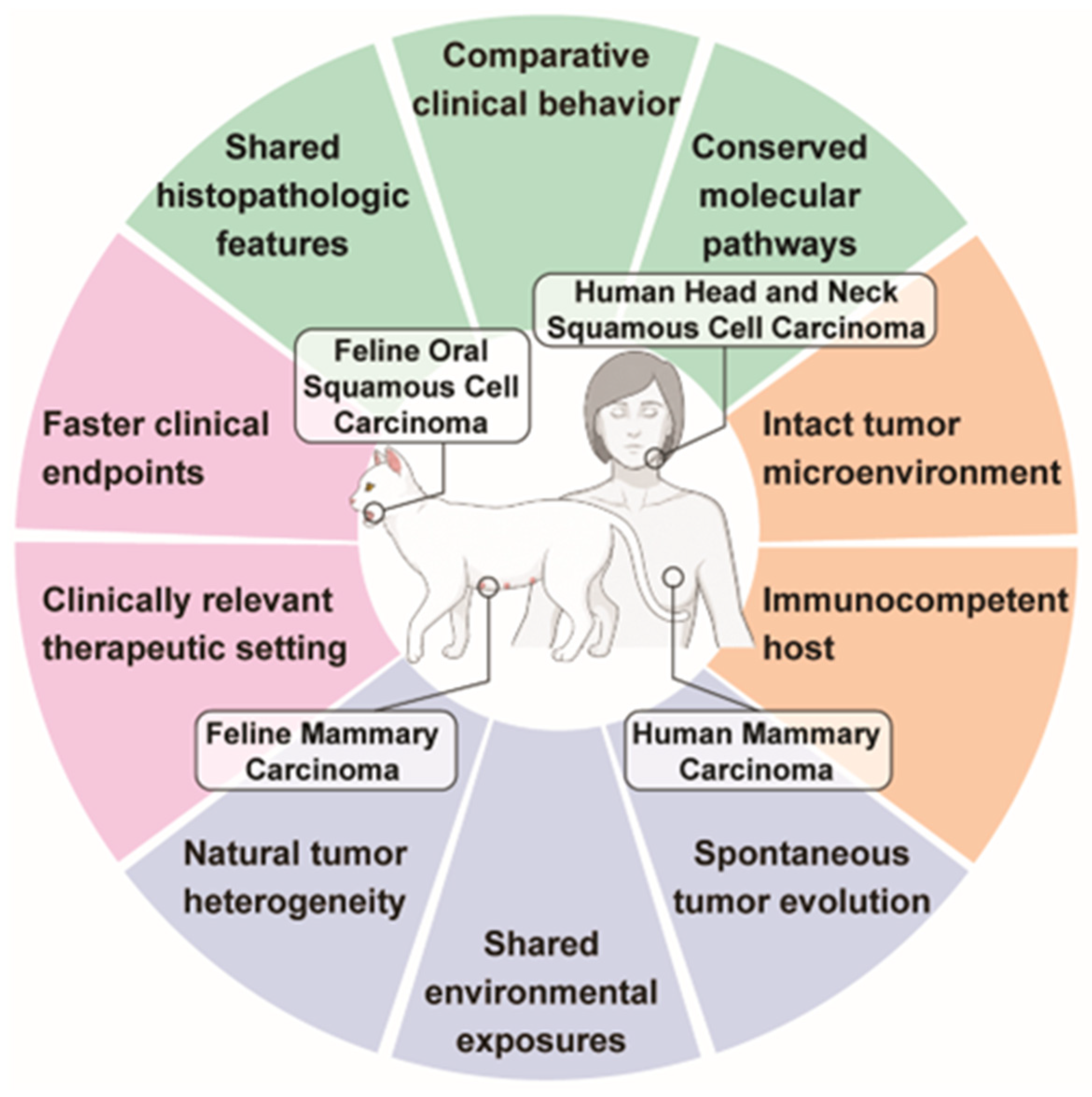
Naturally Occurring Feline Cancers in Comparative Oncology: Translational Insights from Oral Squamous Cell Carcinoma and Mammary Carcinoma

Simple Summary
Abstract
1. Introduction
2. FOSCC and Human HNSCC
2.1. Viral Etiology
2.2. Risk Factors
2.3. Histopathologic Similarities Between Feline and Human Oral Squamous Cell Carcinoma (OSCC)
2.4. Shared Molecular Pathways and Oncogenic Drivers in FOSCC and Human HNSCC

| Molecular Feature | Human HNSCC | FOSCC |
|---|---|---|
| TP53 | Loss-of-function mutation of TP53 is associated with loss of genome surveillance, impaired apoptosis and cell-cycle arrest, with mutations reported in approximately 70% of cases [71,72,73,105,106]. | TP53 mutations have been reported in up to 71% of FOSCC tumors [12,16,34]. |
| MYC | MYC overexpression, with an estimated prevalence of 12% in HNSCC, is linked to aggressive behavior, poorer survival, and therapeutic resistance, particularly in HPV-negative tumors [107,108]. | MYC expression is increased in FOSCC tissue, and recent whole-exome sequencing identified MYC copy-number gain in 31% of FOSCCs [16,25,34]. |
| PTEN | In HNSCC, PTEN loss or inactivation promotes cell proliferation, migration, and survival, with mutations detected in ~10% of patients [109,110,111,112]. | PTEN copy-number loss was identified in 24% of FOSCCs [16]. |
| FAS | FAS pathway dysregulation contributes to apoptosis resistance and immune escape in HNSCC [113,114,115]. | FAS copy-number loss was identified in 21% of FOSCCs [16]. |
| CDKN2A | Loss-of-function mutation of CDKN2A has been identified as a prognostic biomarker in HNSCC, associated with poor overall survival and increased risk of metastasis [116,117,118]. | CDKN2A inactivation has been reported in FOSCC [50,119]. |
| EMT-related pathways | In HNSCC, upregulated EMT contributes to invasion, metastasis, poor prognosis, and treatment resistance [77,78]. | FOSCC RNA-seq shows enrichment of EMT-related pathways [34,79,80]. |
| IL-6/JAK/STAT3 | In HNSCC, hyperactivated IL-6/JAK/STAT3 signaling promotes tumor growth, immune evasion, invasion, treatment resistance, and poor prognosis [82,83,84,85,86]. | The IL-6/JAK/STAT pathway is enriched in FOSCC [41,81]. |
| EGFR signaling | EGFR is up to 90% overexpressed in human HNSCC and is associated with proliferation, invasion, and treatment resistance [89,90,120]. | EGFR is overexpressed in 69–100% of FOSCC cases and is associated with tumor aggressiveness [34,39,88]. |
| COX-2 and VEGF | In HNSCC, upregulation of COX-2 and VEGF is linked to angiogenesis, lymph node metastasis, tumor progression, and poorer survival outcomes [93,94,95,96,121,122]. | COX-2 overexpression has been reported in approximately 33% of FOSCC cases, with VEGF colocalization [97,98,99,100,123]. |
| WNT/β-catenin | WNT/β-catenin signaling is upregulated in HNSCC and contributes to tumor progression, invasion, and treatment resistance [101,102,103,104]. | Upregulation of WNT pathway components has been reported in FOSCC [25]. |
2.5. Translational and Clinical Trial Insights of FOSCC
3. FMC and Human Mammary Carcinoma
3.1. Risk Factors
3.2. Molecular and Biomarker Parallels
| Molecular Feature | HBC | FMC |
|---|---|---|
| TP53 | TP53 is frequently altered in aggressive breast cancer, with mutations reported in approximately 80% of TNBC cases [223]. Loss or alteration of p53 function is associated with high-grade, poor-prognosis tumors and increased metastatic potential [224]. | TP53 mutations have been recurrently reported in FMC [16,160,225,226]. |
| PIK3CA | PIK3CA is one of the most frequently mutated genes in breast cancer [227]. The PI3K/AKT/mTOR pathway promotes tumor-cell growth, survival, and therapeutic vulnerability [227,228,229,230]. Activating mutations are reported in ~26% of invasive breast carcinomas overall and 13–22.1% of TNBC [231,232]. | PIK3CA has been identified as an FMC driver, with mutations reported in approximately 45% of FMCs [16,233]. |
| PTEN | PTEN loss promotes proliferation, invasion, metastatic competence, and treatment resistance, and has been detected in approximately 48.6% of TNBC cases in some cohorts [234,235]. | PTEN is a recurrent FMC driver. Loss of PTEN expression is reported in ~77% of cases and has been associated with poorer prognosis and shorter survival [16,157,211]. |
| FBXW7 | FBXW7 is a tumor suppressor, and low FBXW7 expression has been associated with poorer prognosis in breast cancer patients [236,237,238]. | FBXW7 has been identified as an FMC driver, with mutations reported in approximately 72% of cases [16]. |
| CXCL12/CXCR4 | Overexpression of the CXCL12/CXCR4 axis promotes tumor growth, metastasis, and chemotherapy resistance, particularly in aggressive breast cancer, including TNBC [239,240,241]. | Serum SDF-1/CXCL12 has been proposed as a diagnostic biomarker in FMC; CXCR4 overexpression was detected in 86% of tumors [242]. |
| Leptin/ObR | In HBC, leptin–ObR signaling promotes tumor-cell survival, angiogenesis, inflammation, and disease progression, particularly in obesity-associated breast cancer biology [243,244]. Leptin and ObR expression were detected in 83% and 33.7% of breast cancer tissues, respectively [245]. | ObR is overexpressed in FMC tissues, and serum ObR levels have been correlated with immune downregulation and tumor development [10,29,246]. |
| Immune checkpoint | In HBC, especially TNBC, the PD-1/PD-L1 immune-checkpoint pathway can suppress antitumor immune responses [217,218]. Tumoral PD-L1 staining was reported in 12% of breast cancers overall and 32% of triple-negative cases [247]. | PD-1 and PD-L1 are detectable in FMC, with enrichment reported in aggressive subtypes, including HER2-positive and triple-negative tumors [10,215,216,248]. |
| Bcl-2 | In HBC, high Bcl-2 expression is associated with a favorable prognosis, improved survival, and less aggressive disease [220,221]. | Bcl-2 expression has been associated with longer disease-free interval and overall survival in FMC [219]. |
3.3. Tumor Microenvironment Parallels
3.4. Patient-Derived FMC Organoids
3.5. FMC Therapeutic Studies
4. Current Challenges and Future Directions
Author Contributions
Funding
Institutional Review Board Statement
Informed Consent Statement
Data Availability Statement
Acknowledgments
Conflicts of Interest
Abbreviations
| AKT | Protein kinase B |
| Bcl-2 | B-cell lymphoma 2 |
| CAF | Cancer-associated fibroblast |
| CDKN1A | Cyclin-dependent kinase inhibitor 1A |
| COX-2 | Cyclooxygenase-2 |
| CS3D | Cyclic STAT3 decoy oligonucleotide |
| CXCL12 | C-X-C motif chemokine ligand 12 |
| CXCR4 | C-X-C chemokine receptor type 4 |
| DFMO | Difluoromethylornithine |
| DNA | Deoxyribonucleic acid |
| DPT | Dermatopontin |
| EGFR | Epidermal growth factor receptor |
| EMT | Epithelial-to-mesenchymal transition |
| ER | Estrogen receptor |
| FADS2 | Fatty acid desaturase 2 |
| FAS | Fas cell surface death receptor |
| FBXW7 | F-box and WD repeat domain containing 7 |
| FcaPV | Felis catus papillomavirus |
| FMC | Feline mammary carcinoma |
| FOSCC | Feline oral squamous cell carcinoma |
| FOXM1 | Forkhead box M1 |
| HBC | Human breast cancer |
| HER2 | Human epidermal growth factor receptor 2 |
| HNSCC | Head and neck squamous cell carcinoma |
| HPV | Human papillomavirus |
| IL-6 | Interleukin-6 |
| JAK | Janus kinase |
| LMTK3 | Lemur tyrosine kinase 3 |
| mTOR | Mechanistic target of rapamycin |
| MYC | MYC proto-oncogene, bHLH transcription factor |
| NF-κB | Nuclear factor kappa B |
| ObR | Leptin receptor |
| PCR | Polymerase chain reaction |
| PD-1 | Programmed cell death protein 1 |
| PD-L1 | Programmed death-ligand 1 |
| PD-L2 | Programmed death-ligand 2 |
| PI3K | Phosphoinositide 3-kinase |
| PIK3CA | Phosphatidylinositol-4,5-bisphosphate 3-kinase catalytic subunit alpha |
| PR | Progesterone receptor |
| PTEN | Phosphatase and tensin homolog |
| SCC | Squamous cell carcinoma |
| SDF-1 | Stromal cell-derived factor 1 |
| SNX5 | Sorting nexin 5 |
| STAT3 | Signal transducer and activator of transcription 3 |
| TNBC | Triple-negative breast cancer |
| TNF | Tumor necrosis factor |
| TP53 | Tumor protein p53 |
| TWIST1 | Twist family bHLH transcription factor 1 |
| VEGF | Vascular endothelial growth factor |
| WNT | Wingless/Int-1 signaling pathway |
| ZEB1 | Zinc finger E-box binding homeobox 1 |
References
- Tu, A.Y.; Springer, C.M.; Albright, J.D. Evaluation of Characteristics Associated with Self-Identified Cat or Dog Preference in Pet Owners and Correlation of Preference with Pet Interactions and Care: An Exploratory Study. Animals 2024, 14, 2534. [Google Scholar] [CrossRef]
- MacEwen, E.G. Spontaneous tumors in dogs and cats: Models for the study of cancer biology and treatment. Cancer Metastasis Rev. 1990, 9, 125–136. [Google Scholar] [CrossRef]
- Vail, D.M.; MacEwen, E.G. Spontaneously occurring tumors of companion animals as models for human cancer. Cancer Investig. 2000, 18, 781–792. [Google Scholar] [CrossRef]
- Cannon, C.M. Cats, Cancer and Comparative Oncology. Vet. Sci. 2015, 2, 111–126. [Google Scholar] [CrossRef]
- Oh, J.H.; Cho, J.Y. Comparative oncology: Overcoming human cancer through companion animal studies. Exp. Mol. Med. 2023, 55, 725–734. [Google Scholar] [CrossRef]
- Nance, R.L.; Sajib, A.M.; Smith, B.F. Canine models of human cancer: Bridging the gap to improve precision medicine. Prog. Mol. Biol. Transl. Sci. 2022, 189, 67–99. [Google Scholar] [CrossRef]
- Cardiff, R.D.; Ward, J.M.; Barthold, S.W. ‘One medicine—One pathology’: Are veterinary and human pathology prepared? Lab. Investig. 2008, 88, 18–26. [Google Scholar] [CrossRef]
- King, T.A. The One Medicine concept: Its emergence from history as a systematic approach to re-integrate human and veterinary medicine. Emerg. Top. Life Sci. 2021, 5, 643–654. [Google Scholar] [CrossRef]
- De Haes, C.; de Rooster, H.; Demeyere, K.; Van de Vijver, K.; De Cock, H.; Meyer, E.; Steenbrugge, J. Grade III canine and feline mammary carcinomas are associated with an immunosuppressed, vascularized and proliferative microenvironment. BMC Vet. Res. 2026, 22, 247. [Google Scholar] [CrossRef]
- Rodrigues-Jesus, J.; Canadas-Sousa, A.; Vilhena, H.; Dias-Pereira, P. Inside the Tumor: Decoding the Feline Mammary Tumor Microenvironment and Its Prognostic Value—A Review. Vet. Sci. 2025, 12, 959. [Google Scholar] [CrossRef]
- Alkayyal, A.A. Insights from veterinary models for advancing oncolytic virotherapy through comparative oncology. Front. Mol. Biosci. 2025, 12, 1615393. [Google Scholar] [CrossRef]
- Chu, S.; Skidmore, Z.L.; Warren, W.; Fisk, B.; Kim, D.Y.; Peralta, S.; Rodney, A.; Bryant, J.; Fronick, C.; Skinner, O.; et al. Whole Exome Sequencing of Feline Oral Squamous Cell Carcinoma Reveals Genomic Parallels With Human Head and Neck Squamous Cell Carcinoma. Vet. Comp. Oncol. 2026, 24, 300–312. [Google Scholar] [CrossRef]
- Sommerville, L.; Howard, J.; Evans, S.; Kelly, P.; McCann, A. Comparative gene expression study highlights molecular similarities between triple negative breast cancer tumours and feline mammary carcinomas. Vet. Comp. Oncol. 2022, 20, 535–538. [Google Scholar] [CrossRef]
- Yamamoto, H.; Elbadawy, M.; Tsunedomi, R.; Maeda, N.; Nagano, H.; Ishihara, Y.; Abugomaa, A.; Shiota, Y.; Yu, T.W.; Liu, Y.; et al. Novel organoid-based exploration reveals the role of LMTK3/FADS2 signaling in metastatic breast cancer progression in felines and humans. Sci. Rep. 2025, 15, 45016. [Google Scholar] [CrossRef]
- Alshammari, A.H.; Oshiro, T.; Ungkulpasvich, U.; Yamaguchi, J.; Morishita, M.; Khdair, S.A.; Hatakeyama, H.; Hirotsu, T.; di Luccio, E. Advancing Veterinary Oncology: Next-Generation Diagnostics for Early Cancer Detection and Clinical Implementation. Animals 2025, 15, 389. [Google Scholar] [CrossRef]
- Francis, B.A.; Ludwig, L.; He, C.; Dobromylskyj, M.; Bertram, C.A.; Aupperle-Lellbach, H.; Wong, H.; Foster, A.P.; Arends, M.J.; Suarez-Bonnet, A.; et al. The oncogenome of the domestic cat. Science 2026, 391, 793–799. [Google Scholar] [CrossRef]
- Ahn, S.; Yun, J.H. Comparative Cancer Genetics and Veterinary Therapeutics in Dogs and Cats: A Species-Aware Framework for Comparative Oncology. Life 2026, 16, 430. [Google Scholar] [CrossRef]
- Wong, K.; Ludwig, L.; Krijgsman, O.; Adams, D.J.; Wood, G.A.; van der Weyden, L. Comparison of the oncogenomic landscape of canine and feline hemangiosarcoma shows novel parallels with human angiosarcoma. Dis. Models Mech. 2021, 14, dmm049044. [Google Scholar] [CrossRef]
- The ICGC/TCGA Pan-Cancer Analysis of Whole Genomes Consortium. Pan-cancer analysis of whole genomes. Nature 2020, 578, 82–93. [CrossRef]
- Deneka, A.Y.; Baca, Y.; Serebriiskii, I.G.; Nicolas, E.; Parker, M.I.; Nguyen, T.T.; Xiu, J.; Korn, W.M.; Demeure, M.J.; Wise-Draper, T.; et al. Association of TP53 and CDKN2A Mutation Profile with Tumor Mutation Burden in Head and Neck Cancer. Clin. Cancer Res. 2022, 28, 1925–1937. [Google Scholar] [CrossRef]
- Brown, N.A.; Plouffe, K.R.; Yilmaz, O.; Weindorf, S.C.; Betz, B.L.; Carey, T.E.; Seethala, R.R.; McHugh, J.B.; Tomlins, S.A.; Udager, A.M. TP53 mutations and CDKN2A mutations/deletions are highly recurrent molecular alterations in the malignant progression of sinonasal papillomas. Mod. Pathol. 2021, 34, 1133–1142. [Google Scholar] [CrossRef]
- Shahbandi, A.; Nguyen, H.D.; Jackson, J.G. TP53 Mutations and Outcomes in Breast Cancer: Reading beyond the Headlines. Trends Cancer 2020, 6, 98–110. [Google Scholar] [CrossRef]
- Schaub, F.X.; Dhankani, V.; Berger, A.C.; Trivedi, M.; Richardson, A.B.; Shaw, R.; Zhao, W.; Zhang, X.; Ventura, A.; Liu, Y.; et al. Pan-cancer Alterations of the MYC Oncogene and Its Proximal Network across the Cancer Genome Atlas. Cell Syst. 2018, 6, 282–300.e2. [Google Scholar] [CrossRef]
- Vidotto, T.; Melo, C.M.; Lautert-Dutra, W.; Chaves, L.P.; Reis, R.B.; Squire, J.A. Pan-cancer genomic analysis shows hemizygous PTEN loss tumors are associated with immune evasion and poor outcome. Sci. Rep. 2023, 13, 5049. [Google Scholar] [CrossRef]
- Giuliano, A.; Swift, R.; Arthurs, C.; Marote, G.; Abramo, F.; McKay, J.; Thomson, C.; Beltran, M.; Millar, M.; Priestnall, S.; et al. Quantitative Expression and Co-Localization of Wnt Signalling Related Proteins in Feline Squamous Cell Carcinoma. PLoS ONE 2016, 11, e0161103, Correction in PLoS ONE 2016, 11, e0163838. https://doi.org/10.1371/journal.pone.0163838. [Google Scholar] [CrossRef]
- Groll, T.; Schopf, F.; Denk, D.; Mogler, C.; Schwittlick, U.; Aupperle-Lellbach, H.; Sarker, S.R.J.; Pfarr, N.; Weichert, W.; Matiasek, K.; et al. Bridging the Species Gap: Morphological and Molecular Comparison of Feline and Human Intestinal Carcinomas. Cancers 2021, 13, 5941. [Google Scholar] [CrossRef]
- Thrift, E.; Greenwell, C.; Turner, A.L.; Harvey, A.M.; Maher, D.; Malik, R. Metastatic pulmonary carcinomas in cats (‘feline lung-digit syndrome’): Further variations on a theme. JFMS Open Rep. 2017, 3, 2055116917691069. [Google Scholar] [CrossRef]
- Wiese, D.A.; Thaiwong, T.; Yuzbasiyan-Gurkan, V.; Kiupel, M. Feline mammary basal-like adenocarcinomas: A potential model for human triple-negative breast cancer (TNBC) with basal-like subtype. BMC Cancer 2013, 13, 403. [Google Scholar] [CrossRef]
- Gameiro, A.; Urbano, A.C.; Ferreira, F. Emerging Biomarkers and Targeted Therapies in Feline Mammary Carcinoma. Vet. Sci. 2021, 8, 164. [Google Scholar] [CrossRef]
- Wypij, J.M. A naturally occurring feline model of head and neck squamous cell carcinoma. Pathol. Res. Int. 2013, 2013, 502197. [Google Scholar] [CrossRef]
- Chu, S.; Wylie, T.N.; Wylie, K.M.; Johnson, G.C.; Skidmore, Z.L.; Fleer, M.; Griffith, O.L.; Bryan, J.N. A virome sequencing approach to feline oral squamous cell carcinoma to evaluate viral causative factors. Vet. Microbiol. 2020, 240, 108491. [Google Scholar] [CrossRef]
- Bilgic, O.; Duda, L.; Sanchez, M.D.; Lewis, J.R. Feline Oral Squamous Cell Carcinoma: Clinical Manifestations and Literature Review. J. Vet. Dent. 2015, 32, 30–40. [Google Scholar] [CrossRef]
- Tutu, P.; Daraban Bocaneti, F.; Altamura, G.; Dascalu, M.A.; Horodincu, L.; Soreanu, O.D.; Tanase, O.I.; Borzacchiello, G.; Mares, M. Feline oral squamous cell carcinoma: Recent advances and future perspectives. Front. Vet. Sci. 2025, 12, 1663990. [Google Scholar] [CrossRef]
- Rodney, A.R.; Skidmore, Z.L.; Grenier, J.K.; Griffith, O.L.; Miller, A.D.; Chu, S.; Ahmed, F.; Bryan, J.N.; Peralta, S.; Warren, W.C. Genomic landscape and gene expression profiles of feline oral squamous cell carcinoma. Front. Vet. Sci. 2023, 10, 1079019. [Google Scholar] [CrossRef]
- Sequeira, I.; Pires, M.D.A.; Leitao, J.; Henriques, J.; Viegas, C.; Requicha, J. Feline Oral Squamous Cell Carcinoma: A Critical Review of Etiologic Factors. Vet. Sci. 2022, 9, 558. [Google Scholar] [CrossRef]
- Sparger, E.E.; Murphy, B.G.; Kamal, F.M.; Arzi, B.; Naydan, D.; Skouritakis, C.T.; Cox, D.P.; Skorupski, K. Investigation of immune cell markers in feline oral squamous cell carcinoma. Vet. Immunol. Immunopathol. 2018, 202, 52–62. [Google Scholar] [CrossRef]
- Martin, C.K.; Tannehill-Gregg, S.H.; Wolfe, T.D.; Rosol, T.J. Bone-invasive oral squamous cell carcinoma in cats: Pathology and expression of parathyroid hormone-related protein. Vet. Pathol. 2011, 48, 302–312. [Google Scholar] [CrossRef]
- Soltero-Rivera, M.M.; Krick, E.L.; Reiter, A.M.; Brown, D.C.; Lewis, J.R. Prevalence of regional and distant metastasis in cats with advanced oral squamous cell carcinoma: 49 cases (2005–2011). J. Feline Med. Surg. 2014, 16, 164–169. [Google Scholar] [CrossRef]
- Bergkvist, G.T.; Argyle, D.J.; Morrison, L.; MacIntyre, N.; Hayes, A.; Yool, D.A. Expression of epidermal growth factor receptor (EGFR) and Ki67 in feline oral squamous cell carcinomas (FOSCC). Vet. Comp. Oncol. 2011, 9, 106–117. [Google Scholar] [CrossRef]
- Schlueter, A.; Hanot, C.; Sellon, R.; Fidel, J. Treatment of feline oral squamous cell carcinoma with accelerated radiation and carboplatin with and without follow-up toceranib phosphate. J. Feline Med. Surg. 2025, 27, 1098612X251314343. [Google Scholar] [CrossRef]
- Grandis, J.R.; Skorupski, K.A.; Cheng, N.; Cui, Z.; Li, H.; Woerner, L.C.; Gencel-Augusto, J.; Zeng, Y.; Shiah, J.V.; Bhola, N.E.; et al. Safety and efficacy of a STAT3-targeted cyclic oligonucleotide: From murine models to a phase 1 clinical trial in pet cats with oral cancer. Cancer Cell 2025, 43, 2051–2068.e9. [Google Scholar] [CrossRef]
- Hayes, A.M.; Adams, V.J.; Scase, T.J.; Murphy, S. Survival of 54 cats with oral squamous cell carcinoma in United Kingdom general practice. J. Small Anim. Pract. 2007, 48, 394–399. [Google Scholar] [CrossRef]
- Johnson, D.E.; Burtness, B.; Leemans, C.R.; Lui, V.W.Y.; Bauman, J.E.; Grandis, J.R. Head and neck squamous cell carcinoma. Nat. Rev. Dis. Primers 2020, 6, 92. [Google Scholar] [CrossRef]
- Veeramachaneni, R.; Walker, T.; Revil, T.; Weck, A.; Badescu, D.; O’Sullivan, J.; Higgins, C.; Elliott, L.; Liloglou, T.; Risk, J.M.; et al. Analysis of head and neck carcinoma progression reveals novel and relevant stage-specific changes associated with immortalisation and malignancy. Sci. Rep. 2019, 9, 11992. [Google Scholar] [CrossRef]
- Ferlay, J.; Soerjomataram, I.; Dikshit, R.; Eser, S.; Mathers, C.; Rebelo, M.; Parkin, D.M.; Forman, D.; Bray, F. Cancer incidence and mortality worldwide: Sources, methods and major patterns in GLOBOCAN 2012. Int. J. Cancer 2015, 136, E359–E386. [Google Scholar] [CrossRef]
- Powell, S.F.; Vu, L.; Spanos, W.C.; Pyeon, D. The Key Differences between Human Papillomavirus-Positive and -Negative Head and Neck Cancers: Biological and Clinical Implications. Cancers 2021, 13, 5206. [Google Scholar] [CrossRef]
- Park, R.; Chung, C.H. Advanced Human Papillomavirus-Negative Head and Neck Squamous Cell Carcinoma: Unmet Need and Emerging Therapies. Mol. Cancer Ther. 2024, 23, 1717–1730. [Google Scholar] [CrossRef]
- Qian, X.; Nguyen, D.T.; Dong, Y.; Sinikovic, B.; Kaufmann, A.M.; Myers, J.N.; Albers, A.E.; Graviss, E.A. Prognostic Score Predicts Survival in HPV-Negative Head and Neck Squamous Cell Cancer Patients. Int. J. Biol. Sci. 2019, 15, 1336–1344. [Google Scholar] [CrossRef]
- Mittal, K.; Choi, D.H.; Wei, G.; Kaur, J.; Klimov, S.; Arora, K.; Griffith, C.C.; Kumar, M.; Imhansi-Jacob, P.; Melton, B.D.; et al. Hypoxia-Induced Centrosome Amplification Underlies Aggressive Disease Course in HPV-Negative Oropharyngeal Squamous Cell Carcinomas. Cancers 2020, 12, 517. [Google Scholar] [CrossRef]
- Munday, J.S.; He, Y.; Aberdein, D.; Klobukowska, H.J. Increased p16(CDKN2A), but not p53, immunostaining is predictive of longer survival time in cats with oral squamous cell carcinomas. Vet. J. 2019, 248, 64–70. [Google Scholar] [CrossRef]
- Altamura, G.; Borzacchiello, G. Feline oral squamous cell carcinoma and Felis catus papillomavirus: Is it time to walk the path of human oncology? Front. Vet. Sci. 2023, 10, 1148673. [Google Scholar] [CrossRef]
- Munday, J.S.; Knight, C.G.; French, A.F. Evaluation of feline oral squamous cell carcinomas for p16CDKN2A protein immunoreactivity and the presence of papillomaviral DNA. Res. Vet. Sci. 2011, 90, 280–283. [Google Scholar] [CrossRef]
- Altamura, G.; Cuccaro, B.; Eleni, C.; Strohmayer, C.; Brandt, S.; Borzacchiello, G. Investigation of multiple Felis catus papillomavirus types (-1/-2/-3/-4/-5/-6) DNAs in feline oral squamous cell carcinoma: A multicentric study. J. Vet. Med. Sci. 2022, 84, 881–884. [Google Scholar] [CrossRef]
- Yamashita-Kawanishi, N.; Chang, C.Y.; Chambers, J.K.; Uchida, K.; Sugiura, K.; Kukimoto, I.; Chang, H.W.; Haga, T. Comparison of prevalence of Felis catus papillomavirus type 2 in squamous cell carcinomas in cats between Taiwan and Japan. J. Vet. Med. Sci. 2021, 83, 1229–1233. [Google Scholar] [CrossRef]
- Altamura, G.; Cardeti, G.; Cersini, A.; Eleni, C.; Cocumelli, C.; Bartolome Del Pino, L.E.; Razzuoli, E.; Martano, M.; Maiolino, P.; Borzacchiello, G. Detection of Felis catus papillomavirus type-2 DNA and viral gene expression suggest active infection in feline oral squamous cell carcinoma. Vet. Comp. Oncol. 2020, 18, 494–501. [Google Scholar] [CrossRef]
- Liu, S.; Qin, Z.; Mao, Y.; Zhang, W.; Wang, Y.; Jia, L.; Peng, X. Therapeutic Targeting of MYC in Head and Neck Squamous Cell Carcinoma. Oncoimmunology 2022, 11, 2130583. [Google Scholar] [CrossRef]
- Borgato, G.B.; Borges, G.A.; Souza, A.P.; Squarize, C.H.; Castilho, R.M. Loss of PTEN sensitizes head and neck squamous cell carcinoma to 5-AZA-2′-deoxycytidine. Oral Surg. Oral Med. Oral Pathol. Oral Radiol. 2020, 130, 181–190. [Google Scholar] [CrossRef]
- Hsieh, J.C.; Wang, H.M.; Wu, M.H.; Chang, K.P.; Chang, P.H.; Liao, C.T.; Liau, C.T. Review of emerging biomarkers in head and neck squamous cell carcinoma in the era of immunotherapy and targeted therapy. Head Neck 2019, 41, 19–45. [Google Scholar] [CrossRef]
- Supsavhad, W.; Dirksen, W.P.; Martin, C.K.; Rosol, T.J. Animal models of head and neck squamous cell carcinoma. Vet. J. 2016, 210, 7–16. [Google Scholar] [CrossRef]
- Galati, L.; Chiocca, S.; Duca, D.; Tagliabue, M.; Simoens, C.; Gheit, T.; Arbyn, M.; Tommasino, M. HPV and head and neck cancers: Towards early diagnosis and prevention. Tumour Virus Res. 2022, 14, 200245. [Google Scholar] [CrossRef]
- Frank, D.N.; Qiu, Y.; Cao, Y.; Zhang, S.; Lu, L.; Kofonow, J.M.; Robertson, C.E.; Liu, Y.; Wang, H.; Levens, C.L.; et al. A dysbiotic microbiome promotes head and neck squamous cell carcinoma. Oncogene 2022, 41, 1269–1280. [Google Scholar] [CrossRef]
- Miranda-Galvis, M.; Loveless, R.; Kowalski, L.P.; Teng, Y. Impacts of Environmental Factors on Head and Neck Cancer Pathogenesis and Progression. Cells 2021, 10, 389. [Google Scholar] [CrossRef]
- Bertone, E.R.; Snyder, L.A.; Moore, A.S. Environmental and lifestyle risk factors for oral squamous cell carcinoma in domestic cats. J. Vet. Intern. Med. 2003, 17, 557–562. [Google Scholar]
- Snyder, L.A.; Bertone, E.R.; Jakowski, R.M.; Dooner, M.S.; Jennings-Ritchie, J.; Moore, A.S. p53 expression and environmental tobacco smoke exposure in feline oral squamous cell carcinoma. Vet. Pathol. 2004, 41, 209–214. [Google Scholar] [CrossRef]
- Pai, S.I.; Westra, W.H. Molecular pathology of head and neck cancer: Implications for diagnosis, prognosis, and treatment. Annu. Rev. Pathol. 2009, 4, 49–70. [Google Scholar] [CrossRef]
- Atarbashi-Moghadam, S.; Lotfi, A.; Roghanizadeh, L.; Mirebeigi Jamasbi, S.S.; Akbarzadeh Fathabadi, Z. Infrequent Histopathologic Subtypes of Oral Squamous Cell Carcinoma: A Case Series with Emphasis on Histopathologic Characteristics. J. Dent. 2025, 26, 186–193. [Google Scholar] [CrossRef]
- Goncalves, M.W.A.; de Lima-Souza, R.A.; Ribeiro-de-Assis, M.C.F.; Cattan, M.E.S.; Egal, E.S.A.; Altemani, A.; Mariano, F.V. Prognostic implications across histological subtypes of head and neck squamous cell carcinoma: An update. J. Stomatol. Oral Maxillofac. Surg. 2025, 126, 102149. [Google Scholar] [CrossRef]
- Ozturk-Gurgen, H.; Almilli, O.; Sennazli, G.; Majzoub-Altweck, M. Histopathological Investigation of Feline Oral Squamous Cell Carcinoma and the Possible Role of Papillomavirus Infection. Pak. Vet. J. 2022, 42, 95–101. [Google Scholar] [CrossRef]
- Klobukowska, H.J.; Munday, J.S. High Numbers of Stromal Cancer-Associated Fibroblasts Are Associated With a Shorter Survival Time in Cats With Oral Squamous Cell Carcinoma. Vet. Pathol. 2016, 53, 1124–1130. [Google Scholar] [CrossRef]
- Onken, M.D.; Winkler, A.E.; Kanchi, K.L.; Chalivendra, V.; Law, J.H.; Rickert, C.G.; Kallogjeri, D.; Judd, N.P.; Dunn, G.P.; Piccirillo, J.F.; et al. A surprising cross-species conservation in the genomic landscape of mouse and human oral cancer identifies a transcriptional signature predicting metastatic disease. Clin. Cancer Res. 2014, 20, 2873–2884. [Google Scholar] [CrossRef]
- Miniuk, M.; Reszec-Gielazyn, J.; Bortnik, P.; Borsukiewicz, A.; Mroczek, A. Novel Predictive Biomarkers in the Head and Neck Squamous Cell Carcinoma (HNSCC). J. Clin. Med. 2024, 13, 5876. [Google Scholar] [CrossRef]
- The Cancer Genome Atlas Network. Comprehensive genomic characterization of head and neck squamous cell carcinomas. Nature 2015, 517, 576–582. [Google Scholar] [CrossRef]
- Mahmoud, A.A.; Raih, M.F.; Sage, E.E.; Ali, Q.M.; Suliman, O.H.; Ibrahim, S.A.E.; Mohamed, O.; Abdelrazeg, S.; Mohamed, S.B. The impact of mutations on TP53 protein and MicroRNA expression in HNSCC: Novel insights for diagnostic and therapeutic strategies. PLoS ONE 2025, 20, e0307859. [Google Scholar] [CrossRef]
- Gonzalez-Gonzalez, R.; Ortiz-Sarabia, G.; Molina-Frechero, N.; Salas-Pacheco, J.M.; Salas-Pacheco, S.M.; Lavalle-Carrasco, J.; Lopez-Verdin, S.; Tremillo-Maldonado, O.; Bologna-Molina, R. Epithelial-Mesenchymal Transition Associated with Head and Neck Squamous Cell Carcinomas: A Review. Cancers 2021, 13, 3027. [Google Scholar] [CrossRef]
- Mani, S.A.; Guo, W.; Liao, M.J.; Eaton, E.N.; Ayyanan, A.; Zhou, A.Y.; Brooks, M.; Reinhard, F.; Zhang, C.C.; Shipitsin, M.; et al. The epithelial-mesenchymal transition generates cells with properties of stem cells. Cell 2008, 133, 704–715. [Google Scholar] [CrossRef]
- Ai, J.; Tan, Y.; Liu, B.; Song, Y.; Wang, Y.; Xia, X.; Fu, Q. Systematic establishment and verification of an epithelial-mesenchymal transition gene signature for predicting prognosis of oral squamous cell carcinoma. Front. Genet. 2023, 14, 1113137. [Google Scholar] [CrossRef]
- Pawlicka, M.; Gumbarewicz, E.; Blaszczak, E.; Stepulak, A. Transcription Factors and Markers Related to Epithelial-Mesenchymal Transition and Their Role in Resistance to Therapies in Head and Neck Cancers. Cancers 2024, 16, 1354. [Google Scholar] [CrossRef]
- de Morais, E.F.; de Oliveira, L.Q.R.; Marques, C.E.; Teo, F.H.; Rocha, G.V.; Rodini, C.O.; Gurgel, C.A.; Salo, T.; Graner, E.; Coletta, R.D. Generation and Characterization of Cisplatin-Resistant Oral Squamous Cell Carcinoma Cells Displaying an Epithelial-Mesenchymal Transition Signature. Cells 2025, 14, 1311. [Google Scholar] [CrossRef]
- Harris, K.; Gelberg, H.B.; Kiupel, M.; Helfand, S.C. Immunohistochemical Features of Epithelial-Mesenchymal Transition in Feline Oral Squamous Cell Carcinoma. Vet. Pathol. 2019, 56, 826–839. [Google Scholar] [CrossRef]
- Nasry, W.H.S.; Wang, H.; Jones, K.; Dirksen, W.P.; Rosol, T.J.; Rodriguez-Lecompte, J.C.; Martin, C.K. CD147 and Cyclooxygenase Expression in Feline Oral Squamous Cell Carcinoma. Vet. Sci. 2018, 5, 72. [Google Scholar] [CrossRef]
- Brown, M.E.; Bear, M.D.; Rosol, T.J.; Premanandan, C.; Kisseberth, W.C.; London, C.A. Characterization of STAT3 expression, signaling and inhibition in feline oral squamous cell carcinoma. BMC Vet. Res. 2015, 11, 206. [Google Scholar] [CrossRef]
- Ao, J.; Fei, J.; Wang, G.; Zhang, W.; Yu, S.; Guo, R.; Niu, M.; Chen, H.; Cao, Y.; Xiao, Z.J.; et al. pSTAT3 transactivates EGFR in maintaining EGFR protein homeostasis and EGFR-TKI resistance. Acta Biochim. Biophys. Sin. 2024, 57, 310–316. [Google Scholar] [CrossRef]
- Xiao, L.; Li, X.; Cao, P.; Fei, W.; Zhou, H.; Tang, N.; Liu, Y. Interleukin-6 mediated inflammasome activation promotes oral squamous cell carcinoma progression via JAK2/STAT3/Sox4/NLRP3 signaling pathway. J. Exp. Clin. Cancer Res. 2022, 41, 166. [Google Scholar] [CrossRef]
- Paschold, L.; Schultheiss, C.; Schmidt-Barbo, P.; Klinghammer, K.; Hahn, D.; Tometten, M.; Schafhausen, P.; Blaurock, M.; Brandt, A.; Westgaard, I.; et al. Inflammation and limited adaptive immunity predict worse outcomes on immunotherapy in head and neck cancer. npj Precis. Oncol. 2025, 9, 272. [Google Scholar] [CrossRef]
- Macha, M.A.; Matta, A.; Kaur, J.; Chauhan, S.S.; Thakar, A.; Shukla, N.K.; Gupta, S.D.; Ralhan, R. Prognostic significance of nuclear pSTAT3 in oral cancer. Head Neck 2011, 33, 482–489. [Google Scholar] [CrossRef]
- Sun, Y.; Sang, Z.; Jiang, Q.; Ding, X.; Yu, Y. Transcriptomic characterization of differential gene expression in oral squamous cell carcinoma: A meta-analysis of publicly available microarray data sets. Tumour Biol. 2016, 37, 15913–15924. [Google Scholar] [CrossRef]
- Sriuranpong, V.; Park, J.I.; Amornphimoltham, P.; Patel, V.; Nelkin, B.D.; Gutkind, J.S. Epidermal growth factor receptor-independent constitutive activation of STAT3 in head and neck squamous cell carcinoma is mediated by the autocrine/paracrine stimulation of the interleukin 6/gp130 cytokine system. Cancer Res. 2003, 63, 2948–2956. [Google Scholar]
- Looper, J.S.; Malarkey, D.E.; Ruslander, D.; Proulx, D.; Thrall, D.E. Epidermal growth factor receptor expression in feline oral squamous cell carcinomas. Vet. Comp. Oncol. 2006, 4, 33–40. [Google Scholar] [CrossRef]
- Aggarwal, S.; Devaraja, K.; Sharma, S.C.; Das, S.N. Expression of vascular endothelial growth factor (VEGF) in patients with oral squamous cell carcinoma and its clinical significance. Clin. Chim. Acta 2014, 436, 35–40. [Google Scholar] [CrossRef]
- Marcu, L.G.; Yeoh, E. A review of risk factors and genetic alterations in head and neck carcinogenesis and implications for current and future approaches to treatment. J. Cancer Res. Clin. Oncol. 2009, 135, 1303–1314. [Google Scholar] [CrossRef]
- Wu, G.; Luo, J.; Rana, J.S.; Laham, R.; Sellke, F.W.; Li, J. Involvement of COX-2 in VEGF-induced angiogenesis via P38 and JNK pathways in vascular endothelial cells. Cardiovasc. Res. 2006, 69, 512–519. [Google Scholar] [CrossRef]
- Gallo, O.; Franchi, A.; Magnelli, L.; Sardi, I.; Vannacci, A.; Boddi, V.; Chiarugi, V.; Masini, E. Cyclooxygenase-2 pathway correlates with VEGF expression in head and neck cancer. Implications for tumor angiogenesis and metastasis. Neoplasia 2001, 3, 53–61. [Google Scholar] [CrossRef]
- Kamal, M.V.; Damerla, R.R.; Dikhit, P.S.; Kumar, N.A. Prostaglandin-endoperoxide synthase 2 (PTGS2) gene expression and its association with genes regulating the VEGF signaling pathway in head and neck squamous cell carcinoma. J. Oral Biol. Craniofac. Res. 2023, 13, 567–574. [Google Scholar] [CrossRef]
- Guo, Z.; Li, K.; Liu, P.; Zhang, X.; Lv, J.; Zeng, X.; Zhang, P. Targeted therapy for head and neck squamous cell carcinoma microenvironment. Front. Med. 2023, 10, 1257898. [Google Scholar] [CrossRef]
- Nasry, W.H.S.; Rodriguez-Lecompte, J.C.; Martin, C.K. Role of COX-2/PGE2 Mediated Inflammation in Oral Squamous Cell Carcinoma. Cancers 2018, 10, 348. [Google Scholar] [CrossRef]
- Gallo, O.; Masini, E.; Bianchi, B.; Bruschini, L.; Paglierani, M.; Franchi, A. Prognostic significance of cyclooxygenase-2 pathway and angiogenesis in head and neck squamous cell carcinoma. Hum. Pathol. 2002, 33, 708–714. [Google Scholar] [CrossRef]
- Beam, S.L.; Rassnick, K.M.; Moore, A.S.; McDonough, S.P. An immunohistochemical study of cyclooxygenase-2 expression in various feline neoplasms. Vet. Pathol. 2003, 40, 496–500. [Google Scholar] [CrossRef]
- DiBernardi, L.; Dore, M.; Davis, J.A.; Owens, J.G.; Mohammed, S.I.; Guptill, C.F.; Knapp, D.W. Study of feline oral squamous cell carcinoma: Potential target for cyclooxygenase inhibitor treatment. Prostaglandins Leukot. Essent. Fat. Acids 2007, 76, 245–250. [Google Scholar] [CrossRef]
- Kabak, Y.B.; Sozmen, M.; Devrim, A.K.; Sudagidan, M.; Yildirim, F.; Guvenc, T.; Yarim, M.; Gulbahar, Y.M.; Ahmed, I.; Karaca, E.; et al. Expression levels of angiogenic growth factors in feline squamous cell carcinoma. Acta Vet. Hung. 2020, 68, 37–48. [Google Scholar] [CrossRef]
- Millanta, F.; Andreani, G.; Rocchigiani, G.; Lorenzi, D.; Poli, A. Correlation Between Cyclo-oxygenase-2 and Vascular Endothelial Growth Factor Expression in Canine and Feline Squamous Cell Carcinomas. J. Comp. Pathol. 2016, 154, 297–303. [Google Scholar] [CrossRef]
- Leethanakul, C.; Patel, V.; Gillespie, J.; Pallente, M.; Ensley, J.F.; Koontongkaew, S.; Liotta, L.A.; Emmert-Buck, M.; Gutkind, J.S. Distinct pattern of expression of differentiation and growth-related genes in squamous cell carcinomas of the head and neck revealed by the use of laser capture microdissection and cDNA arrays. Oncogene 2000, 19, 3220–3224. [Google Scholar] [CrossRef]
- Uraguchi, M.; Morikawa, M.; Shirakawa, M.; Sanada, K.; Imai, K. Activation of WNT family expression and signaling in squamous cell carcinomas of the oral cavity. J. Dent. Res. 2004, 83, 327–332. [Google Scholar] [CrossRef]
- Abalkhail, K.A.; Kukuruzinska, M.A. Emerging Insights into Wnt/beta-catenin Signaling in Head and Neck Cancer. J. Dent. Res. 2018, 97, 665–673. [Google Scholar] [CrossRef]
- Polakis, P. The many ways of Wnt in cancer. Curr. Opin. Genet. Dev. 2007, 17, 45–51. [Google Scholar] [CrossRef]
- Hsu, P.C.; Huang, J.H.; Tsai, C.C.; Lin, Y.H.; Kuo, C.Y. Early Molecular Diagnosis and Comprehensive Treatment of Oral Cancer. Curr. Issues Mol. Biol. 2025, 47, 452. [Google Scholar] [CrossRef]
- Li, C.; Fang, Y.; Xu, S.; Zhao, J.; Dong, D.; Li, S. Nanomedicine in HNSCC therapy-a challenge to conventional therapy. Front. Pharmacol. 2024, 15, 1434994. [Google Scholar] [CrossRef]
- Yang, C.; Pang, X.; Teng, S.; Wilson, S.; Gu, X.; Xie, G. MYC Overexpression Enhances Sensitivity to MEK Inhibition in Head and Neck Squamous Cell Carcinoma. Int. J. Mol. Sci. 2025, 26, 588. [Google Scholar] [CrossRef]
- Cyberski, T.F.; Singh, A.; Korzinkin, M.; Mishra, V.; Pun, F.; Shen, L.; Wing, C.; Cheng, X.; Baird, B.; Miao, Y.; et al. Acquired resistance to immunotherapy and chemoradiation in MYC amplified head and neck cancer. npj Precis. Oncol. 2024, 8, 114. [Google Scholar] [CrossRef]
- Wang, R.; Li, S.; Ma, Y.; Da, D.; Zhang, Q.; Dang, C.; Zhao, X.; Liang, S.; Li, H. HPV16-miR-H1 promotes proliferation, migration, and glycolysis in head and neck squamous cell carcinoma cells via the PTEN/AKT pathway. Sci. Rep. 2025, 15, 45047. [Google Scholar] [CrossRef]
- Xu, Q.; Ma, H.; Chang, H.; Feng, Z.; Zhang, C.; Yang, X. The interaction of interleukin-8 and PTEN inactivation promotes the malignant progression of head and neck squamous cell carcinoma via the STAT3 pathway. Cell Death Dis. 2020, 11, 405. [Google Scholar] [CrossRef]
- Izumi, H.; Wang, Z.; Goto, Y.; Ando, T.; Wu, X.; Zhang, X.; Li, H.; Johnson, D.E.; Grandis, J.R.; Gutkind, J.S. Pathway-Specific Genome Editing of PI3K/mTOR Tumor Suppressor Genes Reveals that PTEN Loss Contributes to Cetuximab Resistance in Head and Neck Cancer. Mol. Cancer Ther. 2020, 19, 1562–1571. [Google Scholar] [CrossRef]
- Squarize, C.H.; Castilho, R.M.; Abrahao, A.C.; Molinolo, A.; Lingen, M.W.; Gutkind, J.S. PTEN deficiency contributes to the development and progression of head and neck cancer. Neoplasia 2013, 15, 461–471. [Google Scholar] [CrossRef]
- Fesharaki, S.A.H.; Abari, S.K.; Yazdani, B.; Farajollahi, H.; Kalashami, F.P.; Zadsar, A.; Sirous, H. A comprehensive bioinformatics analysis of fatty acid metabolism-associated genes in the diagnosis and prognosis of head and neck squamous cell carcinoma. Res. Pharm. Sci. 2025, 20, 356–372. [Google Scholar] [CrossRef]
- Costa, E.F.D.; Lima, T.R.P.; Lopes-Aguiar, L.; Nogueira, G.A.S.; Visacri, M.B.; Quintanilha, J.C.F.; Pincinato, E.C.; Calonga, L.; Mariano, F.V.; Altemani, A.; et al. FAS and FASL variations in outcomes of tobacco- and alcohol-related head and neck squamous cell carcinoma patients. Tumour Biol. 2020, 42, 1010428320938494. [Google Scholar] [CrossRef]
- Reichert, T.E.; Strauss, L.; Wagner, E.M.; Gooding, W.; Whiteside, T.L. Signaling abnormalities, apoptosis, and reduced proliferation of circulating and tumor-infiltrating lymphocytes in patients with oral carcinoma. Clin. Cancer Res. 2002, 8, 3137–3145. [Google Scholar]
- Xue, L.; Tang, W.; Zhou, J.; Xue, J.; Li, Q.; Ge, X.; Lin, F.; Zhao, W.; Guo, Y. Next-generation sequencing identifies CDKN2A alterations as prognostic biomarkers in recurrent or metastatic head and neck squamous cell carcinoma predominantly receiving immune checkpoint inhibitors. Front. Oncol. 2023, 13, 1276009. [Google Scholar] [CrossRef]
- Jiang, X.; Ye, J.; Dong, Z.; Hu, S.; Xiao, M. Novel genetic alterations and their impact on target therapy response in head and neck squamous cell carcinoma. Cancer Manag. Res. 2019, 11, 1321–1336. [Google Scholar] [CrossRef]
- Zhou, C.; Shen, Z.; Ye, D.; Li, Q.; Deng, H.; Liu, H.; Li, J. The Association and Clinical Significance of CDKN2A Promoter Methylation in Head and Neck Squamous Cell Carcinoma: A Meta-Analysis. Cell. Physiol. Biochem. 2018, 50, 868–882. [Google Scholar] [CrossRef]
- Supsavhad, W.; Dirksen, W.P.; Hildreth, B.E.; Rosol, T.J. p16, pRb, and p53 in Feline Oral Squamous Cell Carcinoma. Vet. Sci. 2016, 3, 18. [Google Scholar] [CrossRef]
- Kalyankrishna, S.; Grandis, J.R. Epidermal growth factor receptor biology in head and neck cancer. J. Clin. Oncol. 2006, 24, 2666–2672. [Google Scholar] [CrossRef]
- Chen, Y.H.; Chien, C.Y.; Wang, Y.M.; Li, S.H. Serum Levels of Stromal Cell-Derived Factor-1alpha and Vascular Endothelial Growth Factor Predict Clinical Outcomes in Head and Neck Squamous Cell Carcinoma Patients Receiving TPF Induction Chemotherapy. Biomedicines 2022, 10, 803. [Google Scholar] [CrossRef]
- Sridharan, V.; Margalit, D.N.; Lynch, S.A.; Severgnini, M.; Hodi, F.S.; Haddad, R.I.; Tishler, R.B.; Schoenfeld, J.D. Effects of definitive chemoradiation on circulating immunologic angiogenic cytokines in head and neck cancer patients. J. Immunother. Cancer 2016, 4, 32. [Google Scholar] [CrossRef]
- Sugiura, T.; Inoue, Y.; Matsuki, R.; Ishii, K.; Takahashi, M.; Abe, M.; Shirasuna, K. VEGF-C and VEGF-D expression is correlated with lymphatic vessel density and lymph node metastasis in oral squamous cell carcinoma: Implications for use as a prognostic marker. Int. J. Oncol. 2009, 34, 673–680. [Google Scholar] [CrossRef]
- Gendler, A.; Lewis, J.R.; Reetz, J.A.; Schwarz, T. Computed tomographic features of oral squamous cell carcinoma in cats: 18 cases (2002–2008). J. Am. Vet. Med. Assoc. 2010, 236, 319–325. [Google Scholar] [CrossRef]
- Strohmayer, C.; Klang, A.; Kneissl, S. Computed Tomographic and Histopathological Characteristics of 13 Equine and 10 Feline Oral and Sinonasal Squamous Cell Carcinomas. Front. Vet. Sci. 2020, 7, 591437. [Google Scholar] [CrossRef]
- Szturz, P.; Wouters, K.; Kiyota, N.; Tahara, M.; Prabhash, K.; Noronha, V.; Adelstein, D.; Van Gestel, D.; Vermorken, J.B. Low-Dose vs. High-Dose Cisplatin: Lessons Learned From 59 Chemoradiotherapy Trials in Head and Neck Cancer. Front. Oncol. 2019, 9, 86. [Google Scholar] [CrossRef]
- Buglione, M.; Alterio, D.; Maddalo, M.; Greco, D.; Gerardi, M.A.; Tomasini, D.; Pegurri, L.; Augugliaro, M.; Marvaso, G.; Turturici, I.; et al. Three weekly versus weekly concurrent cisplatin: Safety propensity score analysis on 166 head and neck cancer patients. Radiat. Oncol. 2021, 16, 239. [Google Scholar] [CrossRef]
- de Maar, J.S.; Zandvliet, M.; Veraa, S.; Tobon Restrepo, M.; Moonen, C.T.W.; Deckers, R. Ultrasound and Microbubbles Mediated Bleomycin Delivery in Feline Oral Squamous Cell Carcinoma—An In Vivo Veterinary Study. Pharmaceutics 2023, 15, 1166. [Google Scholar] [CrossRef]
- Deprez, J.; Lajoinie, G.; Engelen, Y.; De Smedt, S.C.; Lentacker, I. Opening doors with ultrasound and microbubbles: Beating biological barriers to promote drug delivery. Adv. Drug Deliv. Rev. 2021, 172, 9–36. [Google Scholar] [CrossRef]
- Frinking, P.; Segers, T.; Luan, Y.; Tranquart, F. Three Decades of Ultrasound Contrast Agents: A Review of the Past, Present and Future Improvements. Ultrasound Med. Biol. 2020, 46, 892–908. [Google Scholar] [CrossRef]
- Snipstad, S.; Sulheim, E.; de Lange Davies, C.; Moonen, C.; Storm, G.; Kiessling, F.; Schmid, R.; Lammers, T. Sonopermeation to improve drug delivery to tumors: From fundamental understanding to clinical translation. Expert Opin. Drug Deliv. 2018, 15, 1249–1261. [Google Scholar] [CrossRef]
- Chong, W.K.; Papadopoulou, V.; Dayton, P.A. Imaging with ultrasound contrast agents: Current status and future. Abdom. Radiol. 2018, 43, 762–772. [Google Scholar] [CrossRef]
- Shantz, L.M.; Levin, V.A. Regulation of ornithine decarboxylase during oncogenic transformation: Mechanisms and therapeutic potential. Amino Acids 2007, 33, 213–223. [Google Scholar] [CrossRef]
- Casero, R.A., Jr.; Marton, L.J. Targeting polyamine metabolism and function in cancer and other hyperproliferative diseases. Nat. Rev. Drug Discov. 2007, 6, 373–390. [Google Scholar] [CrossRef]
- Levin, V.A.; Uhm, J.H.; Jaeckle, K.A.; Choucair, A.; Flynn, P.J.; Yung, W.K.A.; Prados, M.D.; Bruner, J.M.; Chang, S.M.; Kyritsis, A.P.; et al. Phase III randomized study of postradiotherapy chemotherapy with alpha-difluoromethylornithine-procarbazine, N-(2-chloroethyl)-N′-cyclohexyl-N-nitrosurea, vincristine (DFMO-PCV) versus PCV for glioblastoma multiforme. Clin. Cancer Res. 2000, 6, 3878–3884. [Google Scholar]
- O’Shaughnessy, J.A.; Demers, L.M.; Jones, S.E.; Arseneau, J.; Khandelwal, P.; George, T.; Gersh, R.; Mauger, D.; Manni, A. Alpha-difluoromethylornithine as treatment for metastatic breast cancer patients. Clin. Cancer Res. 1999, 5, 3438–3444. [Google Scholar]
- Chen, Y.; Weeks, R.S.; Burns, M.R.; Boorman, D.W.; Klein-Szanto, A.; O’Brien, T.G. Combination therapy with 2-difluoromethylornithine and a polyamine transport inhibitor against murine squamous cell carcinoma. Int. J. Cancer 2006, 118, 2344–2349. [Google Scholar] [CrossRef]
- Skorupski, K.A.; O’Brien, T.G.; Guerrero, T.; Rodriguez, C.O.; Burns, M.R. Phase I/II clinical trial of 2-difluoromethyl-ornithine (DFMO) and a novel polyamine transport inhibitor (MQT 1426) for feline oral squamous cell carcinoma. Vet. Comp. Oncol. 2011, 9, 275–282. [Google Scholar] [CrossRef]
- Liu, T.A.; Stewart, T.M.; Casero, R., Jr. The Synergistic Benefit of Combination Strategies Targeting Tumor Cell Polyamine Homeostasis. Int. J. Mol. Sci. 2024, 25, 8173. [Google Scholar] [CrossRef]
- Lewis, J.R.; O’Brien, T.G.; Skorupski, K.A.; Krick, E.L.; Reiter, A.M.; Jennings, M.W.; Jurney, C.H.; Shofer, F.S. Polyamine inhibitors for treatment of feline oral squamous cell carcinoma: A proof-of-concept study. J. Vet. Dent. 2013, 30, 140–145. [Google Scholar] [CrossRef]
- Pinello, K.; Pires, I.; Castro, A.F.; Carvalho, P.T.; Santos, A.; de Matos, A.; Queiroga, F.; Niza-Ribeiro, J. Vet-OncoNet: Developing a Network of Veterinary Oncology and Reporting a Pioneering Portuguese Experience. Vet. Sci. 2022, 9, 72. [Google Scholar] [CrossRef]
- Rodrigues-Jesus, J.; Vilhena, H.; Canadas-Sousa, A.; Dias-Pereira, P. Feline Mammary Tumors: A Comprehensive Review of Histological Classification Schemes, Grading Systems, and Prognostic Factors. Vet. Sci. 2025, 12, 736. [Google Scholar] [CrossRef]
- Egenvall, A.; Bonnett, B.N.; Haggstrom, J.; Strom Holst, B.; Moller, L.; Nodtvedt, A. Morbidity of insured Swedish cats during 1999-2006 by age, breed, sex, and diagnosis. J. Feline Med. Surg. 2010, 12, 948–959. [Google Scholar] [CrossRef]
- Souza, F.R.; Balabram, D.; Moreira, I.S.; Flecher, M.C.; Nakagaki, K.Y.R.; Abreu, C.C.; Ferreira, E.; Cassali, G.D. Lymph node status of felines affected by mammary gland neoplasms: A look beyond the presence or absence of metastasis. Res. Vet. Sci. 2025, 193, 105789. [Google Scholar] [CrossRef]
- Petrucci, G.; Henriques, J.; Gregorio, H.; Vicente, G.; Prada, J.; Pires, I.; Lobo, L.; Medeiros, R.; Queiroga, F. Metastatic feline mammary cancer: Prognostic factors, outcome and comparison of different treatment modalities—A retrospective multicentre study. J. Feline Med. Surg. 2021, 23, 549–556, Correction in J. Feline Med. Surg. 2021, 23, NP1. https://doi.org/10.1177/1098612X20979892. [Google Scholar] [CrossRef]
- Hughes, K. Comparative mammary gland postnatal development and tumourigenesis in the sheep, cow, cat and rabbit: Exploring the menagerie. Semin. Cell Dev. Biol. 2021, 114, 186–195. [Google Scholar] [CrossRef]
- Papadopoulou, P.L.; Patsikas, M.N.; Charitanti, A.; Kazakos, G.M.; Papazoglou, L.G.; Karayannopoulou, M.; Chrisogonidis, I.; Tziris, N.; Dimitriadis, A. The lymph drainage pattern of the mammary glands in the cat: A lymphographic and computerized tomography lymphographic study. Anat. Histol. Embryol. 2009, 38, 292–299. [Google Scholar] [CrossRef]
- Raharison, F.; Sautet, J. Lymph drainage of the mammary glands in female cats. J. Morphol. 2006, 267, 292–299. [Google Scholar] [CrossRef]
- Silver, I.A. The anatomy of the mammary gland of the dog and cat. J. Small Anim. Pract. 1966, 7, 689–696. [Google Scholar] [CrossRef]
- Avera, E.; Valentic, L.; Bui, L. Current understanding and distinct features of multifocal and multicentric breast cancers. Cancer Rep. 2023, 6, e1851. [Google Scholar] [CrossRef]
- Morris, J. Mammary tumours in the cat: Size matters, so early intervention saves lives. J. Feline Med. Surg. 2013, 15, 391–400. [Google Scholar] [CrossRef]
- Rodrigues-Jesus, J.; Canadas-Sousa, A.; Oliveira, P.; Figueira, A.C.; Marrinhas, C.; Petrucci, G.N.; Gregorio, H.; Tinoco, F.; Goulart, A.; Felga, H.; et al. Distribution of Inflammatory Infiltrate in Feline Mammary Lesions: Relationship With Clinicopathological Features. Vet. Comp. Oncol. 2024, 22, 398–409. [Google Scholar] [CrossRef]
- Soares, M.; Madeira, S.; Correia, J.; Peleteiro, M.; Cardoso, F.; Ferreira, F. Molecular based subtyping of feline mammary carcinomas and clinicopathological characterization. Breast 2016, 27, 44–51. [Google Scholar] [CrossRef]
- Kuhn, E.; Gambini, D.; Despini, L.; Asnaghi, D.; Runza, L.; Ferrero, S. Updates on Lymphovascular Invasion in Breast Cancer. Biomedicines 2023, 11, 968. [Google Scholar] [CrossRef]
- van Dooijeweert, C.; van Diest, P.J.; Ellis, I.O. Grading of invasive breast carcinoma: The way forward. Virchows Arch. 2022, 480, 33–43. [Google Scholar] [CrossRef]
- Granados-Soler, J.L.; Bornemann-Kolatzki, K.; Beck, J.; Brenig, B.; Schutz, E.; Betz, D.; Junginger, J.; Hewicker-Trautwein, M.; Murua Escobar, H.; Nolte, I. Analysis of Copy-Number Variations and Feline Mammary Carcinoma Survival. Sci. Rep. 2020, 10, 1003, Correction in Sci. Rep. 2020, 10, 5482. https://doi.org/10.1038/s41598-020-62222-5. [Google Scholar] [CrossRef]
- Adega, F.; Borges, A.; Chaves, R. Cat Mammary Tumors: Genetic Models for the Human Counterpart. Vet. Sci. 2016, 3, 17. [Google Scholar] [CrossRef]
- Maniscalco, L.; Millan, Y.; Iussich, S.; Denina, M.; Sanchez-Cespedes, R.; Gattino, F.; Biolatti, B.; Sasaki, N.; Nakagawa, T.; Di Renzo, M.F.; et al. Activation of mammalian target of rapamycin (mTOR) in triple negative feline mammary carcinomas. BMC Vet. Res. 2013, 9, 80. [Google Scholar] [CrossRef]
- Caliari, D.; Zappulli, V.; Rasotto, R.; Cardazzo, B.; Frassineti, F.; Goldschmidt, M.H.; Castagnaro, M. Triple-negative vimentin-positive heterogeneous feline mammary carcinomas as a potential comparative model for breast cancer. BMC Vet. Res. 2014, 10, 185. [Google Scholar] [CrossRef]
- Ludwig, L.; Dobromylskyj, M.; Wood, G.A.; van der Weyden, L. Feline Oncogenomics: What Do We Know about the Genetics of Cancer in Domestic Cats? Vet. Sci. 2022, 9, 547. [Google Scholar] [CrossRef]
- Lebok, P.; Kopperschmidt, V.; Kluth, M.; Hube-Magg, C.; Ozden, C.; B, T.; Hussein, K.; Mittenzwei, A.; Lebeau, A.; Witzel, I.; et al. Partial PTEN deletion is linked to poor prognosis in breast cancer. BMC Cancer 2015, 15, 963. [Google Scholar] [CrossRef]
- Thorpe, L.M.; Yuzugullu, H.; Zhao, J.J. PI3K in cancer: Divergent roles of isoforms, modes of activation and therapeutic targeting. Nat. Rev. Cancer 2015, 15, 7–24. [Google Scholar] [CrossRef]
- Hayes, H.M., Jr.; Milne, K.L.; Mandell, C.P. Epidemiological features of feline mammary carcinoma. Vet. Rec. 1981, 108, 476–479. [Google Scholar] [CrossRef]
- Overley, B.; Shofer, F.S.; Goldschmidt, M.H.; Sherer, D.; Sorenmo, K.U. Association between ovarihysterectomy and feline mammary carcinoma. J. Vet. Intern. Med. 2005, 19, 560–563. [Google Scholar] [CrossRef]
- Schoemaker, M.J.; Folkerd, E.J.; Jones, M.E.; Rae, M.; Allen, S.; Ashworth, A.; Dowsett, M.; Swerdlow, A.J. Combined effects of endogenous sex hormone levels and mammographic density on postmenopausal breast cancer risk: Results from the Breakthrough Generations Study. Br. J. Cancer 2014, 110, 1898–1907. [Google Scholar] [CrossRef]
- Bravi, F.; Decarli, A.; Russo, A.G. Risk factors for breast cancer in a cohort of mammographic screening program: A nested case-control study within the FRiCaM study. Cancer Med. 2018, 7, 2145–2152. [Google Scholar] [CrossRef]
- Ahi, S.; Ayata, M.K.; Seyhan, M. Evaluation of breast cancer symptoms and risk factors in women: The case of Turkiye. Rev. Esc. Enferm. USP 2025, 59, e20250240. [Google Scholar] [CrossRef]
- Pinello, K.; Pires, I.; Castro, A.F.; Carvalho, P.T.; Santos, A.; de Matos, A.; Queiroga, F.; Canadas-Sousa, A.; Dias-Pereira, P.; Catarino, J.; et al. Cross Species Analysis and Comparison of Tumors in Dogs and Cats, by Age, Sex, Topography and Main Morphologies. Data from Vet-OncoNet. Vet. Sci. 2022, 9, 167. [Google Scholar] [CrossRef]
- Manuali, E.; Forte, C.; Vichi, G.; Genovese, D.A.; Mancini, D.; De Leo, A.A.P.; Cavicchioli, L.; Pierucci, P.; Zappulli, V. Tumours in European Shorthair cats: A retrospective study of 680 cases. J. Feline Med. Surg. 2020, 22, 1095–1102. [Google Scholar] [CrossRef]
- Marshall, T.; Chen, J.; Viloria-Petit, A.M. Adipocyte-Derived Adipokines and Other Obesity-Associated Molecules in Feline Mammary Cancer. Biomedicines 2023, 11, 2309. [Google Scholar] [CrossRef]
- Jacobs, T.M.; Hoppe, B.R.; Poehlmann, C.E.; Ferracone, J.D.; Sorenmo, K.U. Mammary adenocarcinomas in three male cats exposed to medroxyprogesterone acetate (1990–2006). J. Feline Med. Surg. 2010, 12, 169–174. [Google Scholar] [CrossRef]
- Skorupski, K.A.; Overley, B.; Shofer, F.S.; Goldschmidt, M.H.; Miller, C.A.; Sorenmo, K.U. Clinical characteristics of mammary carcinoma in male cats. J. Vet. Intern. Med. 2005, 19, 52–55. [Google Scholar] [CrossRef]
- Giugliano, R.; Dell’Anno, F.; De Paolis, L.; Crescio, M.I.; Ciccotelli, V.; Vivaldi, B.; Razzuoli, E. Mammary gland, skin and soft tissue tumors in pet cats: Findings of the feline tumors collected from 2002 to 2022. Front. Vet. Sci. 2024, 11, 1320696. [Google Scholar] [CrossRef]
- Pickard Price, P.; Stell, A.; O’Neill, D.; Church, D.; Brodbelt, D. Epidemiology and risk factors for mammary tumours in female cats. J. Small Anim. Pract. 2023, 64, 313–320. [Google Scholar] [CrossRef]
- Zappulli, V.; Rasotto, R.; Caliari, D.; Mainenti, M.; Pena, L.; Goldschmidt, M.H.; Kiupel, M. Prognostic evaluation of feline mammary carcinomas: A review of the literature. Vet. Pathol. 2015, 52, 46–60. [Google Scholar] [CrossRef]
- Amorim, F.V.; Souza, H.J.; Ferreira, A.M.; Fonseca, A.B. Clinical, cytological and histopathological evaluation of mammary masses in cats from Rio de Janeiro, Brazil. J. Feline Med. Surg. 2006, 8, 379–388. [Google Scholar] [CrossRef]
- Ito, T.; Kadosawa, T.; Mochizuki, M.; Matsunaga, S.; Nishimura, R.; Sasaki, N. Prognosis of malignant mammary tumor in 53 cats. J. Vet. Med. Sci. 1996, 58, 723–726. [Google Scholar] [CrossRef]
- Souza, F.R.; Moreira, I.S.; Dariva, A.A.; Nakagaki, K.Y.R.; Abreu, C.C.; Balabram, D.; Cassali, G.D. Epidemiologic and Clinicopathological Characterization of Feline Mammary Lesions. Vet. Sci. 2024, 11, 549. [Google Scholar] [CrossRef]
- Travis, R.C.; Key, T.J. Oestrogen exposure and breast cancer risk. Breast Cancer Res. 2003, 5, 239–247. [Google Scholar] [CrossRef]
- Hankinson, S.E.; Colditz, G.A.; Willett, W.C. Towards an integrated model for breast cancer etiology: The lifelong interplay of genes, lifestyle, and hormones. Breast Cancer Res. 2004, 6, 213–218. [Google Scholar] [CrossRef]
- Mahasa, P.S.; Milambo, M.J.P.; Nkosi, S.F.; Mukwada, G.; Nyaga, M.M.; Tesfamichael, S.G. The Relationship Between Climate Change and Breast Cancer and Its Management and Preventative Implications in South Africa. Int. J. Environ. Res. Public Health 2025, 22, 1486. [Google Scholar] [CrossRef]
- Fiore, M.; Palella, M.; Ferroni, E.; Miligi, L.; Portaluri, M.; Marchese, C.A.; Mensi, C.; Civitelli, S.; Tanturri, G.; Mangia, C. Air Pollution and Breast Cancer Risk: An Umbrella Review. Environments 2025, 12, 153. [Google Scholar] [CrossRef]
- Ponce, E.; Louie, M.C.; Sevigny, M.B. Acute and chronic cadmium exposure promotes E-cadherin degradation in MCF7 breast cancer cells. Mol. Carcinog. 2015, 54, 1014–1025. [Google Scholar] [CrossRef]
- Aquino, N.B.; Sevigny, M.B.; Sabangan, J.; Louie, M.C. The role of cadmium and nickel in estrogen receptor signaling and breast cancer: Metalloestrogens or not? J. Environ. Sci. Health C Environ. Carcinog. Ecotoxicol. Rev. 2012, 30, 189–224. [Google Scholar] [CrossRef]
- Franco Jones, C.; Dias, D.; Moreira, A.C.; Goncalves, G.; Cinti, S.; Djamgoz, M.B.A.; Castelo Ferreira, F.; Sanjuan-Alberte, P.; Moreddu, R. Multilevel classification framework for breast cancer cell selection and its integration with advanced disease models. iScience 2025, 28, 113579. [Google Scholar] [CrossRef]
- Carvalho, E.; Canberk, S.; Schmitt, F.; Vale, N. Molecular Subtypes and Mechanisms of Breast Cancer: Precision Medicine Approaches for Targeted Therapies. Cancers 2025, 17, 1102. [Google Scholar] [CrossRef]
- The Cancer Genome Atlas Network. Comprehensive molecular portraits of human breast tumours. Nature 2012, 490, 61–70. [Google Scholar] [CrossRef]
- Perou, C.M.; Sorlie, T.; Eisen, M.B.; van de Rijn, M.; Jeffrey, S.S.; Rees, C.A.; Pollack, J.R.; Ross, D.T.; Johnsen, H.; Akslen, L.A.; et al. Molecular portraits of human breast tumours. Nature 2000, 406, 747–752. [Google Scholar] [CrossRef]
- Santos, I.P.; Martins, C.B.; Batista de Carvalho, L.A.E.; Marques, M.P.M.; Batista de Carvalho, A.L.M. Who’s Who? Discrimination of Human Breast Cancer Cell Lines by Raman and FTIR Microspectroscopy. Cancers 2022, 14, 452. [Google Scholar] [CrossRef]
- Rios-Hoyo, A.; Shan, N.L.; Karn, P.L.; Pusztai, L. Clinical Implications of Breast Cancer Intrinsic Subtypes. Adv. Exp. Med. Biol. 2025, 1464, 435–448. [Google Scholar] [CrossRef]
- Sinn, H.P.; Varga, Z. Triple-negative breast cancer: Classification, current concepts, and therapy-related factors. Pathologie 2023, 44, 32–38. [Google Scholar] [CrossRef]
- Botti, G.; Cantile, M.; Collina, F.; Cerrone, M.; Sarno, S.; Anniciello, A.; Di Bonito, M. Morphological and pathological features of basal-like breast cancer. Transl. Cancer Res. 2019, 8, S503–S509. [Google Scholar] [CrossRef]
- Geyer, F.C.; Pareja, F.; Weigelt, B.; Rakha, E.; Ellis, I.O.; Schnitt, S.J.; Reis-Filho, J.S. The Spectrum of Triple-Negative Breast Disease: High- and Low-Grade Lesions. Am. J. Pathol. 2017, 187, 2139–2151. [Google Scholar] [CrossRef]
- Pareja, F.; Geyer, F.C.; Marchio, C.; Burke, K.A.; Weigelt, B.; Reis-Filho, J.S. Triple-negative breast cancer: The importance of molecular and histologic subtyping, and recognition of low-grade variants. npj Breast Cancer 2016, 2, 16036. [Google Scholar] [CrossRef]
- Xiong, N.; Wu, H.; Yu, Z. Advancements and challenges in triple-negative breast cancer: A comprehensive review of therapeutic and diagnostic strategies. Front. Oncol. 2024, 14, 1405491. [Google Scholar] [CrossRef]
- Fan, Y.; Wang, H.; Zhang, H.; Ma, T.; Zhao, Y. Integrating molecular targeting and immune modulation in triple-negative breast cancer: From mechanistic insights to therapeutic innovation. Front. Immunol. 2025, 16, 1711415. [Google Scholar] [CrossRef]
- Pont, M.; Marques, M.; Sorolla, A. Latest Therapeutical Approaches for Triple-Negative Breast Cancer: From Preclinical to Clinical Research. Int. J. Mol. Sci. 2024, 25, 13518. [Google Scholar] [CrossRef]
- Millanta, F.; Calandrella, M.; Bari, G.; Niccolini, M.; Vannozzi, I.; Poli, A. Comparison of steroid receptor expression in normal, dysplastic, and neoplastic canine and feline mammary tissues. Res. Vet. Sci. 2005, 79, 225–232. [Google Scholar] [CrossRef]
- Muscatello, L.V.; Di Oto, E.; Sarli, G.; Monti, V.; Foschini, M.P.; Benazzi, C.; Brunetti, B. HER2 Amplification Status in Feline Mammary Carcinoma: A Tissue Microarray-Fluorescence In Situ Hydridization-Based Study. Vet. Pathol. 2019, 56, 230–238. [Google Scholar] [CrossRef]
- Soares, M.; Ribeiro, R.; Najmudin, S.; Gameiro, A.; Rodrigues, R.; Cardoso, F.; Ferreira, F. Serum HER2 levels are increased in cats with mammary carcinomas and predict tissue HER2 status. Oncotarget 2016, 7, 17314–17326. [Google Scholar] [CrossRef]
- Granados-Soler, J.L.; Taher, L.; Beck, J.; Bornemann-Kolatzki, K.; Brenig, B.; Nerschbach, V.; Ferreira, F.; Junginger, J.; Hewicker-Trautwein, M.; Murua Escobar, H.; et al. Transcription profiling of feline mammary carcinomas and derived cell lines reveals biomarkers and drug targets associated with metabolic and cell cycle pathways. Sci. Rep. 2022, 12, 17025. [Google Scholar] [CrossRef]
- Rida, P.; Andreae, R.; Bikhazi, N.; Jackson, B.; Wang, I.; Jinna, N. FOXM1 Signaling Network Transcriptionally Upregulates Expression of Proteins Involved in Mitotic Progression to Induce High Proliferation and Chromosomal Instability in Androgen Receptor-Low Triple-Negative Breast Cancer. Int. J. Mol. Sci. 2026, 27, 1823. [Google Scholar] [CrossRef]
- Dibra, D.; Moyer, S.M.; El-Naggar, A.K.; Qi, Y.; Su, X.; Lozano, G. Triple-negative breast tumors are dependent on mutant p53 for growth and survival. Proc. Natl. Acad. Sci. USA 2023, 120, e2308807120. [Google Scholar] [CrossRef]
- Gong, Y.; Ji, P.; Yang, Y.S.; Xie, S.; Yu, T.J.; Xiao, Y.; Jin, M.L.; Ma, D.; Guo, L.W.; Pei, Y.C.; et al. Metabolic-Pathway-Based Subtyping of Triple-Negative Breast Cancer Reveals Potential Therapeutic Targets. Cell Metab. 2021, 33, 51–64.e9. [Google Scholar] [CrossRef]
- Munkacsy, G.; Santarpia, L.; Gyorffy, B. Therapeutic Potential of Tumor Metabolic Reprogramming in Triple-Negative Breast Cancer. Int. J. Mol. Sci. 2023, 24, 6945. [Google Scholar] [CrossRef]
- Reynolds, T.S.; Hu, D.D.; Weaver, S.D.; Ronck, E.C.; Mishra, S.J.; Champion, M.M.; Blagg, B.S.J. Proteomic Analysis of Hsp90beta-Selective Inhibitors Against Triple-Negative Breast Cancer to Gain a Mechanistic Insight. Mol. Cell. Proteom. 2025, 24, 101043. [Google Scholar] [CrossRef]
- Guo, M.; Wang, Y.J.; Shi, J.; Cao, L.X.; Ou, Y.; Jia, X.; Qi, C.C.; Li, Z.X.; Liu, Y.X.; Zuo, S.Y.; et al. Oxidative stress-induced ZEB1 acetylation drives a hybrid epithelial-mesenchymal phenotype and promotes lung metastasis in triple-negative breast cancer. Redox Biol. 2025, 86, 103834. [Google Scholar] [CrossRef]
- Felsheim, B.M.; Fernandez-Martinez, A.; Fan, C.; Pfefferle, A.D.; Hayward, M.C.; Hoadley, K.A.; Rashid, N.U.; Tolaney, S.M.; Somlo, G.; Carey, L.A.; et al. Prognostic and molecular multi-platform analysis of CALGB 40603 (Alliance) and public triple-negative breast cancer datasets. npj Breast Cancer 2025, 11, 24. [Google Scholar] [CrossRef]
- Varzaru, V.B.; Vlad, T.; Popescu, R.; Vlad, C.S.; Moatar, A.E.; Cobec, I.M. Triple-Negative Breast Cancer: Molecular Particularities Still a Challenge. Diagnostics 2024, 14, 1875. [Google Scholar] [CrossRef]
- Nagahashi, M.; Ling, Y.; Toshikawa, C.; Hayashida, T.; Kitagawa, Y.; Futamura, M.; Kuwayama, T.; Nakamura, S.; Yamauchi, H.; Yamauchi, T.; et al. Copy number alteration is an independent prognostic biomarker in triple-negative breast cancer patients. Breast Cancer 2023, 30, 584–595. [Google Scholar] [CrossRef]
- Asproni, P.; Millanta, F.; Ressel, L.; Podesta, F.; Parisi, F.; Vannozzi, I.; Poli, A. An Immunohistochemical Study of the PTEN/AKT Pathway Involvement in Canine and Feline Mammary Tumors. Animals 2021, 11, 365. [Google Scholar] [CrossRef]
- Zhou, Z.; Zhou, Q. Immunotherapy resistance in triple-negative breast cancer: Molecular mechanisms, tumor microenvironment, and therapeutic implications. Front. Oncol. 2025, 15, 1630464. [Google Scholar] [CrossRef]
- Mallick, S.; Duttaroy, A.K.; Dutta, S. The PIK3CA gene and its pivotal role in tumor tropism of triple-negative breast cancer. Transl. Oncol. 2024, 50, 102140. [Google Scholar] [CrossRef]
- Dibra, D.; Xiong, S.; Moyer, S.M.; El-Naggar, A.K.; Qi, Y.; Su, X.; Kong, E.K.; Korkut, A.; Lozano, G. Mutant p53 protects triple-negative breast adenocarcinomas from ferroptosis in vivo. Sci. Adv. 2024, 10, eadk1835. [Google Scholar] [CrossRef]
- Joao, V.S.; Pereira, G.; Vicente, G.; Urbano, A.C.; Correia, J.; Ferreira, J.; Ferreira, F. Serum programmed death ligand 2 is elevated in cats with mammary carcinoma. Sci. Rep. 2026, 16, 8863. [Google Scholar] [CrossRef]
- Franco, M.; Seixas, F.; Pires, M.D.; Alves, A.; Santos, A.; Marrinhas, C.; Vilhena, H.; Santos, J.; Faísca, P.; Dias-Pereira, P.; et al. PD-1, PD-L1, and PD-L2 Expression as Predictive Markers in Rare Feline Mammary Tumors. Vet. Sci. 2025, 12, 731. [Google Scholar] [CrossRef]
- Jin, M.; Fang, J.; Peng, J.; Wang, X.; Xing, P.; Jia, K.; Hu, J.; Wang, D.; Ding, Y.; Wang, X.; et al. PD-1/PD-L1 immune checkpoint blockade in breast cancer: Research insights and sensitization strategies. Mol. Cancer 2024, 23, 266. [Google Scholar] [CrossRef]
- Alkaabi, D.; Arafat, K.; Sulaiman, S.; Al-Azawi, A.M.; Attoub, S. PD-1 Independent Role of PD-L1 in Triple-Negative Breast Cancer Progression. Int. J. Mol. Sci. 2023, 24, 6420. [Google Scholar] [CrossRef]
- Dagher, E.; Abadie, J.; Loussouarn, D.; Fanuel, D.; Campone, M.; Nguyen, F. Bcl-2 expression and prognostic significance in feline invasive mammary carcinomas: A retrospective observational study. BMC Vet. Res. 2019, 15, 25. [Google Scholar] [CrossRef]
- Kedzierska, M.; Bankosz, M. Role of Proteins in Oncology: Advances in Cancer Diagnosis, Prognosis, and Targeted Therapy—A Narrative Review. J. Clin. Med. 2024, 13, 7131. [Google Scholar] [CrossRef]
- Honma, N.; Horii, R.; Ito, Y.; Saji, S.; Younes, M.; Iwase, T.; Akiyama, F. Differences in clinical importance of Bcl-2 in breast cancer according to hormone receptors status or adjuvant endocrine therapy. BMC Cancer 2015, 15, 698. [Google Scholar] [CrossRef]
- Aruvornlop, P.; Ploypetch, S.; Sakcamduang, W.; Sirivisoot, S.; Kasantikul, T.; Roytrakul, S.; Phaonakrop, N.; Arya, N. Tissue Proteomics of Feline Mammary Carcinoma: Differences in Protein Profiles Among Histological Grades Using Liquid Chromatography-Tandem Mass Spectrometry. Vet. Comp. Oncol. 2026, 24, 128–139. [Google Scholar] [CrossRef]
- Darb-Esfahani, S.; Denkert, C.; Stenzinger, A.; Salat, C.; Sinn, B.; Schem, C.; Endris, V.; Klare, P.; Schmitt, W.; Blohmer, J.U.; et al. Role of TP53 mutations in triple negative and HER2-positive breast cancer treated with neoadjuvant anthracycline/taxane-based chemotherapy. Oncotarget 2016, 7, 67686–67698. [Google Scholar] [CrossRef]
- Andrikopoulou, A.; Terpos, E.; Chatzinikolaou, S.; Apostolidou, K.; Ntanasis-Stathopoulos, I.; Gavriatopoulou, M.; Dimopoulos, M.A.; Zagouri, F. TP53 mutations determined by targeted NGS in breast cancer: A case-control study. Oncotarget 2021, 12, 2206–2214. [Google Scholar] [CrossRef]
- Saif, R.; Awan, A.R.; Lyons, L.; Gandolfi, B.; Tayyab, M.; Ellahi Babar, M.; Khalid Mehmood, A.; Ullah, Z.; Wasim, M. Hspb1 and Tp53 Mutation and Expression Analysis in Cat Mammary Tumors. Iran. J. Biotechnol. 2016, 14, 202–212. [Google Scholar] [CrossRef][Green Version]
- Pang, L.Y.; Blacking, T.M.; Else, R.W.; Sherman, A.; Sang, H.M.; Whitelaw, B.A.; Hupp, T.R.; Argyle, D.J. Feline mammary carcinoma stem cells are tumorigenic, radioresistant, chemoresistant and defective in activation of the ATM/p53 DNA damage pathway. Vet. J. 2013, 196, 414–423, Corrigendum in Vet. J. 2021, 276, 105744. https://doi.org/10.1016/j.tvjl.2021.105744. [Google Scholar] [CrossRef]
- Takeshita, T.; Yamamoto, Y.; Yamamoto-Ibusuki, M.; Inao, T.; Sueta, A.; Fujiwara, S.; Omoto, Y.; Iwase, H. Prognostic role of PIK3CA mutations of cell-free DNA in early-stage triple negative breast cancer. Cancer Sci. 2015, 106, 1582–1589. [Google Scholar] [CrossRef]
- Miricescu, D.; Totan, A.; Stanescu, S., II; Badoiu, S.C.; Stefani, C.; Greabu, M. PI3K/AKT/mTOR Signaling Pathway in Breast Cancer: From Molecular Landscape to Clinical Aspects. Int. J. Mol. Sci. 2020, 22, 173. [Google Scholar] [CrossRef]
- Cossu-Rocca, P.; Orru, S.; Muroni, M.R.; Sanges, F.; Sotgiu, G.; Ena, S.; Pira, G.; Murgia, L.; Manca, A.; Uras, M.G.; et al. Analysis of PIK3CA Mutations and Activation Pathways in Triple Negative Breast Cancer. PLoS ONE 2015, 10, e0141763. [Google Scholar] [CrossRef]
- Guo, S.; Loibl, S.; von Minckwitz, G.; Darb-Esfahani, S.; Lederer, B.; Denkert, C. PIK3CA H1047R Mutation Associated with a Lower Pathological Complete Response Rate in Triple-Negative Breast Cancer Patients Treated with Anthracycline-Taxane-Based Neoadjuvant Chemotherapy. Cancer Res. Treat. 2020, 52, 689–696. [Google Scholar] [CrossRef]
- Kriegsmann, M.; Endris, V.; Wolf, T.; Pfarr, N.; Stenzinger, A.; Loibl, S.; Denkert, C.; Schneeweiss, A.; Budczies, J.; Sinn, P.; et al. Mutational profiles in triple-negative breast cancer defined by ultradeep multigene sequencing show high rates of PI3K pathway alterations and clinically relevant entity subgroup specific differences. Oncotarget 2014, 5, 9952–9965. [Google Scholar] [CrossRef]
- Lai, Y.L.; Mau, B.L.; Cheng, W.H.; Chen, H.M.; Chiu, H.H.; Tzen, C.Y. PIK3CA exon 20 mutation is independently associated with a poor prognosis in breast cancer patients. Ann. Surg. Oncol. 2008, 15, 1064–1069. [Google Scholar] [CrossRef]
- Maniscalco, L.; Iussich, S.; de Las Mulas, J.M.; Millan, Y.; Biolatti, B.; Sasaki, N.; Nakagawa, T.; De Maria, R. Activation of AKT in feline mammary carcinoma: A new prognostic factor for feline mammary tumours. Vet. J. 2012, 191, 65–71. [Google Scholar] [CrossRef]
- Prvanovic, M.; Nedeljkovic, M.; Tanic, N.; Tomic, T.; Terzic, T.; Milovanovic, Z.; Maksimovic, Z.; Tanic, N. Role of PTEN, PI3K, and mTOR in Triple-Negative Breast Cancer. Life 2021, 11, 1247. [Google Scholar] [CrossRef]
- Dean, S.J.; Perks, C.M.; Holly, J.M.; Bhoo-Pathy, N.; Looi, L.M.; Mohammed, N.A.; Mun, K.S.; Teo, S.H.; Koobotse, M.O.; Yip, C.H.; et al. Loss of PTEN expression is associated with IGFBP2 expression, younger age, and late stage in triple-negative breast cancer. Am. J. Clin. Pathol. 2014, 141, 323–333. [Google Scholar] [CrossRef]
- Chen, S.; Leng, P.; Guo, J.; Zhou, H. FBXW7 in breast cancer: Mechanism of action and therapeutic potential. J. Exp. Clin. Cancer Res. 2023, 42, 226. [Google Scholar] [CrossRef]
- Hänle-Kreidler, S.; Richter, K.T.; Hoffmann, I. The SCF-FBXW7 E3 ubiquitin ligase triggers degradation of histone 3 lysine 4 methyltransferase complex component WDR5 to prevent mitotic slippage. J. Biol. Chem. 2022, 298, 102703. [Google Scholar] [CrossRef]
- Sailo, B.L.; Banik, K.; Girisa, S.; Bordoloi, D.; Fan, L.; Halim, C.E.; Wang, H.; Kumar, A.P.; Zheng, D.L.; Mao, X.L.; et al. FBXW7 in Cancer: What Has Been Unraveled Thus Far? Cancers 2019, 11, 246. [Google Scholar] [CrossRef]
- Bao, G.; Wang, Z.; Liu, L.; Zhang, B.; Song, S.; Wang, D.; Cheng, S.; Moon, E.S.; Roesch, F.; Zhao, J.; et al. Targeting CXCR4/CXCL12 axis via [(177)Lu]Lu-DOTAGA.(SA.FAPi)(2) with CXCR4 antagonist in triple-negative breast cancer. Eur. J. Nucl. Med. Mol. Imaging 2024, 51, 2744–2757. [Google Scholar] [CrossRef]
- Hayasaka, H.; Yoshida, J.; Kuroda, Y.; Nishiguchi, A.; Matsusaki, M.; Kishimoto, K.; Nishimura, H.; Okada, M.; Shimomura, Y.; Kobayashi, D.; et al. CXCL12 promotes CCR7 ligand-mediated breast cancer cell invasion and migration toward lymphatic vessels. Cancer Sci. 2022, 113, 1338–1351. [Google Scholar] [CrossRef]
- Zielinska, K.A.; Katanaev, V.L. The Signaling Duo CXCL12 and CXCR4: Chemokine Fuel for Breast Cancer Tumorigenesis. Cancers 2020, 12, 3071. [Google Scholar] [CrossRef]
- Marques, C.S.; Soares, M.; Santos, A.; Correia, J.; Ferreira, F. Serum SDF-1 levels are a reliable diagnostic marker of feline mammary carcinoma, discriminating HER2-overexpressing tumors from other subtypes. Oncotarget 2017, 8, 105775–105789. [Google Scholar] [CrossRef]
- Atoum, M.F.; Alzoughool, F.; Al-Hourani, H. Linkage Between Obesity Leptin and Breast Cancer. Breast Cancer 2020, 14, 1178223419898458. [Google Scholar] [CrossRef]
- Linares, R.L.; Benitez, J.G.S.; Reynoso, M.O.; Romero, C.G.; Sandoval-Cabrera, A. Modulation of the leptin receptors expression in breast cancer cell lines exposed to leptin and tamoxifen. Sci. Rep. 2019, 9, 19189. [Google Scholar] [CrossRef]
- Kim, Y.; Kim, S.Y.; Lee, J.J.; Seo, J.; Kim, Y.W.; Koh, S.H.; Yoon, H.J.; Cho, K.S. Effects of the expression of leptin and leptin receptor (OBR) on the prognosis of early-stage breast cancers. Cancer Res. Treat. 2006, 38, 126–132. [Google Scholar] [CrossRef]
- Gameiro, A.; Nascimento, C.; Urbano, A.C.; Correia, J.; Ferreira, F. Serum and Tissue Expression Levels of Leptin and Leptin Receptor Are Putative Markers of Specific Feline Mammary Carcinoma Subtypes. Front. Vet. Sci. 2021, 8, 625147. [Google Scholar] [CrossRef]
- Mills, A.M.; Dill, E.A.; Moskaluk, C.A.; Dziegielewski, J.; Bullock, T.N.; Dillon, P.M. The Relationship Between Mismatch Repair Deficiency and PD-L1 Expression in Breast Carcinoma. Am. J. Surg. Pathol. 2018, 42, 183–191. [Google Scholar] [CrossRef]
- Nascimento, C.; Urbano, A.C.; Gameiro, A.; Ferreira, J.; Correia, J.; Ferreira, F. Serum PD-1/PD-L1 Levels, Tumor Expression and PD-L1 Somatic Mutations in HER2-Positive and Triple Negative Normal-Like Feline Mammary Carcinoma Subtypes. Cancers 2020, 12, 1386. [Google Scholar] [CrossRef]
- Xu, A.; Ayoub, S.; Zhang, H.; Wu, Y.; Rau, M.; Ma, X. Regulatory T Cells in Invasive Breast Cancer: Prognosis, Mechanisms and Therapy. Cancers 2025, 17, 3172. [Google Scholar] [CrossRef]
- Das, B.; Winterbottom, C.W.; Sikandar, S.S. The Trojan Horse Within: Mechanisms of Immune Evasion in Breast Cancer. Cancer Heterog. Plast. 2025, 3, 0001. [Google Scholar] [CrossRef]
- Guimaraes, J.C.M.; Petrucci, G.; Prada, J.; Pires, I.; Queiroga, F.L. Immunohistochemical Expression and Prognostic Value of COX-2 and Alpha-Smooth Muscle Actin-positive Cancer-associated Fibroblasts in Feline Mammary Cancer. In Vivo 2024, 38, 598–605. [Google Scholar] [CrossRef]
- Nascimento, C.; Gameiro, A.; Correia, J.; Ferreira, J.; Ferreira, F. The Landscape of Tumor-Infiltrating Immune Cells in Feline Mammary Carcinoma: Pathological and Clinical Implications. Cells 2022, 11, 2578. [Google Scholar] [CrossRef]
- Rosen, S.; Brisson, B.K.; Durham, A.C.; Munroe, C.M.; McNeill, C.J.; Stefanovski, D.; Sorenmo, K.U.; Volk, S.W. Intratumoral collagen signatures predict clinical outcomes in feline mammary carcinoma. PLoS ONE 2020, 15, e0236516. [Google Scholar] [CrossRef]
- Dagher, E.; Simbault, L.; Abadie, J.; Loussouarn, D.; Campone, M.; Nguyen, F. Identification of an immune-suppressed subtype of feline triple-negative basal-like invasive mammary carcinomas, spontaneous models of breast cancer. Tumour Biol. 2020, 42, 1010428319901052. [Google Scholar] [CrossRef]
- Maller, O.; Drain, A.P.; Barrett, A.S.; Borgquist, S.; Ruffell, B.; Zakharevich, I.; Pham, T.T.; Gruosso, T.; Kuasne, H.; Lakins, J.N.; et al. Tumour-associated macrophages drive stromal cell-dependent collagen crosslinking and stiffening to promote breast cancer aggression. Nat. Mater. 2021, 20, 548–559. [Google Scholar] [CrossRef]
- Zheng, S.; Zou, Y.; Xie, X.; Liang, J.Y.; Yang, A.; Yu, K.; Wang, J.; Tang, H.; Xie, X. Development and validation of a stromal immune phenotype classifier for predicting immune activity and prognosis in triple-negative breast cancer. Int. J. Cancer 2020, 147, 542–553. [Google Scholar] [CrossRef]
- Singh, D.D.; Yadav, D.K.; Shin, D. Targeting the CXCR4/CXCL12 Axis to Overcome Drug Resistance in Triple-Negative Breast Cancer. Cells 2025, 14, 1482. [Google Scholar] [CrossRef]
- Vilela, T.; Valente, S.; Correia, J.; Ferreira, F. Advances in immunotherapy for breast cancer and feline mammary carcinoma: From molecular basis to novel therapeutic targets. Biochim. Biophys. Acta Rev. Cancer 2024, 1879, 189144. [Google Scholar] [CrossRef]
- Pe, K.C.S.; Saetung, R.; Yodsurang, V.; Chaotham, C.; Suppipat, K.; Chanvorachote, P.; Tawinwung, S. Triple-negative breast cancer influences a mixed M1/M2 macrophage phenotype associated with tumor aggressiveness. PLoS ONE 2022, 17, e0273044. [Google Scholar] [CrossRef]
- Madu, C.O.; Wang, S.; Madu, C.O.; Lu, Y. Angiogenesis in Breast Cancer Progression, Diagnosis, and Treatment. J. Cancer 2020, 11, 4474–4494. [Google Scholar] [CrossRef]
- Cortes, J.; Cescon, D.W.; Rugo, H.S.; Nowecki, Z.; Im, S.A.; Yusof, M.M.; Gallardo, C.; Lipatov, O.; Barrios, C.H.; Holgado, E.; et al. Pembrolizumab plus chemotherapy versus placebo plus chemotherapy for previously untreated locally recurrent inoperable or metastatic triple-negative breast cancer (KEYNOTE-355): A randomised, placebo-controlled, double-blind, phase 3 clinical trial. Lancet 2020, 396, 1817–1828. [Google Scholar] [CrossRef]
- Schram, A.M.; Lee, E.K.; Hojgaard, M.; Simpkins, F.; LoRusso, P.; Duska, L.R.; Garrido-Laguna, I.; Weiss, M.C.; Mandilaras, V.; Carneiro, B.A.; et al. Efficacy and safety of the combination PKMYT1-inhibitor lunresertib and ATR-inhibitor camonsertib in patients with ovarian and endometrial cancers: Phase I MYTHIC study (NCT04855656) Free. Cancer Res. 2025, 85, CT262. [Google Scholar] [CrossRef]
- Novosad, C.A.; Bergman, P.J.; O’Brien, M.G.; McKnight, J.A.; Charney, S.C.; Selting, K.A.; Graham, J.C.; Correa, S.S.; Rosenberg, M.P.; Gieger, T.L. Retrospective evaluation of adjunctive doxorubicin for the treatment of feline mammary gland adenocarcinoma: 67 cases. J. Am. Anim. Hosp. Assoc. 2006, 42, 110–120. [Google Scholar] [CrossRef]
- McNeill, C.J.; Sorenmo, K.U.; Shofer, F.S.; Gibeon, L.; Durham, A.C.; Barber, L.G.; Baez, J.L.; Overley, B. Evaluation of adjuvant doxorubicin-based chemotherapy for the treatment of feline mammary carcinoma. J. Vet. Intern. Med. 2009, 23, 123–129. [Google Scholar] [CrossRef]
- Petrucci, G.N.; Henriques, J.; Lobo, L.; Vilhena, H.; Figueira, A.C.; Canadas-Sousa, A.; Dias-Pereira, P.; Prada, J.; Pires, I.; Queiroga, F.L. Adjuvant doxorubicin vs metronomic cyclophosphamide and meloxicam vs surgery alone for cats with mammary carcinomas: A retrospective study of 137 cases. Vet. Comp. Oncol. 2021, 19, 714–723. [Google Scholar] [CrossRef]
- Gameiro, A.; Nascimento, C.; Correia, J.; Ferreira, F. HER2-Targeted Immunotherapy and Combined Protocols Showed Promising Antiproliferative Effects in Feline Mammary Carcinoma Cell-Based Models. Cancers 2021, 13, 2007. [Google Scholar] [CrossRef]
- Savan, N.A.; Saavedra, P.V.; Halim, A.; Yuzbasiyan-Gurkan, V.; Wang, P.; Yoo, B.; Kiupel, M.; Sempere, L.; Medarova, Z.; Moore, A. Case report: MicroRNA-10b as a therapeutic target in feline metastatic mammary carcinoma and its implications for human clinical trials. Front. Oncol. 2022, 12, 959630. [Google Scholar] [CrossRef]
- Millanta, F.; Lazzeri, G.; Mazzei, M.; Vannozzi, I.; Poli, A. MIB-1 labeling index in feline dysplastic and neoplastic mammary lesions and its relationship with postsurgical prognosis. Vet. Pathol. 2002, 39, 120–126. [Google Scholar] [CrossRef]

Disclaimer/Publisher’s Note: The statements, opinions and data contained in all publications are solely those of the individual author(s) and contributor(s) and not of MDPI and/or the editor(s). MDPI and/or the editor(s) disclaim responsibility for any injury to people or property resulting from any ideas, methods, instructions or products referred to in the content. |
© 2026 by the authors. Licensee MDPI, Basel, Switzerland. This article is an open access article distributed under the terms and conditions of the Creative Commons Attribution (CC BY) license.
Share and Cite
Wang, Y.; Yant, J.E.; Pan, X. Naturally Occurring Feline Cancers in Comparative Oncology: Translational Insights from Oral Squamous Cell Carcinoma and Mammary Carcinoma. Cancers 2026, 18, 2136. https://doi.org/10.3390/cancers18132136
Wang Y, Yant JE, Pan X. Naturally Occurring Feline Cancers in Comparative Oncology: Translational Insights from Oral Squamous Cell Carcinoma and Mammary Carcinoma. Cancers. 2026; 18(13):2136. https://doi.org/10.3390/cancers18132136
Chicago/Turabian StyleWang, Yinghua, Jillian Elizabeth Yant, and Xuan Pan. 2026. "Naturally Occurring Feline Cancers in Comparative Oncology: Translational Insights from Oral Squamous Cell Carcinoma and Mammary Carcinoma" Cancers 18, no. 13: 2136. https://doi.org/10.3390/cancers18132136
APA StyleWang, Y., Yant, J. E., & Pan, X. (2026). Naturally Occurring Feline Cancers in Comparative Oncology: Translational Insights from Oral Squamous Cell Carcinoma and Mammary Carcinoma. Cancers, 18(13), 2136. https://doi.org/10.3390/cancers18132136