Article

Cognitive Functioning of Low-Grade Glioma Patients with and Without Adjuvant Treatment Before and One Year After Tumor Resection

by
Eva A. van Breugel
1,2,*,
Iris J. M. Bras
1,2,
Maud J. F. Landers
1,
Nathalie Synhaeve
3,
Geert-Jan Rutten
1,4 and
Karin Gehring
1,2
1
Department of Neurosurgery, Elisabeth-TweeSteden Hospital Tilburg, 5022 GC Tilburg, The Netherlands
2
Department of Cognitive Neuropsychology, Tilburg School of Social and Behavioral Sciences, Tilburg University, 5000 LE Tilburg, The Netherlands
3
Department of Neurology, Elisabeth-TweeSteden Hospital Tilburg, 5022 GC Tilburg, The Netherlands
4
Department of Mathematics and Computer Science, Technical University Eindhoven, 5612 AZ Eindhoven, The Netherlands
*
Author to whom correspondence should be addressed.
Cancers 2026, 18(13), 2113; https://doi.org/10.3390/cancers18132113
Submission received: 4 June 2026 / Revised: 25 June 2026 / Accepted: 25 June 2026 / Published: 29 June 2026
(This article belongs to the Special Issue Brain Tumors—Related Cognitive Impairment)

Simple Summary

Low-grade gliomas are slow-growing brain tumors that are usually treated with surgery and, in selected patients, additionally with radiotherapy and chemotherapy. Radiotherapy and chemotherapy may negatively influence cognitive abilities, but previous studies have used various methods to research this and provided inconsistent findings. In this study, we explored changes in the cognitive functioning of patients with low-grade glioma from before surgery to one year after surgery. We compared 35 patients who underwent surgery only with 25 patients who received surgery plus additional treatment (both radiotherapy and chemotherapy). While most of the tested cognitive functions remained stable in both groups, patients who received additional treatment performed more slowly on a test of information processing speed one year after surgery compared to patients without additional treatment. These findings suggest that the information processing speed of low-grade glioma patients may be sensitive to treatment effects and provide important directions for future research.

Abstract

Background/Objectives: For low-grade glioma (LGG) patients, adjuvant treatment (AT) with radiotherapy and chemotherapy may adversely impact cognition. However, existing evidence is limited by methodological heterogeneity and shortcomings. This study explored the cognitive functioning of LGG patients who underwent resection with both radiotherapy and chemotherapy (AT+) or without AT (AT−), from before resection to one year after resection. Methods: We included patients with World Health Organization 2021 grade 2 isocitrate dehydrogenase-mutated glioma who underwent resection between 2011 and 2024. All patients completed a neuropsychological screening battery one week before (T0) and twelve months after resection (T12), measuring reaction time, attention span, information processing speed, working memory, inhibition, cognitive flexibility, and verbal fluency. We compared cognitive performance between AT+ and AT− patients at T0 and T12, as well as trajectories of cognitive functioning, at the group and individual level. Results: We included 60 LGG patients (M age = 38.8 years; 63.3% male). Compared to AT− patients (n = 35), AT+ patients (n = 25) were significantly older, more frequently had tumors that crossed the midline, and reported more depressive symptoms. At T0, no significant cognitive performance differences existed between AT+ and AT− patients, despite lower observed performance in the AT+ group. At T12, AT+ patients performed significantly worse than AT− patients on mean information processing speed, due to an improvement over time in the AT− group. Conclusions: Patients allocated to AT may show limited cognitive recovery of information processing speed up to 12 months after surgery, without pronounced effects on other cognitive functions. These findings can guide future studies into treatment-related cognitive decline of LGG patients.

1. Introduction

For patients with glioma, the impact of adjuvant treatment (AT) on cognition remains debated [1,2,3,4]. Maintenance of cognitive functioning is especially important for patients with low-grade glioma (LGG; isocitrate dehydrogenase (IDH)-mutated, World Health Organization (WHO) 2021 grade 2), because these patients are relatively young and have a median survival of 10–15 years after diagnosis [5,6]. This is a substantial period during which they may experience the long-term consequences of treatment, such as the burden of cognitive impairment in daily life [7,8]. Treatment for LGG often entails surgical resection [9] and, for some, AT consisting of radiotherapy followed by chemotherapy [10]. AT is advised for patients with LGG who are at risk of worse outcomes, such as those with higher age and larger tumors [10]. Only recently, treatment with IDH inhibitors was developed to prolong the time between surgery and AT [11]. Although AT is well-established to improve progression-free survival [12,13], its effects on the cognitive functioning of patients with LGG are not well-understood [14,15].
Various studies have sought to identify the neurological and cognitive consequences of AT. Within six months after completion of radiotherapy, changes in vascular integrity, inflammation, neurogenesis, and white matter have been identified [14,16,17], reflected in accelerated neural aging [18]. Evidence also suggests that radiotherapy for LGG can result in a decline across various cognitive domains after several months or years [14,19,20,21,22,23,24,25,26]. Reported associations include information processing speed [20,21,22,27,28], attention [21,22], executive functioning [19,20,22,28], verbal fluency [24], episodic verbal memory [21,24,28], and visual memory [19,20,29], with no consistent indication that any single cognitive domain is disproportionately affected. Few studies have examined the cognitive effects of chemotherapy in LGG patients [15]. Chemotherapy has been shown to negatively affect cognition in various other cancer types [30,31,32,33], suggesting that it may have a negative effect on LGG patients as well. In LGG patients, chemotherapy has been compared to radiotherapy, with both treatment groups showing similar courses of cognitive functioning over time [34,35,36,37].
While previous studies provide some insights into the cognitive consequences of AT, contradicting results are reported, and the quality of evidence is low for several reasons [2,15,38]. First, various studies have used the mini-mental state examination (MMSE) to assess cognitive functioning [34,36,37,39], despite its lack of validity and sensitivity for detecting cognitive impairment in the glioma population [40,41]. Second, most studies have used group means only to investigate cognitive functioning, but there is a large heterogeneity in the severity and type of cognitive impairment or decline in individual LGG patients, suggesting the need for individual analyses in research [25,26,42]. Third, various studies were cross-sectional and did not account for pre-treatment cognitive functioning. However, pre-treatment cognitive functioning has consistently been associated with cognitive outcomes after treatment [15], and without accounting for pre-treatment performance, differences between groups could be wrongfully attributed to the treatment instead of pre-existing differences. Fourth, some studies have not included a control group without AT, but this is necessary, as tumor-related factors may influence cognitive functioning over time as well [38]. Moreover, a control group aids in accounting for practice effects in repeated neuropsychological testing [43]. Last, no studies have compared patients treated with resection only to patients treated with resection followed by both radiotherapy and chemotherapy, even though these are the most commonly recommended treatment protocols [10].
Taken together, previous research on the effects of AT on the cognition of LGG patients is limited by methodological shortcomings, heterogeneous study designs, and a lack of studies including both radiotherapy and chemotherapy. Therefore, the present study explored the course of cognitive functioning in LGG patients by assessing reaction time, information processing speed, attention span, working memory, inhibition, cognitive flexibility, and verbal fluency in patients treated with resection only, compared to those with resection and both localized radiotherapy and chemotherapy, from before to one year after tumor resection. We explored both group and individual changes in cognitive functioning over time while accounting for practice effects.

2. Materials and Methods

2.1. Design

Neuropsychological screenings were performed between January 2011 and March 2024 at the neurosurgery department of the Elisabeth-TweeSteden Hospital (ETZ; Tilburg, The Netherlands), and the data were retrospectively obtained. Brain tumor patients underwent neuropsychological screening batteries a week to one day before resection (T0), three months after resection (data not used in this study), and twelve months after resection (T12) as routine clinical care. Before April 2021, T12 was conducted for research purposes and, after that, as part of clinical care. A positive advice was provided for the use of these data for research by the local ethics committees (NW2020-32, METC Brabant, The Netherlands; RP472 Ethics Review Board Tilburg University).

2.2. Participants

Patients were included in the current analyses if they completed the neuropsychological screening battery at T0 and T12, underwent a surgical glioma resection, and received a histopathological diagnosis of WHO grade 2 IDH-mutated glioma [44]. Patients were excluded from the study if they (1) were under 18 years of age, (2) had a history (≤2 years) of psychiatric or neurological disorders, (3) had previously undergone resection for glioma, (4) lacked a basic proficiency in Dutch, (5) received radiotherapy without adjuvant chemotherapy, or (6) completed T12 at more than 18 months after resection. Participants were categorized into two groups: those who had received localized radiotherapy followed by initiation of chemotherapy before T12 (AT+) and those who had received neither adjuvant treatment before T12 (AT−). All participants provided informed consent for the use of their data for research purposes.

2.3. Measures and Procedure

2.3.1. Clinical Data

Histopathological diagnosis, IDH mutation and 1p/19q codeletion status, tumor location, lesion side, use of anti-epileptic medication at T0, psychotropic medication at T0, or dexamethasone at T0, radiotherapy (start and end date, type, fraction and total dose, and volume), and chemotherapy (start and end date, type, dose and number of cycles) were obtained from electronic health records and from The Netherlands Cancer Registry. For tumor volume, automated glioma segmentation was performed with an nnU-Net–based convolutional neural network [45] trained on T1, T1c, and FLAIR MRI scans from the BraTS dataset [46,47]. The resulting segmentations were visually reviewed and manually refined where necessary. Tumor volume was subsequently calculated as the total volume of the segmented voxels.

2.3.2. Sociodemographic Data

Age, sex, and level of education were obtained through a standardized interview at T0. The Dutch Verhage scale for completed level of education was used, and scores were classified as low (0–4), middle (5), or high (6–7) [48].

2.3.3. Psychological Symptoms

The formal Dutch translation of the Hospital Anxiety and Depression Scale (HADS) was used to assess self-reported symptoms of anxiety and depression as part of the neuropsychological screening battery. The HADS is a 14-item scale, in which the scoring of each item ranges from 0 to 3. A total score of 8 or higher on the anxiety or depression subscale denotes the clinical cut-off, which indicates considerable symptoms of anxiety or depression [49].

2.3.4. Cognitive Performance

Cognitive performance was assessed using the formal Dutch translation of the Central Nervous System Vital Signs (CNS VS) computerized test battery (www.cnsvs.com) and two pencil-and-paper tests. The tests were administered on a computer under the continuous supervision of a (neuro)psychologist (in training), in the clinic or via the Microsoft Teams application. Together, the neuropsychological screening battery takes 45–60 min to complete. Data from the following four CNS VS tests were used for this study: Symbol Digit Coding, Stroop I and III, and Shifting Attention Test [50]. We excluded the CNS VS Verbal and Visual Memory tests because of their limited test-retest reliability, as demonstrated in our earlier study [51], which may hamper interpretation of the results. Additionally, we selected data from the Letter Fluency task [52] and the Digit Span (Forward and Backward) test [53] for this study. A description of these tests and scores is presented in Table 1. Raw CNS VS and Digit Span test scores were converted into z-scores adjusted for age, sex, educational level, and practice effects (for T12) using data from our own Dutch normative samples [54,55]. Published norms were used to determine T-scores for Letter Fluency scores, which were converted into z-scores [52].

2.4. Statistical Analyses

2.4.1. Patient Characteristics

Presurgical (T0) differences between AT+ and AT− patients in sociodemographic, clinical, and psychological characteristics were explored with chi-square tests of independence and independent samples t-tests or Mann-Whitney U tests.

2.4.2. Presurgical Cognitive Functioning

Differences between AT+ and AT− patients in mean test performances at T0 were analyzed with independent samples t-tests or Mann-Whitney U tests. Cohen’s d was used as an effect size for independent samples t-tests, with absolute values of 0.20 indicating a small effect, 0.50 indicating a medium effect, and 0.80 indicating a large effect [56]. The r-statistic was used as the effect size for Mann-Whitney U tests, with absolute values ≤0.30 indicating small effects, 0.31–0.50 indicating medium effects, and ≥0.51 indicating large effects.

2.4.3. Course of Cognitive Functioning over Time

Changes in mean neuropsychological test scores of AT+ and AT− patients over time were analyzed with a linear mixed model (LMM) for each of the seven tests. In the models, measurements at each time point (level 1) were nested in the patients (level 2). Group (AT+ or AT−) was adopted in the LMMs as the main effect and as an interaction effect with Time. Intercepts were specified as random effects, which allowed for individual estimations of the data of each patient. Random slopes were added to the model if they significantly improved model fit according to the likelihood ratio test. The Variance Components covariance type was adopted for the random effects of each model. Model parameters were estimated with the maximum likelihood (ML) algorithm. The global fits of the models were compared using the Akaike Information Criterion (AIC) and tested with likelihood ratio tests in case of a significant effect of AT. Effect sizes of the models were estimated using R2 [57], with R2 of 0.01 indicating a small effect, 0.09 a medium effect, and 0.25 a large effect [58]. In case of a significant effect, estimated marginal means were examined with pairwise t-tests for the difference in performance between T0 and T12 in the AT+ and AT− groups separately, using Cohen’s d effect size estimation.
Changes over time in the cognitive functioning of individual patients were assessed using reliable change indices (RCI). RCIs illustrate changes in cognitive performance in individual patients compared with changes in performance of healthy controls, corrected for measurement errors and practice effects [51]. Data from our normative sample of healthy individuals was used to determine the parameters for these formulae [51,54,55]. Improvement in cognitive performance was defined as an RCI score > 1.645 and decline as an RCI score < −1.645 (corresponding with a two-tailed α of 0.10, 90% confidence interval). The number of patients with improved, stable, and declined performances for each cognitive test was compared between AT+ and AT− using chi-square tests of independence. The letter fluency test was excluded from these analyses because this test was not administered in our normative sample, and no RCI formula for letter fluency has been described in the literature.

2.4.4. Cognitive Functioning at Twelve Months Post-Resection

Differences between AT+ and AT− patients in mean test scores at T12 were determined with analyses of covariance (ANCOVA), using the T0 z-scores of the same test as covariates. Scores with non-normal distributions were log-transformed. Partial Eta Squared was used as ES for ANCOVA, with values ≥ 0.02 indicating small effects, ≥0.13 indicating medium effects, and ≥0.26 indicating large effects [56].
Analyses were conducted using SPSS (version 29.0, IBM Corp., Armonk, NY, USA), except for the LMM analysis and figure creation, which were performed in RStudio (version 1.2.1). For comparison of patient characteristics, the alpha level was set to 0.05. Given the aim to explore potential cognitive side effects of AT and the relatively small sample size, a conservative correction for multiple testing may increase the risk of Type II error and obscure clinically relevant adverse effects. Therefore, a less stringent approach was adopted, using the Benjamini-Hochberg (BH) correction for multiple comparisons using a false discovery rate of 0.1 for all other comparisons [59,60].

3. Results

3.1. Patient Characteristics

Out of a total of 60 LGG patients who completed both T0 and T12, 25 patients underwent resection and AT with localized radiotherapy followed by chemotherapy (AT+), and 35 patients underwent resection without subsequent AT at T12 (AT−) (Figure 1).
Compared to AT− patients, AT+ patients were significantly older (t(58) = −3.34, p = 0.002), had larger tumors (t(29) = −2.34, p = 0.027), and more frequently had tumors that crossed the midline (p = 0.040). Additionally, AT+ patients had higher mean HADS depression scores (t(44) = −3.73, p = 0.002) and more AT+ patients scored above the cut-off (p = 0.010). There were no significant differences between AT+ and AT− patients on the other baseline characteristics (Table 2). Out of 25 AT+ patients, 11 (44%) received photon radiotherapy, and 14 (56%) received proton radiotherapy. Most patients underwent 28 fractions with a total dose of 50.4 Gy. By T12, all patients had completed radiotherapy, with a mean time since completion of 7.8 months. At T12, seven patients (28%) had completed or prematurely discontinued chemotherapy, while 18 patients (72%) were still undergoing chemotherapy, with a mean of 1.7 months remaining. Fifteen patients (60%) had been treated with temozolomide (TMZ), while 10 patients (40%) had received procarbazine, lomustine, and vincristine (PCV).

3.2. Presurgical Cognitive Functioning

At T0, the observed differences between AT+ and AT− patients on mean tests performances (Table 3) were not statistically significant (BH-corrected α = 0.014).

3.3. Course of Cognitive Functioning over Time

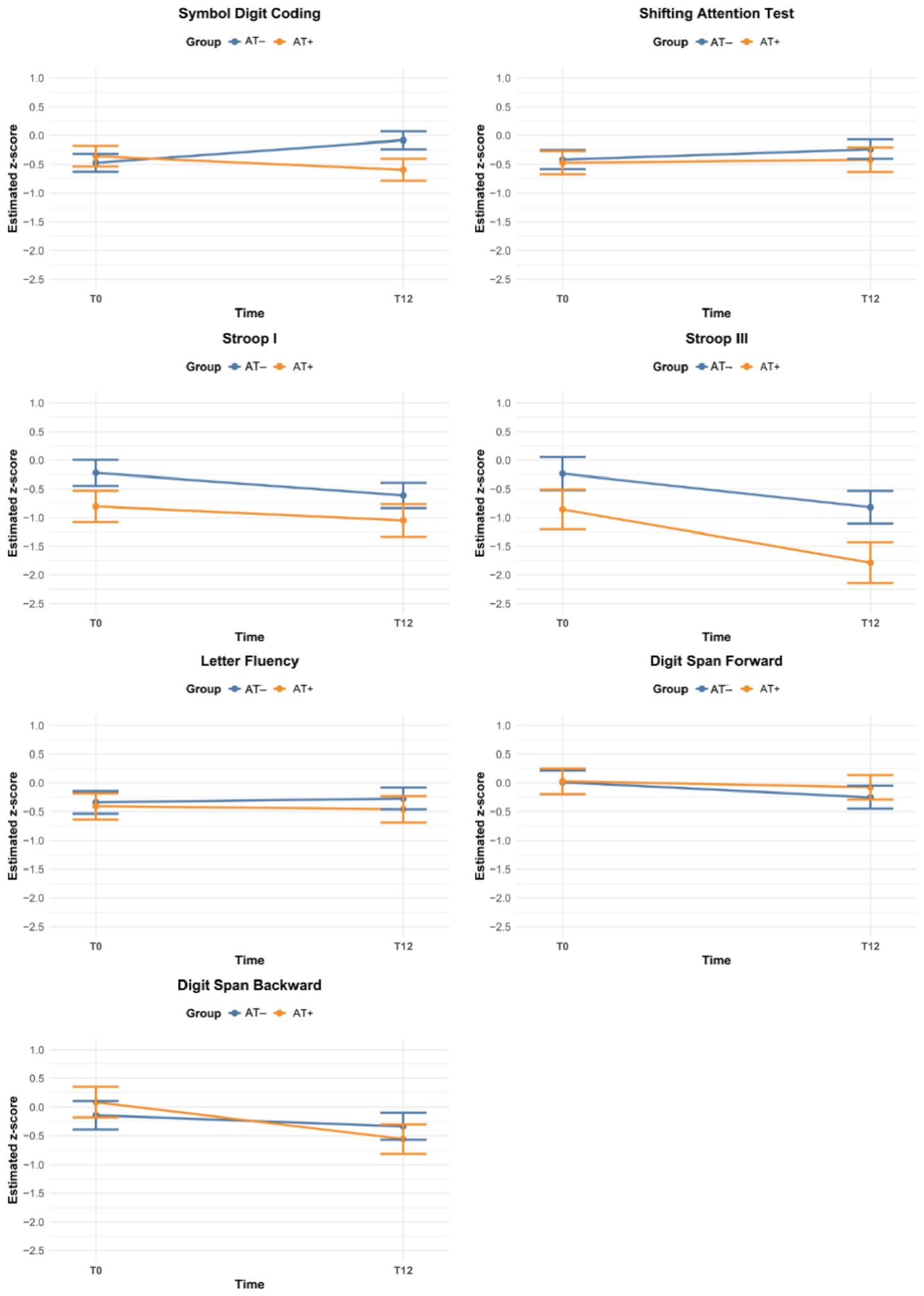
LMM analysis of the Symbol Digit Coding test score showed a significant main effect of time (t(52) = 2.81, p = 0.007), indicating an improvement from before surgery to twelve months after surgery (independent of group), as well as a significant interaction between Time and Group (t(52) = −2.85, p = 0.006), indicating a different trajectory over time between AT+ and AT− patients (Figure 2; Appendix A). Estimated marginal means of the Symbol Digit Coding test exhibited a significant improvement of the AT− group over time (t(52) = −2.81, p = 0.007, d = −0.46), with a medium effect size, and no significant change in the AT+ group after BH-correction (t(52) = 1.40, p = 0.168, d = 0.10). There were no significant predictors or interactions in the LMMs of any of the other test scores (BH-corrected α = 0.014).
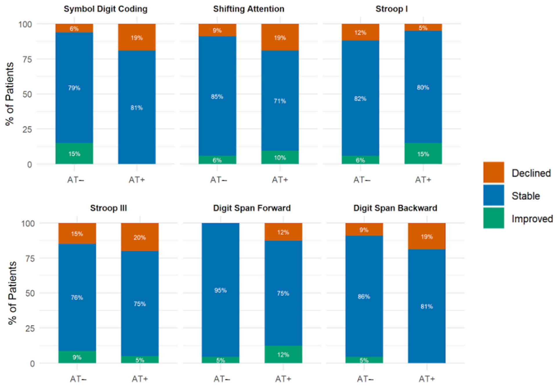
Regarding RCI scores, there were no significant differences between AT+ and AT− patients in proportions of reliably declined, stable, or reliably improved performances over time (BH-corrected α = 0.014) (Figure 3).

3.4. Cognitive Functioning Twelve Months Post-Resection

At T12, AT+ patients performed significantly worse than AT− patients on the Symbol Digit Coding test (after correcting for T0 Symbol Digit Coding performance), with a medium effect size. The observed between-group differences in mean performances on the other tests were not statistically significant (BH-corrected α = 0.014) (Table 4).
Figure 2. Course of observed mean group performance from T0 to T12. Bars represent standard errors for AT− (blue) versus AT+ (orange) groups. Abbreviations: AT = adjuvant treatment.
Cancers 18 02113 g002
Table 4. Comparison of estimated marginal mean z-scores at T12, corrected for T0.
AT−AT+
Cognitive TestEstimated Mean T12 z-Score ± SEnEstimated Mean T12 z-Score ± SEnFp aPartial η2
Symbol Digit Coding−0.03 ± 0.1133−0.56 ± 0.14218.690.005 *0.15
Shifting Attention b−0.26 ± 0.1334−0.38 ± 0.17211.790.1870.03
Stroop I −0.70 ± 0.2434−1.03 ± 0.32200.630.4310.01
Stroop III −1.04 ± 0.2634−1.50 ± 0.34201.170.2850.02
Letter fluency−0.18 ± 0.1628−0.28 ± 0.20180.160.6950.00
Digit Span Forward b−0.31 ± 0.2022−0.03 ± 0.23161.450.2370.04
Digit Span Backward−0.31 ± 0.1622−0.41 ± 0.19160.170.6860.01
a Unadjusted p-values, BH-adjusted α = 0.011. b Scores of the Shifting Attention test and the Digit Span Forward underwent a log transformation due to non-normal distributions. Reported values were back-transformed for interpretability. * Statistically significant p < 0.011. Abbreviations: AT = adjuvant treatment; SE = standard error.
Figure 3. Proportions of patients with declined (orange), stable (blue), or improved (green) performance for each test between T0 and T12 according to the Reliable Change Indices (RCIs). Abbreviations: AT = adjuvant treatment.
Cancers 18 02113 g003

4. Discussion

In this study, we explored the course of cognitive functioning of LGG patients with and without AT, before and one year after tumor resection (on average, eight months after localized radiotherapy, and with 1.7 months of chemotherapy remaining). Although there were no presurgical differences in cognitive performance between the groups, one year after resection, patients with AT performed significantly worse on a measure of information processing speed than patients without AT, with a medium effect size. While the group without AT showed a significant improvement in information processing speed, the group with AT did not show a significant change over time, suggesting that patients who receive AT may show limited postoperative recovery.
Before resection, patients who would receive AT were older, had a larger tumor, more frequently had tumors that crossed the midline, and reported more depressive symptoms than patients who would not receive AT. This is consistent with the indication for AT in LGG, which is based on the estimated risk of tumor progression, influenced by factors such as older age, tumor volume, and bilateral tumor involvement [10,61]. Furthermore, depressive symptoms in the AT group may be partly explained by inflammation, oedema, or the use of dexamethasone. Depressive symptoms are also associated with tumor progression and may have contributed to the indication for AT [62,63]. In the analyses on cognitive functioning, we used age-corrected z-scores, but we did not correct for tumor volume or depressive symptoms. These inherent group differences may therefore confound interpretation of results, as, for example, depressive symptoms may also influence cognitive functioning [64]. However, as tumor volume is closely associated with the indication for AT, correcting for it would remove variance that is inherently tied to group membership, thereby obscuring clinically meaningful differences between the groups. Despite lower observed pre-treatment performances across cognitive tests in the group with AT, possibly due to higher tumor burden, there were no statistically significant differences in cognitive performance between patients with and without AT before surgery. This may be explained by limited statistical power.
Despite the relatively small sample size and inherent group differences, we found medium-sized differences between patients with and without AT in information processing speed over time. The distinct trajectories in information processing speed were also reflected in the frequencies of individual reliable change categories. Patients without AT showed mostly stable performance or improvement, while patients with AT showed only stable performance or decline. Reductions in information processing speed after AT have been reported in previous studies, with follow-up intervals after AT ranging from several months to multiple years [20,21,22,27,28]. Notably, the group difference in information processing speed in this study emerged at eight months after localized radiotherapy, earlier than the delayed cognitive effects previously reported [2,20,21,22,23,43,65]. Twelve months after resection, most patients were still undergoing chemotherapy, with an average of 1.7 months of treatment remaining. Therefore, the group difference in information processing speed may also reflect an acute treatment effect of chemotherapy. Moreover, the observed group difference was not driven by a decline in the AT group but by an improvement in the group without AT. This suggests that patients with AT may have more limited postoperative cognitive recovery and neuroplasticity, possibly through white matter damage in distributed information processing pathways [14,16,27,66,67,68]. These distributed pathways may be particularly sensitive to the treatment-related changes in vascular integrity, inflammation, and myelination [14,16,17]. Therefore, the lack of improvement in the group with AT could also be an early indicator of the later decline, as reported in other studies. Nevertheless, some previous studies did not detect an association between AT and information processing speed [24,29,69], potentially because these did not account for practice effects, pre-treatment performance, or lacked comparison to a group without AT.
We did not observe differences between the groups with and without AT on tests of simple reaction time, attention span, working memory, inhibition, cognitive flexibility, and verbal fluency. Previous studies, however, have reported associations between AT and impairments in executive functioning [19,20,22,28], attention [21,22], and verbal fluency [24]. These discrepancies may reflect small sample sizes, resulting in heterogeneous samples across studies. Cognitive impairment profiles vary substantially among glioma patients [25,26], and differences in treatment location [15] may contribute to variability in cognitive outcomes in this population as well. However, neither cognitive impairment profiles nor treatment location was differentiated in previous nor in the current analyses to preserve power, potentially explaining inconsistencies between studies. Notably, although group differences were not statistically significant, patients with AT observationally showed lower performance and more individual declines over time than patients without AT on most tests at both timepoints. The lack of statistically significant group differences on these tests may reflect substantial heterogeneity in cognitive functioning among patients. While such variability is clinically meaningful and may reflect differences in tumor characteristics and individual recovery patterns, it also reduces the ability to detect subtle between-group differences. In contrast, performance on information processing speed was less heterogeneous, and symbol substitution tests are highly sensitive measures of information processing speed [40], which may partly explain why a significant group difference was found for this test but not for the others.
The findings of this study contribute to the literature on the consequences of AT for LGG patients, with several strengths compared to prior studies. First, we used z-scores that were corrected for demographics and practice effects, thereby reducing potential over- or underestimations of cognitive performances over time [43]. Second, we examined both group-level averages and individual trajectories over time, providing a more complete understanding of treatment effects in a heterogeneous sample of LGG patients [42].
Certain study limitations must also be acknowledged. First, the relatively small sample size reduces statistical power, and results should be interpreted as exploratory, with limited generalizability. Moreover, given the limited sample size, we did not differentiate between types of radiotherapy and chemotherapy, treatment locations, tumor characteristics, or cognitive profiles in order to preserve statistical power. Nevertheless, collecting larger longitudinal cohorts in this population remains challenging, given the low incidence of LGG [70]. Second, the non-randomized design introduced a selection bias. The groups inherently differed at baseline because the indication for AT is based on clinical risk factors, which restricted comparability. Although we used demographically corrected z-scores and accounted for baseline cognitive functioning, residual confounding due to depressive symptoms or tumor characteristics cannot be (entirely) excluded, and results should be interpreted with caution. Moreover, as tumor burden is higher in the group receiving AT, the tumor itself may also have affected cognitive functioning differently in the groups, as it possibly did at baseline [21,38]. At the same time, the findings reflect real-world clinical practice, and it remains challenging to distinguish tumor-related effects on cognition from treatment-related effects in non-randomized research. Third, our approach prioritized breadth across multiple cognitive domains with seven tests due to the lack of consensus in prior literature and the exploratory nature of the current study. To account for these multiple comparisons, we applied a correction with a relatively lenient false discovery rate threshold. This approach balanced the risk of false positives due to multiple testing with the risk of false negatives due to the small sample size. This implies that there may be a higher risk of false positives in the current study compared to more strict multiple comparison corrections. Nevertheless, we prioritized minimizing the risk of false negatives so that clinically relevant adverse effects were not obscured while still applying a correction. Fourth, we conducted cognitive screenings at fixed time points relative to resection rather than AT. Because most patients were still undergoing chemotherapy and the interval between AT and cognitive screening varied between patients, we could not clearly distinguish the acute from the more persistent treatment effects. Studies with a fixed interval around AT or with a longer follow-up are required to distinguish transient from persistent effects. Fifth, we excluded verbal and visual memory tests from our analyses because of the limited test-retest reliability found in our previous study [51]. Nevertheless, declines in both verbal and visual memory have been associated with AT in previous studies [19,20,21,24,28,29]. Future studies may benefit from including sensitive measures of verbal and visual memory to broaden the assessment of cognitive functioning.
This exploratory study suggests that LGG patients who are allocated to AT may have a distinct cognitive trajectory from patients without AT, with the most pronounced differences observed in information processing speed. Although the beneficial effect of AT on progression-free survival is well-established, these possible distinct trajectories may be relevant in treatment planning depending on patient preferences [71]. For example, preliminary evidence suggests that by delaying AT, through IDH inhibitors, for example [11], patients may have a greater opportunity for postoperative neural and functional reorganization, leading to favorable cognitive outcomes [15]. Nevertheless, further research is needed to clarify the cognitive effects of both radiotherapy and chemotherapy in LGG patients. Multicenter collaborations could facilitate larger sample sizes in this rare population, allowing for more comprehensive testing and greater power. Moreover, studies integrating neuroimaging biomarkers with cognitive assessment may provide additional insights into the mechanisms underlying the treatment effects of AT on cognition. Additionally, the scores of neuropsychological performance testing are often only modestly correlated with self-reported cognitive functioning [72,73]. Given the relationship between cognitive complaints and quality of life [72], future studies should integrate both performance-based and self-report measures. Furthermore, a way to capture more subtle cognitive changes and fluctuations over time could be through cognitive ecological momentary assessment. This involves brief, repeated cognitive assessments administered in patients’ daily environments, capturing real-time performance [74].

5. Conclusions

This study explored the cognitive trajectories of LGG patients with and without AT with localized radiotherapy and chemotherapy before and one year after tumor resection. We found significantly worse information processing speed outcomes in the AT group, suggesting that patients allocated to AT may show limited cognitive recovery up to 12 months after surgery. However, our findings are exploratory due to the non-randomized design and small sample, and we cannot distinguish tumor-related effects on cognition from treatment-related effects. Nevertheless, our findings generate clinically relevant hypotheses for future, adequately powered studies. When current literature is expanded with larger, uniform studies, evidence on the effects of treatment for LGG on cognition can be used to support balanced and individualized clinical decision-making.

Author Contributions

Conceptualization, E.A.v.B., K.G. and I.J.M.B., methodology, E.A.v.B., K.G. and I.J.M.B.; formal analysis, E.A.v.B.; writing—original draft preparation, E.A.v.B.; writing—review and editing, K.G., I.J.M.B., M.J.F.L., N.S. and G.-J.R.; visualization, E.A.v.B. All authors have read and agreed to the published version of the manuscript.

Funding

This research was funded by ZonMw, a Dutch organization for health research and development, project numbers 842003007 and 10070012010006.

Institutional Review Board Statement

The study was conducted in accordance with the Declaration of Helsinki and approved by the Ethics Committee of METC Brabant (protocol code NW2020-32, date of approval 2 July 2020) and the Ethics Review Board of Tilburg University (RP472, date of approval 16 March 2021).

Informed Consent Statement

Informed consent was obtained from all subjects involved in the study.

Data Availability Statement

The data that support the findings of this study are not publicly available, as participants did not give written consent for data sharing and the privacy restrictions associated with the data.

Acknowledgments

We would like to thank the patients for participating in the study. Also, we would like to thank all interns for conducting the neuropsychological screening batteries, Winnie van Buijtene for coordination of the study, and Sacha van der Donk for data management of the study.

Conflicts of Interest

The authors declare no conflicts of interest.

Abbreviations

The following abbreviations are used in this manuscript:
LGGLow-grade glioma
IDHIsocitrate dehydrogenase
WHOWorld Health Organization
ATAdjuvant treatment
MMSEMini-Mental State Examination
ETZElisabeth-TweeSteden Hospital
HADSHospital Anxiety and Depression Scale
CNS VSCentral Nervous System Vital Signs
ANCOVAAnalysis of covariance
LMMLinear mixed model
MLMaximum likelihood
AICAkaike Information Criterion
RCIReliable change index
BHBenjamini-Hochberg
TMZTemozolomide
PCVProcarbazine, lomustine, and vincristine
RTRadiotherapy
ChTChemotherapy
SDStandard deviation
SEStandard error

Appendix A

Table A1. Results of the Linear Mixed Model (LMM) analysis of the mean course of functioning between T0 and T12 (Time) of patients with and without adjuvant treatment (Group).
Cognitive TestAICTime a Group b Time × Group R2
b (SE)pb (SE)pb (SE)p
Symbol Digit Coding284.750.39 (0.14)0.007 *0.12 (0.24)0.631−0.63 (0.22)0.006 *0.035
Shifting Attention385.95−0.39 (0.27)0.158−0.59 (0.35)0.1030.15 (0.44)0.7370.015
Stroop I421.37−0.59 (0.25)0.024−0.62 (0.45)0.174−0.34 (0.41)0.4070.037
Stroop III323.120.18 (0.19)0.356−0.05 (0.26)0.837−0.13 (0.31)0.6840.004
Letter fluency295.130.07 (0.16)0.690−0.07 (0.30)0.816−0.12 (0.26)0.6510.008
Digit Span forward245.16−0.26 (0.22)0.2430.02 (0.31)0.9540.16 (0.33)0.6380.021
Digit Span backward274.94−0.19 (0.26)0.4570.23 (0.36)0.535−0.45 (0.39)0.2540.002
a Reference time = T12. b Reference group = AT+. * Significant p-values under BH-corrected α of 0.014. Abbreviations: AIC = Akaike Information Criterion; SE = standard error.

References

  1. Barzilai, O.; Moshe, S.B.; Sitt, R.; Sela, G.; Shofty, B.; Ram, Z. Improvement in cognitive function after surgery for low-grade glioma. J. Neurosurg. 2018, 130, 426–434. [Google Scholar] [CrossRef] [Scilit] [PubMed]
  2. Koutsarnakis, C.; Neromyliotis, E.; Komaitis, S.; Mazarakis, N.; O’Hara, D.J.; Stranjalis, G.; Chumas, P. Effects of brain radiotherapy on cognitive performance in adult low-grade glioma patients: A systematic review. Radiother. Oncol. 2021, 160, 202–211. [Google Scholar] [CrossRef] [Scilit] [PubMed]
  3. Campanella, F.; Palese, A.; Del Missier, F.; Moreale, R.; Ius, T.; Shallice, T.; Fabbro, F.; Skrap, M. Long-term cognitive functioning and psychological well-being in surgically treated patients with low-grade glioma. World Neurosurg. 2017, 103, 799–808. [Google Scholar] [CrossRef] [Scilit] [PubMed]
  4. Klein, M. Neurocognitive functioning in adult WHO grade II gliomas: Impact of old and new treatment modalities. Neuro-Oncology 2012, 14, iv17–iv24. [Google Scholar] [CrossRef] [Scilit] [PubMed]
  5. Mair, M.J.; Geurts, M.; van den Bent, M.J.; Berghoff, A.S. A basic review on systemic treatment options in WHO grade II–III gliomas. Cancer Treat. Rev. 2021, 92, 102124. [Google Scholar] [CrossRef] [Scilit] [PubMed]
  6. Ostrom, Q.T.; Price, M.; Neff, C.; Cioffi, G.; Waite, K.A.; Kruchko, C.; Barnholtz-Sloan, J.S. CBTRUS Statistical Report: Primary Brain and Other Central Nervous System Tumors Diagnosed in the United States in 2015–2019. Neuro-Oncology 2022, 24, v1–v95. [Google Scholar] [CrossRef] [Scilit] [PubMed]
  7. Boele, F.W.; Zant, M.; Heine, E.C.; Aaronson, N.K.; Taphoorn, M.J.; Reijneveld, J.C.; Postma, T.J.; Heimans, J.J.; Klein, M. The association between cognitive functioning and health-related quality of life in low-grade glioma patients. Neuro-Oncol. Pract. 2014, 1, 40–46. [Google Scholar] [CrossRef] [Scilit] [PubMed]
  8. Rimmer, B.; Bolnykh, I.; Dutton, L.; Lewis, J.; Burns, R.; Gallagher, P.; Williams, S.; Araujo-Soares, V.; Menger, F.; Sharp, L. Health-related quality of life in adults with low-grade gliomas: A systematic review. Qual. Life Res. 2023, 32, 625–651. [Google Scholar] [CrossRef] [Scilit] [PubMed]
  9. Karschnia, P.; Young, J.S.; Wijnenga, M.M.; Sciortino, T.; Teske, N.; Corell, A.; Wagner, A.; Youssef, G.; Park, Y.W.; Häni, L. A prognostic classification system for extent of resection in IDH-mutant grade 2 glioma: An international, multicentre, retrospective cohort study with external validation by the RANO resect group. Lancet Oncol. 2025, 26, 1638–1650. [Google Scholar] [CrossRef] [Scilit] [PubMed]
  10. Weller, M.; van den Bent, M.; Preusser, M.; Le Rhun, E.; Tonn, J.C.; Minniti, G.; Bendszus, M.; Balana, C.; Chinot, O.; Dirven, L. EANO guidelines on the diagnosis and treatment of diffuse gliomas of adulthood. Nat. Rev. Clin. Oncol. 2021, 18, 170–186. [Google Scholar] [CrossRef] [Scilit] [PubMed]
  11. Mellinghoff, I.K.; Van Den Bent, M.J.; Blumenthal, D.T.; Touat, M.; Peters, K.B.; Clarke, J.; Mendez, J.; Yust-Katz, S.; Welsh, L.; Mason, W.P. Vorasidenib in IDH1-or IDH2-mutant low-grade glioma. N. Engl. J. Med. 2023, 389, 589–601. [Google Scholar] [CrossRef] [Scilit] [PubMed]
  12. Brown, T.J.; Bota, D.A.; van Den Bent, M.J.; Brown, P.D.; Maher, E.; Aregawi, D.; Liau, L.M.; Buckner, J.C.; Weller, M.; Berger, M.S. Management of low-grade glioma: A systematic review and meta-analysis. Neuro-Oncol. Pract. 2019, 6, 249–258. [Google Scholar] [CrossRef] [Scilit] [PubMed]
  13. Buckner, J.C.; Shaw, E.G.; Pugh, S.L.; Chakravarti, A.; Gilbert, M.R.; Barger, G.R.; Coons, S.; Ricci, P.; Bullard, D.; Brown, P.D. Radiation plus procarbazine, CCNU, and vincristine in low-grade glioma. N. Engl. J. Med. 2016, 374, 1344–1355. [Google Scholar] [CrossRef] [Scilit] [PubMed]
  14. Makale, M.T.; McDonald, C.R.; Hattangadi-Gluth, J.A.; Kesari, S. Mechanisms of radiotherapy-associated cognitive disability in patients with brain tumours. Nat. Rev. Neurol. 2016, 13, 52–64. [Google Scholar] [CrossRef] [Scilit] [PubMed]
  15. Kirkman, M.A.; Hunn, B.H.; Thomas, M.S.; Tolmie, A.K. Influences on cognitive outcomes in adult patients with gliomas: A systematic review. Front. Oncol. 2022, 12, 943600. [Google Scholar] [CrossRef] [Scilit] [PubMed]
  16. Moretti, R.; Caruso, P. An iatrogenic model of brain small-vessel disease: Post-radiation encephalopathy. Int. J. Mol. Sci. 2020, 21, 6506. [Google Scholar] [CrossRef] [Scilit] [PubMed]
  17. Wu, P.H.; Coultrap, S.; Pinnix, C.; Davies, K.D.; Tailor, R.; Ang, K.K.; Browning, M.D.; Grosshans, D.R. Radiation induces acute alterations in neuronal function. PLoS ONE 2012, 7, e37677. [Google Scholar] [CrossRef] [Scilit] [PubMed]
  18. Huisman, S.I.; van der Boog, A.T.; Cialdella, F.; Verhoeff, J.J.; David, S. Quantifying the post-radiation accelerated brain aging rate in glioma patients with deep learning. Radiother. Oncol. 2022, 175, 18–25. [Google Scholar] [CrossRef] [Scilit] [PubMed]
  19. Correa, D.D.; Shi, W.; Thaler, H.T.; Cheung, A.M.; DeAngelis, L.M.; Abrey, L.E. Longitudinal cognitive follow-up in low grade gliomas. J. Neuro-Oncol. 2008, 86, 321–327. [Google Scholar] [CrossRef] [Scilit] [PubMed]
  20. Correa, D.D.; DeAngelis, L.M.; Shi, W.; Thaler, H.T.; Lin, M.; Abrey, L.E. Cognitive functions in low-grade gliomas: Disease and treatment effects. J. Neuro-Oncol. 2007, 81, 175–184. [Google Scholar] [CrossRef] [Scilit] [PubMed]
  21. Klein, M.; Heimans, J.; Aaronson, N.; Van der Ploeg, H.; Grit, J.; Muller, M.; Postma, T.; Mooij, J.; Boerman, R.; Beute, G. Effect of radiotherapy and other treatment-related factors on mid-term to long-term cognitive sequelae in low-grade gliomas: A comparative study. Lancet 2002, 360, 1361–1368. [Google Scholar] [CrossRef] [Scilit] [PubMed]
  22. Douw, L.; Klein, M.; Fagel, S.S.; van den Heuvel, J.; Taphoorn, M.J.; Aaronson, N.K.; Postma, T.J.; Vandertop, W.P.; Mooij, J.J.; Boerman, R.H. Cognitive and radiological effects of radiotherapy in patients with low-grade glioma: Long-term follow-up. Lancet Neurol. 2009, 8, 810–818. [Google Scholar] [CrossRef] [Scilit] [PubMed]
  23. Surma-aho, O.; Niemelä, M.; Vilkki, J.; Kouri, M.; Brander, A.; Salonen, O.; Paetau, A.; Kallio, M.; Pyykkönen, J.; LicPhil; et al. Adverse long-term effects of brain radiotherapy in adult low-grade glioma patients. Neurology 2001, 56, 1285–1290. [Google Scholar] [CrossRef] [Scilit] [PubMed]
  24. Boele, F.W.; Frances, S.M.; Darlix, A.; Ducray, F.; Cartalat, S.; Guerdoux, E.; Tabouret, E.; Burger, M.C.; Rydén, I.; Malmstrom, A. Health-related quality of life and cognitive functioning in survivors of oligodendroglioma: An international cross-sectional investigation. Neuro-Oncology 2025, 27, 2959–2974. [Google Scholar] [CrossRef] [Scilit] [PubMed]
  25. Gorter, M.; Röttgering, J.G.; Belgers, V.; van Lingen, M.R.; de Witt Hamer, P.C.; Douw, L.; Klein, M. Distinct and reproducible neurocognitive profiles in stable diffuse glioma: A data-driven approach to understanding cognitive heterogeneity. Neuro-Oncology 2025, 27, 3250–3259. [Google Scholar] [CrossRef] [Scilit] [PubMed]
  26. Reyes, A.; Stasenko, A.; Hopper, A.; Kohli, J.S.; Helm, J.L.; Salans, M.; Prabhakaran, D.; Kamalyan, L.; Wilkinson, M.; Unnikrishnan, S. Cognitive phenotypes: Unraveling the heterogeneity in cognitive dysfunction among patients with primary brain tumors receiving radiotherapy. Neuro-Oncology 2025, 27, 466–478. [Google Scholar] [CrossRef] [Scilit] [PubMed]
  27. Huynh-Le, M.-P.; Tibbs, M.D.; Karunamuni, R.; Salans, M.; Tringale, K.R.; Yip, A.; Connor, M.; Simon, A.B.; Vitzthum, L.K.; Reyes, A. Microstructural injury to corpus callosum and intrahemispheric white matter tracts correlate with attention and processing speed decline after brain radiation. Int. J. Radiat. Oncol. Biol. Phys. 2021, 110, 337–347. [Google Scholar] [CrossRef] [Scilit] [PubMed]
  28. Haldbo-Classen, L.; Amidi, A.; Wu, L.M.; Lukacova, S.; Oettingen, G.v.; Gottrup, H.; Zachariae, R.; Høyer, M. Long-term cognitive dysfunction after radiation therapy for primary brain tumors. Acta Oncol. 2019, 58, 745–752. [Google Scholar] [CrossRef] [Scilit] [PubMed]
  29. Rydelius, A.; Lätt, J.; Kinhult, S.; Engelholm, S.; Van Westen, D.; Pihlsgård, M.; Bengzon, J.; Sundgren, P.C.; Lilja, Å. Longitudinal study of cognitive function in glioma patients treated with modern radiotherapy techniques and standard chemotherapy. Acta Oncol. 2020, 59, 1091–1097. [Google Scholar] [CrossRef] [Scilit] [PubMed]
  30. Hodgson, K.D.; Hutchinson, A.D.; Wilson, C.J.; Nettelbeck, T. A meta-analysis of the effects of chemotherapy on cognition in patients with cancer. Cancer Treat. Rev. 2013, 39, 297–304. [Google Scholar] [CrossRef] [Scilit] [PubMed]
  31. Lindner, O.C.; Phillips, B.; McCabe, M.G.; Mayes, A.; Wearden, A.; Varese, F.; Talmi, D. A meta-analysis of cognitive impairment following adult cancer chemotherapy. Neuropsychology 2014, 28, 726–740. [Google Scholar] [CrossRef] [Scilit] [PubMed]
  32. Jim, H.S.; Phillips, K.M.; Chait, S.; Faul, L.A.; Popa, M.A.; Lee, Y.-H.; Hussin, M.G.; Jacobsen, P.B.; Small, B.J. Meta-analysis of cognitive functioning in breast cancer survivors previously treated with standard-dose chemotherapy. J. Clin. Oncol. 2012, 30, 3578–3587. [Google Scholar] [CrossRef] [Scilit] [PubMed]
  33. Karschnia, P.; Nelson, T.A.; Dietrich, J. Mechanisms and treatment of cancer therapy-induced peripheral and central neurotoxicity. Nat. Rev. Cancer 2025, 25, 887–909. [Google Scholar] [CrossRef] [Scilit] [PubMed]
  34. Prabhu, R.S.; Won, M.; Shaw, E.G.; Hu, C.; Brachman, D.G.; Buckner, J.C.; Stelzer, K.J.; Barger, G.R.; Brown, P.D.; Gilbert, M.R. Effect of the addition of chemotherapy to radiotherapy on cognitive function in patients with low-grade glioma: Secondary analysis of RTOG 98-02. J. Clin. Oncol. 2014, 32, 535–541. [Google Scholar] [CrossRef] [Scilit] [PubMed]
  35. Klein, M.; Drijver, A.J.; Van Den Bent, M.J.; Bromberg, J.C.; Hoang-Xuan, K.; Taphoorn, M.J.; Reijneveld, J.C.; Ben Hassel, M.; Vauleon, E.; Eekers, D.B. Memory in low-grade glioma patients treated with radiotherapy or temozolomide: A correlative analysis of EORTC study 22033–26033. Neuro-Oncology 2021, 23, 803–811. [Google Scholar] [CrossRef] [Scilit] [PubMed]
  36. Reijneveld, J.C.; Taphoorn, M.J.; Coens, C.; Bromberg, J.E.; Mason, W.P.; Hoang-Xuan, K.; Ryan, G.; Hassel, M.B.; Enting, R.H.; Brandes, A.A. Health-related quality of life in patients with high-risk low-grade glioma (EORTC 22033-26033): A randomised, open-label, phase 3 intergroup study. Lancet Oncol. 2016, 17, 1533–1542. [Google Scholar] [CrossRef] [Scilit] [PubMed]
  37. Wang, M.; Cairncross, G.; Shaw, E.; Jenkins, R.; Scheithauer, B.; Brachman, D.; Buckner, J.; Fink, K.; Souhami, L.; Laperriere, N. Cognition and quality of life after chemotherapy plus radiotherapy (RT) vs. RT for pure and mixed anaplastic oligodendrogliomas: Radiation therapy oncology group trial 9402. Int. J. Radiat. Oncol. Biol. Phys. 2010, 77, 662–669. [Google Scholar] [CrossRef] [Scilit] [PubMed]
  38. Lawrie, T.A.; Gillespie, D.; Dowswell, T.; Evans, J.; Erridge, S.; Vale, L.; Kernohan, A.; Grant, R. Long-term neurocognitive and other side effects of radiotherapy, with or without chemotherapy, for glioma. Cochrane Database Syst. Rev. 2019. [Google Scholar] [CrossRef] [Scilit] [PubMed]
  39. Brown, P.D.; Buckner, J.C.; O’Fallon, J.R.; Iturria, N.L.; Brown, C.A.; O’Neill, B.P.; Scheithauer, B.W.; Dinapoli, R.P.; Arusell, R.M.; Curran, W.J. Effects of radiotherapy on cognitive function in patients with low-grade glioma measured by the folstein mini-mental state examination. J. Clin. Oncol. 2003, 21, 2519–2524. [Google Scholar] [CrossRef] [Scilit] [PubMed]
  40. Boone, M.; Roussel, M.; Chauffert, B.; Le Gars, D.; Godefroy, O. Prevalence and profile of cognitive impairment in adult glioma: A sensitivity analysis. J. Neuro-Oncol. 2016, 129, 123–130. [Google Scholar] [CrossRef] [Scilit] [PubMed]
  41. Meyers, C.A.; Wefel, J.S. The use of the mini-mental state examination to assess cognitive functioning in cancer trials: No ifs, ands, buts, or sensitivity. J. Clin. Oncol. 2003, 21, 3557–3558. [Google Scholar] [CrossRef] [Scilit] [PubMed]
  42. van Loon, E.M.; Heijenbrok-Kal, M.H.; van Loon, W.S.; van den Bent, M.J.; Vincent, A.J.; de Koning, I.; Ribbers, G.M. Assessment methods and prevalence of cognitive dysfunction in patients with low-grade glioma: A systematic review. J. Rehabil. Med. 2015, 47, 481–488. [Google Scholar] [CrossRef] [Scilit] [PubMed]
  43. De Roeck, L.; Gillebert, C.R.; van Aert, R.C.; Vanmeenen, A.; Klein, M.; Taphoorn, M.J.; Gehring, K.; Lambrecht, M.; Sleurs, C. Cognitive outcomes after multimodal treatment in adult glioma patients: A meta-analysis. Neuro-Oncology 2023, 25, 1395–1414. [Google Scholar] [CrossRef] [Scilit] [PubMed]
  44. Louis, D.N.; Perry, A.; Wesseling, P.; Brat, D.J.; Cree, I.A.; Figarella-Branger, D.; Hawkins, C.; Ng, H.; Pfister, S.M.; Reifenberger, G. The 2021 WHO classification of tumors of the central nervous system: A summary. Neuro-Oncology 2021, 23, 1231–1251. [Google Scholar] [CrossRef] [Scilit] [PubMed]
  45. Isensee, F.; Jaeger, P.F.; Kohl, S.A.; Petersen, J.; Maier-Hein, K.H. nnU-Net: A self-configuring method for deep learning-based biomedical image segmentation. Nat. Methods 2021, 18, 203–211. [Google Scholar] [CrossRef] [Scilit] [PubMed]
  46. Bakas, S.; Akbari, H.; Sotiras, A.; Bilello, M.; Rozycki, M.; Kirby, J.S.; Freymann, J.B.; Farahani, K.; Davatzikos, C. Advancing The Cancer Genome Atlas glioma MRI collections with expert segmentation labels and radiomic features. Sci. Data 2017, 4, 170117. [Google Scholar] [CrossRef] [Scilit] [PubMed]
  47. Menze, B.H.; Jakab, A.; Bauer, S.; Kalpathy-Cramer, J.; Farahani, K.; Kirby, J.; Burren, Y.; Porz, N.; Slotboom, J.; Wiest, R.; et al. The multimodal brain tumor image segmentation benchmark (BRATS). IEEE Trans. Med. Imaging 2015, 34, 1993–2024. [Google Scholar] [CrossRef] [Scilit] [PubMed]
  48. Verhage, F. Intelligentie en Leeftijd: Onderzoek bij Nederlanders van Twaalf tot Zevenenzeventig Jaar; van Gorcum: Assen, The Netherlands, 1964. [Google Scholar]
  49. Zigmond, A.S.; Snaith, R.P. The hospital anxiety and depression scale. Acta Psychiatr. Scand. 1983, 67, 361–370. [Google Scholar] [CrossRef] [Scilit] [PubMed]
  50. Gualtieri, C.T.; Johnson, L.G. Reliability and validity of a computerized neurocognitive test battery, CNS Vital Signs. Arch. Clin. Neuropsychol. 2006, 21, 623–643. [Google Scholar] [CrossRef] [Scilit] [PubMed]
  51. Rijnen, S.J.; van der Linden, S.D.; Emons, W.H.; Sitskoorn, M.M.; Gehring, K. Test-retest reliability and practice effects of a computerized neuropsychological battery: A solution-oriented approach. Psychol. Assess. 2018, 30, 1652–1662. [Google Scholar] [CrossRef] [Scilit] [PubMed]
  52. Schmand, B.; Groenink, S.; Van den Dungen, M. Letter fluency: Psychometric properties and Dutch normative data. Tijdschr. Voor Gerontol. En Geriatr. 2008, 39, 64–76. [Google Scholar] [CrossRef] [Scilit] [PubMed]
  53. Wechsler, D. Wechsler Adult Intelligence Scale, 3rd ed.; The Psychological Corporation: San Antonio, TX, USA, 1997. [Google Scholar]
  54. Rijnen, S.J.; Meskal, I.; Emons, W.H.; Campman, C.A.; van der Linden, S.D.; Gehring, K.; Sitskoorn, M.M. Evaluation of normative data of a widely used computerized neuropsychological battery: Applicability and effects of sociodemographic variables in a Dutch sample. Assessment 2020, 27, 373–383. [Google Scholar] [CrossRef] [Scilit] [PubMed]
  55. Schimmel, W.C.; Verhaak, E.; Bakker, M.; Hanssens, P.E.; Sitskoorn, M.M.; Gehring, K. Group and individual change in cognitive functioning in patients with 1 to 10 brain metastases following Gamma Knife radiosurgery. Clin. Oncol. 2021, 33, 314–321. [Google Scholar] [CrossRef] [Scilit] [PubMed]
  56. Cohen, J. Statistical Power Analysis for the Behavioral Sciences, 2nd ed.; Lawrence Erlbaum Associates: Hillsdale, NJ, USA, 1988. [Google Scholar]
  57. Nakagawa, S.; Schielzeth, H. A general and simple method for obtaining R2 from generalized linear mixed-effects models. Methods Ecol. Evol. 2013, 4, 133–142. [Google Scholar] [CrossRef] [Scilit]
  58. Wall Emerson, R. Regression and Effect Size; SAGE Publications: Los Angeles, CA, USA, 2023; pp. 191–192. [Google Scholar]
  59. Benjamini, Y.; Hochberg, Y. Controlling the false discovery rate: A practical and powerful approach to multiple testing. J. R. Stat. Soc. Ser. B (Methodol.) 1995, 57, 289–300. [Google Scholar] [CrossRef] [Scilit]
  60. Glickman, M.E.; Rao, S.R.; Schultz, M.R. False discovery rate control is a recommended alternative to Bonferroni-type adjustments in health studies. J. Clin. Epidemiol. 2014, 67, 850–857. [Google Scholar] [CrossRef] [Scilit] [PubMed]
  61. Richtlijnendatabase. Behandeling Laaggradig Glioom. 2020. Available online: https://richtlijnendatabase.nl/richtlijn/gliomen/behandeling_laaggradig_glioom.html (accessed on 13 January 2026).
  62. Song, L.; Quan, X.; Su, L.; Wang, K.; Wang, H.; Wu, L.; Chen, C.; Li, S.; Xiang, W.; Chen, L. Inflammation and behavioral symptoms in preoperational glioma patients: Is depression, anxiety, and cognitive impairment related to markers of systemic inflammation? Brain Behav. 2020, 10, e01771. [Google Scholar] [CrossRef] [Scilit] [PubMed]
  63. van der Meer, P.B.; Dirven, L.; Hertler, C.; Boele, F.W.; Batalla, A.; Walbert, T.; Rooney, A.G.; Koekkoek, J.A. Depression and anxiety in glioma patients. Neuro-Oncol. Pract. 2023, 10, 335–343. [Google Scholar] [CrossRef] [Scilit] [PubMed]
  64. Rooney, A.G.; Carson, A.; Grant, R. Depression in cerebral glioma patients: A systematic review of observational studies. J. Natl. Cancer Inst. 2011, 103, 61–76. [Google Scholar] [CrossRef] [Scilit] [PubMed]
  65. Park, D.Y.; Tom, M.C.; Chen, Y.; Tewari, S.; Ahluwalia, M.S.; Yu, J.S.; Chao, S.T.; Suh, J.H.; Peereboom, D.M.; Stevens, G.H. Cognitive function after concurrent temozolomide-based chemoradiation therapy in low-grade gliomas. J. Neuro-Oncol. 2022, 158, 341–348. [Google Scholar] [CrossRef] [Scilit] [PubMed]
  66. Schatz, J.; Kramer, J.H.; Ablin, A.; Matthay, K.K. Processing speed, working memory, and IQ: A developmental model of cognitive deficits following cranial radiation therapy. Neuropsychology 2000, 14, 189–200. [Google Scholar] [CrossRef] [PubMed]
  67. Kaiser, J.; Bledowski, C.; Dietrich, J. Neural correlates of chemotherapy-related cognitive impairment. Cortex 2014, 54, 33–50. [Google Scholar] [CrossRef] [Scilit] [PubMed]
  68. Matsos, A.; Loomes, M.; Zhou, I.; Macmillan, E.; Sabel, I.; Rotziokos, E.; Beckwith, W.; Johnston, I. Chemotherapy-induced cognitive impairments: White matter pathologies. Cancer Treat. Rev. 2017, 61, 6–14. [Google Scholar] [CrossRef] [Scilit] [PubMed]
  69. Schlömer, S.; Felsberg, J.; Pertz, M.; Hentschel, B.; Löffler, M.; Schackert, G.; Krex, D.; Juratli, T.; Tonn, J.C.; Schnell, O. Mid-term treatment-related cognitive sequelae in glioma patients. J. Neuro-Oncol. 2022, 159, 65–79. [Google Scholar] [CrossRef] [Scilit] [PubMed]
  70. Ho, V.K.; Reijneveld, J.C.; Enting, R.H.; Bienfait, H.P.; Robe, P.; Baumert, B.G.; Visser, O. Changing incidence and improved survival of gliomas. Eur. J. Cancer 2014, 50, 2309–2318. [Google Scholar] [CrossRef] [Scilit] [PubMed]
  71. Succop, B., Jr.; Richardson, D.R.; Rauf, Y.; Higgins, D.; Catalino, M. Understanding treatment preferences and cognitive outcomes in patients with gliomas. Support. Care Cancer 2024, 32, 673. [Google Scholar] [CrossRef] [Scilit] [PubMed]
  72. Lycke, M.; Lefebvre, T.; Pottel, L.; Pottel, H.; Ketelaars, L.; Stellamans, K.; Van Eygen, K.; Vergauwe, P.; Werbrouck, P.; Cool, L. Subjective, but not objective, cognitive complaints impact long-term quality of life in cancer patients. J. Psychosoc. Oncol. 2019, 37, 427–440. [Google Scholar] [CrossRef] [Scilit] [PubMed]
  73. Savard, J.; Ganz, P.A. Subjective or objective measures of cognitive functioning—what’s more important? JAMA Oncol. 2016, 2, 1263–1264. [Google Scholar] [CrossRef] [Scilit] [PubMed]
  74. Domensino, A.-F.; Lenaert, B.; Verhagen, S.; Bartels, S.L.; van Heugten, C. Real-life cognitive functioning after acquired brain injury: An experience sampling study. Digit. Health 2025, 11, 20552076251387051. [Google Scholar] [CrossRef] [Scilit] [PubMed]
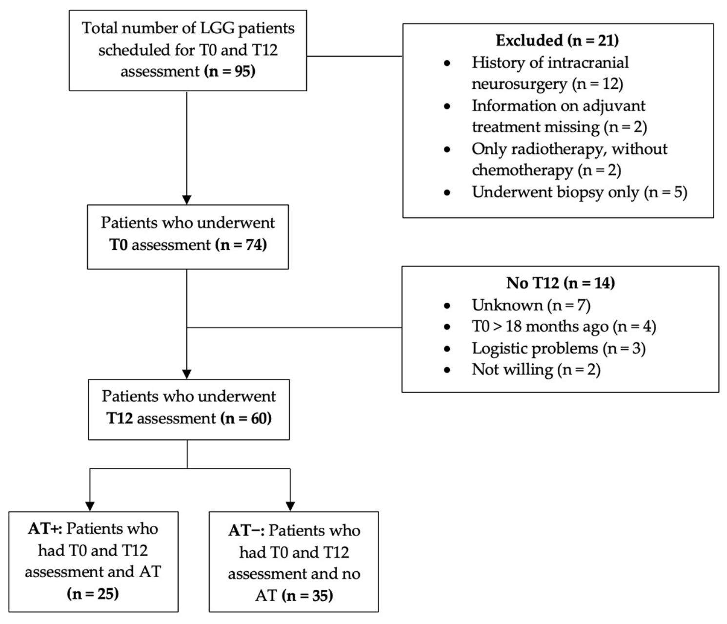
Figure 1. Flowchart of patient inclusion. Abbreviations: LGG = low-grade glioma; AT = adjuvant treatment.
Cancers 18 02113 g001
Table 1. Description of cognitive tests used.
TestDomainContentScore
Symbol Digit Coding
(CNS VS)
Information processing speed Symbols and corresponding numbers are presented at the top of the screen. Participants must match the symbols with the corresponding numbers in the lower part of the screen. Fixed test time of 2 min. Correct responses—incorrect responses
Stroop I
(CNS VS)
Simple reaction timeParticipant presses the space bar when a word is presented on the screen. Fixed total of 16 items.Average reaction time of the correct
responses
Stroop III
(CNS VS)
Inhibitory control Participant presses the space bar if the color of the word does not match the meaning of the word. Fixed total of 48 items.Average reaction time of the correct responses
Shifting
Attention Test
(CNS VS)
Cognitive flexibilityParticipants must match geometric objects either by shape or by color to one of two colored figures at the bottom of the screen using the left and right shift keys. The rules change at random. Fixed test time of 2 min.Correct responses—errors
Letter FluencyVerbal ability and executive controlParticipants are instructed to name as many words as possible starting with a specific letter for 1 min (three trials: D-A-T at T0 and T12, K-O-M at T3).Total number of words named in all three trials
Digit Span Forward Attention spanParticipants are asked to repeat a series of numbers. Every two trials, a longer sequence of numbers is presented. The test is terminated when participants make two consecutive errors.Total number of sequences correct
Digit Span BackwardWorking memoryParticipants are asked to repeat a series of numbers in reverse order. Every two trials, a longer sequence of numbers is presented. The test is terminated when participants make two consecutive errors.Total number of sequences correct
Abbreviations: CNS VS = Central Nervous System Vital Signs [50].
Table 2. Presurgical characteristics of AT− and AT+ patients.
AT− (n = 35)AT+ (n = 25)p
Sex: male/female, n (%)24 (69)/11 (31)14 (52)/11 (48) 0.193
Age at T0 (y): mean ± SD (range)34.6 ± 8.0 (21–54)44.7 ± 13.5 (24–68)0.002 *
Education (y): mean ± SD (range)16.2 ± 2.6 (10–21)16.0 ± 2.7 (8–20)0.825
Histological diagnosis: astrocytoma/oligodendroglioma, n (%)24 (68)/11 (32)17 (68)/8 (32)0.963
1p/19q codeletion: yes, n (%) 11 (32) 8 (33) 0.938
Tumor volume (mm3) **: mean ± SD (range)42,074 ± 69,601
(5668–184,760)
78,382 ± 62,186
(10,496–220,558)
0.027 *
Lesion side: left/right/bilateral, n (%)18 (51)/17 (48)/0 (0)13 (52)/8 (32)/4 (16)0.040 *
Frontal involvement: yes, n (%)27 (77) 17 (68) 0.430
Temporal involvement: yes, n (%)12 (34)10 (40)0.651
Insular involvement: yes, n (%)6 (17)6 (24)0.513
Parietal involvement: yes, n (%)2 (6)2 (8)1.000
Occipital involvement: yes, n (%)1 (3)3 (12)0.298
Awake resection: yes, n (%)17 (49) 12 (48) 0.965
Anti-epileptic medication (T0): yes, n (%)24 (69) 19 (76) 0.529
Dexamethasone use (T0): yes, n (%)6 (17)9 (36)0.096
Psychotropic medication (T0): yes, n (%)8 (23)11 (44)0.083
HADS *** anxiety (T0): mean ± SD (range)5.9 ± 3.3 (0–14)7.5 ± 3.7 (1–14)0.144
   Above clinical cut-off: yes, n (%)9 (31)9 (53)0.142
HADS *** depression (T0): mean ± SD (range)3.4 ± 2.7 (0–11)6.6 ± 3.9 (1–15)0.002 *
   Above clinical cut-off: yes, n (%)3 (10)8 (47)0.010 *
Radiotherapy
   Radiotherapy type: photon/proton, n (%) 11 (44)/14 (56)
   Radiotherapy number of fractions: 25/28/33, n (%) 1 (4)/22 (88)/2 (8)
   Radiotherapy total dose: 45 Gy/50.4 Gy/59.4 Gy, n (%) 1 (4)/22 (88)/2 (8)
   Time since start radiotherapy at T12 (months): mean ± SD (range) 9.0 ± 2.3 (5–14)
   Time since end radiotherapy at T12 (months): mean ± SD (range) 7.8 ± 2.3 (4–13)
   Patients who had completed radiotherapy at T12, n (%) 25 (100)
Chemotherapy
   Chemotherapy type: TMZ/PCV, n (%) 15 (60)/10 (40)
   Time since start chemotherapy at T12 (months): mean ± SD (range) 6.6 ± 2.3 (3–12)
   Time since end chemotherapy at T12 (months): mean ± SD (range) −1.7 ± 3.2 (−7–5)
   Patients who had completed chemotherapy at T12, n (%) 1 (4)
   Patients who prematurely discontinued chemotherapy at T12, n (%) 6 (24)
* Significant p < 0.05. ** Tumor volume missing for seven patients (2 AT−, 5 AT+). *** HADS data missing for 14 patients (6 AT−, 8 AT+). Abbreviations: AT = adjuvant treatment; SD = standard deviation; HADS = hospital anxiety and depression scale; TMZ = temozolomide; PCV = Procarbazine, lomustine, and vincristine.
Table 3. Comparison of mean performance at T0.
AT−AT+
Cognitive TestMean T0 z-Score ± SDnMean T0 z-Score ± SDnt/Zp aEffect Size (Cohen’s d/r-Statistic)
Symbol Digit Coding−0.48 ± 0.9533−0.36 ± 1.0425−0.490.629−0.13
Shifting Attention−0.43 ± 1.0834−0.48 ± 1.2124−0.02 b0.987−0.00 b
Stroop I−0.23 ± 1.0134−0.82 ± 1.3623−1.64 b0.100−0.22 b
Stroop III−0.25 ± 1.3334−0.89 ± 1.6223−1.84 b0.066−0.24 b
Letter fluency−0.29 ± 1.0829−0.33 ± 1.02220.120.9090.03
Digit Span Forward−0.03 ± 0.93220.08 ± 1.0219−0.340.737−0.11
Digit Span Backward−0.15 ± 1.38220.16 ± 1.5719−0.71 b0.480−0.11 b
a Unadjusted p-values, BH-adjusted α = 0.011. b Cognitive tests for which the Mann-Whitney U test was used: z-score and r-statistic. Abbreviations: AT = adjuvant treatment; SD = standard deviation.
Disclaimer/Publisher’s Note: The statements, opinions and data contained in all publications are solely those of the individual author(s) and contributor(s) and not of MDPI and/or the editor(s). MDPI and/or the editor(s) disclaim responsibility for any injury to people or property resulting from any ideas, methods, instructions or products referred to in the content.