Review

MicroRNAs Modulating Cancer Immunotherapy Mechanisms and Therapeutic Synergies

by
Naorem Loya Mangang
1,
Samantha K. Gargasz
2,
Sai Ghanesh Murugan
3,
Munish Kumar
4,
Girish C. Shukla
2 and
Sivakumar Vijayaraghavalu
1,3,*
1
Department of Life Sciences (Zoology), Manipur University (A Central University), Imphal 795003, Manipur, India
2
Center for Gene Regulation in Health and Disease, Department of Biological, Geo and EVS Sciences, 2121 Euclid Ave., Cleveland, OH 44115, USA
3
Department of Medical Sciences and Technology, Indian Institute of Technology Madras, Chennai 600036, Tamil Nadu, India
4
Department of Biochemistry, Faculty of Science, University of Allahabad, Prayagraj 211002, Uttar Pradesh, India
*
Author to whom correspondence should be addressed.
Cancers 2025, 17(24), 3978; https://doi.org/10.3390/cancers17243978

Submission received: 27 October 2025 / Revised: 9 December 2025 / Accepted: 10 December 2025 / Published: 13 December 2025

Simple Summary

Immunotherapy has transformed cancer treatment, but many patients do not respond or develop resistance. microRNAs (miRNAs) play a crucial role in regulating the interaction between cancer cells and immune cells. miRNA can turn immune checkpoint signals on or off, alter the behavior of T cells and macrophages, and travel in tiny vesicles between cells to influence the tumor environment. Because of these functions, miRNAs are being studied as biomarkers to predict who will benefit from immunotherapy, and as drugs themselves, either to block harmful miRNAs or replace protective ones. Although the first clinical trials faced safety and delivery challenges, new technologies such as nanoparticles and exosome-based systems are making miRNA therapies more feasible. This review synthesizes the latest evidence on how miRNAs can enhance cancer immunotherapy and explores future directions for translating these findings into clinical practice.

Abstract

Cancer immunotherapy has transformed oncology, but lasting responses are still limited due to resistance mechanisms within the tumor microenvironment. MicroRNAs (miRNAs) have emerged as critical regulators of immune checkpoint pathways, antigen presentation, T-cell activity, and macrophage polarization. By modulating both tumor-intrinsic and immune cell–intrinsic processes, miRNAs influence the efficacy of immune checkpoint inhibitors, therapeutic vaccines, and adoptive cell therapies. Additionally, circulating and exosomal miRNAs are being investigated as minimally invasive biomarkers to predict patient response and resistance to immunotherapy. Clinical trials of miRNA-based treatments, including mimics and inhibitors, have highlighted both the promise and challenges of translating these molecules into clinical use. Advances in delivery systems, RNA chemistry, and combinatorial strategies are paving the way for their integration into precision immuno-oncology. This review offers a comprehensive overview of the mechanistic, biomarker, and therapeutic roles of miRNAs in cancer immunotherapy, highlighting ongoing clinical progress and prospects.

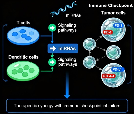
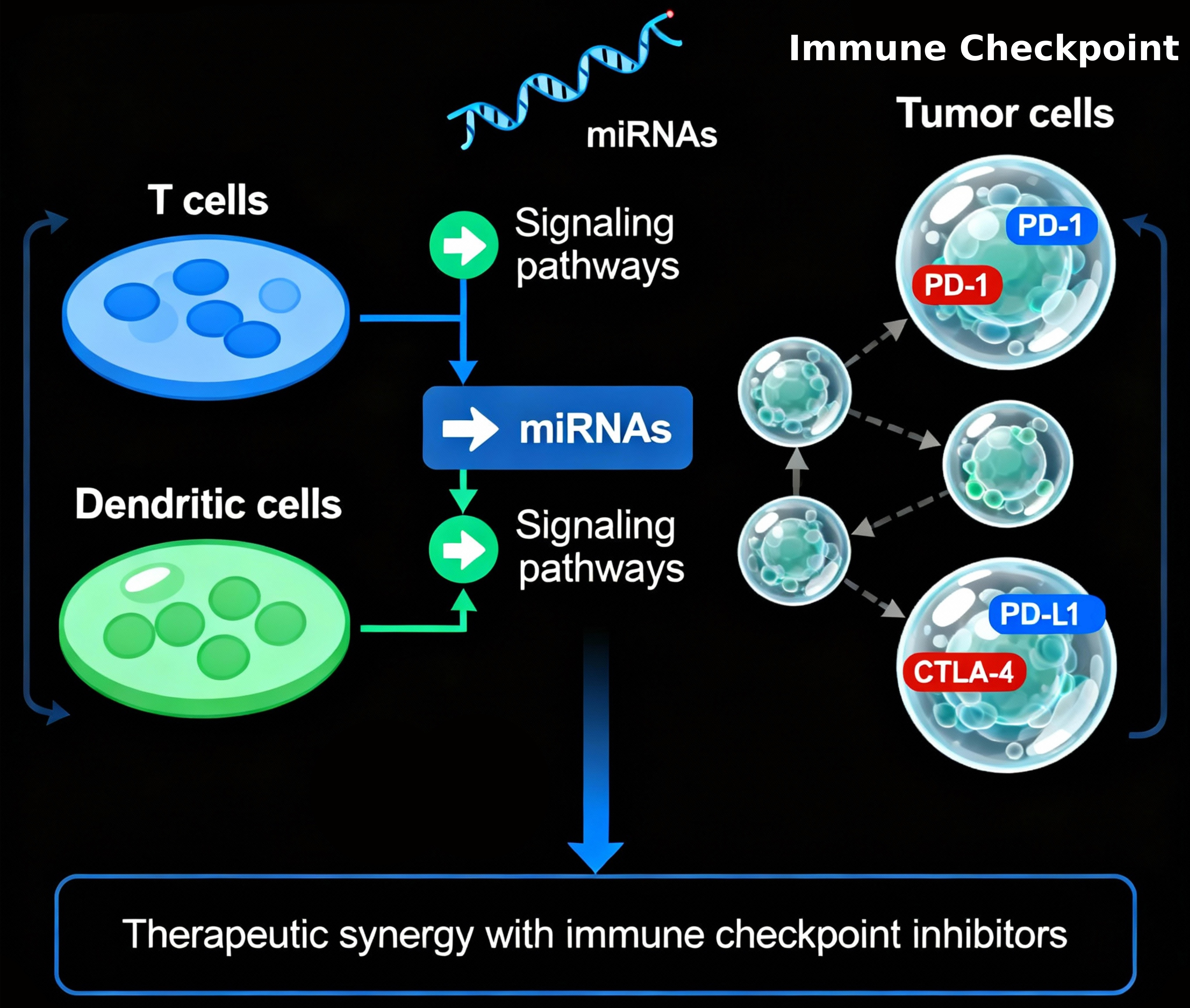
Graphical Abstract

1. Introduction

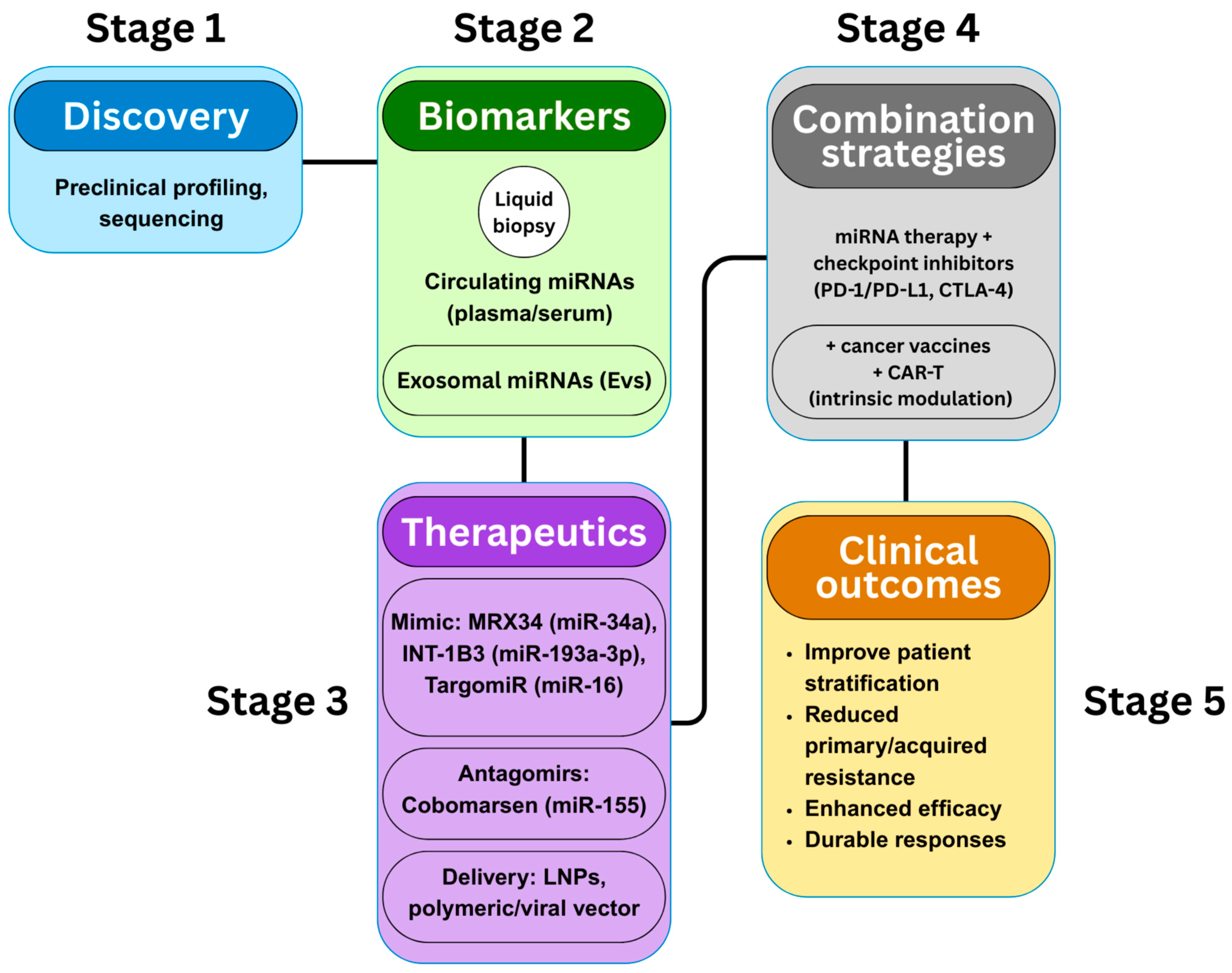
Cancer immunotherapy has established immune checkpoint inhibitors (ICIs), chimeric antigen receptor (CAR) T-cell therapy, and therapeutic vaccines as key approaches in modern oncology [1,2]. Despite their success in treating melanoma, lung, and hematological malignancies, many patients face primary or acquired resistance, highlighting the urgent need for predictive biomarkers and new combination strategies [3]. MicroRNAs (miRNAs), approximately 22-nucleotide non-coding RNAs that control gene expression post-transcription, have emerged as critical modulators of anti-tumor immunity [4]. Dysregulated miRNA expression contributes to immune escape by changing how tumors interact with immune cells and by shaping the tumor microenvironment (TME), ultimately influencing the efficacy of ICIs [5]. Certain miRNAs directly influence checkpoint molecules like PD-1, PD-L1, and CTLA-4; for instance, miR-200 downregulates PD-L1 and predicts ICI response in non-small-cell lung cancer, while oncogenic miRNAs such as miR-21 promote PD-L1 increase and inhibit PTEN, aiding immune evasion [6,7]. Beyond immune checkpoints, miRNAs also control tumor-associated macrophage polarization, T-cell differentiation, and the recruitment of myeloid-derived suppressor cells, thus directing the immunosuppressive environment [4,8]. MiRNAs carried by extracellular vesicles (EVs) facilitate cell-to-cell communication, reprogramming immune and stromal cells to support tumor growth and resistance to immunotherapy [9,10]. Clinically, circulating miRNA profiles are emerging as minimally invasive biomarkers to predict responses to ICIs and the risk of immune-related adverse events [1,3]. Meanwhile, therapeutic strategies using miRNA mimics, antagomirs, and engineered vesicle delivery systems are being developed to enhance effects when combined with ICIs, vaccines, and CAR-T therapies, offering promising translational prospects [7,9,10,11]. This review summarizes current knowledge about miRNA regulation in cancer immunotherapy, examines therapeutic combinations with checkpoint inhibitors and vaccines, emphasizes future directions in biomarker development and delivery platforms, and outlines a five-stage translational pipeline from discovery to clinical application (Figure 1). Unless otherwise specified, the mechanistic findings summarized in this review are derived from a combination of cell-based, animal, and early translational studies, with the model type highlighted when essential for interpretation.

2. Molecular Mechanisms of miRNA-Mediated Immune Regulation

In this section we outline the canonical RISC targeting mechanism, how miRNAs directly and via extracellular vesicles regulate immune checkpoints and co-stimulatory signals, their integration with oncogenic signaling, and cell-type specific roles in T cells, dendritic cells, NK cells and macrophages—ending with a concise synthesis that links mechanisms to therapeutic strategies.

2.1. miRNA-RISC Targeting Mechanism

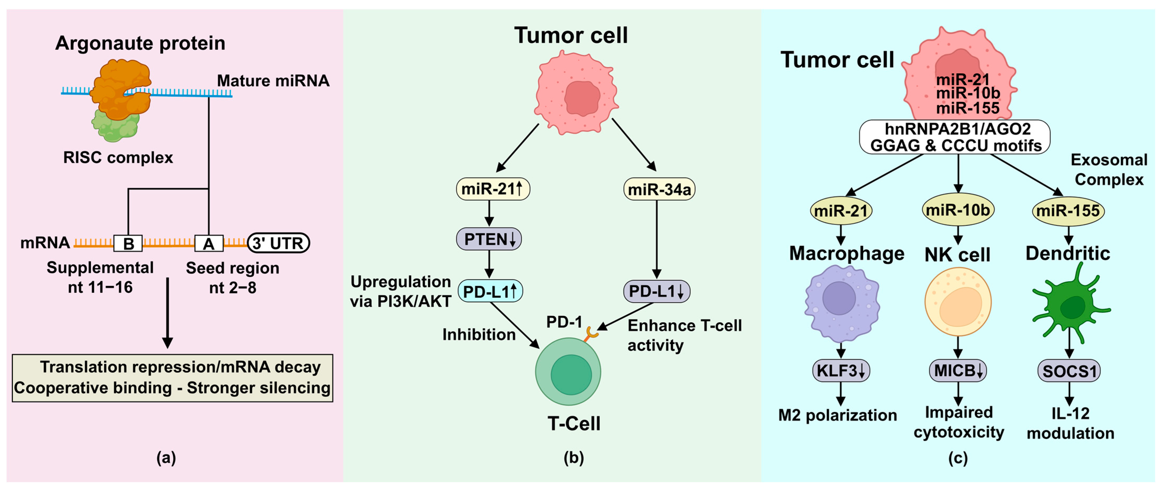
miRNA-mediated immune regulation occurs through sophisticated post-transcriptional control mechanisms centered on the RNA-induced silencing complex (RISC) [12]. Mature miRNAs guide Argonaute proteins to complementary sequences mainly within the 3′ untranslated regions (3′UTRs) of target mRNAs, although functional binding sites can also be found within coding sequences and 5′UTRs. Depending on complementarity and RISC composition, binding causes translational repression or mRNA decay [13]. The canonical targeting mechanism involves the miRNA “seed region” (nucleotides 2–8 from the 5′ end), which shows perfect or near-perfect complementarity to target sites [14]. Recent structural studies demonstrate that supplemental base-pairing beyond the seed region, especially at positions 11–16, improves target specificity and is vital for evolutionarily conserved miRNAs such as let-7 [12,15]. Silencing efficiency increases when multiple RISCs cooperatively bind closely spaced sites (≈15–35 nt), producing synergistic repression. Additionally, “seedless” sites cooperate with canonical binding sites to produce strong target suppression [14]. These basic targeting rules (seed pairing, supplemental pairing and cooperative RISC binding) provide the mechanistic basis for both direct miRNA control of immune checkpoint and co-stimulatory transcripts and for selective miRNA sorting into extracellular vesicles that mediate intercellular reprogramming.

2.2. Direct Targeting of Immune Checkpoints and Co-Stimulatory Molecules

miRNAs regulate anti-tumor immunity through both direct post-transcriptional control of immune checkpoints and extracellular vesicle (EV)-mediated communication that reshapes the tumor microenvironment (TME). Several miRNAs directly target immune checkpoint molecules, such as miR-138-5p, which binds the PD-L1 3′UTR to reduce PD-L1 expression and restore CD8+ T-cell cytotoxicity in non-small cell lung cancer [16,17,18], and miR-34a, a tumor-suppressive miRNA that represses PD-L1, SYT1 and PDGFRA while influencing macrophage polarization through the p53–miR-34a–CSF1R axis [19,20]; therapeutic delivery of miR-34a using antibody–oligonucleotide conjugates effectively lowers PD-L1, enhances macrophage phagocytosis and increases CD8+ T-cell infiltration with minimal systemic toxicity [21,22]. miRNAs also modulate co-stimulatory pathways, exemplified by miR-424, which suppresses CD80 in dendritic cells to disrupt CD80/CTLA-4 interactions, reverse chemoresistance and enhance CD8+ T-cell expansion while reducing Treg and myeloid-derived suppressor-cell recruitment [23,24]. In parallel, tumor-derived EVs transport miRNAs that modulate immune and stromal populations, with selective packaging orchestrated by proteins such as hnRNPA2B1 and AGO2 that recognize specific sequence motifs [25,26,27]. Tumor cells commonly export tumor-suppressive miRNAs to discard them or deliver oncomiRs to promote immunosuppressive states [28]. Exosomal miR-21 drives macrophage M2 polarization by targeting KLF3 and activating Nanog/Oct4 signaling in pancreatic neuroendocrine tumors [29,30,31,32], while breast cancer EVs transfer miR-10b to NK cells, downregulating MICB and impairing NK-mediated cytotoxicity; antagomir-mediated inhibition of miR-10b restores NK activation and eliminates metastases in vivo [33,34,35]. Additional EV-associated miRNAs contribute to TME remodeling, including miR-146a, which limits NF-κB–driven inflammation and fibroblast activation by targeting TRAF6, RIPK2 and PTGES2 [36,37,38], and miR-155, which regulates dendritic-cell IL-12 production and antigen presentation by targeting SOCS-1 [39,40,41]. Collectively, these direct and EV-mediated miRNA networks modulate checkpoint expression, reprogram immune cells and reinforce an immunosuppressive TME, driving tumor progression and shaping responses to immunotherapy. Having described how miRNAs act both cell-intrinsically and via EV transfer to reprogram immune and stromal cells, we now consider how oncogenic signaling pathways feed into these miRNA networks and modulate immune escape.

2.3. Upstream Signaling Integration

miR-21, one of the most extensively studied oncomiRs, modulates the PTEN/AKT pathway in lung cancer by directly targeting PTEN’s 3′UTR, which leads to activation of the PI3K/AKT pathway and downstream effects on cell proliferation, resistance to apoptosis, metastasis, and resistance to immunotherapy [42,43]. miR-21 promotes lung cancer progression through multiple interconnected signaling networks, including the PI3K/AKT, MEK/ERK, TGF-β/SMAD, Hippo, NF-κB, and STAT3 pathways, while also decreasing sensitivity to chemotherapeutic agents like carboplatin, paclitaxel, cisplatin, and gemcitabine by suppressing PTEN and enhancing DNA repair mechanisms [44,45]. The therapeutic significance of miR-21 goes beyond its direct oncogenic effects; it also contributes to immune evasion, as PTEN loss results in increased PD-L1 expression through PI3K/AKT-mediated transcriptional upregulation [46,47]. Conversely, inhibiting miR-21 restores PTEN function, makes cells more sensitive to radiotherapy, and improves combined treatments involving chemotherapy and immunotherapy [48,49]. These mechanistic insights highlight miRNAs as key regulators that connect multiple oncogenic and immune regulatory pathways, demonstrating their potential as both therapeutic targets and biomarkers for predicting treatment responses across various cancer types and therapeutic approaches. It is important to note that miR-21 may exhibit context-dependent roles, including occasional tumor-suppressive effects under specific cellular or inflammatory conditions [50].

2.4. Modulation of the Tumor Microenvironment

The tumor microenvironment is a dynamic ecosystem where miRNAs regulate complex intercellular communication networks through extracellular vesicle (EV)-mediated transfer, fundamentally reshaping the immunological landscape and creating either pro-tumorigenic or anti-tumorigenic conditions depending on the specific miRNA cargo and recipient cell populations [25]. miRNA transfer via extracellular vesicles, particularly exosomes ranging from 30–100 nanometers, has become a sophisticated mechanism of horizontal gene transfer that allows cancer cells to reprogram distant and adjacent stromal components, immune effector cells, and other malignant cells within the tumor microenvironment through the selective packaging and delivery of regulatory miRNAs [26,27]. The molecular machinery governing miRNA sorting into EVs involves complex interactions between miRNA sequences, RNA-binding proteins such as hnRNPA2B1, sumoylated hnRNPA2B1, and Argonaute 2 (AGO2), and the endosomal sorting complex required for transport (ESCRT). Specific sequence motifs (GGAG, CCCU) and secondary structures determine the preferential loading of particular miRNAs into exosomal compartments (Figure 2) [51,52,53]. This selective packaging process allows tumor cells to strategically modulate their microenvironment, either by sequestering tumor-suppressive miRNAs into exosomes for export or loading oncogenic miRNAs for delivery to target cells, thereby maintaining cellular homeostasis while influencing surrounding tissue architecture [54].
Immunosuppressive miRNA transfer is exemplified by miR-21 in pancreatic neuroendocrine tumor (PNET) exosomes, which polarizes tumor-associated macrophages toward the immunosuppressive M2 phenotype by directly targeting Krüppel-like factor 3 (KLF3) and activating stemness-promoting transcription factors like Nanog and Oct4 [29,30,31]. Mechanistically, M2 macrophage-derived exosomes rich in miR-21-5p are easily taken up by pancreatic cancer stem cells (CD24+CD44+EpCAM+), where miR-21 binds to the 3′ UTR of KLF3 mRNA, causing its degradation and leading to increased expression of stem cell transcription factors that enhance sphere formation, colony growth, invasion, migration, and resistance to apoptosis [30,31]. In vivo studies demonstrate that pancreatic cancer stem cells co-cultured with miR-21-overexpressing M2 macrophages display significantly higher tumorigenicity, with increased tumor size and weight, and elevated stemness marker levels, while knocking down miR-21 in M2 macrophage-derived exosomes completely prevents these tumor-promoting effects and reduces Nanog/Oct4 levels in recipient cells [30,32]. This reciprocal communication creates a feed-forward loop where PNET cells initially release factors that polarize macrophages toward M2, which then send out miR-21-enriched exosomes that further enhance cancer stem cell properties and treatment resistance, ultimately contributing to the aggressive behavior and poor prognosis of pancreatic neuroendocrine tumors [55].
NK cell suppression via EV-mediated miRNA delivery is another key mechanism of immune evasion. miR-10b in breast cancer EVs directly targets and downregulates stress-induced cell surface molecules, including MICB (MHC class I polypeptide-related sequence B), a critical ligand for NKG2D receptors on natural killer (NK) cells, which are crucial for recognizing and cytotoxic elimination [33,34]. miR-10b, significantly overexpressed in metastatic breast cancer compared to primary tumors, functions as a master regulator of metastasis by promoting epithelial–mesenchymal transition through HOXD10 suppression, increasing invasive capacity by downregulating E-cadherin, and granting stem cell-like properties that enable chemoresistance and immune evasion [35,56]. Extracellular vesicles from triple-negative breast cancer cell lines (MDA-MB-231), which contain elevated miR-10b, effectively transfer this oncomiR to NK cells. This process results in decreased MICB expression on cancer cells, reduced NK cell activation, impaired cytotoxic granule release, and ultimately compromised immune surveillance within the tumor microenvironment [57]. The therapeutic significance of this mechanism is highlighted by studies demonstrating that magnetic nanoparticle-conjugated anti-miR-10b (MN-anti-miR10b) treatment achieves 99% knockdown of miR-10b in primary tumors and metastatic lesions, restores NK cell function, prevents metastasis formation while eliminating existing metastases when combined with conventional chemotherapy in preclinical models [58].
Tumor-suppressive miRNA regulation of stromal components is exemplified by miR-146a, which inhibits tumor-associated fibroblasts (TAFs) in colorectal cancer by targeting multiple inflammatory signaling intermediates, including TRAF6 (TNF receptor-associated factor 6), RIPK2 (receptor-interacting protein kinase 2), and PTGES2 (prostaglandin E synthase 2). This effectively disrupts the IL-17-mediated inflammatory cascade that drives both colitis-associated and sporadic colorectal carcinogenesis [36,59]. miR-146a acts as a master negative regulator of colonic inflammation by simultaneously targeting RIPK2 in myeloid cells to limit the production of IL-17-inducing cytokines (IL-1β, IL-6, IL-23) and TRAF6 in intestinal epithelial cells to restrict responsiveness to IL-17 signaling. Additionally, targeting PTGES2 reduces prostaglandin E2 synthesis, which promotes tumor growth and angiogenesis [37]. The potential of restoring miR-146a has been validated through preclinical studies showing that systemic administration of miR-146a mimics effectively alleviates both DSS-induced colitis and AOM/DSS-driven colorectal cancer. Treated mice exhibit reduced tumor burden, decreased inflammatory cytokine production, lower Ki67 proliferation indices, and significantly improved survival compared to control animals [60]. Mechanistically, miR-146a deficiency leads to enhanced IL-17 signaling, increased infiltration of inflammatory cells, loss of intestinal barrier function, and progression from low-grade adenomas to invasive adenocarcinomas. Restoring miR-146a or pharmacologically inhibiting its targets (TRAF6, RIPK2) reverses these pathological changes and prevents tumor development [38].
Cytokine regulation through miRNA-mediated control of dendritic cell function is exemplified by miR-155, which modulates IL-12 production by directly targeting SOCS-1 (suppressor of cytokine signaling 1), a negative regulator of JAK/STAT signaling that functions as an antigen presentation attenuator by limiting IL-12p40 and IL-12p70 production in mature dendritic cells [39,40]. During human monocyte differentiation into immature dendritic cells and subsequent maturation, miR-155 expression is progressively upregulated. It reaches peak levels in response to TLR/IL-1 inflammatory stimuli, where it forms part of a negative feedback loop that initially promotes IL-12 production by targeting SOCS-1 but subsequently limits excessive inflammatory cytokine release to prevent tissue damage [61]. The functional significance of miR-155 in dendritic cell biology is demonstrated through studies showing that silencing miR-155 in mature dendritic cells results in increased SOCS-1 protein expression, significantly reduced IL-12p70 secretion, a decreased capacity to activate NK cells for IFN-γ production, and an impaired ability to prime Th1 immune responses [62]. Conversely, forced overexpression of miR-155 in immature dendritic cells enhances IL-12p70 production in response to LPS, poly(I:C), and other maturation stimuli, increases the capacity of dendritic cells to activate autologous NK cells for IFN-γ secretion, and improves their functionality as antigen-presenting cells in cancer immunotherapy contexts [63]. These findings emphasize the potential for miR-155-based approaches to enhance dendritic cell vaccine efficacy by optimizing IL-12 production and improving the activation of both innate and adaptive immune responses against tumor antigens; however, miR-155 also exhibits context-dependent behavior and may promote tumorigenesis under chronic inflammatory conditions [41].
The integration of EV-mediated miRNA transfer with tumor microenvironment modulation represents a paradigm shift in understanding cancer progression and therapeutic resistance, revealing opportunities for intercepting these communication networks through targeted miRNA inhibition, exosome engineering, or combination strategies that simultaneously target multiple miRNA-regulated pathways within the complex tumor ecosystem.

2.5. Oncogenic miRNAs and Immune Evasion

OncomiRs represent a distinct class of miRNAs promoting checkpoint ligand expression and immune evasion [64]. miR-20b functions as a critical regulator of PD-L1 upregulation in glioblastoma through mechanisms that correlate with treatment resistance and immune suppression [65,66]. In the complex glioblastoma microenvironment, where immune infiltration is naturally limited by the blood–brain barrier and immunosuppressive factors, oncomiRs contribute to the establishment of immune privilege by enhancing checkpoint ligand expression and promoting the accumulation of regulatory T cells, creating formidable barriers to the efficacy of immunotherapy, which requires multimodal therapeutic approaches [67]. The identification of oncomiRs reveals therapeutic opportunities through miRNA inhibition strategies, including antisense oligonucleotides, miRNA sponges, and small molecule inhibitors that can reverse immune evasion phenotypes and sensitize tumors to checkpoint blockade [18].

2.6. Context-Specific miRNA Functions

Context-specificity in miRNA function is exemplified by the different roles of hypoxia-induced miR-210 versus normoxic miR-200c in melanoma [28,68]. miR-210 functions as the “hypoxamir,” promoting immune evasion and treatment resistance under low oxygen conditions, while miR-200c acts as a tumor suppressor that enhances drug sensitivity and decreases metastatic potential under normoxic conditions [69,70]. miR-210 is directly upregulated by HIF-1α during hypoxia and promotes cancer stem cell phenotypes, epithelial–mesenchymal transition, and immune suppression by regulating CD24, CD44, CD133, E-cadherin, vimentin, and Snail expression, while simultaneously modulating HIF-1α through negative feedback loops that maintain cellular adaptation to hypoxic stress [71,72]. Conversely, miR-200c shows a decreasing expression pattern from melanocytic nevi to primary melanomas and metastatic lesions, and restoring it significantly reduces cell proliferation, migration, and drug resistance by downregulating BMI-1, ABCG2, ABCG5, and MDR1, while increasing E-cadherin expression, effectively reversing aggressive melanoma phenotypes [73,74]. The opposing roles of these miRNAs in response to microenvironmental conditions highlight the importance of considering tumor oxygenation and metabolic context when developing miRNA-based therapies [75].

2.7. miRNA Regulation of Immune Cell Activation

The orchestration of immune cell activation represents one of the most sophisticated regulatory networks in human biology, where miRNAs function as master conductors that fine-tune the development, differentiation, and effector functions of both innate and adaptive immune cells through precisely timed expression programs and context-dependent targeting mechanisms [76,77]. These small regulatory RNAs control critical checkpoints in immune cell maturation, activation thresholds, cytokine production, and memory formation, ultimately determining the magnitude, duration, and specificity of immune responses in both physiological and pathological contexts, including cancer immunotherapy [78,79,80].

2.7.1. miRNAs in T-Cell Differentiation

The miR-17-92 cluster emerges as a central regulator of CD8+ T-cell effector functions, promoting proliferation and terminal effector differentiation through the upregulation of PI3K-AKT-mTOR signaling pathways [81,82]. This polycistronic miRNA cluster, encoding six individual miRNAs (miR-17, miR-18a, miR-19a, miR-20a, miR-19b, and miR-92a), exhibits dynamic temporal expression patterns during CD8+ T-cell responses, with peak expression occurring during the rapid proliferation phase of effector expansion and subsequent downregulation as cells transition toward memory phenotypes [83]. Mechanistically, miR-17-92 promotes effector differentiation by targeting multiple tumor suppressors, including PTEN, thereby relieving the inhibition of the PI3K-AKT-mTOR axis and driving metabolic reprogramming toward glycolysis and protein synthesis, which are required for rapid cell division and cytotoxic effector functions [84]. Conditional overexpression of miR-17~92 in CD8+ T cells results in enhanced BrdU incorporation, increased proliferation during primary expansion, and preferential differentiation toward short-lived terminal effector cells at the expense of memory precursor cells, while also promoting expression of granzyme B, perforin, and IFN-γ production [85]. Sustained miR-17~92 expression prevents proper memory formation and leads to gradual loss of memory cells over time, providing critical knowledge on the importance of temporal regulation in balancing effector function with long-term protective immunity [86]. From a therapeutic perspective, engineering tumor antigen-specific CD8+ T cells to transiently overexpress miR-17-92 enhances their type-1 effector functions, including IFN-γ and TNF-α production, improves cytotoxic activity against target cells, and increases their capacity to control tumor growth in adoptive transfer models, suggesting potential applications in CAR-T cell therapy and tumor-infiltrating lymphocyte expansion protocols [87].

2.7.2. DC Maturation and Antigen Presentation

miR-155 enhancement of dendritic cell function represents a paradigm for leveraging miRNA biology to improve vaccine efficacy, particularly in the context of mRNA vaccines where enhanced antigen presentation is critical for robust immune responses [88]. During dendritic cell maturation, miR-155 expression is progressively upregulated in response to pathogen-associated molecular patterns (PAMPs) and inflammatory cytokines, where it functions as a master regulator of DC activation by targeting multiple negative regulators, including SOCS-1 (suppressor of cytokine signaling 1), c-Fos, Arg-2, and Jarid2 [89,90,91]. The functional consequences of miR-155 upregulation in DCs include enhanced expression of co-stimulatory molecules (CD80, CD86, and CD40), increased MHC class II presentation, improved migration toward lymph nodes via CCR7 upregulation, and augmented IL-12p70 production, which promotes Th1 polarization and NK cell activation [92,93]. Transgenic overexpression of miR-155 in bone marrow-derived dendritic cells results in superior antigen processing and presentation capabilities, enhanced T cell priming capacity, and improved therapeutic efficacy when used as DC vaccines against established breast cancer tumors, with treated mice exhibiting reduced primary tumor growth, dramatically suppressed lung metastasis, and increased effector T cell infiltration [94,95,96]. In the context of mRNA vaccines, miR-155 enhancement could potentially address the challenge of suboptimal antigen presentation by professional antigen-presenting cells, as host miRNAs may interfere with mRNA vaccine translation and reduce antigen production, thereby weakening the resulting immune response [97]. Recent advances have demonstrated that miR-155-enriched tumor-derived exosomes can be used to prime dendritic cells ex vivo, creating “educated” DCs with enhanced immunostimulatory properties that produce superior antitumor responses compared to conventional DC vaccines [98]. The therapeutic application of miR-155 enhancement in DC vaccines represents a promising strategy for improving immunotherapy outcomes, particularly when combined with other immune adjuvants such as TLR ligands, immune-stimulating cytokines, or checkpoint inhibitors [99].

2.7.3. NK Cell Activity Modulation

The miR-122 targeting of killer immunoglobulin-like receptors (KIRs) in natural killer cells represents a sophisticated mechanism for fine-tuning NK cell activation thresholds and cytotoxic capacity, particularly in the context of liver cancer, where miR-122 functions normally as a hepatocyte-specific tumor suppressor but may also regulate infiltrating immune cells [100,101]. miR-122, which constitutes approximately 70% of the total miRNA population in healthy hepatocytes, is significantly downregulated in about 70% of hepatocellular carcinoma cases, contributing to tumor progression, immune evasion, and treatment resistance through loss of its tumor-suppressive functions [102,103]. In NK cells infiltrating the liver, miR-122 potentially modulates KIR expression, which normally provides inhibitory signals when engaged by MHC class I molecules on target cells, thereby preventing NK cell activation against healthy autologous cells while preserving their ability to eliminate malignant or infected cells that have downregulated MHC class I expression [104]. The therapeutic restoration of miR-122 through various delivery systems, including adeno-associated virus vectors (AAV8) and lipid nanoparticles, has demonstrated significant efficacy in reducing hepatocellular carcinoma tumor growth and improving treatment outcomes in preclinical models, potentially through a combination of direct tumor suppression and enhancement of NK cell-mediated immune surveillance [105,106]. Recent studies have also identified miR-122-5p as a critical regulator of decidual NK (dNK) cell function during pregnancy, where it controls trophoblast invasion and vascular remodeling through targeting of multiple genes involved in cell migration, angiogenesis, and immune tolerance, suggesting broader roles for miR-122 in NK cell biology beyond liver cancer [100,107]. The development of miR-122 replacement therapies using advanced delivery platforms could potentially enhance both direct tumor suppression and NK cell-mediated antitumor immunity, creating synergistic therapeutic effects that address multiple aspects of hepatocellular carcinoma pathogenesis [101].

2.7.4. Macrophage Polarization Control

miR-145 promotion of M1 phenotype in lung cancer represents a critical mechanism for shifting the tumor-associated macrophage population from immunosuppressive M2 polarization toward pro-inflammatory, tumoricidal M1 activation which supports antitumor immunity [44,108]. miR-145, along with miR-130a, functions as a key regulator of myeloid cell reprogramming by targeting transforming growth factor-β receptor II (TβRII) and insulin-like growth factor 1 receptor (IGF1R), both of which are critical mediators of immunosuppressive signaling within the tumor microenvironment [109,110]. Mechanistically, miR-145 disrupts TGF-β signaling cascades that usually promote M2 polarization, angiogenesis, and metastatic progression, while simultaneously inhibiting IGF1R-mediated survival and proliferation signals that support tumor-associated macrophage accumulation [111]. Ectopic expression of miR-145 in Gr-1+CD11b+ myeloid cells effectively reprograms them from a pro-tumorigenic to anti-tumorigenic phenotype, resulting in increased production of pro-inflammatory cytokines (IL-1β, TNF-α, IL-6), enhanced nitric oxide synthase activity, and improved antigen presentation capabilities [112]. In preclinical models of breast cancer metastasis, mice receiving miR-145-engineered hematopoietic stem/progenitor cells exhibited significantly reduced lung metastasis without affecting primary tumor size, demonstrating that miR-145-mediated myeloid cell reprogramming specifically targets the metastatic microenvironment [113]. The therapeutic potential of miR-145 extends beyond direct macrophage polarization to encompass effects on other myeloid-derived suppressor cells and regulatory cell populations, resulting in a comprehensive shift in the immune microenvironment that favors tumor elimination over immune evasion [44]. Recent studies have also demonstrated that miR-145 can be delivered through various nanoparticle platforms and combined with IGF1R inhibitors such as NT157 to achieve synergistic effects on tumor metastasis suppression [109].

2.7.5. Context-Specific Roles in Innate vs. Adaptive Immunity

The context-specific functions of miRNAs in innate versus adaptive immunity reflect the evolutionary sophistication of immune regulation, where the same miRNA can exert opposing effects depending on the cell type, activation state, pathogen context, and tissue environment [114]. In innate immunity, miRNAs primarily regulate rapid response mechanisms, including pathogen recognition, inflammatory cytokine production, and antimicrobial effector functions. MiRNAs such as miR-155, miR-146a, and miR-21 serve as critical regulators of TLR signaling, NF-κB activation, and type I interferon responses [115]. miR-155, for example, promotes M1 macrophage activation and inflammatory cytokine production in response to bacterial lipopolysaccharide by targeting SOCS-1 and other negative regulators, while simultaneously enhancing dendritic cell maturation and IL-12 production, which are required for Th1 priming [116]. In certain contexts, such as hepatitis B virus-associated hepatocellular carcinoma, miR-155 paradoxically promotes M2 macrophage polarization through the miR-155/SHIP1 axis, demonstrating how pathogen-specific factors and tissue environments can reverse the typical functions of miRNAs [117]. In adaptive immunity, miRNAs orchestrate more complex developmental programs including T cell differentiation, B cell maturation, antibody class switching, and memory formation, where precise temporal control is essential for balancing effector function with long-term protective immunity [118]. The miR-17-92 cluster exemplifies this complexity by promoting rapid CD8+ T cell expansion and effector differentiation during acute infections, while requiring subsequent downregulation to enable proper memory cell formation. This process accentuates how the same miRNA network must be dynamically regulated to serve different phases of the adaptive immune response [119]. miR-29 family members exhibit additional context-specific functions by regulating both innate and adaptive immunity through the targeting of distinct gene networks in various cell types, including the promotion of IFN-γ production in T cells, regulation of antibody production in B cells, and modulation of inflammatory cytokine release in macrophages [120]. The clinical implications of these context-specific miRNA functions are profound for cancer immunotherapy, where therapeutic interventions must account for the complex interplay between innate and adaptive immunity, the heterogeneity of tumor microenvironments, and the potential for miRNA-based therapies to have unintended consequences in different cellular contexts [115]. Future therapeutic strategies will likely require sophisticated delivery systems that can target specific cell types and activation states, temporal control mechanisms that modulate miRNA activity during different phases of immune responses, and combination approaches that address the multifaceted roles of miRNAs in both innate and adaptive immunity [119,120].

3. Therapeutic Synergies of miRNAs in Cancer Immunotherapy

3.1. miRNA Mimics and Inhibitors as Immunotherapy Enhancers

For miRNAs such as miR-34a and miR-155, whose mechanistic roles were discussed earlier, this section focuses on their therapeutic synergy without repeating prior mechanistic details. Therapeutic strategies that combine synthetic miRNA mimics or inhibitors with immune checkpoint blockade have demonstrated remarkable synergy in preclinical cancer models by concurrently targeting tumor-intrinsic oncogenic pathways and reshaping the immune microenvironment to overcome resistance [18,121]. For example, the systemic delivery of miR-34a mimics encapsulated in lipid nanoparticles downregulates PD-L1 and oncogenic targets such as BCL2 and MET in triple-negative breast cancer, leading to enhanced CD8+ T-cell infiltration and a 70% greater tumor reduction when combined with anti-PD-1 therapy compared to monotherapy in murine models [122]. Similarly, exosome-mimetic nanovesicles carrying miR-200c sensitize lung adenocarcinoma to anti-CTLA-4 by targeting ZEB1, thereby restoring E-cadherin expression and promoting dendritic cell maturation and antigen cross-presentation, which yields complete tumor regression in 40% of treated mice and long-term immune memory upon rechallenge [123]. In microsatellite-stable colorectal cancer, where high miR-21 expression mediates resistance to PD-1 blockade via PTEN suppression and PI3K-AKT–driven PD-L1 upregulation, anti-miR-21 oligonucleotides restore PTEN, reduce PD-L1 levels, and enhance CD8+ T-cell cytotoxicity, achieving a 60% decrease in tumor burden when combined with anti-PD-1 therapy [124]. Mechanistically, these miRNA-based interventions act through “double-hit” mechanisms: tumor-suppressive mimics directly inhibit checkpoint ligands and oncogenes, while anti-oncomiRs release tumor suppressors and decrease immunosuppressive cytokine production, together converting “cold” tumor microenvironments into immune-permissive states [1]. The translational success of these approaches will depend on advances in delivery platforms—such as targeted lipid nanoparticles, exosome-based carriers, and antibody–miRNA conjugates—that ensure tumor-specific uptake, controlled release kinetics, and minimal off-target activity, thereby maximizing therapeutic efficacy and safety in future clinical applications [125].

3.2. Synergy with Checkpoint Inhibitors and mRNA Vaccines

3.2.1. Enhancement of Immune Checkpoint Inhibitor Efficacy

The strategic integration of miRNAs with immune checkpoint inhibitors has demonstrated remarkable therapeutic synergy through complementary mechanisms that enhance antigen presentation, restore immune surveillance, and overcome resistance pathways limiting single-modality approaches [18]. miR-138-5p exemplifies miRNA-mediated enhancement of checkpoint inhibitor efficacy by directly targeting the 3′ untranslated region of PD-L1 (CD274) mRNA, resulting in a 67% reduction in luciferase reporter activity and significant downregulation of PD-L1 expression in A549 and Calu-6 adenocarcinoma cell lines [16]. This tumor-suppressive miRNA simultaneously prevents T-cell exhaustion by reducing surface PD-1 expression on Jurkat cells co-cultured under tumor microenvironment-mimicking conditions with inflammatory cytokines [122]. When combined with anti-PD-1 therapy in preclinical lung cancer models, miR-138-5p and miR-200c demonstrated remarkable efficacy in preventing both benzo(a)pyrene-induced lung adenomas and N-nitroso-tris-chloroethylurea-induced squamous cell carcinomas without detectable systemic toxicity [126]. Single-cell RNA sequencing and imaging mass cytometry revealed that both miRNAs inhibited PD-L1 expression across tumor cell populations while increasing infiltration of CD4+ and CD8+ T cells and reducing the number of regulatory T cells [127]. The mechanistic basis involves the dual action of miR-138-5p, which directly suppresses checkpoint ligands on tumor cells while simultaneously modulating immune cell activation states, creating a “double-hit” effect that amplifies checkpoint blockade efficacy beyond what either intervention achieves alone [6].

3.2.2. mRNA Vaccine Synergy Enhancement

miRNA-enhanced mRNA vaccine efficacy has been demonstrated through miR-155-mediated dendritic cell optimization, where the overexpression of this master regulatory miRNA significantly improves vaccine-induced antitumor immunity in melanoma and breast cancer models [92,94]. miR-155 functions as a critical regulator of dendritic cell maturation by targeting negative regulators, including SOCS-1, c-Fos, Arg-2, and Jarid2 [41]. This targeting leads to enhanced expression of co-stimulatory molecules (CD80, CD86, CD40), increased MHC class II presentation, improved migration toward lymph nodes via CCR7 upregulation, and augmented IL-12p70 production promoting Th1 polarization [98]. In transgenic mice overexpressing miR-155, dendritic cell vaccines pulsed with tumor antigens resulted in enhanced T-cell priming capacity, increased effector T-cell tumor infiltration, suppressed primary tumor growth, and significantly reduced lung metastasis compared to wild-type DC vaccines [94]. The therapeutic potential of miR-155 enhancement is particularly relevant given recent advances demonstrating that host miRNAs can interfere with mRNA vaccine translation and reduce antigen production, thereby weakening immune responses [96]. By boosting miR-155 expression in dendritic cells, the efficacy of mRNA vaccines could be significantly improved, addressing the suboptimal antigen presentation that currently limits vaccine-driven antitumor immunity [95].

3.2.3. Dual Checkpoint Inhibition Combinations

Combination strategies using dual immune checkpoint inhibitors with miRNA mimics have shown exceptional promise in overcoming resistance to single-agent therapies, especially in immune-cold tumors with low mutational burden [128]. The bispecific antibody cadonilimab (AK104), which targets PD-1 and CTLA-4 simultaneously while lacking an Fc region to prevent inflammatory cytokine secretion, has demonstrated favorable safety profiles and encouraging antitumor activity in cervical cancer, NSCLC, and hepatocellular carcinoma [129]. Preclinical studies combining miR-200c mimics with dual anti-PD-1/CTLA-4 blockade achieved tumor regression rates exceeding 60% in lung adenocarcinoma models, compared to 25% with dual checkpoint inhibition alone, while also inducing robust immunological memory that protects against tumor rechallenge [130]. The mechanistic rationale involves miRNA-driven restoration of immune checkpoint sensitivity through direct targeting of resistance pathways, improved antigen presentation via dendritic cell activation, and reversal of immunosuppressive tumor microenvironments, creating synergistic effects that overcome the limitations of individual therapies [131].

3.3. Advanced Delivery Systems for miRNA Therapeutics

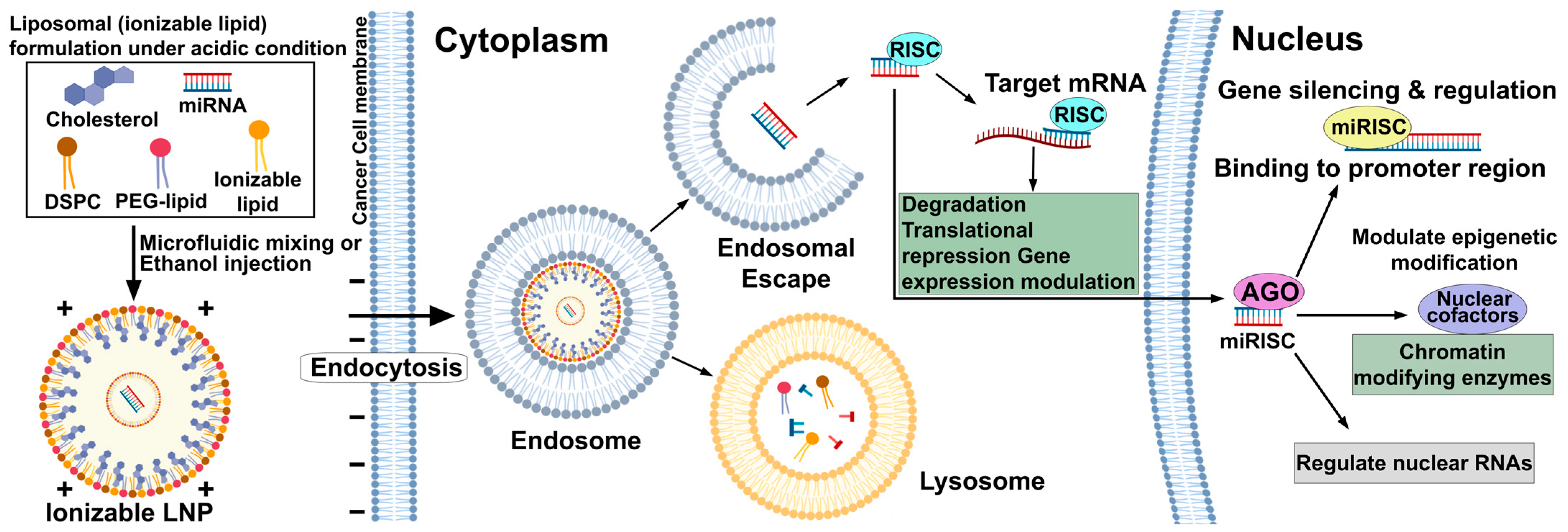
The clinical application of miRNA-based cancer immunotherapy has been fundamentally limited by delivery challenges, leading to extensive research into sophisticated nanocarrier platforms that can overcome miRNAs’ inherent instability, improve tumor targeting accuracy, and reduce off-target effects while preserving therapeutic potency [132]. Nanoparticle-based delivery systems have emerged as the most extensively studied platforms for miRNA therapeutics, with lipid nanoparticles (LNPs) showing great promise for miR-34 a mimics because they can protect miRNAs from ribonuclease degradation and enable efficient cellular uptake and controlled intracellular release [133]. Advanced ionizable lipid-based LNPs that use components such as DODMA (1,2-dioleyloxy-3-dimethylaminopropane) and DLin-MC 3-DMA feature pH-responsive charge switching, allowing for effective miRNA encapsulation at low pH during formulation, endosomal escape upon cellular entry, and minimal toxicity under physiological conditions. This addresses key limitations of earlier cationic lipid formulations that experienced protein aggregation and hemolytic activity [134]. Recent innovations in delivering miR-34 a include chitosan-PLGA nanoparticles with hydrodynamic diameters around 139 nm, which show improved cellular distribution, stability, and encapsulation efficiency of 80–100%. These systems also lead to significant upregulation of p53 and downregulation of SIRT 1 in non-small cell lung cancer models, providing valuable insight into the therapeutic potential of hybrid polymeric systems that blend biodegradability with controlled release kinetics [135]. The effectiveness of nanoparticle-mediated miR-34 a delivery has been confirmed across various cancer types, including the development of folic acid/protamine/miR-34 a/protamine @ nanodiamond nanohybrids (FA/PS/miR-34 a/PS @ NDs) with a 210 nm diameter and −25 mV zeta potential. These targeted the folate receptor on triple-negative breast cancer cells via endocytosis and showed significant anti-tumor effects by inducing apoptosis, inhibiting proliferation, and preventing migration through targeting Activator protein 1 (AP-1) transcription factors [136,137].
Exosome-based delivery systems represent a paradigm shift toward biomimetic nanocarriers that leverage the natural intercellular communication mechanisms of extracellular vesicles to achieve superior biocompatibility, reduced immunogenicity, and enhanced tumor targeting compared to synthetic nanoparticles (Figure 3) [138,139]. Mesenchymal stem cell-derived extracellular vesicles (MSC-EVs) loaded with therapeutic miRNAs, including tumor-derived exosomes carrying miR-146 a, have demonstrated remarkable therapeutic potential through their intrinsic tumor-targeting abilities and capacity for controlled miRNA release within the tumor microenvironment [140,141]. The clinical relevance of miR-146 a delivery via exosomes has been established through studies demonstrating that MSC-derived exosomes engineered to overexpress miR-146 a achieve 67% reduction in tumor weight in ovarian cancer models [142], while mechanistic analyses reveal dual anti-angiogenic effects through direct inhibition of endothelial tube formation (54% reduction) and indirect effects via reduced SERPINE 1 secretion from tumor cells, creating comprehensive anti-tumor activity that targets both cancer cells and supporting vasculature [143]. The therapeutic advantages of exosome-mediated delivery are exemplified by enhanced plant-derived vesicles engineered for improved xenograft penetration and oncolytic effect (HEXPO), which efficiently deliver miR-146 a-5p to the tumor microenvironment and achieve robust tumor growth inhibition through targeting of angiogenesis-related pathways including negative regulation of blood vessel formation, while maintaining excellent biocompatibility and avoiding the immune activation associated with synthetic delivery systems [144]. Importantly, the clinical application of MSC-derived exosomes carrying therapeutic miRNAs has been further validated through studies showing that miR-34 c-overexpressing MSC exosomes with approximately 100 nm particle size effectively attenuate nasopharyngeal carcinoma progression and enhance radiation-induced apoptosis by targeting β-catenin [145,146], demonstrating the potential for combining exosome-mediated miRNA delivery with conventional radiotherapy to overcome treatment resistance [147].
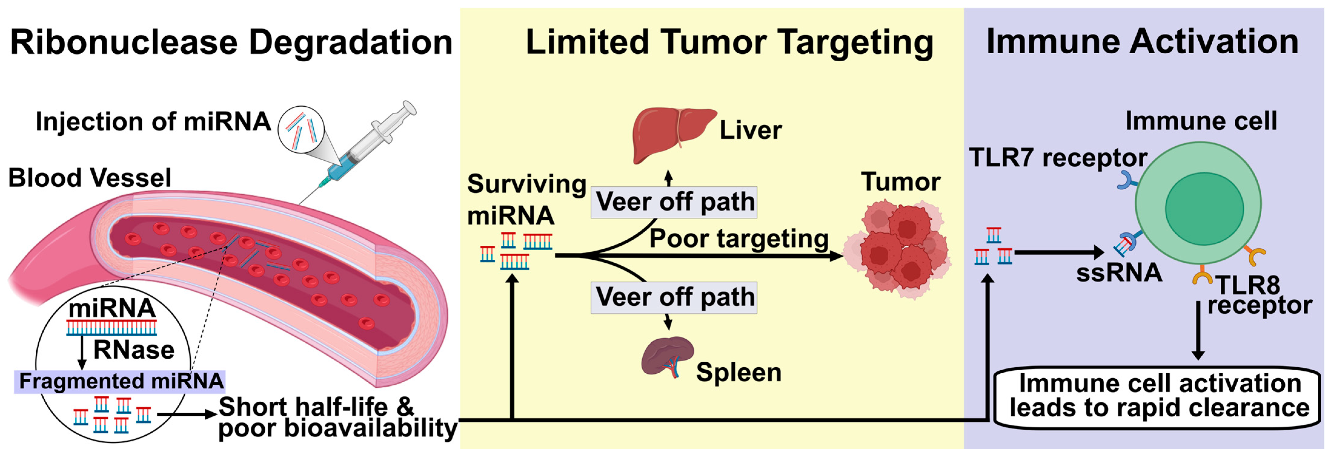
Delivery challenges include three key areas that must be addressed for successful clinical translation: stability issues caused by rapid miRNA degradation by ubiquitous ribonucleases in circulation, resulting in short half-lives and poor bioavailability (Figure 4) [4,148]; limited tumor targeting specificity due to insufficient accumulation at tumor sites and poor cellular uptake by cancer cells [149,150]; and immune activation triggered by recognition of delivery vehicles or miRNA cargo by pattern recognition receptors, which can reduce efficacy and cause adverse effects [99,151]. The stability issue has been partially mitigated through chemical modifications such as 2′-O-methyl, 2′-fluoro, and phosphorothioate modifications that improve nuclease resistance while maintaining miRNA function, though these changes can affect target specificity and require careful optimization to ensure therapeutic effectiveness [152]. Tumor targeting limitations arise from the heterogeneous nature of tumor vasculature, the variable enhanced permeability and retention (EPR) effect across different cancer types, and the necessity for active targeting strategies that can bypass biological barriers, such as the blood–brain barrier, for central nervous system tumors [153,154]. Immune activation presents a dual challenge: delivery systems must avoid triggering innate immune responses that lead to rapid clearance, while also potentially leveraging controlled immune activation to boost anti-tumor immunity. Achieving this balance requires advanced engineering of delivery vehicles to optimize both immune evasion and therapeutic benefit [155].
Advanced delivery innovations have focused on developing stimuli-responsive systems that can overcome traditional delivery limitations through precisely controlled cargo release mechanisms [148]. pH-sensitive nanoparticles utilize the acidic tumor microenvironment (pH 6.0–6.5) to trigger miRNA release specifically at tumor sites, using weakly acidic polymers or lipids that undergo protonation-induced conformational changes to release their therapeutic payload while minimizing systemic exposure to healthy tissues [156]. These systems have been exemplified by PEG-shedding nanoparticles encapsulating both chemotherapeutics and miR-200 for colorectal cancer treatment, demonstrating improved therapeutic effectiveness through site-specific release and reduced off-target toxicity [152]. Aptamer-guided delivery is the most advanced targeting approach, employing single-stranded oligonucleotides that bind with high specificity to overexpressed cell surface receptors on cancer cells, enabling precise delivery of miRNA cargo with minimal off-target effects [157]. The clinical potential of aptamer-mediated miRNA delivery has been shown in studies using GL 21. T and Gint 4. T aptamers that specifically recognize PDGFRα and PDGFRβ, respectively, to deliver anti-miR-222, miR-137, and anti-miR-10 b to glioblastoma cells, achieving receptor-dependent selective modulation of endogenous miRNA levels, increased sensitivity to temozolomide, and inhibition of tumor growth and migration both in vitro and in vivo [4]. The versatility of aptamer-guided delivery is further demonstrated by two-component stick-based methods that allow conjugation of multiple anti-miR sequences (GL 21. T-10 b-222) to single aptamer carriers, providing combination therapeutic effects through targeting multiple oncogenic miRNAs simultaneously while maintaining the structural integrity and binding specificity of both the aptamer and miRNA components [149]. Despite their high specificity and therapeutic potential, aptamer-based delivery systems face clinical translation challenges, including nuclease degradation, high production costs, and the need for better tissue penetration, though ongoing improvements in chemical modifications, in vivo SELEX techniques, and integration with nanoparticle platforms are addressing these issues and moving toward clinical use [158]. The integration of these advanced delivery methods promises to overcome the main barriers limiting miRNA therapeutics, enabling precise, efficient, and safe delivery of therapeutic miRNAs to improve cancer immunotherapy outcomes across various tumor types and clinical scenarios [148,152].

3.4. Integration with CAR T-Cell Therapies

The integration of microRNA engineering with CAR T-cell therapy represents a pioneering approach to overcome ongoing challenges such as T-cell exhaustion, immunosuppressive tumor microenvironments, and limited persistence, which have restricted the effectiveness of engineered T cells against solid tumors [159,160]. miRNAs that enhance CAR T-cell persistence have been most thoroughly studied with the miR-17~92 cluster in glioblastoma, where this polycistronic miRNA cluster promotes Th 1 phenotype differentiation, boosts cytotoxic effector functions, and significantly prolongs long-term tumor control capabilities [161,162]. In glioblastoma patients, CD4+ T cells are typically polarized toward an unfavorable Th 2 phenotype with decreased miR-17-92 expression—a process further worsened by lymphocytes secreting IL-4, which suppresses miR-17-92 expression and lowers the Th 1/Th 2 ratio, an unfavorable prognosis factor for patient survival [163]. Ohno et al. addressed this issue by engineering CAR T cells with additional miR-17-92 transgene expression using a lentiviral vector (FG 12-EF 1 a-miR-17-92) containing dual promoters: EF 1 a controlling miR-17-92 expression and UbiC controlling EGFP expression for assessing transduction efficiency [160]. In preclinical studies, CAR T cells co-transduced with miR-17-92 (3 C + miR) showed superior long-term stability and greater resistance to temozolomide-induced T-cell suppression compared to standard CAR T cells [161]. Notably, when immunocompromised mice were re-challenged with glioblastoma cells 49 days after initial CAR T-cell infusion, all four mice treated with conventional CAR T cells developed tumors, whereas none of the three mice receiving miR-17-92-enhanced CAR T cells exhibited tumor growth-highlighting the significant impact of miRNA engineering on immunological memory and lasting tumor control [160]. The underlying mechanism of this improved efficacy involves increased CAR T-cell survival, heightened interferon-γ secretion, and the maintenance of anti-tumor activity through the promotion of memory T-cell formation via miR-17-92-mediated targeting of pro-apoptotic and exhaustion-related pathways [162,164].
Overcoming tumor microenvironment suppression through miR-155 targeting TGF-β pathways is a sophisticated strategy to make CAR T cells resistant to immunosuppressive signals while boosting their effector functions within hostile tumor environments [165,166]. miR-155 prevents CD8+ T-cell senescence and exhaustion by epigenetically suppressing terminal differentiation factors, achieved by indirectly increasing Polycomb repressor complex 2 (PRC2) activity through promoting Phf19 expression and decreasing Ship1 levels, which inhibit Akt signaling [167]. This pathway is especially important in TGF-β-rich tumor microenvironments, where this cytokine promotes regulatory T-cell differentiation, inhibits effector T-cell functions, and acts as a physical barrier to T-cell infiltration by increasing collagen deposition and stromal remodeling [168]. Recent advances include engineering CAR T cells with chimeric switch receptors that convert inhibitory TGF-β signals into activating ones, along with secreting TGF-β traps that neutralize local TGF-β levels, creating a dual mechanism to resist and overcome tumor immunosuppression [169]. In preclinical solid tumor models, CAR T cells with TGF-β resistance showed significantly better tumor infiltration, sustained proliferation within the tumor microenvironment, and enhanced cytotoxic activity against target cells, while avoiding the T-cell dysfunction usually caused by chronic TGF-β exposure [166,170]. The clinical importance of boosting miR-155 levels in CAR T cells goes beyond TGF-β resistance, as miR-155−/− CD8+ T cells in tumor tissues display reduced proliferation and invasion capabilities, which can be restored with immune checkpoint antibody treatment, indicating that miR-155 regulates pathways essential for tumor immune responses [165,167].
Preclinical models utilizing miRNA-modified CAR T cells in sarcomas have shown strong evidence for this approach’s potential to treat aggressive mesenchymal cancers that are often resistant to traditional immunotherapies [171]. New fourth-generation-like CAR miR cells have been created to release therapeutic miRNAs through exosomes while also targeting tumor antigens, such as IL-13(E12Y) CAR constructs that include precursor miR-34a under a 6xNFAT-IL2 minimal promoter activated after CAR-antigen engagement [172]. These engineered CAR miR cells demonstrate significant upregulation and exportation of miR-34a-5p in exosomes, creating a localized therapeutic miRNA delivery system that improves cytotoxic effects against glioblastoma and sarcoma cell lines compared to standard CAR T cells [173]. Their dual role—in direct tumor cell killing via CAR-mediated cytotoxicity and in indirect tumor inhibition through miRNA delivery—marks a promising direction toward multifunctional therapeutic T cells that can address tumor heterogeneity and resistance mechanisms at once [174]. In sarcoma models, miRNA-modified CAR T cells show promise because specific miRNAs like miR-34a can target multiple oncogenic pathways, including restoring p53, causing cell cycle arrest, and triggering apoptosis, while the CAR component ensures targeting of sarcoma-associated antigens like GD2, HER2, or tumor-specific neoantigens [175]. The production of these advanced CAR miR cells involves lentiviral transduction of T cells with constructs encoding both the CAR and miRNA, followed by expansion under optimized conditions that promote a central memory T-cell phenotype, which offers better persistence and anti-tumor activity in solid tumor settings [176]. Clinical translation faces unique hurdles, such as the diverse nature of sarcoma subtypes, limited expression of universal target antigens, and the dense stromal microenvironment characteristic of these tumors, necessitating combination strategies that incorporate chemotherapy or radiation to improve tumor penetration and miRNA delivery [177]. Nonetheless, early preclinical data suggest that miRNA-enhanced CAR T cells could overcome many barriers that have hindered successful CAR T therapy in solid tumors—like poor persistence, limited tumor infiltration, and resistance from immunosuppressive microenvironments, making this a promising next-generation cellular immunotherapy for patients with advanced sarcomas and other solid cancers [178].

3.5. Clinical Trials of miRNA-Based Therapeutics in Cancer Immunotherapy

The clinical development of miRNA-based therapeutics has provided important insights into their potential for modulating immune responses in cancer [179,180]. The liposomal miR-34a mimic MRX34 was the first miRNA drug to reach clinical testing (NCT01829971), designed to restore tumor-suppressive activity and sensitize tumors to immune attack; however, the trial was terminated due to immune-mediated toxicities, offering valuable information on safety and delivery challenges [181,182]. Subsequent efforts explored TargomiR, a miR-16 mimic encapsulated in bacterial minicells, which demonstrated activity in malignant pleural mesothelioma (NCT02369198) [183], and Cobomarsen (MRG-106), an antisense inhibitor of the oncogenic miR-155, evaluated in cutaneous T-cell lymphoma and other hematological cancers (NCT02580552; NCT03713320), although its development was halted for non-safety-related business reasons [184,185]. More recently, INT-1B3, a lipid nanoparticle-formulated mimic of miR-193a-3p, entered early-phase testing for solid tumors and represents a new generation of RNA therapeutics with improved delivery platforms [186,187]. These trials collectively highlight both the promise and the challenges of miRNA-based drugs in oncology [188]. While early programs faced limitations due to immune-related toxicities and delivery inefficiencies, advances in synthetic chemistry, nanoparticle engineering, and tumor-targeted carriers are paving the way for safer and more effective clinical translation [189]. In particular, combining miRNA therapeutics with checkpoint inhibitors or other immunotherapies may unlock synergistic effects, positioning miRNA-based strategies as valuable tools in the evolving landscape of precision immuno-oncology [180,187].

4. miRNAs as Biomarkers in Cancer Immunotherapy

4.1. Predictive Biomarkers for Immunotherapy Response

Circulating and tissue miRNA signatures have shown strong potential for predicting patient responses to immune checkpoint inhibitors (ICIs) (Table 1), providing non-invasive biomarkers that reflect dynamic tumor–immune interactions [190,191]. In non-small cell lung cancer (NSCLC), higher pretreatment tumor and plasma levels of miR-155 and miR-146a are associated with better responses to anti-PD-1 therapy, with responders exhibiting a 2.5-fold higher median miR-155 expression and a 1.8-fold higher miR-146a level compared to non-responders. Longitudinal monitoring revealed that increasing miR-155 levels during treatment predicts durable clinical benefits and longer progression-free survival [192,193]. In melanoma patients, exosomal miR-21 levels measured before ICI treatment inversely relate to objective response rates. Patients in the lowest quartile of exosomal miR-21 have a 60% response rate, compared to 20% in the highest quartile, suggesting that tumor-derived exosomal miR-21 may act as a marker of an immunosuppressive microenvironment that impairs checkpoint blockade effectiveness [194,195]. These results were reinforced at ASCO 2025, where a prospective multi-center study confirmed that a four-miRNA plasma panel—including miR-155, miR-146a, miR-21, and miR-126—predicts response to anti-PD-1/PD-L1 therapy across 200 patients with melanoma, NSCLC, and renal cell carcinoma, with an area under the receiver operating characteristic curve of 0.87 for distinguishing responders from non-responders [196].

4.2. Biomarkers for Immune-Related Adverse Events

Distinct differential miRNA expression profiles have emerged as early indicators of immune-related adverse events (irAEs), enabling preemptive intervention and personalized immune checkpoint inhibitor (ICI) dosing [123]. In patients receiving anti-PD-1 therapy, increases in circulating miR-122 and miR-206 within two weeks of starting treatment predict severe irAEs—including grade ≥ 3 colitis and hepatitis—with 85% sensitivity and 78% specificity, often preceding clinical symptom onset by a median of 10 days and correlating with cytokine storm markers such as IL-6 and TNF-α [205]. Mechanistically, miR-122 influences hepatocyte innate immune responses by targeting TLR4’s adaptor MyD88, while miR-206 controls skeletal muscle–derived IL-6 production, together contributing to systemic inflammatory amplification that underpins severe irAEs [206]. Using miRNA-guided monitoring has allowed early initiation of immunosuppressive therapy in high-risk patients, reducing irAE-related hospitalizations by 40% in pilot cohorts and supporting personalized dosing strategies that balance tumor suppression with safety [195].

4.3. AI-Driven miRNA Biomarker Discovery

Artificial intelligence and multi-omics integration have transformed miRNA biomarker discovery, allowing for detailed identification of predictive signatures within complex tumor microenvironments [207,208,209]. Machine learning algorithms trained on both bulk and single-cell RNA sequencing datasets have identified TME-specific miRNA modules, such as a macrophage-enriched miR-21/miR-146 b axis and a dendritic cell–specific miR-155/miR-29 signature, which can classify responders to ICI therapy with over 90% accuracy in cross-validation studies [210,211]. Integrating proteomic and genomic data further enhances biomarker panels by connecting miRNA expression to downstream protein networks and mutation landscapes, supporting the development of integrated predictive models that outperform single-modal biomarkers [212,213]. Recent advances in artificial intelligence have led to the creation of specialized platforms that speed up miRNA biomarker discovery by combining multi-omics data and modeling complex molecular interactions [214]. For example, STmiR uses an XGBoost framework (Table 2) to combine bulk transcriptomic data from TCGA and CCLE with spatial transcriptomics profiles, accurately predicting miRNA activity within specific tissue regions and identifying conserved and cell-type–specific regulators across various cancers [207,215]. JointSyn employs a dual-view deep learning architecture that encodes small-molecule chemical descriptors and cell-line molecular signatures simultaneously, delivering strong predictions of personalized miRNA–drug synergies with R2 values around 0.78 and Pearson correlations close to 0.89 [211,216]. SMTRI uses convolutional neural networks to transform miRNA–mRNA duplex secondary structures into simplified numerical formats, enabling rapid in silico screening of small molecules that disrupt specific miRNA–mRNA interactions [208]. Lastly, sChemNET applies graph-based deep learning to learn complex, non-linear relationships between chemical structures and miRNA sequences, facilitating the discovery of new bioactive compounds capable of modulating miRNA function across various cancer types [209]. Collectively, these AI-powered platforms offer powerful tools for identifying predictive miRNA biomarkers and designing combination therapies with remarkable speed and accuracy.
Despite its transformative potential, AI-driven miRNA biomarker discovery faces several challenges. First, heterogeneous data quality and batch effects across different sequencing platforms can introduce technical variability that confounds model training and decreases reproducibility [214,217]. Second, machine learning models trained on limited sample sizes risk overfitting and may not generalize well to independent cohorts, highlighting the need for large, well-annotated datasets and rigorous cross-validation [210,213]. Third, single-cell RNA sequencing data suffer from dropout events and shallow miRNA coverage, which limits the reliable detection of low-abundance miRNAs and makes cell-type–specific signature identification more difficult [218,219]. Fourth, integrating multi-omics layers—including transcriptomics, proteomics, and epigenomics—requires advanced computational frameworks; misalignment between modalities can obscure true biological signals and increase false-positive rates [220,221]. Finally, the “black box” nature of many AI algorithms hampers mechanistic interpretability, making it hard to derive actionable insights or validate candidate biomarkers experimentally without extensive downstream functional studies [208,222].

5. Challenges and Future Directions

MiRNA-based immunotherapy faces the inherent challenge of context specificity, as the same miRNA can have different functions depending on tumor type, microenvironment, and cellular state [223,224]. For example, miR-155 promotes pro-inflammatory M1 macrophage polarization and Th1 T-cell responses in bacterial infection models but promotes M2-like immunosuppressive phenotypes in certain tumor contexts through the miR-155/SHIP1 axis [225]. miR-210 acts as a hypoxamir under low-oxygen conditions to enhance immune evasion, whereas miR-200c functions as a tumor suppressor under normal oxygen conditions to reverse metastatic phenotypes [4]. This functional flexibility complicates predicting therapeutic outcomes and requires careful profiling of miRNA roles in each cancer type [226]. Delivery barriers are also a critical hurdle: unmodified miRNAs are rapidly degraded in circulation, and synthetic carriers risk off-target effects and activating innate immunity [227,228]. Although lipid nanoparticles and exosome-based vehicles improve stability and uptake, active targeting strategies like pH-sensitive nanoparticles and aptamer-guided systems are necessary to ensure precise tumor targeting while avoiding toxic side effects [229,230]. Manufacturing scalability and regulatory standardization of complex nanocarriers further hinder clinical application [231]. Tumors also develop adaptive resistance mechanisms that can undermine miRNA therapies by upregulating compensatory oncomiRs, altering RNA-binding proteins, or remodeling extracellular vesicle landscapes to sequester therapeutic miRNAs [227]. Overcoming these adaptive resistance pathways will require combination strategies that integrate miRNA modulation with immune checkpoint inhibitors, mRNA vaccines, CAR T-cells, and targeted small molecules to achieve lasting antitumor responses across diverse tumor ecosystems [4].
Emerging technologies provide promising solutions. Single-cell and spatial transcriptomics enable high-resolution mapping of miRNA activity across different cell types and niches, uncovering cell-type-specific miRNA regulators that guide precision-targeted therapies [232,233]. CRISPR-based miRNA editing permits direct genomic manipulation of oncogenic or tumor-suppressive miRNAs in situ, with Cas9, Cas12a, and Cas13 platforms offering versatile options for multiplexed miRNA reprogramming [234,235]. AI-driven approaches accelerate the discovery of miRNA targets and synergistic drug combinations by modeling complex miRNA–mRNA–protein interactions and predicting optimal therapeutic pairings from multi-omics datasets [22,236].
Looking ahead, expanding miRNA research into rare cancers such as sarcomas and neuroendocrine tumors is crucial due to their unmet therapeutic needs. Early data suggest that miR-34a and neuroendocrine-specific miRNAs (e.g., miR-375, miR-7) control important oncogenic and hormonal pathways, providing new diagnostic and treatment opportunities. Combining miRNA mimics or inhibitors with immune checkpoint inhibitors, vaccines, and CAR T-cells should be a priority in preclinical and early-stage clinical trials to find the best combinations and dosing strategies. Additionally, moving from research to clinical practice requires standardized protocols for validating miRNA biomarkers, including amplification-free assays and AI-enhanced diagnostics, to support regulatory approval and adoption in personalized cancer care. By overcoming these challenges through collaboration across disciplines, developing advanced delivery systems, and designing innovative trials, miRNA-based immunotherapy can emerge as a cornerstone of precision cancer treatment.

6. Conclusions

MicroRNAs are at the intersection of tumor biology and immune regulation, making them influential modulators of cancer immunotherapy. Growing evidence highlights their ability to regulate immune checkpoints, reprogram the tumor microenvironment, and act as predictive biomarkers of treatment response. Although early clinical trials of miRNA therapeutics faced limitations due to immune-related toxicities and delivery challenges, ongoing innovations in nanoparticle carriers, exosome-based delivery, and chemically stabilized RNA analogs are addressing these issues. Additionally, combining miRNA profiling with multi-omics strategies and artificial intelligence is expected to improve patient stratification and speed up biomarker discovery. Looking forward, combined approaches that use miRNA-based therapies alongside immune checkpoint inhibitors, CAR-T cells, or cancer vaccines show great potential. Continued research and clinical development will reveal whether miRNA-directed treatments can become effective next-generation tools in precision immuno-oncology.

Author Contributions

N.L.M., as the first author, was responsible for data collection and drafting the manuscript. S.K.G. and S.G.M. contributed to data collection and manuscript drafting. M.K. and G.C.S. provided data analysis and guidance during manuscript preparation. S.V. supervised the study, contributed to data analysis, conceptualized the review, supervised the study and finalized the manuscript. All authors have read and agreed to the published version of the manuscript.

Funding

This research received no external funding.

Institutional Review Board Statement

Not applicable. This study did not involve humans or animals. All figures and tables presented in this review article (“MicroRNAs Modulating Cancer Immunotherapy Mechanisms and Therapeutic Synergies”) were originally created by the authors, and no copyrighted or previously published materials were reproduced. Figures were prepared using BioRender.com under an academic license.

Informed Consent Statement

Not applicable. This review article does not involve any studies with human participants or patient data.

Data Availability Statement

This narrative review synthesizes information from previously published studies, which are appropriately cited within the manuscript. No new data was generated or analyzed in this study. Therefore, data sharing is not applicable.

Conflicts of Interest

The authors declare no conflicts of interest.

Abbreviations

The following abbreviations are used in this manuscript:
3′UTRs3′ untranslated regions
AGO2Argonaute 2
AP-1Activator protein 1
ASCOAmerican Society of Clinical Oncology (inferred from context)
AUROCArea under the receiver operating characteristic curve
BCL2B-cell lymphoma 2
BMI-1B lymphoma Mo-MLV insertion region 1 homolog
CARChimeric antigen receptor
CCLECancer Cell Line Encyclopedia
CCR7C-C chemokine receptor type 7
CD274Cluster of differentiation 274 (also known as PD-L1)
CD40Cluster of differentiation 40
CD47Cluster of differentiation 47
CD80Cluster of differentiation 80
CD86Cluster of differentiation 86
CSF1RColony-stimulating factor 1 receptor
CTLA-4Cytotoxic T-lymphocyte-associated protein 4
DCDendritic cell
DLin-MC3-DMA2,2-dilinoleyl-4-(2-dimethylaminoethyl)-dioxolane
DODMA1,2-dioleyloxy-3-dimethylaminopropane
DSSDextran sulfate sodium (in colitis models)
EPREnhanced permeability and retention
ESCRTEndosomal sorting complex required for transport
EVExtracellular vesicle
FA/PS/miR-34a/PS@NDsFolic acid/protamine/miR-34a/protamine@nanodiamond nanohybrids
HEXPOEnhanced plant-derived vesicles for xenograft penetration and oncolytic effect
HIF-1αHypoxia-inducible factor 1-alpha
HOXD10Homeobox D10
ICIImmune checkpoint inhibitor
IFN-γInterferon gamma
IGF1RInsulin-like growth factor 1 receptor
IL-12Interleukin-12
IL-12p70Interleukin-12 p70 subunit
IL-17Interleukin-17
IL-1βInterleukin-1 beta
IL-6Interleukin-6
IL-23Interleukin-23
INT-1B3Investigational miR-193a-3p mimic (lipid nanoparticle-formulated)
irAEsImmune-related adverse events
JAK/STATJanus kinase/signal transducers and activators of transcription
Jarid2Jumonji and AT-rich interaction domain containing 2
KIRKiller immunoglobulin-like receptor
KLF3Krüppel-like factor 3
KLRG1Killer cell lectin-like receptor G1
LNPLipid nanoparticle
LPSLipopolysaccharide
M1Pro-inflammatory macrophage phenotype
M2Immunosuppressive macrophage phenotype
METMesenchymal–epithelial transition factor
MHCMajor histocompatibility complex
MICBMHC class I polypeptide-related sequence B
MN-anti-miR10bMagnetic nanoparticle-conjugated anti-miR-10b
MRG-106Cobomarsen (anti-miR-155)
MRX34Liposomal miR-34a mimic
MSC-EVMesenchymal stem cell-derived extracellular vesicle
MyD88Myeloid differentiation primary response 88
NF-κBNuclear factor kappa-light-chain-enhancer of activated B cells
NFATNuclear factor of activated T-cells
NKNatural killer
NKG2DNatural killer group 2D
NSCLCNon-small cell lung cancer
Oct4Octamer-binding transcription factor 4
PAMPPathogen-associated molecular pattern
PD-1Programmed cell death protein 1
PD-L1Programmed death-ligand 1
PDGFRAPlatelet-derived growth factor receptor alpha
PDGFRαPlatelet-derived growth factor receptor alpha
PDGFRβPlatelet-derived growth factor receptor beta
Phf19PHD finger protein 19
PI3K/AKTPhosphatidylinositol 3-kinase/protein kinase B
PNETPancreatic neuroendocrine tumor
PRC2Polycomb repressor complex 2
PTENPhosphatase and tensin homolog
PTGES2Prostaglandin E synthase 2
RISCRNA-induced silencing complex
RIPK2Receptor-interacting protein kinase 2
RNA-seqRNA sequencing
SELEXSystematic evolution of ligands by exponential enrichment
SERPINE1Serpin family E member 1
SHIP1SH2 domain-containing inositol phosphatase 1
SIRT1Sirtuin 1
SMADMothers against decapentaplegic homolog
SOCS-1Suppressor of cytokine signaling 1
STAT3Signal transducer and activator of transcription 3
SYT1Synaptotagmin I
TAFTumor-associated fibroblast
TCGAThe Cancer Genome Atlas
TGF-βTransforming growth factor beta
Th1T helper 1
Th2T helper 2
TLRToll-like receptor
TMETumor microenvironment
TNF-αTumor necrosis factor alpha
TRAF6TNF receptor-associated factor 6
TβRIITransforming growth factor beta receptor II
UbiCUbiquitin C
ZEB1Zinc finger E-box-binding homeobox 1

References

  1. García-Giménez, J.L.; Saadi, W.; Ortega, A.L.; Lahoz, A.; Suay, G.; Carretero, J.; Pereda, J.; Fatmi, A.; Pallardó, F.V.; Mena-Molla, S. miRNAs Related to Immune Checkpoint Inhibitor Response: A Systematic Review. Int. J. Mol. Sci. 2024, 25, 1737. [Google Scholar] [CrossRef]
  2. Al-Haideri, M.; Tondok, S.B.; Safa, S.H.; Maleki, A.H.; Rostami, S.; Jalil, A.T.; Al-Gazally, M.E.; Alsaikhan, F.; Rizaev, J.A.; Mohammad, T.A.M.; et al. CAR-T Cell Combination Therapy: The next Revolution in Cancer Treatment. Cancer Cell Int. 2022, 22, 365. [Google Scholar] [CrossRef] [PubMed]
  3. Vaxevanis, C.; Bachmann, M.; Seliger, B. Immune Modulatory microRNAs in Tumors, Their Clinical Relevance in Diagnosis and Therapy. J. Immunother. Cancer 2024, 12, e009774. [Google Scholar] [CrossRef] [PubMed]
  4. Martino, M.T.D.; Tagliaferri, P.; Tassone, P. MicroRNA in Cancer Therapy: Breakthroughs and Challenges in Early Clinical Applications. J. Exp. Clin. Cancer Res. 2025, 44, 126. [Google Scholar] [CrossRef] [PubMed]
  5. Yi, M.; Xu, L.; Jiao, Y.; Luo, S.; Li, A.; Wu, K. The Role of Cancer-Derived microRNAs in Cancer Immune Escape. J. Hematol. Oncol. 2020, 13, 25. [Google Scholar] [CrossRef] [PubMed]
  6. Kaneko, A.; Kobayashi, N.; Kubo, S.; Nagaoka, S.; Muraoka, S.; Fukuda, N.; Somekawa, K.; Matsumoto, H.; Katakura, S.; Teranishi, S.; et al. MiR-200a Regulates PD-L1 and Predicts Response to Immune Checkpoint Inhibitors in Advanced Non-Small Cell Lung Cancer. Transl. Lung Cancer Res. 2025, 14, 2522–2536. [Google Scholar] [CrossRef] [PubMed]
  7. Balaraman, A.K.; Arockia Babu, M.; Afzal, M.; Sanghvi, G.; M M, R.; Gupta, S.; Rana, M.; Ali, H.; Goyal, K.; Subramaniyan, V.; et al. Exosome-Based miRNA Delivery: Transforming Cancer Treatment with Mesenchymal Stem Cells. Regen. Ther. 2025, 28, 558–572. [Google Scholar] [CrossRef] [PubMed]
  8. Dong, L.; Tian, X.; Zhao, Y.; Tu, H.; Wong, A.; Yang, Y. The Roles of MiRNAs (MicroRNAs) in Melanoma Immunotherapy. Int. J. Mol. Sci. 2022, 23, 14775. [Google Scholar] [CrossRef]
  9. Kuang, L.; Wu, L.; Li, Y. Extracellular Vesicles in Tumor Immunity: Mechanisms and Novel Insights. Mol. Cancer 2025, 24, 45. [Google Scholar] [CrossRef]
  10. Kim, H.I.; Park, J.; Zhu, Y.; Wang, X.; Han, Y.; Zhang, D. Recent Advances in Extracellular Vesicles for Therapeutic Cargo Delivery. Exp. Mol. Med. 2024, 56, 836–849. [Google Scholar] [CrossRef]
  11. Lin, X.; Yue, L.; Cheng, K.; Rao, L. Engineering Cellular Vesicles for Immunotherapy. Acc. Mater. Res. 2025, 6, 327–339. [Google Scholar] [CrossRef]
  12. Komatsu, S.; Kitai, H.; Suzuki, H.I. Network Regulation of microRNA Biogenesis and Target Interaction. Cells 2023, 12, 306. [Google Scholar] [CrossRef] [PubMed]
  13. Bofill-De Ros, X.; Yang, A.; Gu, S. IsomiRs: Expanding the miRNA Repression Toolbox beyond the Seed. Biochim. Biophys. Acta (BBA)-Gene Regul. Mech. 2020, 1863, 194373. [Google Scholar] [CrossRef] [PubMed]
  14. Kosek, D.M.; Banijamali, E.; Becker, W.; Petzold, K.; Andersson, E.R. Efficient 3′-Pairing Renders microRNA Targeting Less Sensitive to mRNA Seed Accessibility. Nucleic Acids Res. 2023, 51, 11162–11177. [Google Scholar] [CrossRef] [PubMed]
  15. Sheu-Gruttadauria, J.; Xiao, Y.; Gebert, L.F.; MacRae, I.J. Beyond the Seed: Structural Basis for Supplementary Micro RNA Targeting by Human Argonaute2. EMBO J. 2019, 38, e101153. [Google Scholar] [CrossRef]
  16. Rostami, F.; Tavakol Hamedani, Z.; Sadoughi, A.; Mehrabadi, M.; Kouhkan, F. PDL1 Targeting by miR-138-5p Amplifies Anti-Tumor Immunity and Jurkat Cells Survival in Non-Small Cell Lung Cancer. Sci. Rep. 2024, 14, 13542. [Google Scholar] [CrossRef]
  17. Wang, J.; Ge, H.; Yu, Z.; Wu, L. Non-Coding RNAs as Potential Mediators of Resistance to Lung Cancer Immunotherapy and Chemotherapy. Oncol. Res. 2025, 33, 1033–1054. [Google Scholar] [CrossRef]
  18. Zabeti Touchaei, A.; Vahidi, S. MicroRNAs as Regulators of Immune Checkpoints in Cancer Immunotherapy: Targeting PD-1/PD-L1 and CTLA-4 Pathways. Cancer Cell Int. 2024, 24, 102. [Google Scholar] [CrossRef]
  19. Deng, S.; Wang, M.; Wang, C.; Zeng, Y.; Qin, X.; Tan, Y.; Liang, B.; Cao, Y. P53 Downregulates PD-L1 Expression via miR-34a to Inhibit the Growth of Triple-Negative Breast Cancer Cells: A Potential Clinical Immunotherapeutic Target. Mol. Biol. Rep. 2023, 50, 577–587. [Google Scholar] [CrossRef]
  20. Binder, A.K.; Bremm, F.; Dörrie, J.; Schaft, N. Non-Coding RNA in Tumor Cells and Tumor-Associated Myeloid Cells—Function and Therapeutic Potential. Int. J. Mol. Sci. 2024, 25, 7275. [Google Scholar] [CrossRef]
  21. Zhou, Q.; Xiang, J.; Qiu, N.; Wang, Y.; Piao, Y.; Shao, S.; Tang, J.; Zhou, Z.; Shen, Y. Tumor Abnormality-Oriented Nanomedicine Design. Chem. Rev. 2023, 123, 10920–10989. [Google Scholar] [CrossRef] [PubMed]
  22. Gao, S.; Yang, X.; Xu, J.; Qiu, N.; Zhai, G. Nanotechnology for Boosting Cancer Immunotherapy and Remodeling Tumor Microenvironment: The Horizons in Cancer Treatment. ACS Nano 2021, 15, 12567–12603. [Google Scholar] [CrossRef]
  23. Wilczyński, M.; Wilczyński, J.; Nowak, M. MiRNAs as Regulators of Immune Cells in the Tumor Microenvironment of Ovarian Cancer. Cells 2024, 13, 1343. [Google Scholar] [CrossRef]
  24. Di Martino, M.T.; Riillo, C.; Scionti, F.; Grillone, K.; Polerà, N.; Caracciolo, D.; Arbitrio, M.; Tagliaferri, P.; Tassone, P. miRNAs and lncRNAs as Novel Therapeutic Targets to Improve Cancer Immunotherapy. Cancers 2021, 13, 1587. [Google Scholar] [CrossRef]
  25. Huang, Z.; Zhao, X.; Wen, W.; Shi, R.; Liang, G. Exosome miRNA Sorting Controlled by RNA-Binding Protein-Motif Interactions. Extracell. Vesicles Circ. Nucleic Acids 2025, 6, 475–503. [Google Scholar] [CrossRef]
  26. Wu, Q.; Liu, P.; Liu, X.; Li, G.; Huang, L.; Ying, F.; Gong, L.; Li, W.; Zhang, J.; Gao, R.; et al. hnRNPA2B1 Facilitates Ovarian Carcinoma Metastasis by Sorting Cargoes into Small Extracellular Vesicles Driving Myofibroblasts Activation. J. Nanobiotechnol 2025, 23, 273. [Google Scholar] [CrossRef]
  27. Xia, H.-F.; Wang, X.-L.; Zhang, H.-J.; Wang, K.-M.; Zhang, L.-Z.; Yang, Y.; Shi, X.; Chen, G. PCBP2-Dependent Secretion of miRNAs via Extracellular Vesicles Contributes to the EGFR-Driven Angiogenesis. Theranostics 2025, 15, 1255–1271. [Google Scholar] [CrossRef]
  28. Natua, S.; Dhamdhere, S.G.; Mutnuru, S.A.; Shukla, S. Interplay within Tumor Microenvironment Orchestrates Neoplastic RNA Metabolism and Transcriptome Diversity. WIREs RNA 2022, 13, e1676. [Google Scholar] [CrossRef]
  29. Zhang, W.; Zhou, R.; Liu, X.; You, L.; Chen, C.; Ye, X.; Liu, J.; Liang, Y. Key Role of Exosomes Derived from M2 Macrophages in Maintaining Cancer Cell Stemness (Review). Int. J. Oncol. 2023, 63, 126. [Google Scholar] [CrossRef] [PubMed]
  30. Chang, J.; Li, H.; Zhu, Z.; Mei, P.; Hu, W.; Xiong, X.; Tao, J. microRNA-21-5p from M2 Macrophage-Derived Extracellular Vesicles Promotes the Differentiation and Activity of Pancreatic Cancer Stem Cells by Mediating KLF3. Cell Biol. Toxicol. 2022, 38, 577–590. [Google Scholar] [CrossRef] [PubMed]
  31. Giarrizzo, M.; LaComb, J.F.; Bialkowska, A.B. The Role of Krüppel-like Factors in Pancreatic Physiology and Pathophysiology. Int. J. Mol. Sci. 2023, 24, 8589. [Google Scholar] [CrossRef]
  32. Tan, S.; Tang, H.; Wang, Y.; Xie, P.; Li, H.; Zhang, Z.; Zhou, J. Tumor Cell-Derived Exosomes Regulate Macrophage Polarization: Emerging Directions in the Study of Tumor Genesis and Development. Heliyon 2023, 9, e19296. [Google Scholar] [CrossRef] [PubMed]
  33. Kizilpinar, M.; Dastouri, M. Therapeutic Effects of NK Cell-Derived EVs on Cancer: Current Advances and Future Treatment Strategies. Biomed. Adv. 2025, 2, 10–24. [Google Scholar] [CrossRef]
  34. Zhu, B.; Xiang, K.; Li, T.; Li, X.; Shi, F. The Signature of Extracellular Vesicles in Hypoxic Breast Cancer and Their Therapeutic Engineering. Cell Commun. Signal. 2024, 22, 512. [Google Scholar] [CrossRef] [PubMed]
  35. Tsintarakis, A.; Papalouka, C.; Kontarini, C.; Zoumpourlis, P.; Karakostis, K.; Adamaki, M.; Zoumpourlis, V. The Intricate Interplay between Cancer Stem Cells and Oncogenic miRNAs in Breast Cancer Progression and Metastasis. Life 2023, 13, 1361. [Google Scholar] [CrossRef]
  36. Garo, L.P.; Ajay, A.K.; Fujiwara, M.; Gabriely, G.; Raheja, R.; Kuhn, C.; Kenyon, B.; Skillin, N.; Kadowaki-Saga, R.; Saxena, S.; et al. MicroRNA-146a Limits Tumorigenic Inflammation in Colorectal Cancer. Nat. Commun. 2021, 12, 2419. [Google Scholar] [CrossRef]
  37. Ayeldeen, G.; Shaker, O.G.; Khairy, A.M.; Elfert, A.Y.; Hasona, N.A. Signature of Micro RNA 146a/215 and IL-6/TGF-β Levels in a Cross-Link Axis between Obesity and Colorectal Cancer. Non-Coding RNA Res. 2023, 8, 187–191. [Google Scholar] [CrossRef]
  38. Yang, S.-S.; Ma, S.; Dou, H.; Liu, F.; Zhang, S.-Y.; Jiang, C.; Xiao, M.; Huang, Y.-X. Breast Cancer-Derived Exosomes Regulate Cell Invasion and Metastasis in Breast Cancer via miR-146a to Activate Cancer Associated Fibroblasts in Tumor Microenvironment. Exp. Cell Res. 2020, 391, 111983. [Google Scholar] [CrossRef]
  39. Wang, Q.; Tang, B.; Wei, D.; Cun, D.; Wu, T.; Zou, R.; Wang, T.; Su, K.; Wang, L.; Chen, P.; et al. Inhibition of miR-155 Attenuates Dendritic Cell Maturation and Skin Allograft Rejection through SOCS1 in a Rhesus Monkey Model. Cent. Eur. J. Immunol. 2025, 50, 52–76. [Google Scholar] [CrossRef]
  40. Ma, Y.; Shi, R.; Li, F.; Chang, H. Emerging Strategies for Treating Autoimmune Disease with Genetically Modified Dendritic Cells. Cell Commun. Signal. 2024, 22, 262. [Google Scholar] [CrossRef]
  41. Kalkusova, K.; Taborska, P.; Stakheev, D.; Smrz, D. The Role of miR-155 in Antitumor Immunity. Cancers 2022, 14, 5414. [Google Scholar] [CrossRef] [PubMed]
  42. Silvia Lima, R.Q.D.; Vasconcelos, C.F.M.; Gomes, J.P.A.; Bezerra De Menezes, E.D.S.; De Oliveira Silva, B.; Montenegro, C.; Paiva Júnior, S.D.S.L.; Pereira, M.C. miRNA-21, an oncomiR That Regulates Cell Proliferation, Migration, Invasion and Therapy Response in Lung Cancer. Pathol.-Res. Pract. 2024, 263, 155601. [Google Scholar] [CrossRef]
  43. Rascio, F.; Spadaccino, F.; Rocchetti, M.T.; Castellano, G.; Stallone, G.; Netti, G.S.; Ranieri, E. The Pathogenic Role of PI3K/AKT Pathway in Cancer Onset and Drug Resistance: An Updated Review. Cancers 2021, 13, 3949. [Google Scholar] [CrossRef] [PubMed]
  44. Mir, R.; Baba, S.K.; Elfaki, I.; Algehainy, N.; Alanazi, M.A.; Altemani, F.H.; Tayeb, F.J.; Barnawi, J.; Husain, E.; Bedaiwi, R.I.; et al. Unlocking the Secrets of Extracellular Vesicles: Orchestrating Tumor Microenvironment Dynamics in Metastasis, Drug Resistance, and Immune Evasion. J. Cancer 2024, 15, 6383–6415. [Google Scholar] [CrossRef]
  45. Yan, H.; Tang, S.; Tang, S.; Zhang, J.; Guo, H.; Qin, C.; Hu, H.; Zhong, C.; Yang, L.; Zhu, Y.; et al. miRNAs in Anti-Cancer Drug Resistance of Non-Small Cell Lung Cancer: Recent Advances and Future Potential. Front. Pharmacol. 2022, 13, 949566. [Google Scholar] [CrossRef]
  46. Wang, H.; Qi, Y.; Lan, Z.; Liu, Q.; Xu, J.; Zhu, M.; Yang, T.; Shi, R.; Gao, S.; Liang, G. Exosomal PD-L1 Confers Chemoresistance and Promotes Tumorigenic Properties in Esophageal Cancer Cells via Upregulating STAT3/miR-21. Gene Ther. 2023, 30, 88–100. [Google Scholar] [CrossRef]
  47. Wu, Y.; Xiao, Y.; Ding, Y.; Ran, R.; Wei, K.; Tao, S.; Mao, H.; Wang, J.; Pang, S.; Shi, J.; et al. Colorectal Cancer Cell-Derived Exosomal miRNA-372-5p Induces Immune Escape from Colorectal Cancer via PTEN/AKT/NF-κB/PD-L1 Pathway. Int. Immunopharmacol. 2024, 143, 113261. [Google Scholar] [CrossRef] [PubMed]
  48. Kalantari Khandani, N.; Ghahremanloo, A.; Hashemy, S.I. Role of Tumor Microenvironment in the Regulation of PD-L1: A Novel Role in Resistance to Cancer Immunotherapy. J. Cell. Physiol. 2020, 235, 6496–6506. [Google Scholar] [CrossRef] [PubMed]
  49. Zhang, G. Unveiling the Nexus of P53 and PD-L1: Insights into Immunotherapy Resistance Mechanisms in Hepatocellular Carcinoma. Am. J. Cancer Res. 2025, 15, 1410–1435. [Google Scholar] [CrossRef]
  50. Cretella, D.; Digiacomo, G.; Giovannetti, E.; Cavazzoni, A. PTEN Alterations as a Potential Mechanism for Tumor Cell Escape from PD-1/PD-L1 Inhibition. Cancers 2019, 11, 1318. [Google Scholar] [CrossRef]
  51. Chen, Y.; Zhao, Y.; Yin, Y.; Jia, X.; Mao, L. Mechanism of Cargo Sorting into Small Extracellular Vesicles. Bioengineered 2021, 12, 8186–8201. [Google Scholar] [CrossRef]
  52. Corrado, C.; Barreca, M.M.; Zichittella, C.; Alessandro, R.; Conigliaro, A. Molecular Mediators of RNA Loading into Extracellular Vesicles. Cells 2021, 10, 3355. [Google Scholar] [CrossRef]
  53. Fabbiano, F.; Corsi, J.; Gurrieri, E.; Trevisan, C.; Notarangelo, M.; D’Agostino, V.G. RNA Packaging into Extracellular Vesicles: An Orchestra of RNA-binding Proteins? J. Extracell. Vesicle 2020, 10, e12043. [Google Scholar] [CrossRef]
  54. Qiu, Y.; Li, P.; Zhang, Z.; Wu, M. Insights Into Exosomal Non-Coding RNAs Sorting Mechanism and Clinical Application. Front. Oncol. 2021, 11, 664904. [Google Scholar] [CrossRef] [PubMed]
  55. Lin, F.; Luo, H.; Wang, J.; Li, Q.; Zha, L. Macrophage-Derived Extracellular Vesicles as New Players in Chronic Non-Communicable Diseases. Front. Immunol. 2025, 15, 1479330. [Google Scholar] [CrossRef]
  56. Gomarasca, M.; Maroni, P.; Banfi, G.; Lombardi, G. microRNAs in the Antitumor Immune Response and in Bone Metastasis of Breast Cancer: From Biological Mechanisms to Therapeutics. Int. J. Mol. Sci. 2020, 21, 2805. [Google Scholar] [CrossRef]
  57. Sereno, M.; Videira, M.; Wilhelm, I.; Krizbai, I.A.; Brito, M.A. miRNAs in Health and Disease: A Focus on the Breast Cancer Metastatic Cascade towards the Brain. Cells 2020, 9, 1790. [Google Scholar] [CrossRef] [PubMed]
  58. Park, J.H.; Theodoratou, E.; Calin, G.A.; Shin, J.I. From Cell Biology to Immunology: Controlling Metastatic Progression of Cancer via microRNA Regulatory Networks. OncoImmunology 2016, 5, e1230579. [Google Scholar] [CrossRef]
  59. Wang, D.; Wang, X.; Song, Y.; Si, M.; Sun, Y.; Liu, X.; Cui, S.; Qu, X.; Yu, X. Exosomal miR-146a-5p and miR-155-5p Promote CXCL12/CXCR7-Induced Metastasis of Colorectal Cancer by Crosstalk with Cancer-Associated Fibroblasts. Cell Death Dis. 2022, 13, 380. [Google Scholar] [CrossRef]
  60. Chen, R.; Coleborn, E.; Bhavsar, C.; Wang, Y.; Alim, L.; Wilkinson, A.N.; Tran, M.A.; Irgam, G.; Atluri, S.; Wong, K.; et al. miR-146a Inhibits Ovarian Tumor Growth in Vivo via Targeting Immunosuppressive Neutrophils and Enhancing CD8+ T Cell Infiltration. Mol. Ther. Oncol. 2023, 31, 100725. [Google Scholar] [CrossRef]
  61. Holvoet, P. Noncoding RNAs Controlling Oxidative Stress in Cancer. Cancers 2023, 15, 1155. [Google Scholar] [CrossRef]
  62. Xu, W.-D.; Feng, S.-Y.; Huang, A.-F. Role of miR-155 in Inflammatory Autoimmune Diseases: A Comprehensive Review. Inflamm. Res. 2022, 71, 1501–1517. [Google Scholar] [CrossRef]
  63. Veiga, R.N.; Zambalde, É.P.; Cox, L.; Jucoski, T.S.; Kohler, A.F.; Carvalho, T.M.; Rodrigues, A.C.; Ludwig, B.; Crowley, K.; De Oliveira, J.C.; et al. Regulation of Immune Cells by microRNAs and microRNA-Based Cancer Immunotherapy. In Systems Biology of MicroRNAs in Cancer; Schmitz, U., Wolkenhauer, O., Vera-González, J., Eds.; Advances in Experimental Medicine and Biology; Springer International Publishing: Cham, Switzerland, 2022; Volume 1385, pp. 75–108. ISBN 978-3-031-08355-6. [Google Scholar]
  64. Srinivasan, C.; Karthikeyan, M.C.; Jeyaprakash, A.; Arockiam, A.J.V. AAAGUGC Seed-Containing miRNAs: Master Regulators of Cancer Pathways and Therapeutic Resistance. MicroRNA 2025, 14, 177–198. [Google Scholar] [CrossRef]
  65. Tomei, S.; Ibnaof, O.; Ravindran, S.; Ferrone, S.; Maccalli, C. Cancer Stem Cells Are Possible Key Players in Regulating Anti-Tumor Immune Responses: The Role of Immunomodulating Molecules and MicroRNAs. Cancers 2021, 13, 1674. [Google Scholar] [CrossRef]
  66. Jung, E.; Choi, J.; Kim, J.-S.; Han, T.-S. MicroRNA-Based Therapeutics for Drug-Resistant Colorectal Cancer. Pharmaceuticals 2021, 14, 136. [Google Scholar] [CrossRef] [PubMed]
  67. Kipkeeva, F.; Muzaffarova, T.; Korotaeva, A.; Mansorunov, D.; Apanovich, P.; Nikulin, M.; Malikhova, O.; Stilidi, I.; Karpukhin, A. The Features of Immune Checkpoint Gene Regulation by microRNA in Cancer. Int. J. Mol. Sci. 2022, 23, 9324. [Google Scholar] [CrossRef]
  68. Ye, Q.; Li, Z.; Li, Y.; Li, Y.; Zhang, Y.; Gui, R.; Cui, Y.; Zhang, Q.; Qian, L.; Xiong, Y.; et al. Exosome-Derived microRNA: Implications in Melanoma Progression, Diagnosis and Treatment. Cancers 2022, 15, 80. [Google Scholar] [CrossRef]
  69. Cheng, W.; Xiao, X.; Liao, Y.; Cao, Q.; Wang, C.; Li, X.; Jia, Y. Conducive Target Range of Breast Cancer: Hypoxic Tumor Microenvironment. Front. Oncol. 2022, 12, 978276. [Google Scholar] [CrossRef] [PubMed]
  70. Kabakov, A.E.; Yakimova, A.O. Hypoxia-Induced Cancer Cell Responses Driving Radioresistance of Hypoxic Tumors: Approaches to Targeting and Radiosensitizing. Cancers 2021, 13, 1102. [Google Scholar] [CrossRef]
  71. Zhang, Q.; Han, Z.; Zhu, Y.; Chen, J.; Li, W. Role of Hypoxia Inducible Factor-1 in Cancer Stem Cells (Review). Mol. Med. Rep. 2020, 23, 17. [Google Scholar] [CrossRef] [PubMed]
  72. Wang, M.; Zheng, Y.; Hao, Q.; Mao, G.; Dai, Z.; Zhai, Z.; Lin, S.; Liang, B.; Kang, H.; Ma, X. Hypoxic BMSC-Derived Exosomal miR-210-3p Promotes Progression of Triple-Negative Breast Cancer Cells via NFIX-Wnt/β-Catenin Signaling Axis. J. Transl. Med. 2025, 23, 39. [Google Scholar] [CrossRef]
  73. Chengizkhan, G.; Thangavelu, S.K.; Muthusami, S.; Banerjee, A.; Pathak, S.; Natarajan, G.; Ramalingam, S.; Queimado, L.; Kumaran, R.I.; Ramachandran, I. Regulation of Cancer Stemness, Cell Signaling, Reactive Oxygen Species, and microRNAs in Cancer Stem Cells. In Cancer Stem Cells and Signaling Pathways; Elsevier: Amsterdam, The Netherlands, 2024; pp. 243–263. ISBN 978-0-443-13212-4. [Google Scholar]
  74. Mazurkiewicz, J.; Simiczyjew, A.; Dratkiewicz, E.; Ziętek, M.; Matkowski, R.; Nowak, D. Stromal Cells Present in the Melanoma Niche Affect Tumor Invasiveness and Its Resistance to Therapy. Int. J. Mol. Sci. 2021, 22, 529. [Google Scholar] [CrossRef] [PubMed]
  75. Gajos-Michniewicz, A.; Czyz, M. Role of miRNAs in Melanoma Metastasis. Cancers 2019, 11, 326. [Google Scholar] [CrossRef] [PubMed]
  76. Pottoo, F.H.; Iqubal, A.; Iqubal, M.K.; Salahuddin, M.; Rahman, J.U.; AlHajri, N.; Shehadeh, M. miRNAs in the Regulation of Cancer Immune Response: Effect of miRNAs on Cancer Immunotherapy. Cancers 2021, 13, 6145. [Google Scholar] [CrossRef] [PubMed]
  77. Pesce, S.; Greppi, M.; Ferretti, E.; Obino, V.; Carlomagno, S.; Rutigliani, M.; Thoren, F.B.; Sivori, S.; Castagnola, P.; Candiani, S.; et al. miRNAs in NK Cell-Based Immune Responses and Cancer Immunotherapy. Front. Cell Dev. Biol. 2020, 8, 119. [Google Scholar] [CrossRef]
  78. Nguyen, M.-H.T.; Luo, Y.-H.; Li, A.-L.; Tsai, J.-C.; Wu, K.-L.; Chung, P.-J.; Ma, N. miRNA as a Modulator of Immunotherapy and Immune Response in Melanoma. Biomolecules 2021, 11, 1648. [Google Scholar] [CrossRef]
  79. Caserta, S.; Pera, A. Editorial: Immune Responses to Persistent or Recurrent Antigens: Implications for Immunological Memory and Immunotherapy. Front. Immunol. 2021, 12, 643989. [Google Scholar] [CrossRef]
  80. Guram, K.; Kim, S.S.; Wu, V.; Sanders, P.D.; Patel, S.; Schoenberger, S.P.; Cohen, E.E.W.; Chen, S.-Y.; Sharabi, A.B. A Threshold Model for T-Cell Activation in the Era of Checkpoint Blockade Immunotherapy. Front. Immunol. 2019, 10, 491. [Google Scholar] [CrossRef]
  81. Zabeti Touchaei, A.; Vahidi, S. Unraveling the Interplay of CD8 + T Cells and microRNA Signaling in Cancer: Implications for Immune Dysfunction and Therapeutic Approaches. J. Transl. Med. 2024, 22, 1131. [Google Scholar] [CrossRef]
  82. Wu, T.; Wieland, A.; Araki, K.; Davis, C.W.; Ye, L.; Hale, J.S.; Ahmed, R. Temporal Expression of microRNA Cluster miR-17-92 Regulates Effector and Memory CD8+ T-Cell Differentiation. Proc. Natl. Acad. Sci. USA 2012, 109, 9965–9970. [Google Scholar] [CrossRef]
  83. Alshahrani, S.H.; Ibrahim, Y.S.; Jalil, A.T.; Altoum, A.A.; Achmad, H.; Zabibah, R.S.; Gabr, G.A.; Ramírez-Coronel, A.A.; Alameri, A.A.; Qasim, Q.A.; et al. Metabolic Reprogramming by miRNAs in the Tumor Microenvironment: Focused on Immunometabolism. Front. Oncol. 2022, 12, 1042196. [Google Scholar] [CrossRef] [PubMed]
  84. Nazari, N.; Jafari, F.; Ghalamfarsa, G.; Hadinia, A.; Atapour, A.; Ahmadi, M.; Dolati, S.; Rostamzadeh, D. The Emerging Role of microRNA in Regulating the mTOR Signaling Pathway in Immune and Inflammatory Responses. Immunol. Cell Biol. 2021, 99, 814–832. [Google Scholar] [CrossRef]
  85. Franzese, O.; Ancona, P.; Bianchi, N.; Aguiari, G. Apoptosis, a Metabolic “Head-to-Head” between Tumor and T Cells: Implications for Immunotherapy. Cells 2024, 13, 924. [Google Scholar] [CrossRef]
  86. Grinkevich, L.N. The Role of microRNAs in Learning and Long-Term Memory. Vestn. VOGiS 2020, 24, 885–896. [Google Scholar] [CrossRef] [PubMed]
  87. Veerasamy, V.; Veeran, V.; Nagini, S. Dysregulated PI3K/AKT Signaling in Oral Squamous Cell Carcinoma: The Tumor Microenvironment and Epigenetic Modifiers as Key Drivers. Oncol. Res. 2025, 33, 1835–1860. [Google Scholar] [CrossRef] [PubMed]
  88. Haralambieva, I.H.; Ratishvili, T.; Goergen, K.M.; Grill, D.E.; Simon, W.L.; Chen, J.; Ovsyannikova, I.G.; Poland, G.A.; Kennedy, R.B. Effect of Lymphocyte miRNA Expression on Influenza Vaccine-Induced Immunity. Vaccine 2025, 55, 127023. [Google Scholar] [CrossRef]
  89. Jin, P.; Han, T.H.; Ren, J.; Saunders, S.; Wang, E.; Marincola, F.M.; Stroncek, D.F. Molecular Signatures of Maturing Dendritic Cells: Implications for Testing the Quality of Dendritic Cell Therapies. J. Transl. Med. 2010, 8, 4. [Google Scholar] [CrossRef]
  90. De Rosa, F.; Fanini, F.; Guidoboni, M.; Vannini, I.; Amadori, D.; Ridolfi, R.; Ridolfi, L.; Fabbri, M. MicroRNAs and Dendritic Cell-Based Vaccination in Melanoma Patients. Melanoma Res. 2014, 24, 181–189. [Google Scholar] [CrossRef]
  91. Kim, G.-Y.; Jeong, H.; Yoon, H.-Y.; Yoo, H.-M.; Lee, J.Y.; Park, S.H.; Lee, C.-E. Anti-Inflammatory Mechanisms of Suppressors of Cytokine Signaling Target ROS via NRF-2/Thioredoxin Induction and Inflammasome Activation in Macrophages. BMB Rep. 2020, 53, 640–645. [Google Scholar] [CrossRef]
  92. Yang, P.; Cao, X.; Cai, H.; Chen, X.; Zhu, Y.; Yang, Y.; An, W.; Jie, J. Upregulation of microRNA-155 Enhanced Migration and Function of Dendritic Cells in Three-Dimensional Breast Cancer Microenvironment. Immunol. Investig. 2021, 50, 1058–1071. [Google Scholar] [CrossRef]
  93. Holmstrøm, K.; Pedersen, A.W.; Claesson, M.H.; Zocca, M.-B.; Jensen, S.S. Identification of a microRNA Signature in Dendritic Cell Vaccines for Cancer Immunotherapy. Hum. Immunol. 2010, 71, 67–73. [Google Scholar] [CrossRef]
  94. Hodge, J.; Wang, F.; Wang, J.; Liu, Q.; Saaoud, F.; Wang, Y.; Singh, U.P.; Chen, H.; Luo, M.; Ai, W.; et al. Overexpression of microRNA-155 Enhances the Efficacy of Dendritic Cell Vaccine against Breast Cancer. OncoImmunology 2020, 9, 1724761. [Google Scholar] [CrossRef] [PubMed]
  95. Wang, J.; Iwanowycz, S.; Yu, F.; Jia, X.; Leng, S.; Wang, Y.; Li, W.; Huang, S.; Ai, W.; Fan, D. microRNA-155 Deficiency Impairs Dendritic Cell Function in Breast Cancer. OncoImmunology 2016, 5, e1232223. [Google Scholar] [CrossRef]
  96. Asadirad, A.; Hashemi, S.M.; Baghaei, K.; Ghanbarian, H.; Mortaz, E.; Zali, M.R.; Amani, D. Phenotypical and Functional Evaluation of Dendritic Cells after Exosomal Delivery of miRNA-155. Life Sci. 2019, 219, 152–162. [Google Scholar] [CrossRef] [PubMed]
  97. Wu, J.; Zhang, H.; Zheng, Y.; Jin, X.; Liu, M.; Li, S.; Zhao, Q.; Liu, X.; Wang, Y.; Shi, M.; et al. The Long Noncoding RNA MALAT1 Induces Tolerogenic Dendritic Cells and Regulatory T Cells via miR155/Dendritic Cell-Specific Intercellular Adhesion Molecule-3 Grabbing Nonintegrin/IL10 Axis. Front. Immunol. 2018, 9, 1847. [Google Scholar] [CrossRef] [PubMed]
  98. Taghikhani, A.; Hassan, Z.M.; Ebrahimi, M.; Moazzeni, S. microRNA Modified Tumor-derived Exosomes as Novel Tools for Maturation of Dendritic Cells. J. Cell. Physiol. 2019, 234, 9417–9427. [Google Scholar] [CrossRef] [PubMed]
  99. Sareen, G.; Mohan, M.; Mannan, A.; Dua, K.; Singh, T.G. A New Era of Cancer Immunotherapy: Vaccines and miRNAs. Cancer Immunol. Immunother. 2025, 74, 163. [Google Scholar] [CrossRef]
  100. Manea, M.; Apostol, D.; Constantinescu, I. The Connection between MiR-122 and Lymphocytes in Patients Receiving Treatment for Chronic Hepatitis B Virus Infection. Microorganisms 2023, 11, 2731. [Google Scholar] [CrossRef]
  101. Vianello, C.; Monti, E.; Leoni, I.; Galvani, G.; Giovannini, C.; Piscaglia, F.; Stefanelli, C.; Gramantieri, L.; Fornari, F. Noncoding RNAs in Hepatocellular Carcinoma: Potential Applications in Combined Therapeutic Strategies and Promising Candidates of Treatment Response. Cancers 2024, 16, 766. [Google Scholar] [CrossRef]
  102. Romeo, M.; Dallio, M.; Scognamiglio, F.; Ventriglia, L.; Cipullo, M.; Coppola, A.; Tammaro, C.; Scafuro, G.; Iodice, P.; Federico, A. Role of Non-Coding RNAs in Hepatocellular Carcinoma Progression: From Classic to Novel Clinicopathogenetic Implications. Cancers 2023, 15, 5178. [Google Scholar] [CrossRef]
  103. Wischhusen, J.C.; Chowdhury, S.M.; Lee, T.; Wang, H.; Bachawal, S.; Devulapally, R.; Afjei, R.; Sukumar, U.K.; Paulmurugan, R. Ultrasound-Mediated Delivery of miRNA-122 and Anti-miRNA-21 Therapeutically Immunomodulates Murine Hepatocellular Carcinoma in Vivo. J. Control. Release 2020, 321, 272–284. [Google Scholar] [CrossRef] [PubMed]
  104. Zhang, J.; Luo, Q.; Li, X.; Guo, J.; Zhu, Q.; Lu, X.; Wei, L.; Xiang, Z.; Peng, M.; Ou, C.; et al. Novel Role of Immune-Related Non-Coding RNAs as Potential Biomarkers Regulating Tumour Immunoresponse via MICA/NKG2D Pathway. Biomark Res. 2023, 11, 86. [Google Scholar] [CrossRef] [PubMed]
  105. Yang, F.; Chen, Y.; Luo, L.; Nong, S.; Li, T. circFOXO3 Induced by KLF16 Modulates Clear Cell Renal Cell Carcinoma Growth and Natural Killer Cell Cytotoxic Activity through Sponging miR-29a-3p and miR-122-5p. Dis. Markers 2022, 2022, 6062236. [Google Scholar] [CrossRef] [PubMed]
  106. Dosil, S.G.; Lopez-Cobo, S.; Rodriguez-Galan, A.; Fernandez-Delgado, I.; Ramirez-Huesca, M.; Milan-Rois, P.; Castellanos, M.; Somoza, A.; Gómez, M.J.; Reyburn, H.T.; et al. Natural Killer (NK) Cell-Derived Extracellular-Vesicle Shuttled microRNAs Control T Cell Responses. eLife 2022, 11, e76319. [Google Scholar] [CrossRef] [PubMed]
  107. Abdelbary, R.; Ragheb, M.; El Sobky, S.A.; El-Badri, N.; Aboud, N.; Tawheed, A.; Gomaa, A.; Zidan, M.; Aziz, R.K.; Abouzid, A.E.; et al. MiR-216a-3p Inhibits the Cytotoxicity of Primary Natural Killer Cells. Front. Oncol. 2025, 14, 1523068. [Google Scholar] [CrossRef]
  108. Wang, S.; Yu, H.; Liu, S.; Liu, Y.; Gu, X. Regulation of Idiopathic Pulmonary Fibrosis: A Cross-Talk between TGF-β Signaling and MicroRNAs. Front. Med. 2024, 11, 1415278. [Google Scholar] [CrossRef] [PubMed]
  109. Pan, G.; Liu, Y.; Shang, L.; Zhou, F.; Yang, S. EMT-associated microRNAs and Their Roles in Cancer Stemness and Drug Resistance. Cancer Commun. 2021, 41, 199–217. [Google Scholar] [CrossRef] [PubMed]
  110. Yu, M.; Yu, H.; Wang, H.; Xu, X.; Sun, Z.; Chen, W.; Yu, M.; Liu, C.; Jiang, M.; Zhang, X. Tumor-associated Macrophages Activated in the Tumor Environment of Hepatocellular Carcinoma: Characterization and Treatment (Review). Int. J. Oncol. 2024, 65, 100. [Google Scholar] [CrossRef] [PubMed]
  111. Niu, L.; Yang, W.; Duan, L.; Wang, X.; Li, Y.; Xu, C.; Liu, C.; Zhang, Y.; Zhou, W.; Liu, J.; et al. Biological Implications and Clinical Potential of Metastasis-Related miRNA in Colorectal Cancer. Mol. Ther.-Nucleic Acids 2021, 23, 42–54. [Google Scholar] [CrossRef]
  112. Armstrong, L.; Willoughby, C.E.; McKenna, D.J. The Suppression of the Epithelial to Mesenchymal Transition in Prostate Cancer through the Targeting of MYO6 Using MiR-145-5p. Int. J. Mol. Sci. 2024, 25, 4301. [Google Scholar] [CrossRef]
  113. Padinharayil, H.; Varghese, J.; John, M.C.; Rajanikant, G.K.; Wilson, C.M.; Al-Yozbaki, M.; Renu, K.; Dewanjee, S.; Sanyal, R.; Dey, A.; et al. Non-Small Cell Lung Carcinoma (NSCLC): Implications on Molecular Pathology and Advances in Early Diagnostics and Therapeutics. Genes Dis. 2023, 10, 960–989. [Google Scholar] [CrossRef]
  114. Chandan, K.; Gupta, M.; Sarwat, M. Role of Host and Pathogen-Derived MicroRNAs in Immune Regulation During Infectious and Inflammatory Diseases. Front. Immunol. 2020, 10, 3081. [Google Scholar] [CrossRef]
  115. Gaál, Z. Role of microRNAs in Immune Regulation with Translational and Clinical Applications. Int. J. Mol. Sci. 2024, 25, 1942. [Google Scholar] [CrossRef] [PubMed]
  116. Alwani, A.; Andreasik, A.; Szatanek, R.; Siedlar, M.; Baj-Krzyworzeka, M. The Role of miRNA in Regulating the Fate of Monocytes in Health and Cancer. Biomolecules 2022, 12, 100. [Google Scholar] [CrossRef] [PubMed]
  117. Holvoet, P. miRNAs and T Cell-Mediated Immune Response inDisease. Yale J. Biol. Med. 2025, 98, 187–202. [Google Scholar] [CrossRef] [PubMed]
  118. Roy, U.; Raghavan, S.C. Regulation of B-Cell Development and Differentiation by microRNAs during Immune Response and Their Implications in Immunological Disorders. J. Immunol. 2025, vkaf203. [Google Scholar] [CrossRef]
  119. Fuertes, T.; Salgado, I.; De Yébenes, V.G. microRNA Fine-Tuning of the Germinal Center Response. Front. Immunol. 2021, 12, 660450. [Google Scholar] [CrossRef] [PubMed]
  120. Yee Mon, K.J.; Zhu, H.; Daly, C.W.P.; Vu, L.T.; Smith, N.L.; Patel, R.; Topham, D.J.; Scheible, K.; Jambo, K.; Le, M.T.N.; et al. MicroRNA-29 Specifies Age-Related Differences in the CD8+ T Cell Immune Response. Cell Rep. 2021, 37, 109969. [Google Scholar] [CrossRef]
  121. Skafi, N.; Fayyad-Kazan, M.; Badran, B. Immunomodulatory Role for MicroRNAs: Regulation of PD-1/PD-L1 and CTLA-4 Immune Checkpoints Expression. Gene 2020, 754, 144888. [Google Scholar] [CrossRef]
  122. Huemer, F.; Leisch, M.; Geisberger, R.; Zaborsky, N.; Greil, R. miRNA-Based Therapeutics in the Era of Immune-Checkpoint Inhibitors. Pharmaceuticals 2021, 14, 89. [Google Scholar] [CrossRef]
  123. Zhou, H.; Jia, W.; Lu, L.; Han, R. MicroRNAs with Multiple Targets of Immune Checkpoints, as a Potential Sensitizer for Immune Checkpoint Inhibitors in Breast Cancer Treatment. Cancers 2023, 15, 824. [Google Scholar] [CrossRef]
  124. Yadav, R.; Khatkar, R.; Yap, K.C.-H.; Kang, C.Y.-H.; Lyu, J.; Singh, R.K.; Mandal, S.; Mohanta, A.; Lam, H.Y.; Okina, E.; et al. The miRNA and PD-1/PD-L1 Signaling Axis: An Arsenal of Immunotherapeutic Targets against Lung Cancer. Cell Death Discov. 2024, 10, 414. [Google Scholar] [CrossRef]
  125. Huang, J.; Xiao, K. Nanoparticles-Based Strategies to Improve the Delivery of Therapeutic Small Interfering RNA in Precision Oncology. Pharmaceutics 2022, 14, 1586. [Google Scholar] [CrossRef]
  126. Zhang, Q.; Pan, J.; Xiong, D.; Zheng, J.; McPherson, K.N.; Lee, S.; Huang, M.; Xu, Y.; Chen, S.; Wang, Y.; et al. Aerosolized miR-138-5p and miR-200c Targets PD-L1 for Lung Cancer Prevention. Front. Immunol. 2023, 14, 1166951. [Google Scholar] [CrossRef]
  127. Shadbad, M.A.; Ghorbaninezhad, F.; Hassanian, H.; Ahangar, N.K.; Hosseinkhani, N.; Derakhshani, A.; Shekari, N.; Brunetti, O.; Silvestris, N.; Baradaran, B. A Scoping Review on the Significance of Programmed Death-Ligand 1-Inhibiting microRNAs in Non-Small Cell Lung Treatment: A Single-Cell RNA Sequencing-Based Study. Front. Med. 2022, 9, 1027758. [Google Scholar] [CrossRef]
  128. Yu, J.; Kong, X.; Feng, Y. Tumor Microenvironment-Driven Resistance to Immunotherapy in Non-Small Cell Lung Cancer: Strategies for Cold-to-Hot Tumor Transformation. Cancer Drug Resist. 2025, 8, 21. [Google Scholar] [CrossRef] [PubMed]
  129. Li, H.; Zhao, W.; Li, C.; Shen, H.; Li, M.; Wang, C.; Han, C.; Yi, C.; Wang, J.; Meng, X.; et al. The Efficacy and Safety of a Novel PD-1/CTLA-4 Bispecific Antibody Cadonilimab (AK104) in Advanced Non-Small Cell Lung Cancer: A Multicenter Retrospective Observational Study. Thorac. Cancer 2024, 15, 2327–2338. [Google Scholar] [CrossRef] [PubMed]
  130. Liang, Y.; Wang, L.; Ma, P.; Ju, D.; Zhao, M.; Shi, Y. Enhancing Anti-Tumor Immune Responses through Combination Therapies: Epigenetic Drugs and Immune Checkpoint Inhibitors. Front. Immunol. 2023, 14, 1308264. [Google Scholar] [CrossRef]
  131. Fujiwara, Y.; Mittra, A.; Naqash, A.R.; Takebe, N. A Review of Mechanisms of Resistance to Immune Checkpoint Inhibitors and Potential Strategies for Therapy. Cancer Drug Resist. 2020, 3, 252–275. [Google Scholar] [CrossRef] [PubMed]
  132. Fawzy, M.P.; Hassan, H.A.F.M.; Sedky, N.K.; Nafie, M.S.; Youness, R.A.; Fahmy, S.A. Revolutionizing Cancer Therapy: Nanoformulation of miRNA-34–Enhancing Delivery and Efficacy for Various Cancer Immunotherapies: A Review. Nanoscale Adv. 2024, 6, 5220–5257. [Google Scholar] [CrossRef] [PubMed]
  133. Iqbal, M.J.; Javed, Z.; Sadia, H.; Mehmood, S.; Akbar, A.; Zahid, B.; Nadeem, T.; Roshan, S.; Varoni, E.M.; Iriti, M.; et al. Targeted Therapy Using Nanocomposite Delivery Systems in Cancer Treatment: Highlighting miR34a Regulation for Clinical Applications. Cancer Cell Int. 2023, 23, 84. [Google Scholar] [CrossRef]
  134. Sharma, P.; Dando, I.; Strippoli, R.; Kumar, S.; Somoza, A.; Cordani, M.; Tafani, M. Nanomaterials for Autophagy-Related miRNA-34a Delivery in Cancer Treatment. Front. Pharmacol. 2020, 11, 1141. [Google Scholar] [CrossRef]
  135. Sharma, S.; Pukale, S.; Sahel, D.K.; Singh, P.; Mittal, A.; Chitkara, D. Folate Targeted Hybrid Lipo-Polymeric Nanoplexes Containing Docetaxel and miRNA-34a for Breast Cancer Treatment. Mater. Sci. Eng. C 2021, 128, 112305. [Google Scholar] [CrossRef]
  136. Bardania, H.; Baneshi, M.; Mahmoudi, R.; Khosravani, F.; Safari, F.; Khalvati, B.; Poursamad, A.; Alipour, M. Synergistic Breast Cancer Therapy with RGD-Decorated Liposomes Co-Delivering Mir-34a and Cisplatin. Cancer Nano 2024, 15, 60. [Google Scholar] [CrossRef]
  137. Fu, J.; Imani, S.; Wu, M.-Y.; Wu, R.-C. MicroRNA-34 Family in Cancers: Role, Mechanism, and Therapeutic Potential. Cancers 2023, 15, 4723. [Google Scholar] [CrossRef]
  138. Goyal, A.; Afzal, M.; Goyal, K.; Ganesan, S.; Kumari, M.; Sunitha, S.; Dash, A.; Saini, S.; Rana, M.; Gupta, G.; et al. MSC-Derived Extracellular Vesicles: Precision miRNA Delivery for Overcoming Cancer Therapy Resistance. Regen. Ther. 2025, 29, 303–318. [Google Scholar] [CrossRef]
  139. Dalmizrak, A.; Dalmizrak, O. Mesenchymal Stem Cell-Derived Exosomes as New Tools for Delivery of miRNAs in the Treatment of Cancer. Front. Bioeng. Biotechnol. 2022, 10, 956563. [Google Scholar] [CrossRef] [PubMed]
  140. Lin, Z.; Wu, Y.; Xu, Y.; Li, G.; Li, Z.; Liu, T. Mesenchymal Stem Cell-Derived Exosomes in Cancer Therapy Resistance: Recent Advances and Therapeutic Potential. Mol. Cancer 2022, 21, 179. [Google Scholar] [CrossRef] [PubMed]
  141. Jahangiri, B.; Khalaj-Kondori, M.; Asadollahi, E.; Kian Saei, A.; Sadeghizadeh, M. Dual Impacts of Mesenchymal Stem Cell-Derived Exosomes on Cancer Cells: Unravelling Complex Interactions. J. Cell Commun. Signal. 2023, 17, 1229–1247. [Google Scholar] [CrossRef] [PubMed]
  142. Sun, Z.; Zhang, J.; Li, J.; Li, M.; Ge, J.; Wu, P.; You, B.; Qian, H. Roles of Mesenchymal Stem Cell-Derived Exosomes in Cancer Development and Targeted Therapy. Stem Cells Int. 2021, 2021, 9962194. [Google Scholar] [CrossRef]
  143. Tang, J.-Y.; Chuang, Y.-T.; Shiau, J.-P.; Yen, C.-Y.; Chang, F.-R.; Tsai, Y.-H.; Farooqi, A.A.; Chang, H.-W. Connection between Radiation-Regulating Functions of Natural Products and miRNAs Targeting Radiomodulation and Exosome Biogenesis. Int. J. Mol. Sci. 2023, 24, 12449. [Google Scholar] [CrossRef]
  144. Aldoghachi, A.F.; Chong, Z.X.; Yeap, S.K.; Cheong, S.K.; Ho, W.Y.; Ong, A.H.K. Stem Cells for Cancer Therapy: Translating the Uncertainties and Possibilities of Stem Cell Properties into Opportunities for Effective Cancer Therapy. Int. J. Mol. Sci. 2023, 24, 1012. [Google Scholar] [CrossRef]
  145. Shan, C.; Liang, Y.; Wang, K.; Li, P. Mesenchymal Stem Cell-Derived Extracellular Vesicles in Cancer Therapy Resistance: From Biology to Clinical Opportunity. Int. J. Biol. Sci. 2024, 20, 347–366. [Google Scholar] [CrossRef]
  146. Nittayaboon, K.; Molika, P.; Bissanum, R.; Leetanaporn, K.; Chumsuwan, N.; Navakanitworakul, R. Bone Marrow Mesenchymal Stem Cell-Derived Exosomes Modulate Chemoradiotherapy Response in Cervical Cancer Spheroids. Pharmaceuticals 2025, 18, 1050. [Google Scholar] [CrossRef] [PubMed]
  147. Sadeghi, Z.; Malekzadeh, M.; Sharifi, M.; Hashemibeni, B. The Role of miR-16 and miR-34a Family in the Regulation of Cancers: A Review. Heliyon 2025, 11, e42733. [Google Scholar] [CrossRef] [PubMed]
  148. Bravo-Vázquez, L.A.; Méndez-García, A.; Rodríguez, A.L.; Sahare, P.; Pathak, S.; Banerjee, A.; Duttaroy, A.K.; Paul, S. Applications of Nanotechnologies for miRNA-Based Cancer Therapeutics: Current Advances and Future Perspectives. Front. Bioeng. Biotechnol. 2023, 11, 1208547. [Google Scholar] [CrossRef]
  149. Serra, M.; Buccellini, A.; Paolillo, M. Knocking on Cells’ Door: Strategic Approaches for miRNA and siRNA in Anticancer Therapy. Int. J. Mol. Sci. 2025, 26, 8703. [Google Scholar] [CrossRef] [PubMed]
  150. Telkoparan-Akillilar, P.; Chichiarelli, S.; Tucci, P.; Saso, L. Integration of MicroRNAs with Nanomedicine: Tumor Targeting and Therapeutic Approaches. Front. Cell Dev. Biol. 2025, 13, 1569101. [Google Scholar] [CrossRef] [PubMed]
  151. Chen, R.; Zou, J.; Chen, J.; Zhong, X.; Kang, R.; Tang, D. Pattern Recognition Receptors: Function, Regulation and Therapeutic Potential. Signal Transduct. Target. Ther. 2025, 10, 216. [Google Scholar] [CrossRef]
  152. Lou, Y.; Wang, Y.; Lu, J.; Chen, X. MicroRNA-Targeted Nanoparticle Delivery Systems for Cancer Therapy: Current Status and Future Prospects. Nanomedicine 2025, 20, 1181–1194. [Google Scholar] [CrossRef]
  153. Zhang, Y.; Zhang, M.; Song, H.; Dai, Q.; Liu, C. Tumor Microenvironment-Responsive Polymer-Based RNA Delivery Systems for Cancer Treatment. Small Methods 2025, 9, 2400278. [Google Scholar] [CrossRef]
  154. Fallatah, M.M.; Alradwan, I.; Alfayez, N.; Aodah, A.H.; Alkhrayef, M.; Majrashi, M.; Jamous, Y.F. Nanoparticles for Cancer Immunotherapy: Innovations and Challenges. Pharmaceuticals 2025, 18, 1086. [Google Scholar] [CrossRef] [PubMed]
  155. Magoola, M.; Niazi, S.K. Current Progress and Future Perspectives of RNA-Based Cancer Vaccines: A 2025 Update. Cancers 2025, 17, 1882. [Google Scholar] [CrossRef] [PubMed]
  156. Pandey, V.; Pandey, T. Mechanistic Understanding of pH as a Driving Force in Cancer Therapeutics. J. Mater. Chem. B 2025, 13, 2640–2657. [Google Scholar] [CrossRef]
  157. Yin, X.; He, Z.; Ge, W.; Zhao, Z. Application of Aptamer Functionalized Nanomaterials in Targeting Therapeutics of Typical Tumors. Front. Bioeng. Biotechnol. 2023, 11, 1092901. [Google Scholar] [CrossRef]
  158. Afonin, K.A.; Dobrovolskaia, M.A.; Ke, W.; Grodzinski, P.; Bathe, M. Critical Review of Nucleic Acid Nanotechnology to Identify Gaps and Inform a Strategy for Accelerated Clinical Translation. Adv. Drug Deliv. Rev. 2022, 181, 114081. [Google Scholar] [CrossRef]
  159. Zarychta, J.; Kowalczyk, A.; Marszołek, A.; Zawitkowska, J.; Lejman, M. Strategies to Overcome Tumor Microenvironment Immunosuppressive Effect on the Functioning of CAR-T Cells in High-Grade Glioma. Ther. Adv. Med. Oncol. 2024, 16, 17588359241266140. [Google Scholar] [CrossRef] [PubMed]
  160. Hong, M.; Clubb, J.D.; Chen, Y.Y. Engineering CAR-T Cells for Next-Generation Cancer Therapy. Cancer Cell 2020, 38, 473–488. [Google Scholar] [CrossRef]
  161. Wang, M.; Wang, X.; Jin, X.; Zhou, J.; Zhang, Y.; Yang, Y.; Liu, Y.; Zhang, J. Cell-Based and Cell-Free Immunotherapies for Glioblastoma: Current Status and Future Directions. Front. Immunol. 2023, 14, 1175118. [Google Scholar] [CrossRef]
  162. Basu, A.; Ramamoorthi, G.; Albert, G.; Gallen, C.; Beyer, A.; Snyder, C.; Koski, G.; Disis, M.L.; Czerniecki, B.J.; Kodumudi, K. Differentiation and Regulation of TH Cells: A Balancing Act for Cancer Immunotherapy. Front. Immunol. 2021, 12, 669474. [Google Scholar] [CrossRef]
  163. Papaioannou, E.; González-Molina, M.D.P.; Prieto-Muñoz, A.M.; Gámez-Reche, L.; González-Martín, A. Regulation of Adaptive Tumor Immunity by Non-Coding RNAs. Cancers 2021, 13, 5651. [Google Scholar] [CrossRef]
  164. Shi, W.; Hu, J.; Wang, H.; Zhong, H.; Zhang, W.; Wang, J.; Shao, H.; Shen, H.; Bo, H.; Tao, C.; et al. miR-143-3p Promotes TSCM Differentiation and Inhibits Progressive T Cell Differentiation via Inhibiting ABL2 and PAG1. Genes 2025, 16, 466. [Google Scholar] [CrossRef]
  165. Zhang, Z.; Su, M.; Jiang, P.; Wang, X.; Tong, X.; Wu, G. Unlocking Apoptotic Pathways: Overcoming Tumor Resistance in CAR -T-Cell Therapy. Cancer Med. 2024, 13, e70283. [Google Scholar] [CrossRef]
  166. Zheng, S.; Che, X.; Zhang, K.; Bai, Y.; Deng, H. Potentiating CAR-T Cell Function in the Immunosuppressive Tumor Microenvironment by Inverting the TGF-β Signal. Mol. Ther. 2025, 33, 688–702. [Google Scholar] [CrossRef]
  167. Labib Salem, M.; Zidan, A.-A.A.; Ezz El-Din El-Naggar, R.; Attia Saad, M.; El-Shanshory, M.; Bakry, U.; Zidan, M. Myeloid-Derived Suppressor Cells and Regulatory T Cells Share Common Immunoregulatory Pathways-Related microRNAs That Are Dysregulated by Acute Lymphoblastic Leukemia and Chemotherapy. Hum. Immunol. 2021, 82, 36–45. [Google Scholar] [CrossRef] [PubMed]
  168. Khalaf, K.; Hana, D.; Chou, J.T.-T.; Singh, C.; Mackiewicz, A.; Kaczmarek, M. Aspects of the Tumor Microenvironment Involved in Immune Resistance and Drug Resistance. Front. Immunol. 2021, 12, 656364. [Google Scholar] [CrossRef]
  169. Tan, S.; Xia, L.; Yi, P.; Han, Y.; Tang, L.; Pan, Q.; Tian, Y.; Rao, S.; Oyang, L.; Liang, J.; et al. Exosomal miRNAs in Tumor Microenvironment. J. Exp. Clin. Cancer Res. 2020, 39, 67. [Google Scholar] [CrossRef]
  170. Guo, Q.; Jin, Y.; Chen, X.; Ye, X.; Shen, X.; Lin, M.; Zeng, C.; Zhou, T.; Zhang, J. NF-κB in Biology and Targeted Therapy: New Insights and Translational Implications. Signal Transduct. Target. Ther. 2024, 9, 53. [Google Scholar] [CrossRef] [PubMed]
  171. Alvanou, M.; Lysandrou, M.; Christophi, P.; Psatha, N.; Spyridonidis, A.; Papadopoulou, A.; Yannaki, E. Empowering the Potential of CAR-T Cell Immunotherapies by Epigenetic Reprogramming. Cancers 2023, 15, 1935. [Google Scholar] [CrossRef] [PubMed]
  172. Yeware, A.; Helton, A.; Dong, Y.; Dong, C.; Pritchard, J.; Mineishi, S.; Minagawa, K.; Schell, T.; Hayes, D. Novel Fourth Generation-like CARmiR Cells Release Therapeutic miRNA via Exosomes and Enhance Glioblastoma Cell Killing Activity. Biochem. Eng. J. 2023, 199, 109068. [Google Scholar] [CrossRef]
  173. Lyu, C.; Sun, H.; Sun, Z.; Liu, Y.; Wang, Q. Roles of Exosomes in Immunotherapy for Solid Cancers. Cell Death Dis. 2024, 15, 106. [Google Scholar] [CrossRef]
  174. Lu, Y.; Zheng, J.; Lin, P.; Lin, Y.; Zheng, Y.; Mai, Z.; Chen, X.; Xia, T.; Zhao, X.; Cui, L. Tumor Microenvironment-Derived Exosomes: A Double-Edged Sword for Advanced T Cell-Based Immunotherapy. ACS Nano 2024, 18, 27230–27260. [Google Scholar] [CrossRef]
  175. Pandey, R.; Chiu, C.-C.; Wang, L.-F. Immunotherapy Study on Non-Small-Cell Lung Cancer (NSCLC) Combined with Cytotoxic T Cells and miRNA34a. Mol. Pharm. 2024, 21, 1364–1381. [Google Scholar] [CrossRef] [PubMed]
  176. Sandhanam, K.; Tamilanban, T.; Bhattacharjee, B.; Manasa, K. Exploring miRNA Therapies and Gut Microbiome–Enhanced CAR-T Cells: Advancing Frontiers in Glioblastoma Stem Cell Targeting. Naunyn-Schmiedeberg’s Arch. Pharmacol. 2025, 398, 2169–2207. [Google Scholar] [CrossRef]
  177. Yuan, Z.; He, W.; Luo, W.; Huang, C.; Li, M.; You, J.; Wu, J.; Yang, K.; Yang, L. CD8 + T Cells in Gastrointestinal Cancer: A Perspective on Targeting MicroRNA. J. Mol. Med. 2025, 103, 1019–1042. [Google Scholar] [CrossRef] [PubMed]
  178. Wu, Y.; Han, W.; Dong, H.; Liu, X.; Su, X. The rising roles of exosomes in the tumor microenvironment reprogramming and cancer immunotherapy. MedComm 2024, 5, e541. [Google Scholar] [CrossRef]
  179. Arenas, A.M.; Andrades, A.; Patiño-Mercau, J.R.; Sanjuan-Hidalgo, J.; Cuadros, M.; García, D.J.; Peinado, P.; Rodriguez, M.I.; Baliñas-Gavira, C.; Álvarez-Perez, J.C.; et al. Opportunities of miRNAs in Cancer Therapeutics. In MicroRNA in Human Malignancies; Elsevier: Amsterdam, The Netherlands, 2022; pp. 153–164. ISBN 978-0-12-822287-4. [Google Scholar]
  180. Kim, T.; Croce, C.M. MicroRNA: Trends in Clinical Trials of Cancer Diagnosis and Therapy Strategies. Exp. Mol. Med. 2023, 55, 1314–1321. [Google Scholar] [CrossRef] [PubMed]
  181. Piergentili, R.; Sechi, S. Targeting Regulatory Noncoding RNAs in Human Cancer: The State of the Art in Clinical Trials. Pharmaceutics 2025, 17, 471. [Google Scholar] [CrossRef] [PubMed]
  182. Hwang, T.I.-S.; Chang, A.-C. Tumor Suppressor MiRNAs in Bladder Tumors: From Preclinical Research to Therapeutic Applications. Urol. Sci. 2025, 36, 54–60. [Google Scholar] [CrossRef]
  183. Bartolucci, D.; Pession, A.; Hrelia, P.; Tonelli, R. Precision Anti-Cancer Medicines by Oligonucleotide Therapeutics in Clinical Research Targeting Undruggable Proteins and Non-Coding RNAs. Pharmaceutics 2022, 14, 1453. [Google Scholar] [CrossRef] [PubMed]
  184. Sell, M.C.; Ramlogan-Steel, C.A.; Steel, J.C.; Dhungel, B.P. MicroRNAs in Cancer Metastasis: Biological and Therapeutic Implications. Expert Rev. Mol. Med. 2023, 25, e14. [Google Scholar] [CrossRef]
  185. Panni, S. The Relevance of the Accurate Annotation of Micro and Long Non-Coding RNA Interactions for the Development of Therapies. Genes 2025, 16, 262. [Google Scholar] [CrossRef] [PubMed]
  186. Kumar, R.; Tiwari, S.; Harilal, S. MicroRNA-Based Clinical Trials for Head and Neck Cancer. In Diagnostic, Prognostic, and Therapeutic Role of MicroRNAs in Head and Neck Cancer; Elsevier: Amsterdam, The Netherlands, 2024; pp. 325–335. ISBN 978-0-443-15968-8. [Google Scholar]
  187. Wang, Z.; Wang, H.; Zhou, S.; Mao, J.; Zhan, Z.; Duan, S. miRNA Interplay: Mechanisms and Therapeutic Interventions in Cancer. MedComm–Oncology 2024, 3, e93. [Google Scholar] [CrossRef]
  188. Wei, Y.; Wang, Z.; Qin, Z.; Wan, Q.; Li, Y.; Tay, F.R.; Wang, C.; Zhang, T.; Niu, L. The Contribution of Extracellular RNA and Its Derived Biomaterials in Disease Management. BMEMat 2025, 3, e12127. [Google Scholar] [CrossRef]
  189. Kotecki, N.; Opdam, F.; Robbrecht, D.; Strijbos, M.; Kroon, K.; Janicot, M.; Yahyanejad, S.; Telford, B.; Van Den Bosch, M.; Alemdehy, F.; et al. Phase I/Ib Study with INT-1B3, a Novel LNP-Formulated Micro-RNA (miR-193a-3p Mimic) Therapeutic for Patients with Advanced Solid Cancer. J. Clin. Oncol. 2021, 39, TPS2666. [Google Scholar] [CrossRef]
  190. Deng, H.; Yang, Y.; Yang, Y.; Liang, Y.; Wang, F.; Yang, L.; Ma, K.; Mo, J.; Chenli, Z.; Wu, J.; et al. Novel Circulating microRNA Signature for Early Detection and Prognostication of Checkpoint Inhibitor-Related Pneumonitis. J. Immunother. Cancer 2025, 13, e012270. [Google Scholar] [CrossRef] [PubMed]
  191. Bartoszewska, E.; Misiąg, P.; Czapla, M.; Rakoczy, K.; Tomecka, P.; Filipski, M.; Wawrzyniak-Dzierżek, E.; Choromańska, A. The Role of microRNAs in Lung Cancer: Mechanisms, Diagnostics and Therapeutic Potential. Int. J. Mol. Sci. 2025, 26, 3736. [Google Scholar] [CrossRef]
  192. Monastirioti, A.; Papadaki, C.; Kalapanida, D.; Rounis, K.; Michaelidou, K.; Papadaki, M.A.; Mavroudis, D.; Agelaki, S. Plasma-Based microRNA Expression Analysis in Advanced Stage NSCLC Patients Treated with Nivolumab. Cancers 2022, 14, 4739. [Google Scholar] [CrossRef]
  193. Wei, X.; Xiong, X.; Chen, Z.; Chen, B.; Zhang, C.; Zhang, W. MicroRNA155 in Non-Small Cell Lung Cancer: A Potential Therapeutic Target. Front. Oncol. 2025, 15, 1517995. [Google Scholar] [CrossRef] [PubMed]
  194. Motti, M.L.; Minopoli, M.; Di Carluccio, G.; Ascierto, P.A.; Carriero, M.V. MicroRNAs as Key Players in Melanoma Cell Resistance to MAPK and Immune Checkpoint Inhibitors. Int. J. Mol. Sci. 2020, 21, 4544. [Google Scholar] [CrossRef] [PubMed]
  195. Lim, S.Y.; Boyd, S.C.; Diefenbach, R.J.; Rizos, H. Circulating MicroRNAs: Functional Biomarkers for Melanoma Prognosis and Treatment. Mol. Cancer 2025, 24, 99. [Google Scholar] [CrossRef]
  196. Nucera, F.; Ruggeri, P.; Spagnolo, C.C.; Santarpia, M.; Ieni, A.; Monaco, F.; Tuccari, G.; Pioggia, G.; Gangemi, S. MiRNAs and Microbiota in Non-Small Cell Lung Cancer (NSCLC): Implications in Pathogenesis and Potential Role in Predicting Response to ICI Treatment. Int. J. Mol. Sci. 2024, 25, 6685. [Google Scholar] [CrossRef]
  197. Shadbad, M.A.; Safaei, S.; Brunetti, O.; Derakhshani, A.; Lotfinejad, P.; Mokhtarzadeh, A.; Hemmat, N.; Racanelli, V.; Solimando, A.G.; Argentiero, A.; et al. A Systematic Review on the Therapeutic Potentiality of PD-L1-Inhibiting MicroRNAs for Triple-Negative Breast Cancer: Toward Single-Cell Sequencing-Guided Biomimetic Delivery. Genes 2021, 12, 1206. [Google Scholar] [CrossRef]
  198. Grimaldi, A.M.; Salvatore, M.; Incoronato, M. miRNA-Based Therapeutics in Breast Cancer: A Systematic Review. Front. Oncol. 2021, 11, 668464. [Google Scholar] [CrossRef]
  199. Yang, H.; Liu, Y.; Chen, L.; Zhao, J.; Guo, M.; Zhao, X.; Wen, Z.; He, Z.; Chen, C.; Xu, L. MiRNA-Based Therapies for Lung Cancer: Opportunities and Challenges? Biomolecules 2023, 13, 877. [Google Scholar] [CrossRef] [PubMed]
  200. Schwarzenbach, H. Clinical Implementation of MicroRNAs in Cancer Immunology. Int. J. Transl. Med. 2024, 4, 53–71. [Google Scholar] [CrossRef]
  201. Li, T.; Lei, Z.; Wei, L.; Yang, K.; Shen, J.; Hu, L. Tumor Necrosis Factor Receptor-Associated Factor 6 and Human Cancer: A Systematic Review of Mechanistic Insights, Functional Roles, and Therapeutic Potential. J. Cancer 2024, 15, 560–576. [Google Scholar] [CrossRef] [PubMed]
  202. Kuznetsova, A.B.; Kolesova, E.P.; Parodi, A.; Zamyatnin, A.A.; Egorova, V.S. Reprogramming Tumor-Associated Macrophage Using Nanocarriers: New Perspectives to Halt Cancer Progression. Pharmaceutics 2024, 16, 636. [Google Scholar] [CrossRef]
  203. Gupta, M.; Akhtar, J.; Sarwat, M. MicroRNAs: Regulators of Immunological Reactions in Hepatocellular Carcinoma. Semin. Cell Dev. Biol. 2022, 124, 127–133. [Google Scholar] [CrossRef]
  204. Bracamonte-Baran, W.; Kim, S.T. The Current and Future of Biomarkers of Immune Related Adverse Events. Rheum. Dis. Clin. N. Am. 2024, 50, 201–227. [Google Scholar] [CrossRef]
  205. Les, I.; Martínez, M.; Pérez-Francisco, I.; Cabero, M.; Teijeira, L.; Arrazubi, V.; Torrego, N.; Campillo-Calatayud, A.; Elejalde, I.; Kochan, G.; et al. Predictive Biomarkers for Checkpoint Inhibitor Immune-Related Adverse Events. Cancers 2023, 15, 1629. [Google Scholar] [CrossRef]
  206. Khanam, A.; Tang, L.S.Y.; Kottilil, S. Programmed Death 1 Expressing CD8+CXCR5+ Follicular T Cells Constitute Effector Rather than Exhaustive Phenotype in Patients with Chronic Hepatitis B. Hepatology 2022, 75, 690–708. [Google Scholar] [CrossRef]
  207. Mei, T.; Wang, T.; Zhou, Q. Multi-Omics and Artificial Intelligence Predict Clinical Outcomes of Immunotherapy in Non-Small Cell Lung Cancer Patients. Clin. Exp. Med. 2024, 24, 60. [Google Scholar] [CrossRef]
  208. Ali, H. Artificial Intelligence in Multi-Omics Data Integration: Advancing Precision Medicine, Biomarker Discovery and Genomic-Driven Disease Interventions. Int. J. Sci. Res. Arch. 2023, 8, 1012–1030. [Google Scholar] [CrossRef]
  209. Aswathy, R.; Chalos, V.A.; Suganya, K.; Sumathi, S. Advancing miRNA Cancer Research through Artificial Intelligence: From Biomarker Discovery to Therapeutic Targeting. Med. Oncol. 2024, 42, 30. [Google Scholar] [CrossRef]
  210. Tao, W.; Sun, Q.; Xu, B.; Wang, R. Towards the Prediction of Responses to Cancer Immunotherapy: A Multi-Omics Review. Life 2025, 15, 283. [Google Scholar] [CrossRef] [PubMed]
  211. Biswas, N.; Chakrabarti, S. Artificial Intelligence (AI)-Based Systems Biology Approaches in Multi-Omics Data Analysis of Cancer. Front. Oncol. 2020, 10, 588221. [Google Scholar] [CrossRef] [PubMed]
  212. Wei, L.; Niraula, D.; Gates, E.D.H.; Fu, J.; Luo, Y.; Nyflot, M.J.; Bowen, S.R.; El Naqa, I.M.; Cui, S. Artificial Intelligence (AI) and Machine Learning (ML) in Precision Oncology: A Review on Enhancing Discoverability through Multiomics Integration. Br. J. Radiol. 2023, 96, 20230211. [Google Scholar] [CrossRef]
  213. Li, Y.; Wu, X.; Fang, D.; Luo, Y. Informing Immunotherapy with Multi-Omics Driven Machine Learning. npj Digit. Med. 2024, 7, 67. [Google Scholar] [CrossRef] [PubMed]
  214. Gschwind, A.; Ossowski, S. AI Model for Predicting Anti-PD1 Response in Melanoma Using Multi-Omics Biomarkers. Cancers 2025, 17, 714. [Google Scholar] [CrossRef] [PubMed]
  215. Donisi, C.; Pretta, A.; Pusceddu, V.; Ziranu, P.; Lai, E.; Puzzoni, M.; Mariani, S.; Massa, E.; Madeddu, C.; Scartozzi, M. Immunotherapy and Cancer: The Multi-Omics Perspective. Int. J. Mol. Sci. 2024, 25, 3563. [Google Scholar] [CrossRef]
  216. Xiao, Y.; Bi, M.; Guo, H.; Li, M. Multi-Omics Approaches for Biomarker Discovery in Early Ovarian Cancer Diagnosis. eBioMedicine 2022, 79, 104001. [Google Scholar] [CrossRef]
  217. Athanasopoulou, K.; Michalopoulou, V.-I.; Scorilas, A.; Adamopoulos, P.G. Integrating Artificial Intelligence in Next-Generation Sequencing: Advances, Challenges, and Future Directions. Curr. Issues Mol. Biol. 2025, 47, 470. [Google Scholar] [CrossRef]
  218. Alhamrani, S.Q.; Ball, G.R.; El-Sherif, A.A.; Ahmed, S.; Mousa, N.O.; Alghorayed, S.A.; Alatawi, N.A.; Ali, A.M.; Alqahtani, F.A.; Gabre, R.M. Machine Learning for Multi-Omics Characterization of Blood Cancers: A Systematic Review. Cells 2025, 14, 1385. [Google Scholar] [CrossRef] [PubMed]
  219. Van De Sande, B.; Lee, J.S.; Mutasa-Gottgens, E.; Naughton, B.; Bacon, W.; Manning, J.; Wang, Y.; Pollard, J.; Mendez, M.; Hill, J.; et al. Applications of Single-Cell RNA Sequencing in Drug Discovery and Development. Nat. Rev. Drug Discov. 2023, 22, 496–520. [Google Scholar] [CrossRef] [PubMed]
  220. Hai, L.; Jiang, Z.; Zhang, H.; Sun, Y. From Multi-Omics to Predictive Biomarker: AI in Tumor Microenvironment. Front. Immunol. 2024, 15, 1514977. [Google Scholar] [CrossRef] [PubMed]
  221. Liu, Y.; Gao, F.; Cheng, Y.; Qi, L.; Yu, H. Applications and Advances of Multi-Omics Technologies in Gastrointestinal Tumors. Front. Med. 2025, 12, 1630788. [Google Scholar] [CrossRef]
  222. Hassija, V.; Chamola, V.; Mahapatra, A.; Singal, A.; Goel, D.; Huang, K.; Scardapane, S.; Spinelli, I.; Mahmud, M.; Hussain, A. Interpreting Black-Box Models: A Review on Explainable Artificial Intelligence. Cogn. Comput 2024, 16, 45–74. [Google Scholar] [CrossRef]
  223. Segal, M.; Slack, F.J. Challenges Identifying Efficacious miRNA Therapeutics for Cancer. Expert Opin. Drug Discov. 2020, 15, 987–991. [Google Scholar] [CrossRef] [PubMed]
  224. Wang, M.; Yu, F.; Zhang, Y. Present and Future of Cancer Nano-Immunotherapy: Opportunities, Obstacles and Challenges. Mol. Cancer 2025, 24, 26. [Google Scholar] [CrossRef]
  225. Sabit, H.; Rashwan, S.; Albrahim, Y.; Wadan, A.-H.S.; Radwan, F.; Alqosaibi, A.I.; Abdel-Ghany, S.; Arneth, B. Targeting Resistance Pathways in Breast Cancer Through Precision Oncology: Nanotechnology and Immune Modulation Approaches. Biomedicines 2025, 13, 1691. [Google Scholar] [CrossRef] [PubMed]
  226. Bandara, S.; Raveendran, S. Current Landscape and Future Directions in Cancer Immunotherapy: Therapies, Trials, and Challenges. Cancers 2025, 17, 821. [Google Scholar] [CrossRef]
  227. Parayath, N.N.; Gandham, S.K.; Amiji, M.M. Tumor-Targeted miRNA Nanomedicine for Overcoming Challenges in Immunity and Therapeutic Resistance. Nanomedicine 2022, 17, 1355–1373. [Google Scholar] [CrossRef] [PubMed]
  228. Katopodi, T.; Petanidis, S.; Tsavlis, D.; Anestakis, D.; Charalampidis, C.; Chatziprodromidou, I.; Eskitzis, P.; Zarogoulidis, P.; Kosmidis, C.; Matthaios, D.; et al. Engineered Multifunctional Nanocarriers for Controlled Drug Delivery in Tumor Immunotherapy. Front. Oncol. 2022, 12, 1042125. [Google Scholar] [CrossRef] [PubMed]
  229. Deshmukh, R.; Sethi, P.; Singh, B.; Shiekmydeen, J.; Salave, S.; Patel, R.J.; Ali, N.; Rashid, S.; Elossaily, G.M.; Kumar, A. Recent Review on Biological Barriers and Host–Material Interfaces in Precision Drug Delivery: Advancement in Biomaterial Engineering for Better Treatment Therapies. Pharmaceutics 2024, 16, 1076. [Google Scholar] [CrossRef] [PubMed]
  230. Baker, A.; Lorch, J.; VanderWeele, D.; Zhang, B. Smart Nanocarriers for the Targeted Delivery of Therapeutic Nucleic Acid for Cancer Immunotherapy. Pharmaceutics 2023, 15, 1743. [Google Scholar] [CrossRef]
  231. Sabit, H.; Pawlik, T.M.; Radwan, F.; Abdel-Hakeem, M.; Abdel-Ghany, S.; Wadan, A.-H.S.; Elzawahri, M.; El-Hashash, A.; Arneth, B. Precision Nanomedicine: Navigating the Tumor Microenvironment for Enhanced Cancer Immunotherapy and Targeted Drug Delivery. Mol. Cancer 2025, 24, 160. [Google Scholar] [CrossRef] [PubMed]
  232. Baysoy, A.; Bai, Z.; Satija, R.; Fan, R. The Technological Landscape and Applications of Single-Cell Multi-Omics. Nat. Rev. Mol. Cell Biol. 2023, 24, 695–713. [Google Scholar] [CrossRef] [PubMed]
  233. Alamri, A.M.; Assiri, A.A.; Khan, B.; Khan, N.U. Next-Generation Oncology: Integrative Therapeutic Frontiers at the Crossroads of Precision Genomics, Immuno-Engineering, and Tumor Microenvironment Modulation. Med. Oncol. 2025, 42, 482. [Google Scholar] [CrossRef]
  234. Menon, A.V.; Song, B.; Chao, L.; Sriram, D.; Chansky, P.; Bakshi, I.; Ulianova, J.; Li, W. Unraveling the Future of Genomics: CRISPR, Single-Cell Omics, and the Applications in Cancer and Immunology. Front. Genome Ed. 2025, 7, 1565387. [Google Scholar] [CrossRef]
  235. Zhou, Z.; Wang, J.; Wang, J.; Yang, S.; Wang, R.; Zhang, G.; Li, Z.; Shi, R.; Wang, Z.; Lu, Q. Deciphering the Tumor Immune Microenvironment from a Multidimensional Omics Perspective: Insight into next-Generation CAR-T Cell Immunotherapy and Beyond. Mol. Cancer 2024, 23, 131. [Google Scholar] [CrossRef] [PubMed]
  236. Liu, X.-H.; Wang, G.-R.; Zhong, N.-N.; Wang, W.-Y.; Liu, B.; Li, Z.; Bu, L.-L. Multi-Omics in Immunotherapy Research for HNSCC: Present Situation and Future Perspectives. NPJ Precis. Oncol. 2025, 9, 93. [Google Scholar] [CrossRef] [PubMed]
Figure 1. Translational framework illustrates the progression from miRNA discovery and biomarker development to therapeutic formulation, combination with immunotherapies, and ultimate clinical outcomes in cancer immunotherapy.
Cancers 17 03978 g001
Figure 2. Mechanistic overview of miRNA-mediated regulation of antitumor immunity. (a) Canonical RISC targeting. Mature miRNAs are incorporated into the Argonaute (AGO)-containing RISC complex, where the seed region (nt 2–8) and supplemental pairing stabilize binding to target mRNAs at the 3′UTR, resulting in translation repression or mRNA decay. (b) Tumor-intrinsic regulation. Within tumor cells, oncogenic miR-21 suppresses PTEN, increasing PD-L1 expression and promoting CD8+ T-cell inhibition. Conversely, miR-34a targets the PD-L1 3′UTR, reducing PD-L1 levels and enhancing cytotoxic T-cell activity. (c) EV-mediated horizontal transfer. Tumor cells selectively package miRNAs (e.g., miR-21, miR-10b, miR-155) into extracellular vesicles via AGO2/hnRNPA2B1-dependent sorting motifs and release them into the tumor microenvironment. These EV-delivered miRNAs reprogram recipient immune cells: miR-21 induces M2 macrophage polarization, miR-10b reduces MICB expression and impairs NK-cell cytotoxicity, and miR-155 modulates SOCS-1 and IL-12 signaling in dendritic cells. Together, these pathways cooperatively reshape antitumor immunity and promote an immunosuppressive microenvironment.
Cancers 17 03978 g002
Figure 3. The formulation, cellular uptake and functional mechanisms of miRNA-loaded lipid nanoparticles (LNPs) in cancer therapy. Ionizable LNPs are prepared under acidic conditions using microfluidic mixing or ethanol injection, typically comprising ionizable lipids, helper lipids (DSPC and cholesterol), and PEG-lipids for stability. Following systemic administration, the LNPs are internalized by cancer cells through endocytosis and subsequently escape from endosomes into the cytoplasm, where the released miRNA associates with the RNA-induced silencing complex (RISC) to mediate mRNA degradation, translational repression, and gene expression modulation. A fraction of the miRNA–Argonaute (AGO) complex translocates into the nucleus, where it binds to promoter regions to regulate gene transcription or interacts with chromatin-modifying enzymes and nuclear cofactors to modulate epigenetic states.
Cancers 17 03978 g003
Figure 4. The major biological barriers that limit the efficacy of unmodified miRNA delivery in cancer therapy. Following intravenous injection, miRNA molecules are rapidly degraded by circulating ribonucleases, resulting in fragmented RNA and reduced bioavailability. The small fraction of surviving miRNAs exhibits poor tumor accumulation due to nonspecific biodistribution to off-target organs such as the liver and spleen. Free miRNAs can activate Toll-like receptors (TLR7 and TLR8) on immune cells, leading to immune stimulation and accelerated systemic clearance.
Cancers 17 03978 g004
Table 1. Key microRNAs demonstrating mechanistic roles in cancer immunotherapy, summarizing their targets, functional effects and therapeutic contexts.
miRNAMolecular Target(s)Functional EffectCancer TypeTherapeutic ContextReferences
miR-138-5pPD-L1 (CD274)↓ PD-L1 expression; enhances CD8+ T-cell activityNon-small cell lung cancerSynergy with anti-PD-1; aerosol delivery[18,122,197]
miR-34aPD-L1, BCL2, MET↓ checkpoint ligands & oncogenes; ↑ apoptosisTriple-negative breast cancerCombined with anti-PD-1; lipid nanoparticles[67,122,198]
miR-200cZEB1, E-cadherin (indirect)Restores epithelial phenotype; ↑ antigen presentationLung adenocarcinomaSynergy with anti-CTLA-4; exosome-mimetic nanovesicles[199,200]
miR-21PTENRestores tumor suppressor; ↓ PD-L1 expressionMicrosatellite-stable colorectal cancerAnti-miR-21 + anti-PD-1 combination[47]
miR-155SOCS-1, c-Fos, Arg-2, Jarid2↑ DC maturation; ↑ IL-12p70; ↑ Th1 polarizationMelanoma, breast cancer (vaccine models)miR-155-enriched DC vaccines; mRNA vaccine adjuvant[41]
miR-17~92PTEN (indirect via PI3K-AKT)↑ CD8+ T-cell proliferation; ↑ memory formationGlioblastomamiR-17~92-enhanced CAR T-cell persistence[176]
miR-146aTRAF6, RIPK2, PTGES2↓ inflammation; ↓ angiogenesis; ↓ tumor-associated fibroblastsColorectal cancerExosome-mediated delivery; anti-TAF strategy[201]
miR-145TGFβRII, IGF1RReprograms macrophages to M1 phenotypeLung cancer (metastasis models)Nanoparticle delivery; combined with IGF1R inhibitors[202]
miR-122KIR (putative), MyD88Modulates NK activation; ↓ cytokine storm markersHepatocellular carcinomamiR-122 replacement + checkpoint blockade[203]
miR-206IL-6 (indirect via muscle source)Predicts irAE risk; modulates cytokine stormsNSCLC (anti-PD-1 therapy)Biomarker for irAE monitoring[204]
↓—Downregulation. ↑—Upregulation.
Table 2. AI-driven platforms for miRNA biomarker discovery and therapeutic synergy prediction.
PlatformMethodologyIntegrated DataApplicationPerformance MetricsReferences
STmiRXGBoost modelBulk RNA-seq (TCGA, CCLE) + spatial transcriptomicsPredicts spatially resolved miRNA activity and identifies conserved and cell-type–specific regulatorsSpearman’s ρ > 0.8 across multiple cancer types[207,215]
JointSynDual-view deep learningSmall-molecule chemical descriptors + cell-line molecular profilesPredicts personalized miRNA–drug synergy combinationsR2 ≈ 0.78; Pearson r ≈ 0.89[211,216]
SMTRIConvolutional neural networkSimplified numerical representations of miRNA–mRNA duplex secondary structuresScreens small molecules targeting specific miRNA–mRNA interactionsHigh predictive accuracy (AUROC > 0.85)[208]
sChemNETGraph-based deep learningSmall-molecule structural features + miRNA sequence dataDe novo prediction of bioactive compounds modulating miRNA functionCross-validated accuracy > 0.9[209]
Disclaimer/Publisher’s Note: The statements, opinions and data contained in all publications are solely those of the individual author(s) and contributor(s) and not of MDPI and/or the editor(s). MDPI and/or the editor(s) disclaim responsibility for any injury to people or property resulting from any ideas, methods, instructions or products referred to in the content.