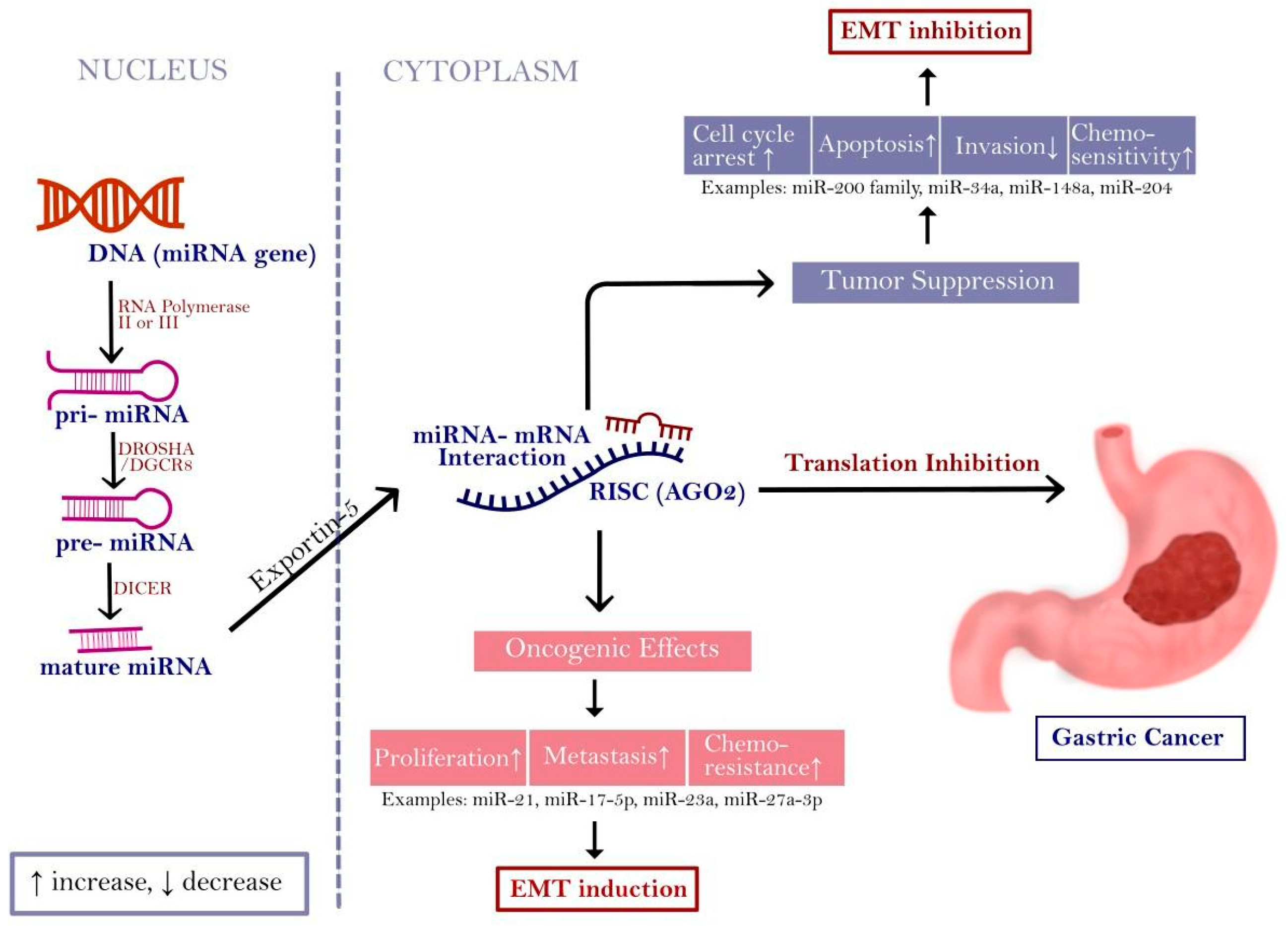
Gastric Cancer Epithelial-Mesenchymal Transition-The Role of Micro-RNA
 ,
,  , , , , and
Simple Summary
Abstract
1. Introduction
2. Epithelial-Mesenchymal Transition
2.1. Epithelial-Mesenchymal Transition in Gastric Carcinogenesis
2.2. Wnt/β-Catenin Signaling in EMT in GC
2.3. TGF-β Signaling in EMT in GC
3. Micro-RNAs in Carcinogenesis
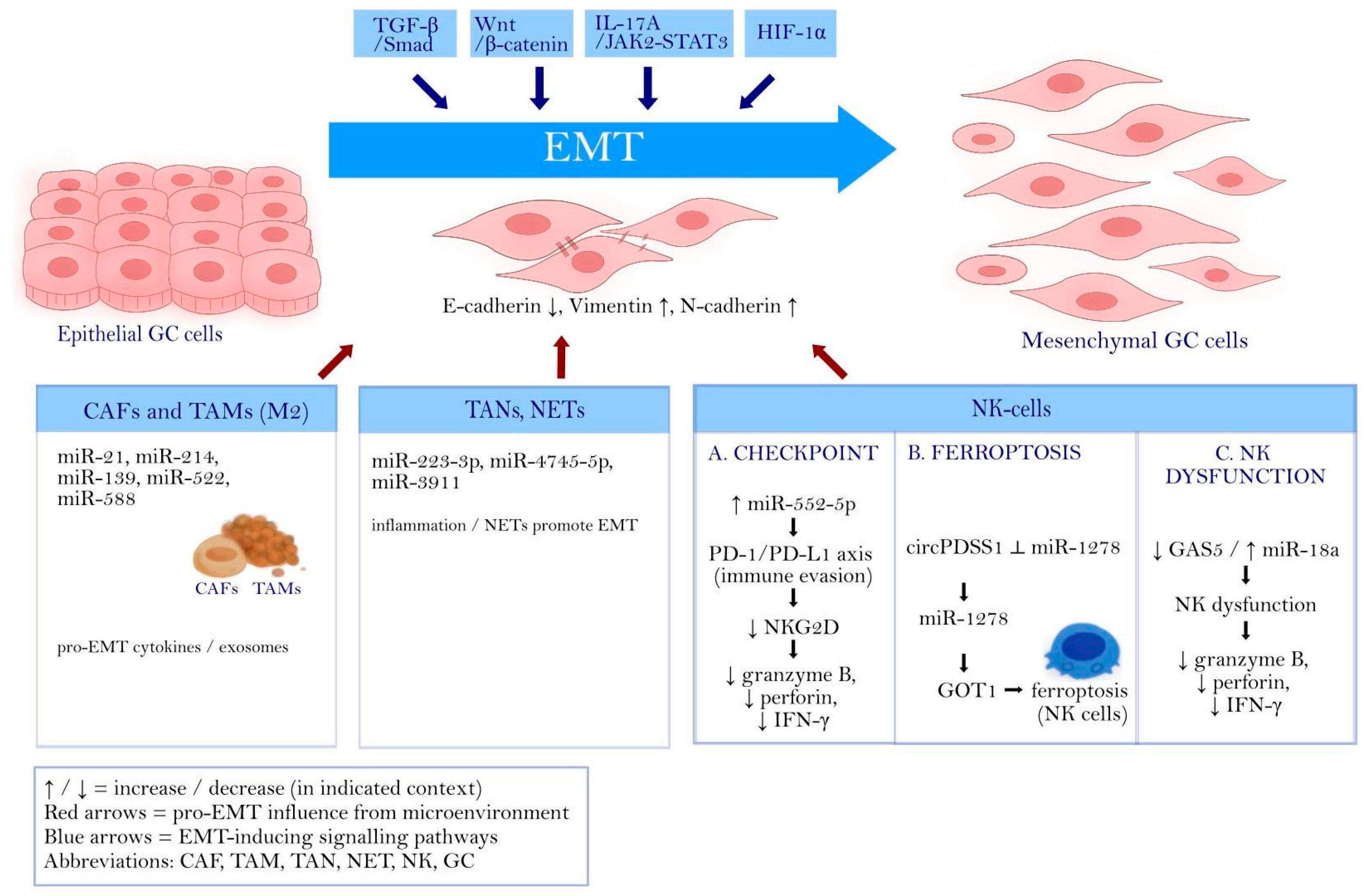
4. Micro-RNAs in Gastric Cancer Epithelial-Mesenchymal Transition
4.1. Cancer-Associated Fibroblasts
4.2. Tumor-Associated Macrophages
4.3. Tumor-Associated Neutrophils
4.4. Natural Killer Cells
4.5. Systemic Inflammatory Indices and Neutrophil-Derived Mediator FAM3C
4.6. Hierarchy of Evidence and Robustness of EMT-miRNA Claims
4.7. Context Dependence, Conflicting Reports, and EMT Plasticity
4.8. Cohort Consistency Across Populations and Experimental Systems
4.9. Methodological Weaknesses and Standardization Gaps
4.10. Translational Barriers: Pleiotropy, Off-Target Effects, and Delivery Constraints
4.11. Future Directions
5. Conclusions
Author Contributions
Funding
Institutional Review Board Statement
Informed Consent Statement
Data Availability Statement
Conflicts of Interest
Abbreviations
| ABCB1 | ATP-binding cassette subfamily B member 1 (P-glycoprotein) |
| ABL2 | Abelson tyrosine-protein kinase 2 |
| AGO | Argonaute protein |
| AKT | protein kinase B |
| ALEX1 | armadillo repeat-containing protein X-linked 1 |
| AML | acute myeloid leukaemia |
| AUC | area under the curve |
| CAF | cancer-associated fibroblast |
| CAFs | cancer-associated fibroblasts |
| CCL20 | C-C motif chemokine ligand 20 |
| CCL22 | C-C motif chemokine ligand 22 |
| CCL5 | C-C motif chemokine ligand 5 |
| Cdks | cyclin-dependent kinases |
| CI | confidence interval |
| CIN | chromosomal instability |
| CLL | chronic lymphocytic leukaemia |
| CRISPR | clustered regularly interspaced short palindromic repeats |
| CSC | cancer stem cell |
| Ct | cycle threshold |
| CXCL8 | C-X-C motif chemokine ligand 8 (interleukin-8) |
| DDP | cisplatin |
| DFS | disease-free survival |
| DGCR8 | DiGeorge syndrome critical region 8 |
| DICER | endoribonuclease Dicer |
| DROSHA | Drosha ribonuclease III |
| ECM | extracellular matrix |
| EMT | epithelial-mesenchymal transition |
| EMT-TFs | Epithelial-Mesenchymal Transition Transcription Factors |
| FAM3C | family with sequence similarity 3 member C |
| GAS5 | growth arrest-specific 5 |
| GC | gastric cancer |
| GLOBOCAN | Global Cancer Observatory (Global Cancer Statistics database) |
| GOT1 | glutamic-oxaloacetic transaminase 1 |
| GS | genomically stable |
| H. pylori | Helicobacter pylori |
| HGF | hepatocyte growth factor |
| HIF-1α | hypoxia-inducible factor 1-alpha |
| IFN-γ | interferon gamma |
| IGF | insulin-like growth factor |
| IL-17A | interleukin-17A |
| IL-1β | interleukin-1 beta |
| IL-6 | interleukin-6 |
| IL-8 | interleukin-8 |
| ILEI | interleukin-like EMT inducer |
| JAK/STAT | Janus kinase/signal transducer and activator of transcription pathway |
| JAK2/STAT3 | Janus kinase 2/signal transducer and activator of transcription |
| JNK | c-Jun N-terminal kinase |
| lncRNA | long non-coding RNA |
| MALT | mucosa-associated lymphoid tissue |
| MAPK | mitogen-activated protein kinase |
| MAPK1 | mitogen-activated protein kinase 1 |
| MET | mesenchymal-epithelial transition |
| MIF | macrophage migration inhibitory factor |
| miRNA | microRNA |
| MMPs | matrix metalloproteinases |
| mRNA | messenger RNA |
| MSI | microsatellite instability |
| mTOR | mechanistic target of rapamycin |
| ncRNA | non-coding RNA |
| NET | neutrophil extracellular trap |
| NK | natural killer (cell) |
| NKG2D | natural killer group 2D receptor |
| NLR | neutrophil-to-lymphocyte ratio |
| NTN4 | netrin-4 |
| PD-1 | programmed cell death protein 1 |
| PD-L1 | programmed death-ligand 1 |
| PI3K | phosphatidylinositol 3-kinase |
| PLR | platelet-to-lymphocyte ratio |
| pre-miRNA | precursor microRNA |
| pri-miRNA | primary microRNA transcript |
| PTEN | phosphatase and tensin homolog |
| qRT-PCR | quantitative reverse transcription PCR |
| RISC | RNA-induced silencing complex |
| sens | sensitivity |
| SNAI2 | Snail family transcriptional repressor 2 (Slug) |
| spec | specificity |
| TAM | tumor-associated macrophage |
| TAN | tumor-associated neutrophil |
| TCGA | The Cancer Genome Atlas |
| TGF-β | transforming growth factor beta |
| TNF | tumor necrosis factor |
| TNF-α | tumor necrosis factor alpha |
| TNM | tumor-node-metastasis (staging system) |
| Treg | regulatory T cell |
| UTR | untranslated region |
| VEGF | vascular endothelial growth factor |
| VEGF-C | vascular endothelial growth factor C |
| WGS | whole-genome sequencing |
| WT | wild-type |
References
- Yang, W.-J.; Zhao, H.-P.; Yu, Y.; Wang, J.-H.; Guo, L.; Liu, J.-Y.; Pu, J.; Lv, J. Updates on global epidemiology, risk and prognostic factors of gastric cancer. World J. Gastroenterol. 2023, 29, 2452–2468. [Google Scholar] [CrossRef] [Scilit]
- Kong, W.; Sun, Y.; Qin, X.; Zhu, G.; Zhu, X.; Kuang, Z.; Xiao, Z.; Li, J. The Burden and Risk Factors of Gastric Cancer in Eastern Asia From 1990 to 2021: Longitudinal Observational Study of the Global Burden of Disease Study 2021. JMIR Cancer 2025, 11, e75728. [Google Scholar] [CrossRef] [Scilit]
- Ilic, M.; Ilic, I. Epidemiology of stomach cancer. World J. Gastroenterol. 2022, 28, 1187–1203. [Google Scholar] [CrossRef] [Scilit] [PubMed]
- Chandanos, E.; Lagergren, J. Oestrogen and the enigmatic male predominance of gastric cancer. Eur. J. Cancer 2008, 44, 2397–2403. [Google Scholar] [CrossRef] [Scilit] [PubMed]
- Mamun, T.I.; Younus, S.; Rahman, M.H. Gastric cancer-Epidemiology, modifiable and non-modifiable risk factors, challenges and opportunities: An updated review. Cancer Treat. Res. Commun. 2024, 41, 100845. [Google Scholar] [CrossRef] [Scilit]
- Wroblewski, L.E.; Peek, R.M., Jr.; Wilson, K.T. Helicobacter pylori and gastric cancer: Factors that modulate disease risk. Clin. Microbiol. Rev. 2010, 23, 713–739. [Google Scholar] [CrossRef] [Scilit] [PubMed]
- Reyes, V.E. Helicobacter pylori and its role in gastric cancer. Microorganisms 2023, 11, 1312. [Google Scholar] [CrossRef] [Scilit]
- Jaroenlapnopparat, A.; Bhatia, K.; Coban, S. Inflammation and gastric cancer. Diseases 2022, 10, 35. [Google Scholar] [CrossRef] [Scilit]
- He, C.; Chen, M.; Liu, J.; Yuan, Y. Host genetic factors respond to pathogenic step-specific virulence factors of Helicobacter pylori in gastric carcinogenesis. Mutat. Res. Rev. Mutat. Res. 2014, 759, 14–26. [Google Scholar] [CrossRef] [Scilit]
- Karimi, P.; Islami, F.; Anandasabapathy, S.; Freedman, N.D.; Kamangar, F. Gastric cancer: Descriptive epidemiology, risk factors, screening, and prevention. Cancer Epidemiol. Biomark. Prev. 2014, 23, 700–713. [Google Scholar] [CrossRef] [Scilit]
- Machlowska, J.; Baj, J.; Sitarz, M.; Maciejewski, R.; Sitarz, R. Gastric cancer: Epidemiology, risk factors, classification, genomic characteristics and treatment strategies. Int. J. Mol. Sci. 2020, 21, 4012. [Google Scholar] [CrossRef] [Scilit] [PubMed]
- Li, B.; Zhang, F.; Niu, Q.; Liu, J.; Yu, Y.; Wang, P.; Zhang, S.; Zhang, H.; Wang, Z. A molecular classification of gastric cancer associated with distinct clinical outcomes and validated by an XGBoost-based prediction model. Mol. Ther. Nucleic Acids 2023, 31, 224–240. [Google Scholar] [CrossRef] [Scilit]
- Ma, J.; Shen, H.; Kapesa, L.; Zeng, S. Lauren classification and individualized chemotherapy in gastric cancer. Oncol. Lett. 2016, 11, 2959–2964. [Google Scholar] [CrossRef] [Scilit]
- Yu, L.; Cao, C.; Li, X.; Zhang, M.; Gu, Q.; Gao, H.; Balic, J.J.; Xu, D.; Zhang, L.; Ying, L.; et al. Complete loss of miR-200 family induces EMT associated cellular senescence in gastric cancer. Oncogene 2022, 41, 26–36. [Google Scholar] [CrossRef] [Scilit]
- Xie, Y.; Wang, X.; Wang, W.; Pu, N.; Liu, L. Epithelial-mesenchymal transition orchestrates tumor microenvironment: Current perceptions and challenges. J. Transl. Med. 2025, 23, 386. [Google Scholar] [CrossRef] [Scilit] [PubMed]
- Peng, Z.; Wang, C.X.; Fang, E.H.; Wang, G.B.; Tong, Q. Role of epithelial-mesenchymal transition in gastric cancer initiation and progression. World J. Gastroenterol. 2014, 20, 5403–5410. [Google Scholar] [CrossRef] [Scilit]
- Kozak, J.; Forma, A.; Czeczelewski, M.; Kozyra, P.; Sitarz, R.; Radzikowska-Büchner, E.; Sitarz, M.; Baj, J. Inhibition or reversal of the epithelial-mesenchymal transition in gastric cancer: Pharmacological approaches. Int. J. Mol. Sci. 2021, 22, 277. [Google Scholar] [CrossRef] [Scilit] [PubMed]
- Sannino, G.; Marchetto, A.; Kirchner, T.; Grünewald, T.G.P. Epithelial-to-mesenchymal and mesenchymal-to-epithelial transition in mesenchymal tumors: A paradox in sarcomas? Cancer Res. 2017, 77, 4556–4561. [Google Scholar] [CrossRef] [Scilit]
- Ma, H.Y.; Liu, X.Z.; Liang, C.M. Inflammatory microenvironment contributes to epithelial-mesenchymal transition in gastric cancer. World J. Gastroenterol. 2016, 22, 6619–6628. [Google Scholar] [CrossRef] [Scilit]
- Baj, J.; Brzozowska, K.; Forma, A.; Maani, A.; Sitarz, E.; Portincasa, P. Immunological aspects of the tumor microenvironment and epithelial-mesenchymal transition in gastric carcinogenesis. Int. J. Mol. Sci. 2020, 21, 2544. [Google Scholar] [CrossRef] [Scilit]
- Baj, J.; Korona-Głowniak, I.; Forma, A.; Maani, A.; Sitarz, E.; Rahnama-Hezavah, M.; Radzikowska, E.; Portincasa, P. Mechanisms of the epithelial-mesenchymal transition and tumor microenvironment in Helicobacter pylori-induced gastric cancer. Cells 2020, 9, 1055. [Google Scholar] [CrossRef] [Scilit]
- Wang, M.; Zhang, R.; Zhang, S.; Xu, R.; Yang, Q. MicroRNA-574-3p regulates epithelial mesenchymal transition and cisplatin resistance via targeting ZEB1 in human gastric carcinoma cells. Gene 2019, 700, 110–119. [Google Scholar] [CrossRef] [Scilit]
- Liu, Z.; Chen, L.; Zhang, X.; Xu, X.; Xing, H.; Zhang, Y.; Li, W.; Yu, H.; Zeng, J.; Jia, J. RUNX3 regulates vimentin expression via miR-30a during epithelial-mesenchymal transition in gastric cancer cells. J. Cell. Mol. Med. 2014, 18, 610–623. [Google Scholar] [CrossRef] [Scilit]
- Chen, D.D.; Cheng, J.T.; Chandoo, A.; Sun, X.W.; Zhang, L.; Lu, M.D.; Sun, W.J.; Huang, Y.P. MicroRNA-33a prevents epithelial-mesenchymal transition, invasion, and metastasis of gastric cancer cells through the Snail/Slug pathway. Am. J. Physiol. Gastrointest. Liver Physiol. 2019, 317, G147–G160. [Google Scholar] [CrossRef] [Scilit] [PubMed]
- Mirzaei, S.; Baghaei, K.; Parivar, K.; Hashemi, M.; Asadzadeh Aghdaei, H. The expression level changes of microRNAs 200a/205 in the development of invasive properties in gastric cancer cells through epithelial-mesenchymal transition. Eur. J. Pharmacol. 2019, 857, 172426. [Google Scholar] [CrossRef] [Scilit] [PubMed]
- Li, H.; Xu, L.; Li, C.; Zhao, L.; Ma, Y.; Zheng, H.; Li, Z.; Zhang, Y.; Wang, R.; Liu, Y.; et al. Ubiquitin ligase Cbl-b represses IGF-I-induced epithelial mesenchymal transition via ZEB2 and microRNA-200c regulation in gastric cancer cells. Mol. Cancer 2014, 13, 136. [Google Scholar] [CrossRef] [Scilit]
- Li, J.; Li, T.; Lu, Y.; Shen, G.; Guo, H.; Wu, J.; Lei, C.; Du, F.; Zhou, F.; Zhao, X.; et al. MiR-2392 suppresses metastasis and epithelial-mesenchymal transition by targeting MAML3 and WHSC1 in gastric cancer. FASEB J. 2017, 31, 3774–3786. [Google Scholar] [CrossRef] [Scilit] [PubMed]
- Wu, C.; Zhuang, Y.; Jiang, S.; Liu, S.; Zhou, J.; Wu, J.; Teng, Y.; Xia, B.; Wang, R.; Zou, X. Interaction between Wnt/β-catenin pathway and microRNAs regulates epithelial-mesenchymal transition in gastric cancer (Review). Int. J. Oncol. 2016, 48, 2236–2246. [Google Scholar] [CrossRef] [Scilit]
- Zhang, X.; Peng, Y.; Huang, Y.; Yang, M.; Yan, R.; Zhao, Y.; Cheng, Y.; Liu, X.; Deng, S.; Feng, X.; et al. SMG-1 inhibition by miR-192/-215 causes epithelial-mesenchymal transition in gastric carcinogenesis via activation of Wnt signaling. Cancer Med. 2018, 7, 146–156. [Google Scholar] [CrossRef] [Scilit]
- Lei, Z.N.; Teng, Q.X.; Tian, Q.; Chen, W.; Xie, Y.; Wu, K.; Zeng, Q.; Zeng, L.; Pan, Y.; Chen, Z.S.; et al. Signaling pathways and therapeutic interventions in gastric cancer. Signal Transduct. Target. Ther. 2022, 7, 358. [Google Scholar] [CrossRef] [Scilit]
- Kim, Y.; Bae, Y.J.; Kim, J.H.; Kim, H.; Shin, S.J.; Jung, D.H.; Park, H. Wnt/β-catenin pathway is a key signaling pathway to trastuzumab resistance in gastric cancer cells. BMC Cancer 2023, 23, 922. [Google Scholar] [CrossRef] [Scilit]
- Jang, J.H.; Jung, J.; Kang, H.G.; Kim, W.; Kim, W.J.; Lee, H.; Cho, J.Y.; Hong, R.; Kim, J.W.; Chung, J.Y.; et al. Kindlin-1 promotes gastric cancer cell motility through the Wnt/β-catenin signaling pathway. Sci. Rep. 2025, 15, 2481. [Google Scholar] [CrossRef] [Scilit]
- Shi, R.; Cao, Z.; Li, J.; Ji, R.; Guo, Z. Targeting the Wnt/β-catenin pathway and epithelial-mesenchymal transition in gastric cancer: Mechanisms, therapeutic strategies, and clinical challenges. Front. Oncol. 2025, 15, 1633699. [Google Scholar] [CrossRef] [Scilit]
- Zhang, Y.E. Non-Smad signaling pathways of the TGF-β family. Cold Spring Harb. Perspect. Biol. 2017, 9, a022129. [Google Scholar] [CrossRef] [Scilit]
- Veen, L.M.; Skrabanja, T.L.P.; Derks, S.; de Gruijl, T.D.; Bijlsma, M.F.; van Laarhoven, H.W.M. The role of transforming growth factor β in upper gastrointestinal cancers: A systematic review. Cancer Treat. Rev. 2021, 100, 102285. [Google Scholar] [CrossRef] [Scilit]
- Qin, S.; Guo, Q.; Liu, Y.; Zhang, X.; Huang, P.; Yu, H.; Xia, L.; Leng, W.; Li, D. A novel TGFbeta/TGILR axis mediates crosstalk between cancer-associated fibroblasts and tumor cells to drive gastric cancer progression. Cell Death Dis. 2024, 15, 368. [Google Scholar] [CrossRef] [Scilit] [PubMed]
- O’Brien, J.; Hayder, H.; Zayed, Y.; Peng, C. Overview of microRNA biogenesis, mechanisms of actions, and circulation. Front. Endocrinol. 2018, 9, 402. [Google Scholar] [CrossRef] [Scilit] [PubMed]
- Hammond, S.M. An overview of microRNAs. Adv. Drug Deliv. Rev. 2015, 87, 3–14. [Google Scholar] [CrossRef] [Scilit]
- Su, E.C.; Chen, Y.S.; Tien, Y.C.; Liu, J.; Ho, B.C.; Yu, S.L.; Singh, S. ChemiRs: A web application for microRNAs and chemicals. BMC Bioinform. 2016, 17, 167. [Google Scholar] [CrossRef] [Scilit]
- Huntley, R.P.; Sitnikov, D.; Orlic-Milacic, M.; Balakrishnan, R.; D’Eustachio, P.; Gillespie, M.E.; Howe, D.; Kalea, A.Z.; Maegdefessel, L.; Osumi-Sutherland, D.; et al. Guidelines for the functional annotation of microRNAs using the Gene Ontology. RNA 2016, 22, 667–676. [Google Scholar] [CrossRef] [Scilit] [PubMed]
- Reddy, K.B. MicroRNA (miRNA) in cancer. Cancer Cell Int. 2015, 15, 38. [Google Scholar] [CrossRef] [Scilit]
- Hussen, B.M.; Hidayat, H.J.; Salihi, A.; Sabir, D.K.; Taheri, M.; Ghafouri-Fard, S. MicroRNA: A signature for cancer progression. Biomed. Pharmacother. 2021, 138, 111528. [Google Scholar] [CrossRef] [Scilit]
- Mogilyansky, E.; Rigoutsos, I. The miR-17/92 cluster: A comprehensive update on its genomics, genetics, functions and increasingly important and numerous roles in health and disease. Cell Death Differ. 2013, 20, 1603–1614. [Google Scholar] [CrossRef] [Scilit]
- Babaei, K.; Shams, S.; Keymoradzadeh, A.; Vahidi, S.; Hamami, P.; Khaksar, R.; Norollahi, S.E.; Samadani, A.A. An insight of microRNAs performance in carcinogenesis and tumorigenesis; an overview of cancer therapy. Life Sci. 2020, 240, 117077. [Google Scholar] [CrossRef] [Scilit]
- Montano, M. MicroRNAs: miRRORS of health and disease. Transl. Res. 2011, 157, 157–162. [Google Scholar] [CrossRef] [Scilit]
- Said, M.N.; Muawia, S.; Helal, A.; Fawzy, A.; Allam, R.M.; Shafik, N.F. Regulation of CDK inhibitor p27 by microRNA 222 in breast cancer patients. Exp. Mol. Pathol. 2021, 123, 104718. [Google Scholar] [CrossRef] [Scilit]
- Gruszka, R.; Zakrzewski, K.; Liberski, P.P.; Zakrzewska, M. mRNA and miRNA expression analyses of the MYC/E2F/miR-17-92 network in the most common pediatric brain tumors. Int. J. Mol. Sci. 2021, 22, 543. [Google Scholar] [CrossRef] [Scilit]
- Ozdogan, S.; Yaltirik, C.K.; Yilmaz, S.G.; Akdeniz, F.T.; Sumerkent, K.; Duzkalir, A.H.; Ture, U.; Isbir, T. Investigation of the effects of microRNA-221 expression levels in glioblastoma multiforme tumors. Anticancer Res. 2020, 40, 3265–3270. [Google Scholar] [CrossRef] [Scilit]
- Fiori, M.E.; Villanova, L.; Barbini, C.; De Angelis, M.L.; De Maria, R. miR-663 sustains NSCLC by inhibiting mitochondrial outer membrane permeabilization (MOMP) through PUMA/BBC3 and BTG2. Cell Death Dis. 2018, 9, 49. [Google Scholar] [CrossRef] [Scilit]
- Hanahan, D. Hallmarks of cancer: New dimensions. Cancer Discov. 2022, 12, 31–46. [Google Scholar] [CrossRef] [Scilit]
- Sargolzaei, J.; Etemadi, T.; Alyasin, A. The P53/microRNA network: A potential tumor suppressor with a role in anticancer therapy. Pharmacol. Res. 2020, 160, 105179. [Google Scholar] [CrossRef] [Scilit]
- Pekarsky, Y.; Balatti, V.; Croce, C.M. BCL2 and miR-15/16: From gene discovery to treatment. Cell Death Differ. 2018, 25, 21–26. [Google Scholar] [CrossRef] [Scilit]
- Sadri, F.; Hosseini, S.F.; Aghayei, A.; Fereidouni, M.; Rezaei, Z. The tumor suppressor roles and mechanisms of miR-491 in human cancers. DNA Cell Biol. 2022, 41, 810–823. [Google Scholar] [CrossRef] [Scilit]
- Moro, M.; Fortunato, O.; Bertolini, G.; Mensah, M.; Borzi, C.; Centonze, G.; Andriani, F.; Di Paolo, D.; Perri, P.; Ponzoni, M.; et al. MiR-486-5p targets CD133+ lung cancer stem cells through the p85/AKT pathway. Pharmaceuticals 2022, 15, 297. [Google Scholar] [CrossRef] [Scilit]
- Liu, X.; Li, Y.; Li, Z.; Hou, T. miR-155 promotes proliferation and epithelial-mesenchymal transition of MCF-7 cells. Exp. Ther. Med. 2021, 21, 218. [Google Scholar] [CrossRef] [Scilit]
- Mongroo, P.S.; Rustgi, A.K. The role of the miR-200 family in epithelial-mesenchymal transition. Cancer Biol. Ther. 2010, 10, 219–222. [Google Scholar] [CrossRef]
- Liu, Y.W.; Sun, M.; Xia, R.; Zhang, E.B.; Liu, X.H.; Zhang, Z.H.; Xu, T.P.; De, W.; Liu, B.R.; Wang, Z.X. LincHOTAIR epigenetically silences miR34a by binding to PRC2 to promote the epithelial-to-mesenchymal transition in human gastric cancer. Cell Death Dis. 2015, 6, e1802. [Google Scholar] [CrossRef] [Scilit]
- Xu, T.; Xie, F.; Xu, D.; Xu, W.; Ge, X.; Lv, S.; Li, S. MiR-200b suppresses gastric cancer cell migration and invasion by inhibiting NRG1 through ERBB2/ERBB3 signaling. J. Oncol. 2021, 2021, 4470778. [Google Scholar] [CrossRef] [Scilit]
- Wang, S.H.; Li, X.; Zhou, L.S.; Cao, Z.W.; Shi, C.; Zhou, C.Z.; Wen, Y.G.; Shen, Y.; Li, J.K. MicroRNA-148a suppresses human gastric cancer cell metastasis by reversing epithelial-to-mesenchymal transition. Tumour Biol. 2013, 34, 3705–3712. [Google Scholar] [CrossRef] [Scilit]
- Zhang, L.; Wang, X.; Chen, P. MiR-204 down regulates SIRT1 and reverts SIRT1-induced epithelial-mesenchymal transition, anoikis resistance and invasion in gastric cancer cells. BMC Cancer 2013, 13, 290. [Google Scholar] [CrossRef] [Scilit]
- Wang, L.L.; Zhang, X.H.; Zhang, X.; Chu, J.K. MiR-30a increases cisplatin sensitivity of gastric cancer cells through suppressing epithelial-to-mesenchymal transition (EMT). Eur. Rev. Med. Pharmacol. Sci. 2016, 20, 1733–1739. [Google Scholar]
- Imamura, T.; Komatsu, S.; Ichikawa, D.; Miyamae, M.; Okajima, W.; Ohashi, T.; Kiuchi, J.; Nishibeppu, K.; Kosuga, T.; Konishi, H.; et al. Low plasma levels of miR-101 are associated with tumor progression in gastric cancer. Oncotarget 2017, 8, 106538–106550. [Google Scholar] [CrossRef] [Scilit]
- Wang, G.; Fu, Y.; Liu, G.; Ye, Y.; Zhang, X. miR-218 inhibits proliferation, migration, and EMT of gastric cancer cells by targeting WASF3. Oncol. Res. 2017, 25, 355–364. [Google Scholar] [CrossRef] [Scilit]
- Deng, M.; Tang, H.L.; Lu, X.H.; Liu, M.Y.; Lu, X.M.; Gu, Y.X.; Liu, J.F.; He, Z.M. miR-26a suppresses tumor growth and metastasis by targeting FGF9 in gastric cancer. PLoS ONE 2013, 8, e72662. [Google Scholar] [CrossRef] [Scilit]
- Lin, X.M.; Wang, Z.J.; Lin, Y.X.; Chen, H. Decreased exosome-delivered miR-486-5p is responsible for the peritoneal metastasis of gastric cancer cells by promoting EMT progress. World J. Surg. Oncol. 2021, 19, 312. [Google Scholar] [CrossRef] [Scilit]
- Liu, Y.; Cao, J.; Zhu, L.; Zhao, W.; Zhou, Y.; Shao, C.; Shao, S. Circular RNA circPGD contributes to gastric cancer progression via the sponging miR-16-5p/ABL2 axis and encodes a novel PGD-219aa protein. Cell Death Discov. 2022, 8, 384. [Google Scholar] [CrossRef] [Scilit]
- Ni, H.; Qin, H.; Sun, C.; Liu, Y.; Ruan, G.; Guo, Q.; Xi, T.; Xing, Y.; Zheng, L. MiR-375 reduces the stemness of gastric cancer cells through triggering ferroptosis. Stem Cell Res. Ther. 2021, 12, 325. [Google Scholar] [CrossRef] [Scilit] [PubMed]
- Chen, G.; Liao, J.; Xu, Y.; Chen, Y.; Li, J.; Bu, G.; Li, Q. LINC01232 promotes metastasis and EMT by regulating miR-506-5p/PAK1 axis in gastric cancer. Cancer Manag. Res. 2022, 14, 1729–1740. [Google Scholar] [CrossRef] [Scilit]
- Sakimura, S.; Sugimachi, K.; Kurashige, J.; Ueda, M.; Hirata, H.; Nambara, S.; Komatsu, H.; Saito, T.; Takano, Y.; Uchi, R.; et al. The miR-506-induced epithelial-mesenchymal transition is involved in poor prognosis for patients with gastric cancer. Ann. Surg. Oncol. 2015, 22, 1436–1443. [Google Scholar] [CrossRef] [Scilit] [PubMed]
- Huang, N.; Wu, Z.; Lin, L.; Zhou, M.; Wang, L.; Ma, H.; Xia, J.; Bin, J.; Liao, Y.; Liao, W. MiR-338-3p inhibits epithelial-mesenchymal transition in gastric cancer cells by targeting ZEB2 and MACC1/Met/Akt signaling. Oncotarget 2015, 6, 15222–15234. [Google Scholar] [CrossRef] [Scilit]
- Dai, Z.T.; Xiang, Y.; Duan, Y.Y.; Wang, J.; Li, J.P.; Zhang, H.M.; Cheng, C.; Wang, Q.; Zhang, T.C.; Liao, X.H. MiR-17-5p and MKL-1 modulate stem cell characteristics of gastric cancer cells. Int. J. Biol. Sci. 2021, 17, 2278–2293. [Google Scholar] [CrossRef] [Scilit]
- Wu, Q.; Luo, G.; Yang, Z.; Zhu, F.; An, Y.; Shi, Y.; Fan, D. miR-17-5p promotes proliferation by targeting SOCS6 in gastric cancer cells. FEBS Lett. 2014, 588, 2055–2062. [Google Scholar] [CrossRef] [Scilit]
- Zhu, Z.; Yang, Q.; Zhang, B.; Wu, W.; Yuan, F.; Zhu, Z. miR-106b promotes metastasis of early gastric cancer by targeting ALEX1 in vitro and in vivo. Cell. Physiol. Biochem. 2019, 52, 606–616. [Google Scholar]
- Li, L.; Zhang, J.; Pan, Q.; Lei, C. MicroRNA-23a regulates cell migration and invasion by targeting PTEN in gastric cancer. Int. J. Clin. Exp. Pathol. 2016, 9, 877–887. [Google Scholar]
- Hu, W.; Zheng, X.; Liu, J.; Zhang, M.; Liang, Y.; Song, M. MicroRNA miR-130a-3p promotes gastric cancer by targeting glucosaminyl N-acetyl transferase 4 (GCNT4) to regulate the TGF-β1/SMAD3 pathway. Bioengineered 2021, 12, 11634–11647. [Google Scholar] [CrossRef] [Scilit]
- Pan, Y.; Shu, X.; Sun, L.; Yu, L.; Sun, L.; Yang, Z.; Ran, Y. miR-196a-5p modulates gastric cancer stem cell characteristics by targeting Smad4. Int. J. Oncol. 2017, 50, 1965–1976. [Google Scholar] [CrossRef] [Scilit] [PubMed]
- Lu, Q.; Chen, Y.; Sun, D.; Wang, S.; Ding, K.; Liu, M.; Zhang, Y.; Miao, Y.; Liu, H.; Zhou, F. MicroRNA-181a functions as an oncogene in gastric cancer by targeting Caprin-1. Front. Pharmacol. 2019, 9, 1565. [Google Scholar] [CrossRef] [Scilit]
- Wu, Z.H.; Lin, C.; Liu, C.C.; Jiang, W.W.; Huang, M.Z.; Liu, X.; Guo, W.J. MiR-616-3p promotes angiogenesis and EMT in gastric cancer via the PTEN/AKT/mTOR pathway. Biochem. Biophys. Res. Commun. 2018, 501, 1068–1073. [Google Scholar] [CrossRef] [Scilit]
- Xia, X.; Wang, S.; Ni, B.; Xing, S.; Cao, H.; Zhang, Z.; Yu, F.; Zhao, E.; Zhao, G. Hypoxic gastric cancer-derived exosomes promote progression and metastasis via miR-301a-3p/PHD3/HIF-1α positive feedback loop. Oncogene 2020, 39, 6231–6244. [Google Scholar] [CrossRef] [Scilit] [PubMed]
- Peng, Y.; Zhang, X.; Lin, H.; Deng, S.; Qin, Y.; He, J.; Hu, F.; Zhu, X.; Feng, X.; Wang, J.; et al. Dual activation of Hedgehog and Wnt/β-catenin signaling pathway caused by downregulation of SUFU targeted by miRNA-150 in human gastric cancer. Aging 2021, 13, 10749–10769. [Google Scholar] [CrossRef] [Scilit]
- Li, K.; Zhu, X.; Chen, X.; Wang, X. MicroRNA-27a-3p promotes epithelial-mesenchymal transition by targeting NOVA alternative splicing regulator 1 in gastric cancer. Mol. Med. Rep. 2020, 21, 1615–1622. [Google Scholar] [CrossRef] [Scilit]
- Song, J.; Lin, Z.; Liu, Q.; Huang, S.; Han, L.; Fang, Y.; Zhong, P.; Dou, R.; Xiang, Z.; Zheng, J.; et al. MiR-192-5p/RB1/NF-κBp65 signaling axis promotes IL-10 secretion during gastric cancer EMT to induce Treg cell differentiation in the tumour microenvironment. Clin. Transl. Med. 2022, 12, e992. [Google Scholar] [CrossRef] [Scilit]
- Sun, H.; Wang, X.; Wang, X.; Xu, M.; Sheng, W. The role of cancer-associated fibroblasts in tumorigenesis of gastric cancer. Cell Death Dis. 2022, 13, 874. [Google Scholar] [CrossRef] [Scilit]
- Yan, Y.; Wang, L.F.; Wang, R.F. Role of cancer-associated fibroblasts in invasion and metastasis of gastric cancer. World J. Gastroenterol. 2015, 21, 9717–9726. [Google Scholar] [CrossRef] [Scilit]
- Zhai, J.; Shen, J.; Xie, G.; Wu, J.; He, M.; Gao, L.; Zhang, Y.; Yao, X.; Shen, L. Cancer-associated fibroblasts-derived IL-8 mediates resistance to cisplatin in human gastric cancer. Cancer Lett. 2019, 454, 37–43. [Google Scholar] [CrossRef] [Scilit]
- Valcz, G.; Sipos, F.; Tulassay, Z.; Molnar, B.; Yagi, Y. Importance of carcinoma-associated fibroblast-derived proteins in clinical oncology. J. Clin. Pathol. 2014, 67, 1026–1031. [Google Scholar] [CrossRef] [Scilit]
- Madar, S.; Goldstein, I.; Rotter, V. ‘Cancer associated fibroblasts’-More than meets the eye. Trends Mol. Med. 2013, 19, 447–453. [Google Scholar] [CrossRef] [Scilit]
- Martinez-Outschoorn, U.E.; Lisanti, M.P.; Sotgia, F. Catabolic cancer-associated fibroblasts transfer energy and biomass to anabolic cancer cells, fueling tumor growth. Semin. Cancer Biol. 2014, 25, 47–60. [Google Scholar] [CrossRef] [Scilit]
- Karakasheva, T.A.; Lin, E.W.; Tang, Q.; Qiao, E.; Waldron, T.J.; Soni, M.; Klein-Szanto, A.J.; Sahu, V.; Basu, D.; Ohashi, S.; et al. IL-6 mediates cross-talk between tumor cells and activated fibroblasts in the tumor microenvironment. Cancer Res. 2018, 78, 4957–4970. [Google Scholar] [CrossRef] [Scilit]
- Li, J.; Sun, J.; Zeng, Z.; Liu, Z.; Ma, M.; Zheng, Z.; He, Y.; Kang, W. Tumour-associated macrophages in gastric cancer: From function and mechanism to application. Clin. Transl. Med. 2023, 13, e1386. [Google Scholar] [CrossRef] [Scilit]
- Zhang, J.; Hu, C.; Zhang, R.; Xu, J.; Zhang, Y.; Yuan, L.; Zhang, S.; Pan, S.; Cao, M.; Qin, J.; et al. The role of macrophages in gastric cancer. Front. Immunol. 2023, 14, 1282176. [Google Scholar] [CrossRef] [Scilit]
- Li, W.; Zhang, X.; Wu, F.; Zhou, Y.; Bao, Z.; Li, H.; Zheng, P.; Zhao, S. Gastric cancer-derived mesenchymal stromal cells trigger M2 macrophage polarization that promotes metastasis and EMT in gastric cancer. Cell Death Dis. 2019, 10, 918. [Google Scholar] [CrossRef] [Scilit]
- Chen, L.; Shi, Y.; Zhu, X.; Guo, W.; Zhang, M.; Che, Y.; Tang, L.; Yang, X.; You, Q.; Liu, Z. IL-10 secreted by cancer-associated macrophages regulates proliferation and invasion in gastric cancer cells via c-Met/STAT3 signaling. Oncol. Rep. 2019, 42, 595–604. [Google Scholar] [CrossRef] [Scilit]
- Guo, J.; Yan, Y.; Yan, Y.; Guo, Q.; Zhang, M.; Zhang, J.; Goltzman, D. Tumor-associated macrophages induce the expression of FOXQ1 to promote epithelial-mesenchymal transition and metastasis in gastric cancer cells. Oncol. Rep. 2017, 38, 2003–2010. [Google Scholar] [CrossRef] [Scilit]
- Ucaryilmaz Metin, C.; Ozcan, G. The HIF-1α as a potent inducer of the hallmarks in gastric cancer. Cancers 2022, 14, 2711. [Google Scholar] [CrossRef] [Scilit]
- Sun, Q.; Liu, H.; Guo, X.; Zhang, H.; Qin, C. Exosomal miR-15b-5p derived from gastric cancer cells enhances immune infiltrating of tumor-associated macrophages through the WIF1/WNT5A axis. Aging 2021, 13, 203192. [Google Scholar]
- Xiao, H.; Fu, J.; Liu, R.; Yan, L.; Zhou, Z.; Yuan, J. Gastric cancer cell-derived exosomal miR-541-5p induces M2 macrophage polarization through DUSP3/JAK2/STAT3 pathway. BMC Cancer 2024, 24, 957. [Google Scholar] [CrossRef] [Scilit]
- Yang, X.; Cai, S.; Shu, Y.; Deng, X.; Zhang, Y.; He, N.; Wan, L.; Chen, X.; Qu, Y.; Yu, S. Exosomal miR-487a derived from M2 macrophage promotes the progression of gastric cancer. Cell Cycle 2021, 20, 434–444. [Google Scholar] [CrossRef] [Scilit]
- Cui, H.Y.; Rong, J.S.; Chen, J.; Guo, J.; Zhu, J.Q.; Ruan, M.; Zuo, R.R.; Zhang, S.S.; Qi, J.M.; Zhang, B.H. Exosomal microRNA-588 from M2 polarized macrophages contributes to cisplatin resistance of gastric cancer cells. World J. Gastroenterol. 2021, 27, 6079–6092. [Google Scholar] [CrossRef] [Scilit]
- Zheng, P.; Chen, L.; Yuan, X.; Luo, Q.; Liu, Y.; Xie, G.; Ma, Y.; Shen, L. Exosomal transfer of tumor-associated macrophage-derived miR-21 confers cisplatin resistance in gastric cancer cells. J. Exp. Clin. Cancer Res. 2017, 36, 53. [Google Scholar] [CrossRef] [Scilit]
- Chen, C.W.; Wang, H.C.; Tsai, I.M.; Chen, I.S.; Chen, C.J.; Hou, Y.C.; Shan, Y.S. CD204-positive M2-like tumor-associated macrophages increase migration of gastric cancer cells by upregulating miR-210 to reduce NTN4 expression. Cancer Immunol. Immunother. 2024, 73, 1. [Google Scholar] [CrossRef] [Scilit]
- Yun, Z.; Tan, Y.; Wang, Y.; Ding, W.; Chu, Y.; Xu, J.; Jiang, R.; Xu, X. Hypoxia decreases macrophage glycolysis and M1 percentage by targeting microRNA-30c and mTOR in human gastric cancer. Cancer Sci. 2019, 110, 2368–2377. [Google Scholar]
- Qiu, Y.; Lu, G.; Li, N.; Hu, Y.; Tan, H.; Jiang, C. Exosome-mediated communication between gastric cancer cells and macrophages: Implications for tumor microenvironment. Front. Immunol. 2024, 15, 1327281. [Google Scholar] [CrossRef] [Scilit]
- Li, S.; Cong, X.; Gao, H.; Lan, X.; Li, Z.; Wang, W.; Song, S.; Wang, Y.; Li, C.; Zhang, H.; et al. Correction to: Tumor-associated neutrophils induce EMT by IL-17a to promote migration and invasion in gastric cancer cells. J. Exp. Clin. Cancer Res. 2019, 38, 177. [Google Scholar] [CrossRef] [Scilit]
- Shi, H.; Yu, S.; Li, W.; Li, X.; Wu, X.; Qu, X.; Ma, Y.; Zheng, C.; Che, X. Neutrophils in the tumor microenvironment: Role in tumor progression and potential targeted therapeutic strategy. J. Cancer Metastasis Treat. 2024, 10, 13. [Google Scholar] [CrossRef] [Scilit]
- Zhu, T.; Zou, X.; Yang, C.; Li, L.; Wang, B.; Li, R.; Li, H.; Xu, Z.; Huang, D.; Wu, Q. Neutrophil extracellular traps promote gastric cancer metastasis by inducing epithelial-mesenchymal transition. Int. J. Mol. Med. 2021, 48, 127. [Google Scholar] [CrossRef] [Scilit]
- Xia, X.; Zhang, Z.; Zhu, C.; Ni, B.; Wang, S.; Yang, S.; Yu, F.; Zhao, E.; Li, Q.; Zhao, G. Author Correction: Neutrophil extracellular traps promote metastasis in gastric cancer patients with postoperative abdominal infectious complications. Nat. Commun. 2025, 16, 2381. [Google Scholar] [CrossRef] [Scilit]
- Guo, X.; Wu, K.; Dang, L.; Liu, S.; Xu, J.; Wang, R.; Xu, J.; Zhong, Y.; Chen, Z.; Wu, B. Initial insights into the NET-associated ceRNA network in pulpitis: Transcriptomic and functional exploration. Int. Dent. J. 2025, 75, 100958. [Google Scholar] [CrossRef] [Scilit]
- Zhang, J.; Yu, D.; Ji, C.; Wang, M.; Fu, M.; Qian, Y.; Zhang, X.; Ji, R.; Li, C.; Gu, J.; et al. Exosomal miR-4745-5p/3911 from N2-polarized tumor-associated neutrophils promotes gastric cancer metastasis by regulating SLIT2. Mol. Cancer 2024, 23, 198. [Google Scholar] [CrossRef] [Scilit]
- Yu, D.; Zhang, J.; Wang, M.; Ji, R.; Qian, H.; Xu, W.; Zhang, H.; Gu, J.; Zhang, X. Exosomal miRNAs from neutrophils act as accurate biomarkers for gastric cancer diagnosis. Clin. Chim. Acta 2024, 554, 117773. [Google Scholar] [CrossRef] [Scilit]
- Tang, C.W.; Yang, J.H.; Qin, J.W.; Wu, H.J.; Cui, H.P.; Ge, L.Y.; Liu, A.Q. Regulation of the PD-1/PD-L1 axis and NK cell dysfunction by exosomal miR-552-5p in gastric cancer. Dig. Dis. Sci. 2024, 69, 3276–3289. [Google Scholar]
- Dong, H.; Ye, C.; Yu, X.; Shao, Y. Non-coding RNAs in gastric cancer immunotherapy: Mechanisms and clinical implications. Front. Immunol. 2025, 16, 1688181. [Google Scholar] [CrossRef] [Scilit]
- Qin, J.; Yang, J.; Cui, H.; Feng, C.; Liu, A. Exosomal miR-552-5p regulates the role of NK cells in EMT of gastric cancer via the PD-1/PD-L1 axis. J. Cancer 2025, 16, 406–416. [Google Scholar] [CrossRef] [Scilit]
- Yu, X.; Zhang, Y.; Luo, F.; Zhou, Q.; Zhu, L. The role of microRNAs in the gastric cancer tumor microenvironment. Mol. Cancer 2024, 23, 170. [Google Scholar] [CrossRef] [Scilit]
- Wei, M.F.; Gu, Z.S.; Zheng, L.L.; Zhao, M.X.; Wang, X.J. Long non-coding RNA GAS5 promotes natural killer cell cytotoxicity against gastric cancer by regulating miR-18a. Neoplasma 2020, 67, 1085–1093. [Google Scholar] [CrossRef] [Scilit]
- Li, Y.; Zhu, Y.; Sheng, P.; Guo, X.; Li, X.; Li, R.; Zhao, Z.; Li, L.; Gong, K. Mechanism of gastric cancer cell-derived exosomal circPDSS1 promoting ferroptosis of natural killer cells by regulating the miR-1278/GOT1 molecular axis. Res. Sq. 2024; preprint. [CrossRef] [Scilit]
- Yasui, S.; Takata, T.; Kamitani, Y.; Mae, Y.; Kurumi, H.; Ikebuchi, Y.; Yoshida, A.; Kawaguchi, K.; Yashima, K.; Isomoto, H. Neutrophil-to-lymphocyte ratio is a useful marker for predicting histological types of early gastric cancer. J. Clin. Med. 2021, 10, 791. [Google Scholar] [CrossRef] [Scilit] [PubMed]
- Szor, D.J.; Dias, A.R.; Pereira, M.A.; Ramos, M.F.K.P.; Zilberstein, B.; Cecconello, I.; Ribeiro-Júnior, U. Prognostic role of neutrophil/lymphocyte ratio in resected gastric cancer: A systematic review and meta-analysis. Clinics 2018, 73, e360. [Google Scholar] [CrossRef] [Scilit]
- Li, L.L.; Pan, L.S. Prognostic value of neutrophil-to-lymphocyte ratio in gastric cancer patients treated with immune checkpoint inhibitors: A meta-analysis. Kaohsiung J. Med. Sci. 2023, 39, 842–852. [Google Scholar] [CrossRef] [Scilit]
- Yin, S.; Chen, F.; Ye, P.; Yang, G. Overexpression of FAM3C protein as a novel biomarker for epithelial-mesenchymal transition and poor outcome in gastric cancer. Int. J. Clin. Exp. Pathol. 2018, 11, 4247–4256. [Google Scholar]
- Noguchi, K.; Dalton, A.C.; Howley, B.V.; McCall, B.J.; Yoshida, A.; Diehl, J.A.; Howe, P.H. Interleukin-like EMT inducer regulates partial phenotype switching in MITF-low melanoma cell lines. PLoS ONE 2017, 12, e0177830. [Google Scholar] [CrossRef] [Scilit]
- Wang, Y.; Li, X.; Zhang, T.; Li, F.; Shen, Y.; He, Y.; You, Q.; Zhang, Y.; Zhai, J.; Yao, X.; et al. Neutrophils promote tumor invasion via FAM3C-mediated epithelial-to-mesenchymal transition in gastric cancer. Int. J. Biol. Sci. 2023, 19, 1352–1368. [Google Scholar] [CrossRef] [Scilit]
- Jin, L.L.; Zhang, S.J.; Lu, G.X.; Lv, F.; Shang, R.; Yang, J. miR-574-3p inhibits proliferation and invasion in esophageal cancer by targeting FAM3C and MAPK1. Kaohsiung J. Med. Sci. 2020, 36, 318–327. [Google Scholar] [CrossRef] [Scilit]
- Zhu, Y.; Pu, Z.; Wang, G.; Li, Y.; Wang, Y.; Li, N.; Peng, F. FAM3C: An emerging biomarker and potential therapeutic target for cancer. Biomark. Med. 2021, 15, 373–384. [Google Scholar] [CrossRef] [Scilit]
- Bakir, B.; Chiarella, A.M.; Pitarresi, J.R.; Rustgi, A.K. EMT, MET, plasticity, and tumor metastasis. Trends Cell Biol. 2020, 30, 764–776. [Google Scholar] [CrossRef] [Scilit]
- Ban, E.; Song, E.J. Considerations and suggestions for the reliable analysis of miRNA in plasma using qRT-PCR. Genes 2022, 13, 328. [Google Scholar] [CrossRef] [Scilit]
- Xiang, M.; Zeng, Y.; Yang, R.; Xu, H.; Chen, Z.; Zhong, J.; Xie, H.; Xu, Y.; Zeng, X. U6 is not a suitable endogenous control for the quantification of circulating microRNAs. Biochem. Biophys. Res. Commun. 2014, 454, 210–214. [Google Scholar] [CrossRef] [Scilit]
- Hong, D.S.; Kang, Y.K.; Borad, M.; Sachdev, J.; Ejadi, S.; Lim, H.Y.; Brenner, A.J.; Park, K.; Lee, J.L.; Kim, T.Y.; et al. Phase 1 study of MRX34, a liposomal miR-34a mimic, in patients with advanced solid tumours. Br. J. Cancer 2020, 122, 1630–1637. [Google Scholar] [CrossRef] [Scilit]
- Kim, T.; Croce, C.M. MicroRNA: Trends in clinical trials of cancer diagnosis and therapy strategies. Exp. Mol. Med. 2023, 55, 1314–1321. [Google Scholar] [CrossRef] [Scilit]


| Downregulated EMT-Related miRNAs in Gastric Cancer | ||||||
| miRNA | Study Type | Sample Type | miRNA Detection Platform | Normalization | Key Findings | Limitations |
| miR-34a [57] | Human + in vitro + in vivo | Human tissues: matched tumor vs. adjacent non-tumor from 61 GC patients (no neoadjuvant therapy). In vivo: tail vein model using BGC-823 cells injected into 7 athymic male mice. | qRT-PCR (SYBR, PrimeScript RT + SYBR Premix Ex Taq; ABI platform) | U6 (miR-34a); GAPDH (HOTAIR) | miR-34a is downregulated in GC tissues and negatively correlated with HOTAIR; HOTAIR knockdown increases miR-34a (~4.2-fold), reverses EMT (E-cad↑, N-cad/VIM↓), and reduces migration/invasion. Mechanistically, HOTAIR recruits PRC2 (EZH2/SUZ12) to enforce H3K27me3 at the miR-34a promoter; restoring miR-34a suppresses c-Met/Snail and EMT; sh-HOTAIR reduces lung metastases in vivo. | Single-center human cohort; no independent external clinical validation cohort; tmetastasis assessed mainly via experimental tail-vein model |
| miR-200 family (miR-200a/200b/200c/141/429) [14] | In vitro + in vivo + in silico (multiplex CRISPR/Cas9 complete miR-200 family KO in GC cell lines + xenografts; TCGA/ACRG transcriptomic comparison) | In vitro: human GC cell lines AGS (single-cell cloning; 39 clones screened) + MKN28 validation. In vivo xenografts: NSG mice with n = 4 per group (A-NTC vs. A-31 FKO vs. A-26 residual miR-200). | qPCR for miR-200 family validation (proxy screening: miR-200c + miR-429; extended panel); RNA-seq (steady-state transcriptome); WGS used to assess CRISPR off-targets | miRNA qPCR used miR-21 as control (explicitly stated for the miR-200 panel) | Complete miR-200 loss induced EMT-like changes (junction/cytoskeleton disruption; EMT genes ↑) but simultaneously triggered strong senescence phenotype (↑SA-β-Gal, G1/S arrest, ↑p21/p53 axis), aberrant metabolic reprogramming, and SASP/TGF-β + TNF-α pathway enrichment. In vivo, miR-200 FKO clones formed slower-growing xenografts and showed stromal recruitment signatures; “low-miR-200” TCGA tumors aligned transcriptomically with ACRG EMT subtype. | Mechanistic preclinical design (CRISPR editing) → potential confounding from DSB/p53 activation (authors discuss); NSG mice immunodeficient, limiting immune-TME inference; normalization/qPCR workflow not fully detailed in main text |
| miR-200b [58] | Humans + in vitro | Human cohort: 60 GC resection patients (tumor vs. “normal control” tissues). Patients stratified into high miR-200b (n = 29) vs. low miR-200b (n = 31) for clinicopathological correlations. In vitro: GC cell lines MGC-803 + BGC-823 vs. normal GES-1; HEK-293T for luciferase assays. | qRT-PCR for miR-200b (±mRNA targets); WB for EMT/NRG1 axis; Transwell migration/invasion; dual-luciferase (NRG1 3′UTR) | qRT-PCR normalized to U6 (miRNA) and GAPDH (mRNA/protein WB control); relative expression via 2−ΔΔCt | miR-200b was reduced in GC tissues/cells; ectopic miR-200b suppressed migration/invasion, promoted epithelial phenotype (↑E-cadherin, ↓vimentin), and directly targeted NRG1 (luciferase validation), consistent with inhibition of NRG1-ERBB2/ERBB3 signaling. Clinically, lower miR-200b associated with adverse clinicopathologic features and worse survival (as reported by authors). | Single-centre cohort; no in vivo validation; survival analysis appears largely univariate (risk of confounding); cut-off defined by mean expression; qPCR-only quantification (no orthogonal assay like ISH in cohort). |
| miR-148a [59] | Human + in vitro | Human: 60 GC patients, paired tumor + adjacent non-tumor mucosa from all cases (gastrectomy; no pre-op therapy stated). In vitro: MKN-45 (stable miR-148a overexpression via lentivirus; inhibitor experiments also performed); HEK-293T (luciferase + lentiviral packaging). | TaqMan miRNA Assay (Applied Biosystems, Foster, CA, USA) for miR-148a qRT-PCR; mRNAs (SMAD2, E-cadherin, vimentin) by SYBR Green qRT-PCR | miR-148a: RNU48; mRNAs: GAPDH; WB loading control: β-actin | miR-148a was significantly downregulated in GC tissues vs. matched non-tumor mucosa (54/60 tumors showed downregulation). Low miR-148a was associated with adverse clinicopathological features (reported: lymph node metastasis/N stage/blood vessel invasion). Stable miR-148a overexpression reduced migration/invasion and reversed EMT markers (E-cadherin↑, vimentin↓). SMAD2 was validated as a direct functional target (luciferase + reduced SMAD2 mRNA/protein). | Single-centre cohort (n = 60); no in vivo metastasis/therapy validation; EMT readouts mainly limited to E-cadherin/vimentin; clinical outcomes (e.g., survival) not reported in the excerpted sections. |
| miR-204 [60] | Human + in vitro (TGF-β-induced EMT model + functional assays) | Human: 24 matched GC tissues (tumor + adjacent non-tumor mucosa; resection; no pre-op chemo/RT). Subgroup comparison reported: metastasis group n = 14 vs. no-metastasis group (remaining cases). In vitro: AGS + BGC GC cell lines; HEK293/HEK293T for luciferase; EMT-like transformation induced by TGF-β1 (10 ng/mL, 21 days) to generate AGS-T/BGC-T. | miRNA detection by poly(A) tailing + SYBR Green qRT-PCR (miR-204); initial screening also included conventional RT-PCR. mRNAs by SYBR Green qRT-PCR; luciferase reporter assay for SIRT1 3′UTR | miR-204: U6 internal control; mRNAs: GAPDH; luciferase: pRL-TK Renilla as transfection control; WB loading control: β-actin | miR-204 was downregulated in GC tissues vs. matched normal mucosa and was lower in the reported metastasis subgroup. miR-204 directly targeted SIRT1 (WT vs. MUT 3′UTR luciferase). miR-204 restoration reduced invasion, reversed EMT markers (E-cadherin↑, vimentin↓) in TGF-β-transformed cells, and reduced anoikis resistance (Annexin V/PI on poly-HEMA). SIRT1 knockdown phenocopied miR-204 effects; authors link pathway to LKB1 regulation. | Small human cohort (n = 24); metastasis subgroup definition is cohort-specific (no external validation); experiments limited to two cell lines and in vitro EMT induction; no in vivo validation of metastasis or therapeutic delivery. |
| miR-30a [23,61] | Humans + in vitro + in vivo | Human tissues: primary GC n = 55 (tumor + surrounding normal mucosa); RUNX3 protein quantified in subset n = 25. Cell lines: AGS, BGC-823, SGC-7901, KATOIII, GES-1. In vivo: nude mice tail-vein metastasis model using stable BGC-823 (6 × 10⁶ cells/mouse) | miRNA profiling by miRCURY LNA Array v16.0 (scanned with Axon GenePix 4000B, Molecular Devices, Inc., Sunnyvale, CA, USA); qRT-PCR for miR-30a; luciferase reporter (vimentin 3′UTR WT/MUT); ChIP-PCR for RUNX3 binding at miR-30a promoter; Western blot | RUNX3 protein in tissues expressed as RUNX3/GAPDH (WB). miRNA qRT-PCR normalization not explicitly stated in the accessible text. | miR-30a directly targeted vimentin 3′UTR and lowered vimentin protein; miR-30a inhibitor reversed RUNX3-driven suppression of invasion and vimentin downregulation. Clinically, RUNX3 was reduced in 35/55 (63.6%) tumors; RUNX3 correlated positively with miR-30a and negatively with vimentin. | Mouse experiment group size not reported; patient cohort used mainly for correlation (no survival/outcome modeling); single setting; miRNA qRT-PCR reference control not clearly described, which affects reproducibility. |
| Humans + in vitro | Human: advanced GC patients n = 20 → chemo-sensitive (n = 13) vs. chemo-resistant (n = 7). Cells: cisplatin-resistant SGC-7901/DDP vs. parental SGC-7901; functional rescue with miR-30a mimic/inhibitor. | TaqMan miRNA assays (Applied Biosystems) | 2−ΔΔCt approach reported; endogenous control for miRNA qPCR not specified in the extracted text | miR-30a is reduced in cisplatin resistance and its restoration increases cisplatin sensitivity. Mechanistically, miR-30a targets YAP1, reducing downstream pro-survival/EMT-associated behavior, and shifting cells toward a more chemo-responsive phenotype. | Small clinical cohort (especially resistant group n = 7). Predominantly one resistant cell-line model, and no in vivo validation reported here. Missing/unclear reporting for miRNA qPCR endogenous control reduces methodological transparency. | |
| miR-101 [62] | Humans + in vitro | Plasma: GC n = 128 vs. healthy controls n = 80; Exosomes: GC n = 4 vs. healthy n = 4; Tissue: GC n = 8 vs. normal mucosa n = 8; GC cell lines (multiple; functional work mainly in MKN45) | TaqMan qRT-PCR (Applied Biosystems) in plasma/exosomes/tissue/cells; ROC analysis; functional assays (CCK-8 growth, colony formation, FACS cell cycle/apoptosis, Transwell migration/invasion); Western blot | Plasma qRT-PCR normalized using cel-miR-39 spike-in (2−ΔΔCt); tissue/cells normalized to U6 (RNU6B) | Plasma miR-101 was significantly downregulated in GC and associated with advanced T stage, advanced TNM stage, and peritoneal metastasis; low plasma miR-101 predicted worse prognosis (HR ~3.07 independent of TNM stage). Diagnostic performance: AUC ~0.740, sens 56.3%, spec 82.5% (cut-off 8.64). Functionally, miR-101 restoration induced apoptosis via MCL1 suppression and reduced migration/invasion via ZEB1 downregulation (with EMT shift, incl. ↑E-cadherin). | Clinical part described as relatively small retrospective single-institute cohort; limited exosome/tissue subset sizes; no independent external validation cohort. |
| miR-218 [63] | In vitro | GC cell lines SGC7901, BGC823 vs. normal gastric epithelial GES-1; functional assays after miR-218 mimic/inhibitor transfection; experiments performed ≥ triplicate | qRT-PCR (SYBR Green) on ABI Prism 7500; dual-luciferase reporter assay (WASF3 3′UTR WT/MUT); Western blot | miR-218 normalized to U6; WASF3 mRNA normalized to GAPDH; WB control β-actin; luciferase internal Renilla (pRL-TK) | miR-218 was lower in GC cell lines, while WASF3 was higher. miR-218 overexpression suppressed proliferation and migration and shifted EMT markers toward epithelial phenotype (↑E-cadherin; ↓N-cadherin/vimentin/TWIST1). WASF3 confirmed as a direct target (3′UTR reporter); restoring WASF3 partially rescued miR-218 effects. | No human clinical cohort and no animal validation; limited number of cell lines; mainly short-term functional assays (e.g., MTT, scratch); generalizability/clinical relevance not assessed. |
| miR-26a [64] | Humans + in vitro + in vivo | Human tissues: 40 paired GC + adjacent non-tumor tissues (qRT-PCR). Tissue microarrays: 126 GC + 41 adjacent normal (ISH). In vivo: subcutaneous xenograft n = 5/group; tail-vein metastasis model n = 6/group. | Expression: qRT-PCR (miR-26a), in situ hybridization (ISH) on TMA. Functional assays: proliferation, colony formation, migration/invasion (in vitro). Target validation: luciferase reporter, Western blot for FGF9. In vivo tumor growth and metastasis models. | qRT-PCR for miRNA normalized to U6 snRNA. ISH used U6 as positive control. | miR-26a was downregulated in GC vs. adjacent non-tumor tissues and associated with advanced clinical features. Overexpression of miR-26a suppressed proliferation, migration, invasion and metastasis. FGF9 identified and validated as a functional target: miR-26a decreased FGF9 protein and inhibited luciferase activity of FGF9 3′UTR reporter. In vivo, miR-26a reduced tumor growth (n = 5/group) and tail-vein metastasis burden (n = 6/group). | Moderate-sized clinical cohort (qRT-PCR n = 40; ISH cohort larger but still single setting). Mainly preclinical functional endpoints; broader translational robustness (independent cohorts/circulating miRNA) not assessed. Mechanistic axis centered on FGF9; other potential targets not deeply explored. |
| miR-486-5p [65] | In vitro | Cell-line exosomes: GC9811 vs. GC9811-P (peritoneal metastatic subline). Recipient cells: HMrSV5 (human peritoneal mesothelial cells). No human samples. | Exosome isolation (ExoQuick-TC). Exosome validation: TEM, NTA (NanoSight NS300) (Malvern Panalytical, Malvern, UK), WB (CD9, CD63). miRNA profiling: Agilent Human miRNA microarray. Validation: qRT-PCR. | Microarray normalized in GeneSpring (quantile normalization + baseline transformation). qRT-PCR: RUN6-1 (U6) (miRNA) and GAPDH (mRNA). | PM-derived exosomes (GC9811-P-Exo) induced stronger EMT-like changes in mesothelial cells vs. control exosomes. miR-486-5p and miR-132-3p were downregulated in PM-Exo, while miR-132-5p was upregulated. miR-486-5p overexpression attenuated EMT-related phenotype (e.g., α-SMA changes) and reduced expression of candidate downstream molecules (SMAD2, CDK4, ACTR3). | Entirely cell-line based (no patient/clinical validation). Targeting relationships mainly associative (no definitive luciferase confirmation of direct binding for proposed targets). Exosome isolation via precipitation kit may increase co-isolation/impurities vs. ultracentrifugation. |
| miR-16-5p [66] | Human + in vitro + in vivo | Human qRT-PCR cohort: 50 paired GC + matched paracancer tissues (fresh; no neoadjuvant therapy). IHC cohort: 57 GC patients. Cell lines: GES-1 + GC lines (BGC-823, HGC-27, MKN-45, SGC-7901, MGC-803, AGS). In vivo xenograft: MGC-803 (sh-circPGD/sh-ABL2/control), n = 4/group, 2 × 10⁶ cells. In vivo metastasis: tail-vein BGC-823 (GFP-labelled), n = 4/group, 2 × 10⁶ cells. | qRT-PCR (ABI Step One Plus) (Thermo Fisher Scientific, Waltham, MA, USA), RNase R validation, nuclear/cytoplasmic fractionation + RNA-FISH. Dual-luciferase reporter (circPGD-miR-16-5p & miR-16-5p-ABL2). Western blot / IF; functional assays (Transwell, wound healing, colony formation, CCK-8, apoptosis). LC-MS/MS to confirm PGD-219aa peptide. | qRT-PCR quantification: 2−ΔΔCt, endogenous reference GAPDH | circPGD sponges miR-16-5p, releasing repression of ABL2, with downstream involvement of SMAD2/3 and YAP signaling. circPGD also encodes PGD-219aa, which enhances migration/growth and supports EMT-related protein changes. In vivo: circPGD/ABL2 knockdown reduces xenograft growth; circPGD overexpression increases metastatic aggressiveness. | Single-center patient cohorts (50 qRT-PCR; 57 IHC) and small mouse group size (n = 4/group). |
| miR-375 [67] | Human + in vitro + in vivo | Human tissue microarray: 17 primary GC, 12 adjacent tissues, 16 metastatic GC, 5 normal gastric mucosa. Cell lines: HGC-27, MGC-803, BGC-823, SGC-7901. In vivo: BALB/c nude mice; tail-vein lung metastasis model (n = 3/group, 2 × 10⁶ SGC-7901 cells). Tumor initiation/limiting dilution: subcutaneous injections (1 × 10⁷/5 × 106/2.5 × 106 cells; mice number not specified). | qRT-PCR for miR-375 & mRNAs; Affymetrix Clariom™ D Assay 2.0 gene expression microarray (SGC-7901 ± miR-375 overexpression); IHC (SLC7A11), RNA-FISH (miR-375) on tissue chip | miRNA qRT-PCR: U6; GAPDH | miR-375 directly targets SLC7A11, promoting ferroptosis; in vivo, miR-375 overexpression reduced lung metastasis burden in the tail-vein model | Authors explicitly note limited tissue sample size, and discuss that their observation about SLC7A11/metastasis may appear contradictory to the conventional view; mouse numbers were small in the metastasis model (n = 3/group). |
| miR-506/miR-506-5p [68,69] | Humans + in vitro | Human tumors: 141 GC tumors (qRT-PCR for miR-506). Groups: low (n = 85) vs. high (n = 56). Subgroup without peritoneal metastasis: low (n = 71) vs. high (n = 47). IHC subset: 39 GC patients (SNAI2 protein). Cell lines: MKN7, MKN45 (pre-miR-506 experiments); additional lines referenced for baseline miR-506/SNAI2 comparison. | qRT-PCR for miR-506 and mRNA targets; luciferase reporter assay for SNAI2 3′UTR binding; WB/qPCR for E-cadherin; functional assays for proliferation/migration. | Not explicitly detailed in the main text for miR-506 qRT-PCR. For mechanistic assays: luciferase readouts vs. negative controls; E-cadherin assessed at mRNA/protein level post-transfection. | Low miR-506 expression associated with poorer differentiation and worse overall survival. In multivariate analysis, miR-506 was an independent prognostic factor (relative risk 1.78, 95% CI 1.00-3.30, p = 0.049). Mechanistically, miR-506 directly represses SNAI2, and miR-506 overexpression increases E-cadherin (mRNA and protein), supporting EMT inhibition. | Human data are observational (expression-outcome associations). No in vivo validation. IHC performed only in a subset (n = 39). Some experimental protocol specifics (incl. normalization details) are not fully shown in the main text. |
| Humans + in vitro + in silico | Human tissues: 46 paired gastric adenocarcinoma vs. matched para-cancer tissues (qRT-PCR for LINC01232 and miR-506-5p). Cell lines (expression): GES-1 vs. AGS, BGC-823, HGC-27, SGC-7901. Functional assays: mainly AGS + SGC-7901. 293T used for luciferase binding assays. | qRT-PCR (SYBR, Bio-Rad CTF100) (Bio-Rad Laboratories, Inc., Hercules, CA, USA); WB for EMT proteins (E-cadherin/vimentin) and PAK1; transwell migration, wound healing; dual-luciferase reporter assays for LINC01232-miR-506-5p and miR-506-5p-PAK1 interactions. | qRT-PCR normalization: U6 for LINC01232 + miR-506-5p, GAPDH for PAK1; luciferase normalized to Renilla. WB normalized to GAPDH. | LINC01232 is upregulated in GC (TCGA + 46 paired samples) and acts as a competitive endogenous RNA binding miR-506-5p (luciferase confirmation). miR-506-5p is downregulated in GC tissues/cell lines and suppresses GC cell proliferation/migration/EMT-like phenotypes; PAK1 validated as a miR-506-5p target, and PAK1 knockdown attenuates LINC01232-driven migration. | No in vivo model. Small patient sample (n = 46). | |
| miR-338-3p [70] | Humans + in vitro | Human tissues: 20 paired GC vs. adjacent non-cancerous tissues (qRT-PCR; also ISH/IHC performed on tissue sections). Staging: I-II (n = 9) vs. III-IV (n = 11). Cell lines: AGS, MKN-28 (main functional assays); additional GC lines tested for baseline expression; HEK-293T for luciferase assays. | qRT-PCR (SYBR-based), ISH (LNA probes) for miR-338-3p localization, dual-luciferase reporter assays (ZEB2/MACC1 3′UTR), WB for EMT markers; wound healing, Transwell, 3D culture. | qRT-PCR normalized to snRNA U6 (miRNA) and β-actin (mRNA); 2−ΔΔCt method. | miR-338-3p is reduced in GC tissues and lower in advanced stage. miR-338-3p overexpression suppressed migration/invasion and shifted EMT markers toward epithelial phenotype (E-cadherin↑; mesenchymal markers↓). Mechanistically, miR-338-3p directly targets ZEB2 and MACC1 (luciferase validation), implicating inhibition of EMT-driving circuitry. | Small clinical cohort (n = 20), limited power for clinicopathologic stratification. No in vivo model. Mostly 2 main GC cell lines used for functional validation. |
| miR-2392 [27] | Human + in vitro + in vivo | Human tissues: tissue microarrays with 84 paired GC + adjacent normal tissues (ISH for miR-2392; IHC for MAML3/WHSC1; clinicopath + survival analyses). Normal gastric tissues also collected from gastroscopy patients. In vitro: GC cell lines AGS, SGC7901, BGC823, GC9811, MKN45 + nonmalignant GES; HEK293T for luciferase. In vivo metastasis: tail-vein injection of BGC823-luc cells; nude mice n = 6/group (miR-2392 agomir vs. negative control), lung metastasis assessed at 4 weeks by IVIS + H&E. | qPCR (LightCycler 480 (Roche, CHE); SYBR Green master mix) for miR-2392 + target genes; ISH (miRCURY LNA probe) for miR-2392 on TMAs; IHC for MAML3/WHSC1; dual-luciferase (psiCHECK-2) for MAML3/WHSC1 3′UTRs; WB for EMT/TF markers | miR-2392 qPCR normalized to U6; mRNAs normalized to β-actin; WB loading control β-actin; luciferase normalized to Renilla | miR-2392 was downregulated in GC tissues and cell lines, lower in stage III-IV vs. stage I-II, and associated with more aggressive clinicopathologic features. Low miR-2392 expression predicted worse OS and remained an independent prognostic factor in Cox analysis. Functionally, miR-2392 inhibited migration/invasion (no major effect on proliferation/cell cycle). Mechanism: miR-2392 directly suppressed MAML3 and WHSC1, leading to reduced Slug/Twist1, increased E-cadherin, decreased vimentin, and EMT inhibition; knockdown of MAML3/WHSC1 phenocopied miR-2392 effects. In vivo, miR-2392 overexpression markedly reduced lung metastasis after tail-vein injection. | Single-center tissue resource (n = 84 pairs) and a tail-vein metastasis model (non-orthotopic). Mechanistic validation largely in selected GC cell lines; therapeutic feasibility/delivery not addressed beyond agomir transfection prior to injection. |
| Upregulated EMT-Related miRNAs in Gastric Cancer | ||||||
| miRNA | Study Type | Sample Type | miRNA Detection Platform | Normalization | Key Findings | Limitations |
| miR-17-5p [71,72] | Human + in vitro + in vivo | Human tissues: paired gastric tumor vs. adjacent normal from 28 patients (miR-17-5p higher in 18/28 tumors). Cells: SGC7901, MKN28 (stable lenti-miR-17-5p overexpression + inhibitor). In vivo: BALB/c nude mice xenografts, 6 mice/group (lenti-miR-17-5p vs. lenti-NC). | qRT-PCR for miR-17-5p; luciferase reporter (SOCS6 3′UTR WT/MUT); Western blot/IHC, MTT + colony assays; in vivo IVIS bioluminescence imaging. | U6 (miRNA); β-actin (SOCS6 mRNA). | miR-17-5p was overexpressed in GC tissues vs. adjacent normal. Overexpression increased proliferation (MTT/colony) and enhanced xenograft tumorigenicity. SOCS6 confirmed as direct target (3′UTR luciferase; SOCS6 protein down with miR-17-5p). Restoring SOCS6 (without 3′UTR) attenuated miR-17-5p pro-proliferative effects. | Human cohort relatively small (n = 28) and clinicopathologic associations were limited. Focused mainly on proliferation, not a full metastasis program. Single-center tissue set; limited independent external validation. |
| Bioinformatics + in vitro + in vivo | TCGA/UALCAN analyses only (no independent patient tissue validation). Cells: SGC-7901, MGC-803, AGS (stemness assays mainly emphasize MGC-803). In vivo: subcutaneous xenografts in 18 male BALB/c nude mice (4 weeks old); group allocation NR. | RT-PCR (SYBR-green); dual-luciferase for promoter/3′UTR assays; ChIP (MKL-1 binding to promoters), RIP/RNA pull-down; WB/IHC; sphere formation + drug resistance assays. | U6 (miRNA); GAPDH (mRNA). | TCGA analysis suggested miR-17-5p and MKL-1 are increased in GC, with worse prognosis in high-expression groups (database-level). Mechanistically, MKL-1 activated the promoters of CD44, EpCAM, and miR-17, promoting stem-like traits. miR-17-5p targeted MKL-1 3′UTR (luciferase + RIP/pull-down), inhibiting MKL-1 expression. In xenografts, miR-17-5p/MKL-1 modulation supported effects on tumor stemness markers (CD44/EpCAM) and tumor growth-related outputs. | No primary human cohort (clinical part relies on public databases). Xenograft group sizes/design not clearly specified beyond total n = 18. Broad mechanistic model with many assays may limit reproducibility without external validation. | |
| miR-106b-5p [73] | Human + in vitro + in vivo | Human tissues: metastatic vs. non-metastatic EGC tumor tissues (sample size NR). Cells: AGS gastric cancer cells. In vivo: AGS xenograft BALB/c nude mice; 5 groups (Control/NC/GLPG0634/miR-106b inhibitor/inhibitor + GLPG0634), n per group NR. | qRT-PCR (TaqMan miRNA assay) + FISH for tissues; qRT-PCR in cells. | U6 reference gene (2−ΔΔCt). | miR-106b was higher in metastatic EGC tissues (~2-fold vs. non-metastatic). miR-106b mimics increased migration/invasion and EMT-related proteins; inhibitor increased apoptosis. ALEX1 validated as a direct target (luciferase); miR-106b inhibition reduced pJAK1/pSTAT3, and JAK1 overexpression rescued apoptosis effects. In xenografts, miR-106b inhibition reduced tumor growth, enhanced by GLPG0634. | Clinical tissue n not reported; xenograft group sizes not reported. Functional work largely in one GC cell line (AGS). No external clinical validation cohort/prognostic endpoint testing reported. |
| miR-23a [74] | Human + in vitro + in vivo | Clinical samples: expression correlation reported using 38 stage I GC patients (miR-23a by RT-PCR + PTEN by IHC). DFS analysis reported for stage I cohort with follow-up data (n = 91, stages II-III not analyzed due to limited numbers). In vitro: human GC AGS, SGC-7901, normal gastric GES-1. In vivo: (1) subcutaneous tumor-bearing study in C57BL/6 mice, 5 mice/group (murine gastric adenocarcinoma cells; intratumoral miR-23a precursor injections); (2) intraperitoneal model reported in BALB/c nu/nu mice, n = 3/group; (3) subcutaneous xenograft model reported in nude mice, n = 10/group using transfected cells (as described in figure legend). | TaqMan miRNA qRT-PCR on 7900HT Real-Time PCR System (Applied Biosystems, Foster, CA, USA); dual-luciferase PTEN 3′UTR; WB/IHC for PTEN and pathway markers; functional Transwell invasion assays. | miR-23a qRT-PCR normalized to RNU6B (U6); WB loading control actin; luciferase normalized to Renilla. | miR-23a inhibition reduced GC cell migration/invasion. miR-23a directly targeted PTEN (3′UTR luciferase) and PTEN loss promoted invasion. miR-23a-mediated PTEN suppression activated AKT/ERK signaling and EMT-associated changes, and was associated with enhanced xenograft growth in vivo. Clinically, high miR-23a with low PTEN was proposed as a risk factor; PTEN status associated with DFS in stage I subgroup (as reported). | Clinical analyses restricted to stage I and small correlation cohort (n = 38); DFS analysis appears limited and not extended to stages II-III; multiple animal experiments reported with differing models/cell types and some internal inconsistencies in descriptions (must be interpreted cautiously); no external cohort validation. |
| miR-130a-3p [75] | Human + in vitro + in vivo | Human tissues: 45 paired gastric tumor vs. adjacent normal tissues (same patients; n = 45 vs. n = 45). In vitro: GC cell lines HGC-27, MKN45, AGS (functional assays mainly in MKN45 & AGS), normal gastric GES-1. In vivo: BALB/c nude mouse subcutaneous xenografts using MKN45 (5 × 10⁵ cells); antagomiR vs antagomiR-NC treatment every 48 h. | RT-qPCR (SYBR-based) for miR-130a-3p and GCNT4; dual-luciferase (GCNT4 3′UTR); RNA pull-down; WB for TGF-β1/SMAD3 signaling. | miR-130a-3p normalized to U6; GCNT4 normalized to GAPDH; 2−ΔΔCt used; dual-luciferase reported as firefly/Renilla. | miR-130a-3p was upregulated in GC tissues/cell lines, while GCNT4 was downregulated. miR-130a-3p enhanced proliferation/migration/invasion in vitro and promoted tumor growth in xenografts; GCNT4 overexpression counteracted these effects. Mechanistically, miR-130a-3p directly suppressed GCNT4, with downstream activation of TGF-β1/SMAD3 pathway (↑p-SMAD3). | Single-center cohort (n = 45); in vivo group size not reported; outcomes focus on growth/motility signaling (no metastasis model); heavy reliance on RT-qPCR/WB (limited orthogonal clinical validation). |
| miR-196a-5p [76] | Human + in vitro | In vitro: CD44(+) vs. CD44(−) cells sorted by FACS from SNU-5 and BGC-823 (microarray performed on CD44(+) vs. CD44(−) SNU-5). Human tissue: 95 paired GC vs. adjacent non-tumorous tissues assessed by IHC for SMAD4 (clinical association analyses for SMAD4). | miRNA microarray: Affymetrix GeneChip miRNA 2.0. Validation: Bulge-Loop miRNA qRT-PCR (ABI7500); SMAD4 mRNA by qRT-PCR; dual-luciferase 3′UTR assays. | miRNA qRT-PCR normalized to U6; mRNA qRT-PCR normalized to GAPDH. | miR-196a-5p was upregulated in CD44(+) stem-like GC cells and promoted sphere formation and invasion. miR-196a-5p directly suppressed SMAD4 (3′UTR targeting). Inhibition of miR-196a-5p reduced invasion and reversed EMT marker profile (↑E-cadherin; ↓N-cadherin/vimentin/slug/snail), while SMAD4 overexpression antagonized EMT/invasion in CD44(+) cells. SMAD4 protein was reduced in GC tissues vs. adjacent and correlated with tumor differentiation/TNM depth parameters. | Predominantly cell-line CSC model (no in vivo metastasis model for miR-196a-5p); clinical dataset centers on SMAD4 IHC, not miR-196a-5p tissue levels; microarray discovery performed in one cell line context (SNU-5 CD44±). |
| miR-181a [77] | Human + in vitro + in vivo | Human: 90 GC tissues + 30 adjacent non-tumor tissues (adenocarcinoma; no pre-op chemo/RT); survival analysis: high vs. low miR-181a split by median (reported as 45 vs. 45). In vitro: GC cell lines MKN45, SGC-7901, MGC803, BGC-823; normal gastric GES-1; HEK293T luciferase. In vivo: NU/NU nude mice-subcutaneous tumor growth 3 mice/group (1 × 10⁶ cells; 5-week endpoint) and tail-vein lung metastasis 4 mice/group (5 × 10⁶ cells; 12-week endpoint). | qRT-PCR (miR-181a + caprin-1 mRNA); IHC for caprin-1 in tissues; dual-luciferase (caprin-1 3′UTR); WB (caprin-1) | miR-181a normalized to U6; caprin-1 mRNA normalized to β-actin; WB loading control GAPDH | miR-181a was overexpressed in GC tissues and cell lines, associated with larger tumor size, LN/distant metastasis and higher TNM stage; higher miR-181a linked to worse OS. miR-181a downregulation reduced proliferation, migration/invasion and increased apoptosis in vitro, and suppressed xenograft growth and lung metastasis in vivo. caprin-1 confirmed as a direct miR-181a target (luciferase); caprin-1 siRNA rescued the phenotypes. | “Control” tissues are fewer (30) than tumor samples (90) and not explicitly described as paired; mechanistic axis focuses on caprin-1 but EMT-specific marker panel is not a central endpoint; animal models use small group sizes (3-4/group). |
| miR-616-3p [78] | Human + in vitro | Human: 63 paired GC tumor vs. adjacent non-cancerous tissues (surgery 2008-2009; no pre-op chemo/RT). In vitro: GC cell lines MKN-28, AGS, SGC-7901, MGC-803; normal gastric GES-1; HEK293T for luciferase; HUVECs for tube formation (3 × 10⁴ cells/well; assays ≥ 3 independent experiments). | qRT-PCR (SYBR Premix Ex Taq) on Applied Biosystems Prism 7900; PTEN targeting validated by dual-luciferase reporter assay | miR-616-3p qRT-PCR normalized to U6; WB endogenous control β-actin | miR-616-3p was upregulated in GC tissues and higher expression predicted worse OS and RFS (median cut-off). Gain-/loss-of-function showed miR-616-3p increased migration/invasion and promoted EMT marker changes (E-cadherin↓; vimentin/snail/slug↑). miR-616-3p enhanced angiogenesis (HUVEC tube formation; VEGFA/VEGFR2 ↑). Mechanistically, miR-616-3p directly suppressed PTEN, activating AKT/mTOR; PTEN restoration reversed EMT/angiogenesis effects. | No in vivo tumor/metastasis validation; single-center patient cohort (n = 63) with survival stratification by median; mechanistic work mainly relies on cell models and HUVEC tube formation assays. |
| miR-301a-3p [79] | Human + in vitro + in vivo | Human: primary GC tissues with peritoneal metastasis (n = 10) vs. without (n = 10); serum exosomes: healthy controls (n = 10) vs. GC without peritoneal metastasis (n = 10) vs. GC with peritoneal metastasis (n = 10). In vitro: GC cell lines (incl. hypoxia-conditioned exosomes), endothelial assays. In vivo: mouse metastasis models (n = 5/group). | RT-qPCR (exosomal miRNA + cellular/tissue miRNA quantification) | U6 (cellular/tissue miRNA); cel-miR-39 spike-in | Hypoxic GC cells secrete exosomes enriched in miR-301a-3p, which can be transferred to recipient cells and enhance malignant phenotypes. Mechanistically, miR-301a-3p promotes HIF-1α-driven pro-metastatic signaling by targeting a negative regulator (reported as PHD3), contributing to invasion/metastasis and pro-angiogenic effects. Clinically, serum exosomal miR-301a-3p is higher in peritoneal metastasis compared with non-metastatic GC and controls. | Small clinical cohorts (10 per group). Experimental models use engineered exosome transfer and xenograft/metastasis assays that may not fully reproduce natural metastatic evolution. Limited detail on broader clinical confounders (e.g., treatment effects) within the sampling design. |
| miR-150 [80] | Human + in vitro + in vivo | Human tissues: paired GC vs. adjacent (n = 50 pairs for miR-150); n = 34 pairs used for SUFU correlation. Cells: multiple GC cell lines + normal epithelial cells. In vivo: nude mice xenografts (n = 6/group), local treatment with miR-150 inhibitor vs. control. | RT-qPCR (miRNA); WB/qPCR for pathway targets; luciferase reporter for target validation | WB loading control GAPDH | miR-150 acts as an oncogenic miRNA in GC: promotes proliferation, migration, and EMT. SUFU is validated as a direct target (3′UTR reporter), and miR-150 activates both Hedgehog and Wnt/β-catenin signaling, consistent with pro-EMT and tumor progression. In vivo, miR-150 inhibition reduced tumor growth (including complete regression in a subset of treated mice). | Survival association not significant in the full TCGA cohort (trend only in late-stage subsets). IHC in vivo was frequently negative due to long-term storage of tumor samples, limiting protein-level validation in tissues. Xenograft work relies on limited models (and mechanistic interpretation is mainly from cell-line experiments). |
| miR-27a-3p [81] | Human + in vitro | Human miRNA qPCR: 10 paired GC tissues vs. adjacent normal tissues (>3 cm from tumor). Human IHC: 108 GC samples stained for NOVA1 (clinicopath correlations + OS). In vitro: AGS gastric cancer cells (pre-miR-27a-3p overexpression; NOVA1 shRNA knockdown). | RT-qPCR (SYBR Green) using mirVana RT-qPCR miRNA Detection kit; NOVA1 targeting assessed by dual-luciferase reporter assay (NOVA1 3′UTR). | miR-27a-3p qPCR normalized to U6; mRNA qPCR normalized to GAPDH; luciferase normalized by co-transfected Renilla; WB loading control β-actin. | miR-27a-3p was upregulated in GC tissues (10 paired samples) and higher tumor miR-27a-3p was associated with shorter OS (comparison of OS <40 vs. ≥40 months). In AGS cells, miR-27a-3p overexpression induced EMT-like phenotype with E-cadherin/keratin 8 ↓ and N-cadherin/fibronectin ↑. miR-27a-3p directly suppressed NOVA1 protein (3′UTR luciferase + WB) without reducing NOVA1 mRNA, consistent with translational repression. NOVA1 knockdown phenocopied EMT marker changes, and low NOVA1 protein in tumors (IHC, n = 108) correlated with lymph node metastasis, TNM stage, and worse OS. | Human miR-27a-3p expression analysis was performed in a small subset (10 pairs) despite a larger IHC cohort for NOVA1; functional validation relies on a single GC cell line (AGS); no in vivo metastasis model and no independent external clinical validation cohort for miR-27a-3p. |
| miR-192-5p [82] | Human + in vitro + in vivo | Human tissues: 30 paired GC + adjacent normal tissues (miR-192-5p and RB1 qRT-PCR); RB1 WB shown in 6 paired samples. Peripheral blood: 30 GC patients + 40 healthy volunteers. In vitro: GC cell lines BGC-823 + MKN45 (miR-192-5p mimic/inhibitor; RB1 OE/siRNA), PBMC co-culture 1:1 ratio (2 × 10⁵ cells/mL; 96 h). In vivo: (1) BGC-823 xenograft in BALB/c nude mice + intratumoral miR-192-5p antagomir/NC + PBMC implantation; n = 5/group. (2) MFC tumor model in C57BL/6 mice with treatment arms incl. antagomir ± anti-CD25 ± anti-IL-10 (flow cytometry of tumor/spleen; n = 5/group reported for several readouts). | miRNA quantification by qRT-PCR; target validation by RIP (Ago2) + dual-luciferase (RB1 3′UTR). EMT/axis validation by WB (E-cadherin, vimentin, RB1, p65/p-p65, IL-10), ELISA (secreted IL-10), flow cytometry (CD4+CD25+FOXP3+ Tregs; PD-1+FOXP3+ Tregs), ChIP (p65 binding to miR-192-5p promoter), Co-IP (RB1-p65 interaction). | miR-192-5p qRT-PCR normalized to U6; RB1 mRNA normalized to GAPDH; WB loading control GAPDH. Luciferase assays used reporter constructs (WT/MUT binding sites) with standard internal control design. | miR-192-5p is overexpressed in GC and associated with poor prognosis in their clinical subset. miR-192-5p directly binds RB1 3′UTR and suppresses RB1, promoting EMT (E-cadherin↓, vimentin↑) plus proliferation/migration/invasion. Mechanistically, RB1 restrains NF-κB p65 transcriptional activity; loss of RB1 (via miR-192-5p) increases p65 activity, driving IL-10 secretion. Tumor cells with activated miR-192-5p/RB1 axis increased Treg differentiation and PD-1+ Treg fraction in PBMC co-culture; effects were reduced by IL-10 neutralization or p65 inhibition (BAY11-7082). In vivo, miR-192-5p antagomir reduced tumor growth, decreased IL-10 and Treg infiltration, and shifted EMT markers toward epithelial state. | Single-center human cohort with limited size (30 paired tissues) and heavy reliance on mechanistic cell-line models. Immune conclusions derive from PBMC co-culture and PBMC implantation approaches rather than fully endogenous humanised immunity. Authors note a key limitation: lack of direct corroboration that Treg deficiency inhibits tumor progression by affecting immune cells and tumor cells, and further work is needed to dissect immune mechanisms and PD-1/PD-L1 regulation. |
| miR-192/miR-215 [29] | Human + in vitro + in vivo | Human (protein-level, IHC): tissue microarray with 90 paired GC + para-cancer tissues; 5-6 years follow-up clinicopath data. “Fresh GC samples” were collected for RNA work, but not specified in the paper. In vitro: BGC-823 (GC) + HFE145 (gastric epithelial) with miR-192/215 mimics/inhibitors; SMG-1 siRNAs. In vivo: subcutaneous BGC-823 xenografts, n = 5/group, arms: inhibitor NC vs. miR-192 inhibitor vs. miR-215 inhibitor (local intratumoral injection q3 days × 2 weeks). | No endogenous miRNA profiling platform reported (miR-192/215 were functionally modulated using synthetic mimics/inhibitors, incl. cholesterol-conjugated inhibitors for in vivo). Target discovery used Agilent whole-genome cDNA microarrays (4 × 44 K) in cell models. Target validation: dual-luciferase 3′UTR assay (psiCHECK-2) for SMG-1 | Luciferase: firefly normalized to Renilla. WB loading control: β-actin. IHC scored by intensity-based scoring by two blinded pathologists | SMG-1 was identified as a candidate target from microarray screening and confirmed as a direct miR-192/215 target by 3′UTR luciferase and WB (mimics ↓SMG-1; inhibitors ↑SMG-1). In GC tissue microarrays, SMG-1 protein was lower in tumors vs. matched para-cancer tissues, and low SMG-1 was associated with larger tumor size and serosal invasion, though not with OS. Functionally, miR-192/215 inhibition reduced proliferation/colony formation and migration/invasion, while SMG-1 knockdown rescued these inhibitory effects. In vivo, miR-192/215 inhibitors reduced xenograft growth. Mechanistically, SMG-1 loss promoted Wnt pathway activation (↑cyclin D1/CD44/MMP-7) and EMT marker shift (E-cadherin↓, N-cadherin↑), reversible by miR inhibition and SMG-1 siRNA co-transfection. | Human data are protein-level (SMG-1 IHC) without a matched, explicitly reported clinical miR-192/215 quantification in this manuscript. Functional work uses limited models (primarily BGC-823 for loss-of-function and HFE145 for gain-of-function), and the in vivo experiment is a subcutaneous growth model (no orthotopic or metastasis endpoints). Wnt/EMT involvement is inferred from marker changes and downstream targets rather than pathway-wide validation. |
Disclaimer/Publisher’s Note: The statements, opinions and data contained in all publications are solely those of the individual author(s) and contributor(s) and not of MDPI and/or the editor(s). MDPI and/or the editor(s) disclaim responsibility for any injury to people or property resulting from any ideas, methods, instructions or products referred to in the content. |
© 2026 by the authors. Licensee MDPI, Basel, Switzerland. This article is an open access article distributed under the terms and conditions of the Creative Commons Attribution (CC BY) license.
Share and Cite
Biskupski, M.; Brachet, A.; Hunek, G.; Karabin, A.; Czerski, M.; Bojarska, W.; Karpiński, R.; Teresiński, G.; Forma, A.; Baj, J. Gastric Cancer Epithelial-Mesenchymal Transition-The Role of Micro-RNA. Cancers 2026, 18, 462. https://doi.org/10.3390/cancers18030462
Biskupski M, Brachet A, Hunek G, Karabin A, Czerski M, Bojarska W, Karpiński R, Teresiński G, Forma A, Baj J. Gastric Cancer Epithelial-Mesenchymal Transition-The Role of Micro-RNA. Cancers. 2026; 18(3):462. https://doi.org/10.3390/cancers18030462
Chicago/Turabian StyleBiskupski, Maciej, Adam Brachet, Gabriela Hunek, Agnieszka Karabin, Michał Czerski, Wiktoria Bojarska, Robert Karpiński, Grzegorz Teresiński, Alicja Forma, and Jacek Baj. 2026. "Gastric Cancer Epithelial-Mesenchymal Transition-The Role of Micro-RNA" Cancers 18, no. 3: 462. https://doi.org/10.3390/cancers18030462
APA StyleBiskupski, M., Brachet, A., Hunek, G., Karabin, A., Czerski, M., Bojarska, W., Karpiński, R., Teresiński, G., Forma, A., & Baj, J. (2026). Gastric Cancer Epithelial-Mesenchymal Transition-The Role of Micro-RNA. Cancers, 18(3), 462. https://doi.org/10.3390/cancers18030462