Commentary

The Central Role of Ribosomal Proteins in p53 Regulation

by
Mikael S. Lindström
Department of Medical Biochemistry and Biophysics, Division of Genome Biology, Science for Life Laboratory, Karolinska Institutet, SE-171 21 Stockholm, Sweden
Cancers 2025, 17(10), 1597; https://doi.org/10.3390/cancers17101597
Submission received: 2 April 2025 / Revised: 30 April 2025 / Accepted: 6 May 2025 / Published: 8 May 2025
(This article belongs to the Special Issue Insights from the Editorial Board Member)

Simple Summary

Ribosomal proteins are essential components of the ribosome known for their role in protein synthesis. However, several ribosomal proteins function outside the ribosome and influence the activity of the p53 tumor suppressor protein. The intracellular RPL5-RPL11-5S rRNA complex blocks the MDM2-mediated degradation of p53. Whether ribosomal proteins inhibit MDM4, which suppresses p53 transcriptional activity, has remained less clear. Several recent studies elegantly demonstrate that RPL22 controls MDM4 pre-mRNA splicing to boost p53 activity, revealing an additional layer of p53 regulation. RPL22 is frequently mutated in certain cancer types. Ribosomal protein-mediated control of MDM2 and MDM4 has implications for how cancer cells respond to chemotherapy.

Abstract

The tumor suppressor protein p53 prevents the malignant transformation of cells by responding to DNA damage, oncogene activation, and abnormal growth signals including ribosome assembly defects. Under normal conditions, p53 activity is controlled by the regulatory proteins MDM2 and MDM4, which suppress its function through ubiquitin-mediated degradation and transcriptional inhibition. A subset of ribosomal proteins initiates the p53 response to impaired ribosome biogenesis. The ability of some ribosomal proteins to control MDM2 and MDM4 activities, and thereby p53, underscores an intriguing aspect of cell biology: proteins primarily known for their roles in ribosome function can exert extra-ribosomal functions. One notable example is the cellular RNA-protein complex involving RPL5, RPL11, and 5S rRNA (5S RNP) which inhibits MDM2 and stabilizes p53. Another RP, RPL22, is frequently mutated in cancers with microsatellite instability and its paralog RPL22L1 is often amplified. Recent studies have revealed that RPL22 directly modulates the alternative splicing of MDM4 to promote p53 activation, suggesting that the ribosomal protein-p53 relationship is more complex than previously thought. Cellular responses to ribosome biogenesis inhibition extend beyond general alterations in transcription and translation to actively determine cancer cell fate by selectively engaging tumor-suppressor pathways. RPL22’s effect on MDM4 and other mRNA splicing events is a striking example. A better understanding of the mechanisms involved could guide the development of improved cancer treatments.

1. Introduction

The ribosome, composed of ribosomal RNA (rRNA) and ribosomal proteins (RPs), is essential for protein synthesis and cell growth. Mounting evidence reveals that RPs have functions beyond the ribosome and mRNA translation [1]. Somatic mutations and deletions affecting RPs, such as RPL5, RPL10, RPL22, and RPS15, have been identified across multiple cancer types [2,3,4,5,6,7]. Alterations in other RPs have been observed as well [8,9]. These alterations are likely to be involved in tumorigenesis through several mechanisms, including changes in mRNA translation, inactivation of tumor suppressors, increased oxidative stress, and genome instability [10]. A subset of RPs operates in the p53 tumor suppressor pathway, e.g., through the 5S ribonucleoprotein complex (5S RNP), which restrains MDM2-mediated p53 degradation [11]. Recent studies show that RPL22 (eL22) extends this network by affecting the splicing of MDM4 (MDMX), thus modulating p53 through a different mechanism [12]. This commentary discusses the emerging dual roles of RPs in ribosome biogenesis and tumor suppression, with a focus on RPL22’s role in the splicing of MDM4 [5,13,14]. Understanding how RPL22 controls p53 may have future implications for cancer prognosis and therapy.

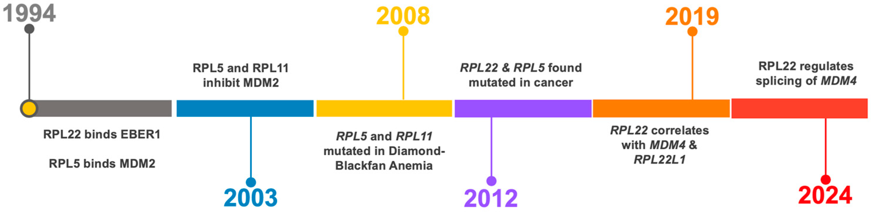
2. The p53—Ribosome Connection

p53 acts as a guardian of cellular homeostasis by responding to DNA damage, oxidative stress, and impaired ribosome biogenesis [15]. As mentioned, p53 is suppressed by its negative regulators, MDM2 and MDM4; the inactivation of either is embryonically lethal [16,17]. MDM2, an E3 ubiquitin ligase, promotes p53 degradation, while MDM4 inhibits p53’s transcriptional activity [18]. MDM2 and MDM4 are often amplified in cancers leading to the abnormal suppression of p53 [19]. The disruption of ribosome biogenesis is one of the most potent triggers of the p53 pathway [20,21,22,23]. This is thought to be mediated at least in part by the 5S RNP complex, composed of RPL5 (uL18), RPL11 (uL5), and 5S rRNA, a precursor in ribosome assembly within the nucleus [24]. Under normal cell growth conditions, 5S RNP is assembled into ribosomes, but if the ribosome assembly pathway is dysfunctional, its free form binds to MDM2, blocking its ability to degrade p53, thereby inducing cell cycle arrest (Figure 1) [11,25,26]. The loss of RPL5 or RPL11, two RPs that work in tandem, disables this p53 checkpoint [11,26,27,28]. A recent study provided much-needed biochemical insights into the 5S RNP-MDM2 complex and described a physical association with the SURF2 (Surfeit 2) protein [29]. SURF2 acts as a buffering component within the 5S RNP by antagonizing MDM2. The depletion of SURF2 activates p53 by allowing more MDM2 to be tethered to 5S RNP. Other proteins bound to this complex include HEATR3 and La/Sjögren syndrome type B antigen [29].
Ribosomal protein RPL22 has been suspected to influence p53 function. Earlier studies showed that RPL22 can physically bind to MDM2, inhibiting its ability to degrade p53 [30], and RPL22 was also reported to bind Trp53 mRNA and negatively regulate its translation [31]. While other RPs, such as RPL26 and RPL23, have similarly been implicated in modulating the MDM2-p53 axis, these mechanisms often involve general effects on MDM2 E3 ligase activity or mRNA translation [32,33]. RPs can also act in p53-independent cellular stress responses [34,35]. In contrast, and as will be discussed below, RPL22 exerts a more specific function by controlling the alternative splicing of MDM4, thereby adding a new layer to the regulation of p53 activity.

3. RPL22 and RPL22L1 Paralog Pair

While RPs are highly conserved, some have paralogs that in rare cases can compensate for the loss of the main variant [36]. RPL22 and its paralog RPL22L1 (RPL22 Like-1) provide an interesting example of compensation, and in this case, it also extends beyond translation to influence cancer-related signaling pathways. The existence of RP paralogs may allow for the fine-tuning of protein synthesis, adapting to the specific needs of different cell types or various stress conditions [37,38]. In cancer, the illegitimate expression of paralogs may lead to changes in ribosome composition, potentially changing the translation of specific mRNAs affecting cell growth or survival. However, the extent to which paralog expression occurs and its relevance in human cells including cancer cells are subjects of ongoing investigations and debate [10,39,40].
RPL22L1 shares 73% amino acid sequence homology with RPL22. RPL22 is normally incorporated into the 60S large ribosomal subunit but RPL22L1 can substitute for RPL22 in ribosome assembly [41]. This compensatory capacity explains why mice lacking Rpl22 are viable and exhibit only a mild phenotype—Rpl22-L1—which compensates for the loss of Rpl22 [41]. The compensation mechanism is complex, and this paralog pair has been studied in several organisms including yeast, flies, fish, and mammals [41,42,43,44,45]. In mouse cells, Rpl22 can repress the expression of its own paralog Rpl22L1 by binding directly to its mRNA, preventing translation through splicing alterations [41]. RPL22L1 is spliced into two or more variants: RPL22L1a, the predominant stable form and incorporated into ribosomes, and a shorter, truncated form, RPL22L1b, which may perform extra-ribosomal functions [46]. A detailed discussion of the intriguing dynamics of the RPL22-RPL22L1 pair is beyond the scope of this commentary, but for further reading and examples see references [47,48,49].

4. RPL22 and RPL22L1 Alterations in Cancer

Frequent RPL22 mutations were discovered in T-cell acute lymphoblastic leukemia (T-ALL) [50], gastric cancer [51], and endometrial cancer as reported at the end of 2012 [52]. This coincided with the identification of somatic mutations in RPL5 and RPL10 in T-ALL [53]. RPL22 is often point-mutated, causing frameshifts or harboring other missense mutations, and is also deleted. Additional cancer types with alterations on RPL22 include ovarian cancer, adrenocortical carcinomas, hepatocellular carcinomas, and colon adenocarcinomas. See for example, cBioPortal, available online: https://www.cbioportal.org (accessed on 4 May 2025). Mutations and the downregulation of RPL22 expression have been observed in other malignancies as well. RPL22 point mutations are especially prevalent in cancers classified as microsatellite instability-high (MSI-H) [5]. These cancers often display the point-mutated allele RPL22 p.K15fs, explained by the fact that RPL22 has vulnerable coding mononucleotide repeats. At the time these mutations were initially reported, the functional and clinical significance was not clear, however, a tumor-suppressive role in T-cell lymphoma was suggested [50]. The correlation between mutant RPL22, RPL22L1, and MDM4 splicing pattern was described a few years later [54]. RPL22L1 expression is typically low in normal tissues but is upregulated in response to RPL22 loss of function. Moreover, RPL22L1 is frequently amplified in certain cancer types. See for example, cBioPortal, available online: https://www.cbioportal.org (accessed on 4 May 2025).

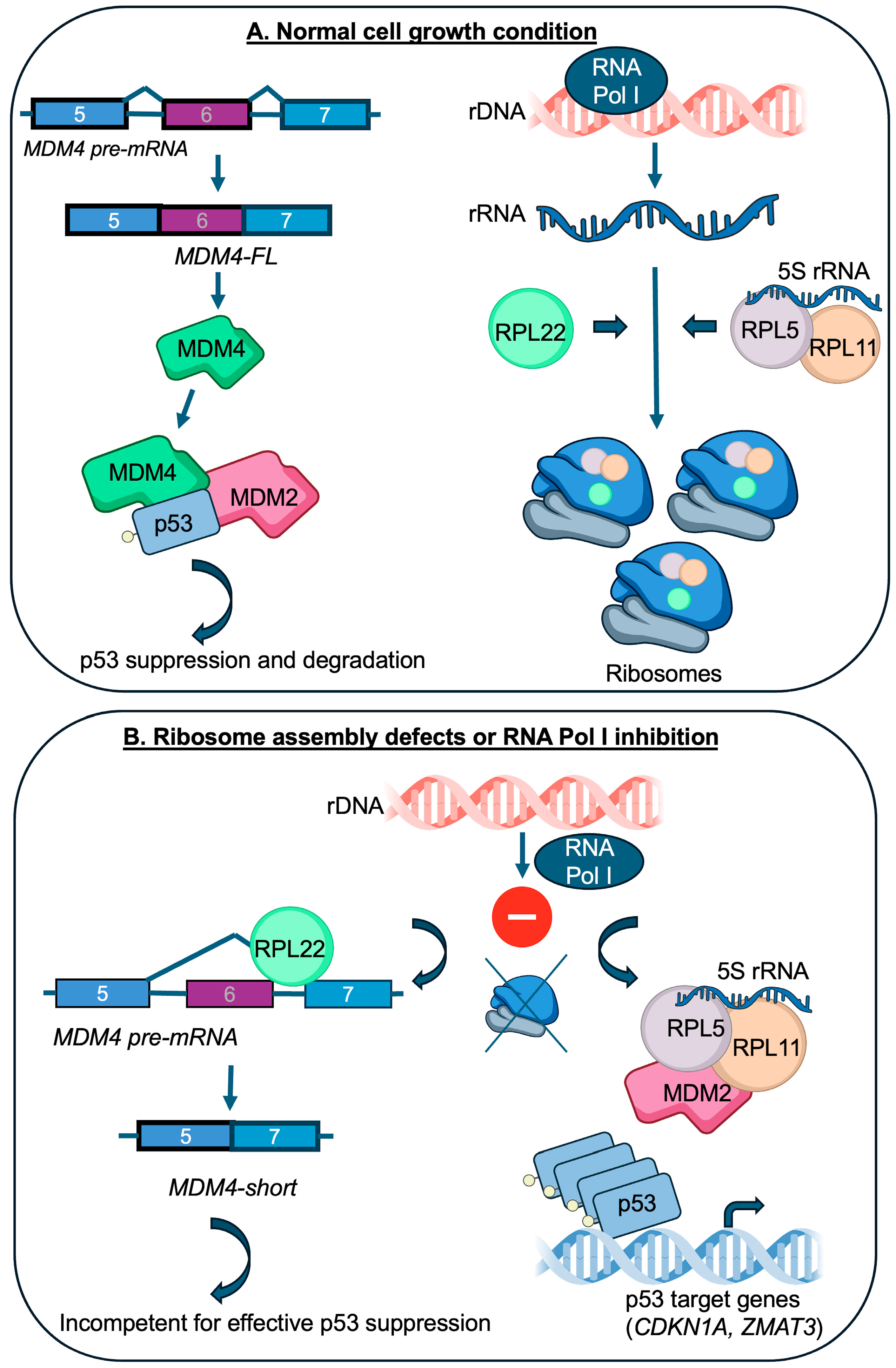
5. RPL22 Becomes Connected to MDM4 and p53

Splicing is an important mechanism affecting MDM4 function [19,55]. The inclusion of exon 6 results in the production of full-length MDM4, which effectively suppresses p53. In contrast, the exclusion of exon 6 produces a shorter, unstable MDM4 isoform that fails to inhibit p53 effectively [55]. The splicing of MDM4 is governed by multiple proteins, including the p53 target ZMAT3 (previously known as Wig-1), which promotes exon 6 skipping [56]. In 2023 and 2024, several groups published exciting findings that clarified the functional link between RPL22 and MDM4 (see timeline in Figure 1). As a prelude, Howard et al. (2023) provided a ribosome-centered angle on MDM4 and p53 signaling [13]. Their lab studied inhibitors targeting the chromatin-associated protein WDR5 (WD repeat domain 5). Interesting findings on their own, inhibiting WDR5 disrupts RP gene transcription, causing ribosome biogenesis stress and the activation of p53 in leukemia cells [13]. Treatment with the WDR5 inhibitors led to changes in MDM4 splicing (exclusion of exon 6) correlated with reduced levels of RPL22L1 [13].
Weinstein et al., (2024), identified RPL22 as a tumor suppressor in MSI-H cancers and demonstrated that it alters MDM4 splicing by directly binding to its pre-mRNA. They also showed that RPL22 controls the splicing of other pre-mRNAs, including RPL22L1 and UBAP2L (Ubiquitin-Associated Protein 2 Like) resulting in decreased levels [5]. The deletion of RPL22 led to increased inclusion of MDM4 exon 6, augmenting full length and active MDM4 protein expression. Furthermore, reduced expression of RPL22 was associated with increased proliferation of cancer cells, and resistance to Nutlin-3a, an MDM2 inhibitor. In the same issue, Jansen et al., (2024), elegantly dissected the full mechanism by which RPL22 regulates MDM4 splicing to activate p53 [14]. This team could show how RPL22 binds to specific elements, stem-loop structures, within MDM4 intron 6, promoting exon 6 skipping, which then leads to the predominant production of the unstable MDM4 isoform enhancing p53 activation in response to 5-fluorouracil (a cytostatic compound inducing nucleolar stress). Jansen et al. also described UBAP2L and RPL22L1 splicing by RPL22. Importantly, Weinstein et al., (2024), and Jansen et al., (2024), confirmed the direct binding of RPL22 to MDM4 pre-mRNA using cross-linking and immunoprecipitation [5,14]. Fan et al., (2024), pre-print, also demonstrated that RPL22 interacts with mRNA splice junctions, affecting the splicing of RPL22L1 and MDM4 following RNA Polymerase I (RNA Pol I) inhibition [57]. The study indicates numerous (hundreds) potential splicing changes in response to the targeting of RNA Pol I, suggesting effects on splicing beyond RPL22L1 and MDM4.
Collectively, these studies establish RPL22 as a novel regulator of MDM4 with possible future implications for cancer prognosis and therapy. They also pinpoint RPL22L1 as a compensatory paralog whose expression increases when RPL22 expression is decreased. It should be noted that the studies mostly rely on cancer cell lines and knockout cell line models in vitro, which may not fully capture the complexity in vivo. While RPL22L1 can partially compensate for RPL22, altered expression level of RPL22 may result in changes in other RPs, including RPL5 and RPL11, because the synthesis of RPs is regulated in a coordinated and balanced manner [58,59]. RPL22’s role in MDM4 splicing is now clearer, but how it coordinates with other splicing factors is still poorly understood, although there are some clues. SRSF1 splicing factor has already been linked to RPL5 and 5S RNP [60]. Another example is SRSF3 that is connected to MDM4 splicing [61], and SRSF4 to the splicing of RPL22L1 [46]. Current evidence presented does not support a direct role for RPL22 in regulating MDM2 or TP53 splicing, and it remains to be seen whether there are effects on splicing of p53 target genes.
In summary, the disruption of ribosome biogenesis (inhibition of RNA Pol I) sets free more RPL22 in the nucleus that becomes available to bind MDM4 pre-mRNA. This is likely to occur in parallel with an increase in free RPL5 and RPL11 that engage MDM2 as is illustrated in the lower panel of Figure 1. In this setting, RPL11 and RPL5 increase both the stability and activity of the p53 protein by inhibition of MDM2, whereas RPL22 has little effect on stress-induced p53 protein stabilization mainly influencing its transcriptional activity. Future work should take care to separate the consequences of RPL22 loss of function and exchange with RPL22L1 in the ribosome from its extra-ribosomal roles in splicing. A more general experimental challenge in the field is whether the observed effects of drugs or mutant RPs are due to altered ribosome content, ribosomal stress responses, or extra-ribosomal functions [39].

6. RPL22-MDM4 and Implications for Cancer Biology

Evidently, RPL22 and RPL5 have now emerged as important regulators of p53 signaling. It is a curiosity that RPL22 and RPL5 were both identified as binding to non-ribosomal targets in the early 1990s and later found mutated in cancers in the 2010s (see a timeline of discoveries in Figure 2). RPL22 was found to associate with Epstein–Barr virus (EBV)-expressed small RNAs (EBERs) [62,63,64,65], while RPL5 was shown to bind MDM2 in association with 5S rRNA [66]. These early findings hinted at extra-ribosomal functions for both proteins, though the biological significance remained unclear at the time. The impact of RP-mediated p53 regulation appears to vary across cancer types and likely has multiple explanations. In some cancers MDM2-mediated p53 degradation may dominate, while others may depend more on the MDM4 suppression of p53 activity [18]. This variation could determine the relative importance of specific RPs in different cancer types. In tumors with mutant p53, RP-mediated control may become less relevant, although keep in mind that MDM2 and MDM4 possess p53-independent functions.
The discovery of the RPL22-MDM4 connection may help resolve several observations in the field that have been difficult to fit within the model of RP-MDM2 dynamics. First, it was observed that MDM4 levels decreased following the inhibition of ribosome biogenesis [71]. That RPL22 directly controls MDM4 pre-mRNA splicing [12] now emerges as a likely explanation. Second, the activation of the p53-p21 link or induction of p53-dependent apoptosis has occasionally been observed under conditions of ribosomal stress despite the inactivation of the 5S RNP-p53 control [72,73]. It is tempting to speculate that RPL22-MDM4 may be involved in such situations, but this needs to be experimentally tested. Third, the link to MDM4 may resolve some issues on the complex and essential roles of RPL22 and RPL22L1 seen in the development of B and T cells, and the activation of p53 [47,48]. A disruption in the RPL22:RPL22L1 ratio may impact ribosome homeostasis and p53 via MDM4 in hematopoietic stem cells, B and T cells. Fourth, the binding of EBERs to RPL22 with the subsequent induction of RPL22L1 may have a role in modulating growth patterns during EBV latency [74,75], with implications for EBV-positive Burkitt lymphoma.
While RPL22 is mutated in cancer and clearly implicated in MDM4 splicing, what is the evidence regarding RPL5 and RPL11 in cancer? RPL5 heterozygous point mutations or heterozygous deletions, which often show an anti-correlation with TP53 mutations, are observed in glioblastomas, multiple myelomas, breast cancer, and several other cancer types. This supports the idea that some tumors may selectively bypass 5S RNP-mediated p53 activation by reducing the function and/or expression of RPL5 [7]. In contrast, RPL11 mutations are much less frequent and show no clear correlation with p53 status [7]. Mutations in the MDM2 zinc finger, which disrupt its interaction with the 5S RNP complex, have been very useful in biochemical studies, but are exceedingly rare and unlikely to play a major role in cancer. The scarcity of MDM2 or RPL11 mutations that would impair 5S RNP control of MDM2 function suggests that tumors rarely target this pathway directly, aside from RPL5 or p53. However, the regulation of the 5S RNP-MDM2 complex could occur through alternative mechanisms, such as altered levels of SURF2 [29]. Importantly, the deletion of Rpl11 or inactivation of the Mdm2 zinc finger region accelerates lymphoma development in mouse models [76,77]. Furthermore, a study investigating clonal dynamics in hematopoietic cell colonies from individuals with Schwachman–Diamond syndrome (a ribosome disorder with the activation of p53) found that mutations in TP53, RPL5, and RPL22 appeared rather frequently as escape mechanisms to overcome p53-imposed growth arrest [78].

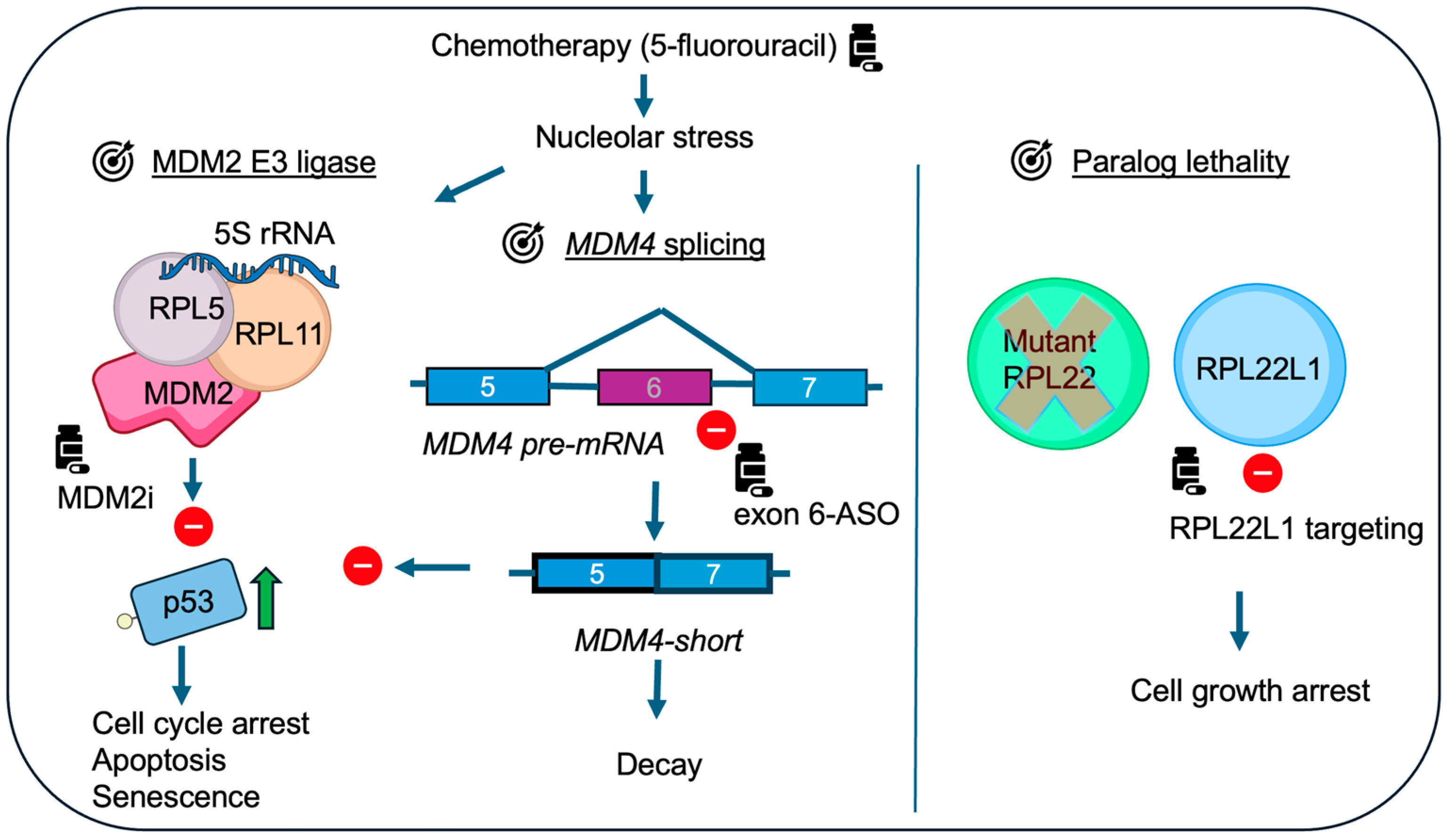
7. RPL22-MDM4 and Implications for Cancer Treatment

One of the key take-home messages from the studies by Weinstein et al., Jansen et al., and Howard et al. is that RPL22 loss-of-function reduces sensitivity to rather different ribosome biogenesis inhibitors and Nutlin-3a, suggesting a shared putative resistance mechanism across distinct classes of chemotherapies [5,13,14]. RPs including RPL5 and RPL22 influence chemotherapy response in vitro, particularly in cancer cell lines that retain wild-type p53 [5,13,14,79,80]. Given their role in the regulation of p53-MDM2-MDM4, these RPs may affect sensitivity to small molecule MDM2 inhibitors. For example, cancers with reduced RPL5 might show increased dependence on MDM2 and thus potentially render them more susceptible to MDM2 inhibition. However, the efficacy of MDM2 inhibitors in such context needs to be determined. One also has to keep in mind that alterations in RPL5 are less frequent, and MDM2 inhibitors are not yet approved for clinical use [18]. Restoring MDM4 exon skipping, resulting in reduced levels of full-length MDM4 and enhanced p53 activation, has been suggested as an experimental strategy (Figure 3). Splicing modulators such as SF3B1 inhibitors have been explored in preclinical models to shift splicing patterns in favor of p53 activation, though they can have toxic side effects in normal tissues [81]. In addition to p53-MDM2-MDM4 dynamics, broader alterations in ribosome assembly caused by the loss of RPL5 or RPL22, and general effects on transcription and translation are likely to contribute to chemotherapy responses and resistance.
The roles of RPL22 and RPL22L1 in cancer are of interest beyond their connection to MDM4. Their functional dynamics are likely context-dependent, varying by tissue and cancer type. RPL22L1 is frequently amplified in cancers, but is this a reflection of a direct oncogenic role (that is increasing MDM4)? It could be argued that the loss of RPL22 is compensated by RPL22L1 in a way that is not necessarily oncogenic but simply serves to maintain ribosome function. However, the overexpression of RPL22L1 has been associated with enhanced malignant phenotypes in cancer such as increased cell proliferation and resistance to chemotherapeutic agents, such as sorafenib in hepatocellular carcinomas [82], temozolomide in glioblastomas [83], and 5-fluorouracil in colorectal cancer [84]. RPL22L1 expression promotes cell proliferation and invasion, in part, through the ERK signaling pathway [82]. While some of these effects can be potentially explained by unrestrained MDM4 action on p53, findings from studies on alternatively spliced isoforms of RPL22L1 in glioblastoma [46] suggest the presence of additional more complex mechanisms. Given that the reduction in RPL22 is compensated for by RPL22L1, targeting RPL22L1 may exploit a paralog synthetic lethality approach as indicated [85]. RNA-targeting drugs could be tested experimentally to selectively reduce RPL22L1 expression (Figure 3). It is important to keep in mind that most RPs are pan-essential and targeting any of them would be expected to result in toxic side effects in normal tissues. But, reliance on paralogs opens an interesting window of opportunity in cancer cells. Finally, RPs are candidates as clinically relevant biomarkers in oncology. RPL22 loss of function correlates with poor prognosis in T-ALL [50,86] and in aggressive microsatellite instability-high (MSI-H) tumors [5], while RPL22L1 overexpression predicts resistance to sorafenib in hepatocellular carcinoma [82]. High RPL22L1 expression in colorectal cancer correlates with poor prognosis [84] and in lung adenocarcinomas [87,88]. These examples underscore the potential of RP-based biomarkers for prognosis, patient stratification, and treatment selection.

8. Conclusions

As our understanding of RP biology expands, it becomes increasingly clear that RP mutations and extra-ribosomal RP activities are not merely collateral to tumor progression. First, studies on RPL22 add to a growing body of evidence demonstrating that many RPs have extra-ribosomal functions. Second, RPL22 and RPL22L1 exemplify the complexity of RP paralogs, indicating the broader significance of RP-regulated networks. Third, the role of RPL22 in regulating MDM4 splicing introduces a new dimension to the p53 pathway, which has traditionally focused on the RP-MDM2 axis. As is now clear, RPL22 mutations disable an important control of MDM4-mediated p53 suppression. The latest studies thus position RPL22 at the center of a ribosome-/nucleolus-related regulatory axis, distinct from yet integrated with, the 5S RNP-MDM2-p53 module. From a therapeutic standpoint, modulating MDM4 splicing or exploiting RPL22 paralog synthetic lethality may offer strategies for reactivating p53 in cancers with intact but suppressed wild-type function. Even in the context of mutant p53, targeting RPL22L1 could be an option. Several important questions remain: what are the precise mechanisms governing MDM2-5S RNP dynamics, MDM4 splicing, and p53 mRNA translation? In addition, there are more fundamental questions: to what extent do these mechanisms drive tumor development versus determine chemotherapy response in vitro and in vivo? And, a question that often comes to mind is why did p53 evolve such an intimate relationship with the ribosome and RPs? A true understanding of these connections may provide insight into cancer development.

Funding

No funding has been received for this commentary.

Institutional Review Board Statement

This work did not require ethical approval.

Informed Consent Statement

Not applicable.

Data Availability Statement

This commentary has not generated new data.

Acknowledgments

I thank past and present members of Jiri Bartek group in Stockholm, Karolinska Institutet, for discussions and support. I also thank Siniša Volarević, Univ. of Rijeka, Croatia, for interesting discussions on ribosomal proteins and p53 in cancer.

Conflicts of Interest

The author declares no conflicts of interest.

References

  1. Warner, J.R.; McIntosh, K.B. How Common Are Extraribosomal Functions of Ribosomal Proteins? Mol. Cell 2009, 34, 3–11. [Google Scholar] [CrossRef] [PubMed]
  2. Fancello, L.; Kampen, K.R.; Hofman, I.J.F.; Verbeeck, J.; De Keersmaecker, K. The Ribosomal Protein Gene RPL5 Is a Haploinsufficient Tumor Suppressor in Multiple Cancer Types. Oncotarget 2017, 8, 14462–14478. [Google Scholar] [CrossRef] [PubMed]
  3. Ljungström, V.; Cortese, D.; Young, E.; Pandzic, T.; Mansouri, L.; Plevova, K.; Ntoufa, S.; Baliakas, P.; Clifford, R.; Sutton, L.-A.; et al. Whole-Exome Sequencing in Relapsing Chronic Lymphocytic Leukemia: Clinical Impact of Recurrent RPS15 Mutations. Blood 2016, 127, 1007–1016. [Google Scholar] [CrossRef] [PubMed]
  4. Kampen, K.R.; Sulima, S.O.; Verbelen, B.; Girardi, T.; Vereecke, S.; Rinaldi, G.; Verbeeck, J.; Op de Beeck, J.; Uyttebroeck, A.; Meijerink, J.P.P.; et al. The Ribosomal RPL10 R98S Mutation Drives IRES-Dependent BCL-2 Translation in T-ALL. Leukemia 2019, 33, 319–332. [Google Scholar] [CrossRef]
  5. Weinstein, H.N.W.; Hu, K.; Fish, L.; Chen, Y.-A.; Allegakoen, P.; Pham, J.H.; Hui, K.S.F.; Chang, C.-H.; Tutar, M.; Benitez-Rivera, L.; et al. RPL22 Is a Tumor Suppressor in MSI-High Cancers and a Splicing Regulator of MDM4. Cell Rep. 2024, 43, 114622. [Google Scholar] [CrossRef]
  6. Ferreira, A.M.; Tuominen, I.; van Dijk-Bos, K.; Sanjabi, B.; van der Sluis, T.; van der Zee, A.G.; Hollema, H.; Zazula, M.; Sijmons, R.H.; Aaltonen, L.A.; et al. High Frequency of RPL22 Mutations in Microsatellite-Unstable Colorectal and Endometrial Tumors. Hum. Mutat. 2014, 35, 1442–1445. [Google Scholar] [CrossRef]
  7. Oršolić, I.; Bursać, S.; Jurada, D.; Drmić Hofman, I.; Dembić, Z.; Bartek, J.; Mihalek, I.; Volarević, S. Cancer-Associated Mutations in the Ribosomal Protein L5 Gene Dysregulate the HDM2/P53-Mediated Ribosome Biogenesis Checkpoint. Oncogene 2020, 39, 3443–3457. [Google Scholar] [CrossRef]
  8. Ajore, R.; Raiser, D.; McConkey, M.; Jöud, M.; Boidol, B.; Mar, B.; Saksena, G.; Weinstock, D.M.; Armstrong, S.; Ellis, S.R.; et al. Deletion of Ribosomal Protein Genes Is a Common Vulnerability in Human Cancer, Especially in Concert with TP53 Mutations. EMBO Mol. Med. 2017, 9, 498–507. [Google Scholar] [CrossRef]
  9. Kulkarni, S.; Dolezal, J.M.; Wang, H.; Jackson, L.; Lu, J.; Frodey, B.P.; Dosunmu-Ogunbi, A.; Li, Y.; Fromherz, M.; Kang, A.; et al. Ribosomopathy-like Properties of Murine and Human Cancers. PLoS ONE 2017, 12, e0182705. [Google Scholar] [CrossRef]
  10. Caruso, M.; De Keersmaecker, K. Ribosome Specialization by Cancer-Associated Ribosomal Protein Mutations: Progress Made and Open Questions. Philos. Trans. R. Soc. Lond. B Biol. Sci. 2025, 380, 20230380. [Google Scholar] [CrossRef]
  11. Sloan, K.E.; Bohnsack, M.T.; Watkins, N.J. The 5S RNP Couples P53 Homeostasis to Ribosome Biogenesis and Nucleolar Stress. Cell Rep. 2013, 5, 237–247. [Google Scholar] [CrossRef] [PubMed]
  12. Jansen, J.; Dobbelstein, M. MDM4 Exon Skipping upon Dysfunctional Ribosome Assembly. Trends Cell Biol. 2024, S0962-8924(24)00212-5. [Google Scholar] [CrossRef] [PubMed]
  13. Howard, G.C.; Wang, J.; Rose, K.L.; Jones, C.; Patel, P.; Tsui, T.; Florian, A.C.; Vlach, L.; Lorey, S.L.; Grieb, B.C.; et al. Ribosome Subunit Attrition and Activation of the P53-MDM4 Axis Dominate the Response of MLL-Rearranged Cancer Cells to WDR5 WIN Site Inhibition. eLife 2024, 12, RP90683. [Google Scholar] [CrossRef] [PubMed]
  14. Jansen, J.; Bohnsack, K.E.; Böhlken-Fascher, S.; Bohnsack, M.T.; Dobbelstein, M. The Ribosomal Protein L22 Binds the MDM4 Pre-mRNA and Promotes Exon Skipping to Activate P53 upon Nucleolar Stress. Cell Rep. 2024, 43, 114610. [Google Scholar] [CrossRef]
  15. Liu, Y.; Su, Z.; Tavana, O.; Gu, W. Understanding the Complexity of P53 in a New Era of Tumor Suppression. Cancer Cell 2024, 42, 946–967. [Google Scholar] [CrossRef]
  16. Gu, J.; Kawai, H.; Nie, L.; Kitao, H.; Wiederschain, D.; Jochemsen, A.G.; Parant, J.; Lozano, G.; Yuan, Z.-M. Mutual Dependence of MDM2 and MDMX in Their Functional Inactivation of P53. J. Biol. Chem. 2002, 277, 19251–19254. [Google Scholar] [CrossRef]
  17. Parant, J.; Chavez-Reyes, A.; Little, N.A.; Yan, W.; Reinke, V.; Jochemsen, A.G.; Lozano, G. Rescue of Embryonic Lethality in MDM4-Null Mice by Loss of Trp53 Suggests a Nonoverlapping Pathway with MDM2 to Regulate P53. Nat. Genet. 2001, 29, 92–95. [Google Scholar] [CrossRef]
  18. Peuget, S.; Zhou, X.; Selivanova, G. Translating P53-Based Therapies for Cancer into the Clinic. Nat. Rev. Cancer 2024, 24, 192–215. [Google Scholar] [CrossRef]
  19. Wu, J.; Lu, G.; Wang, X. MDM4 Alternative Splicing and Implication in MDM4 Targeted Cancer Therapies. Am. J. Cancer Res. 2021, 11, 5864–5880. [Google Scholar]
  20. Hannan, K.M.; Soo, P.; Wong, M.S.; Lee, J.K.; Hein, N.; Poh, P.; Wysoke, K.D.; Williams, T.D.; Montellese, C.; Smith, L.K.; et al. Nuclear Stabilization of P53 Requires a Functional Nucleolar Surveillance Pathway. Cell Rep. 2022, 41, 111571. [Google Scholar] [CrossRef]
  21. Deisenroth, C.; Franklin, D.A.; Zhang, Y. The Evolution of the Ribosomal Protein-MDM2-P53 Pathway. Cold Spring Harb. Perspect. Med. 2016, 6, a026138. [Google Scholar] [CrossRef] [PubMed]
  22. Rubbi, C.P.; Milner, J. Disruption of the Nucleolus Mediates Stabilization of P53 in Response to DNA Damage and Other Stresses. EMBO J. 2003, 22, 6068–6077. [Google Scholar] [CrossRef] [PubMed]
  23. Pestov, D.G.; Strezoska, Z.; Lau, L.F. Evidence of P53-Dependent Cross-Talk between Ribosome Biogenesis and the Cell Cycle: Effects of Nucleolar Protein Bop1 on G(1)/S Transition. Mol. Cell. Biol. 2001, 21, 4246–4255. [Google Scholar] [CrossRef]
  24. Steitz, J.A.; Berg, C.; Hendrick, J.P.; La Branche-Chabot, H.; Metspalu, A.; Rinke, J.; Yario, T. A 5S rRNA/L5 Complex Is a Precursor to Ribosome Assembly in Mammalian Cells. J. Cell Biol. 1988, 106, 545–556. [Google Scholar] [CrossRef]
  25. Castillo Duque de Estrada, N.M.; Thoms, M.; Flemming, D.; Hammaren, H.M.; Buschauer, R.; Ameismeier, M.; Baßler, J.; Beck, M.; Beckmann, R.; Hurt, E. Structure of Nascent 5S RNPs at the Crossroad between Ribosome Assembly and MDM2-P53 Pathways. Nat. Struct. Mol. Biol. 2023, 30, 1119–1131. [Google Scholar] [CrossRef]
  26. Donati, G.; Peddigari, S.; Mercer, C.A.; Thomas, G. 5S Ribosomal RNA Is an Essential Component of a Nascent Ribosomal Precursor Complex That Regulates the Hdm2-P53 Checkpoint. Cell Rep. 2013, 4, 87–98. [Google Scholar] [CrossRef]
  27. Teng, T.; Mercer, C.A.; Hexley, P.; Thomas, G.; Fumagalli, S. Loss of Tumor Suppressor RPL5/RPL11 Does Not Induce Cell Cycle Arrest but Impedes Proliferation Due to Reduced Ribosome Content and Translation Capacity. Mol. Cell. Biol. 2013, 33, 4660–4671. [Google Scholar] [CrossRef]
  28. Bursać, S.; Brdovčak, M.C.; Pfannkuchen, M.; Orsolić, I.; Golomb, L.; Zhu, Y.; Katz, C.; Daftuar, L.; Grabušić, K.; Vukelić, I.; et al. Mutual Protection of Ribosomal Proteins L5 and L11 from Degradation Is Essential for P53 Activation upon Ribosomal Biogenesis Stress. Proc. Natl. Acad. Sci. USA 2012, 109, 20467–20472. [Google Scholar] [CrossRef]
  29. Tagnères, S.; Santo, P.E.; Radermecker, J.; Rinaldi, D.; Froment, C.; Provost, Q.; Bongers, M.; Capeille, S.; Watkins, N.; Marcoux, J.; et al. SURF2 Is a MDM2 Antagonist in Triggering the Nucleolar Stress Response. Nat. Commun. 2024, 15, 8404. [Google Scholar] [CrossRef]
  30. Cao, B.; Fang, Z.; Liao, P.; Zhou, X.; Xiong, J.; Zeng, S.; Lu, H. Cancer-Mutated Ribosome Protein L22 (RPL22/eL22) Suppresses Cancer Cell Survival by Blocking P53-MDM2 Circuit. Oncotarget 2017, 8, 90651–90661. [Google Scholar] [CrossRef]
  31. Rashkovan, M.; Vadnais, C.; Ross, J.; Gigoux, M.; Suh, W.-K.; Gu, W.; Kosan, C.; Möröy, T. Miz-1 Regulates Translation of Trp53 via Ribosomal Protein L22 in Cells Undergoing V(D)J Recombination. Proc. Natl. Acad. Sci. USA 2014, 111, E5411–E5419. [Google Scholar] [CrossRef] [PubMed]
  32. Takagi, M.; Absalon, M.J.; McLure, K.G.; Kastan, M.B. Regulation of P53 Translation and Induction after DNA Damage by Ribosomal Protein L26 and Nucleolin. Cell 2005, 123, 49–63. [Google Scholar] [CrossRef] [PubMed]
  33. Dai, M.-S.; Zeng, S.X.; Jin, Y.; Sun, X.-X.; David, L.; Lu, H. Ribosomal Protein L23 Activates P53 by Inhibiting MDM2 Function in Response to Ribosomal Perturbation but Not to Translation Inhibition. Mol. Cell. Biol. 2004, 24, 7654–7668. [Google Scholar] [CrossRef] [PubMed]
  34. Russo, A.; Russo, G. Ribosomal Proteins Control or Bypass P53 during Nucleolar Stress. Int. J. Mol. Sci. 2017, 18, 140. [Google Scholar] [CrossRef]
  35. James, A.; Wang, Y.; Raje, H.; Rosby, R.; DiMario, P. Nucleolar Stress with and without P53. Nucleus 2014, 5, 402–426. [Google Scholar] [CrossRef]
  36. Milenkovic, I.; Novoa, E.M. Ribosomal Protein Paralogues in Ribosome Specialization. Philos. Trans. R. Soc. Lond. B Biol. Sci. 2025, 380, 20230387. [Google Scholar] [CrossRef]
  37. Ghulam, M.M.; Catala, M.; Abou Elela, S. Differential Expression of Duplicated Ribosomal Protein Genes Modifies Ribosome Composition in Response to Stress. Nucleic Acids Res. 2020, 48, 1954–1968. [Google Scholar] [CrossRef]
  38. Malik Ghulam, M.; Catala, M.; Reulet, G.; Scott, M.S.; Abou Elela, S. Duplicated Ribosomal Protein Paralogs Promote Alternative Translation and Drug Resistance. Nat. Commun. 2022, 13, 4938. [Google Scholar] [CrossRef]
  39. Ramalho, S.; Dopler, A.; Faller, W.J. Ribosome Specialization in Cancer: A Spotlight on Ribosomal Proteins. NAR Cancer 2024, 6, zcae029. [Google Scholar] [CrossRef]
  40. Fuentes, P.; Pelletier, J.; Gentilella, A. Decoding Ribosome Complexity: Role of Ribosomal Proteins in Cancer and Disease. NAR Cancer 2024, 6, zcae032. [Google Scholar] [CrossRef]
  41. O’Leary, M.N.; Schreiber, K.H.; Zhang, Y.; Duc, A.-C.E.; Rao, S.; Hale, J.S.; Academia, E.C.; Shah, S.R.; Morton, J.F.; Holstein, C.A.; et al. The Ribosomal Protein RPL22 Controls Ribosome Composition by Directly Repressing Expression of Its Own Paralog, RPL22L1. PLoS Genet. 2013, 9, e1003708. [Google Scholar] [CrossRef] [PubMed]
  42. Abrhámová, K.; Nemčko, F.; Libus, J.; Převorovský, M.; Hálová, M.; Půta, F.; Folk, P. Introns Provide a Platform for Intergenic Regulatory Feedback of RPL22 Paralogs in Yeast. PLoS ONE 2018, 13, e0190685. [Google Scholar] [CrossRef] [PubMed]
  43. Gabunilas, J.; Chanfreau, G. Splicing-Mediated Autoregulation Modulates RPL22p Expression in Saccharomyces Cerevisiae. PLoS Genet. 2016, 12, e1005999. [Google Scholar] [CrossRef] [PubMed]
  44. Kearse, M.G.; Chen, A.S.; Ware, V.C. Expression of Ribosomal Protein L22e Family Members in Drosophila Melanogaster: RPL22-like Is Differentially Expressed and Alternatively Spliced. Nucleic Acids Res. 2011, 39, 2701–2716. [Google Scholar] [CrossRef]
  45. Zhang, Y.; O’Leary, M.N.; Peri, S.; Wang, M.; Zha, J.; Melov, S.; Kappes, D.J.; Feng, Q.; Rhodes, J.; Amieux, P.S.; et al. Ribosomal Proteins RPL22 and RPL22L1 Control Morphogenesis by Regulating Pre-mRNA Splicing. Cell Rep. 2017, 18, 545–556. [Google Scholar] [CrossRef]
  46. Larionova, T.D.; Bastola, S.; Aksinina, T.E.; Anufrieva, K.S.; Wang, J.; Shender, V.O.; Andreev, D.E.; Kovalenko, T.F.; Arapidi, G.P.; Shnaider, P.V.; et al. Alternative RNA Splicing Modulates Ribosomal Composition and Determines the Spatial Phenotype of Glioblastoma Cells. Nat. Cell Biol. 2022, 24, 1541–1557. [Google Scholar] [CrossRef]
  47. Fahl, S.P.; Sertori, R.; Zhang, Y.; Contreras, A.V.; Harris, B.; Wang, M.; Perrigoue, J.; Balachandran, S.; Kennedy, B.K.; Wiest, D.L. Loss of Ribosomal Protein Paralog RPL22-like1 Blocks Lymphoid Development without Affecting Protein Synthesis. J. Immunol. 2022, 208, 870–880. [Google Scholar] [CrossRef]
  48. Fahl, S.P.; Harris, B.; Coffey, F.; Wiest, D.L. RPL22 Loss Impairs the Development of B Lymphocytes by Activating a P53-Dependent Checkpoint. J. Immunol. 2015, 194, 200–209. [Google Scholar] [CrossRef]
  49. Fahl, S.P.; Wang, M.; Zhang, Y.; Duc, A.-C.E.; Wiest, D.L. Regulatory Roles of RPL22 in Hematopoiesis: An Old Dog with New Tricks. Crit. Rev. Immunol. 2015, 35, 379–400. [Google Scholar] [CrossRef]
  50. Rao, S.; Lee, S.-Y.; Gutierrez, A.; Perrigoue, J.; Thapa, R.J.; Tu, Z.; Jeffers, J.R.; Rhodes, M.; Anderson, S.; Oravecz, T.; et al. Inactivation of Ribosomal Protein L22 Promotes Transformation by Induction of the Stemness Factor, Lin28B. Blood 2012, 120, 3764–3773. [Google Scholar] [CrossRef]
  51. Nagarajan, N.; Bertrand, D.; Hillmer, A.M.; Zang, Z.J.; Yao, F.; Jacques, P.-É.; Teo, A.S.M.; Cutcutache, I.; Zhang, Z.; Lee, W.H.; et al. Whole-Genome Reconstruction and Mutational Signatures in Gastric Cancer. Genome Biol. 2012, 13, R115. [Google Scholar] [CrossRef] [PubMed]
  52. Novetsky, A.P.; Zighelboim, I.; Thompson, D.M.; Powell, M.A.; Mutch, D.G.; Goodfellow, P.J. Frequent Mutations in the RPL22 Gene and Its Clinical and Functional Implications. Gynecol. Oncol. 2013, 128, 470–474. [Google Scholar] [CrossRef] [PubMed]
  53. De Keersmaecker, K.; Atak, Z.K.; Li, N.; Vicente, C.; Patchett, S.; Girardi, T.; Gianfelici, V.; Geerdens, E.; Clappier, E.; Porcu, M.; et al. Exome Sequencing Identifies Mutation in CNOT3 and Ribosomal Genes RPL5 and RPL10 in T-Cell Acute Lymphoblastic Leukemia. Nat. Genet. 2013, 45, 186–190. [Google Scholar] [CrossRef]
  54. Ghandi, M.; Huang, F.W.; Jané-Valbuena, J.; Kryukov, G.V.; Lo, C.C.; McDonald, E.R.; Barretina, J.; Gelfand, E.T.; Bielski, C.M.; Li, H.; et al. Next-Generation Characterization of the Cancer Cell Line Encyclopedia. Nature 2019, 569, 503–508. [Google Scholar] [CrossRef]
  55. Marine, J.-C.; Jochemsen, A.G. MDMX (MDM4), a Promising Target for P53 Reactivation Therapy and Beyond. Cold Spring Harb. Perspect. Med. 2016, 6, a026237. [Google Scholar] [CrossRef]
  56. Bieging-Rolett, K.T.; Kaiser, A.M.; Morgens, D.W.; Boutelle, A.M.; Seoane, J.A.; Van Nostrand, E.L.; Zhu, C.; Houlihan, S.L.; Mello, S.S.; Yee, B.A.; et al. Zmat3 Is a Key Splicing Regulator in the P53 Tumor Suppression Program. Mol. Cell 2020, 80, 452–469.e9. [Google Scholar] [CrossRef]
  57. Fan, W.; Liu, H.; Stachelek, G.C.; Begum, A.; Davis, C.E.; Dorado, T.E.; Ernst, G.; Reinhold, W.C.; Ozbek, B.; Zheng, Q.; et al. Ribosomal RNA Transcription Governs Splicing through Ribosomal Protein RPL22. bioRxiv 2024. bioRxiv:2024.08.15.608201. [Google Scholar] [CrossRef]
  58. Perry, R.P. Balanced Production of Ribosomal Proteins. Gene 2007, 401, 1–3. [Google Scholar] [CrossRef]
  59. Ni, C.; Buszczak, M. The Homeostatic Regulation of Ribosome Biogenesis. Semin. Cell Dev. Biol. 2023, 136, 13–26. [Google Scholar] [CrossRef]
  60. Fregoso, O.I.; Das, S.; Akerman, M.; Krainer, A.R. Splicing-Factor Oncoprotein SRSF1 Stabilizes P53 via RPL5 and Induces Cellular Senescence. Mol. Cell 2013, 50, 56–66. [Google Scholar] [CrossRef]
  61. Xiong, J.; Chen, Y.; Wang, W.; Sun, J. Biological Function and Molecular Mechanism of SRSF3 in Cancer and Beyond. Oncol. Lett. 2022, 23, 21. [Google Scholar] [CrossRef] [PubMed]
  62. Dobbelstein, M.; Shenk, T. In Vitro Selection of RNA Ligands for the Ribosomal L22 Protein Associated with Epstein-Barr Virus-Expressed RNA by Using Randomized and cDNA-Derived RNA Libraries. J. Virol. 1995, 69, 8027–8034. [Google Scholar] [CrossRef] [PubMed]
  63. Toczyski, D.P.; Matera, A.G.; Ward, D.C.; Steitz, J.A. The Epstein-Barr Virus (EBV) Small RNA EBER1 Binds and Relocalizes Ribosomal Protein L22 in EBV-Infected Human B Lymphocytes. Proc. Natl. Acad. Sci. USA 1994, 91, 3463–3467. [Google Scholar] [CrossRef] [PubMed]
  64. Toczyski, D.P.; Steitz, J.A. EAP, a Highly Conserved Cellular Protein Associated with Epstein-Barr Virus Small RNAs (EBERs). EMBO J. 1991, 10, 459–466. [Google Scholar] [CrossRef]
  65. Teramoto, N.; Szekely, L.; Klein, G. Differential Expression and Localization of EBER-1 and EBER-2 in Epstein-Barr Virus-Carrying Cells. J. Hum. Virol. 1998, 1, 307–313. [Google Scholar]
  66. Marechal, V.; Elenbaas, B.; Piette, J.; Nicolas, J.C.; Levine, A.J. The Ribosomal L5 Protein Is Associated with Mdm-2 and Mdm-2-P53 Complexes. Mol. Cell. Biol. 1994, 14, 7414–7420. [Google Scholar] [CrossRef]
  67. Gazda, H.T.; Sheen, M.R.; Vlachos, A.; Choesmel, V.; O’Donohue, M.-F.; Schneider, H.; Darras, N.; Hasman, C.; Sieff, C.A.; Newburger, P.E.; et al. Ribosomal Protein L5 and L11 Mutations Are Associated with Cleft Palate and Abnormal Thumbs in Diamond-Blackfan Anemia Patients. Am. J. Hum. Genet. 2008, 83, 769–780. [Google Scholar] [CrossRef]
  68. Lohrum, M.A.E.; Ludwig, R.L.; Kubbutat, M.H.G.; Hanlon, M.; Vousden, K.H. Regulation of HDM2 Activity by the Ribosomal Protein L11. Cancer Cell 2003, 3, 577–587. [Google Scholar] [CrossRef]
  69. Zhang, Y.; Wolf, G.W.; Bhat, K.; Jin, A.; Allio, T.; Burkhart, W.A.; Xiong, Y. Ribosomal Protein L11 Negatively Regulates Oncoprotein MDM2 and Mediates a P53-Dependent Ribosomal-Stress Checkpoint Pathway. Mol. Cell. Biol. 2003, 23, 8902–8912. [Google Scholar] [CrossRef]
  70. Dai, M.-S.; Lu, H. Inhibition of MDM2-Mediated P53 Ubiquitination and Degradation by Ribosomal Protein L5. J. Biol. Chem. 2004, 279, 44475–44482. [Google Scholar] [CrossRef]
  71. Gilkes, D.M.; Chen, L.; Chen, J. MDMX Regulation of P53 Response to Ribosomal Stress. EMBO J. 2006, 25, 5614–5625. [Google Scholar] [CrossRef] [PubMed]
  72. Franklin, D.A.; Liu, S.; Jin, A.; Cui, P.; Guo, Z.; Arend, K.C.; Moorman, N.J.; He, S.; Wang, G.G.; Wan, Y.Y.; et al. Ribosomal Protein RPL11 Haploinsufficiency Causes Anemia in Mice via Activation of the RP-MDM2-P53 Pathway. J. Biol. Chem. 2023, 299, 102739. [Google Scholar] [CrossRef] [PubMed]
  73. Fukui, Y.; Hayano, S.; Kawanabe, N.; Wang, Z.; Shimada, A.; Saito, M.K.; Asaka, I.; Kamioka, H. Investigation of the Molecular Causes Underlying Physical Abnormalities in Diamond-Blackfan Anemia Patients with RPL5 Haploinsufficiency. Pathol. Int. 2021, 71, 803–813. [Google Scholar] [CrossRef] [PubMed]
  74. Houmani, J.L.; Davis, C.I.; Ruf, I.K. Growth-Promoting Properties of Epstein-Barr Virus EBER-1 RNA Correlate with Ribosomal Protein L22 Binding. J. Virol. 2009, 83, 9844–9853. [Google Scholar] [CrossRef]
  75. Paudel, S.; Lee, N. Epstein-Barr Virus Noncoding RNA EBER1 Promotes the Expression of a Ribosomal Protein Paralog to Boost Oxidative Phosphorylation. J. Med. Virol. 2024, 96, e29869. [Google Scholar] [CrossRef]
  76. Morgado-Palacin, L.; Varetti, G.; Llanos, S.; Gómez-López, G.; Martinez, D.; Serrano, M. Partial Loss of Rpl11 in Adult Mice Recapitulates Diamond-Blackfan Anemia and Promotes Lymphomagenesis. Cell Rep. 2015, 13, 712–722. [Google Scholar] [CrossRef]
  77. Macias, E.; Jin, A.; Deisenroth, C.; Bhat, K.; Mao, H.; Lindström, M.S.; Zhang, Y. An ARF-Independent c-MYC-Activated Tumor Suppression Pathway Mediated by Ribosomal Protein-Mdm2 Interaction. Cancer Cell 2010, 18, 231–243. [Google Scholar] [CrossRef]
  78. Machado, H.E.; Øbro, N.F.; Williams, N.; Tan, S.; Boukerrou, A.Z.; Davies, M.; Belmonte, M.; Mitchell, E.; Baxter, E.J.; Mende, N.; et al. Convergent Somatic Evolution Commences in Utero in a Germline Ribosomopathy. Nat. Commun. 2023, 14, 5092. [Google Scholar] [CrossRef]
  79. Sun, X.-X.; Dai, M.-S.; Lu, H. 5-Fluorouracil Activation of P53 Involves an MDM2-Ribosomal Protein Interaction. J. Biol. Chem. 2007, 282, 8052–8059. [Google Scholar] [CrossRef]
  80. Sun, X.-X.; Dai, M.-S.; Lu, H. Mycophenolic Acid Activation of P53 Requires Ribosomal Proteins L5 and L11. J. Biol. Chem. 2008, 283, 12387–12392. [Google Scholar] [CrossRef]
  81. Hepburn, L.A.; McHugh, A.; Fernandes, K.; Boag, G.; Proby, C.M.; Leigh, I.M.; Saville, M.K. Targeting the Spliceosome for Cutaneous Squamous Cell Carcinoma Therapy: A Role for c-MYC and Wild-Type P53 in Determining the Degree of Tumour Selectivity. Oncotarget 2018, 9, 23029–23046. [Google Scholar] [CrossRef] [PubMed]
  82. Zhang, D.; Zhou, Y.; Ma, Y.; Jiang, P.; Lv, H.; Liu, S.; Mu, Y.; Zhou, C.; Xiao, S.; Ji, G.; et al. Ribosomal Protein L22-like1 (RPL22L1) Mediates Sorafenib Sensitivity via ERK in Hepatocellular Carcinoma. Cell Death Discov. 2022, 8, 365. [Google Scholar] [CrossRef] [PubMed]
  83. Chen, Y.; Mu, Y.; Guan, Q.; Li, C.; Zhang, Y.; Xu, Y.; Zhou, C.; Guo, Y.; Ma, Y.; Zhao, M.; et al. RPL22L1, a Novel Candidate Oncogene Promotes Temozolomide Resistance by Activating STAT3 in Glioblastoma. Cell Death Dis. 2023, 14, 757. [Google Scholar] [CrossRef] [PubMed]
  84. Rao, S.; Peri, S.; Hoffmann, J.; Cai, K.Q.; Harris, B.; Rhodes, M.; Connolly, D.C.; Testa, J.R.; Wiest, D.L. RPL22L1 Induction in Colorectal Cancer Is Associated with Poor Prognosis and 5-FU Resistance. PLoS ONE 2019, 14, e0222392. [Google Scholar] [CrossRef]
  85. McDonald, E.R.; de Weck, A.; Schlabach, M.R.; Billy, E.; Mavrakis, K.J.; Hoffman, G.R.; Belur, D.; Castelletti, D.; Frias, E.; Gampa, K.; et al. Project DRIVE: A Compendium of Cancer Dependencies and Synthetic Lethal Relationships Uncovered by Large-Scale, Deep RNAi Screening. Cell 2017, 170, 577–592.e10. [Google Scholar] [CrossRef]
  86. Rao, S.; Cai, K.Q.; Stadanlick, J.E.; Greenberg-Kushnir, N.; Solanki-Patel, N.; Lee, S.-Y.; Fahl, S.P.; Testa, J.R.; Wiest, D.L. Ribosomal Protein RPL22 Controls the Dissemination of T-Cell Lymphoma. Cancer Res. 2016, 76, 3387–3396. [Google Scholar] [CrossRef]
  87. Wu, Y.; Yao, N.; Du, B.; Zhu, Y.; Ji, X.; Lv, C.; Lai, J. Ribosomal Protein L22 like 1: A Promising Biomarker for Lung Adenocarcinoma. J. Cancer 2024, 15, 2549–2560. [Google Scholar] [CrossRef]
  88. Xing, S.; Li, D.; Zhao, Q. RPL22L1 Is a Novel Biomarker for Prognosis and Immune Infiltration in Lung Adenocarcinoma, Promoting the Growth and Metastasis of LUAD Cells by Inhibiting the MDM2/P53 Signaling Pathway. Aging 2024, 16, 12392–12413. [Google Scholar] [CrossRef]
Figure 1. Control of p53 by ribosomal proteins. Under normal cell growth, MDM2 and MDM4 suppress p53 activity, and ribosome biogenesis is ongoing with the incorporation of RPL22, RPL5, and RPL11 into maturing ribosomes (upper panel A). In the case of the inhibition or disruption of ribosome biogenesis (RNA Pol I inhibition), RPL22 increasingly binds MDM4 pre-mRNA, preventing the inclusion of exon 6. This produces a short form of MDM4 that fails to inhibit p53 (lower panel B). At the same time, RPL5 and RPL11 together with 5S rRNA tethers MDM2 to prevent it from targeting p53 for degradation. For simplicity, additional proteins associated with the 5S RNP complex are not shown. A few items in the figure are from the NIAID NIH BIOART source available online: https://bioart.niaid.nih.gov (accessed on 4 May 2025). This include item numbers 123, 449, 452, 481, and 473.
Cancers 17 01597 g001
Figure 2. Timeline of discoveries on the role of RPL5 and RPL22 in Diamond–Blackfan anemia [67], cancer [51,52,53], and in the regulation of p53 [5,14,54,68,69,70].
Cancers 17 01597 g002
Figure 3. Examples of possible therapeutic strategies in wild-type p53 cancer cells expressing mutant RPL22. Chemotherapy inducing nucleolar stress, e.g., 5-fluorouracil engages the p53 pathway but is less effective due to mutations in RPL22. The inhibition of MDM2 function by MDM2i (e.g., Nutlin-3a) or targeting of MDM4 splicing to prevent the inclusion of exon 6 can serve to boost p53 activity in this setting (exon 6-ASO: exon 6 antisense-oligonucleotides). Active p53 triggers cell cycle arrest, apoptosis, or other cell fates. The targeting of RPL22L1 may induce growth arrest or lethality in cancer cells regardless of p53 status. A few items in the figure are from the NIAID NIH BIOART source available online: https://bioart.niaid.nih.gov (accessed on 3 May 2025), including item numbers 452, 481, and 473.
Cancers 17 01597 g003
Disclaimer/Publisher’s Note: The statements, opinions and data contained in all publications are solely those of the individual author(s) and contributor(s) and not of MDPI and/or the editor(s). MDPI and/or the editor(s) disclaim responsibility for any injury to people or property resulting from any ideas, methods, instructions or products referred to in the content.

