Article

Association of MGMT Promoter and Enhancer Methylation with Genetic Variants, Clinical Parameters, and Demographic Characteristics in Glioblastoma

1
Department of Analytical Chemistry, Faculty of Chemistry, University of Vienna, 1090 Vienna, Austria
2
Division of Neuropathology, Department of Pathology and Molecular Pathology, Kepler University Hospital GmbH, Johannes Kepler University, 4040 Linz, Austria
3
Department of Neurosurgery, Kepler University Hospital GmbH, Johannes Kepler University, 4040 Linz, Austria
*
Author to whom correspondence should be addressed.
Cancers 2023, 15(24), 5777; https://doi.org/10.3390/cancers15245777
Submission received: 3 November 2023 / Revised: 27 November 2023 / Accepted: 5 December 2023 / Published: 9 December 2023

Abstract

:

Simple Summary

Glioblastoma is the most common and most aggressive brain tumor in adults. The chemotherapeutic substance temozolomide plays an important role in glioblastoma therapy. However, the response of glioblastoma patients to temozolomide depends on the expression level of the repair protein O6-methylguanine-DNA methyltransferase (MGMT) in the tumor. Since the DNA methylation status of the MGMT promoter has an impact on MGMT expression, it serves as biomarker for predicting the response to temozolomide and prognosis of glioblastoma. We investigated if the DNA methylation status of enhancers, other important regulatory elements, is also associated with MGMT expression. We found the methylation status of several regions of enhancers to be associated with MGMT expression. In addition, we found associations with common genetic variants and clinical parameters, including overall survival.

Abstract

The response of glioblastoma (GBM) patients to the alkylating agent temozolomide (TMZ) vitally depends on the expression level of the repair protein O6-methylguanine-DNA methyltransferase (MGMT). Since MGMT is strongly regulated by promoter methylation, the methylation status of the MGMT promoter has emerged as a prognostic and predictive biomarker for GBM patients. By determining the methylation levels of the four enhancers located within or close to the MGMT gene, we recently found that enhancer methylation contributes to MGMT regulation. In this study, we investigated if methylation of the four enhancers is associated with SNP rs16906252, TERT promoter mutations C228T and C250T, TERT SNP rs2853669, proliferation index Ki-67, overall survival (OS), age, and sex of the patients. In general, associations with genetic variants, clinical parameters, and demographic characteristics were caused by a complex interplay of multiple CpGs in the MGMT promoter and of multiple CpGs in enhancer regions. The observed associations for intragenic enhancer 4, located in intron 2 of MGMT, differed from associations observed for the three intergenic enhancers. Some findings were restricted to subgroups of samples with either methylated or unmethylated MGMT promoters, underpinning the relevance of the MGMT promoter status in GBMs.

1. Introduction

According to the recent World Health Organization (WHO) classification of tumors of the central nervous system, glioblastomas (GBMs) are isocitrate dehydrogenase (IDH)-wildtype diffuse astrocytomas WHO grade 4 [1]. GBMs are characterized by high aggressiveness, poor prognosis, and limited treatment options. Standard therapy consists of radiotherapy plus concomitant and adjuvant therapy with the alkylating agent temozolomide (TMZ) [2]. However, response to TMZ vitally depends on the expression level of O6-methylguanine-DNA methyltransferase (MGMT), a protein repairing DNA damage by transferring the alkyl group from DNA to a cysteine residue in its active center [3]. Tumor cells lacking MGMT expression are frequently sensitive to TMZ, whereas MGMT-expressing tumors are commonly resistant to alkylating agents, including TMZ [4].
DNA methylation plays a crucial role in MGMT regulation. Analogous to various other genes containing a cytosine guanine dinucleotide (CpG) island in their promoter, methylation of the MGMT promoter results in transcriptional silencing [5,6]. Several studies indicate that MGMT promoter methylation confers a survival benefit to GBM patients treated with TMZ [7,8,9]. Thus, the MGMT promoter methylation status has emerged as a predictive biomarker for the response to TMZ, in particular in newly diagnosed patients of higher age [10]. In addition, MGMT promoter methylation is considered a suitable prognostic biomarker for GBM [11,12].
However, growing evidence suggests that not all tumors with an unmethylated MGMT promoter express MGMT, whereas in other tumors, MGMT expression was detected in spite of MGMT promoter methylation [13]. In a recent study, we investigated if enhancer methylation is involved in MGMT regulation [14]. We selected four enhancers that had previously been associated with MGMT, including three intergenic enhancers (“enhancer 1”, hs737, [15]; “enhancer 2”, identified by Chen et al. [16]; “enhancer 3” (hs699, [15]) and one intragenic enhancer located in MGMT intron 2 (“enhancer 4”, hs696, [15]). For DNA methylation analysis, we developed assays based on polymerase chain reaction (PCR) and pyrosequencing (PSQ), targeting eight out of 27 CpGs of enhancer 1, 19 out of 46 CpGs of enhancer 2, eight out of 33 CpGs of enhancer 3, and 14 out of 26 CpGs of enhancer 4. Application of the assays to primary human tumor cell lines derived from GBM patients revealed that the DNA methylation status of MGMT enhancers 2, 3, and 4 is associated with MGMT promoter methylation and/or MGMT protein expression. In addition, we found the methylation status of two CpG regions of enhancer 4 to be associated with the overall survival (OS) of GBM patients, suggesting that enhancer methylation is a potential prognostic biomarker for GBM [14].
In this study, we investigated if MGMT enhancer methylation is associated with common genetic variants in GBM patients, e.g., MGMT SNP rs16906252 C>T and telomerase reverse transcriptase (TERT) promoter mutations C228T and C250T, clinical parameters, e.g., proliferation index Ki-67, postoperative Karnofsky Performance Score (KPS), progression-free survival (PFS), and overall survival (OS), as well as age and sex of the patients.

2. Materials and Methods

2.1. Samples and Cell Culturing

The sample set consisted of primary human tumor cell lines derived from 38 GBM patients who underwent surgery between 2001 and 2020 at the Department of Neurosurgery, Kepler University Hospital GmbH, Johannes Kepler University Linz. Primary cell lines were established as described previously [17]. For 35 patients (GBM01-GBM35), stable cell cultures were obtained, whereas GBM derived cell cultures for three patients (GBM36–GBM38) did not develop into stable cell lines but ceased growth after several passages. The study was approved by the local Ethics Commission of the Faculty of Medicine at the Johannes Kepler University Linz (application number E-39-15). Informed written consent was obtained from all patients.
To closely mirror the original tumor, all cell cultures were used at passages between 2 and 6. Cell lines were cultured in RPMI-1640, 7% fetal calf serum (FCS), and 1% glutamine without antibiotics (all Sigma-Aldrich, Schnelldorf, Germany) at 37 °C in a humidified 5% CO2 incubator. Cells were harvested before reaching the confluence between passages 2 and 6 and pelleted by centrifugation. Cell pellets were stored at −80 °C until DNA extraction.

2.2. DNA Methylation Analysis

DNA methylation analysis was performed previously [14]. In brief, genomic DNA was isolated using the QIAamp DNA Blood Mini Kit (Qiagen, Hilden, Germany), and bisulfite was converted using the EpiTect Fast Bisulfite Conversion Kit (Qiagen, Germany). After amplifying the target regions by PCR, the DNA methylation status of individual CpGs was determined by PSQ using the PyroMark Q24 Vacuum Workstation, PyroMark Q24 Advanced instrument with PyroMark Q24 Advanced Accessories, and PyroMark Q24 Advanced CpG Reagents (all Qiagen).
The numbering of the 98 CpGs in the MGMT promoter was done according to the CpG island given in the University of California at Santa Cruz Genome (UCSC) Browser (GRCh38/hg38) [18] as suggested by Wick et al. [19].

2.3. Determination of Genetic Variants and Clinical Parameters

TERT promoter mutations C228T, C229A, and C250T, as well as SNP rs2853669 T>C>G genotypes, were determined previously by sequencing using BigDye Terminator v1.1 Cycle Sequencing Kit (Applied Biosystems, Waltham, MA, USA) and a 3130 Genetic Analyzer (Applied Biosystems) following standard procedures [20]. Genotypes for MGMT SNP rs16906252 C>T were determined previously for samples GBM01–GBM18 [21]; the genotyping of samples GBM19–GBM38 was done in this study by high-resolution melting (HRM) coupled with PSQ using the “3 SNPs assay” as described by Zappe et al. [21].
Relative MGMT protein expression (related to cell line GL80; expression in GL80 was set as 1) was determined by Western blot analysis [20].
Corresponding clinical parameters (age, sex, therapy, KPS, OS) were available for all patients. OS was defined as the period between the date of surgery and death. Progression-free survival (PFS), defined as the time between diagnosis and disease progression, was only available for 20 patients. In addition, the proliferation index Ki-67 was assessed during routine histopathologic diagnostics by immunohistochemistry.

2.4. Data Analysis and Statistics

Data were analyzed and presented graphically using R version 3.6.2 [22]. A Student’s t-test and one-way ANOVA (analysis of variance) followed by a post-hoc t-test corrected for multiple testing by Holm’s p-value adjustment was applied to test for significant differences between groups. Groups consisting of only one member were excluded from testing. Effects of multiple variables were analyzed by two-way ANOVA. Scatterplots and Pearson’s correlation coefficients were used to assess the relationship between two quantitative variables. The Kaplan–Meier estimator and Cox proportional hazards models with log-rank tests were used to analyze survival data.
Data obtained for unstable cell cultures (GBM36–GBM38) were excluded from statistical analysis and are discussed separately (Section 3.14).

3. Results

3.1. Clinical Parameters and Demographic Characteristics of the Patient Cohort

Clinical characteristics of the 35 GBM patients for which stable cell cultures could be established are summarized in Table 1. The age of patients at surgery ranged from 44 years to 85 years (mean 62.2 years, median 64.0 years). The Ki-67 index, specified for 15 GBM patients, ranged from 15% to 90%. For a further nine patients, the Ki-67 status was available semi-quantitatively. Thirteen (54.2%) patients showed a Ki-67 index > 50%. Twenty-one GBM patients underwent radio-chemotherapy, seven radiotherapy, one patient received chemotherapy only, and seven patients did not receive any primary therapy after surgery. Fourteen (40.0%) of the 35 GBM patients were females, 21 (60.0%) were males. The median PFS was 5.26 months. KPS ranged from 40 to 100%. OS was in the range of 0.89 months to 52.50 months (mean 15.31 months, median 11.15 months).
Clinical and demographic data of GBM patients for which stable cell cultures could not be established are summarized in Supplementary Table S1.

3.2. Methylation Status of the MGMT Promoter and Enhancers for the Patient Cohort

DNA methylation levels determined for the patient cohort were published recently [14]. DNA methylation data were provided for 49 CpGs in MGMT enhancers, including eight CpGs (CpGs 12–19) of enhancer 1 (hs737, [15]), 19 CpGs (CpGs 05–08, CpGs 11–18, CpGs 24–27, and CpGs 37–39) of enhancer 2 (identified by Chen et al. [16]), eight CpGs (CpGs 15–22) of enhancer 3 (hs699, [15]), and 14 CpGs (CpGs 01–03, 07–08, 09–13, and 19–22) of enhancer 4 (hs696, [15]). In addition, the paper provided the methylation status of 12 CpGs (CpGs 72–83) in the MGMT promoter [14].

3.3. Association between MGMT Promoter/Enhancer Methylation and MGMT SNP rs16906252 C>T

By genotyping MGMT SNP rs16906252, 32 (91.4%) GBM samples were found to be homozygous C (wildtype), two (5.7%) samples (GBM20 and GBM27) were homozygous T, and for one (2.9%) sample (GBM06), an (atypical) heterozygous genotype was determined. Homozygous C and homozygous T samples did not differ (p ≥ 0.269) in their MGMT promoter methylation status (Figure 1a). In addition, wildtype samples and homozygous SNP carriers did not differ (p = 0.059) in the methylation status of enhancer 1 if all eight CpGs targeted (CpGs 12–19) were taken into account (Figure 1b). However, individual CpGs 13, 14, and 15 of enhancer 1 were significantly more highly methylated in homozygous SNP carriers than in wildtype samples (Figure 1b). In line with enhancer 1, homozygous SNP carriers showed higher methylation status of enhancer 3 compared to wildtype samples (Figure 1c).
Significant differences between wildtype and homozygous SNP carrying samples were also found for CpGs in enhancer 2 (Figure 1d) and enhancer 4 (Figure 1e). However, in contrast to enhancer 1 and enhancer 3, methylation levels of enhancer 2 and enhancer 4 methylation were lower in homozygous SNP carriers than in wildtype samples.

3.4. Association between MGMT Promoter/Enhancer Methylation and TERT Promoter Mutations C228T and C250T

Only three (8.6%) of the 35 GBM samples were lacking TERT promoter mutations. C228T and C250T mutations were detected in 21 (60.0%) and eleven (31.4%) tumors, respectively. None of the GBM samples had the C229A mutation.
In the subgroup of tumors with methylated MGMT promoter, C250T mutation carriers showed significantly higher methylation of CpGs 72–83 in the MGMT promoter compared to wildtype tumors and C228T mutation carriers (Figure 2a). No significant differences were found for individual CpGs (p ≥ 0.198). Since GBM18 was the only sample with an unmethylated MGMT promoter lacking TERT promoter mutation, it was excluded from statistical analysis.
Methylation levels of enhancer 1, enhancer 2, and enhancer 3 were also associated with TERT promoter mutations (Figure 2b–d). For enhancer 4, we did not find any association between the methylation status and TERT promoter mutations (p ≥ 0.079).

3.5. Association between MGMT Promoter/Enhancer Methylation and TERT SNP rs2853669

By genotyping the patient cohort for TERT SNP rs2853669, 19 (55.9%) GBM samples were found to have the wildtype TT genotype and 15 (44.1%) were heterozygous CT. The homozygous CC genotype was not detected in any samples.
GBM patients of the CT genotype had a significantly lower MGMT promoter methylation status (CpG 72–83) than those of the TT genotype (Figure 3a). For individual CpG, no significant differences were found (p ≥ 0.090).
Significant differences were also found for the four enhancers investigated (Figure 3b–e). The three intergenic enhancers were found to be more highly methylated in samples of the CT genotype compared to wildtype samples. In contrast, enhancer 4 was significantly lower methylated in samples of the CT genotype compared to those of the TT genotype. The difference was predominantly observed for samples with an unmethylated MGMT promoter. In samples with a methylated MGMT promoter, individual CpG 09 was lower methylated in heterozygous samples than in those showing the wildtype (Figure 3e).

3.6. Association between MGMT Promoter/Enhancer Methylation and Proliferation Index Ki-67

We did not find a significant correlation between the Ki-67 index and the methylation status of the MGMT promoter (p ≥ 0.067), enhancer 3 (p ≥ 0.073) or enhancer 4 (p ≥ 0.094), independent if GBM samples were stratified by their MGMT promoter methylation status or not. However, we found the Ki-67 index to be associated with the methylation status of individual CpGs of enhancer 1 and enhancer 2. Three CpGs of enhancer 1 (CpG 12 (r = 0.84, p = 0.019), CpG 13 (r = 0.76, p = 0.047), and CpG 18 (r = 0.83, p = 0.020)) and ten CpGs of enhancer 2 (CpG 05 (r = 0.90, p = 0.005), CpG 07 (r = 0.93, p = 0.002), CpG 08, (r = 0.95, p = 0.001), CpG 11 (r = 0.92, p = 0.004), CpG 12 (r = 0.85, p = 0.015), CpG 14 (r = 0.77, p = 0.043), CpG 15 (r = 0.80, p = 0.032), CpG 18 (r = 0.80, p = 0.030), CpG 37 (r = 0.97, p = 0.002), and CpG 38 (r = 0.96, p = 0.002)) showed a significantly positive correlation with Ki-67 (Figure 4a). In the case of enhancer 2, mean methylation levels of CpGs 05–08 (r = 0.94, p = 0.002), CpGs 11–14 (r = 0.82, p = 0.023), CpGs 15–18 (r = 0.79, p = 0.035), and CpGs 37–39 (r = 0.94, p = 0.005) also significantly correlated with the Ki-67 index. Interestingly, these correlations were exclusively found for GBM samples with an unmethylated MGMT promoter, as shown for the CpG 18 of enhancer 1 (Figure 4b) and for the CpG 08 of enhancer 2 (Figure 4c).
Next, we investigated if GBM samples stratified by their Ki-67 index differed in MGMT promoter and/or enhancer methylation. By setting a cut-off of 50%, we found significant differences between GBM samples with high (>50%) and those with low (≤50%) Ki-67 index for the MGMT promoter but also for enhancer regions (Figure 4d–g). Methylation levels of CpGs 72–83 in the MGMT promoter (Figure 4d), CpGs 12–19 of enhancer 1 (Figure 4e), and CpGs 05–08 and CpGs 37–39 of enhancer 2 (Figure 4f) were significantly higher in GBM patients with high Ki-67 index (>50%) compared to those with low (≤50%) Ki-67 index. The difference was also significant for individual CpG 08 of enhancer 2 (Figure 4f). In samples with an unmethylated MGMT promoter, Ki-67 status > 50% was associated with significantly higher methylation of CpGs 05–08, CpGs 24–27, CpGs 37–39, mean of CpGs 37–39, and individual CpG 08 and CpG 37 (Figure 4f). In GBM samples with methylated MGMT promoter, methylation of CpGs 15–22 of enhancer 3 was higher in patients with Ki-67 > 50% (Figure 4g).
Among all CpGs targeted, only CpGs 15–18 of enhancer 2 were significantly lower methylated in patients with high Ki-67 status but exclusively in samples with methylated MGMT promoter (Figure 4f).

3.7. Association between MGMT Promoter/Enhancer Methylation and KPS

We did not find MGMT promoter or enhancer methylation to be correlated with KPS in our patient cohort (p ≥ 0.070).

3.8. Association between MGMT Promoter/Enhancer Methylation and PFS

Neither MGMT promoter nor enhancer methylation was correlated with PFS if GBM samples were not stratified by their MGMT promoter methylation status (p > 0.087). However, in samples with an unmethylated MGMT promoter, methylation of CpG 01 in enhancer 4 significantly negatively correlated with PFS (r = −0.84, p = 0.009).

3.9. Association between MGMT Promoter/Enhancer Methylation and OS

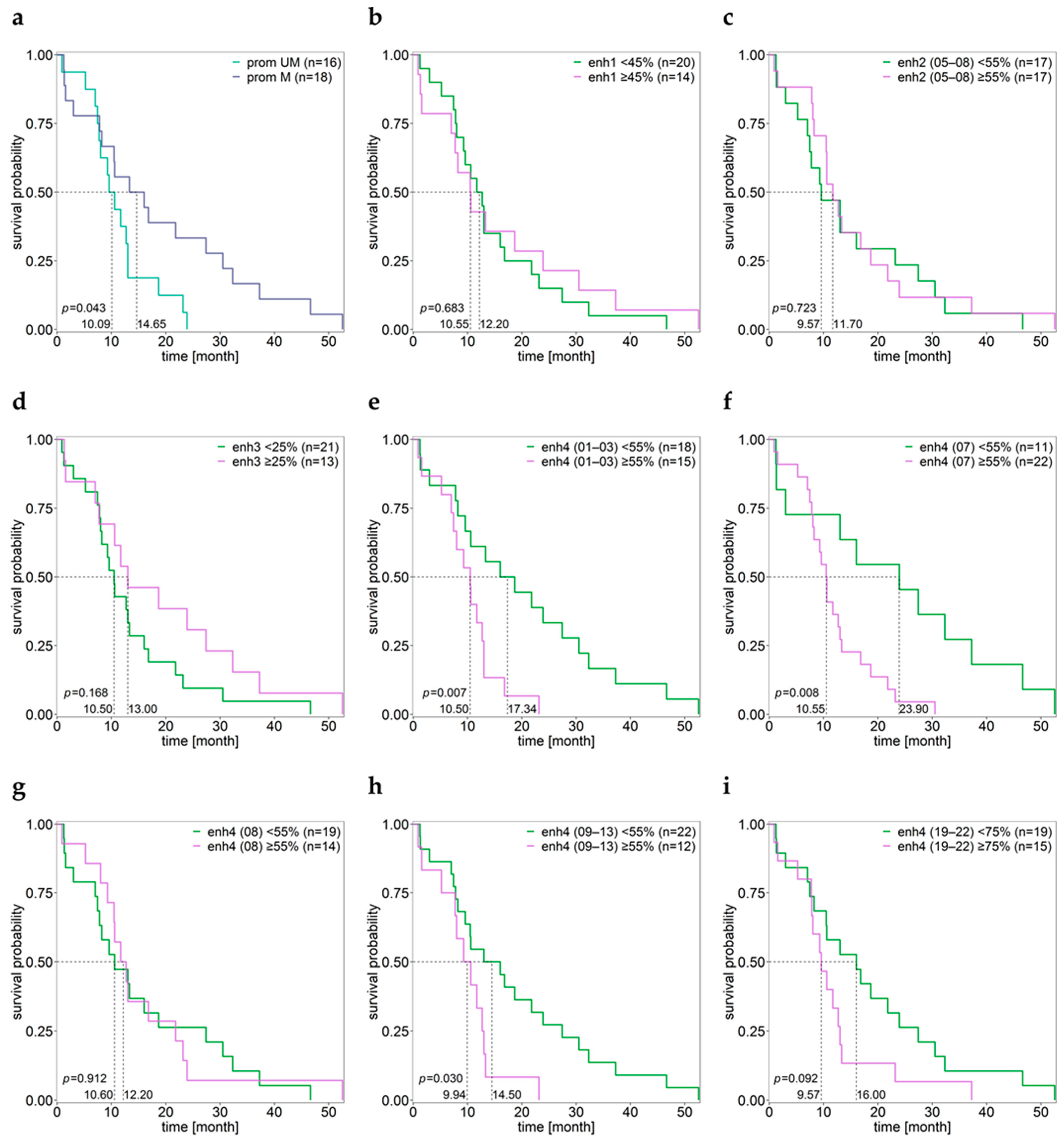
By using a cut-off of 8%, patients with methylated MGMT promoter had significantly longer OS compared to patients with an unmethylated MGMT promoter (Figure 5a).
No significant differences in OS were found for the methylation of enhancer 1 (Figure 5b), enhancer 2 (Figure 5c), and enhancer 3 (Figure 5d). However, for enhancer 4 we found significantly longer OS for patients showing low methylation of CpGs 01–03, CpGs 09–13, and individual CpG 07, but not for individual CpG 08 (Figure 5e–i) and mean methylation of CpGs 07–08 (cut-off 55%, p = 0.117).
The hazard ratio for individual CpGs of the MGMT promoter and enhancer 4 is given in Table 2. Methylation levels ≥8% of the CpGs targeted in the MGMT promoter as well as methylation levels <55% of the mean of CpGs 01–03, mean of CpGs 09–13, and methylation levels of CpG 03, CpG 07, and CpG 13 offer survival benefit for GBM patients.

3.10. Associations of Various Clinical Parameters

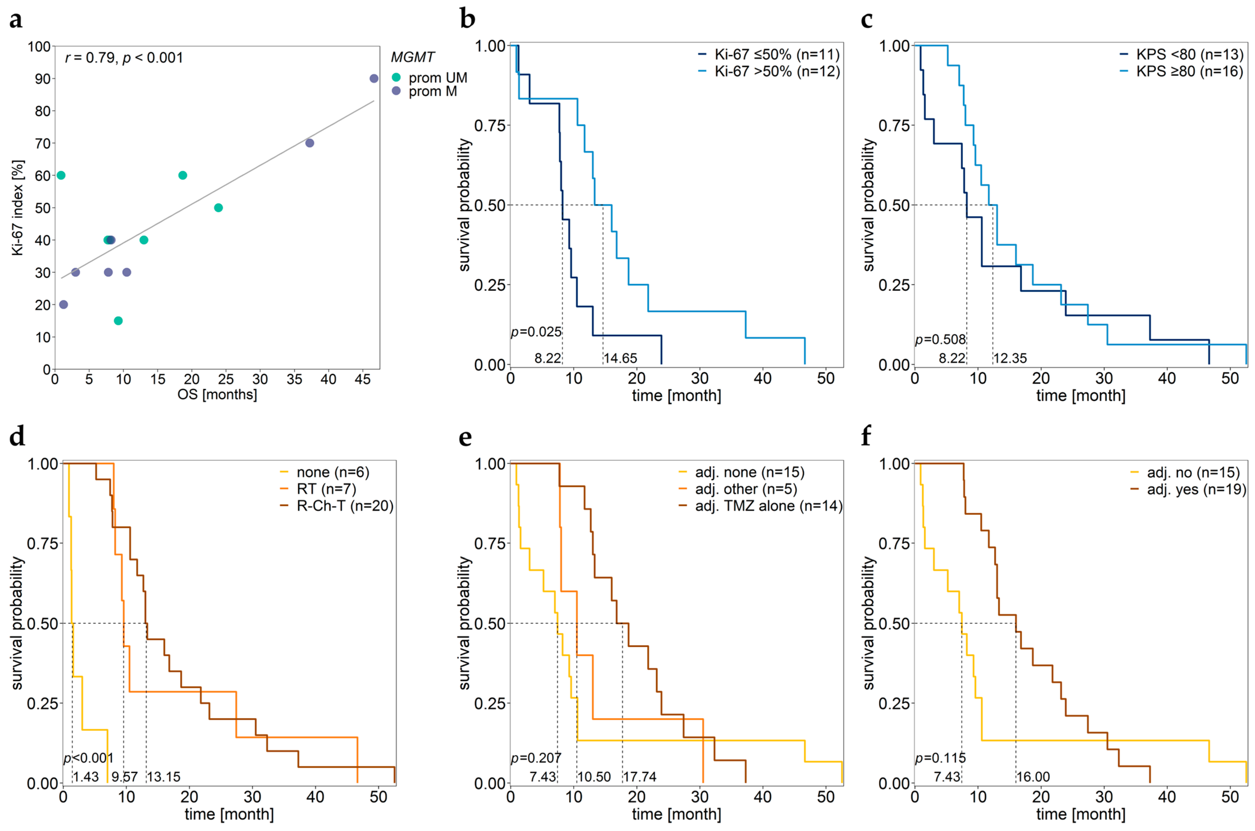
Surprisingly, for 15 GBM patients for which the Ki-67 index was available quantitatively, the Ki-67 index was significantly positively correlated with OS (r = 0.79, p < 0.001) (Figure 6a). GBM patients with a Ki-67 index > 50% had significantly longer OS compared to patients with a low (≤50%) Ki-67 index (Figure 6b, Table 3). The significant positive association between the Ki-67 index and OS has already been reported in previous studies, but there are others reporting the opposite (see Section 4.)
No difference in OS was found between patients with low (<80) and patients with high (≥80) KPS (Figure 6c, Table 3). However, for GBM patients with methylated MGMT promoter, OS significantly positively correlated with KPS (r = 0.55, p = 0.044).
Patients who did not receive any primary therapy after surgery had significantly lower OS than those undergoing either radiotherapy (p < 0.001) or radio-chemotherapy (p < 0.001). No significant difference was found between patients receiving radiotherapy and those who received radio-chemotherapy (p = 0.620) (Figure 6d, Table 3). Moreover, no significant difference in OS was found between patients without adjuvant therapy and those with adjuvant therapy (Figure 6e,f, Table 3).

3.11. Impact of Age

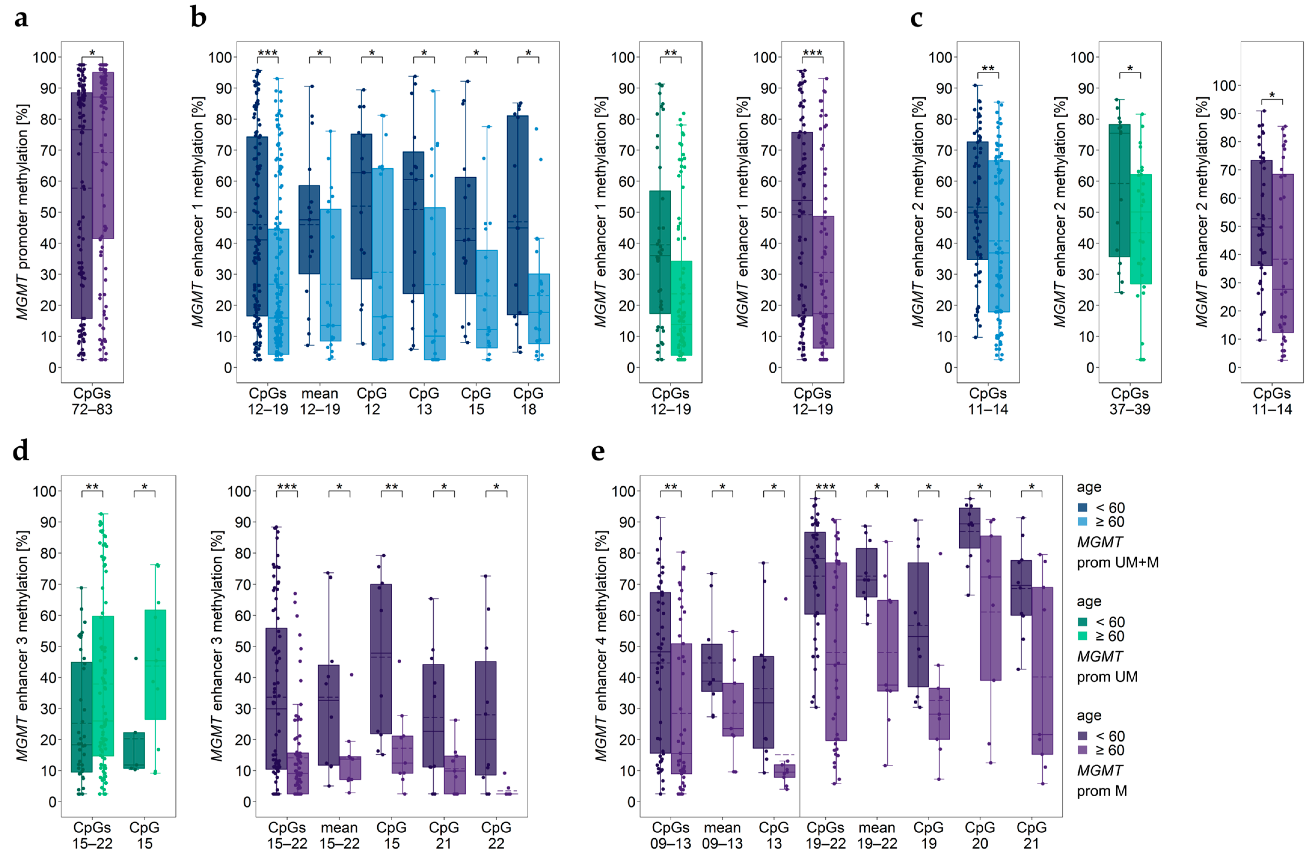
We found significant differences in MGMT promoter and enhancer methylation between patients <60 years and those ≥60 years (Figure 7). In the subgroup of patients with methylated MGMT promoter, patients ≥60 years had significantly higher methylation levels of CpGs 72–83 in the MGMT promoter than younger ones (Figure 7a).
Significant differences in the methylation status between older and younger patients were also found for the four enhancers (Figure 7b–e). Methylation levels of enhancer 1, enhancer 2, and enhancer 4 were lower in older GBM patients than in younger ones (Figure 7b,c,e). In the case of enhancer 4, significant differences between age subgroups were only found for tumors with methylated MGMT promoter (Figure 7e). The association between enhancer 3 methylation and age depended on MGMT promoter methylation. In patients with an unmethylated MGMT promoter, older patients showed significantly higher methylation levels, whereas in patients with a methylated MGMT promoter, older patients showed lower enhancer 3 methylation levels compared to younger patients (Figure 7d).
In our patient cohort, the proliferation index Ki-67 did not correlate with age of the patients (r = −0.48, p = 0.068). However, patients with Ki-67 ≤ 50% had significantly higher ages than those with Ki-67 > 50% (Figure 8a).
For GBM patients with methylated MGMT promoter, we found a significantly negative correlation between KPS and the age of the patients (r = −0.64, p = 0.010, Figure 8b). In the subgroup of patients with methylated MGMT promoter, patients with KPS ≤ 80% were older than those with KPS > 80% (Figure 8c).
Patients <60 years had longer OS compared to those ≥60 years (Figure 8d, Table 3). In samples with methylated MGMT promoter, age negatively correlated with OS (r = −0.53, p = 0.024, Figure 8e).
Our patient cohort contained three long-term survivors (GBM03, GBM04, and GBM23). Long-term survivors are generally defined as GBM patients who survive longer than three years after surgery [23]. We found significant differences in MGMT promoter methylation (Supplementary Figure S1). In addition, the methylation of enhancer 1, enhancer 2, and enhancer 4 was different between long-term survivors and non-long-term survivors of our cohort. For all three enhancers, significant differences were found in case all samples were included in statistical analysis but also for samples with methylated MGMT promoter (Supplementary Figure S1).

3.12. Impact of Sex

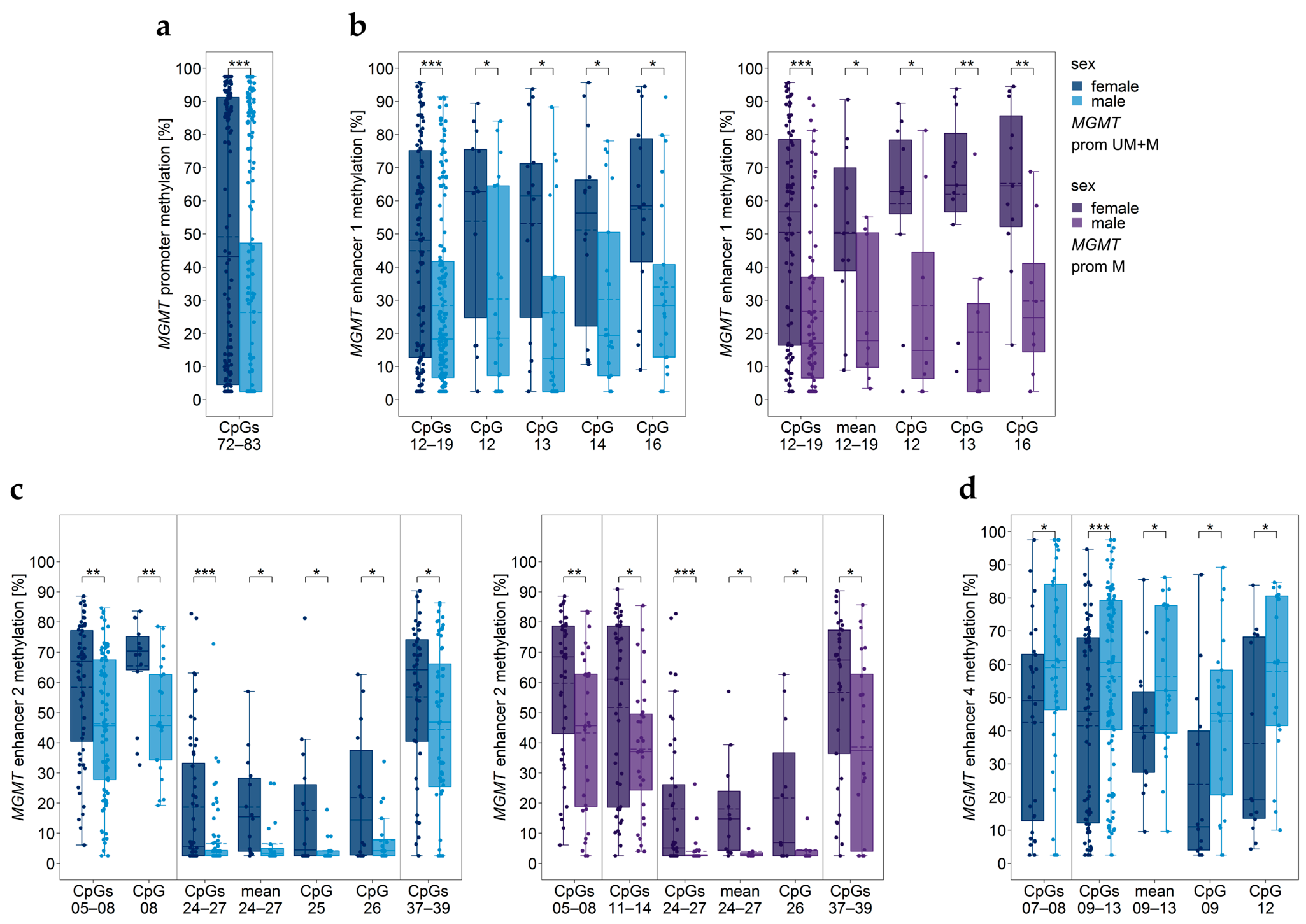
CpGs 72–83 in the MGMT promoter were significantly more highly methylated in female than in male patients (Figure 9a). However, for none of the individual CpGs a significant difference was found (p ≥ 0.051).
Significantly higher methylation levels in female patients were also found for enhancer 1 (Figure 9b) and enhancer 2 (Figure 9c). In contrast, several CpGs of enhancer 4 were methylated significantly lower in female than in male patients (Figure 9d).

3.13. Two-Way ANOVA Considering Age and Sex

Two-way ANOVA was performed to verify if differences in MGMT promoter or enhancer methylation between TERT promoter wildtype and mutations C228T and C250T, between TERT rs2853669 genotypes, and between patients with low (≤50%) and high (>50%) Ki-67 index found by one-way ANOVA were also significant when considering age (<60 and ≥60 years) or sex.
Interaction effects of TERT promoter status with age were found in MGMT promoter methylated samples for promoter and enhancer 3 (Table S2). Few interactions with sex were identified for enhancer 2 and enhancer 3. All significant associations between MGMT promoter/enhancer methylation and TERT promoter mutations identified by one-way analysis (Section 3.4) were also found in the two-way approach, with the exception of the methylation status of CpG 12–19 of MGMT enhancer 1 when considering sex (Table S3).
An interaction between the TERT rs2853669 genotype and age was only found for CpGs 19–22 of enhancer 4 in samples with an unmethylated MGMT promoter (Table S2). An interaction with sex was found for CpGs 72–83 of the MGMT promoter in samples not stratified for the MGMT promoter methylation status, for CpGs 12–19 of enhancer 1 independent of the MGMT promoter methylation status, as well as for CpGs 15–22 of enhancer 3 in samples with an unmethylated MGMT promoter. All significant associations between MGMT promoter/enhancer methylation and the TERT rs2853669 genotype identified by one-way analysis (Section 3.5) were confirmed by two-way ANOVA. However, for a few CpGs, no main effect was found when considering age or sex (Table S3).
The Ki-67 status was found to interact with age for the methylation levels of CpGs 05–08 and CpGs 37–39 of enhancer 2. An interaction with sex was found for CpGs of enhancer 1 and CpGs 05–08, CpG 08, and CpGs 37–39 of enhancer 2, and exclusively in samples with methylated MGMT promoter, CpGs 15–22 of enhancer 3 (Table S2). In general, Ki-67 status was a main effect when considering age, with the exception of CpGs 12–19 of enhancer 1. No main effect was found for CpGs 72–83 of the MGMT promoter when sex was considered (Table S3).
ANOVA could not be applied for differences between MGMT SNP rs16906252 genotypes due to a too-low number of representatives of the TT genotype.

3.14. Cell Lines Resulting in Unstable Cell Cultures

Three primary GBM cell lines (GBM36–GBM38) did not result in stable cell cultures (Table S1). In general, results obtained for these samples were quite different from those obtained for samples resulting in stable cell cultures and were therefore excluded from the statistical analyses described above. All three (100%) samples had an unmethylated MGMT promoter, compared to 16 (45.7%) samples resulting in stable cultures (Figures S2a and S3a). In general, CpGs in enhancers 1–4 had significantly higher in samples GBM36-38 than in samples GBM01–35 (Figures S2b–d and S3b–d). Regarding MGMT SNP rs16906252, two (66.6%) samples were heterozygous CT, and one (33.3%) showed the homozygous C wildtype genotype. In the case of GBM01–35, 32 (91.4%) GBM samples were homozygous C, and an (atypical) heterozygous genotype was only determined for one (2.9%) sample. GBM36–38 (100%) showed the TERT promoter wildtype, whereas this genotype was only found for three (8.6%) of the samples GBM01–35.

4. Discussion

Promoter methylation plays a crucial role in regulating MGMT protein expression in GBMs. However, MGMT in all GBMs is not silenced, although the promoter is methylated. Vice versa, some tumors don’t express MGMT despite a demonstrable lack of promoter methylation. These findings suggest that there are further regulatory mechanisms in addition to MGMT promoter methylation.
In a very recent study, we investigated if enhancer methylation is one of the mechanisms contributing to MGMT regulation [14]. By determining methylation levels of the MGMT promoter and four enhancers, we found that the methylation status of several enhancer regions was actually associated with MGMT promoter methylation and/or MGMT expression. In samples with an unmethylated MGMT promoter, methylation of enhancer 2 correlated with MGMT expression. In addition, the results suggested that methylation of CpGs of enhancer 4 (hs696, [15]), located in intron 2 of the MGMT gene, could serve as a potential prognostic biomarker in GBMs [14]. In this study, we investigated if methylation levels of the four enhancers targeted previously are associated with common genetic variants in GBMs, clinical parameters, and demographic characteristics of GBM patients. To date, studies dealing with the association of enhancer methylation with genetic variants, clinical parameters, and/or demographic characteristics in cancer are limited. For MGMT in GBM, there are some studies reporting enhancer methylation data, but these studies refer to the cis-acting enhancer element within the MGMT promoter [23]. To the best of our knowledge, this is the first paper investigating the methylation of MGMT enhancers located outside of the MGMT promoter in terms of genetic variants, clinical parameters, and demographic characteristics.
SNP rs16906252, located within the cis-acting enhancer element in the MGMT promoter, is one of the genetic variants that has been associated with MGMT promoter methylation and MGMT expression [24,25]. The presence of the T allele of rs16906252 was linked to MGMT promoter methylation and reduced activity of the MGMT promoter. Moreover, among GBM patients with a methylated MGMT promoter, carriers of the T allele showed a significant survival benefit compared to patients lacking the T allele [24,25]. In our patient cohort, the prevalence of the rs16906252 T allele was very low. Only 5.7% of the GBM samples were homozygous T, and only 2.9% of the samples showed an (atypical) heterozygous genotype. In contrast to previous studies targeting CpGs 74–78 [24,25], MGMT promoter methylation was not associated with the rs16906252 genotype. However, the methylation status of CpGs in MGMT enhancers was linked to MGMT SNP rs16906252. Methylation of intragenic enhancer 4 (CpGs 02, 03, and 07) was significantly lower in samples of the homozygous T genotype compared to those being homozygous C. In our previous study, we found enhancer 4 methylation negatively correlated with MGMT promoter methylation, with CpG 03 showing a particularly strong correlation [14]. Among the three intergenic enhancers, enhancer 2 (mean CpGs 24–27; CpGs 24–27; CpGs 24, 26, and 27) was also significantly lower methylated in homozygous T compared to homozygous C samples. In contrast, enhancer 1 (CpGs 13–15) and enhancer 3 (CpGs 15–22, CpG 16) were significantly more highly methylated in homozygous T compared to homozygous C samples. Our results suggest an association between methylation levels of the enhancers and the MGMT SNP rs16906252 genotype. However, due to the low prevalence of the T allele in our patient cohort, further studies are required to confirm our findings.
Mutations in the TERT promoter commonly occur in malignant tumors [26,27,28]. TERT encodes a rate-limiting catalytic subunit of the enzyme telomerase, which regulates the length of telomeres [29]. Transcriptional activation of TERT by mutations plays a crucial role in tumorigenesis [28]. In GBMs, TERT is frequently activated by mutation C250T or C228T, located 146 bp and 124 bp upstream of the translation start site (TSS), respectively. Increased telomerase activity, reported for >60–70% of GBM patients, has been associated with poor prognosis [26]. In addition to TERT mutations, the SNP rs2853669, located 245 bp upstream of the TSS, has been associated with TERT activity. In contrast to mutations C250T and C228T, the less frequent (C) allele of SNP rs2853669 is linked to low TERT activity [30,31].
In our patient cohort, TERT mutations C228T and C250T were detected in 21 (60.0%) and 11 (31.4%) of the tumors, respectively. In line with the literature [32], the mutations occurred mutually exclusive. Regarding TERT SNP rs2853669, 19 (55.9%) GBM samples had the wildtype TT genotype and 15 (44.1%) the heterozygous CT genotype.
Previous findings on associations between MGMT promoter methylation, TERT promoter mutations, and/or TERT SNP rs2853669 are controversial, with some studies reporting associations [33,34], whereas others did not [20,26]. In the subgroup of tumors with methylated MGMT promoter, carriers of the C250T mutation showed significantly higher MGMT promoter methylation compared to wildtype tumors and C228T mutation carriers. GBM patients, being heterozygous for the SNP rs2853669, had a significantly lower MGMT promoter methylation status than those of the TT genotype. Thus, our results are in accordance with previous papers reporting associations between MGMT promoter methylation, TERT promoter mutations, and TERT SNP rs2853669 [33,34]. However, the biological mechanism for these interactions remains to be elucidated.
In addition to MGMT promoter methylation, methylation of the three intergenic enhancers but not that of intragenic enhancer 4 was associated with TERT mutations. Tumors with TERT promoter mutation showed significantly higher methylation of enhancer 1 (C228T, CpGs 12–19) and enhancer 2 (C228T and C250T, CpGs 37–39) compared to tumors lacking TERT mutation. In the subgroup of tumors with methylated MGMT promoter, methylation of enhancer 2 (CpG 37, CpG 38, mean methylation of CpGs 37–39) was significantly higher in C228T mutation carriers than in wildtype tumors. In GBM patients with an unmethylated MGMT promoter, carriers of C228T mutation showed significantly higher methylation of enhancer 3 (CpGs 15–22) than carriers of C250T mutation. For GBM patients with a methylated MGMT promoter, TERT C228T mutation was associated with lower methylation of CpGs 15–22 than tumors of wildtype or those with C250T mutation. In the case of TERT SNP rs2853669, significant differences between wildtype and SNP genotype were found for all four enhancers investigated. The three intergenic enhancers were more highly methylated in samples of the CT genotype compared to the wildtype TT genotype. In contrast, enhancer 4 was significantly lower methylated in samples of the CT genotype compared to wildtype samples. The difference was more pronounced in samples with an unmethylated MGMT promoter. In samples with methylated MGMT promoter, a significant difference was found for CpG 09, with heterozygous samples showing lower methylation than wildtype samples.
Ki-67 is a nuclear protein that is expressed in all cell cycle phases except the resting cell phase G0 [35]. Thus, it is widely used as a proliferation marker for human tumor cells [36]. In the context of brain tumors, the Ki-67 index was found to be applicable for differentiating between high and low-grade gliomas, but its relevance as a marker for GBMs is controversially discussed [37]. In this study, the Ki-67 index was specified only for 15 patients; for a further nine patients, the proliferation status of the tumor was available in a semi-quantitative manner. Thirteen (54.2%) patients showed a Ki-67 index >50%, which is rather high compared to other studies, reporting mean Ki-67 values of about 20% [38,39]. Differences in reported Ki-67 values have been mainly attributed to differences in Ki-67 determination, including the use of different antibody clones, antibody formats, and/or staining platforms [40]. In addition, heterogeneity of tumor tissues, e.g., the presence of non-neoplastic cells, activated microglia or macrophages, has an impact on overall proliferative activity [37,41]. Furthermore, differences in Ki-67 data may be caused by interobserver variability [38].
MGMT promoter methylation was not significantly correlated with the Ki-67 index. However, we found a significant difference in MGMT promoter methylation between tumors with high and low Ki-67 index. Tumors with a high Ki-67 index (>50%) showed significantly higher MGMT promoter methylation than tumors with a low (≤50%) Ki-67 index. Due to the high Ki-67 values found for our patient cohort, we set the cut-off at 50%. In other studies, the Ki-67 cut-off was set at lower values, e.g., at 20% [37,39] or 27% [35,42]. Thus, our results are not comparable with previous findings.
The three intergenic enhancers investigated are located between the Ki-67 gene (upstream) and the MGMT gene (downstream). For enhancer 1 (CpG 12, CpG 13, and CpG 18) and enhancer 2 (mean methylation of CpGs 05–08, CpGs 11–14, CpGs 15–18, and CpGs 37–39 as well as ten individual CpGs), the two enhancers being closest to Ki-67, we found significantly positive correlation between enhancer methylation and Ki-67 index, but exclusively for GBM samples with an unmethylated MGMT promoter. In addition, methylation of enhancer 2 (CpGs 37–39, CpG 08) was significantly higher in GBM patients with a high Ki-67 index (>50%) compared to those with a low (≤50%) Ki-67 index. In samples with unmethylated MGMT promoter, we found significant differences for CpGs 05–08, CpGs 24–27, CpGs 37–39, mean of CpGs 37–39, and individual CpG 08 and CpG 37. Methylation of enhancer 1 (CpGs 12–19) was significantly higher in GBM patients with a high Ki-67 index (>50%) compared to those with a low (≤50%) Ki-67 index. Methylation of enhancer 3 (CpGs 15–22) was also significantly higher in patients with high Ki-67 index (Ki-67>50%), but only for GBM samples with methylated MGMT promoter. Chen et al., who identified enhancer 2, have already linked enhancer 2 with Ki-67. Deletion of the enhancer region resulted in reduced expression of not only MGMT but also Ki-67, although to a lesser extent [16]. Thus, Chen et al. concluded that Ki-67 and MGMT are in the same topologically associating domain (TAD). TADs are genomic regions with maximal intradomain interactions and minimal interactions with neighboring regions [43,44]. Our results support the hypothesis of Chen et al. In contrast to intergenic enhancers 1, 2, and 3, the methylation status of enhancer 4, located in intron 2 of MGMT, was not associated with the Ki-67 index.
By using a cut-off of 8% for MGMT promoter methylation, OS was significantly longer for patients with methylated MGMT promoter compared to those with an unmethylated MGMT promoter, which is in accordance with previous studies [45,46]. For individual CpGs 75, 78, and 80, we found a significant positive correlation with OS [14]. In a recent study by Leske et al., methylation levels of CpGs 72, 74, 75, 81, 83, 85, and 93 in the MGMT promoter were found to be strongly associated with the prognosis of GBM patients [23]. In addition to the MGMT promoter, we found enhancer 4 to be associated with OS. By setting a cut-off value of 55%, we revealed significantly longer OS for patients with higher enhancer 4 methylation (mean methylation of CpGs 01–03, CpG 03, CpG 07, mean of CpGs 09–13, and CpG 13) compared to those with lower methylation status. In contrast to the MGMT promoter and enhancer 4, neither mean methylation levels nor methylation levels of individual CpGs of intergenic enhancers 1–3 were associated with OS of the patients.
Growing evidence suggests that MGMT promoter methylation is one of the factors linked to long-term survival, the phenomenon that GBM patients survive longer than three years after surgery [23,47]. In the three long-term survivors of our patient cohort, the MGMT promoter was methylated, which is in accordance with the literature. Moreover, we found long-term survival to be associated with methylation of enhancer 1, enhancer 2, and enhancer 4. Since our patient cohort only contained three long-term survivors, further studies are required to confirm our findings.
In our patient cohort, neither MGMT promoter nor enhancer methylation correlated with KPS. In terms of survival, only enhancer 4 methylation (CpG 01) negatively correlated with PFS, but exclusively in samples with an unmethylated MGMT promoter.
In accordance with previous studies [45], MGMT promoter methylation was not associated with the age of the patients at surgery if samples were not stratified by their MGMT promoter methylation status. However, in the subgroup of tumors with methylated MGMT promoter, patients ≥60 years showed significantly higher MGMT promoter methylation than younger ones (<60 years). We also found MGMT enhancer methylation to be associated with age. Patients ≥60 years showed significantly lower methylation levels of intragenic enhancer 4 and intergenic enhancers 1 and 2. In the case of enhancer 3, the association with the age of the patients depended on the MGMT promoter methylation status. In the subgroup of patients with an unmethylated MGMT promoter, younger patients showed significantly lower enhancer 3 methylation levels compared to older ones. For the subgroup of patients with methylated MGMT promoter, the opposite finding was observed.
Our patient cohort comprised fourteen (40%) females and 21 (60%) males. We found the MGMT promoter was significantly more highly methylated in female patients than in male patients. Sex-specific differences in MGMT promoter methylation have already been reported in the literature [48]. Methylation levels of intergenic enhancer 1 (CpGs 12-19, CpG 12, CpG 13, CpG 14, and CpG 16) and enhancer 2 (CpGs 05–08, CpGs 24–27, CpGs 37–39, mean methylation of CpGs 24–27, CpG08, CpGs 25, CpG 26) were also higher in females than in males. In contrast, enhancer 4 methylation (CpGs 07–08, CpGs 09–13, mean methylation CpGs 09–13, CpG 09, CpG 12) was significantly lower in female than in male patients.
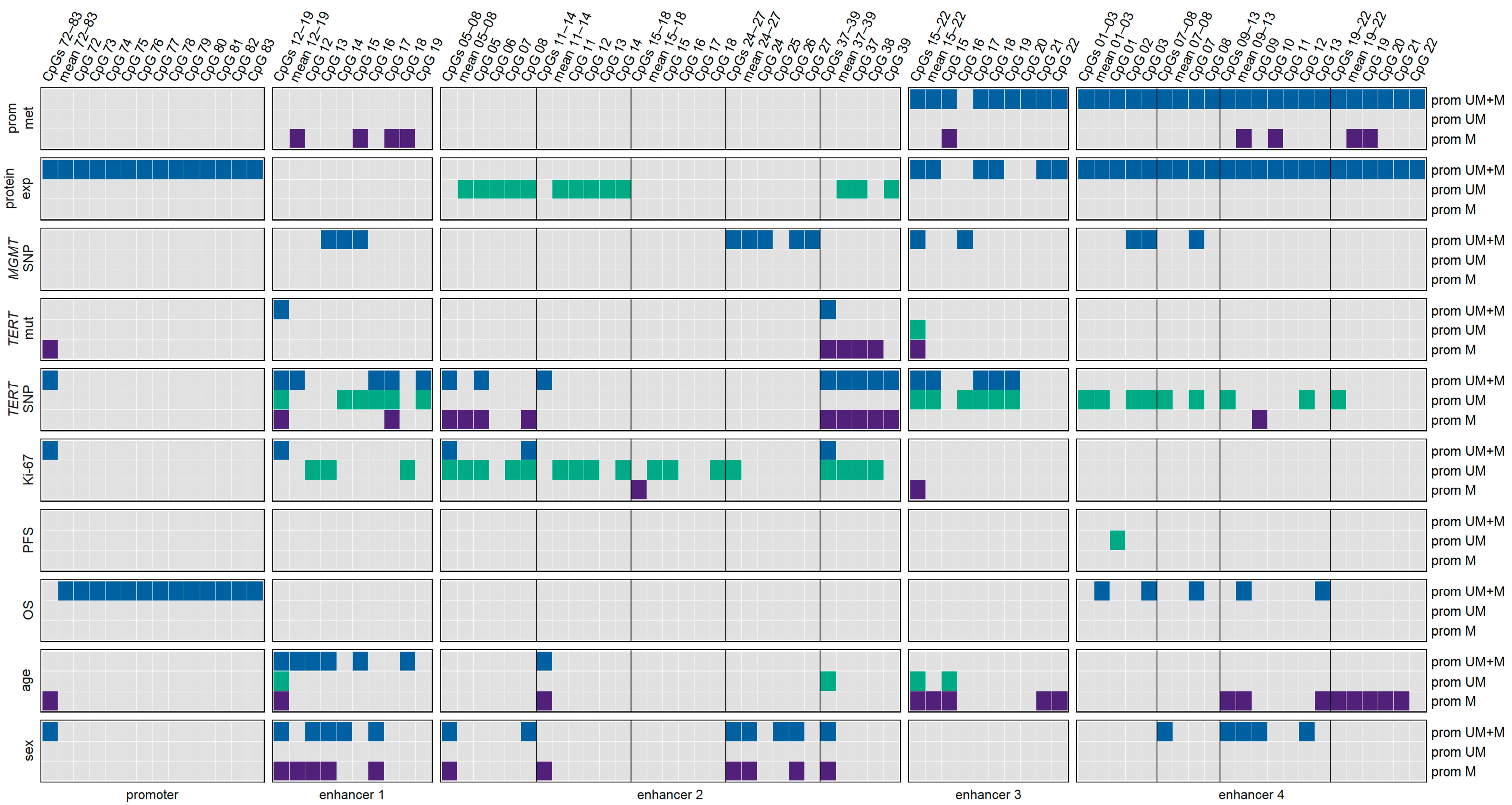
Figure 10 summarizes the significant associations between MGMT promoter methylation and/or MGMT enhancer methylation with MGMT expression, the occurrence of genetic variants, clinical parameters, and demographic characteristics that we found for our patient cohort. With the exception of MGMT protein expression and OS, associations for the MGMT promoter were only found when all twelve CpGs were taken into account. We could not identify one enhancer, one enhancer region, nor one individual CpG within the enhancers that was relevant for all parameters and characteristics investigated. The figure clearly indicates the strong impact of the MGMT promoter methylation status on the associations found. For example, associations between enhancer 2 methylation and MGMT protein expression and the Ki-67 index were predominantly found for samples with an unmethylated MGMT promoter and those with TERT mutations, mainly for samples with methylated MGMT promoter. In the case of enhancer 4, associations with the TERT SNP genotype and age were strongly influenced by the MGMT promoter methylation status.
We also found associations between various clinical parameters for our patient cohort, with most of them being in accordance with previous studies. Patients who did not receive any primary therapy after surgery had significantly lower OS than those undergoing either radiotherapy or radio-chemotherapy, which is in line with the literature [12,48]. OS was longer in patients undergoing adjuvant therapy with TMZ alone than in those without adjuvant therapy. In a number of studies, higher age has been associated with worse clinical outcomes in GBM [8,47]. This association was also observed in our study.
For our patient cohort, we did not find a significant association between sex and OS, which is also in accordance with previous studies [8,35,49].
Surprisingly, Ki-67 levels positively correlated with OS for the 15 GBM patients for which quantitative Ki-67 data were available. In addition, GBM patients with a Ki-67 index >50% had significantly longer OS compared to patients with a low (≤50%) Ki-67 index. One would obviously expect that higher proliferation activity of the tumor is associated with a worse survival prognosis. In the literature, findings on Ki-67 and OS are controversial. A few studies also linked higher Ki-67 values to longer OS in GBM [42,50]. However, the majority of studies report a negative association between Ki-67 and OS [39,49,51,52]. There are also studies that did not find the Ki-67 index to be linked to OS [53]. Controversial discussion on associations between Ki-67 and OS is not limited to GBM but is also ongoing for other cancer types, e.g., melanoma [54] and breast cancer [55]. Longer OS of patients with higher Ki-67 index could be explained by the fact that highly proliferating tumors are particularly prone to cytotoxic effects of chemotherapy or radio-chemotherapy [42].
Growing evidence suggests that radio chemotherapy improves the treatment efficacy for GBM patients compared to radiotherapy alone [56]. However, for our patient cohort, we did not find a significant difference in OS between patients receiving radiotherapy and those who received radio-chemotherapy. Most likely, the subgroup receiving radiotherapy alone was too small in our patient cohort.
Our original patient cohort contained three patients for which stable cell cultures could not be obtained. These samples were excluded from statistical analysis because they differed in many data from samples for which stable cell cultures were achieved. Among others, all three samples showed the TERT promoter wildtype, which could explain why cells could not achieve immortality. However, for other cells showing the TERT promoter wildtype, stable cell cultures could be achieved, suggesting that additional factors were involved.

5. Conclusions

Our results show that enhancer methylation does not only play a crucial role in regulating gene expression. The findings clearly indicate that methylation of the enhancers investigated is associated with genetic variants, clinical parameters, and demographic characteristics in GBM.
In general, the associations identified for intragenic enhancer 4, located in intron 2 of MGMT, were quite different from those found for the three intergenic enhancers. Intragenic enhancer 4 was the only enhancer whose methylation levels were negatively correlated with MGMT promoter methylation, as shown previously. In addition, an association of the methylation status with OS was exclusively found for enhancer 4. Thus, enhancer 4 methylation could be a potential prognostic biomarker for GBM. In contrast to the three intergenic enhancers, enhancer 4 methylation was neither associated with TERT mutations nor with the Ki-67 index. The methylation status of enhancer 4 was significantly lower in female than in male patients, whereas for the three intergenic enhancers, the opposite finding was observed.
Among the three intergenic enhancers, enhancer 2 methylation could be a potential biomarker in GBM. Methylation of several CpGs of enhancer 2 was positively correlated with MGMT expression, as shown previously and/or the Ki-67 index, as shown in this work. Both associations were observed for the subgroup of samples with an unmethylated MGMT promoter. Associations with genetic variants, clinical parameters, and demographic characteristics were also observed for enhancer 1 and enhancer 3.
In general, the associations with genetic variants, clinical parameters, and demographic characteristics were caused by enhancer regions rather than by individual CpGs, hinting at a complex interplay of multiple CpGs and enhancer regions.
Some of our findings were obtained without stratifying samples by their MGMT promoter methylation status. However, other associations were restricted to subgroups, either samples with methylated or samples with an unmethylated MGMT promoter, underpinning the relevance of the MGMT promoter status in GBMs.
We would like to stress that our findings are based on a rather heterogenous sample set, especially regarding primary and adjuvant therapy of the patients. Discrepancies in treatment are expected to have an impact on PFS and OS and thus may confound the robustness of the associations between MGMT methylation levels and these clinical parameters. Further studies are required to confirm our findings.

Supplementary Materials

The following supporting information can be downloaded at: https://www.mdpi.com/article/10.3390/cancers15245777/s1, Figure S1. Significantly different MGMT promoter/enhancer methylation levels between non-long-term survivors (≤36 months) and long-term survivors (>36 months); Figure S2. MGMT promoter/enhancer methylation levels of stable and unstable cell cultures; Figure S3. MGMT promoter/enhancer methylation levels of stable and unstable cell cultures in MGMT promoter unmethylated samples; Table S1. Clinical and demographic data of GBM patients for which stable cell cultures could not be established; Table S2. Interaction effects found by two-way ANOVA for significantly different (one-way ANOVA) MGMT promoter/enhancer methylation levels between TERT promoter wildtype and mutations, between TERT rs2853669 genotypes, and between patients with low (≤50%) and high (>50%) Ki-67 index; Table S3. Non-significant main effects identified by two-way ANOVA for significantly different (one-way ANOVA) MGMT promoter/enhancer methylation levels between TERT promoter wildtype and mutations, between TERT rs2853669 genotypes, and between patients with low (≤50%) and high (>50%) Ki-67 index.

Author Contributions

Conceptualization, M.C.-M., K.Z. and S.S.-K.; methodology, investigation, and validation, K.Z., K.P., S.S.-K., S.W., S.P. and D.B.; resources, S.S.-K.; writing—original draft preparation, K.Z. and M.C.-M.; writing—review and editing, K.Z., M.C.-M. and S.S.-K.; visualization, K.Z.; project administration, M.C.-M.; funding acquisition, M.C.-M. All authors have read and agreed to the published version of the manuscript.

Funding

This article was supported by the Open Access Publishing Fund of the University of Vienna.

Institutional Review Board Statement

The study was approved by the local Ethics Commission of the Faculty of Medicine at the Johannes Kepler University Linz (application number E-39-15).

Informed Consent Statement

Informed consent was obtained from all subjects involved in the study.

Data Availability Statement

The datasets generated during the current study are available from the corresponding author on reasonable request.

Conflicts of Interest

The authors declare no conflict of interest.

Abbreviations

CpG—CpG dinucleotide, GBM—glioblastoma, HRM—high-resolution melting, IDH—isocitrate dehydrogenase, MGMT—O6-methylguanine-DNA methyltransferase, OS—overall survival, PCR polymerase chain reaction, PSQ—pyrosequencing, SD—standard deviation, SNP—single nucleotide polymorphism, TMZ—temozolomide

References

  1. Louis, D.N.; Perry, A.; Wesseling, P.; Brat, D.J.; Cree, I.A.; Figarella-Branger, D.; Hawkins, C.; Ng, H.K.; Pfister, S.M.; Reifenberger, G.; et al. The 2021 WHO classification of tumors of the central nervous system: A summary. Neuro Oncol. 2021, 23, 1231–1251. [Google Scholar] [CrossRef]
  2. Stupp, R.; Mason, W.P.; Van Den Bent, M.J.; Weller, M.; Fisher, B.; Taphoorn, M.J.B.; Belanger, K.; Brandes, A.A.; Marosi, C.; Bogdahn, U.; et al. Radiotherapy plus concomitant and adjuvant temozolomide for glioblastoma. N. Engl. J. Med. 2005, 352, 987–996. [Google Scholar] [CrossRef]
  3. Soll, J.M.; Sobol, R.W.; Mosammaparast, N. Regulation of DNA Alkylation Damage Repair: Lessons and Therapeutic Opportunities. Trends Biochem. Sci. 2017, 42, 206–218. [Google Scholar] [CrossRef]
  4. Lee, S.Y. Temozolomide resistance in glioblastoma multiforme. Genes Dis. 2016, 3, 198–210. [Google Scholar] [CrossRef]
  5. Deaton, A.M.; Bird, A. CpG islands and the regulation of transcription. Gene Dev. 2011, 25, 1010–1022. [Google Scholar] [CrossRef]
  6. Nakagawachi, T.; Soejima, H.; Urano, T.; Zhao, W.; Higashimoto, K.; Satoh, Y.; Matsukura, S.; Kudo, S.; Kitajima, Y.; Harada, H.; et al. Silencing effect of CpG island hypermethylation and histone modifications on O6-methylguanine-DNA methyltransferase (MGMT) gene expression in human cancer. Oncogene 2003, 22, 8835–8844. [Google Scholar] [CrossRef]
  7. Hegi, M.E.; Diserens, A.C.; Gorlia, T.; Hamou, M.F.; De Tribolet, N.; Weller, M.; Kros, J.M.; Hainfellner, J.A.; Mason, W.; Mariani, L.; et al. MGMT gene silencing and benefit from temozolomide in glioblastoma. N. Engl. J. Med. 2005, 352, 997–1003. [Google Scholar] [CrossRef]
  8. Brandes, A.A.; Franceschi, E.; Tosoni, A.; Benevento, F.; Scopece, L.; Mazzocchi, V.; Bacci, A.; Agati, R.; Calbucci, F.; Ermani, M. Temozolomide concomitant and adjuvant to radiotherapy in elderly patients with glioblastoma: Correlation with MGMT promoter methylation status. Cancer 2009, 115, 3512–3518. [Google Scholar] [CrossRef]
  9. Malmström, A.; Grønberg, B.H.; Marosi, C.; Stupp, R.; Frappaz, D.; Schultz, H.; Abacioglu, U.; Tavelin, B.; Lhermitte, B.; Hegi, M.E.; et al. Temozolomide versus standard 6-week radiotherapy versus hypofractionated radiotherapy in patients older than 60 years with glioblastoma: The Nordic randomised, phase 3 trial. Lancet Oncol. 2012, 13, 916–926. [Google Scholar] [CrossRef] [PubMed]
  10. Reifenberger, G.; Hentschel, B.; Felsberg, J.; Schackert, G.; Simon, M.; Schnell, O.; Westphal, M.; Wick, W.; Pietsch, T.; Loeffler, M.; et al. Predictive impact of MGMT promoter methylation in glioblastoma of the elderly. Int. J. Cancer 2012, 131, 1342–1350. [Google Scholar] [CrossRef] [PubMed]
  11. Binabaj, M.M.; Bahrami, A.; ShahidSales, S.; Joodi, M.; Joudi Mashhad, M.; Hassanian, S.M.; Anvari, K.; Avan, A. The prognostic value of MGMT promoter methylation in glioblastoma: A meta-analysis of clinical trials. J. Cell. Physiol. 2018, 233, 378–386. [Google Scholar] [CrossRef] [PubMed]
  12. Brown, N.F.; Ottaviani, D.; Tazare, J.; Gregson, J.; Kitchen, N.; Brandner, S.; Fersht, N.; Mulholland, P. Survival Outcomes and Prognostic Factors in Glioblastoma. Cancers 2022, 14, 3161. [Google Scholar] [CrossRef] [PubMed]
  13. Kreth, S.; Thon, N.; Eigenbrod, S.; Lutz, J.; Ledderose, C.; Egensperger, R.; Tonn, J.C.; Kretzschmar, H.A.; Hinske, L.C.; Kreth, F.W. O6-methylguanine-DNA methyltransferase (MGMT) mRNA expression predicts outcome in malignant glioma independent of MGMT promoter methylation. PLoS ONE 2011, 6, e17156. [Google Scholar] [CrossRef] [PubMed]
  14. Zappe, K.; Pühringer, K.; Pflug, S.; Berger, D.; Böhm, A.; Spiegl-Kreinecker, S.; Cichna-Markl, M. Association between MGMT Enhancer Methylation and MGMT Promoter Methylation, MGMT Protein Expression, and Overall Survival in Glioblastoma. Cells 2023, 12, 1639. [Google Scholar] [CrossRef]
  15. Visel, A.; Minovitsky, S.; Dubchak, I.; Pennacchio, L.A. VISTA Enhancer Browser—A database of tissue-specific human enhancers. Nucleic Acids Res. 2007, 35, D88–D92. [Google Scholar] [CrossRef]
  16. Chen, X.; Zhang, M.; Gan, H.; Wang, H.; Lee, J.H.; Fang, D.; Kitange, G.J.; He, L.; Hu, Z.; Parney, I.F.; et al. A novel enhancer regulates MGMT expression and promotes temozolomide resistance in glioblastoma. Nat. Commun. 2018, 9, 2949. [Google Scholar] [CrossRef]
  17. Spiegl-Kreinecker, S.; Pirker, C.; Marosi, C.; Buchroithner, J.; Pichler, J.; Silye, R.; Fischer, J.; Micksche, M.; Berger, W. Dynamics of chemosensitivity and chromosomal instability in recurrent glioblastoma. Br. J. Cancer 2007, 96, 960–969. [Google Scholar] [CrossRef]
  18. James Kent, W.; Sugnet, C.W.; Furey, T.S.; Roskin, K.M.; Pringle, T.H.; Zahler, A.M.; Haussler, D. The human genome browser at UCSC. Genome Res. 2002, 12, 996–1006. [Google Scholar] [CrossRef]
  19. Wick, W.; Weller, M.; Van Den Bent, M.; Sanson, M.; Weiler, M.; Von Deimling, A.; Plass, C.; Hegi, M.; Platten, M.; Reifenberger, G. MGMT testing—The challenges for biomarker-based glioma treatment. Nat. Rev. Neurol. 2014, 10, 372–385. [Google Scholar] [CrossRef]
  20. Spiegl-Kreinecker, S.; Lötsch, D.; Ghanim, B.; Pirker, C.; Mohr, T.; Laaber, M.; Weis, S.; Olschowski, A.; Webersinke, G.; Pichler, J.; et al. Prognostic quality of activating TERT promoter mutations in glioblastoma: Interaction with the rs2853669 polymorphism and patient age at diagnosis. Neuro Oncol. 2015, 17, 1231–1240. [Google Scholar] [CrossRef]
  21. Zappe, K.; Pirker, C.; Miedl, H.; Schreiber, M.; Heffeter, P.; Pfeiler, G.; Hacker, S.; Haslik, W.; Spiegl-Kreinecker, S.; Cichna-Markl, M. Discrimination between 34 of 36 possible combinations of three C>T SNP genotypes in the MGMT promoter by high resolution melting analysis coupled with pyrosequencing using a single primer set. Int. J. Mol. Sci. 2021, 22, 12527. [Google Scholar] [CrossRef]
  22. R Core Team. R: A Language and Environment for Statistical Computing. Available online: https://www.R-project.org/ (accessed on 15 August 2023).
  23. Leske, H.; Camenisch Gross, U.; Hofer, S.; Neidert, M.C.; Leske, S.; Weller, M.; Lehnick, D.; Rushing, E.J. MGMT methylation pattern of long-term and short-term survivors of glioblastoma reveals CpGs of the enhancer region to be of high prognostic value. Acta Neuropathol. Commun. 2023, 11, 139. [Google Scholar] [CrossRef] [PubMed]
  24. McDonald, K.L.; Rapkins, R.W.; Olivier, J.; Zhao, L.; Nozue, K.; Lu, D.; Tiwari, S.; Kuroiwa-Trzmielina, J.; Brewer, J.; Wheeler, H.R.; et al. The T genotype of the MGMT C>T (rs16906252) enhancer single-nucleotide polymorphism (SNP) is associated with promoter methylation and longer survival in glioblastoma patients. Eur. J. Cancer 2013, 49, 360–368. [Google Scholar] [CrossRef] [PubMed]
  25. Rapkins, R.W.; Wang, F.; Nguyen, H.T.N.; Cloughesy, T.F.; Lai, A.; Ha, W.; Nowak, A.K.; Hitchins, M.P.; McDonald, K.L. The MGMT promoter SNP rs16906252 is a risk factor for MGMT methylation in glioblastoma and is predictive of response to temozolomide. Neuro Oncol. 2015, 17, 1589–1598. [Google Scholar] [CrossRef] [PubMed]
  26. Simon, M.; Hosen, I.; Gousias, K.; Rachakonda, S.; Heidenreich, B.; Gessi, M.; Schramm, J.; Hemminki, K.; Waha, A.; Kumar, R. TERT promoter mutations: A novel independent prognostic factor in primary glioblastomas. Neuro Oncol. 2015, 17, 45–52. [Google Scholar] [CrossRef] [PubMed]
  27. Olympios, N.; Gilard, V.; Marguet, F.; Clatot, F.; Fiore, F.D.; Fontanilles, M. TERT promoter alterations in glioblastoma: A systematic review. Cancers 2021, 13, 1147. [Google Scholar] [CrossRef] [PubMed]
  28. Bell, R.J.A.; Rube, H.T.; Xavier-Magalhães, A.; Costa, B.M.; Mancini, A.; Song, J.S.; Costello, J.F. Understanding TERT promoter mutations: A common path to immortality. Mol. Cancer Res. 2016, 14, 315–323. [Google Scholar] [CrossRef]
  29. Heidenreich, B.; Rachakonda, P.S.; Hemminki, K.; Kumar, R. TERT promoter mutations in cancer development. Curr. Opin. Genet. Dev. 2014, 24, 30–37. [Google Scholar] [CrossRef]
  30. Nencha, U.; Rahimian, A.; Giry, M.; Sechi, A.; Mokhtari, K.; Polivka, M.; Schmitt, Y.; Di Stefano, A.L.; Alentorn, A.; Labussière, M.; et al. TERT promoter mutations and rs2853669 polymorphism: Prognostic impact and interactions with common alterations in glioblastomas. J. Neurooncol. 2016, 126, 441–446. [Google Scholar] [CrossRef]
  31. Giunco, S.; Padovan, M.; Angelini, C.; Cavallin, F.; Cerretti, G.; Morello, M.; Caccese, M.; Rizzo, B.; d’Avella, D.; Puppa, A.D.; et al. Prognostic role and interaction of TERT promoter status, telomere length and MGMT promoter methylation in newly diagnosed IDH wild-type glioblastoma patients. ESMO Open 2023, 8, 101570. [Google Scholar] [CrossRef]
  32. Gao, K.; Li, G.; Qu, Y.; Wang, M.; Cui, B.; Ji, M.; Shi, B.; Hou, P. TERT promoter mutations and long telomere length predict poor survival and radiotherapy resistance in gliomas. Oncotarget 2016, 7, 8712–8725. [Google Scholar] [CrossRef] [PubMed]
  33. Arita, H.; Yamasaki, K.; Matsushita, Y.; Nakamura, T.; Shimokawa, A.; Takami, H.; Tanaka, S.; Mukasa, A.; Shirahata, M.; Shimizu, S.; et al. A combination of TERT promoter mutation and MGMT methylation status predicts clinically relevant subgroups of newly diagnosed glioblastomas. Acta Neuropathol. Commun. 2016, 4, 79. [Google Scholar] [CrossRef] [PubMed]
  34. Nguyen, H.N.; Lie, A.; Li, T.; Chowdhury, R.; Liu, F.; Ozer, B.; Wei, B.; Green, R.M.; Ellingson, B.M.; Wang, H.J.; et al. Human TERT promoter mutation enables survival advantage from MGMT promoter methylation in IDH1 wild-type primary glioblastoma treated by standard chemoradiotherapy. Neuro Oncol. 2017, 19, 394–404. [Google Scholar] [CrossRef] [PubMed]
  35. Alkhaibary, A.; Alassiri, A.H.; AlSufiani, F.; Alharbi, M.A. Ki-67 labeling index in glioblastoma; does it really matter? Hematology Oncol. Stem Cell Ther. 2019, 12, 82–88. [Google Scholar] [CrossRef] [PubMed]
  36. Sun, X.; Kaufman, P.D. Ki-67: More than a proliferation marker. Chromosoma 2018, 127, 175–186. [Google Scholar] [CrossRef] [PubMed]
  37. Armocida, D.; Frati, A.; Salvati, M.; Santoro, A.; Pesce, A. Is Ki-67 index overexpression in IDH wild type glioblastoma a predictor of shorter Progression Free survival? A clinical and Molecular analytic investigation. Clin. Neurol. Neurosurg. 2020, 198, 106126. [Google Scholar] [CrossRef] [PubMed]
  38. Nielsen, L.A.G.; Bangsø, J.A.; Lindahl, K.H.; Dahlrot, R.H.; Hjelmborg, J.V.B.; Hansen, S.; Kristensen, B.W. Evaluation of the proliferation marker Ki-67 in gliomas: Interobserver variability and digital quantification. Diagn. Pathol. 2018, 13, 38. [Google Scholar] [CrossRef] [PubMed]
  39. Henker, C.; Kriesen, T.; Schneider, B.; Glass, Ä.; Scherer, M.; Langner, S.; Erbersdobler, A.; Piek, J. Correlation of Ki-67 Index with Volumetric Segmentation and its Value as a Prognostic Marker in Glioblastoma. World Neurosurg. 2019, 125, e1093–e1103. [Google Scholar] [CrossRef]
  40. Aung, T.N.; Acs, B.; Warrell, J.; Bai, Y.; Gaule, P.; Martinez-Morilla, S.; Vathiotis, I.; Shafi, S.; Moutafi, M.; Gerstein, M.; et al. A new tool for technical standardization of the Ki67 immunohistochemical assay. Modern Pathol. 2021, 34, 1261–1270. [Google Scholar] [CrossRef]
  41. Dahlrot, R.H.; Bangsø, J.A.; Petersen, J.K.; Rosager, A.M.; Sørensen, M.D.; Reifenberger, G.; Hansen, S.; Kristensen, B.W. Prognostic role of Ki-67 in glioblastomas excluding contribution from non-neoplastic cells. Sci. Rep. 2021, 11, 17918. [Google Scholar] [CrossRef]
  42. Bredel, M.; Piribauer, M.; Marosi, C.; Birner, P.; Gatterbauer, B.; Fischer, I.; Ströbel, T.; Rössler, K.; Budka, H.; Hainfellner, J.A. High expression of DNA topoisomerase IIα and Ki-67 antigen is associated with prolonged survival in glioblastoma patients. Eur. J. Cancer 2002, 38, 1343–1347. [Google Scholar] [CrossRef] [PubMed]
  43. Cavalheiro, G.R.; Pollex, T.; Furlong, E.E. To loop or not to loop: What is the role of TADs in enhancer function and gene regulation? Curr. Opin. Genet. Dev. 2021, 67, 119–129. [Google Scholar] [CrossRef] [PubMed]
  44. Panigrahi, A.; O’Malley, B.W. Mechanisms of enhancer action: The known and the unknown. Genome Biol. 2021, 22, 108. [Google Scholar] [CrossRef] [PubMed]
  45. Dunn, J.; Baborie, A.; Alam, F.; Joyce, K.; Moxham, M.; Sibson, R.; Crooks, D.; Husband, D.; Shenoy, A.; Brodbelt, A.; et al. Extent of MGMT promoter methylation correlates with outcome in glioblastomas given temozolomide and radiotherapy. Br. J. Cancer 2009, 101, 124–131. [Google Scholar] [CrossRef] [PubMed]
  46. Gurrieri, L.; De Carlo, E.; Gerratana, L.; De Maglio, G.; Macerelli, M.; Pisa, F.E.; Masiero, E.; Aprile, G.; Follador, A.; Puglisi, F.; et al. MGMT pyrosequencing-based cut-off methylation level and clinical outcome in patients with glioblastoma multiforme. Future Oncol. 2018, 14, 699–707. [Google Scholar] [CrossRef]
  47. Madhugiri, V.S.; Moiyadi, A.V.; Shetty, P.; Gupta, T.; Epari, S.; Jalali, R.; Subeikshanan, V.; Dutt, A.; Sasidharan, G.M.; Roopesh Kumar, V.R.; et al. Analysis of Factors Associated with Long-Term Survival in Patients with Glioblastoma. World Neurosurg. 2021, 149, e758–e765. [Google Scholar] [CrossRef]
  48. Smits, A.; Lysiak, M.; Magnusson, A.; Rosell, J.; Söderkvist, P.; Malmström, A. Sex disparities in mgmt promoter methylation and survival in glioblastoma: Further evidence from clinical cohorts. J. Clin. Med. 2021, 10, 556. [Google Scholar] [CrossRef]
  49. Delgado-López, P.D.; Corrales-García, E.M. Survival in glioblastoma: A review on the impact of treatment modalities. Clin. Transl. Oncol. 2016, 18, 1062–1071. [Google Scholar] [CrossRef]
  50. Wong, E.; Nahar, N.; Hau, E.; Varikatt, W.; Gebski, V.; Ng, T.; Jayamohan, J.; Sundaresan, P. Cut-point for Ki-67 proliferation index as a prognostic marker for glioblastoma. Asia Pac. J. Clin. Oncol. 2019, 15, 5–9. [Google Scholar] [CrossRef]
  51. Chen, W.J.; He, D.S.; Tang, R.X.; Ren, F.H.; Chen, G. Ki-67 is a valuable prognostic factor in gliomas: Evidence from a systematic review and meta-analysis. Asian Pac. J. Cancer Prev. 2015, 16, 411–420. [Google Scholar] [CrossRef]
  52. Dumke, R.; Dumke, C.; Eberle, F.; Nimsky, C.; Keber, U.; Engenhart-Cabillic, R.; Lautenschläger, S. Monocentric evaluation of Ki-67 labeling index in combination with a modified RPA score as a prognostic factor for survival in IDH-wildtype glioblastoma patients treated with radiochemotherapy. Strahlenther. Und Onkol. 2022, 198, 892–906. [Google Scholar] [CrossRef] [PubMed]
  53. Maskowitz, S.I.; Jin, T.; Prayson, R.A. Role of MIB1 in predicting survival in patients with glioblastomas. J. Neurooncol. 2006, 76, 193–200. [Google Scholar] [CrossRef] [PubMed]
  54. Liu, Q.; Peng, Z.; Shen, L.; Shen, L. Prognostic and Clinicopathological Value of Ki-67 in Melanoma: A Meta-Analysis. Front. Oncol. 2021, 11, 737760. [Google Scholar] [CrossRef] [PubMed]
  55. Denkert, C.; Budczies, J.; von Minckwitz, G.; Wienert, S.; Loibl, S.; Klauschen, F. Strategies for developing Ki67 as a useful biomarker in breast cancer. Breast 2015, 24, S67–S72. [Google Scholar] [CrossRef]
  56. Stupp, R.; Hegi, M.E.; Mason, W.P.; van den Bent, M.J.; Taphoorn, M.J.; Janzer, R.C.; Ludwin, S.K.; Allgeier, A.; Fisher, B.; Belanger, K.; et al. Effects of radiotherapy with concomitant and adjuvant temozolomide versus radiotherapy alone on survival in glioblastoma in a randomised phase III study: 5-year analysis of the EORTC-NCIC trial. Lancet Oncol. 2009, 10, 459–466. [Google Scholar] [CrossRef]
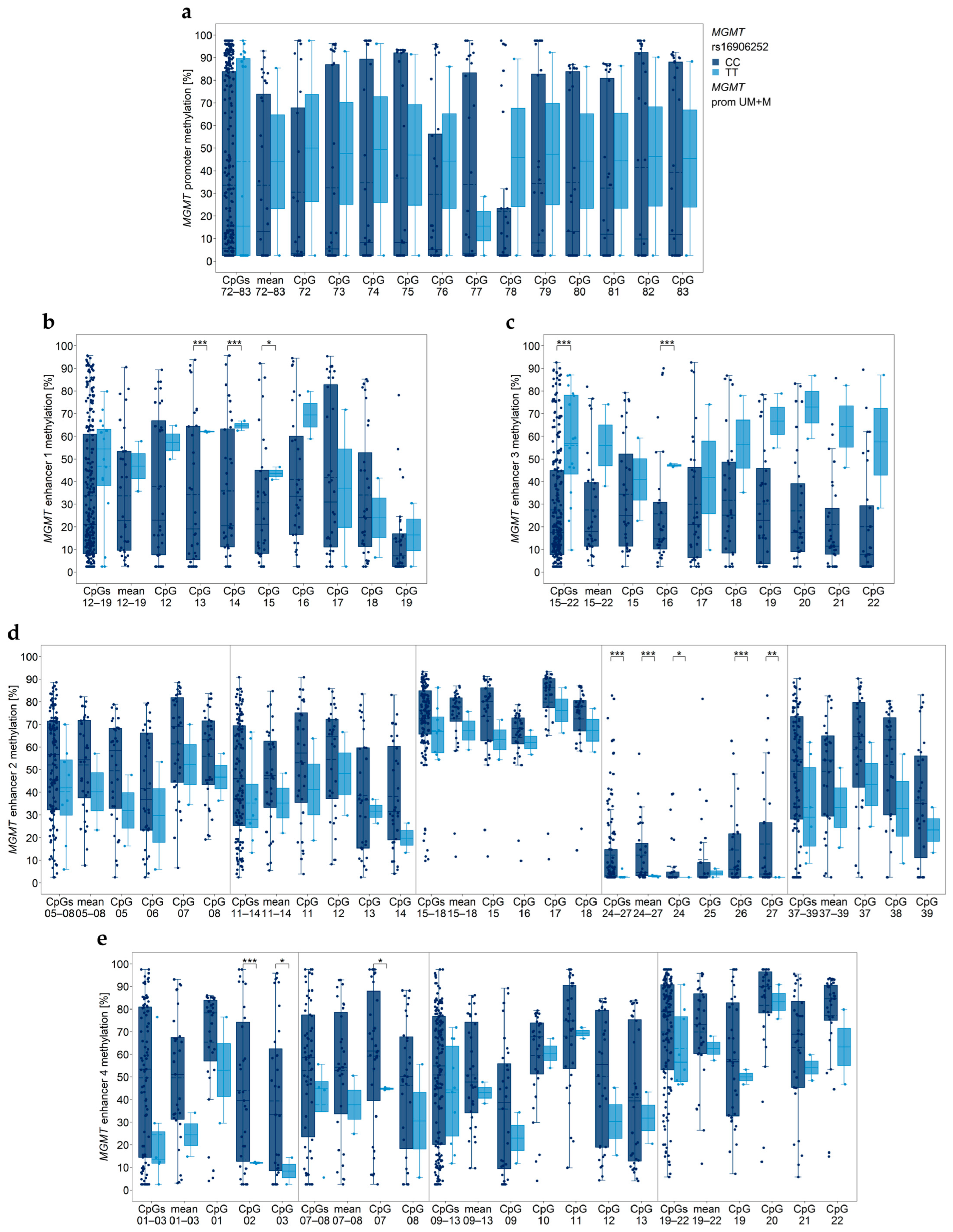
Figure 1. Association between MGMT promoter/enhancer methylation and MGMT SNP rs16906252 genotypes. MGMT (a) promoter, (b) enhancer 1, (c) enhancer 3, (d) enhancer 2, and (e) enhancer 4 regions. Gray vertical lines separate data obtained with different assays. Significance levels: * p ≤ 0.05, ** p ≤ 0.01, *** p ≤ 0.001. Data points represent the mean of two independent PCR-PSQ runs.
Cancers 15 05777 g001
Figure 2. Significantly different MGMT promoter/enhancer methylation levels between TERT promoter (prom) wildtype (wt) and mutations C228T and C250T. MGMT (a) promoter, (b) enhancer 1, (c) enhancer 2, and (d) enhancer 3 regions. prom M—promoter methylated (purple), UM—unmethylated (green), UM + M (blue) patients. GBM18 was the only sample with an unmethylated MGMT promoter lacking C228T and C250T mutation; it was not included in statistical analysis. Significance levels—* p ≤ 0.05, ** p ≤ 0.01, *** p ≤ 0.001. Data points represent the mean of two independent PCR-PSQ runs.
Cancers 15 05777 g002
Figure 3. Significantly different MGMT promoter/enhancer methylation levels between TERT rs2853669 genotypes. MGMT (a) promoter, (b) enhancer 1, (c) enhancer 2, (d) enhancer 3, and (e) enhancer 4 regions. prom M—promoter methylated (purple), UM—unmethylated (green), UM + M (blue) patients. Significance levels: * p ≤ 0.05, ** p ≤ 0.01, *** p ≤ 0.001. Data points represent the mean of two independent PCR-PSQ runs.
Cancers 15 05777 g003
Figure 4. Association of MGMT promoter/enhancer methylation with Ki-67 index. (ac) Correlation plots and two exemplary scatterplots for MGMT (b) enhancer 1 (CpG18) and (c) enhancer 2 (CpG08) indicating correlations in patients with an unmethylated MGMT promoter. (dg) Significant differences between patients with low (≤50%) and high (>50%) Ki-67 index for MGMT (d) promoter, (e) enhancer 1, (f) enhancer 2, and (g) enhancer 3 regions. Pearson’s correlation coefficients are shown in color ranges of dark red (−1.0)–dark blue (1.0); point size (0–±1.0), significant coefficients are highlighted in yellow. Prom M—promoter methylated (purple), UM—unmethylated (green), UM + M (blue) patients. Significance levels: * p ≤ 0.05, ** p ≤ 0.01, *** p ≤ 0.001. Data points represent the mean of two independent PCR-PSQ runs.
Cancers 15 05777 g004
Figure 5. Kaplan–Meier survival analysis. Samples were grouped by the mean methylation status of MGMT (a) promoter CpGs 72–83, (b) enhancer 1 CpGs 12–19, (c) enhancer 2 CpGs 05–08, (d) enhancer 3 CpGs 15–22, and (ei) enhancer 4 for CpGs (e) 01–03, (f) 07, (g) 08, (h) 09–13, and (i) 19–22. enh—enhancer, prom—promoter. Patient GBM27 was excluded due to a lack of OS data.
Cancers 15 05777 g005
Figure 6. Association of Ki-67 index, KPS, and therapy with OS. Scatterplot showing the (a) correlation between Ki-67 index and OS in patients independent of their MGMT promoter methylation status. (bf) Kaplan–Meier survival analysis of samples grouped by (b) Ki-67 index, (c) KPS, (d) primary therapy, and (e,f) adjuvant therapy status. Patient GBM27 was excluded due to a lack of OS data. Prom M—promoter methylated (purple), UM—unmethylated (green), UM + M (gray) samples.
Cancers 15 05777 g006
Figure 7. Significantly different MGMT promoter/enhancer methylation levels between younger (<60 years) and older (≥60 years) patients. MGMT (a) promoter, (b) enhancer 1, (c) enhancer 2, (d) enhancer 3, and (e) enhancer 4 regions. prom M—promoter methylated (purple), UM—unmethylated (green), UM + M (blue) samples. Significance levels: * p ≤ 0.05, ** p ≤ 0.01, *** p ≤ 0.001. Data points represent the mean of two independent PCR-PSQ runs.
Cancers 15 05777 g007
Figure 8. Association of age with Ki-67 index, KPS, and OS. (a) Significant difference in age between patients with low (≤50%) and high (>50%) Ki-67 index. (b) Correlation between age and KPS in patients with methylated MGMT promoter. (c) Significant difference in age of patients with low (≤80%) and high (>80%) KPS. (d) Kaplan–Meier survival analysis of patients grouped by age (<60 years, ≥60 years). Patient GBM27 was excluded from the survival analysis due to a lack of OS data. (e) Correlation between age and OS in patients with methylated MGMT promoter. Significance levels: * p ≤ 0.05, ** p ≤ 0.01. Prom M—promoter methylated (purple), UM—unmethylated (green), UM + M (gray) patients.
Cancers 15 05777 g008
Figure 9. Significantly different MGMT promoter/enhancer methylation levels between female and male patients. MGMT (a) promoter, (b) enhancer 1, (c) enhancer 2, and (d) enhancer 4 regions. prom M—promoter methylated (purple), UM—unmethylated, UM + M (blue) samples. Significance levels: * p ≤ 0.05, ** p ≤ 0.01, *** p ≤ 0.001. Data points represent the mean of two independent PCR-PSQ runs.
Cancers 15 05777 g009
Figure 10. Summary of associations found for MGMT promoter and MGMT enhancer methylation. Significant associations are highlighted for prom M—promoter methylated (purple), UM—unmethylated (green), and UM + M (blue) samples.
Cancers 15 05777 g010
Table 1. Clinical and genetic data of GBM patients for which stable cell cultures could be established.
Patient
Age [year]Sex
KPS [%]Ki-67
[%]
Primary TherapyAdjuvant
Therapy
OS [m]PFS [m]MGMT ExpMGMT rs16906252TERT
Prom
TERT rs2853669
GBM0164f60n.s.R-Ch-Tnone7.434.000.00CCC250TTT
GBM0285m40≤50 (30)nonenone3.00n.s.0.00CCwtTT
GBM0353f100n.s.R-Ch-Tnone52.504.500.00CCC228TCT
GBM0467f70>50 (90)RTnone46.630.530.00CCC228TTT
GBM0557f100≤50 (30)RTCCNU10.506.000.00CCC228TTT
GBM0646m90n.s.R-Ch-TTMZ + CCNU30.504.000.00CT (atyp.)C228TTT
GBM0750f90n.s.RTTMZ27.40n.s.0.00CCC250Tn.s.
GBM0874f70≤50 (30)R-Ch-T 1Avastin + TMZ7.79n.s.0.00CCC228TCT
GBM0948m60n.s.nonenone1.55n.s.0.00CCwtTT
GBM1064m80>50R-Ch-TTMZ11.70n.s.0.20CCC228TCT
GBM1173f60>50R-Ch-Tno10.60n.s.0.16CCC228TTT
GBM1244m80>50R-Ch-TTMZ13.009.001.10CCC250TCT
GBM1365m90≤50 (15)RT 1none9.27n.s.1.20CCC228TTT
GBM1469m90≤50 (40)RTTMZ + CCNU8.00n.s.0.04CCC250TTT
GBM1573f100n.s.nonenone7.002.631.10CCC250TCT
GBM1683m90≤50RTnone9.579.001.00CCC228TCT
GBM1774m90n.s.R-Ch-Tnone5.19n.s.1.32CCC250TCT
GBM1844m100n.s.R-Ch-TTMZ23.15n.s.0.25CCwtTT
GBM1948m100>50 (60)R-Ch-TTMZ18.674.540.09CCC228TCT
GBM2075m60≤50 (50)Ch-TTMZ23.905.980.06TTC228TCT
GBM2160m90≤50 (40)R-Ch-TAvastin13.003.000.40CCC228TTT
GBM2253m70>50 (60)nonenone0.89n.s.0.54CCC250TTT
GBM2347f70>50 (70)R-Ch-TTMZ37.258.000.00CCC250TCT
GBM2464m90≤50 (40)R-Ch-TTMZ7.73n.s.0.49CCC228TCT
GBM2567f70>50R-Ch-TTMZ16.8010.000.00CCC228TTT
GBM2675f70≤50 (40)RTnone8.22n.s.0.00CCC228TCT
GBM2752f90>50 (70)R-Ch-TTMZn.s.n.s.0.00TTC250TTT
GBM2863mn.s.>50R-Ch-TTMZ21.808.000.00CCC250TCT
GBM2979f40>50nonenone1.31n.s.0.00CCC228TCT
GBM3058m90>50R-Ch-TTMZ16.004.310.00CCC228TTT
GBM3158fn.s.>50R-Ch-TTMZ13.304.000.00CCC228TTT
GBM3271mn.s.≤50 (20)nonenone1.25n.s.0.00CCC228TTT
GBM3357mn.s.n.s.R-Ch-TTMZ12.706.000.20CCC250TCT
GBM3453mn.s.n.s.R-Ch-TTMZ32.3022.000.00CCC228TTT
GBM3564m40n.s.R-Ch-Tnone10.60n.s.0.00CCC228TTT
GBM01–35—glioblastoma multiforme patients. Applied drugs were bevacizumab (Avastin), Lomustine (CCNU—(1-(2-chloroethyl)-3-cyclohexyl-1-nitrosourea)), and temozolomide (TMZ). atyp—atypical HRM-PSQ result [21], Ch-T—chemotherapy, exp—expression, f—female, KPS—Karnofsky Performance Score, m—male, mut—mutation, n.s.—not specified, OS—overall survival, PFS—progression-free survival, R-Ch-T—radio-chemotherapy, RT—radiotherapy, wt—wildtype. 1 Therapy had to be discontinued.
Table 2. Cox proportional hazards models for MGMT promoter and enhancer 4 regions.
PromoterEnhancer 4
Cut-OffCpGHazard Ratio
(95% CI)
p-ValueCut-OffCpGHazard Ratio
(95% CI)
p-Value
(<8% vs. ≥8%)mean 72–832.18 (1.01–4.71)0.042(≥55% vs. <55%)mean 01–032.97 (1.31–6.74)0.007
722.81 (1.23–6.40)0.011 010.71 (0.31–1.60)0.402
732.55(1.15–5.66)0.018 022.05 (0.97–4.30)0.054
742.52 (1.16–5.50)0.017 032.27 (1.01–5.10)0.042
752.18(1.01–4.71)0.042 mean 07–081.81 (0.25–1.12)0.118
762.76 (1.24–6.14)0.010 073.27 (1.32–8.12)0.008
772.76 (1.24–6.14)0.010 080.96 (0.47–1.97)0.915
783.35 (1.43–7.85)0.004 mean 09–132.34 (1.06–5.13)0.030
792.36 (1.08–5.17)0.028 092.19 (0.94–5.08)0.063
802.18 (1.01–4.71)0.042 101.93 (0.87–4.27)0.102
812.18 (1.01–4.71)0.042 111.83 (0.76–4.37)0.172
822.36 (1.08–5.17)0.028 121.18 (0.59–2.35)0.641
832.18 (1.01–4.71)0.042 132.30 (1.05–5.07)0.033
(≥75% vs. <75%)mean 19–221.84 (0.90–3.75)0.092
191.43 (0.69–3.00)0.337
201.22 (0.52–2.83)0.647
211.11 (0.56–2.23)0.762
220.85 (0.38–1.91)0.696
bold—significant (p ≤ 0.05), underlined—Hazard Ratio given for this group.
Table 3. Univariate Cox proportional hazards model.
CovariateHazard Ratio (95% CI)p-Value
Ki-67 index (≤50% vs. >50%)2.72 (1.11–6.70)0.024
KPS (≥80% vs. <80%)0.78 (0.37–1.64)0.507
primary therapy (R-Ch-T vs. none or RT)0.01 (0.001–0.11)<0.001
adjuvant therapy (TMZ alone vs. none or other)0.50 (0.23–1.09)0.201
adjuvant therapy (yes vs. no)0.56 (0.27–1.16)0.113
age (≥60 vs. <60)2.28 (1.11–4.70)0.022
bold—significant (p ≤ 0.05), underlined—Hazard Ratio given for this group.
Disclaimer/Publisher’s Note: The statements, opinions and data contained in all publications are solely those of the individual author(s) and contributor(s) and not of MDPI and/or the editor(s). MDPI and/or the editor(s) disclaim responsibility for any injury to people or property resulting from any ideas, methods, instructions or products referred to in the content.