A Deep Learning Approach for Automatic Segmentation during Daily MRI-Linac Radiotherapy of Glioblastoma
by
 , ,
, ,
Adrian L. Breto
1,†, 
Kaylie Cullison
1,†,
Evangelia I. Zacharaki
1,
Veronica Wallaengen
1,
Danilo Maziero
1,2,
Kolton Jones
1,3,
Alessandro Valderrama
1,
Macarena I. de la Fuente
4,
Jessica Meshman
1,
Gregory A. Azzam
1,
John C. Ford
1,
Radka Stoyanova
1 and 
Eric A. Mellon
1,* 1
Department of Radiation Oncology, Sylvester Comprehensive Cancer Center, Miller School of Medicine, University of Miami, Miami, FL 33136, USA
2
Department of Radiation Medicine & Applied Sciences, UC San Diego Health, La Jolla, CA 92093, USA
3
West Physics, Atlanta, GA 30339, USA
4
Department of Neurology, Sylvester Comprehensive Cancer Center, Miller School of Medicine, University of Miami, Miami, FL 33136, USA
*
Author to whom correspondence should be addressed.
†
These authors contributed equally to this work.
Cancers 2023, 15(21), 5241; https://doi.org/10.3390/cancers15215241
Submission received: 5 October 2023
/
Revised: 25 October 2023
/
Accepted: 30 October 2023
/
Published: 31 October 2023
(This article belongs to the Special Issue Radiation Therapy for Brain Tumors)
Simple Summary
Current auto-segmentation methods for glioblastoma utilize mainly pre-operative 1.5T and 3T MRI. The first commercial MRI-linear accelerator (linac) radiation treatment platform acquires low-field (0.35T) post-operative MRI at the delivery of each treatment. This study presents the first automatic brain lesion segmentation network developed for MRI-linac to track tumor changes during radiotherapy. The tumors and resection cavities are automatically segmented using a deep learning network, allowing for daily monitoring of tumor volume changes, creation of tools necessary for adaptive radiotherapy of glioblastoma, and providing MRI regions of interest for further analyses that may discover prognostic markers.
Abstract
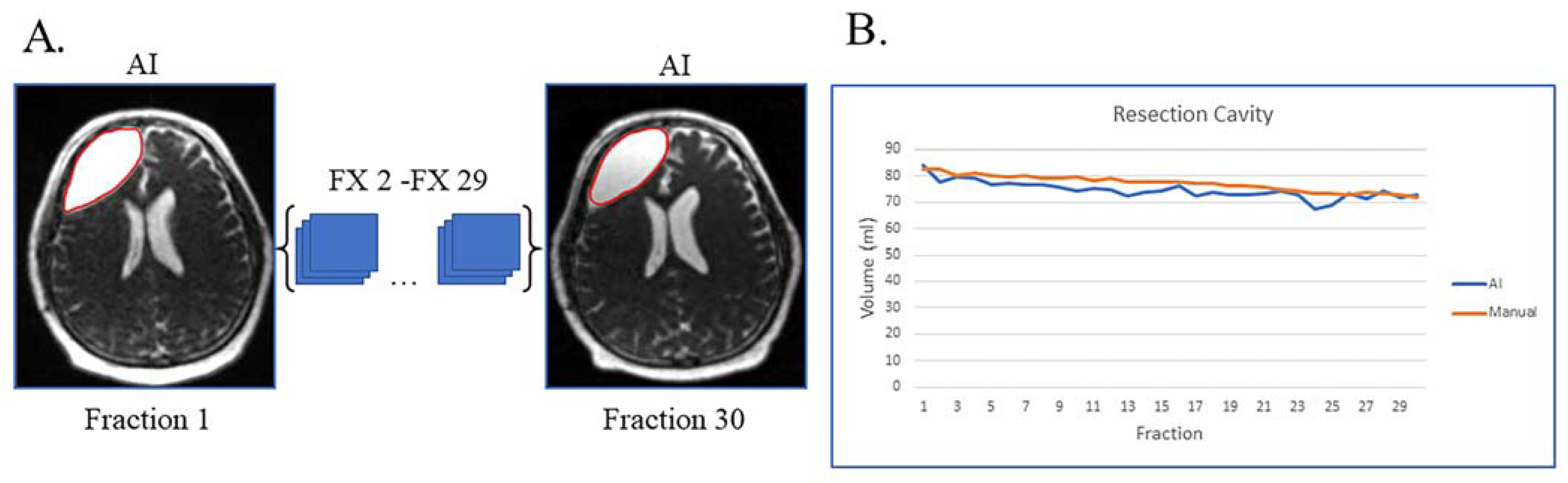
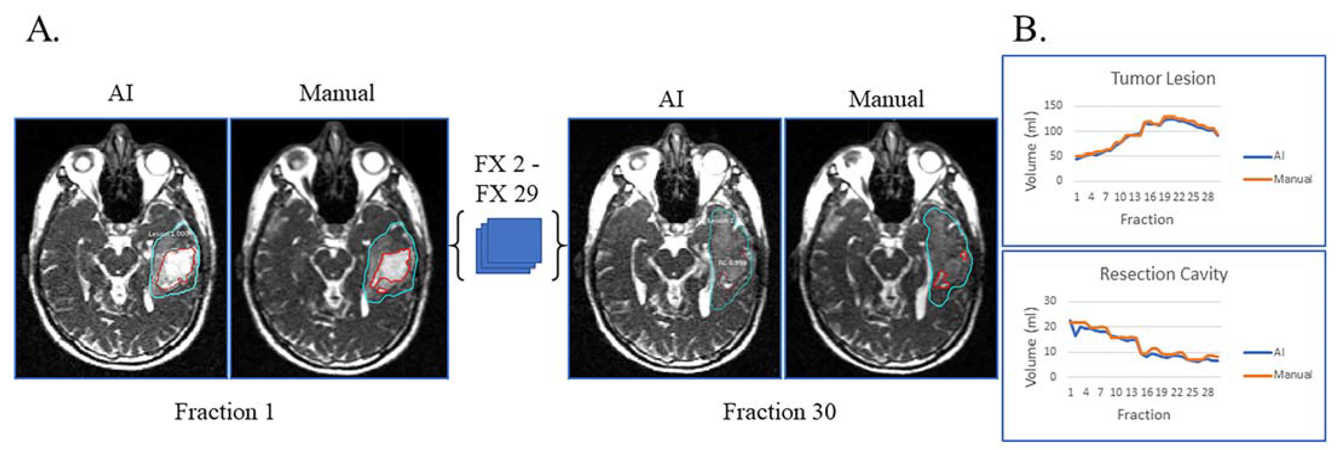
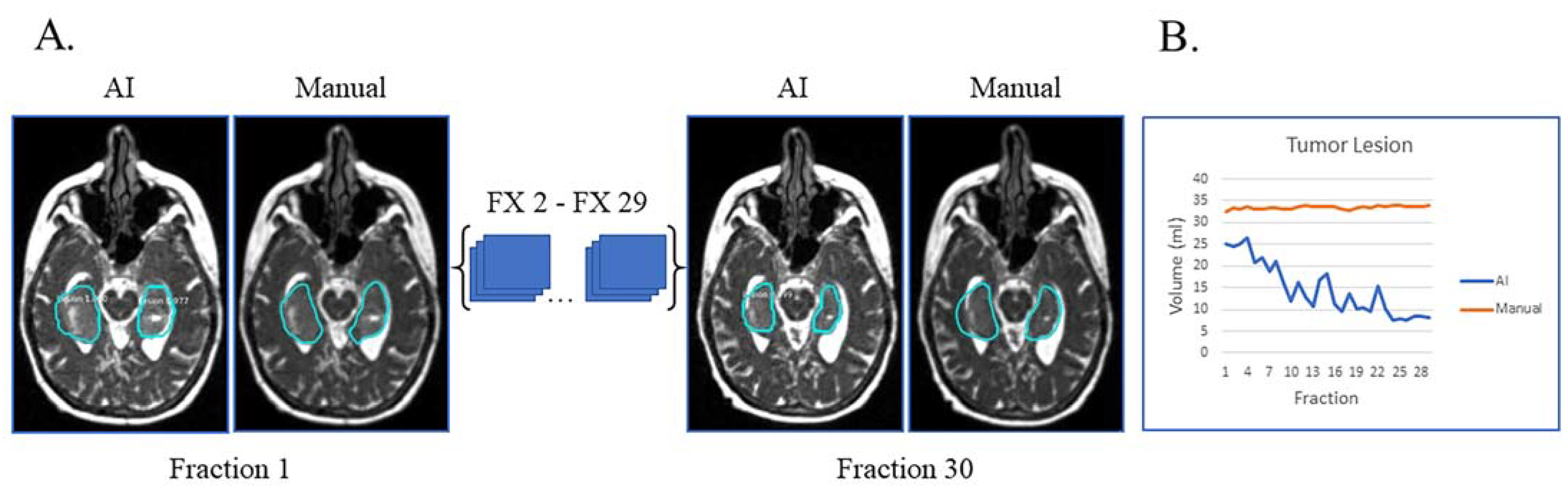
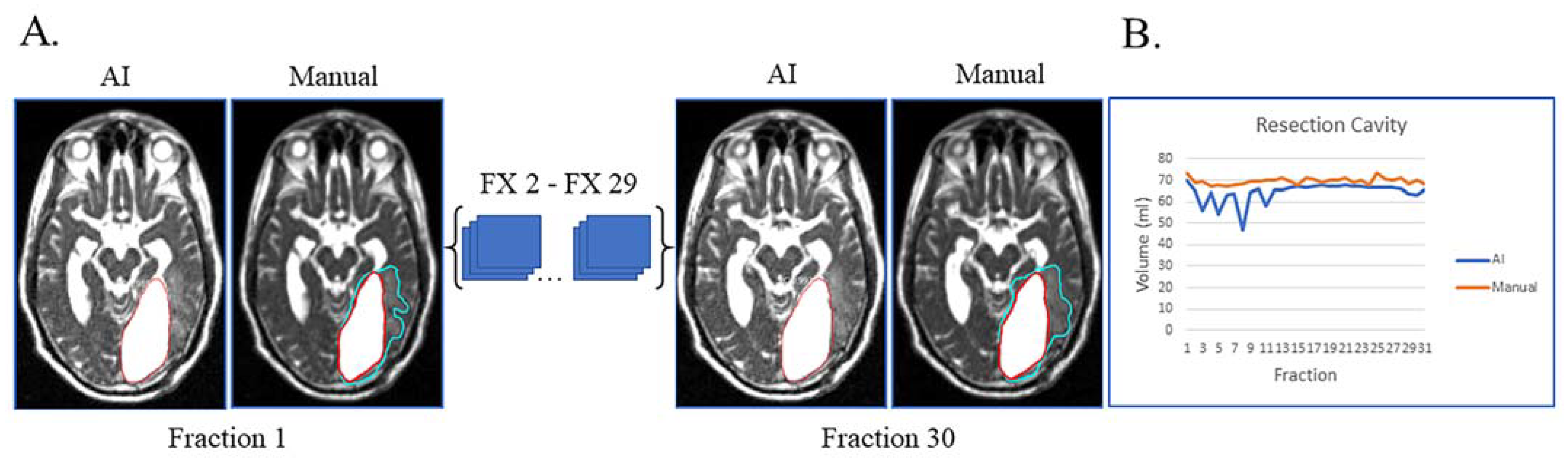
Glioblastoma changes during chemoradiotherapy are inferred from high-field MRI before and after treatment but are rarely investigated during radiotherapy. The purpose of this study was to develop a deep learning network to automatically segment glioblastoma tumors on daily treatment set-up scans from the first glioblastoma patients treated on MRI-linac. Glioblastoma patients were prospectively imaged daily during chemoradiotherapy on 0.35T MRI-linac. Tumor and edema (tumor lesion) and resection cavity kinetics throughout the treatment were manually segmented on these daily MRI. Utilizing a convolutional neural network, an automatic segmentation deep learning network was built. A nine-fold cross-validation schema was used to train the network using 80:10:10 for training, validation, and testing. Thirty-six glioblastoma patients were imaged pre-treatment and 30 times during radiotherapy (n = 31 volumes, total of 930 MRIs). The average tumor lesion and resection cavity volumes were 94.56 ± 64.68 cc and 72.44 ± 35.08 cc, respectively. The average Dice similarity coefficient between manual and auto-segmentation for tumor lesion and resection cavity across all patients was 0.67 and 0.84, respectively. This is the first brain lesion segmentation network developed for MRI-linac. The network performed comparably to the only other published network for auto-segmentation of post-operative glioblastoma lesions. Segmented volumes can be utilized for adaptive radiotherapy and propagated across multiple MRI contrasts to create a prognostic model for glioblastoma based on multiparametric MRI.
Keywords:
glioblastoma; auto-segmentation; MRI-linac; radiation therapy; deep learning; MRI