Review

Long Non-Coding RNAs as Emerging Targets in Lung Cancer

by
Jovanka Gencel-Augusto
1,2,
Wei Wu
1,2,* and
Trever G. Bivona
1,2,3,*
1
Department of Medicine, University of California San Francisco (UCSF), San Francisco, CA 94158, USA
2
UCSF Hellen Diller Comprehensive Cancer Center, San Francisco, CA 94158, USA
3
Chan-Zuckerberg Biohub, San Francisco, CA 94158, USA
*
Authors to whom correspondence should be addressed.
Cancers 2023, 15(12), 3135; https://doi.org/10.3390/cancers15123135
Submission received: 9 May 2023 / Revised: 4 June 2023 / Accepted: 8 June 2023 / Published: 10 June 2023

Abstract

:

Simple Summary

Long non-coding RNAs (LncRNAs) are non-protein coding molecules longer than 200 nucleotides. They play essential roles in normal cell function and development, and can contribute to diseases such as cancer when dysregulated. Although lncRNAs have oncogenic or tumor-suppressive properties in lung cancer and can serve as stable biomarkers, this is still an understudied field. Here, we discuss recent evidence for lncRNAs role in lung cancer development, therapy resistance, biomarker potential, and therapeutic strategies. We conclude that understanding the interplay between non-coding and coding molecules in lung cancer should be explored further and may open up new avenues for treatment.

Abstract

Long non-coding RNAs (LncRNAs) are mRNA-like molecules that do not encode for proteins and that are longer than 200 nucleotides. LncRNAs play important biological roles in normal cell physiology and organism development. Therefore, deregulation of their activities is involved in disease processes such as cancer. Lung cancer is the leading cause of cancer-related deaths due to late stage at diagnosis, distant metastasis, and high rates of therapeutic failure. LncRNAs are emerging as important molecules in lung cancer for their oncogenic or tumor-suppressive functions. LncRNAs are highly stable in circulation, presenting an opportunity for use as non-invasive and early-stage cancer diagnostic tools. Here, we summarize the latest works providing in vivo evidence available for lncRNAs role in cancer development, therapy-induced resistance, and their potential as biomarkers for diagnosis and prognosis, with a focus on lung cancer. Additionally, we discuss current therapeutic approaches to target lncRNAs. The evidence discussed here strongly suggests that investigation of lncRNAs in lung cancer in addition to protein-coding genes will provide a holistic view of molecular mechanisms of cancer initiation, development, and progression, and could open up a new avenue for cancer treatment.

1. Introduction

Long non-coding RNAs (LncRNAs) are broadly defined as RNAs that usually do not encode for proteins and that are longer than 200 nucleotides. These are messenger RNA (mRNA)-like molecules that are transcribed by polymerase II, 5′ capped, and have a 3′ poly-A tail. Because they can form complex secondary structures, they often have functions. Many lncRNAs are preferentially found in the nucleus, where they participate in the regulation of chromatin organization and transcription, often by forming lncRNA-DNA triplex [1,2,3], as well as through the formation of nuclear speckles and regulation of splicing. In the cytoplasm, lncRNAs can regulate mRNA stability, bind to other non-coding RNAs, and modulate protein post-translational modifications and protein function [4,5,6,7,8].
LncRNAs have been studied in mammals since the early 1990s due to their involvement in developmental processes. For example, the Xist (X-inactive specific transcript) lncRNA contributes to reshaping the architecture of chromatin to achieve X chromosome-silencing in early embryonic development [9]. The H19 lncRNA is involved in genomic imprinting and regulation of the insulin growth factor 2 (IGF2) and other genes involved in embryonic growth [10,11]. The study of HOX genes, master regulators of embryonic development, led to the discovery of the lncRNA HOTAIR (Homeobox transcript antisense intergenic RNA). HOTAIR is transcribed from the antisense strand of the HOXC gene.
HOTAIR has been reported to repress the transcription of the HOXD loci via interaction with PRC2 (polycomb repressive complex 2) [12], although in vivo models developed later report conflicting results regarding HOXC or HOXD genes regulation by HOTAIR [13,14]. Because of the importance of physiological context to understand lncRNA molecular function, more recently, other groups have reported exclusively in vivo approaches using animal models of lncRNA genetic ablation. For example, a comprehensive study developed 18 knock-out (KO) mouse models for less well-known lncRNAs with human orthologs. Certain lncRNAs’ expression was highly tissue-specific (such as Fendrr, Manr and linc–Cox2 expressed mainly in lung), supporting a unique physiological role, while other lncRNAs were more ubiquitously expressed. Three of the analyzed lncRNAs were required for embryonic development (Fendrr, Mdgt, Peril), speaking to their fundamental functions [15]. Thus, the involvement of lncRNAs in normal cell physiology and organism development suggests that these may also control disease-related processes such as cancer.
Lung cancer is the leading cause of cancer-related mortality in the U.S., and non-small cell lung cancer (NSCLC) is the most common subtype. Lung cancer is commonly diagnosed in late stages, where patients present distant metastasis with 9% having a 5-year survival rate [16]. Although the development of targeted therapies (e.g., tyrosine kinase inhibitors, TKIs) has improved patient outcomes, their clinical efficacy is often limited by both innate and acquired resistance, permitting tumor progression and recurrence leading to poor survival rates [17]. It is imperative to better understand molecular drivers of tumorigenesis, metastasis, and therapy resistance in lung cancer to develop improved therapeutic strategies. LncRNAs are emerging as important molecules in cancer due to their oncogenic or tumor-suppressive functions. Here, we summarize the latest works providing in vivo evidence available for the role of lncRNAs in lung cancer, therapy resistance, and their potential as biomarkers.

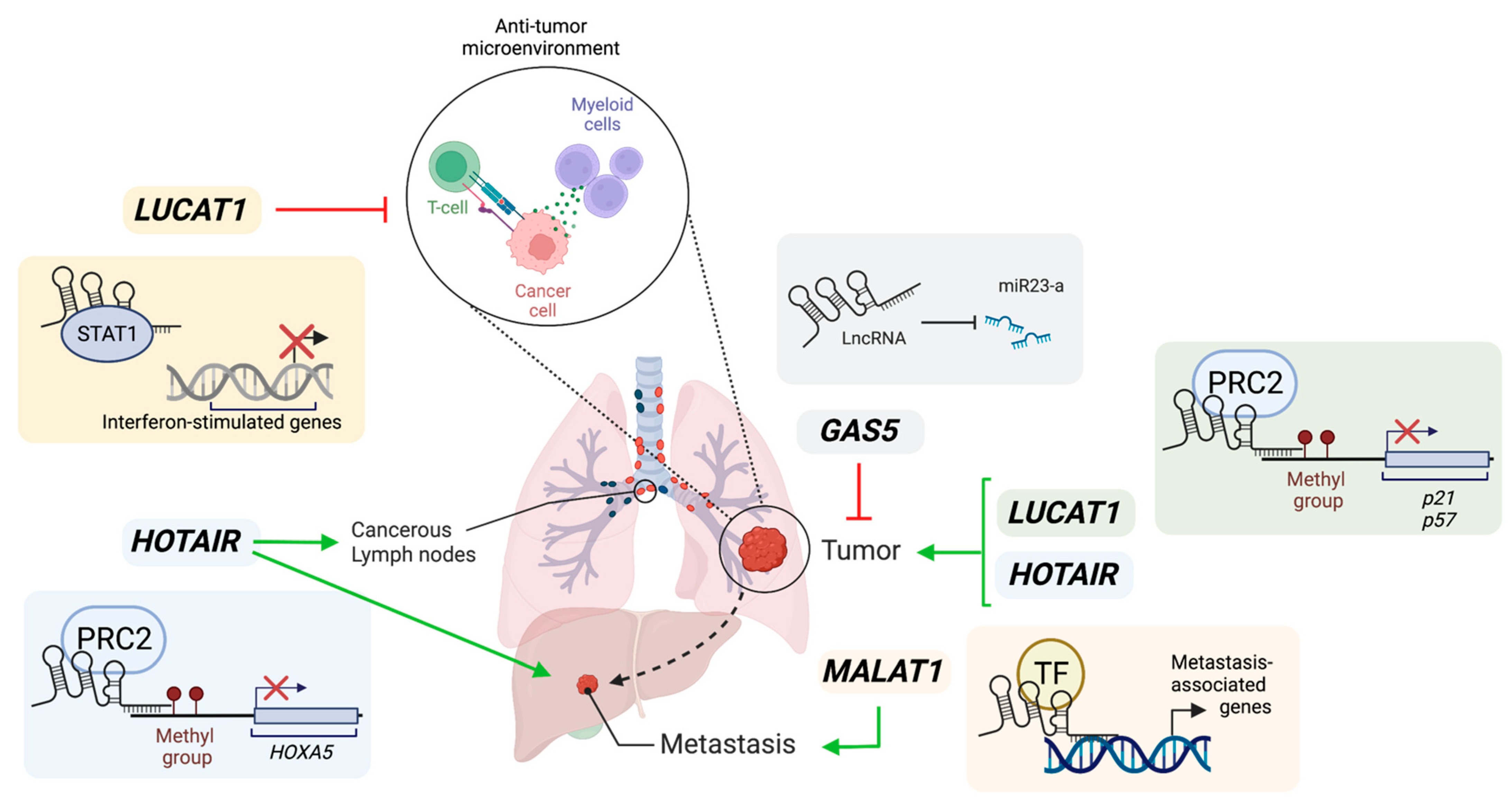
2. Role of lncRNAs in Lung Cancer

Because of the role of lncRNAs in regulating a diverse array of cellular functions, deregulation of their activities is involved in cancer. Some mechanisms reported for lncRNAs functions in cancer are as follows: acting as miRNA sponges to modulate activity on their targets; interacting with histone-modifier enzymes to modulate known oncogene/tumor suppressor gene expression; interacting with transcription factors to repress/activate their transcriptional programs; acting as anti-sense molecules for tumor suppressor mRNAs, among other mechanisms [2,18]. Importantly, regulation of lncRNA expression and function in cancer follows similar principles to that of known oncogenes and tumor suppressors, as it can be mediated by DNA methylation [19], amplification or deletion [20], and mutation or SNPs of DNA sequences [21,22]. In this section, we provide examples of well-studied lncRNAs that are reported to have oncogenic or tumor-suppressive roles in lung cancer, as well as those with controversial functions.
MALAT1 (Metastasis-Associated Lung Adenocarcinoma Transcript 1) was one of the first lncRNAs described to be associated with cancer. In 2003, Ji et al. analyzed the gene expression profile in human primary lung cancer tumors that subsequently metastasized or those that did not metastasize and compared their transcriptional signatures. They identified a Metastasis-Associated Lung Adenocarcinoma Transcript 1, named MALAT1, for its higher expression in primary tumors that metastasized. They also found a significant correlation between higher level of MALAT1 expression in stage I lung cancer and worse survival outcomes [23]. Several studies since then have described MALAT1 function in normal physiology and cancer [24,25,26]. Three independent groups developed an in vivo approach to describing MALAT1 physiological function. They found that Malat1 is highly abundant in several mouse tissues and highly conserved across species. Genetic perturbation of the Malat1 locus in mice (using genetic deletion [27,28] or genetic inactivation approaches [29]) did not alter animal development, nuclear speckle formation, splicing, or mRNA stability. However, they described a role for Malat1 in controlling neighboring genes expression in a tissue-specific manner that was not consistent between the three studies, especially that of Neat1, another lncRNA [28,29]. In the context of lung cancer, MALAT1-silencing did not show effects on lung cancer cell proliferation or viability in vitro [27]. To further understand the role of MALAT1 in lung cancer metastasis, Gutschner et al. implanted EBC-1 lung cancer cells into nude mice and treated them with subcutaneous administration of an anti-sense oligonucleotide (ASO) targeting MALAT1. After five weeks of treatment, all primary tumors were excised. Metastasis nodules were analyzed at 12 weeks, indicating fewer and smaller metastatic nodules in the treated group, suggesting a role for MALAT1 in promoting metastasis. Through additional in vitro studies, they report MALAT1 inhibition results in aberrant expression of metastasis-associated genes in cell lines [30]. Although these studies support a role for MALAT1 in promoting metastasis and regulating certain genes expression, other evidence exists for different roles. Kim et al. describe an elegant study that challenges previously reported roles for MALAT1 in metastasis. Using the Malat1 knock-out (KO) mouse model developed by Nakagawa et al. (LacZ and Poly-A sequences were used as transcriptional terminators inserted 69 bp downstream of the transcription start site of Malat1 without deletion of the DNA sequence), Kim et al. crossed these mice with a breast cancer model driven by MMTV-PyMT that mimics human disease. Surprisingly, they found a 7.2-fold increase in metastatic foci and 31-fold increase in the percent of lung areas with metastatic lesions in Malat1-KO mice as compared to Malat1 WT mice, suggesting a role for Malat1 in suppressing breast cancer metastasis to the lung. This phenotype was rescued by transgenic expression of Malat1, suggesting that the RNA product itself diminished metastasis. Additionally, they identified Malat1 interaction with TEAD (transcriptional enhanced associate domain) proteins in mouse-derived tumors and cell lines, which suppressed TEAD-YAP interaction and, therefore, inhibited their pro-metastatic transcriptional program [31]. This study and others highlight the importance of context (lung model vs. breast model) as well as experimental methodology to approach lncRNAs’ functional characterization (such as lncRNA genomic DNA loss vs. RNA loss reviewed in detail elsewhere [24]). Of note, DNA elements themselves within a lncRNA locus may be responsible for regulatory functions that are independent from transcript function [32,33,34,35]. In summary, MALAT1 promotes metastasis in lung cancer, but may show opposite functions in different types of cancer depending on cellular context.
The GAS5 (growth arrest-specific 5) gene was first described as a G0-specific gene that is inhibited by serum and growth factors [36]. In vivo, Gas5 genetic deletion (Gas5+/−) in mice decreased bone mass and impaired bone repair, leading to osteoporosis. Mechanistically, Gas5 positively influenced proper cell differentiation through interaction with UPF1 (a DNA/RNA helicase) to accelerate SMAD7 mRNA decay [37]. In lung cancer, GAS5 was found down-regulated in 72 NSCLC tumor samples as compared to their paired adjacent normal tissues, suggesting a tumor-suppressive role. Additionally, low GAS5 expression was correlated with larger tumor size, lower differentiation levels, and higher staging of tumor–node metastasis [38]. A xenograft model of GAS5 overexpression (OE) showed that GAS5 OE markedly decreased tumor size compared with control [38]. Although this study did not explore a mechanism for GAS5-mediated tumor suppression, other studies implicate a role for GAS5 as a miRNA sponge to negatively influence cell cycle activator genes [39] or positively influence PTEN levels [40]. Additionally, a recent study revealed GAS5 is partially localized to the mitochondria where it modulates energy homeostasis by promoting the de-acetylation of malate dehydrogenase, suppressing breast cancer [41]. Taken together, a role for GAS5 in halting the cell cycle as well as promoting cell differentiation in normal cells supports its tumor-suppressive role reported in lung cancer. Besides GAS5, other lncRNAs have been studied for their tumor-suppressive functions, such as MEG3 and TUG1, reviewed elsewhere [42].
LUCAT1 (Lung Cancer-Associated Transcript 1), first identified as smoke-induced and cancer-associated lncRNA1 (SCAL1) [43], has higher expression in lung cancer as compared to normal controls, and is also found to be overexpressed in several cancer types [44]. To our knowledge, a LUCAT1-deficient mouse model has not been reported. Additionally, patients with tumors that express high levels of LUCAT1 showed poorer overall survival as compared to those with lower LUCAT1 expression. Moreover, high LUCAT1 levels were associated with late staging in tumor–lymph node metastasis and higher tumor volume. In NSCLC cell lines A549 and SPC-A1, LUCAT1 modulates p21 and p57 expression by promoting loci methylation through PRC2 [45].
More recently, a role for LUCAT1 in regulating immune responses has been described. LUCAT1 genetic deletion in myeloid cells is found to enhance interferon-mediated gene transcription. LUCAT1 acts as an immune suppressor by interacting with STAT1 and chromatin in the nucleus. It may also act by inhibiting NF-kB functions [46]. These findings suggest a tumor-promoting role for LUCAT1 that is tumor-cell-intrinsic, in addition to a potential non-cell autonomous mechanism via the inhibition of immune surveillance, although this mechanism remains to be explored in the lung cancer context.
HOTAIR has been vastly studied in cancer contexts [47,48]. In NSCLC, tumor samples and cell lines expressed higher HOTAIR levels as compared to normal counterparts [49]. Additionally, high HOTAIR levels correlated with higher tumor grade and presence of lymph node metastases [49,50]. In vitro, HOTAIR has been reported as a direct target of the hypoxia-inducible-factor-1α (HIF-1α), therefore enhancing A549 NSCLC cells’ proliferation, migration, and invasion [51]. In vivo, tail vein injections of SPC-A1 cells with or without siRNA targeting HOTAIR showed that the knock-down condition reduced the number of metastasis nodules found in the lungs of immunocompromised mice [49]. HOTAIR-silencing resulted in a decrease in matrix metalloproteinases (MMPs, which promote invasion and migration) expression and an increase in HOXA5 levels (a tumor suppressor) in cell lines, suggesting HOTAIR acts through the regulation of expression of cancer-related genes [49]. Although this xenograft assay does not account for all steps required for a tumor cell to achieve metastatic colonization (extravasation, survival in blood, seeding of new site, proliferation in new site), and they measured colonization of lungs using a lung cancer cell line (same tissue), it raises the possibility that HOTAIR may be involved in the seeding and survival of cancer cells. Additionally, the absence of a competent immune system challenges interpretation of these results. Development of a HOTAIR transgenic mouse model to understand in vivo implications in lung cancer initiation and progression is necessary, similar to a HOTAIR inducible system recently reported for breast cancer [52]. This model showed that sustained HOTAIR overexpression promotes breast cancer metastasis to lungs. Overall, with the data available, HOTAIR seems to play an oncogenic role in lung cancer; however, robust mechanisms through which this lncRNA function remain to be uncovered.
Most studies focus on the contribution of a single lncRNA to cancer phenotypes. However, whether the lncRNAs described above are expressed simultaneously in tumors with unique or redundant functions remains to be explored in depth. For example, Esposito et al. showed that at least 80 oncogenic lncRNAs are active in NSCLC through a lncRNA-focused CRISPR screen. By further dissecting the role of two candidate lncRNAs, CHiLL1 and GCAWKR, they showed these have distinct cellular localization and non-overlapping targets. Importantly, ASOs targeting both these lncRNAs yielded additive effects, suggesting that they have cooperating functions in NSCLC progression [53]. LncRNAs are generally expressed at lower levels than protein-coding genes [54]. Because of this, we speculate that lncRNAs with redundant functions may be expressed simultaneously to compensate for a higher expression of their targets in disease conditions. By examining available TCGA lung adenocarcinoma datasets containing mRNA expression data, we did not find significant correlations (negative or positive) among the expression of lncRNAs described here. However, such an analysis in combination with functional studies could shed light on mutual exclusivity relationships between certain lncRNAs. Additionally, whether certain lncRNAs are predominantly expressed at different stages of tumor progression remains to be explored. A new online resource, lncRNAfunc https://ccsm.uth.edu/lncRNAfunc (accessed on 7 May 2023), provides insights on differentially expressed lncRNAs across different cancer types and stages available in TCGA, as well as functional predictions [55]. Although this analysis did not detect any correlations between the lncRNAs mentioned here and lung cancer stages, possibly due to lack of sufficient sample sizes, these lncRNAs did show correlation with stage in other cancers; for example, LUCAT1 was correlated with cancer stage in kidney cancer.
In summary, lncRNAs have oncogenic and tumor-suppressive roles in lung cancer, illustrated in Figure 1. LncRNAs interact with protein-coding molecules, resulting in the activation or inactivation of specific signaling pathways in cancer cells. We speculate more lung-cancer-specific lncRNAs will be identified with genome-wide transcriptomic studies.

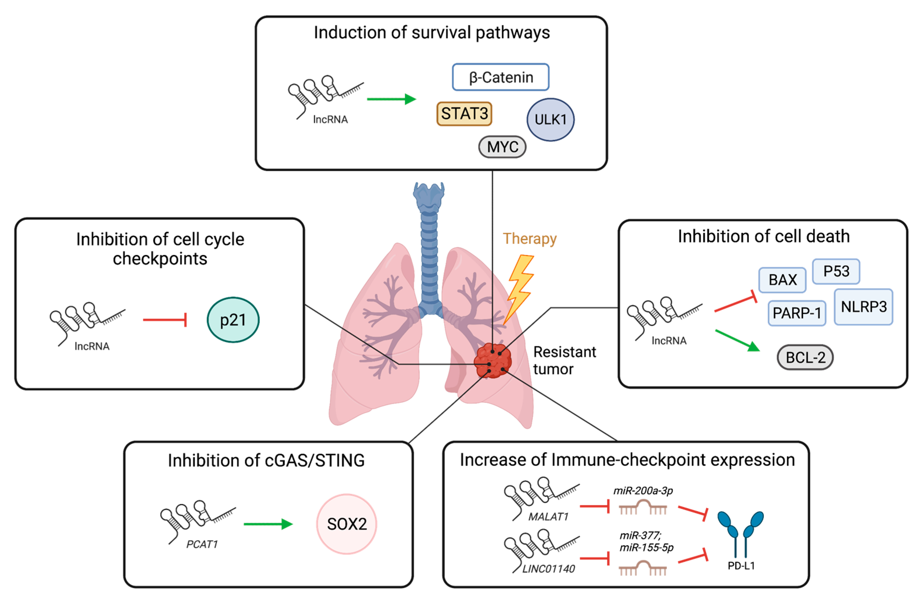
3. LncRNAs in Lung Cancer Therapy Resistance

The role of non-coding RNAs in resistance to cancer therapies has been documented [48,56,57]. Mechanistically, lncRNAs can contribute to therapy resistance by promoting cell survival pathways (including autophagy, DNA repair), inhibiting apoptosis and cell cycle checkpoints, increasing self-renewal capacity and epithelial to mesenchymal transition (EMT), modulating the tumor microenvironment as well as the cellular xenobiotic stress response (drug efflux mechanisms), among others [48,58]. Here, we provide evidence for the roles of the lncRNAs discussed above in resistance to chemotherapy, radiotherapy, and targeted therapy in lung cancer. Additionally, we discuss new advances in understanding the role of lncRNAs in immune checkpoint therapy in lung cancer.

3.1. Role of lncRNAs in Resistance to Chemotherapy, Radiotherapy, and Targeted Therapy in Lung Cancer

In a tumor xenograft model, silencing of MALAT1 in cisplatin resistant A549 cells led to decreased growth in nude mice (subsequently treated with cisplatin), as compared to non-targeting control. Similarly, overexpression of MALAT1 in cisplatin-sensitive A549 lung cancer cells increased tumor volume as compared to empty vector control [59]. These data suggest a role for MALAT1 in promoting cisplatin resistance in lung cancer. The authors suggest that modulation of STAT-3 function by MALAT1 drives this phenotype, although a direct interaction was not confirmed [59]. While additional lung-cancer-focused studies are lacking, the MALAT1 role in cisplatin resistance was also found in a xenograft model of oral squamous cell carcinoma [60]. In radiotherapy, MALAT1 also promotes resistance, although this function has not been explored in lung cancer. In a xenograft model of esophageal squamous cell carcinoma, MALAT1 levels were found to be reduced upon radiation in tumors that respond to treatment. Additionally, overexpressing MALAT1 in xenografts did not decrease their size upon radiation exposure, while controls showed regression [61]. Furthermore, in colorectal carcinoma cell lines, MALAT1 knockdown enhanced radiosensitivity [62]. Therefore, MALAT1 can impact sensitivity to radiation therapy in cancer. To our knowledge, there are no robust in vivo studies addressing the role of MALAT1 in lung cancer targeted therapy resistance (TKIs), only those in cell lines. Cheng et al. characterized differentially expressed lncRNAs in gefitinib (EGFR TKI)-sensitive PC9 cells and gefitinib-resistant PC9 cells. They found H19 and BC200 lncRNAs to be up-regulated in resistant cells vs. sensitive ones, while MALAT1 and HOTAIR were down-regulated in the resistant setting. These data suggest that MALAT1 may promote sensitivity to targeted therapy in lung cancer cells [63]. However, opposite roles for MALAT1 in targeted therapy resistance in other cancers have been described. For example, MALAT1 is overexpressed in Sutinib-resistant renal cell carcinoma tumors vs. sensitive tumors [64]. These findings suggest MALAT1 plays a role in therapy resistance that can be highly context-specific in regard to the type of therapy (cisplatin vs. targeted therapy) or cancer primary site (lung vs. kidney) and support the need to study these functions and mechanisms in physiologically relevant settings.
GAS5, a tumor-suppressive lncRNA, plays a role in the sensitization of lung cancer cells to therapy. In cisplatin-resistant A549 and H1299 cells, GAS5 overexpression reduced IC50 (half-maximal inhibitory concentration) values to cisplatin. In vivo, cisplatin-resistant A549 cells stably overexpressing GAS5 yielded lower tumor volumes when injected into nude mice as compared to vector controls [65]. Additionally, a role for GAS5 has been reported in sensitivity to targeted therapy (gefitinib, EGFR TKI). A xenograft mouse model of GAS5 overexpression (OE), GAS5 OE plus gefitinib, gefitinib alone, or vehicle, showed that GAS5 OE plus gefitinib yielded the best tumor size reduction outcomes. This suggests that GAS5 can synergize with targeted therapy to achieve better clinical outcomes [38]. Lastly, roles for GAS5 in sensitizing lung cancer cells to radiotherapy have also been reported [66]. This evidence suggests GAS5 as a promising target to sensitize lung tumors to cancer therapy.
LUCAT1 can contribute to cisplatin resistance in NSCLC. Shen et al. describe a role of LUCAT1 as sponge of miR-514-3p, whose target is ULK1, a protein involved in autophagy. Therefore, LUCAT1 promotes cisplatin resistance by modulating autophagy [67]. Although further investigation in the context of lung cancer is limited, LUCAT1 is known to promote resistance to DNA-damaging agents in colorectal carcinomas [68]. Because LUCAT1 has been recently reported to play a role in immune cell regulation [46,69], its role in immune checkpoint blockade therapies should be explored in detail as a possible target for combination therapy.
The role of HOTAIR in therapy resistance, similar to findings described for MALAT1, can be complex. For example, HOTAIR was found in higher levels in cisplatin-resistant NSCLC tumors as compared to sensitive ones [50]. In radiotherapy, HOTAIR can promote resistance to radiation therapy through the inhibition of p21 in cervical cancer [70], it can modulate β-catenin signaling in Lewis lung cancer tumors [71], and it modulates Akt signaling in breast cancer cell lines [72]. In contrast, in targeted therapy, HOTAIR was reported down-regulated in tumors derived from acquired and primary resistant states to EGFR-TKIs as compared to treatment naïve tumors. Here, higher levels of HOTAIR expression were correlated with better survival outcomes [73]. Consistent with these findings, HOTAIR was down-regulated in gefinitib-resistant PC9 cells vs. sensitive ones [63]. However, another study reports higher HOTAIR expression in gefitinib-resistant PC9 cells as compared to gefitinib-sensitive cells [74]. Lastly, in vitro assays suggest that HOTAIR may mediate Crizotinib (ALK/ROS1 inhibitor) resistance through the up-regulation of autophagy [75]. Taken together, HOTAIR promotes resistance to cisplatin and radiotherapy in lung cancer. In targeted therapy, the type of drug and differences in experimental methodologies employed may account for the confounding role of HOTAIR.
Programmed cell death is crucial in attaining effective therapeutic responses regardless of the type of therapy employed. LncRNAs play a significant role in modulating cell death through several mechanisms, influencing therapeutic failure and resistance. For instance, certain lncRNAs can inhibit pro-apoptotic proteins such as P53, BAX and PARP-1, while others can promote anti-apoptotic proteins such as BCL-2 directly or through activating MYC [76,77,78]. Both mechanisms lead to the inhibition of apoptosis, contributing to therapy resistance. Ferroptosis is another type of programmed cell death that uses iron-dependent accumulation of reactive oxygen species (ROS) to induce death. LncRNAs can interfere with ferroptosis by modifying levels of key proteins, such as inhibiting ACSL4 or increasing GPX4 [79]. Pyroptosis is a type of cell death that triggers an inflammatory response and its role in cancer is controversial. LncRNAs can also exert an impact on pyroptosis. In particular, XIST can sequester SMAD2 in the cytoplasm, impeding the transcription of NLRP3, an essential mediator of pyroptosis. This finding was associated with an increased resistance to cisplatin in NSCLC [78]. In summary, understanding the role of lncRNAs in regulating programmed cell death, including apoptosis, ferroptosis and pyroptosis, is key for shedding light on their contribution to therapy resistance and developing strategies to overcome it.

3.2. Role of lncRNAs in Immunotherapy Responses in Lung Cancer

The role of lncRNAs in resistance to immune checkpoint inhibitors (ICI) is emerging as a field of study in many cancers [80]. ICIs target immune inhibitory molecules such as PD-1, PD-L1, and CTLA-4 with the goal of re-activating immune surveillance and tumor-cell killing [81]. Even though these therapies have favorable outcomes in certain tumor types, an effective and durable response in lung cancer is achieved only in ~25% of cases [82]. Therefore, understanding the underlying molecular mechanisms of response or resistance to ICIs is critical to improve lung cancer outcomes.
MALAT1 may be involved in regulating responses to immunotherapy. In a study of 113 NSCLC tumor samples, MALAT1 expression was positively correlated with PD-L1 mRNA as well as PD-L1 protein levels [83]. Here, the authors propose MALAT1 acts as a sponge of miR-200a-3p, whose target is PD-L1 [83]. Similarly, another study proposed LINC01140 directly represses two miRNAs (miR-377 and miR-155-5p) whose target is PD-L1. Therefore, LINC01140 expression promotes PD-L1 expression and a potential pro-tumorigenic microenvironment [84]. In a co-culture assay, LINC01140-silencing in lung cancer cells promoted higher IFN-γ secretion from cytokine-induced killer cells, as compared to non-targeting control. In a xenograft model, lung cancer cells with knock-down of LINC01140 were injected into immunocompromised mice and received peritumoral administration of cytokine-induced killer cells upon tumor establishment. Further tumor growth was inhibited in the knock-down condition as compared to non-targeting controls. Importantly, higher levels of pro-inflammatory cytokines were found in the circulation of mice injected with LINC01140 knock-down tumor cells compared to controls [84]. Moreover, a recent study reports an unbiased approach to understanding lncRNAs’ relationship to the tumor immune microenvironment and prediction of response to immune checkpoint therapy in NSCLC [85]. Based on lncRNAs that were correlated with immune-checkpoint expression, and taking into account overall survival data, Zhang et al. identified a signature of ten lncRNAs that they used to separate patients into “low”- and “high”-risk groups. They analyzed immune infiltrates in tumor samples and found a significantly higher density of T-cells (CD4+ and CD8+) and dendritic cells in the low-risk group (suggesting responsiveness to immunotherapy), while macrophages were higher in the high-risk group tumors (suggesting unresponsiveness to immunotherapy) [85]. Although the gold-standards for prediction of immunotherapy response are still levels of immune-checkpoint molecules, the evidence discussed here suggests there is potential for lncRNAs to function as biomarkers to predict immunotherapy response in lung cancer, as well as to be therapeutic targets in combination with immune checkpoint inhibitors.
More recently, the cGAS/STING pathway has gained importance in modulating cancer immunotherapy responses. This pathway provides a defense against microbial pathogens and malignant cells [86]. Therefore, approaches to activate it have recently emerged, such as STING agonists. These approaches have shown to synergize with immunotherapy (anti PD-L1) and achieve better responses in cancer [87]. Importantly, lncRNAs can also regulate the cGAS/STING pathway. In NSCLC, the lncRNA PCAT1 was reported to activate the transcription of SOX2, therefore inhibiting cGAS/STING-dependent interferon responses and causing immunosuppression [88]. In non-cancer contexts, MALAT1 has been reported to activate cGAS/STING through CREB, therefore promoting inflammatory lung conditions [89]. It is imperative to continue researching the role of lncRNAs in modulating cGAS/STING as their de-regulation may impact cancer patient responses to immunotherapy.
In summary, the lncRNAs discussed above have all been reported to modulate resistance to cancer therapies (illustrated in Figure 2), although evidence for some therapy types is limited to other cancer settings. A caveat in the data presented is the focus on association studies, in vitro and xenograft assays (immunocompromised mice) without strong mechanistic insights.

4. LncRNAs as Biomarkers in NSCLC

LncRNAs are highly stable molecules that can be found in the systemic circulation. They are resistant to degradation due in part to their secondary structures, transport by exosomes, and stabilizing post-translational modifications [90,91]. Therefore, the study of lncRNAs in circulation is a plausible non-invasive method of detecting and following cancer progression. Currently, the most commonly used biomarkers for NSCLC diagnosis from circulation are carcinoembryonic antigen (CEA), cytokeratin-19 fragment (CYFRA21-1), squamous cell carcinoma antigen (SCCA), prolactin (PRL), and carbohydrate antigen 125 (CA125), which can be used individually or combined as a signature [92,93]. Additionally, lncRNAs can also be stably found in urine [94] and even in nasal mucosa [95], although the latter has not been explored in the context of cancer. In this section, we review evidence for the potential of lncRNAs as disease biomarkers in lung cancer.
Exosomes are small vesicles that facilitate the transfer of cargo from one cell to another. One of the first lncRNAs found in exosomes was PARTICLE. It was observed not only in exosomes isolated from the MDA-MB-361 metastatic breast adenocarcinoma cell line but also in plasma samples obtained from patients exposed to radiation [2]. In lung cancer, exosomal lncRNAs have been found to play a significant role in cancer development and metastasis. For example, the exosomal lncRNA UFC1 was demonstrated to enhance lung cancer cell proliferation and metastasis by interacting with EZH2 and subsequently reducing PTEN expression [96]. Due to their packaging into exosomes, lncRNAs exhibit increased stability and can travel to distant sites, having significant potential effects in biological processes of both cancer and non-cancer cells. For instance, as reported for miRNAs, exosomal lncRNAs may sustain proliferation of cancer cells at the same time that they modify pre-metastatic niches by remodeling the microenvironment towards tumor-promoting immune cells or fibroblasts, as well as increasing vessel leakiness and angiogenesis [97]. A specific example is exosomal HOTAIRM1, which has been observed to modulate the expression of SPON2, an extracellular matrix protein, in cancer-associated fibroblasts. This modulation promotes cancer cell migration and invasion [98]. Additionally, PCAT6 derived from NSCLC exosomes influences macrophages polarization, promoting the shift towards M2 phenotypes that support tumor growth [99]. These findings highlight the pivotal role of exosomal lncRNAs in intercellular communication and their implications in cancer progression.
Tang et al. analyzed lncRNAs expression in the blood from 232 patients diagnosed with NSCLC as compared to healthy controls. Expression levels of three lncRNAs (RP11-397D12.4, AC007403.1, ERICH1-AS1) were higher in disease versus health states. Importantly, this expression pattern was stable even after freeze–thaw cycles [100]. In a similar study, the lncRNAs SPRY4-IT1, ANRIL, and NEAT1 were found overexpressed in NSCLC versus healthy controls (N = 50/group), and were detected stably after several freeze–thaw cycles and after the samples’ exposure to room temperature for up to 24 h [101]. A more recent study analyzed exosomal lncRNAs from the blood of NSCLC patients vs. tuberculosis patients and healthy controls. They report higher levels of RP5-977B1 in NSCLC compared to the two non-cancer groups. The diagnostic power of this lncRNA was greater than that of conventional biomarkers such as CEA and CYFRA21-1, and additionally worked for early-stage NSCLC, speaking to the promise of lncRNAs to detect early disease [102]. HOTAIR has also been evaluated for its diagnostic value for pathological staging of NSCLC. It was determined to have a power similar to that of biomarkers CEA and CYFRA21-1 [103]. ESCCAL-1 is an oncogenic lncRNA, initially identified in esophageal cancer [104]. The results from a large cohort lung cancer screen show that ESCCAL-1 has increased expression in serum samples from lung cancer patients as compared to serum from patients with benign nodules or healthy individuals. These studies support the use of elevated lncRNAs expression individually or as signatures as biomarkers to predict disease or as staging tools in NSCLC. Clinical trials that evaluate the use of lncRNAs as biomarkers are currently ongoing for other cancers, according to the ClinicalTrials.gov database (accessed on 12 April 2023).
Comparable to tumor-promoting lncRNAs, tumor-suppressive lncRNAs such as GAS5 also show potential as biomarkers in NSCLC. Liang et al. compared GAS5 levels in the plasma of 90 NSCLC patients vs. 33 healthy controls. They detected significantly lower levels of GAS5 in plasma derived from cancer patients. Additionally, they measured the dynamics of GAS5 before and after surgery, detecting an increase in GAS5 levels seven days after the patients had surgery [105]. As discussed above, GAS5 has a clear tumor-suppressive role in lung cancer; therefore, these studies are consistent and support the use of GAS5 as biomarker for diagnosis and for responses to clinical intervention.

5. LncRNAs as Therapeutic Targets

The main approaches to targeting lncRNAs are similar to protein-coding genes: inhibiting oncogenic lncRNAs or restoring the function of tumor-suppressive lncRNAs. Here, we briefly describe advances in lncRNA therapeutics as well as some challenges.
Therapeutic approaches to target lncRNAs with oncogenic functions mainly use ASOs (anti-sense oligonucleotides). The core mechanisms of action of ASOs involve promoting the cleavage of their RNA targets, impeding their translation, or modulating splicing [106]. There are several types of ASOs depending on their chemical modifications, such as locked nucleic acids and morpholinos, that may make them more resistant to degradation, provide better cellular availability, and lower off-target events [106]. As mentioned in the sections above, ASOs are a strategy widely used in cell line and animal-based assays to elucidate lncRNAs role in cancer [30]. Although ASOs that target miRNAs have entered clinical trials [7,107], to our knowledge, there are no therapies in clinical trials targeting lncRNAs directly. Small molecules are another strategy to interfere with oncogenic lncRNAs function [108]. For example, quercetin, a recently developed small molecule, binds to a MALAT1 triplex and modulates its transcript levels and functions in vitro [3]. Another small molecule recently identified, AC1NOD4Q, blocks the interaction between HOTAIR and EZH2 (a PRC2 subunit), impeding methylation of downstream targets [109]. Moreover, emerging strategies to silence lncRNAs in pre-clinical models use CRISPR-based approaches, although these have not reached the clinical setting [110].
Restoring the function of tumor-suppressive lncRNAs can be achieved via gene therapy or administration of synthetic RNA molecules approaches. Although gene therapy has not reached the clinic for lncRNAs as targets, there are pre-clinical models exploring it in non-cancer contexts. For example, the lncRNA LeXis has been explored for exogenous administration using an adeno-associated virus vector in a murine model of familial hypercholesterolemia [111].
A main limitation in translating lncRNA targeting approaches into the clinic is their relatively poor sequence conservation between humans and other species [112]. However, it is noteworthy that MALAT1 sequence exhibits significant conservation among various species [113], rendering it a compelling candidate for in-depth investigation into its potential as a therapeutic target. Furthermore, it is worth noting that lncRNAs are highly tissue-specific thereby minimizing the likelihood of off-target effects when employed for therapeutic purposes. Several pre-clinical models have been used to explore MALAT1 targeting, including genetically engineered mouse models, patient-derived cell lines, organoids, and xenografts. For instance, in a MMTV-PyMT breast cancer mouse model, subcutaneous delivery of Malat1 ASO led to metastasis reduction and higher levels of cancer cell differentiation compared to non-targeting controls. Similarly, in a 3D breast cancer organoid model, Malat1 ASOs inhibited branching morphogenesis [114]. In lung cancer, systemic administration of Malat1 ASO in nude mice yielded a marked reduction in the colonization of patient-derived lung cancer cells in the lungs, as compared to non-targeting controls [30].
A more recent approach to targeting lncRNAs is the use of nanoparticles containing siRNA against a specific oncogenic lncRNA or nanoparticle-conjugated tumor-suppressive lncRNAs. Nanoconjugates of the tumor suppressor MEG3 delivered intrahepatically to animals with liver cancer showed improvement in histopathology and tumor-associated biomarkers that was superior to that achieved with unconjugated MEG3. Although this study did not analyze differences in blood retention or side effects, it is important to consider these factors when assessing the superiority of nanoparticles as therapeutic approaches [115]. In another study, siRNA targeting DANCR, a tumor-promoting lncRNA, packed into nanoparticles was delivered systemically into breast cancer-bearing nude mice. This approach was effective in suppressing tumor progression with no significant changes in animal body weight and in morphology and histology of liver, kidneys, and lungs [116]. Although this strategy has not been used in in vivo models of lung cancer, its use in cell lines shows promising results as it was effective in silencing DANCR and reducing migration and invasion in vitro, supporting its further exploration in animal models [117].
There are several other challenges for RNA-based therapies reported from the field of miRNAs. For example, in the phase-I clinical trial for MRX34, a double-stranded miR-34a mimic encapsulated in liposomal nanoparticles, serious adverse events were immune-mediated toxicities, such as cytokine release syndrome, which resulted in four patient deaths and caused the trial to terminate early [118]. The delivery of these therapies is another challenge, as they may accumulate in detoxifying organs such as kidneys and liver, causing associated toxicities [119,120]. Additionally, off-target events and on-target events in non-tumor tissues can account for toxicities. Therefore, there is a need to detect immune-related toxicities in pre-clinical studies and design strategies to reduce their prevalence in order to engineer delivery strategies that target organs of interest with lower off-site effects, as well as to increase target molecule specificity.

6. Conclusions/Perspectives

In summary, lncRNAs regulate key biological pathways of lung cancer such as tumor development, metastasis, and resistance to current therapies, summarized in Figure 1 and Table 1. Although the lncRNAs described in this review have been studied for at least 10 years, several aspects (such as roles in immunotherapy responses) remain to be explored in the context of lung cancer (Table 1). Because of the ability of lncRNAs to modulate function of other biomolecules, a comprehensive approach to studying their role in signaling pathways is necessary and should take into account the interactome between coding and non-coding molecules. Such approach has been recently reported for neural cell differentiation processes using CRISPRi and single-cell RNA-seq approaches [121]. Additionally, with the increasing access to single-cell sequencing technologies, it would be important to question cell of origin of lncRNAs expression within a tumor, such as for LUCAT1, reported to modulate immune responses.
LncRNAs have highly context-dependent roles. Therefore, robust in vivo studies to dissect mechanisms that account for physiological functions of lncRNAs are essential. Additionally, studies should be conducted in the presence of a competent immune system. Assays relying on xenografts (using immunocompromised mice) have two important caveats: (1) they do not account for potential physical interactions between tumor cells and microenvironment that may alter outcomes; and (2) they fail to predict potential side effects of lncRNA-targeting therapies that are immune-system-dependent. This gap in knowledge presents an opportunity for the field to develop such approaches.
Lastly, even though the role of lncRNAs in therapy resistance is documented, a role in the persister cell state (minimal residual disease) has not been explored. Such studies could shed light on important survival mechanisms that drive therapeutic failure and disease recurrence. LncRNAs show promise as disease biomarkers based on their highly stable nature in circulation. An important aspect to explore deeper is their potential to detect disease in early stages and temporal circular detection of lncRNAs to monitor the therapeutic response. Such an approach could be implemented for routine surveillance in advanced cancer patient groups.

Author Contributions

Conceptualization, J.G.-A., W.W. and T.G.B.; investigation, J.G.-A.; data curation, J.G.-A.; writing—original draft preparation, J.G.-A.; writing—review and editing, W.W. and T.G.B.; supervision, W.W. and T.G.B. All authors have read and agreed to the published version of the manuscript.

Funding

T.G.B. is supported by funding from NIH/NCI and the Chan-Zuckerberg Biohub. J.G.-A. is a recipient of a University of California President’s Post-Doctoral Fellowship.

Acknowledgments

The authors acknowledge the NIH/NCI and Chan-Zuckerberg Biohub for research support.

Conflicts of Interest

T.G.B. received funding support from Revolution Medicines, Kinnate, and Verastem and is an advisor to Revolution Medicines, Relay, Rain, Scorpion, Engine, Deciphera.

References

  1. Leisegang, M.S.; Bains, J.K.; Seredinski, S.; Oo, J.A.; Krause, N.M.; Kuo, C.C.; Gunther, S.; Sentruk Cetin, N.; Warwick, T.; Cao, C.; et al. HIF1alpha-AS1 is a DNA:DNA:RNA triplex-forming lncRNA interacting with the HUSH complex. Nat. Commun. 2022, 13, 6563. [Google Scholar] [CrossRef]
  2. O’Leary, V.B.; Ovsepian, S.V.; Carrascosa, L.G.; Buske, F.A.; Radulovic, V.; Niyazi, M.; Moertl, S.; Trau, M.; Atkinson, M.J.; Anastasov, N. PARTICLE, a Triplex-Forming Long ncRNA, Regulates Locus-Specific Methylation in Response to Low-Dose Irradiation. Cell Rep. 2015, 11, 474–485. [Google Scholar] [CrossRef] [Green Version]
  3. Rakheja, I.; Ansari, A.H.; Ray, A.; Chandra Joshi, D.; Maiti, S. Small molecule quercetin binds MALAT1 triplex and modulates its cellular function. Mol. Ther. Nucleic Acids 2022, 30, 241–256. [Google Scholar] [CrossRef]
  4. Kung, J.T.; Colognori, D.; Lee, J. Long noncoding RNAs: Past, present, and future. Genetics 2013, 193, 651–669. [Google Scholar] [CrossRef] [Green Version]
  5. Yao, R.W.; Wang, Y.; Chen, L.L. Cellular functions of long noncoding RNAs. Nat. Cell Biol. 2019, 21, 542–551. [Google Scholar] [CrossRef] [PubMed]
  6. Guo, C.J.; Xu, G.; Chen, L.L. Mechanisms of Long Noncoding RNA Nuclear Retention. Trends Biochem. Sci. 2020, 45, 947–960. [Google Scholar] [CrossRef]
  7. Chen, B.; Dragomir, M.P.; Yang, C.; Li, Q.; Horst, D.; Calin, G.A. Targeting non-coding RNAs to overcome cancer therapy resistance. Signal Transduct. Target. Ther. 2022, 7, 121. [Google Scholar] [CrossRef] [PubMed]
  8. Long, Y.; Wang, X.; Youmans, D.T.; Cech, T.R. How do lncRNAs regulate transcription? Sci. Adv. 2017, 3, eaao2110. [Google Scholar] [CrossRef] [PubMed] [Green Version]
  9. Wang, W.; Min, L.; Qiu, X.; Wu, X.; Liu, C.; Ma, J.; Zhang, D.; Zhu, L. Biological Function of Long Non-coding RNA (LncRNA) Xist. Front. Cell Dev. Biol. 2021, 9, 645647. [Google Scholar] [CrossRef]
  10. Monnier, P.; Martinet, C.; Pontis, J.; Stancheva, I.; Ait-Si-Ali, S.; Dandolo, L. H19 lncRNA controls gene expression of the Imprinted Gene Network by recruiting MBD. Proc. Natl. Acad. Sci. USA 2013, 110, 20693–20698. [Google Scholar] [CrossRef] [Green Version]
  11. Ripoche, M.A.; Kress, C.; Poirier, F.; Dandolo, L. Deletion of the H19 transcription unit reveals the existence of a putative imprinting control element. Genes Dev. 1997, 11, 1596–1604. [Google Scholar] [CrossRef] [PubMed] [Green Version]
  12. Rinn, J.L.; Kertesz, M.; Wang, J.K.; Squazzo, S.L.; Xu, X.; Brugmann, S.A.; Goodnough, L.H.; Helms, J.A.; Farnham, P.J.; Segal, E.; et al. Functional demarcation of active and silent chromatin domains in human HOX loci by noncoding RNAs. Cell 2007, 129, 1311–1323. [Google Scholar] [CrossRef] [Green Version]
  13. Li, L.; Liu, B.; Wapinski, O.L.; Tsai, M.C.; Qu, K.; Zhang, J.; Carlson, J.C.; Lin, M.; Fang, F.; Gupta, R.A.; et al. Targeted disruption of Hotair leads to homeotic transformation and gene derepression. Cell Rep. 2013, 5, 3–12. [Google Scholar] [CrossRef] [Green Version]
  14. Amandio, A.R.; Necsulea, A.; Joye, E.; Mascrez, B.; Duboule, D. Hotair Is Dispensible for Mouse Development. PLoS Genet. 2016, 12, e1006232. [Google Scholar] [CrossRef] [Green Version]
  15. Sauvageau, M.; Goff, L.A.; Lodato, S.; Bonev, B.; Groff, A.F.; Gerhardinger, C.; Sanchez-Gomez, D.B.; Hacisuleyman, E.; Li, E.; Spence, M.; et al. Multiple knockout mouse models reveal lincRNAs are required for life and brain development. Elife 2013, 2, e01749. [Google Scholar] [CrossRef] [Green Version]
  16. Cronin, K.A.; Lake, A.J.; Scott, S.; Sherman, R.L.; Noone, A.M.; Howlader, N.; Henley, S.J.; Anderson, R.N.; Firth, A.U.; Ma, J.; et al. Annual Report to the Nation on the Status of Cancer, part I: National cancer statistics. Cancer 2018, 124, 2785–2800. [Google Scholar] [CrossRef] [PubMed] [Green Version]
  17. Sabnis, A.J.; Bivona, T.G. Principles of Resistance to Targeted Cancer Therapy: Lessons from Basic and Translational Cancer Biology. Trends Mol. Med. 2019, 25, 185–197. [Google Scholar] [CrossRef]
  18. Zhang, X.Z.; Liu, H.; Chen, S.R. Mechanisms of Long Non-Coding RNAs in Cancers and Their Dynamic Regulations. Cancers 2020, 12, 1245. [Google Scholar] [CrossRef] [PubMed]
  19. Shen, S.; Chen, J.; Li, H.; Jiang, Y.; Wei, Y.; Zhang, R.; Zhao, Y.; Chen, F. Large-scale integration of the non-coding RNAs with DNA methylation in human cancers. Cell Rep. 2023, 42, 112261. [Google Scholar] [CrossRef]
  20. Aprile, M.; Katopodi, V.; Leucci, E.; Costa, V. LncRNAs in Cancer: From garbage to Junk. Cancers 2020, 12, 3220. [Google Scholar] [CrossRef]
  21. Tong, G.; Tong, W.; He, R.; Cui, Z.; Li, S.; Zhou, B.; Yin, Z. MALAT1 Polymorphisms and Lung Cancer Susceptibility in a Chinese Northeast Han Population. Int. J. Med. Sci. 2022, 19, 1300–1306. [Google Scholar] [CrossRef]
  22. Ren, M.M.; Xu, S.; Wei, Y.B.; Yang, J.J.; Yang, Y.N.; Sun, S.S.; Li, Y.J.; Wang, P.Y.; Xie, S.Y. Roles of HOTAIR in lung cancer susceptibility and prognosis. Mol. Genet. Genom. Med. 2020, 8, e1299. [Google Scholar] [CrossRef]
  23. Ji, P.; Diederichs, S.; Wang, W.; Boing, S.; Metzger, R.; Schneider, P.M.; Tidow, N.; Brandt, B.; Buerger, H.; Bulk, E.; et al. MALAT-1, a novel noncoding RNA, and thymosin beta4 predict metastasis and survival in early-stage non-small cell lung cancer. Oncogene 2003, 22, 8031–8041. [Google Scholar] [CrossRef] [Green Version]
  24. Sun, Y.; Ma, L. New Insights into Long Non-Coding RNA MALAT1 in Cancer and Metastasis. Cancers 2019, 11, 216. [Google Scholar] [CrossRef] [PubMed] [Green Version]
  25. Hou, J.; Zhang, G.; Wang, X.; Wang, Y.; Wang, K. Functions and mechanisms of lncRNA MALAT1 in cancer chemotherapy resistance. Biomark. Res. 2023, 11, 23. [Google Scholar] [CrossRef] [PubMed]
  26. Jiang, L.; Li, Z.; Wang, R. Long non-coding RNAs in lung cancer: Regulation patterns, biologic function and diagnosis implications (Review). Int. J. Oncol. 2019, 55, 585–596. [Google Scholar] [CrossRef] [Green Version]
  27. Eissmann, M.; Gutschner, T.; Hammerle, M.; Gunther, S.; Caudron-Herger, M.; Gross, M.; Schirmacher, P.; Rippe, K.; Braun, T.; Zornig, M.; et al. Loss of the abundant nuclear non-coding RNA MALAT1 is compatible with life and development. RNA Biol. 2012, 9, 1076–1087. [Google Scholar] [CrossRef] [PubMed] [Green Version]
  28. Zhang, B.; Arun, G.; Mao, Y.S.; Lazar, Z.; Hung, G.; Bhattacharjee, G.; Xiao, X.; Booth, C.J.; Wu, J.; Zhang, C.; et al. The lncRNA Malat1 is dispensable for mouse development but its transcription plays a cis-regulatory role in the adult. Cell Rep. 2012, 2, 111–123. [Google Scholar] [CrossRef] [Green Version]
  29. Nakagawa, S.; Ip, J.Y.; Shioi, G.; Tripathi, V.; Zong, X.; Hirose, T.; Prasanth, K.V. Malat1 is not an essential component of nuclear speckles in mice. RNA 2012, 18, 1487–1499. [Google Scholar] [CrossRef] [Green Version]
  30. Gutschner, T.; Hammerle, M.; Eissmann, M.; Hsu, J.; Kim, Y.; Hung, G.; Revenko, A.; Arun, G.; Stentrup, M.; Gross, M.; et al. The noncoding RNA MALAT1 is a critical regulator of the metastasis phenotype of lung cancer cells. Cancer Res. 2013, 73, 1180–1189. [Google Scholar] [CrossRef] [Green Version]
  31. Kim, J.; Piao, H.L.; Kim, B.J.; Yao, F.; Han, Z.; Wang, Y.; Xiao, Z.; Siverly, A.N.; Lawhon, S.E.; Ton, B.N.; et al. Long noncoding RNA MALAT1 suppresses breast cancer metastasis. Nat. Genet. 2018, 50, 1705–1715. [Google Scholar] [CrossRef] [PubMed]
  32. Anderson, K.M.; Anderson, D.M.; McAnally, J.R.; Shelton, J.M.; Bassel-Duby, R.; Olson, E.N. Transcription of the non-coding RNA upperhand controls Hand2 expression and heart development. Nature 2016, 539, 433–436. [Google Scholar] [CrossRef] [Green Version]
  33. Cho, S.W.; Xu, J.; Sun, R.; Mumbach, M.R.; Carter, A.C.; Chen, Y.G.; Yost, K.E.; Kim, J.; He, J.; Nevins, S.A.; et al. Promoter of lncRNA Gene PVT1 Is a Tumor-Suppressor DNA Boundary Element. Cell 2018, 173, 1398–1412.e22. [Google Scholar] [CrossRef] [Green Version]
  34. Engreitz, J.M.; Haines, J.E.; Perez, E.M.; Munson, G.; Chen, J.; Kane, M.; McDonel, P.E.; Guttman, M.; Lander, E.S. Local regulation of gene expression by lncRNA promoters, transcription and splicing. Nature 2016, 539, 452–455. [Google Scholar] [CrossRef] [Green Version]
  35. Nunez-Martinez, H.N.; Recillas-Targa, F. Emerging Functions of lncRNA Loci beyond the Transcript Itself. Int. J. Mol. Sci. 2022, 23, 6258. [Google Scholar] [CrossRef] [PubMed]
  36. Schneider, C.; King, R.M.; Philipson, L. Genes specifically expressed at growth arrest of mammalian cells. Cell 1988, 54, 787–793. [Google Scholar] [CrossRef]
  37. Li, M.; Xie, Z.; Li, J.; Lin, J.; Zheng, G.; Liu, W.; Tang, S.; Cen, S.; Ye, G.; Li, Z.; et al. GAS5 protects against osteoporosis by targeting UPF1/SMAD7 axis in osteoblast differentiation. Elife 2020, 9, e59079. [Google Scholar] [CrossRef] [PubMed]
  38. Dong, S.; Qu, X.; Li, W.; Zhong, X.; Li, P.; Yang, S.; Chen, X.; Shao, M.; Zhang, L. The long non-coding RNA, GAS5, enhances gefitinib-induced cell death in innate EGFR tyrosine kinase inhibitor-resistant lung adenocarcinoma cells with wide-type EGFR via downregulation of the IGF-1R expression. J. Hematol. Oncol. 2015, 8, 43. [Google Scholar] [CrossRef] [Green Version]
  39. Mei, Y.; Si, J.; Wang, Y.; Huang, Z.; Zhu, H.; Feng, S.; Wu, X.; Wu, L. Long Noncoding RNA GAS5 Suppresses Tumorigenesis by Inhibiting miR-23a Expression in Non-Small Cell Lung Cancer. Oncol. Res. 2017, 25, 1027–1037. [Google Scholar] [CrossRef]
  40. Guo, C.; Song, W.Q.; Sun, P.; Jin, L.; Dai, H.Y. LncRNA-GAS5 induces PTEN expression through inhibiting miR-103 in endometrial cancer cells. J. Biomed. Sci. 2015, 22, 100. [Google Scholar] [CrossRef] [Green Version]
  41. Sang, L.; Ju, H.Q.; Yang, Z.; Ge, Q.; Zhang, Z.; Liu, F.; Yang, L.; Gong, H.; Shi, C.; Qu, L.; et al. Mitochondrial long non-coding RNA GAS5 tunes TCA metabolism in response to nutrient stress. Nat. Metab. 2021, 3, 90–106. [Google Scholar] [CrossRef] [PubMed]
  42. Ye, R.; Tang, R.; Gan, S.; Li, R.; Cheng, Y.; Guo, L.; Zeng, C.; Sun, Y. New insights into long non-coding RNAs in non-small cell lung cancer. Biomed. Pharmacother. 2020, 131, 110775. [Google Scholar] [CrossRef]
  43. Thai, P.; Statt, S.; Chen, C.; Liang, E.; Campbell, C.; Wu, R. Characterization of a novel long noncoding RNA, SCAL1, induced by cigarette smoke and elevated in lung cancer cell lines. Am. J. Respir. Cell Mol. Biol. 2013, 49, 204–211. [Google Scholar] [CrossRef] [Green Version]
  44. Xing, C.; Sun, S.G.; Yue, Z.Q.; Bai, F. Role of lncRNA LUCAT1 in cancer. Biomed. Pharmacother. 2021, 134, 111158. [Google Scholar] [CrossRef] [PubMed]
  45. Sun, Y.; Jin, S.D.; Zhu, Q.; Han, L.; Feng, J.; Lu, X.Y.; Wang, W.; Wang, F.; Guo, R.H. Long non-coding RNA LUCAT1 is associated with poor prognosis in human non-small lung cancer and regulates cell proliferation via epigenetically repressing p21 and p57 expression. Oncotarget 2017, 8, 28297–28311. [Google Scholar] [CrossRef] [Green Version]
  46. Agarwal, S.; Vierbuchen, T.; Ghosh, S.; Chan, J.; Jiang, Z.; Kandasamy, R.K.; Ricci, E.; Fitzgerald, K.A. The long non-coding RNA LUCAT1 is a negative feedback regulator of interferon responses in humans. Nat. Commun. 2020, 11, 6348. [Google Scholar] [CrossRef] [PubMed]
  47. Mahpour, A.; Mullen, A.C. Our emerging understanding of the roles of long non-coding RNAs in normal liver function, disease, and malignancy. JHEP Rep. 2021, 3, 100177. [Google Scholar] [CrossRef] [PubMed]
  48. Zhu, C.; Wang, X.; Wang, Y.; Wang, K. Functions and underlying mechanisms of lncRNA HOTAIR in cancer chemotherapy resistance. Cell Death Discov. 2022, 8, 383. [Google Scholar] [CrossRef]
  49. Liu, X.H.; Liu, Z.L.; Sun, M.; Liu, J.; Wang, Z.X.; De, W. The long non-coding RNA HOTAIR indicates a poor prognosis and promotes metastasis in non-small cell lung cancer. BMC Cancer 2013, 13, 464. [Google Scholar] [CrossRef] [Green Version]
  50. Liu, M.Y.; Li, X.Q.; Gao, T.H.; Cui, Y.; Ma, N.; Zhou, Y.; Zhang, G.J. Elevated HOTAIR expression associated with cisplatin resistance in non-small cell lung cancer patients. J. Thorac. Dis. 2016, 8, 3314–3322. [Google Scholar] [CrossRef] [Green Version]
  51. Zhou, C.; Ye, L.; Jiang, C.; Bai, J.; Chi, Y.; Zhang, H. Long noncoding RNA HOTAIR, a hypoxia-inducible factor-1alpha activated driver of malignancy, enhances hypoxic cancer cell proliferation, migration, and invasion in non-small cell lung cancer. Tumour Biol. 2015, 36, 9179–9188. [Google Scholar] [CrossRef]
  52. Ma, Q.; Yang, L.; Tolentino, K.; Wang, G.; Zhao, Y.; Litzenburger, U.M.; Shi, Q.; Zhu, L.; Yang, C.; Jiao, H.; et al. Inducible lncRNA transgenic mice reveal continual role of HOTAIR in promoting breast cancer metastasis. Elife 2022, 11, e79126. [Google Scholar] [CrossRef]
  53. Esposito, R.; Polidori, T.; Meise, D.F.; Pulido-Quetglas, C.; Chouvardas, P.; Forster, S.; Schaerer, P.; Kobel, A.; Schlatter, J.; Kerkhof, E.; et al. Multi-hallmark long noncoding RNA maps reveal non-small cell lung cancer vulnerabilities. Cell Genom. 2022, 2, 100171. [Google Scholar] [CrossRef]
  54. Pacholewska, A.; Sung, M.H. lncRNA expression predicts mRNA abundance. Epigenomics 2019, 11, 1121–1128. [Google Scholar] [CrossRef] [PubMed]
  55. Yang, M.; Lu, H.; Liu, J.; Wu, S.; Kim, P.; Zhou, X. lncRNAfunc: A knowledgebase of lncRNA function in human cancer. Nucleic Acids Res. 2022, 50, D1295–D1306. [Google Scholar] [CrossRef]
  56. Mondal, P.; Meeran, S.M. Emerging role of non-coding RNAs in resistance to platinum-based anti-cancer agents in lung cancer. Front. Pharmacol. 2023, 14, 1105484. [Google Scholar] [CrossRef]
  57. Wang, W.T.; Han, C.; Sun, Y.M.; Chen, T.Q.; Chen, Y.Q. Noncoding RNAs in cancer therapy resistance and targeted drug development. J. Hematol. Oncol. 2019, 12, 55. [Google Scholar] [CrossRef] [PubMed]
  58. Xie, Y.; Han, J.; Xie, K.; Gou, Q. LncRNAs as biomarkers for predicting radioresistance and survival in cancer: A meta-analysis. Sci. Rep. 2022, 12, 18494. [Google Scholar] [CrossRef]
  59. Fang, Z.; Chen, W.; Yuan, Z.; Liu, X.; Jiang, H. LncRNA-MALAT1 contributes to the cisplatin-resistance of lung cancer by upregulating MRP1 and MDR1 via STAT3 activation. Biomed. Pharmacother. 2018, 101, 536–542. [Google Scholar] [CrossRef]
  60. Wang, R.; Lu, X.; Yu, R. lncRNA MALAT1 Promotes EMT Process and Cisplatin Resistance of Oral Squamous Cell Carcinoma via PI3K/AKT/m-TOR Signal Pathway. OncoTargets Ther. 2020, 13, 4049–4061. [Google Scholar] [CrossRef] [PubMed]
  61. Li, Z.; Zhou, Y.; Tu, B.; Bu, Y.; Liu, A.; Kong, J. Long noncoding RNA MALAT1 affects the efficacy of radiotherapy for esophageal squamous cell carcinoma by regulating Cks1 expression. J. Oral Pathol. Med. 2017, 46, 583–590. [Google Scholar] [CrossRef]
  62. Yao, P.A.; Wu, Y.; Zhao, K.; Li, Y.; Cao, J.; Xing, C. The feedback loop of ANKHD1/lncRNA MALAT1/YAP1 strengthens the radioresistance of CRC by activating YAP1/AKT signaling. Cell Death Dis. 2022, 13, 103. [Google Scholar] [CrossRef]
  63. Cheng, N.; Li, X.; Zhao, C.; Ren, S.; Chen, X.; Cai, W.; Zhao, M.; Zhang, Y.; Li, J.; Wang, Q.; et al. Microarray expression profile of long non-coding RNAs in EGFR-TKIs resistance of human non-small cell lung cancer. Oncol. Rep. 2015, 33, 833–839. [Google Scholar] [CrossRef] [PubMed] [Green Version]
  64. Wang, Z.; Chang, X.; Zhu, G.; Gao, X.; Chang, L. Depletion of lncRNA MALAT1 inhibited sunitinib resistance through regulating miR-362-3p-mediated G3BP1 in renal cell carcinoma. Cell Cycle 2020, 19, 2054–2062. [Google Scholar] [CrossRef]
  65. Yang, X.; Meng, L.; Zhong, Y.; Hu, F.; Wang, L.; Wang, M. The long intergenic noncoding RNA GAS5 reduces cisplatin-resistance in non-small cell lung cancer through the miR-217/LHPP axis. Aging 2021, 13, 2864–2884. [Google Scholar] [CrossRef] [PubMed]
  66. Lambrou, G.I.; Hatziagapiou, K.; Zaravinos, A. The Non-Coding RNA GAS5 and Its Role in Tumor Therapy-Induced Resistance. Int. J. Mol. Sci. 2020, 21, 7633. [Google Scholar] [CrossRef]
  67. Shen, Q.; Xu, Z.; Xu, S. Long non-coding RNA LUCAT1 contributes to cisplatin resistance by regulating the miR-514a-3p/ULK1 axis in human non-small cell lung cancer. Int. J. Oncol. 2020, 57, 967–979. [Google Scholar] [CrossRef] [PubMed]
  68. Huan, L.; Guo, T.; Wu, Y.; Xu, L.; Huang, S.; Xu, Y.; Liang, L.; He, X. Hypoxia induced LUCAT1/PTBP1 axis modulates cancer cell viability and chemotherapy response. Mol. Cancer 2020, 19, 11. [Google Scholar] [CrossRef] [Green Version]
  69. Vierbuchen, T.; Agarwal, S.; Johnson, J.; Galia, L.; Lei, X.; Stein, K.; Olagnier, D.; Gaede, K.I.; Herzmann, C.; Holm, C.K.; et al. The lncRNA LUCAT1 is elevated in inflammatory disease and restrains inflammation by regulating the splicing and stability of NR4A. Proc. Natl. Acad. Sci. USA 2023, 120, e2213715120. [Google Scholar] [CrossRef]
  70. Jing, L.; Yuan, W.; Ruofan, D.; Jinjin, Y.; Haifeng, Q. HOTAIR enhanced aggressive biological behaviors and induced radio-resistance via inhibiting p21 in cervical cancer. Tumour Biol. 2015, 36, 3611–3619. [Google Scholar] [CrossRef]
  71. Chen, J.; Shen, Z.; Zheng, Y.; Wang, S.; Mao, W. Radiotherapy induced Lewis lung cancer cell apoptosis via inactivating beta-catenin mediated by upregulated HOTAIR. Int. J. Clin. Exp. Pathol. 2015, 8, 7878–7886. [Google Scholar]
  72. Zhou, Y.; Wang, C.; Liu, X.; Wu, C.; Yin, H. Long non-coding RNA HOTAIR enhances radioresistance in MDA-MB231 breast cancer cells. Oncol. Lett. 2017, 13, 1143–1148. [Google Scholar] [CrossRef] [Green Version]
  73. Wang, Q.; Li, X.; Ren, S.; Su, C.; Li, C.; Li, W.; Yu, J.; Cheng, N.; Zhou, C. HOTAIR induces EGFR-TKIs resistance in non-small cell lung cancer through epithelial-mesenchymal transition. Lung Cancer 2020, 147, 99–105. [Google Scholar] [CrossRef]
  74. Liu, Y.; Jiang, H.; Zhou, H.; Ying, X.; Wang, Z.; Yang, Y.; Xu, W.; He, X.; Li, Y. Lentivirus-mediated silencing of HOTAIR lncRNA restores gefitinib sensitivity by activating Bax/Caspase-3 and suppressing TGF-alpha/EGFR signaling in lung adenocarcinoma. Oncol. Lett. 2018, 15, 2829–2838. [Google Scholar]
  75. Yang, Y.; Jiang, C.; Yang, Y.; Guo, L.; Huang, J.; Liu, X.; Wu, C.; Zou, J. Silencing of LncRNA-HOTAIR decreases drug resistance of Non-Small Cell Lung Cancer cells by inactivating autophagy via suppressing the phosphorylation of ULK1. Biochem. Biophys. Res. Commun. 2018, 497, 1003–1010. [Google Scholar] [CrossRef]
  76. Tang, X.D.; Zhang, D.D.; Jia, L.; Ji, W.; Zhao, Y.S. lncRNA AFAP1-AS1 Promotes Migration and Invasion of Non-Small Cell Lung Cancer via Up-Regulating IRF7 and the RIG-I-Like Receptor Signaling Pathway. Cell Physiol. Biochem. 2018, 50, 179–195. [Google Scholar] [CrossRef]
  77. Jing, C.; Wang, Z.; Lou, R.; Wu, J.; Shi, C.; Chen, D.; Ma, R.; Liu, S.; Cao, H.; Feng, J. Nedaplatin reduces multidrug resistance of non-small cell lung cancer by downregulating the expression of long non-coding RNA MVIH. J. Cancer 2020, 11, 559–569. [Google Scholar] [CrossRef] [PubMed] [Green Version]
  78. Xu, X.; Zhou, X.; Chen, Z.; Gao, C.; Zhao, L.; Cui, Y. Silencing of lncRNA XIST inhibits non-small cell lung cancer growth and promotes chemosensitivity to cisplatin. Aging 2020, 12, 4711–4726. [Google Scholar] [CrossRef] [PubMed]
  79. Wu, H.; Liu, A. Long non-coding RNA NEAT1 regulates ferroptosis sensitivity in non-small-cell lung cancer. J. Int. Med. Res. 2021, 49, 300060521996183. [Google Scholar] [CrossRef] [PubMed]
  80. Pan, X.; Li, C.; Feng, J. The role of LncRNAs in tumor immunotherapy. Cancer Cell Int. 2023, 23, 30. [Google Scholar] [CrossRef]
  81. Sharma, P.; Allison, J.P. Dissecting the mechanisms of immune checkpoint therapy. Nat. Rev. Immunol. 2020, 20, 75–76. [Google Scholar] [CrossRef] [PubMed]
  82. Hong, L.; Aminu, M.; Li, S.; Lu, X.; Petranovic, M.; Saad, M.B.; Chen, P.; Qin, K.; Varghese, S.; Rinsurongkawong, W.; et al. Efficacy and clinicogenomic correlates of response to immune checkpoint inhibitors alone or with chemotherapy in non-small cell lung cancer. Nat. Commun. 2023, 14, 695. [Google Scholar] [CrossRef] [PubMed]
  83. Wei, S.; Wang, K.; Huang, X.; Zhao, Z.; Zhao, Z. LncRNA MALAT1 contributes to non-small cell lung cancer progression via modulating miR-200a-3p/programmed death-ligand 1 axis. Int. J. Immunopathol. Pharmacol. 2019, 33, 2058738419859699. [Google Scholar] [CrossRef] [PubMed] [Green Version]
  84. Xia, R.; Geng, G.; Yu, X.; Xu, Z.; Guo, J.; Liu, H.; Li, N.; Li, Z.; Li, Y.; Dai, X.; et al. LINC01140 promotes the progression and tumor immune escape in lung cancer by sponging multiple microRNAs. J. Immunother. Cancer 2021, 9, e002746. [Google Scholar] [CrossRef]
  85. Zhang, H.; Liu, M.; Du, G.; Yu, B.; Ma, X.; Gui, Y.; Cao, L.; Li, X.; Tan, B. Immune checkpoints related-LncRNAs can identify different subtypes of lung cancer and predict immunotherapy and prognosis. J. Cancer Res. Clin. Oncol. 2022, 148, 1597–1612. [Google Scholar] [CrossRef] [PubMed]
  86. Woo, S.R.; Fuertes, M.B.; Corrales, L.; Spranger, S.; Furdyna, M.J.; Leung, M.; Duggan, R.; Wang, Y.; Barber, G.N.; Fitzgerald, K.A.; et al. STING-dependent cytosolic DNA sensing mediates innate immune recognition of immunogenic tumors. Immunity 2014, 41, 830–842. [Google Scholar] [CrossRef] [Green Version]
  87. Wu, Y.T.; Fang, Y.; Wei, Q.; Shi, H.; Tan, H.; Deng, Y.; Zeng, Z.; Qiu, J.; Chen, C.; Sun, L.; et al. Tumor-targeted delivery of a STING agonist improvescancer immunotherapy. Proc. Natl. Acad. Sci. USA 2022, 119, e2214278119. [Google Scholar] [CrossRef]
  88. Gao, Y.; Zhang, N.; Zeng, Z.; Wu, Q.; Jiang, X.; Li, S.; Sun, W.; Zhang, J.; Li, Y.; Li, J.; et al. LncRNA PCAT1 activates SOX2 and suppresses radioimmune responses via regulating cGAS/STING signalling in non-small cell lung cancer. Clin. Transl. Med. 2022, 12, e792. [Google Scholar] [CrossRef]
  89. Chen, J.H.; Feng, D.D.; Chen, Y.F.; Yang, C.X.; Juan, C.X.; Cao, Q.; Chen, X.; Liu, S.; Zhou, G.P. Long non-coding RNA MALAT1 targeting STING transcription promotes bronchopulmonary dysplasia through regulation of CREB. J. Cell Mol. Med. 2020, 24, 10478–10492. [Google Scholar] [CrossRef]
  90. Badowski, C.; He, B.; Garmire, L.X. Blood-derived lncRNAs as biomarkers for cancer diagnosis: The Good, the Bad and the Beauty. NPJ Precis. Oncol. 2022, 6, 40. [Google Scholar] [CrossRef]
  91. Fan, T.; Sun, N.; He, J. Exosome-Derived LncRNAs in Lung Cancer. Front. Oncol. 2020, 10, 1728. [Google Scholar] [CrossRef] [PubMed]
  92. Fang, R.; Zhu, Y.; Khadka, V.S.; Zhang, F.; Jiang, B.; Deng, Y. The Evaluation of Serum Biomarkers for Non-small Cell Lung Cancer (NSCLC) Diagnosis. Front. Physiol. 2018, 9, 1710. [Google Scholar] [CrossRef]
  93. Sutic, M.; Vukic, A.; Baranasic, J.; Forsti, A.; Dzubur, F.; Samarzija, M.; Jakopovic, M.; Brcic, L.; Knezevic, J. Diagnostic, Predictive, and Prognostic Biomarkers in Non-Small Cell Lung Cancer (NSCLC) Management. J. Pers. Med. 2021, 11, 1102. [Google Scholar] [CrossRef] [PubMed]
  94. Du, L.; Duan, W.; Jiang, X.; Zhao, L.; Li, J.; Wang, R.; Yan, S.; Xie, Y.; Yan, K.; Wang, Q.; et al. Cell-free lncRNA expression signatures in urine serve as novel non-invasive biomarkers for diagnosis and recurrence prediction of bladder cancer. J. Cell Mol. Med. 2018, 22, 2838–2845. [Google Scholar] [CrossRef] [PubMed]
  95. Wei, X.; Xu, M.; Wang, C.; Fang, S.; Zhang, Y.; Wang, W. Genome-wide analysis of long noncoding RNA expression profile in nasal mucosa with allergic rhinitis. BMC Med. Genom. 2021, 14, 100. [Google Scholar] [CrossRef]
  96. Entezari, M.; Ghanbarirad, M.; Taheriazam, A.; Sadrkhanloo, M.; Zabolian, A.; Goharrizi, M.; Hushmandi, K.; Aref, A.R.; Ashrafizadeh, M.; Zarrabi, A.; et al. Long non-coding RNAs and exosomal lncRNAs: Potential functions in lung cancer progression, drug resistance and tumor microenvironment remodeling. Biomed. Pharmacother. 2022, 150, 112963. [Google Scholar] [CrossRef]
  97. Kanada, M.; Bachmann, M.H.; Contag, C.H. Signaling by Extracellular Vesicles Advances Cancer Hallmarks. Trends Cancer 2016, 2, 84–94. [Google Scholar] [CrossRef]
  98. Chen, Z.; Bian, C.; Huang, J.; Li, X.; Chen, L.; Xie, X.; Xia, Y.; Yin, R.; Wang, J. Tumor-derived exosomal HOTAIRM1 regulates SPON2 in CAFs to promote progression of lung adenocarcinoma. Discov. Oncol. 2022, 13, 92. [Google Scholar] [CrossRef]
  99. Chen, Y.; Hong, C.; Qu, J.; Chen, J.; Qin, Z. Knockdown of lncRNA PCAT6 suppresses the growth of non-small cell lung cancer cells by inhibiting macrophages M2 polarization via miR-326/KLF1 axis. Bioengineered 2022, 13, 12834–12846. [Google Scholar] [CrossRef]
  100. Tang, Q.; Ni, Z.; Cheng, Z.; Xu, J.; Yu, H.; Yin, P. Three circulating long non-coding RNAs act as biomarkers for predicting NSCLC. Cell Physiol. Biochem. 2015, 37, 1002–1009. [Google Scholar] [CrossRef]
  101. Hu, X.; Bao, J.; Wang, Z.; Zhang, Z.; Gu, P.; Tao, F.; Cui, D.; Jiang, W. The plasma lncRNA acting as fingerprint in non-small-cell lung cancer. Tumour Biol. 2016, 37, 3497–3504. [Google Scholar] [CrossRef] [PubMed]
  102. Min, L.; Zhu, T.; Lv, B.; An, T.; Zhang, Q.; Shang, Y.; Yu, Z.; Zheng, L.; Wang, Q. Exosomal LncRNA RP5-977B1 as a novel minimally invasive biomarker for diagnosis and prognosis in non-small cell lung cancer. Int. J. Clin. Oncol. 2022, 27, 1013–1024. [Google Scholar] [CrossRef] [PubMed]
  103. Yao, X.; Wang, T.; Sun, M.Y.; Yuming, Y.; Guixin, D.; Liu, J. Diagnostic value of lncRNA HOTAIR as a biomarker for detecting and staging of non-small cell lung cancer. Biomarkers 2022, 27, 526–533. [Google Scholar] [CrossRef] [PubMed]
  104. Cao, W.; Lee, H.; Wu, W.; Zaman, A.; McCorkle, S.; Yan, M.; Chen, J.; Xing, Q.; Sinnott-Armstrong, N.; Xu, H.; et al. Multi-faceted epigenetic dysregulation of gene expression promotes esophageal squamous cell carcinoma. Nat. Commun. 2020, 11, 3675. [Google Scholar] [CrossRef] [PubMed]
  105. Liang, W.; Lv, T.; Shi, X.; Liu, H.; Zhu, Q.; Zeng, J.; Yang, W.; Yin, J.; Song, Y. Circulating long noncoding RNA GAS5 is a novel biomarker for the diagnosis of nonsmall cell lung cancer. Medicine 2016, 95, e4608. [Google Scholar] [CrossRef]
  106. Dhuri, K.; Bechtold, C.; Quijano, E.; Pham, H.; Gupta, A.; Vikram, A.; Bahal, R. Antisense Oligonucleotides: An Emerging Area in Drug Discovery and Development. J. Clin. Med. 2020, 9, 2004. [Google Scholar] [CrossRef]
  107. Winkle, M.; El-Daly, S.M.; Fabbri, M.; Calin, G.A. Noncoding RNA therapeutics—Challenges and potential solutions. Nat. Rev. Drug Discov. 2021, 20, 629–651. [Google Scholar] [CrossRef]
  108. Pedram Fatemi, R.; Salah-Uddin, S.; Modarresi, F.; Khoury, N.; Wahlestedt, C.; Faghihi, M.A. Screening for Small-Molecule Modulators of Long Noncoding RNA-Protein Interactions Using AlphaScreen. J. Biomol. Screen 2015, 20, 1132–1141. [Google Scholar] [CrossRef] [Green Version]
  109. Ren, Y.; Wang, Y.; Zhang, J.; Wang, Q.X.; Han, L.; Mei, M.; Kang, C.S. Targeted design and identification of AC1NOD4Q to block activity of HOTAIR by abrogating the scaffold interaction with EZH2. Clin. Epigenetics 2019, 11, 29. [Google Scholar] [CrossRef]
  110. Zhen, S.; Li, X. Application of CRISPR-Cas9 for Long Noncoding RNA Genes in Cancer Research. Hum. Gene Ther. 2019, 30, 3–9. [Google Scholar] [CrossRef]
  111. Tontonoz, P.; Wu, X.; Jones, M.; Zhang, Z.; Salisbury, D.; Sallam, T. Long Noncoding RNA Facilitated Gene Therapy Reduces Atherosclerosis in a Murine Model of Familial Hypercholesterolemia. Circulation 2017, 136, 776–778. [Google Scholar] [CrossRef]
  112. Huang, C.K.; Kafert-Kasting, S.; Thum, T. Preclinical and Clinical Development of Noncoding RNA Therapeutics for Cardiovascular Disease. Circ. Res. 2020, 126, 663–678. [Google Scholar] [CrossRef] [PubMed]
  113. Ma, X.Y.; Wang, J.H.; Wang, J.L.; Ma, C.X.; Wang, X.C.; Liu, F.S. Malat1 as an evolutionarily conserved lncRNA, plays a positive role in regulating proliferation and maintaining undifferentiated status of early-stage hematopoietic cells. BMC Genom. 2015, 16, 676. [Google Scholar] [CrossRef] [PubMed] [Green Version]
  114. Arun, G.; Diermeier, S.; Akerman, M.; Chang, K.C.; Wilkinson, J.E.; Hearn, S.; Kim, Y.; MacLeod, A.R.; Krainer, A.R.; Norton, L.; et al. Differentiation of mammary tumors and reduction in metastasis upon Malat1 lncRNA loss. Genes Dev. 2016, 30, 34–51. [Google Scholar] [CrossRef] [PubMed] [Green Version]
  115. Elzallat, M.; Hassan, M.; Elkramani, N.; Aboushousha, T.; AbdelLatif, A.; Helal, N.; Abu-Taleb, H.; El-Ahwany, E. Nanoconjugated long non-coding RNA MEG3 as a new therapeutic approach for Hepatocellular carcinoma. Heliyon 2023, 9, e15288. [Google Scholar] [CrossRef]
  116. Vaidya, A.M.; Sun, Z.; Ayat, N.; Schilb, A.; Liu, X.; Jiang, H.; Sun, D.; Scheidt, J.; Qian, V.; He, S.; et al. Systemic Delivery of Tumor-Targeting siRNA Nanoparticles against an Oncogenic LncRNA Facilitates Effective Triple-Negative Breast Cancer Therapy. Bioconjug Chem. 2019, 30, 907–919. [Google Scholar] [CrossRef]
  117. Nicolescu, C.; Vaidya, A.; Schilb, A.; Lu, Z.R. Regulating Oncogenic LncRNA DANCR with Targeted ECO/siRNA Nanoparticles for Non-Small Cell Lung Cancer Therapy. ACS Omega 2022, 7, 22743–22753. [Google Scholar] [CrossRef]
  118. Hong, D.S.; Kang, Y.K.; Borad, M.; Sachdev, J.; Ejadi, S.; Lim, H.Y.; Brenner, A.J.; Park, K.; Lee, J.L.; Kim, T.Y.; et al. Phase 1 study of MRX34, a liposomal miR-34a mimic, in patients with advanced solid tumours. Br. J. Cancer 2020, 122, 1630–1637. [Google Scholar] [CrossRef]
  119. Reda El Sayed, S.; Cristante, J.; Guyon, L.; Denis, J.; Chabre, O.; Cherradi, N. MicroRNA Therapeutics in Cancer: Current Advances and Challenges. Cancers 2021, 13, 2680. [Google Scholar] [CrossRef]
  120. Segal, M.; Biscans, A.; Gilles, M.E.; Anastasiadou, E.; De Luca, R.; Lim, J.; Khvorova, A.; Slack, F.J. Hydrophobically Modified let-7b miRNA Enhances Biodistribution to NSCLC and Downregulates HMGA2 In Vivo. Mol. Ther. Nucleic Acids 2020, 19, 267–277. [Google Scholar] [CrossRef]
  121. Wu, D.; Poddar, A.; Ninou, E.; Hwang, E.; Cole, M.A.; Liu, S.J.; Horlbeck, M.A.; Chen, J.; Replogle, J.M.; Carosso, G.A.; et al. Dual genome-wide coding and lncRNA screens in neural induction of induced pluripotent stem cells. Cell Genom. 2022, 2, 100177. [Google Scholar] [CrossRef] [PubMed]
  122. Pan, J.; Bian, Y.; Cao, Z.; Lei, L.; Pan, J.; Huang, J.; Cai, X.; Lan, X.; Zheng, H. Long noncoding RNA MALAT1 as a candidate serological biomarker for the diagnosis of non-small cell lung cancer: A meta-analysis. Thorac. Cancer 2020, 11, 329–335. [Google Scholar] [CrossRef] [PubMed]
  123. Wang, L.; Xie, Y.; Wang, J.K.; Zhang, Y.; Liu, S.; Zhan, Y.; Zhao, Y.; Li, J.; Li, P.; Wang, C. Characterization of a Novel LUCAT1/miR-4316/VEGF-A Axis in Metastasis and Glycolysis of Lung Adenocarcinoma. Front. Cell Dev. Biol. 2022, 10, 833579. [Google Scholar] [CrossRef] [PubMed]
  124. Tsai, M.C.; Manor, O.; Wan, Y.; Mosammaparast, N.; Wang, J.; Lan, F.; Shi, Y.; Segal, E.; Chang, H.Y. Long noncoding RNA as modular scaffold of histone modification complexes. Science 2010, 329, 689–693. [Google Scholar] [CrossRef] [PubMed] [Green Version]
Figure 1. Illustration of roles of lncRNAs in lung cancer. GAS5 acts as a miRNA sponge to activate their mRNA targets and inhibit cancer cell proliferation, therefore acting as a tumor suppressor. LUCAT1 can promote cancer cell proliferation via epigenetic silencing of p21 and p57 loci. Additionally, LUCAT1 may create a pro-tumorigenic microenvironment by inhibiting interferon-mediated responses through the sequestration of STAT1. HOTAIR can promote cancer cell proliferation and metastasis via the recruitment of PRC2 to methylate loci and repress gene expression, such as HOXA5 which is a tumor suppressor. MALAT1 stimulates lung cancer metastasis by potential recruitment of transcription factors (TF) to promoters of metastasis-associated genes. Green arrows represent positive influence. Red arrows represent inhibition. Created with Biorender.com (accessed on 4 June 2023).
Cancers 15 03135 g001
Figure 2. Illustration of lncRNAs contribution to therapy resistance. LncRNAs can induce survival pathways through activation of several proteins such as β-catenin, STAT3, MYC, or ULK1. LncRNAs inhibit cell death by inducing anti-apoptotic proteins such as BCL-2 or inhibiting pro-apoptotic proteins such as P53, BAX, PARP-1, and NLRP3. LncRNAs promote increase in PD-L1 levels by inhibiting miRNAs that target PD-L1. PCAT1 induces SOX2 to inhibit cGAS/STING pathway and diminish interferon responses, thereby modulating immune-therapy efficacy. LncRNAs may inhibit cell cycle checkpoints by modulating p21, although this has not been explored in the context of lung cancer. Green arrows represent induction, red arrows represent inhibition. Created with Biorender.com (accessed on 4 June 2023).
Cancers 15 03135 g002
Table 1. Summary of roles of lncRNAs GAS5, MALAT1, LUCAT1, and HOTAIR in normal physiology and in lung cancer.
LncRNANormal FunctionRole in Lung CancerRole in ChemotherapyRole in RadiotherapyRole in Targeted TherapyRole in ImmunotherapyPotential as Biomarker
In Lung Cancer
GAS5Cell cycle inhibition [36], cell differentiation [37]Tumor suppressor [38]Promotes sensitivity to cisplatin [65]Promotes sensitivity [66]Promotes sensitivity to EGFR TKI [38]Not exploredYes—lower levels in NSCLC [105]
MALAT1Regulation of neighboring genes expression [28,29] Oncogene [23,30]Promotes resistance to cisplatin [59]Not explored May promote sensitivity: Down-regulated in EGFR-TKI resistant PC9 cells [63]May be associated with therapeutic failure: correlated with PD-L1 expression [83]Yes—higher levels in NSCLC [122]
LUCAT1Inhibition of immune responses [46,69]Oncogene [43,45]Promotes resistance to cisplatin [67]Not exploredNot exploredNot exploredYes—higher levels in LUAD [123]
HOTAIRRegulation of HOX genes expression [12,13,14] by recruitment of histone-modifier enzymes [124]Oncogene [49,50,72]Promotes resistance to cisplatin [50]Promotes resistance [71]Controversial roles:
  • Down-regulated in EGFR-TKI resistant tumors [73];
  • Down-regulated in EGFR-TKI resistant PC9 cells [63];
  • Up-regulated in EGFR-TKI resistant PC9 cells [74];
  • Promotes resistance to Crizotinib (ALK/ROS1 inhibitor) [75].
Not exploredYes—higher levels in NSCLC [103]
TKI = tyrosine-kinase inhibitor, NSCLC = non-small cell lung cancer, LUAD = lung adenocarcinoma.
Disclaimer/Publisher’s Note: The statements, opinions and data contained in all publications are solely those of the individual author(s) and contributor(s) and not of MDPI and/or the editor(s). MDPI and/or the editor(s) disclaim responsibility for any injury to people or property resulting from any ideas, methods, instructions or products referred to in the content.

