Article

Cytotoxic Activity, Anti-Migration and In Silico Study of Black Ginger (Kaempferia parviflora) Extract against Breast Cancer Cell

by
Indah Hairunisa
1,2,*,
Mohd Fadzelly Abu Bakar
1,*,
Muhammad Da’i
3,
Fazleen Izzany Abu Bakar
1 and
Eka Siswanto Syamsul
4
1
Faculty of Applied Sciences and Technology, Universiti Tun Hussein Onn Malaysia (UTHM), Muar 84600, Malaysia
2
Faculty of Pharmacy, Universitas Muhammadiyah Kalimantan Timur (UMKT), Samarinda 75124, Indonesia
3
Faculty of Pharmacy, Universitas Muhammadiyah Surakarta (UMS), Solo 57162, Indonesia
4
Sekolah Tinggi Ilmu Kesehatan Samarinda (STIKSAM), Samarinda 75242, Indonesia
*
Authors to whom correspondence should be addressed.
Cancers 2023, 15(10), 2785; https://doi.org/10.3390/cancers15102785
Submission received: 2 March 2023 / Revised: 28 April 2023 / Accepted: 3 May 2023 / Published: 17 May 2023
(This article belongs to the Special Issue New Insights into Targeted Drugs for Breast Cancer)

Abstract

:

Simple Summary

Breast cancer that has metastasized to other parts of the body still has a high incidence and mortality rate. Furthermore, treating metastatic breast cancer remains a major medical undertaking. This is due to the limited treatment options for patients with metastatic breast cancer, as well as the occurrence of drug resistance when long-term treatment is used. The search for drug candidates using natural medicine is currently accelerating, particularly in Asia. Plants that have traditionally been used to improve health must be studied and scientifically proven in order to ensure their efficacy. This study demonstrates that Kaemferia parviflora may have the scientific potential to be developed as a cytotoxic and anti-migration agent for breast cancer. This research also predicts the bioactive compound responsible for this activity. To complete it out, this article discusses the potential mechanism of action of Kaemferia parviflora’s potential components. The findings of this study could pave the way for Kaemferia parviflora to be developed as an anticancer agent.

Abstract

Metastatic breast cancer remains the leading cause of death in women worldwide. This condition necessitates extensive research to find an effective treatment, one of which is the natural medicine approach. Kaempferia parviflora (KP) is a plant believed to possess anticancer properties. Therefore, this study aims to determine KP’s bioactive compound, cytotoxic, and anti-migration activity in the highly metastatic breast cancer cell line model 4T1, also in the breast cancer cell model MCF-7 and noncancerous cell line NIH-3T3. Maceration with ethanol (EEKP) and infusion with distilled water (EWKP) was used for extraction. The MTT assay was used to test for cytotoxicity, and the scratch wound healing assay was used to test for the inhibition of migration. Phytochemical profiling of EEKP was performed using UHPLC-MS, and the results were studied for in silico molecular docking. Result showed that EEKP had a better cytotoxic activity than EWKP with an IC50 value of 128.33 µg/mL (24 h) and 115.09 µg/mL (48 h) on 4T1 cell line, and 138.43 µg/mL (24 h) and 124.81 µg/mL (48 h) on MCF-7 cell line. Meanwhile, no cytotoxic activity was observed at concentrations ranging from 3–250 µg/mL in NIH-3T3. EEKP also showed anti-migration activity in a concentration of 65 µg/mL. Mass Spectrophotometer (MS) structures from EEKP are 5-Hydroxy-7,4′-dimethoxyflavanone (HDMF), 5-Hydro-7,8,2′-trimethoxyflavanone (HTMF), Retusine, and Denbinobin. The in silico docking was investigated for receptors Bcl-2, Bcl-XL, ERK2, and FAK, as well as their activities. In silico result indicates that HTMF and denbinobin are bioactive compounds responsible for EEKP’s cytotoxic and anti-migration activity. These two compounds and standardized plant extract can be further studied as potential breast cancer treatment candidates.

1. Introduction

Breast cancer remains a leading cause of death in women worldwide [1]. In 2020, there are 355,457 incidents and 91,826 deaths in the EU [2]. This number is expected to rise annually. Breast cancer is responsible for 24.5% of all female cancers and half of these incidences occurred in Asia [3].
In addition, breast cancer in its advanced stages has the potential to spread, known as metastasis [4]. Cancer cells migrate and settle in distant organs, thereby forming new colonies as a result of this property. Moreover, breast cancer has a high incidence of metastasis [5]. Approximately, 15–25% of all breast cancer cases will progress to metastatic breast cancer [6]. Metastatic breast cancer is also known as the terminal stage of breast cancer.
However, breast cancer treatment can be a complicated process because of the nature of metastases. In general, systemic treatments such as hormonal therapy, chemotherapy, immunotherapy, and targeted therapy are used to treat metastatic breast cancer [7]. In addition to systemic treatment, radiation therapy and surgery are also good options [8]. However, the efficacy of this treatment remains challenging in the treatment of metastatic breast cancer [9]. These treatments initially produce positive results, but long-term and repeated treatments can result in resistance [10]. Therefore, it is necessary to seek out new and effective treatments to address this issue.
There has been an extensive search for active ingredients for cancer treatment derived from natural ingredients [11]. Kaempferia parviflora (KP), also known as black ginger, is a Zingiberaceae plant that can be potentially developed as an anticancer and antimetastatic agent. Kaempferia parviflora has numerous pharmacological activities that are extremely beneficial to health, including neuroprotective, antibiotic, aphrodisiac, anti-obesity, antidiabetic, anti-inflammation, and anticancer [12,13,14,15,16,17,18]. This plant is also commonly used in traditional drinks and as the primary raw material in the production of traditional medicinal preparations.
In addition, KP, in the form of crude extract and its pure compound, has a cytotoxic effect on several cancer cell models. KP inhibited the proliferation of HeLa cells [14]. It also showed antiproliferative and antimetastatic effects on a highly aggressive ovarian cancer cell SKOV3 [15]. This cytotoxic activity is thought to be caused by KP’s influence on the apoptotic pathway, in which KP induces apoptosis via the intrinsic pathway by influencing the ratio of Bcl-2, Bax, as well as Caspases 3 and 9 [16]. Based on this, KP is expected to have cytotoxic activity in breast cancer.
Moreover, KP’s potential is linked not only to its antiproliferative but also to its antimetastatic activity. Several studies on this activity have been carried out. KP demonstrated antimetastatic activity in Human bile duct cancer (HuCCA-1 and RMCCA-1), ovarian cancer cells (SKOV3), and Cervical Cancer HeLa [19,20]. These three cancers have a high proclivity to spread. In general, cancer cell metastasis is aided by the production of matrix metalloproteinases 2 and 9, which work by breaking down matrix proteins when cancer cells are about to metastasise [21]. KP demonstrated antimetastatic activity in HeLa cells and the mechanism is due to the inhibition of matrix metalloproteinase-2 production by KP [18]. Similarly, KP inhibited the activity of MMP-2 and MMP-9 at concentrations of 0.01–0.05 μg/mL in ovarian cancer cells (SKOV3) [15]. On the contrary, in human bile duct cancer (HuCCA-1 and RMCCA-1), KP could inhibit cancer cell movement when compared to controls, however, this study did not investigate the possible mechanism of KP [17]. Besides being an anticancer, KP can also be an anti-migration agent.
Based on the above-mentioned, previous research has revealed the high number of cases of metastatic breast cancer and the limited treatment options, as well as the potential of KP as an anticancer and anti-migration agent. Thus, this study aims to look into KP’s cytotoxic and anti-migration activity in the highly metastatic 4T1 breast cancer cell model, MCF-7 breast cancer model, and its safety on noncancerous cell line NIH-3T3. Furthermore, a molecular docking approach was used on the active compounds in KP against several proteins involved in the apoptotic and metastatic pathways. Furthermore, the findings of this study can be used to demonstrate KP’s cytotoxic and anti-migration activity; these two activities can then be used to evaluate KP’s antimetastatic activity.

2. Materials and Methods

2.1. Materials

This study used the rhizome of K. parviflora, 96% ethanol (Merck, Rahway, NJ, USA), 96% methanol PA (Merck), distilled water (Onelab, PT. Jayamas Medica Industri, Indonesia), RPMI Medium (Gibco, Billings, MT, USA), DMEM High Glucose (Gibco), MTT Reagent (Merck), Trypsin (Gibco), Fetal Bovine Serum (Gibco), sodium bicarbonate, Hepes, penicillin–streptomycin (Thermo Fischer, Waltham, MA, USA), 96-well plate (Biologic, Seyssinet-Pariset, France), micropipette (Dragonlab), rotary evaporator (RV 10), vortex (B-One, Cedar Knolls, NJ, USA), Elisa Reader (Epoch, Fremont, CA, USA), glassware (Iwaki, Tokyo, Japan), and freeze dryer.

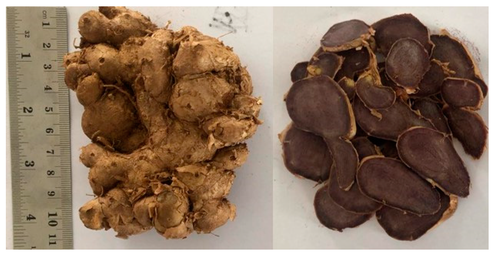
2.2. Sample Preparation

The rhizome of K. parviflora was obtained from Johor, Malaysia. Sample authentication was carried out at the Laboratory of Ecology and Conservation of Tropical Forest Biodiversity at the University of Mulawarman, Indonesia with the herbarium number of 12/UNI7.4.08/LL/2021. The sample was cleaned using a wet cloth. Then, it was cut into small pieces for further drying in an oven at 40 °C. Subsequently, the dried sample was powdered using a blender and then sieved to obtain a sample size of less than 2 mm. Finally, samples that were uniform in size were used for extraction.

2.3. Sample Extraction

Samples were extracted using the following two different methods: maceration with 96% ethanol and infusion with distilled water. Maceration was carried out for 72 h at a sample-to-solvent ratio of 1:8, with stirring every 24 h. The maceration results were concentrated using a rotary evaporator to obtain a thick extract. The second extraction used an infusion technique (boiling at 90 °C for 15 min) with distilled water as a solvent [22], and the extraction results were dried using a freeze dryer.

2.4. Determination of Secondary Metabolites

Determination of secondary metabolites was carried out on the samples, including saponins, tannins, alkaloids, flavonoids, polyphenols, terpenoids, steroids, glycosides, and quinones. The phytochemical content was determined following standard procedures [23]. After specific reagents were added to the solution, tests were carried out by visual observation of colour change or precipitate formation.

2.5. Determination of Cytotoxic Activity Using MTT Assay

The MTT assay was carried out based on previous study with modifications on 4T1, MCF-7, and NIH-3T3 cell line [19]. 4T1 and MCF-7 cells are breast cancer cell models, whereas NIH-3T3 cells are noncancerous cell line models. All cell lines were kindly provided by Prof. Dr. Muhammad Dai (Cell Culture Laboratory, Faculty of Pharmacy, Universitas Muhammadiyah Surakarta, Solo, Indonesia). All the cells were grown in 96 well plates. Cell cultures were incubated for 24 h in RPMI medium (for 4T1 cell line) and DMEM (for MCF-7 and NIH-3T3 cell line) and incubated at 37 °C with a CO2 level of 5%. Then, 100 µL of sample (EWKP, EEKP, and Doxorubicin) solution with the concentration in the range 3–250 µg/mL was placed into the well, then incubated for 24 and 48 h. Afterward, cells were washed with 100 µL of PBS from each well, then 0.5 mg/mL of MTT reagent was added to the culture medium in each well. Incubated again for 2.5 h. Subsequently, the media containing MTT was stopped by adding a stopper reagent (10% SDS in 0.01 N HCl). The plates were wrapped in aluminum foil and left overnight at room temperature. The following day, the absorbance was read using an ELISA reader (EPOCH) with a wavelength of 595 nm. The absorbance data of a single treatment was converted into percent cell viability using this formulation:
Percent   cell   viability   ( % ) = ( A b s   s a m p l e A b s   m e d i u m ) ( A b s   c o n t r o l   c e l l A b s   m e d i u m ) × 100 % .
Make a linear regression equation of concentration vs. cell viability, then calculate the IC50 by changing the value of y = 50.

2.6. Determination of Anti-Migration Effect Using Scratch Wound Healing Assay

A total of 4 × 10⁴ 4T1 cells were distributed into 96 well plates, then incubated for 24 h at 37 °C, 5% CO2 cells were washed using PBS 1× and given starvation medium (a culture medium with 0.5% FBS) and then incubated for 24 h. Then, each well was scratched vertically using a white tip and treated with Doxorubicin 0.5 µg/mL (positive control) and EEKP 16.25 µg/mL, 32.5 µg/mL, and 65 µg/mL. Finally, cells were observed under a light microscope (Olympus, Olympus Corporation, Japan) each time at 0 h, 8 h, and 24 h and captured by the camera [19].

2.7. Secondary Metabolites Profiling Using Ultra-High Performance Liquid Chromatography Mass Spectrophotometry (UHPLC-MS)

Secondary metabolite studies were carried out with reference to procedure in previous study with modifications [20]. Ultra-High Performance Liquid Chromatography Mass Spectrophotometry (UHPLC-MS) was performed on the ACQUITY UPLC I-Class system from Waters. A total of 1 µL sample was injected into column ACQUITY UPLC HSS T3 (100 mm × 2.1 mm × 1.8 μm) also from Waters, and maintained at 40 °C. The mobile phase used was water and acetonitrile (0.1% formic acid) as solvents A and B, respectively. The mobile phase was flowed using a gradient technique as follows: 0 min, 1% B; 0.5 min, 1% B; 16.00 min, 35% B; 18.00 min, 100% B; and 20.00 min, 1% B. The flow rate was set to 0.6 mL/min. The ion source was operated in positive electrospray ionisation (ESI) mode under the following specific conditions: capillary voltage, 1.50 kV; reference capillary voltage, 3.00 kV; source temperature, 120 °C; desolvation gas temperature, 550 °C; desolvation gas flow, 800 L/h; and cone gas flow, 50 L/h. Nitrogen (>99.5%) was employed as desolvation and cone gas. Data were acquired in high-definition MS E (HDMS E) mode in the range of m/z 50–1500 at 0.1 s/scan. Thus, two independent scans with different collision energies (Ces) were alternatively acquired during the run: a low-energy (LE) scan at a fixed CE of 4 eV, and a high-energy (HE) scan where the CE was ramped from 10 to 40 eV. Argon (99.999%) was used as collision-induced dissociation (CID) gas. Peak identification was accomplished by comparing it with the WATER UNIFI Natural Product Database.

2.8. Molecular Docking

The XP (extra precision) option was used to dock an inflexible protein structure with a stretchy ligand (chemical structures used to support target receptors) utilising Schrodinger Glide-v 7.4. A total of 100 poses were created for each docking calculation. Thus, their Van Der Waals (VDW) radius was adjusted as 1.0 with a net atomic charge cut off of 0.25 subunits to soften the potential of nonpolarity elements of protein and ligands, whereas the Van der Waals radius of the other elements was not adjusted. Glide docking uses a conventional cluster-based method to find the best ligand-binding regions in a specified receptor grid field. The ligand with the least XP glide score has the strongest binding affinity for the enzymes [24]. Software was used to study molecular docking for the four target proteins, Bcl-2, Bcl-XL, ERK2, and FAK.

2.9. Statistical Analysis

Scratch analysis was figured out by quantifying the cell migration distance using ImageJ software by comparing the distance between untreated and treated cells. Data from multiple scratches within the same test group were analysed using the Analysis of Variance (ANOVA) test to analyse the difference between experimental groups. Data were presented as mean ± S.D. The p < 0.05 was considered significant. Other data were analyzed using MS Excel 2010 in Acer Aspire 5 laptop (Acer Inc. New Taipei, Taiwan)

3. Results

3.1. Extraction Process and Secondary Metabolites Analysis

The extraction process from the samples was carried out using the following two different extraction techniques: maceration using 96% ethanol and infusion with distilled water. The KP rhizome used appears dark black and has an odour that is not typical of ginger varieties (Figure 1). Other tests confirmed that the sample used was also black ginger. The ethanol extract yielded 12.98%, while the aqueous extract yielded 12.49%. The two extracted extracts were both black (Table 1).
In addition, secondary metabolite analysis was performed using specific reagents that are dropped on the sample, thereby causing a change in colour or formation of precipitate that can indicate the presence of a class of secondary metabolites. Results of secondary metabolite analysis revealed that both ethanol and water extracts contained secondary metabolites of alkaloids, flavonoids, and polyphenols. Meanwhile, the water extract contained quinone and saponin compounds, whereas the ethanol extract did not (Table 2).

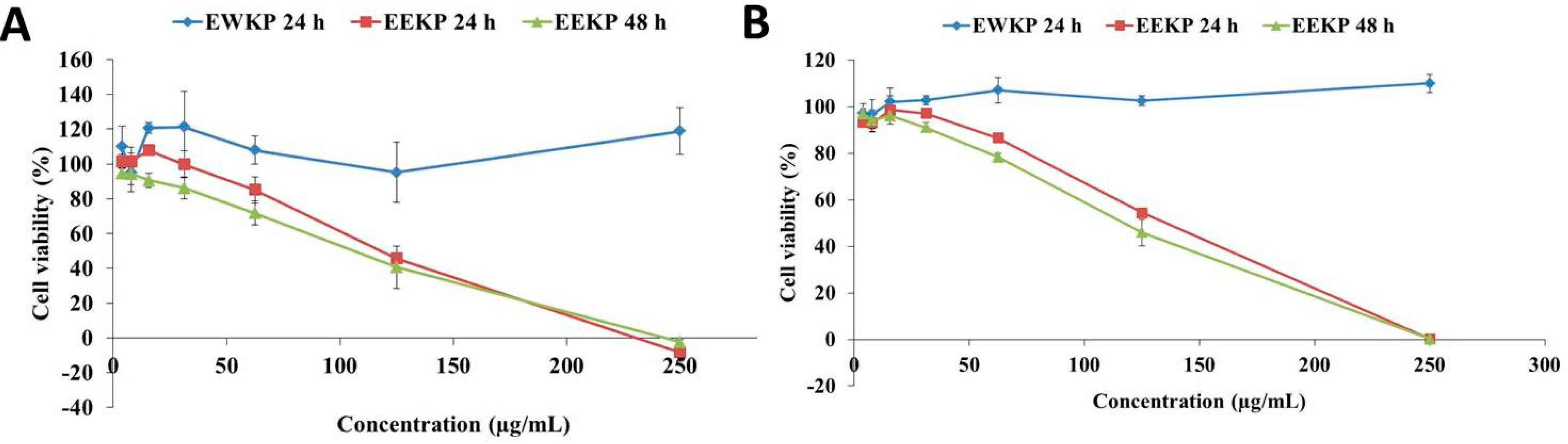
3.2. Cytotoxic Activity of Kaempferia parviflora

MTT assay was used to perform a cytotoxic test on the samples to determine their cytotoxic activity. This cytotoxic test activity of EWKP and EEKP was performed against the breast cancer cell line 4T1 and MCF-7, also in noncancerous cell line NIH-3T3 for 24 h and 48 h. During the 24 h incubation period, results showed that the EWKP did not exhibit any cytotoxic activity both in 4T1 and MCF-7 cell line. Consequently, the EWKP was not tested during the 48 h incubation period (Figure 2). Meanwhile, in NIH-3T3 cytotoxic testing, EWKP and EEKP also had no cytotoxic effect at the highest tested concentration (250 µg/mL). According to the findings of this study, increasing doses of EEKP demonstrated cytotoxic activity at both 24 h and 48 h. Based on the calculations, the IC50 of EEKP was 128.33 µg/mL (24 h) and 115.09 µg/mL (48 h) on 4T1 cell line, and 138.43 µg/mL (24 h) and 124.81 µg/mL (48 h) on MCF-7 cell line.

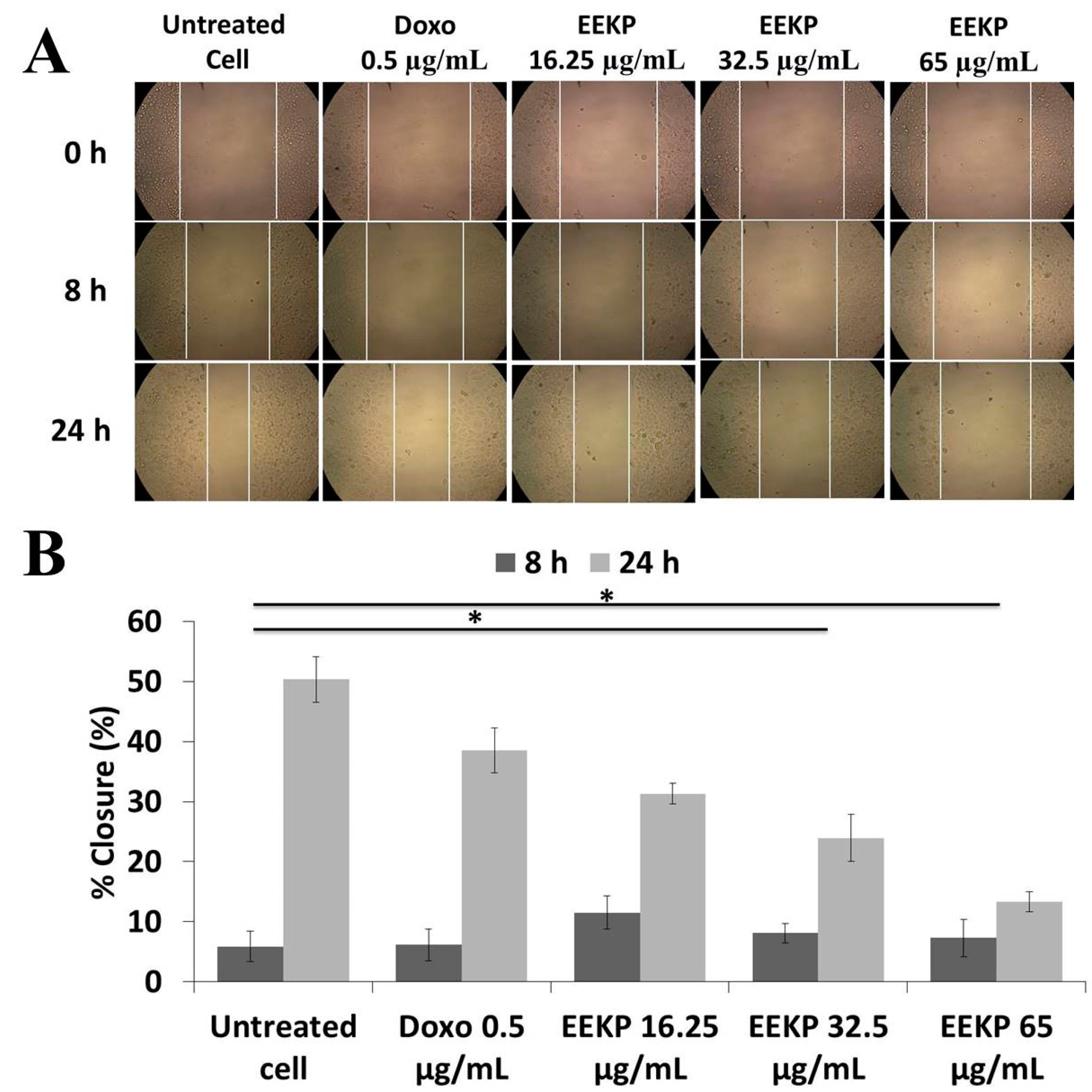
3.3. Anti-Migration Activity on 4T1 Breast Cancer Cell Line

In addition to a cytotoxic test, to determine the anti-migration activity, the scratch wound healing assay technique was performed as part of this study. Based on the cytotoxic activity findings, only EEKP is continued in this study, because only EEKP has cytotoxic activity. Three nontoxic doses of EEKP were used in the anti-migration study, namely 16.25 µg/mL, 32.5 µg/mL, and 65, µg/mL as well as a positive control of doxorubicin. The experiment lasted for 24 h, with cell movement monitored at 8 h and 24 h. EEKP inhibited cell movement based on the findings. When compared to control cells, the higher the concentration, the greater the inhibition (Figure 3). This anti-migration activity test only uses 4T1 cells because this cell line is a model of highly metastatic breast cancer.

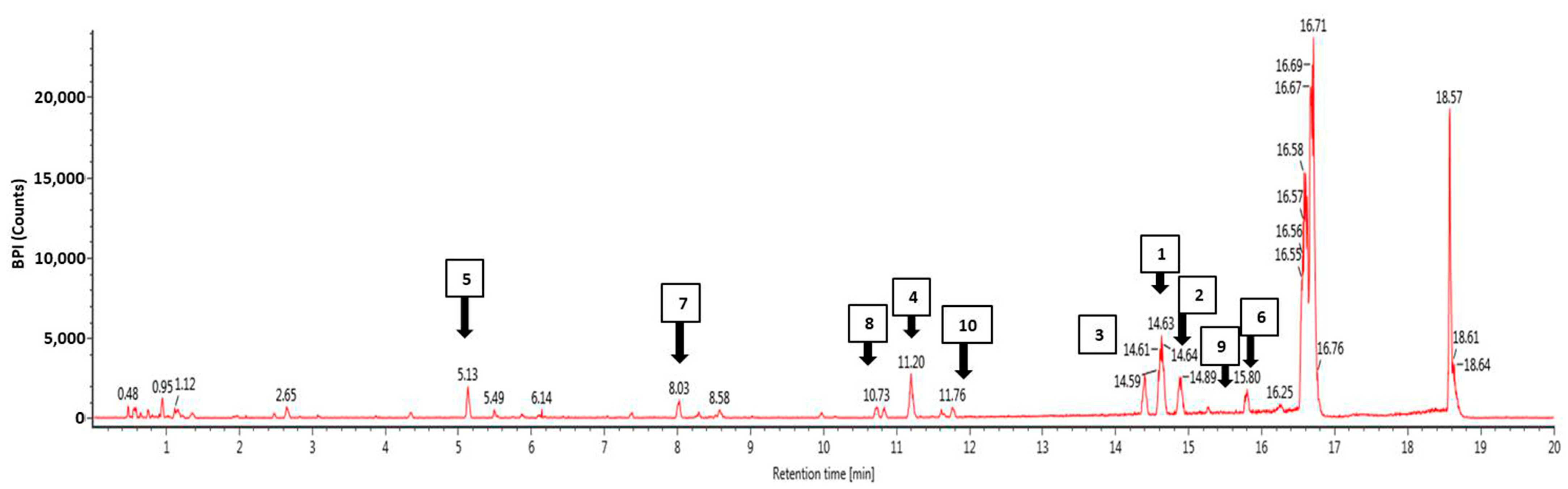
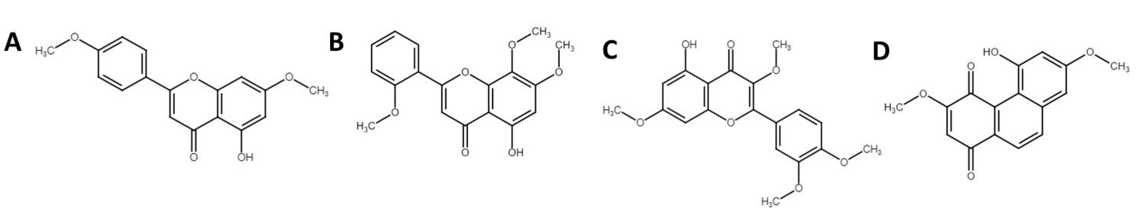
3.4. Phytochemical Profiling Using Ultra-High Performance Liquid Chromatography Mass Spectrophotometry (UHPLC-MS)

The profile of the bioactive compounds found in EEKP was revealed using UHPLC-MS. This study was carried out to investigate previous findings and determine which compound is responsible for the activities. The result showed the EEKP is expected to contain at least 75 bioactive compounds (Figure 4). Then, we chose 10 compounds with the highest response, and four of these were chosen for further in silico molecular docking studies: 5-Hydroxy-7,4′-dimethoxyflavanone (HDMF), 5-Hydro-7,8,2′-trimethoxyflavanone (HTMF), Retusine, and Denbinobin (Table 3 and Figure 4).

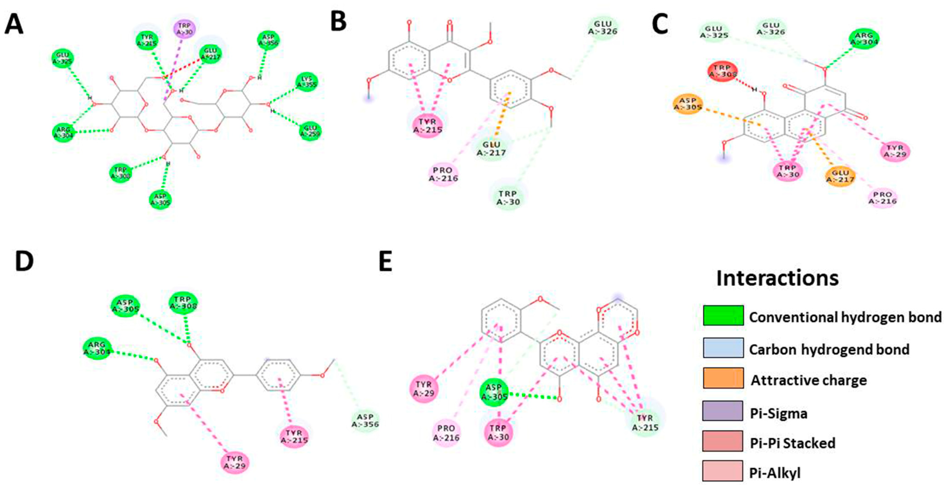
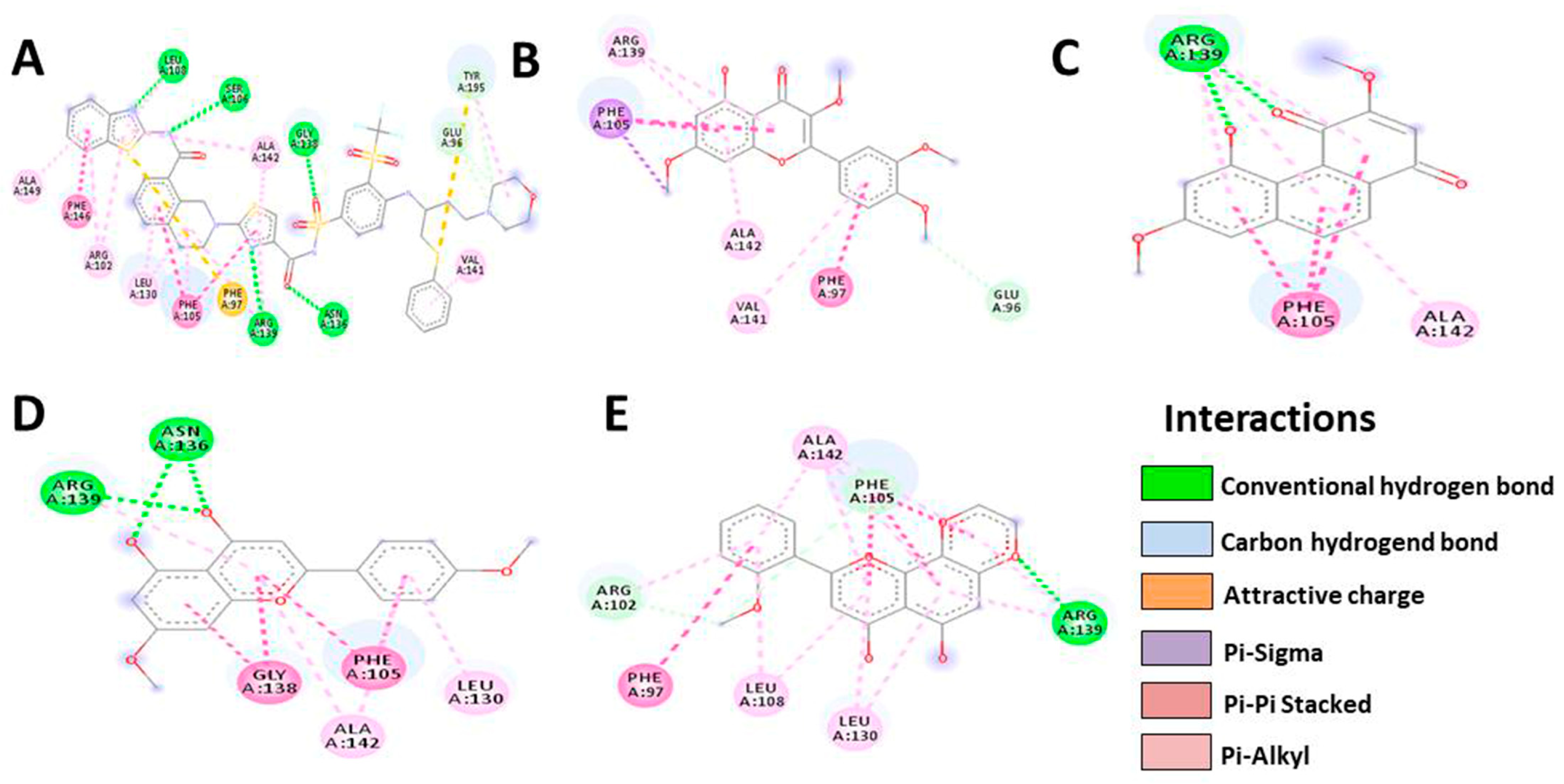
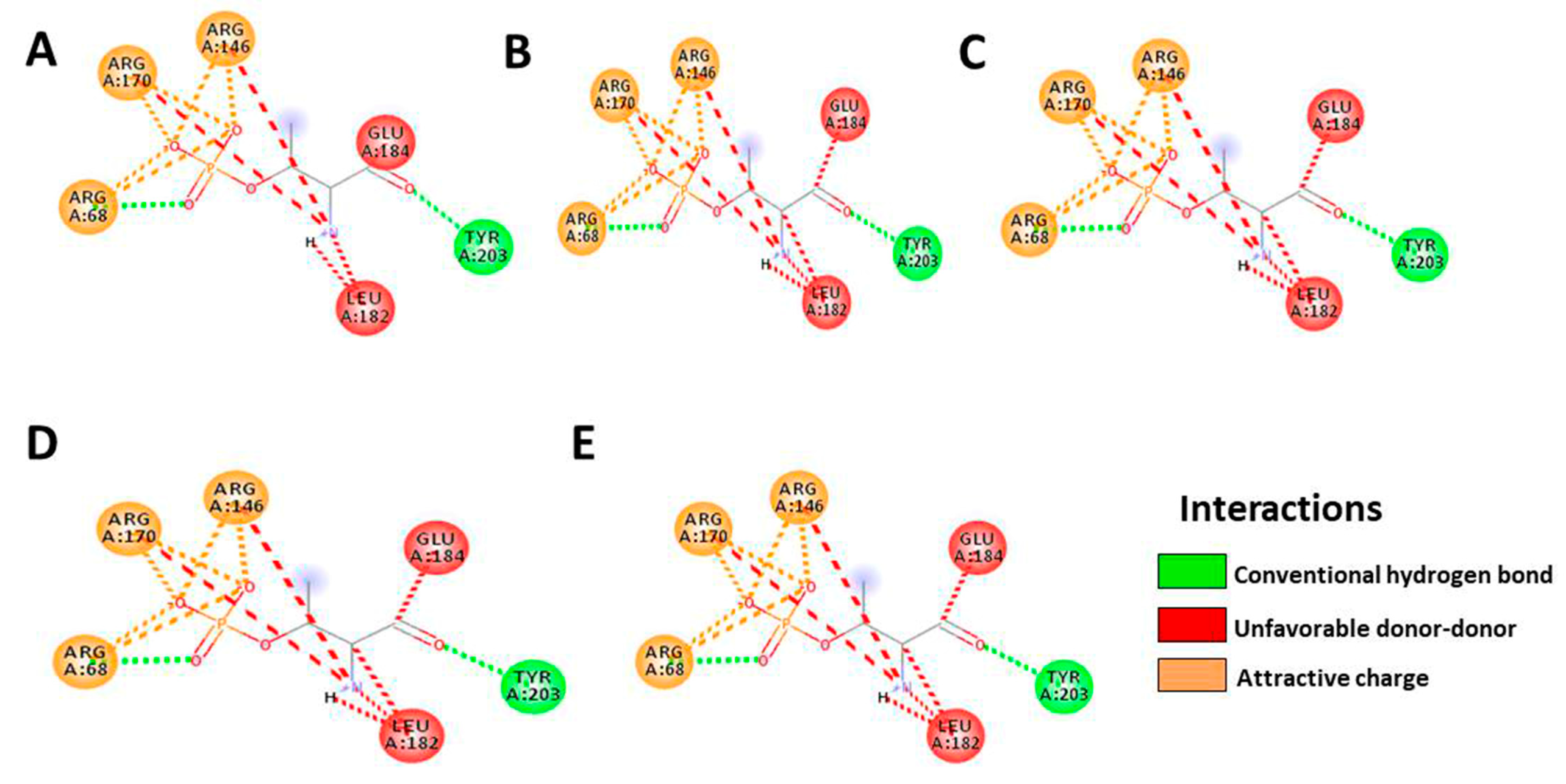
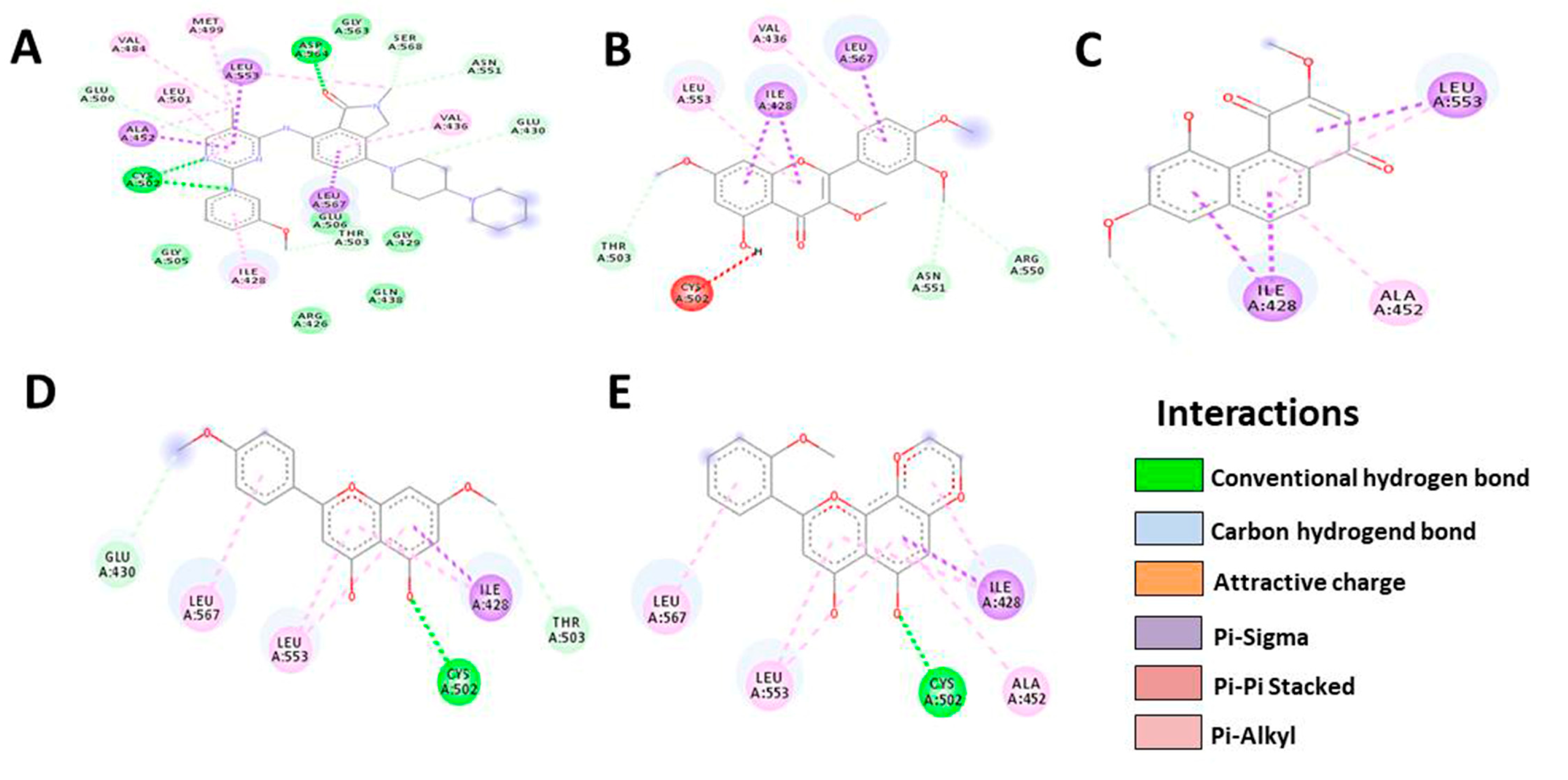
3.5. In Silico Molecular Docking
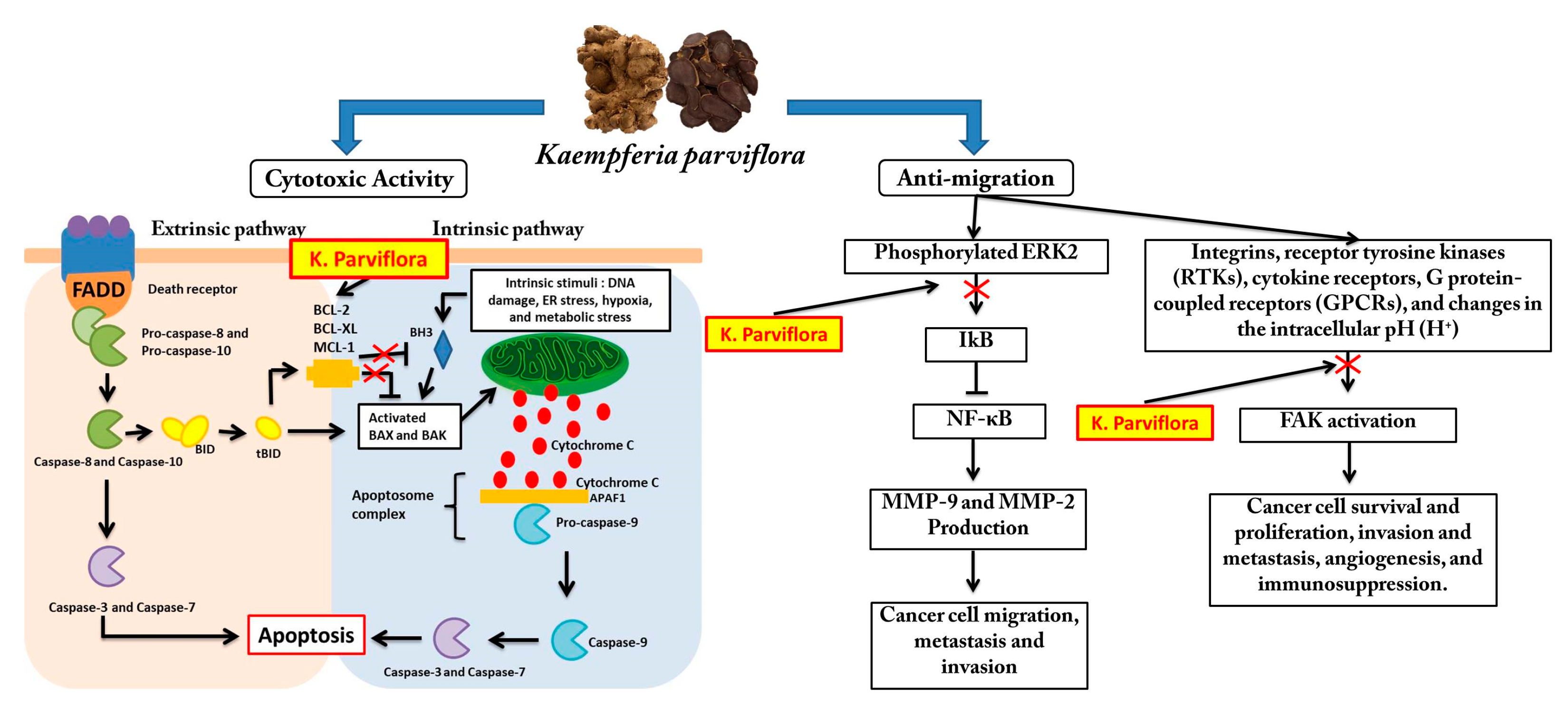
An in silico molecular docking study was performed on the following four compounds: 5-Hydroxy-7,4′-dimethoxyflavanone (HDMF), 5-Hydroxy-7,8,2′-trimethoxyflavanone (HTMF), Retusine, and Denbinobin to predict the bioactive compounds responsible for cytotoxic and antimetastatic activity based on UHPLC-MS prediction results (Figure 5). This in silico study was carried out to predict the compound’s potential binding strength to the chosen receptor. The receptors used in this study are proteins that are involved in the apoptotic pathway (Bcl-2, Bcl-XL) and metastasis (ERK2, FAK). Furthermore, this receptor was chosen based on numerous studies that show KP causes cytotoxicity via intrinsic apoptosis and antimetastatic effects via MMP-9 and MMP-2 production inhibition.
Molecular docking was used to model the possible binding mode of phytochemicals found in KP towards proteins involved in the cytotoxic and metastasis processes. The 3D structures of matrices Bcl-2, Bcl-XL, ERK2, and FAK were obtained from the RCSB protein data bank. Afterwards, the data was prepared by removing crystallographic water and any co-crystallised ligand. Molecular docking simulation was carried out using AutoDock Vina (Vina) with the default settings. The best-docked conformation ranked by vina scoring was used for visual analysis [25]. Pose View, which is accessible through Protein PDB, was used to infer the intermolecular interaction of a protein–ligand complex.
Meanwhile, a molecular docking study was carried out assuming a model where the protein and ligand were considered rigid and flexible, respectively, during the docking procedure [26]. Unfortunately, the sameness in the estimated docking score, the number of H-bonds, and interacting residues are presented in Table 4, Table 5, Table 6 and Table 7. A possible explanation could be the limitation of the docking model to explain how the compounds could arrive at the active site. In all molecular docking experiments, the procedure starts with the ligand located in the site active.
Based on the results, HTMF has a higher docking score than the native ligand in the Bcl-2 protein, with a value of −9.4, but not in the Bcl-XL protein (Table 4 and Table 5, Figure 6 and Figure 7). Meanwhile, Retusine and Denbinobin are the compounds with the best binding to proteins related to cancer cell migration activity (Table 6 and Table 7, Figure 8 and Figure 9). Consequently, it can be predicted that the compound involved in KP’s cytotoxic activity is 5-Hydro-7,8,2′-trimethoxyflavanone (HTMF), while those involved in antimetastasis are Retusine and Denbinobin.

4. Discussion

Breast cancer still has a high incidence and mortality rate today. The treatment of breast cancer also remains a challenge in the health sector [27]. Furthermore, if the breast cancer has migrated and metastasised to other organs, the cancer has progressed to the final stage. This condition has implications for treatment challenges and resistance [28].
Thus, this study primarily aims to identify potential anticancer candidates derived from natural ingredients that can be used in the treatment of metastatic breast cancer. Consequently, black ginger (Kaempferia parviflora) has been chosen as a natural ingredient to test on a highly metastatic 4T1 breast cancer cell model, breast cancers cell model MCF-7, and noncancerous cell model NIH-3T3. Even though there has not been much research done on this plant, the literature review suggested that the material used can be developed as an anticancer and anti-migration agent.
This research began with sample extraction using two different extraction procedures and solvents. This extraction process results in two types of extracts: ethanol extract of KP (EEKP) and water extract of KP (EWKP). We tested these two extracts for cytotoxic and anti-migration activity in the highly metastatic 4T1 breast cancer cell model, breast cancers cell model MCF-7, and noncancerous cell model NIH-3T3 in vitro. The result showed that EEKP displayed better cytotoxic activity during the 24 and 48 h treatment periods. Consequently, EEKP was chosen to be studied further.
The cytotoxic activity test results showed that EEKP had cytotoxic activity, with an IC50 value of 128.33 g/mL (24 h) and 115.09 g/mL (48 h) in 4T1 cell line and 138.43 µg/mL (24 h) and 124.81 µg/mL (48 h) on MCF-7 cell line. This showed that EEKP has promising anticancer potential. In addition, KP displayed a cytotoxic effect on HeLa, SKOV3, HuCCA-1, RMCCA-1, and T24 cancer cell lines [18,19,21,22,23]. Given that the cytotoxic activity of KP in highly metastatic breast cancer cells has never been reported before, our findings are novel. The water extract from KP had an IC50 value that was 19 times lower than the EEKP in the T24 human urinary bladder cancer model [13]. In line with this, it has been shown that EWKP has no cytotoxic activity in 4T1 and MCF-7 cells when concentration was tested.
The next study reported the EEKP’s anti-migration activity. This experiment was carried out using the scratch wound healing assay technique in highly metastasis breast cancer cell line model 4T1. This assay allows for the visualisation of the inhibition of cell movement in the intentional wound area. Three concentration series were chosen for this test based on the previously obtained IC50 values. The concentration that was used was nontoxic because scratch wound healing assay studies must use concentrations that allow at least 80% of cells to survive. Our results show that there is a barrier to cell movement, as indicated by EEKP at a dose of 65 g/mL with a 13.53% closure value. This result significantly differs from the % closure value in untreated cells (50.34%). The higher the dose, the greater the inhibitory of EEKP on migration.
Several researchers have also reported KP’s anti-migration and antimetastatic activity, including in HeLa, SKOV3, HuCCA-1, and RMCCA-1 cell lines [20,22,29]. These three studies demonstrated that the ethanol extract of KP inhibited MMP-9 and MMP-2 production. However, the bioactive compound responsible for this activity is not particularly mentioned.
Methoxyflavones, including dimethoxyflavones (DMF), trimethoxyflavones (TMF), tertramethoxyflavones (TMF), and pentamethoxyflavones (PMF) are compounds that provide anticancer activity from KP [30]. These compounds are biomarkers and major components of KP. UHPLC-MS was used to perform phytochemical profiling on EEKP to determine the bioactive compounds responsible for EEKP′s cytotoxic and antimetastatic activity. Based on the WATER UNIFI Natural Product Database, the results of this profiling are predictions of the phytochemical content of the EEKP.
Results of the UHPLC-MS profiling showed the presence of at least 75 bioactive compounds, and we also discovered the presence of methoxyflavones in the form of 5-Hydroxy-7,4′-dimethoxyflavanone (HDMF) at the highest concentration. Besides HDMF, there are previously unknown methoxyflavones, 5-Hydro-7,8,2′-trimethoxyflavanone (HTMF) and two other compounds, Retusine, and Denbinobin. Then, the four compounds were investigated and chosen for in silico molecular docking assays.
Thus, in silico studies were performed to predict which compounds were responsible for the cytotoxic and anti-migration activities. 5-Hydroxy-7,4′-dimethoxyflavanone (HDMF), 5-Hydroxy-7,8,2′-trimethoxyflavanone (HTMF), Retusine, and Denbinobin were selected. Afterwards, these four compounds were tested on several proteins involved in apoptosis (Bcl-2, Bcl-XL) and metastasis (ERK2, FAK).
Various studies on the cytotoxic activity of KP ethanol extract on various cancer cells concluded that KP’s cytotoxic activity was associated with apoptosis induction, particularly in the intrinsic pathway [16]. The intrinsic pathway of apoptosis, also known as the mitochondrial pathway, is where apoptosis occurs primarily as a result of cell-based triggers such as DNA damage, oxidative stress, or metabolic stress. This trigger normally causes the cell to ‘decide’ to die to save other cells and maintain optimal conditions [31].
The Bcl-2 protein family is involved in the regulation of apoptotic on cell [32]. This family consist of pro-apoptotic and anti-apoptotic members [33]. Bcl-2 and Bcl-XL are the example of anti-apoptotic, which block the process of apoptotic by forming complexes with other proteins. Several anticancer studies have been conducted that target Bcl-2 and Bcl-XL as sites of action for the developed anticancer candidates [29,34]. For example, BM-1197, a molecules that targeted Bcl-2 and inhibited the role of this protein on apoptosis, has been developed for the treatment of Diffuse large B cell lymphoma (DLBCL) [35]. This protein is also used as a target of action in the development and search for breast cancer treatments [36].
During the process, the induction ofs BAX and BAK by the BH3 protein has a strong influence on the intrinsic pathway apoptosis process. Other proteins, particularly Bcl-2 and Bcl-XL, have a strong influence on BH3 activity. The presence of these two proteins in their free form can inhibit the process of apoptosis [37]. Thus, inhibition in the form of ‘bonding’ between Bcl-2 and Bcl-XL with a compound will immediately have implications for the initiation of apoptotic events. Our findings show that HTMF binds strongly to the Bcl-2 protein, with a docking value of −9.4. This docking value is greater than that of the native ligand, which is only −9.1. Consequently, we anticipate that HTMF significantly contributes to KP’s cytotoxic activity.
Since migration is a part of cancer cell metastasis, so we used in silico studies of two proteins, ERK2 and FAK. These two proteins are proteins that play a role in cancer cell migration and metastasis. According to the previous study, KP has antimetastatic activity by inhibiting MMP-9 and MMP-2 production. Both of these proteins play critical roles in the invasion of cancer cells. MMP-9 and MMP-2 production can be activated in various ways, one of which is by activating NF-κB via the MEK1 and ERK1/2 pathways [38]. This pathway begins by phosphorylating the MEK1/ERK1/2 cascade, and then ERK1/2 is phosphorylated, hence, it activates NF-κB by removing IkB. Finally, the activated NF-κB/p65/p50 complex moves into the nucleus, thereby causing transcription and Pro-MMP-9 production [39]. Consequently, if a compound can bind to ERK 1/2, MMP-9 production and cell invasion can be inhibited.
Focal adhesion kinase (FAK), such as MMP-9, is a protein tyrosine kinase that regulates cellular adhesion, motility, proliferation, and survival in various cell types. FAK, which promotes cancer progression and metastasis, is activated and/or overexpressed in advanced cancers. Consequently, FAK has emerged as a potential therapeutic target in cancer [40]. Inhibiting the FAK protein is also thought to aid in cancer healing, particularly in combination treatments. FAK influences the occurrence of cell survival, angiogenesis, and drug resistance in cancer cells in addition to metastasis. In its molecular process, FAK can be activated by presence of Integrins, receptor tyrosine kinases (RTKs), cytokine receptors, G protein-coupled receptors (GPCRs), and changes in the intracellular pH (H+) [41]. Based on this, we tested our compound on the FAK protein in addition to the ERK2 protein. We discovered that Retusine and Denbinobin provided a better docking score than the native ligand, namely −8.8 and −9.0 for ERK2, respectively, whereas, in FAK, HTMF provided the best binding value, but not better than the native ligand. As a result of these findings, we predict that HTMF, Retusine, and Denbinobin may play an important role in KP′s anti-migration activity. As a result of our findings, we predict the possible mechanism of KP activity, which is depicted in Figure 10.
5-Hydroxy-7,8,2′-trimethoxyflavone (HTMF) is a methoxyflavone derivative. This compound is the main component of KP and has been shown to be responsible for KP′s biological activity, including anticancer activity [42]. In general, Methoxyflavones are known to be poorly soluble in water, have low lipophilic property, and have low bioavailability [43,44]. This physical character causes methoxyflavones to have a low oral bioavailability of 1% to 4%, with the liver having the highest levels, followed by the kidney. They were also discovered in the lungs, testicles, and brain. As a result, methoxyflavones are primarily eliminated through urine as demethylated, sulfated, and glucuronidated products, and as demethylated metabolites in the faeces [45].
Denbinobin is cytotoxic and antimetastatic in breast and gastric cancer cells [46] and colon cancer cell [47]. This compound is thought to have anti-migration properties via Src-mediated signalling pathways [48]. Meanwhile, Retusine has cytotoxic and antibacterial activity, though this is only based on computer simulations [49]. Thus, we predict that HTMF, Retusine, and Denbinobin play important roles in the cytotoxic and anti-migration activity of KP in breast cancer cells. However, even though our studies have revealed bioactive compounds that play an important role in cytotoxic and anti-migration activities, more research is needed to confirm this. Because our study still relies on predictive studies such as in silico studies, it is preferable if the results are confirmed by other tests such as the transwell migration assay and protein expression assays such as western blotting.

5. Conclusions

In conclusion, in vitro studies suggest that KP has anti-migration and cytotoxic activity in breast cancer cells. Based on in silico studies on the Bcl-2, Bcl-XL, ERK2, and FAK receptors, the compounds HTMF, Denbinobin, and Retusine are predicted to contribute to this activity. However, further research is needed to confirm this finding.

Author Contributions

This study was co-authored by all of the authors. I.H. designed the study, collected samples, extracted them, tested them for cytotoxicity and antimetastatic potential, and wrote the manuscript. M.F.A.B., M.D., and F.I.A.B. designed the study, assisted with data analysis, and wrote the manuscripts. A molecular docking study was carried out by E.S.S. All authors have read and agreed to the published version of the manuscript.

Funding

This research was funded by Matching RE-SIP grant with vot number of M069 entitled “In vitro anticancer potential of Black Ginger (Kaempferia parviflora) against breast cancer cell lines”.

Institutional Review Board Statement

Not applicable.

Informed Consent Statement

Not applicable.

Data Availability Statement

Data will be made available to any party upon request.

Acknowledgments

The researchers would like to acknowledge the sponsership by Universiti Tun Hussein Onn Malaysia (UTHM) through Matching RE-SIP grant (vot M069). The researchers would also like to thank Universitas Muhammadiyah Kalimantan Timur (UMKT), Universitas Muhammadiyah Surakarta (UMS), Universiti Tun Hussein Onn Malaysia (UTHM), and Universiti Teknologi Malaysia (UTM) for the laboratory facilities that were provided.

Conflicts of Interest

The authors declare no conflict of interest.

References

  1. Wilkinson, L.; Gathani, T. Understanding Breast Cancer as a Global Health Concern. Br. J. Radiol. 2022, 95, 7–9. [Google Scholar] [CrossRef] [PubMed]
  2. European Cancer Information System. ECIS Breast Cancer Burden in EU-27 2020 New Cases (Incidence) and Deaths (Mortality) Estimates New Cases Deaths 1 in 11. Jt. Res. Cent. 2020, 44–45. Available online: https://ecis.jrc.ec.europa.eu/pdf/Breast_cancer_factsheet-Oct_2020.pdf (accessed on 1 March 2023).
  3. Lim, Y.X.; Lim, Z.L.; Ho, P.J.; Li, J. Breast Cancer in Asia: Incidence, Mortality, Early Detection, Mammography Programs, and Risk-Based Screening Initiatives. Cancers 2022, 14, 4218. [Google Scholar] [CrossRef] [PubMed]
  4. Tungsukruthai, S.; Petpiroon, N.; Chanvorachote, P. Molecular Mechanisms of Breast Cancer Metastasis and Potential Anti-Metastatic Compounds. Anticancer Res. 2018, 38, 2607–2618. [Google Scholar] [CrossRef] [PubMed]
  5. Marino, N.; Woditschka, S.; Reed, L.T.; Nakayama, J.; Mayer, M.; Wetzel, M.; Steeg, P.S. Breast Cancer Metastasis: Issues for the Personalization of Its Prevention and Treatment. Am. J. Pathol. 2013, 183, 1084–1095. [Google Scholar] [CrossRef]
  6. Bray, F.; Ferlay, J.; Soerjomataram, I.; Siegel, R.L.; Torre, L.A.; Jemal, A. Global Cancer Statistics 2018: GLOBOCAN Estimates of Incidence and Mortality Worldwide for 36 Cancers in 185 Countries. CA. Cancer J. Clin. 2018, 68, 394–424. [Google Scholar] [CrossRef]
  7. Rubovszky, G.; Kocsis, J.; Boér, K.; Chilingirova, N.; Dank, M.; Kahán, Z.; Kaidarova, D.; Kövér, E.; Krakovská, B.V.; Máhr, K.; et al. Systemic Treatment of Breast Cancer. 1st Central-Eastern European Professional Consensus Statement on Breast Cancer. Pathol. Oncol. Res. 2022, 28, 1610383. [Google Scholar] [CrossRef]
  8. Waks, A.G.; Winer, E.P. Breast Cancer Treatment: A Review. JAMA—J. Am. Med. Assoc. 2019, 321, 288–300. [Google Scholar] [CrossRef]
  9. Lim, B.; Hortobagyi, G.N. Current Challenges of Metastatic Breast Cancer. Cancer Metastasis Rev. 2016, 35, 495–514. [Google Scholar] [CrossRef]
  10. Vasan, N.; Baselga, J.; Hyman, D.M. A View on Drug Resistance in Cancer. Nature 2019, 575, 299–309. [Google Scholar] [CrossRef]
  11. Huang, M.; Lu, J.J.; Ding, J. Natural Products in Cancer Therapy: Past, Present and Future. Nat. Prod. Bioprospect. 2021, 11, 5–13. [Google Scholar] [CrossRef]
  12. Natsume, N.; Yamano, A.; Watanabe, A.; Yonezawa, T.; Woo, J.T.; Yamakuni, T.; Teruya, T. Effect of Methoxyflavones Contained in Kaempferia Parviflora on CRE-Mediated Transcription in PC12D Cells. Bioorg. Med. Chem. Lett. 2020, 30, 127606. [Google Scholar] [CrossRef] [PubMed]
  13. Tangjitjaroenkun, J.; Yahayo, W.; Supabphol, S.; Supabphol, R. Selective Cytotoxicity of Kaempferia Parviflora Extracts in Human Cell Lines. Asian Pac. J. Cancer Prev. 2021, 22, 73–79. [Google Scholar] [CrossRef] [PubMed]
  14. Suradej, B.; Sookkhee, S.; Panyakaew, J.; Mungkornasawakul, P.; Wikan, N.; Smith, D.R.; Potikanond, S.; Nimlamool, W. Kaempferia Parviflora Extract Inhibits STAT3 Activation and Interleukin-6 Production in Hela Cervical Cancer Cells. Int. J. Mol. Sci. 2019, 20, 4226. [Google Scholar] [CrossRef] [PubMed]
  15. Paramee, S.; Sookkhee, S.; Sakonwasun, C.; Na Takuathung, M.; Mungkornasawakul, P.; Nimlamool, W.; Potikanond, S. Anti-Cancer Effects of Kaempferia Parviflora on Ovarian Cancer SKOV3 Cells. BMC Complement. Altern. Med. 2018, 12, 12. [Google Scholar] [CrossRef]
  16. Kim, H.; Moon, J.Y.; Burapan, S.; Han, J.; Cho, S.K. Induction of ER Stress-Mediated Apoptosis by the Major Component 5,7,4′-Trimethoxyflavone Isolated from Kaempferia Parviflora Tea Infusion. Nutr. Cancer 2018, 70, 984–996. [Google Scholar] [CrossRef]
  17. Leardkamolkarn, V.; Tiamyuyen, S.; Sripanidkulchai, B.O. Pharmacological Activity of Kaempferia Parviflora Extract against Human Bile Duct Cancer Cell Lines. Asian Pac. J. Cancer Prev. 2009, 10, 695–698. [Google Scholar]
  18. Potikanond, S.; Sookkhee, S.; Takuathung, M.N.; Mungkornasawakul, P.; Wikan, N.; Smith, D.R.; Nimlamool, W. Kaempferia Parviflora Extract Exhibits Anti-Cancer Activity against HeLa Cervical Cancer Cells. Front. Pharmacol. 2017, 8, 630. [Google Scholar] [CrossRef]
  19. Hairunisa, I.; Utomo, R.Y.; Ertanto, Y.; Jenie, R.I.; Meiyanto, E. Pentagamaboronon-0 Fructose Inhibited Migration and Overexpression of Matrix Metalloproteinases 9 on MCF-7/HER2 Breast Cancer Cells. Indones. J. Cancer Chemoprevention 2018, 9, 134. [Google Scholar] [CrossRef]
  20. Asamenew, G.; Kim, H.W.; Lee, M.K.; Lee, S.H.; Kim, Y.J.; Cha, Y.S.; Yoo, S.M.; Kim, J.B. Characterization of Phenolic Compounds from Normal Ginger (Zingiber officinale Rosc.) and Black Ginger (Kaempferia parviflora Wall.) Using UPLC-DAD-QToF-MS. Eur. Food Res. Technol. 2019, 245, 653–665. [Google Scholar] [CrossRef]
  21. Miyata, Y.; Tatsuzaki, J.; Yang, J.; Kosano, H. Potential Therapeutic Agents, Polymethoxylated Flavones Isolated from Kaempferia Parviflora for Cataract Prevention through Inhibition of Matrix Metalloproteinase-9 in Lens Epithelial Cells. Biol. Pharm. Bull. 2019, 42, 1658–1664. [Google Scholar] [CrossRef]
  22. Health Ministry of Republic Indonesia Keputusan Menteri Kesehatan Republik Indonesia Nomor HK.01.07/MENKES/187/2017 Tentang Formularium Ramuan Obat Tradisional Indonesia. 2017. Available online: http://hukor.kemkes.go.id/uploads/produk_hukum/KMK_No._HK_.01_.07-MENKES-187-2017_ttg_Formularium_Ramuan_Obat_Tradisional_Indonesia_.pdf (accessed on 22 October 2022).
  23. Saifudin, A. Senyawa Alam Metabolit Sekunder (Teori, Konsep Dan Teknik Pemurnian); Deepublish: Yogyakarta, Indonesia, 2014; ISBN 9786022804727. [Google Scholar]
  24. Ramírez-Salinas, G.L.; García-Machorro, J.; Rojas-Hernández, S.; Campos-Rodríguez, R.; de Oca, A.C.M.; Gomez, M.M.; Luciano, R.; Zimic, M.; Correa-Basurto, J. Bioinformatics Design and Experimental Validation of Influenza A Virus Multi-Epitopes That Induce Neutralizing Antibodies. Arch. Virol. 2020, 165, 891–911. [Google Scholar] [CrossRef] [PubMed]
  25. Pettersen, E.F.; Goddard, T.D.; Huang, C.C.; Couch, G.S.; Greenblatt, D.M.; Meng, E.C.; Ferrin, T.E. UCSF Chimera—A Visualization System for Exploratory Research and Analysis. J. Comput. Chem. 2004, 25, 1605–1612. [Google Scholar] [CrossRef] [PubMed]
  26. Syamsul, E.S.; Umar, S.; Wahyuni, F.S.; Martien, R.; Hamidi, D. Anti-Aging Activity, In Silico Modeling and Molecular Docking from Sonneratia Caseolaris. Open Access Maced. J. Med. Sci. 2022, 10, 1471–1477. [Google Scholar] [CrossRef]
  27. Raikwar, S.; Jain, K.S. Opportunities and Challenges in Breast Cancer. Int. J. Pharm. Life Sci. 2020, 11, 6858–6873. [Google Scholar]
  28. Kilmister, E.J.; Koh, S.P.; Weth, F.R.; Gray, C.; Tan, S.T. Cancer Metastasis and Treatment Resistance: Mechanistic Insights and Therapeutic Targeting of Cancer Stem Cells and the Tumor Microenvironment. Biomedicines 2022, 10, 2988. [Google Scholar] [CrossRef] [PubMed]
  29. Barillé-Nion, S.; Lohard, S.; Juin, P.P. Targeting of Bcl-2 Family Members during Anticancer Treatment: A Necessary Compromise between Individual Cell and Ecosystemic Responses? Biomolecules 2020, 10, 1109. [Google Scholar] [CrossRef]
  30. Elshamy, A.I.; Mohamed, T.A.; Essa, A.F.; Abd-Elgawad, A.M.; Alqahtani, A.S.; Shahat, A.A.; Yoneyama, T.; Farrag, A.R.H.; Noji, M.; El-Seedi, H.R.; et al. Recent Advances in Kaempferia Phytochemistry and Biological Activity: A Comprehensive Review. Nutrients 2019, 11, 2396. [Google Scholar] [CrossRef]
  31. Obeng, E. Apoptosis (Programmed Cell Death) and Its Signals—A Review. Braz. J. Biol. 2021, 81, 1133–1143. [Google Scholar] [CrossRef]
  32. Tsujimoto, Y. Role of Bcl-2 Family Proteins in Apoptosis: Apoptosomes or Mitochondria? Genes Cells 1998, 3, 697–707. [Google Scholar] [CrossRef]
  33. O’Brien, M.A.; Kirby, R. Apoptosis: A Review of pro-Apoptotic and Anti-Apoptotic Pathways and Dysregulation in Disease. J. Vet. Emerg. Crit. Care 2008, 18, 572–585. [Google Scholar] [CrossRef]
  34. Campbell, K.J.; Tait, S.W.G. Targeting BCL-2 Regulated Apoptosis in Cancer. Open Biol. 2018, 8, 180002. [Google Scholar] [CrossRef]
  35. Sun, Y.L.; Jiang, W.Q.; Luo, Q.Y.; Yang, D.J.; Cai, Y.C.; Huang, H.Q.; Sun, J. A Novel Bcl-2 Inhibitor, BM-1197, Induces Apoptosis in Malignant Lymphoma Cells through the Endogenous Apoptotic Pathway. BMC Cancer 2019, 20, 1. [Google Scholar] [CrossRef]
  36. Wen, M.; Deng, Z.K.; Jiang, S.L.; Guan, Y.D.; Wu, H.Z.; Wang, X.L.; Xiao, S.S.; Zhang, Y.; Yang, J.M.; Cao, D.S.; et al. Identification of a Novel Bcl-2 Inhibitor by Ligand-Based Screening and Investigation of Its Anti-Cancer Effect on Human Breast Cancer Cells. Front. Pharmacol. 2019, 10, 391. [Google Scholar] [CrossRef] [PubMed]
  37. Hussar, P. Apoptosis Regulators Bcl-2. Encylopedia 2022, 2, 1624–1636. [Google Scholar] [CrossRef]
  38. Napoli, S.; Scuderi, C.; Gattuso, G.; Di Bella, V.; Candido, S.; Basile, M.S.; Libra, M.; Falzone, L. Functional Roles of Matrix Metalloproteinases and Their Inhibitors in Melanoma. Cells 2020, 9, 1151. [Google Scholar] [CrossRef]
  39. Wang, T.; Jin, X.; Liao, Y.; Sun, Q.; Luo, C.; Wang, G.; Zhao, F.; Jin, Y. Association of NF-ΚB and AP-1 with MMP-9 Overexpression in 2-Chloroethanol Exposed Rat Astrocytes. Cells 2018, 7, 96. [Google Scholar] [CrossRef]
  40. Yoon, H.; Dehart, J.P.; Murphy, J.M.; Lim, S.T.S. Understanding the Roles of FAK in Cancer: Inhibitors, Genetic Models, and New Insights. J. Histochem. Cytochem. 2015, 63, 114–128. [Google Scholar] [CrossRef]
  41. Chuang, H.H.; Zhen, Y.Y.; Tsai, Y.C.; Chuang, C.H.; Hsiao, M.; Huang, M.S.; Yang, C.J. FAK in Cancer: From Mechanisms to Therapeutic Strategies. Int. J. Mol. Sci. 2022, 23, 1726. [Google Scholar] [CrossRef]
  42. Wongsrikaew, N.; Kim, H.; Vichitphan, K.; Cho, S.K.; Han, J. Antiproliferative Activity and Polymethoxyflavone Composition Analysis of Kaempferia Parviflora Extracts. J. Korean Soc. Appl. Biol. Chem. 2012, 55, 813–817. [Google Scholar] [CrossRef]
  43. Sutthanut, K.; Lu, X.; Jay, M.; Sripanidkulchai, B. Solid Lipid Nanoparticles for Topical Administration of Kaempferia parviflora Extracts. J. Biomed. Nanotechnol. 2009, 5, 224–232. [Google Scholar] [CrossRef]
  44. Mekjaruskul, C.; Yang, Y.T.; Leed, M.G.D.; Sadgrove, M.P.; Jay, M.; Sripanidkulchai, B. Novel Formulation Strategies for Enhancing Oral Delivery of Methoxyflavones in Kaempferia Parviflora by SMEDDS or Complexation with 2-Hydroxypropyl-β- Cyclodextrin. Int. J. Pharm. 2013, 445, 1–11. [Google Scholar] [CrossRef] [PubMed]
  45. Mekjaruskul, C.; Jay, M.; Sripanidkulchai, B. Pharmacokinetics, Bioavailability, Tissue Distribution, Excretion, and Metabolite Identification of Methoxyflavones in Kaempferia parviflora Extract in Rats. Drug Metab. Dispos. 2012, 40, 2342–2353. [Google Scholar] [CrossRef] [PubMed]
  46. Song, J.I.; Kang, Y.J.; Yong, H.Y.; Kim, Y.C.; Moon, A. Denbinobin, a Phenanthrene from Dendrobium Nobile, Inhibits Invasion and Induces Apoptosis in SNU-484 Human Gastric Cancer Cells. Oncol. Rep. 2012, 27, 813–818. [Google Scholar] [CrossRef] [PubMed]
  47. Yang, K.C.; Uen, Y.H.; Suk, F.M.; Liang, Y.C.; Wang, Y.J.; Ho, Y.S.; Li, I.H.; Lin, S.Y. Molecular Mechanisms of Denbinobin-Induced Anti-Tumorigenesis Effect in Colon Cancer Cells. World J. Gastroenterol. 2005, 11, 3040–3045. [Google Scholar] [CrossRef] [PubMed]
  48. Chen, P.H.; Peng, C.Y.; Pai, H.C.; Teng, C.M.; Chen, C.C.; Yang, C.R. Denbinobin Suppresses Breast Cancer Metastasis through the Inhibition of Src-Mediated Signaling Pathways. J. Nutr. Biochem. 2011, 22, 732–740. [Google Scholar] [CrossRef]
  49. Zhanzhaxina, A.; Suleimen, Y.; Metwaly, A.M.; Eissa, I.H.; Elkaeed, E.B.; Suleimen, R.; Ishmuratova, M.; Akatan, K.; Luyten, W. In Vitro and in Silico Cytotoxic and Antibacterial Activities of a Diterpene from Cousinia alata Schrenk. J. Chem. 2021, 2021, 5542455. [Google Scholar] [CrossRef]
Figure 1. Whole rhizome and cross-sectional appearance of black ginger (Kaempferia parviflora) rhizome.
Cancers 15 02785 g001
Figure 2. The cytotoxic activity result of EWKP and EEKP against 4T1 breast cancer cell line (A) and MCF-7 breast cancer cell line (B) in 24 h and 48 h.
Cancers 15 02785 g002
Figure 3. Effect of EEKP and Doxorubicin treatment on cell migration in 4T1 cells (A) Morphology of 4T1 cells after being treated with EEKP doses of 16.25 µg/mL, 32.5 µg/mL, and 65 µg/mL and doxorubicin 0.5 µg/mL. Observations were made at 0 h, 8 h, and 24 h using an inverted microscope with 100× magnification. (B) Percentage of 4T1 cell closure after 24 h of treatment. The wound area was analyzed using ImageJ software, then the % closure was calculated according to the analytical procedure. (*) indicates a statistically significant difference at the 95% confidence level (n = 3).
Cancers 15 02785 g003
Figure 4. Ultra-High Performance Liquid Chromatography Mass Spectrophotometry (UHPLC-MS) chromatogram, acquired in the positive ion mode, from EEKP where peak labelling represents the compounds identified and listed in Table 3.
Cancers 15 02785 g004
Figure 5. (A) 5-Hydroxy-7,4′-dimethoxyflavanone (HDMF), (B) 5-Hydro-7,8,2′-trimethoxyflavanone (HTMF), (C) Retusine, and (D) Denbinobin.
Cancers 15 02785 g005
Figure 6. Illustration of bond prediction between native ligands (A), Retusine (B), Denbinobin (C), 5-Hydroxy-7,4′-dimethoxyflavone (HDMF) (D), 5-Hydroxy-7,8,2′-trimethoxyflavone (HTMF) (E) with Bcl-2 receptor.
Cancers 15 02785 g006
Figure 7. Illustration of bond prediction between native ligands (A), Retusine (B), Denbinobin (C), 5-Hydroxy-7,4′-dimethoxyflavone (HDMF) (D), 5-Hydroxy-7,8,2′-trimethoxyflavone (HTMF) (E) with Bcl-XL receptor.
Cancers 15 02785 g007
Figure 8. Illustration of bond prediction between native ligands (A), Retusine (B), Denbinobin (C), 5-Hydroxy-7,4′-dimethoxyflavone (HDMF) (D), 5-Hydroxy-7,8,2′-trimethoxyflavone (HTMF) (E) with ERK2 receptor.
Cancers 15 02785 g008
Figure 9. Illustration of bond prediction between native ligands (A), Retusine (B), Denbinobin (C), 5-Hydroxy-7,4′-dimethoxyflavone (HDMF) (D), 5-Hydroxy-7,8,2′-trimethoxyflavone (HTMF) (E) with FAK receptor.
Cancers 15 02785 g009
Figure 10. The possible molecular mechanism of Kaempferia parviflora’s cytotoxic and anti-migration activity.
Cancers 15 02785 g010
Table 1. Results of the extraction process from Kaempferia parviflora.
Kaempferia parviflora
EthanolWater
Yield (%)12.9812.49
Organoleptic
OdorOdorlessSmoky
ColorBlackBlack
Table 2. The results of phytochemical analysis on ethanol and water extract of Kaempferia parviflora.
Type of Secondary MetabolitesKaempferia parvifloraType of the Test
EthanolWater
Saponin−ve+veSaponin test
Tannin+ve−veTannin test
Steroid−ve+veLieberman Buchard’s test
Alkaloid
Mayer+ve+veMayer’s test
Dragendorf+ve+veDragendorf’s test
Buchardat+ve−veBuchardat’s test
Flavonoid+ve+veLead acetate test
Glycosides−ve−veKeller–Kiliani’s test
Quinone−ve+veQuinone test
Polyphenol+ve+vePolyphenol test
Table 3. Tentative identification of chemical constituents in the K. parviflora ethanol extract.
NoComponent NameFormulaObserved
m/z
Observed RT
(min)
Response
15-Hydroxy-7,4′-dimethoxyflavanoneC17H14O5297.076414.6281,895
21,3-Dihydroxy-2-ethoxymethyl-anthraquinoneC17H14O5297.076214.8836,111
3RetusineC19H18O7357.097514.435,908
4DenbinobinC16H12O5283.060511.226,614
5TectorusideC21H30O13489.16115.1324,405
65-Hydro-7,8,2′-trimethoxyflavanoneC18H16O6327.086915.7924,310
7Acacetin-7-galactosideC22H22O10445.11378.0217,504
8Viscumneoside ⅥC24H26O12505.134610.8311,985
95-Hydro-7,8,2′-trimethoxyflavanoneC18H16O6327.086915.2710,317
103′,5-Dihydroxy-7,4′-dimethoxy flavoneC17H14O6313.071111.778933
Table 4. The docking score, number of H-bonds, and interacting residues of the selected compounds, Bcl-2.
Bcl-2NativeDenbinobinRetusineHDMFHTMF
Docking score−9.1−8.8−8.4−8.1−9.4
ResidueGLU325GLU325GLU326ARG304TYR29
TYR215GLU326TRP30ASP305PRO216
TRP30ARG304GLU217TRP308ASP305
GLU217TRP308PRO216ASP356TRP30
ASP356ASP305TYR215TYR215TYR215
LYS355TRP30 TYR29
GLU259GLU217
ASP305TYR29
TRP308PRO216
ARG304
Number of H-bonds109565
Table 5. The docking score, number of H-bonds, and interacting residues of the selected compounds, Bcl-XL.
Bcl-XLNativeDenbinobinRetusineHDMFHTMF
Docking score−13.4−8.2−8.1−7.9−8.0
ResidueLEU108ARG139PHE105ARG139ALA142
SER106PHE105ARG139ASN136PHE105
ALA142ALA142ALA142GLY138ARG139
GLY138 VAL141ALA142LEU130
GLU96 PHE97PHE105LEU108
TYR195 GLU96LEU130PHE97
VAL141 ARG102
Number of H-bonds73667
Table 6. The docking score, number of H-bonds, and interacting residues of the selected compounds, ERK2.
ERK2NativeDenbinobinRetusineHDMFHTMF
Docking score−8.7−9.0−8.8−7.5−7.9
ResidueARG68ARG68ARG68ARG68ARG68
ARG170ARG170ARG170ARG170ARG170
ARG146ARG146ARG146ARG146ARG146
GLU184GLU184GLU184GLU184GLU184
LEU182LEU182LEU182LEU182LEU182
TYR203TYR203TYR203TYR203TYR203
number of H-bonds66666
Table 7. The docking score, number of H-bonds, and interacting residues, of the selected compounds, FAK.
FAKNativeDenbinobinRetusineHDMFHTMF
Docking score−9.8−7.9−7.8−7.3−8.1
ResidueMET499LEU553VAL436GLU430LEU567
VAL484ALA452LEU567LEU567LEU553
LEU501ILE428ILE428LEU553CYS502
LEU553THR503LEU553CYS502ALA452
ASP564 THR503ILE428ILE428
GLY563 CYS502THR503
SER568 ASN551
ASN551 ARG550
Number of H-bonds84865
Disclaimer/Publisher’s Note: The statements, opinions and data contained in all publications are solely those of the individual author(s) and contributor(s) and not of MDPI and/or the editor(s). MDPI and/or the editor(s) disclaim responsibility for any injury to people or property resulting from any ideas, methods, instructions or products referred to in the content.

