Article

Thermal Ablation versus Stereotactic Ablative Body Radiotherapy to Treat Unresectable Colorectal Liver Metastases: A Comparative Analysis from the Prospective Amsterdam CORE Registry

by
Sanne Nieuwenhuizen
1,*,
Madelon Dijkstra
1,
Robbert S. Puijk
1,
Florentine E. F. Timmer
1,
Irene M. Nota
1,
Jip Opperman
2,
Bente van den Bemd
1,
Bart Geboers
1,
Alette H. Ruarus
1,
Evelien A. C. Schouten
1,
Jan J. J. de Vries
1,
Hester J. Scheffer
1,
Anne M. van Geel
2,
Jan Hein T. M. van Waesberghe
1,
Rutger-Jan Swijnenburg
3,
Kathelijn S. Versteeg
4,
Birgit I. Lissenberg-Witte
5,
M. Petrousjka van den Tol
4,
Cornelis J. A. Haasbeek
6 and
Martijn R. Meijerink
1
1
Department of Radiology and Nuclear Medicine, Amsterdam UMC, Cancer Center Amsterdam, VU University, 1081 HV Amsterdam, The Netherlands
2
Department of Radiology and Nuclear Medicine, Noordwest Ziekenhuisgroep, 1815 JD Alkmaar, The Netherlands
3
Department of Surgery, Amsterdam UMC, Cancer Center Amsterdam, VU University, 1081 HV Amsterdam, The Netherlands
4
Department of Medical Oncology, Amsterdam UMC, Cancer Center Amsterdam, VU University, 1081 HV Amsterdam, The Netherlands
5
Department of Epidemiology and Data Science, Amsterdam UMC, Cancer Center Amsterdam, VU University, 1081 HV Amsterdam, The Netherlands
6
Department of Radiation Oncology, Amsterdam UMC, Cancer Center Amsterdam, VU University, 1081 HV Amsterdam, The Netherlands
*
Author to whom correspondence should be addressed.
Cancers 2021, 13(17), 4303; https://doi.org/10.3390/cancers13174303
Submission received: 12 July 2021 / Revised: 18 August 2021 / Accepted: 20 August 2021 / Published: 26 August 2021
(This article belongs to the Special Issue Management of Colorectal Cancer Metastatic Disease)

Abstract

:

Simple Summary

Approximately 30–50% of colorectal cancer patients will develop colorectal liver metastases (CRLM) in the course of their disease. Though partial hepatectomy is considered the historical gold standard, complete removal of all metastases is only feasible in 20–30% of patients. Thermal ablation and stereotactic ablative radiotherapy (SABR) are techniques to eradicate unresectable CRLM. This AmCORE based study compares the safety, efficacy and survival outcomes of these treatment methods. In this study thermal ablation was superior to SABR with regard to overall survival, local tumor progression-free survival and local control. However, there was a slightly higher risk of serious adverse events after thermal ablation. Further studies are required to assess whether the worse outcomes following SABR were the effect of true differences in ablative treatment or a result of residual confounding.

Abstract

Thermal ablation and stereotactic ablative radiotherapy (SABR) are techniques to eradicate colorectal liver metastases (CRLM). This study compares the safety, efficacy and long-term oncological outcomes of these treatment methods. All prospectively registered patients (AmCORE registry) treated with thermal ablation or SABR alone for unresectable CRLM between 2007 and 2020 were analyzed using multivariate Cox-proportional hazard regression. In total 199 patients were included for analysis: 144 (400 CRLM) thermal ablation; 55 (69 CRLM) SABR. SABR patients were characterized by older age (p = 0.006), extrahepatic disease at diagnosis (p = 0.004) and larger tumors (p < 0.001). Thermal ablation patients were more likely to have synchronous disease, higher clinical risk scores (p = 0.030) and higher numbers of CRLMs treated (p < 0.001). Mortality was zero and morbidity low in both groups: no serious adverse events were recorded following SABR (n = 0/55) and nine (n = 9/144 [6.3%]; all CTCAE grade 3) after thermal ablation. SABR was associated with an inferior overall survival (OS) (median OS 53.0 months vs. 27.4 months; HR = 1.29, 95% CI 1.12–1.49; p = 0.003), local tumor progression-free survival (LTPFS) per-tumor (HR = 1.24, 95% CI 1.01–1.52; p = 0.044) and local control per-patient (HR = 1.57, 95% CI 1.20–2.04; p = 0.001) and per-tumor (HR = 1.89, 95% CI 1.44–2.49; p < 0.001). In this study thermal ablation was superior to SABR with regard to OS, LTPFS and local control, albeit at the cost of a limited risk of serious adverse events. Further studies are required to assess whether the worse outcomes following SABR were the effect of true differences in ablative treatment or a result of residual confounding.

1. Introduction

Colorectal cancer (CRC) ranks third among cancers in terms of incidence and is the second most common cause of cancer-related mortality in the world, accounting for about one in 10 cancer cases and deaths [1]. The liver is the most common site of distant spread. Approximately 15–25% of CRC patients will have colorectal liver metastases (CRLM) at the time of diagnosis of the primary tumor and another 18–25% patients will develop distant metastases within five years [2,3,4]. A multidisciplinary approach to managing CRLM has been pivotal to recent improvements in survival. Though partial hepatectomy is considered the historical gold standard, with five-year survival rates nowadays reaching 40–55% [5,6,7], complete removal of all metastases is only feasible in 20–30% of patients [2,3,8]. For patients with CRLM that are partially or completely considered unresectable, several other local eradicative treatments have emerged.
The most well-known and widely adopted treatments are thermal ablation techniques, such as radiofrequency ablation (RFA) and microwave ablation (MWA), and non-thermal ablative methods, such as stereotactic ablative radiotherapy (SABR) and most recently irreversible electroporation (IRE). Because of its safety profile and good local control rate, (inter)national multidisciplinary guidelines and expert consensus groups generally recommend the use of thermal ablation for small-size unresectable CRLM [9,10,11] and the reservation of SABR for CRLMs that are unsuitable for partial hepatectomy and thermal ablation, either due to a certain anatomical location, i.e., near critical structures like major blood vessels or bile ducts, or due to a poor general health status [12,13]. Furthermore, thermal ablation is restricted by tumor size as efficacy decreases exponentially for tumors >3 cm [14,15,16,17,18,19]. Hypothetically, SABR can overcome some of these limitations as it does not require the placement of needle electrodes or antennas, is presumably less affected by tumor size and by proximity of large blood vessels, and may be less susceptible to operator experience [20].
In the absence of randomized controlled trials or robust retrospective comparative series, claims of superiority of one technique over the other seem unsubstantiated. The aim of this study was to compare the safety, local control rate and long-term oncological outcome of thermal ablation versus SABR for unresectable CRLM by analyzing per-patient, per-procedure and per-tumor data from the prospective Amsterdam colorectal liver metastases registry (AmCORE).

2. Materials and Methods

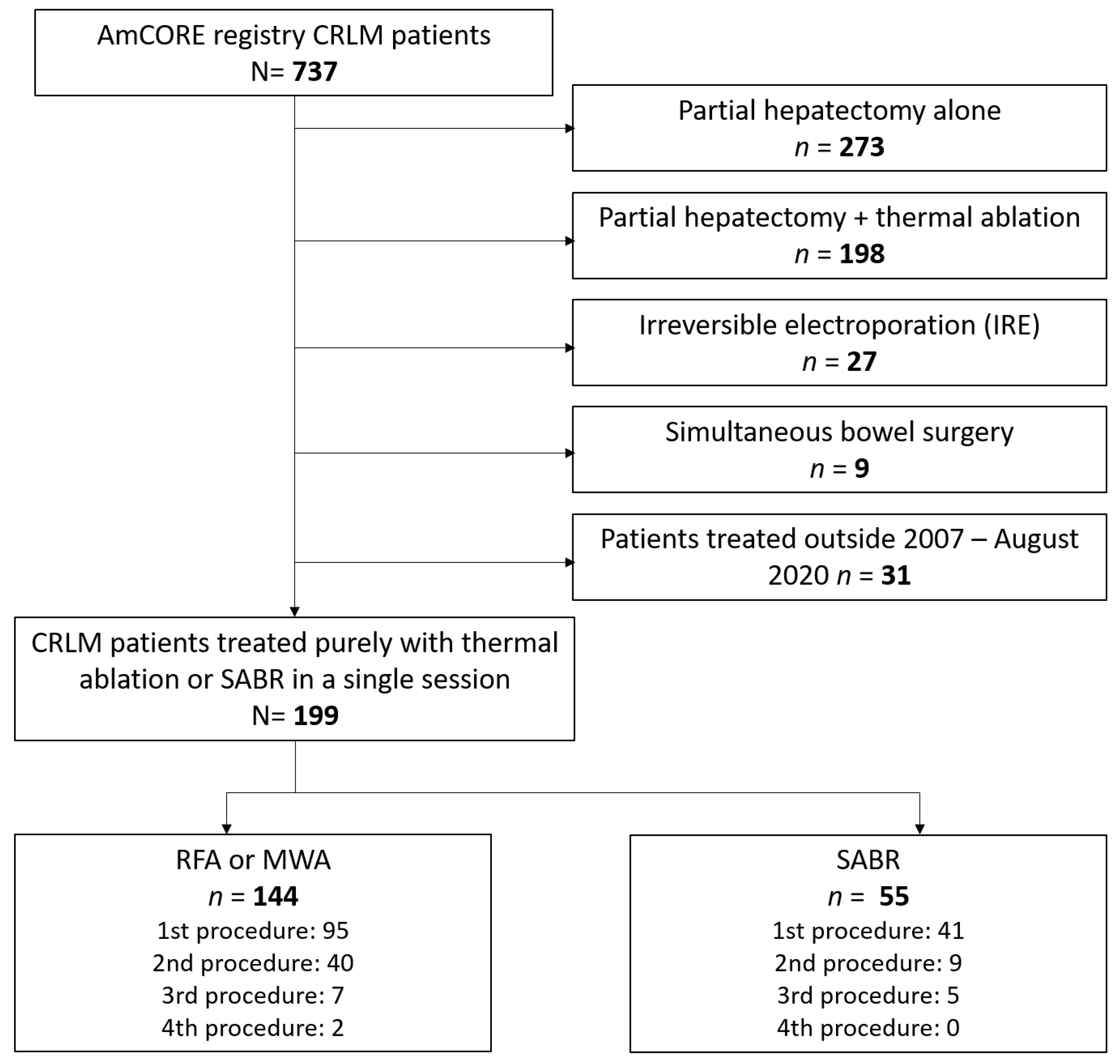
2.1. Study Design and Population

AmCORE is a prospectively maintained database that currently contains coded per-patient, per-procedure and per-tumor data of 737 CRLM patients treated with partial hepatectomy, thermal ablation and/or non-thermal ablation techniques in one of the Amsterdam University Medical Centers (location VUmc) from January 2007 onwards. From that registry, all patients with a history of solely thermal ablation(s) or SABR for previously untreated CRLM within a single session were selected, disallowing concomitant resections for additional CRLM within that session or as a first or second stage to that procedure. A history of previous partial hepatectomy (+/− thermal ablations) was allowed, as long as the CRLMs assessed were newly appearing and hence local treatment-naïve. If a patient had both a treatment-site recurrence from that earlier surgical procedure and a new CRLM, the treatment-site recurrence was disregarded in the evaluation. In case of multiple thermal ablation or SABR sessions, only the first ablative procedure, and the (new) metastases treated in that specific procedure, was analyzed. Patients who had simultaneous bowel surgery were excluded to avoid including complications unrelated to the ablative procedure. To update the registry with missing information, the regional hospitals’ electronic patient databases were consulted. To confirm radiology and nuclear medicine reports when pathology confirmation was unavailable, two independent abdominal radiologists (JHvW and IN, who have 15 years and 3 years of board certified experience, respectively) re-assessed the cross-sectional imaging exams of patients. Imaging re-evaluations were performed independently; discrepancies were noted and resolved using consensus. The institutional review board judged that the study was not subject to the Medical Research Involving Human Subjects Act (METc VUmc: 2020.454).

2.2. Radiofrequency Ablation, Microwave Ablation and Stereotactic Ablative Radiotherapy

All patients were discussed at a multidisciplinary tumor board, with the routine presence of interventional radiologists, radiation oncologists, hepatopancreaticobiliary surgeons, medical oncologists, diagnostic radiologists, nuclear medicine physicians and pathologists. Partial hepatectomy was considered the standard treatment method. Conforming to (inter)national guidelines, thermal ablation and SABR were considered appropriate for anatomically unresectable CRLM and for patients with poor performance status and/or major comorbidities. In recent years patients with solitary and deep-seated CRLMs, where resection would require major hepatectomy, have been offered thermal ablation or SABR as a fair alternative to partial hepatectomy. Though thermal ablation was considered the standard for unresectable CRLM, direct referral to radiation oncology from an external center, specific patient’s preference, high age, poor performance status, major cardiopulmonary comorbidities or a perivascular, peribiliary or difficult-to-reach anatomical location were relative criteria to opt for SABR over thermal ablation.
As this study addresses procedures performed over a period of >10 years, both the stereotactic ablative radiotherapy systems and specific dosing and sequencing and the thermal ablation generators, needle electrodes and antenna design, as well as the quality of work-up and follow-up cross-sectional anatomical and molecular imaging, anesthesia techniques and the methods used for real-time image-guidance, needle navigation and ablation confirmation, have undergone substantial technological and methodological developments.

2.3. Thermal Ablation

RFA and MWA procedures were preferably performed using a real-time fluoroscopy computed tomography (CT)-guided (+/− ultrasound) percutaneous approach. An ultrasound-guided open or laparoscopic approach was used for (a) potentially resectable CRLM, (b) multiple (>3) CRLM or (c) distancing of certain delicate structures such as intestines if a pneumo- or hydrodissection was unfeasible. Percutaneous thermal ablation procedures were performed under general anesthesia, midazolam sedation or propofol sedation in an interventional oncology treatment room which accommodated an angiography system, CT scanner, ultrasound apparatus and anesthetic facilities. RFA was most often performed using the RF3000 generator with expandable LeVeen electrodes (Boston Scientific, Marlborough, MA, USA), or the RITA system with compatible expandable electrodes (AngioDynamics BV, Amsterdam, The Netherlands) and MWA using the Evident or Emprint (Medtronic-Covidien, Minneapolis, MN, USA) or Solero (AngioDynamics BV, Amsterdam, The Netherlands) generators with compatible antennas. The ablations were performed according to the instructions for use provided by the manufacturer. Overlapping ablations were allowed to treat residual unablated tumor tissue in case of presumed insufficiently ablated margins. As computational techniques to confirm tumor-free margins using image fusion and registration were not available in the earlier years of this assessment, the documented minimum tumor free margins for procedures performed >5 years ago were eyeballed and estimated. After the procedure, patients were admitted to the post-anesthesia care unit to monitor vital parameters and patients remained admitted at the short-stay interventional oncology ward for one night.

2.4. Stereotactic Ablative Radiotherapy

Patients were simulated and treated in supine positions according to institutional protocol. The use of gated treatment or breath hold was determined at the discretion of the treating radiation oncologist. SABR was delivered in an image-guided hypofractionated scheme of 60Gy in 3, 5, 8 or 12 fractions, prescribed to 95% of the planning target volume (PTV). Target volumes were delineated by the treating radiation oncologist by means of the available cross-sectional imaging studies. A margin of 3–10 mm was used to delineate the PTV, depending on treatment technique and tumor visibility. Many different irradiation devices from several providers were used over the years. Since 2016 most patients were treated during breath hold using the ViewRay MRIdian system (ViewRay, Oakwood Village, OH, USA). The duodenum, stomach, bowel, liver, gallbladder, kidneys, and spinal cord were contoured as structures to avoid.

2.5. Follow-Up

Recommended follow-up after thermal ablation consisted of three-monthly serum CEA and [18F]-fluoro-2-deoxy-D-glucose (18F-FDG) positron emission tomography (PET)-ceCT scans for the first year of follow-up, changing to six-monthly for the second and third years and then to annually up to five years following the procedure. Follow-up recommendations after SABR were more heterogeneous, but consisted at the very minimum of a six-monthly cross-sectional ceCT scan and serum CEA for the first three years changing to annually up to five years. Contrast enhanced Magnetic Resonance Imaging (ceMRI) including high B-value diffusion-weighted images (DWI) and/or image-guided core biopsies were used as problem solver to confirm or exclude the presence of viable tumor tissue at the treatment site. If histopathological confirmation was not available, local tumor progression (LTP) following thermal ablation and SABR was defined as an unequivocal solid and enlarging, preferably nodular and ring-enhancing, mass at the surface of the ablated tumor and/or a new or enlarging marginal focus of 18F-FDG PET avidity, without signs of inflammation or infection.
Adverse events, defined as any actual or potential injury related to the ablative procedure, were determined according to the Common Terminology Criteria for Adverse Events 5.0 (CTCAE). Time-to-event endpoints were defined and calculated in accordance with the standardization of short-, mid- and long-term oncological outcome measures in image-guided tumor ablation consensus guidelines. Overall survival (OS) and distant progression-free survival (DPFS) were defined as the times elapsed from the (start of) the ablative treatment to death or to any disease recurrence distant from the ablation site. Local tumor progression-free survival (LTPFS) was defined as the time between (start of) the ablative treatment and LTP per-tumor treated (per-tumor analysis) or per-patient treated (per-patient analysis). For patients alive without LTP at the end of assessment, the date of the last cross-sectional imaging reliably excluding LTP was used as the censoring date. Death without LTP was not considered an event in the LTPFS analysis (it was treated as a competing risk). Local control per-tumor and per-patient is herein defined as the time elapsed between the (start of) the first ablation and the latest date of detection of LTP considered unfeasible for repeat local treatment, including repeat treatments whenever performed; simultaneous discovery of widespread distant disease progression was considered a competing risk. Though potentially correlated, the presence of multiple CRLM in one unique patient was ignored in the per-tumor LTPFS analysis and a per-patient LTPFS analysis was added conforming to recommendations by Puijk et al. [21]. Median follow-up time was calculated for patients alive at the time of analysis.

2.6. Statistical Analyses

Baseline patient, disease, tumor and treatment characteristics were analyzed using the Chi-square test for categorical variables and the independent two-sample t-test, ANOVA or Mann–Whitney U test for continuous variables. Characteristics assessed were: age, sex, simplified Charlson’s comorbidity index (CCI < 5, 5–8 or >8), BMI, number and size of metastases, primary tumor location, histopathological T-, N- and M-stage following primary tumor resection, Fong’s clinical risk score (low risk if CRS 0–2; high risk if CRS 3–5; 1 point for baseline CEA level > 200 ng/mL, for node positivity, for >1 CRLM, for largest CRLM > 5 cm and for detection of CRLM within 12-months following primary tumor diagnosis), extrahepatic disease at time of diagnosis of CRLM, history of partial hepatectomy and the use of induction or neo-adjuvant systemic therapy for the CRLM. OS, DPFS and LTPFS were estimated using Kaplan–Meier survival curves, log-rank tests and Cox proportional hazards survival models. Factors with p ≤ 0.10 in univariate analysis were entered into the multivariate analysis model to adjust for potential confounders. Statistical analyses were performed using SPSS version 26.0 and R version 4.0.4.

3. Results

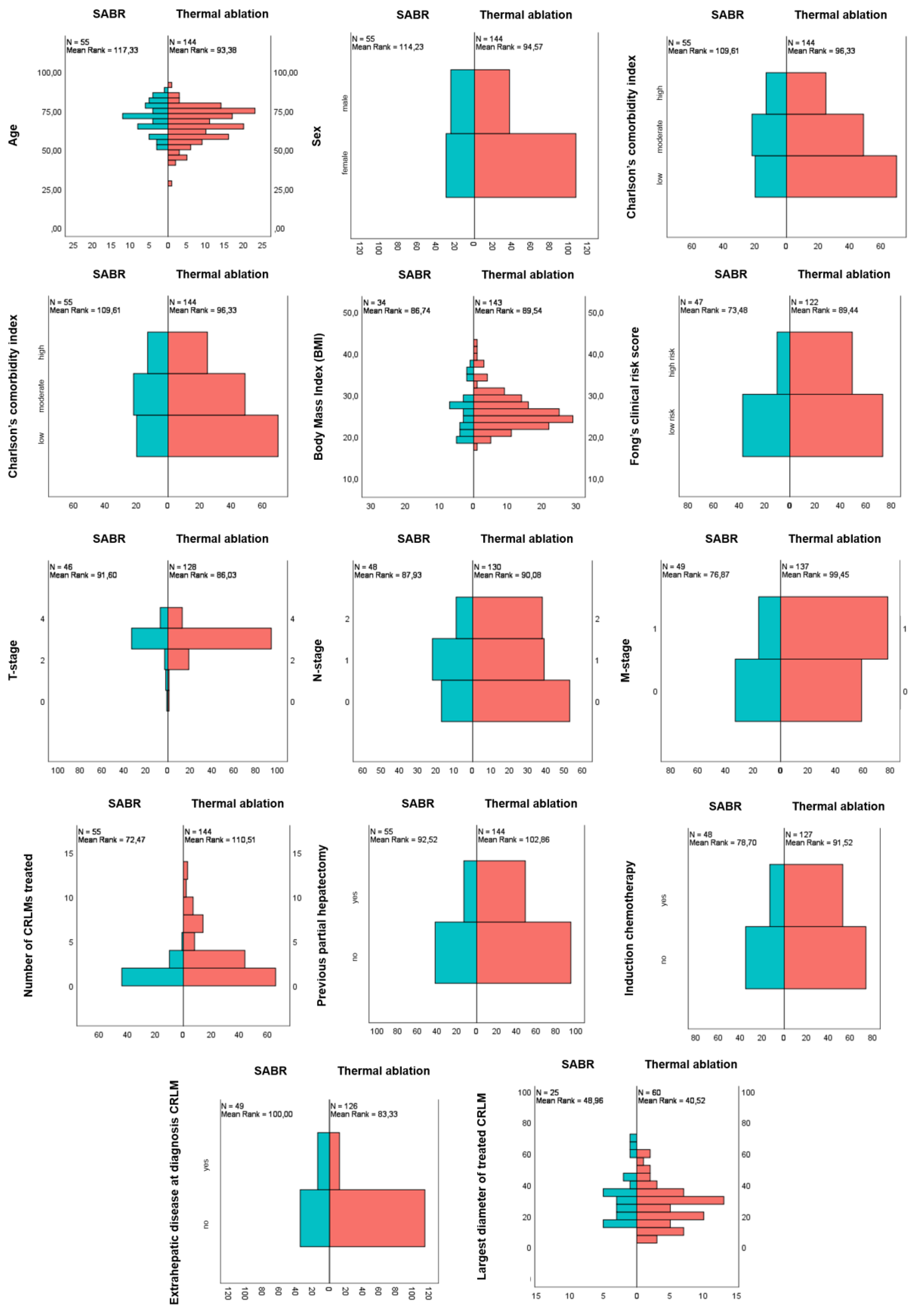
3.1. Baseline and Treatment Characteristics

Between January 2007 and August 2020, 737 registered patients underwent local treatment for CRLM (Figure 1). Of these, 536 patients were excluded because they underwent partial hepatectomy alone (n = 273), thermal ablation simultaneous with partial hepatectomy (n = 198), with bowel surgery (n = 9) or with IRE of at least one of the CRLM within the procedure assessed (n = 27). The remaining 199 patients (137 male/62 female; mean age 67.3 ± 10.5 years) were included for full analysis: 144 patients with 400 CRLM underwent thermal ablation and 55 patients with 69 CRLM underwent SABR.
Median follow-up time was 29.3 months for the whole cohort, 29.6 months (range 3.8–168.1) for thermal ablation and 27.6 months (range 11.8–91.3) for SABR. The thermal ablation approach was percutaneous in 55.6% (n = 80; 152 CRLM) and open in 44.4% (n = 64; 247 CRLM); 63 patients received treatment with RFA (n = 63/144; 43.8%), all prior to 2017, and 81 patients (n = 81/144; 56.3%) had MWA. In the SABR group 31 patients (n = 31/55; 56.4%) were treated with conventional SABR and 24 patients (n = 24/55; 43.6%) with MR-guided SABR, all after 2014. Table 1 shows the clinicopathological characteristics of included patients and their distribution over the two treatment groups.
The following potentially confounding parameters were evenly distributed over both treatment groups: CCI, previous chemotherapy regimens and the rate of patients with a history of earlier treatments for CRLM (Figure 2). There appeared to be a trend towards higher CCI scores in the SABR group, however this was not statistically significant. Patients in the SABR group were more often characterized by female sex (p = 0.005), higher age (p = 0.006), higher rates of extrahepatic disease at diagnosis of CRLM (p = 0.004) and larger tumors (p < 0.001). Patients treated with thermal ablation had higher Fong clinical risk scores (p = 0.030), higher rates of synchronous disease (p = 0.005) and higher number of tumors treated (p = 0.0004).

3.2. Adverse Events and Hospital Stay

Ninety-day mortality was 0% in both groups. Nine patients (n = 9/144; 6.3%) developed serious adverse events, all following thermal ablation (all CTCAE grade 3): bile leakage requiring percutaneous drainage (n = 2; 1.4%), intrahepatic abscess requiring percutaneous drainage (n = 2/144; 1.4%), hepatic hemorrhage requiring coil embolization (n = 2/144; 1.4%), bacteremia (n = 1/144; 0.7%), ileus (n = 1/144; 0.7%) and intraoperative stomach perforation (n = 1/144; 0.7%). Lower grade adverse events reported were pain requiring pain medication (n = 2/144; 1.4%), abscess requiring antibiotics (n = 1/144; 0.7%), hematoma (n = 1/144; 0.7%) and pneumothorax requiring no intervention (n = 4/144; 2.8%), which amounts to a total of eight CTCAE grade 1 or 2 adverse events (n = 8/144; 5.6%). Following SABR, no serious adverse events were registered, and the reported lower-grade adverse events were nausea requiring anti-emetics (n = 5/55; 9.1%), pain requiring additional pain medication (n = 2/55; 3.6%), rib insufficiency fracture (n = 1/55; 1.8%), dysphagia (n = 1/55; 1.8%) and fatigue (n = 9/55; 16.4%), which totals seventeen CTCAE grade 1 or 2 adverse events (n = 17/55; 30.9%). Though more adverse events were reported with SABR (30.9% vs. 11.8%; p = 0.0014), thermal ablation was associated with more serious complications that required an intervention (0% vs. 6.3%; p = 0.058).
Median hospital stay for thermal ablation was one day (range 1–35 days): one day for percutaneous ablations and five days for open ablations, with one reported hospital readmission. SABR was always performed in the outpatient clinic and patients underwent a median of eight (range 3–12) radiation cycles (not including the planning CT hospital visit) within a median of 16 days (range 5–24 days).

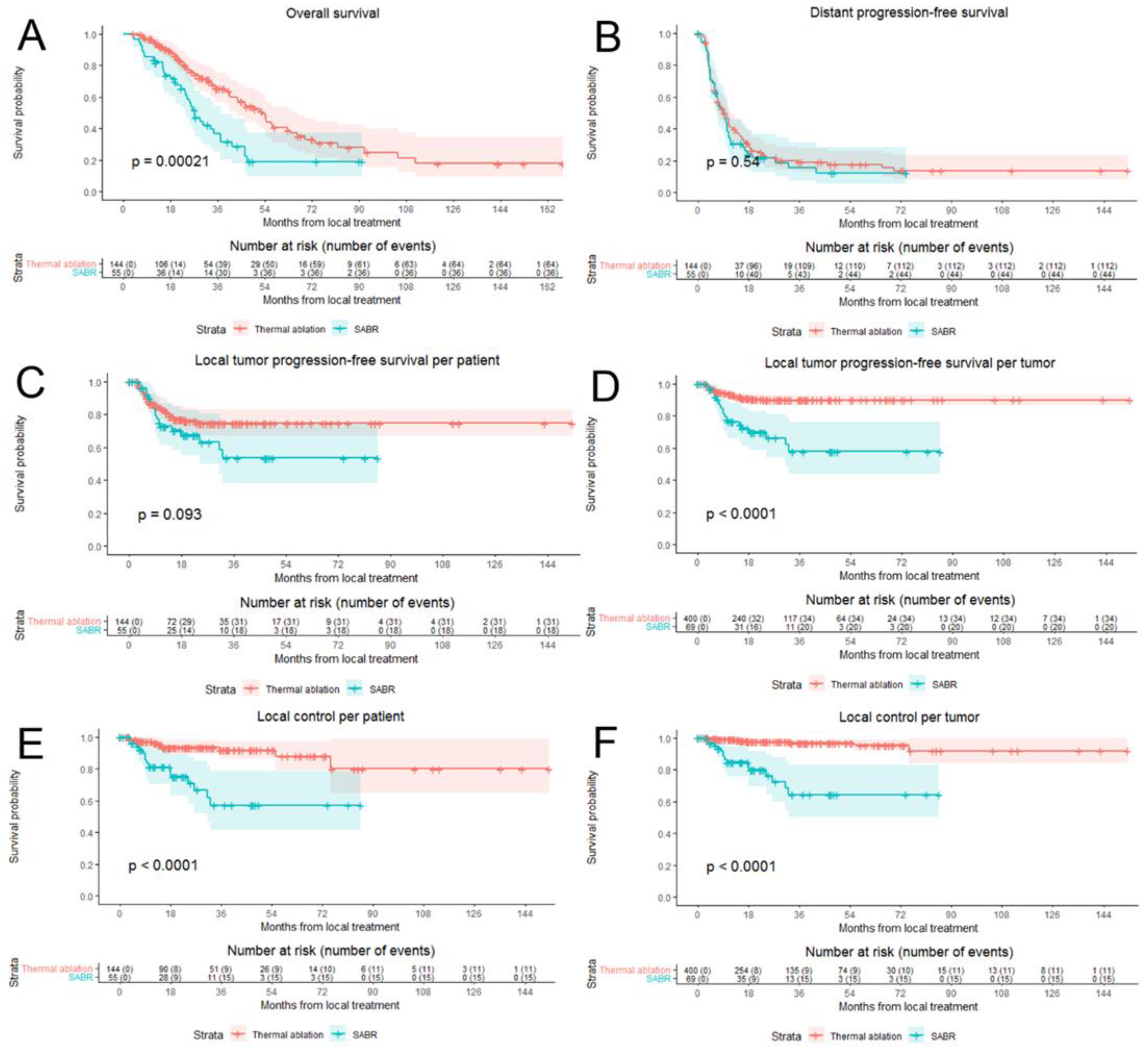
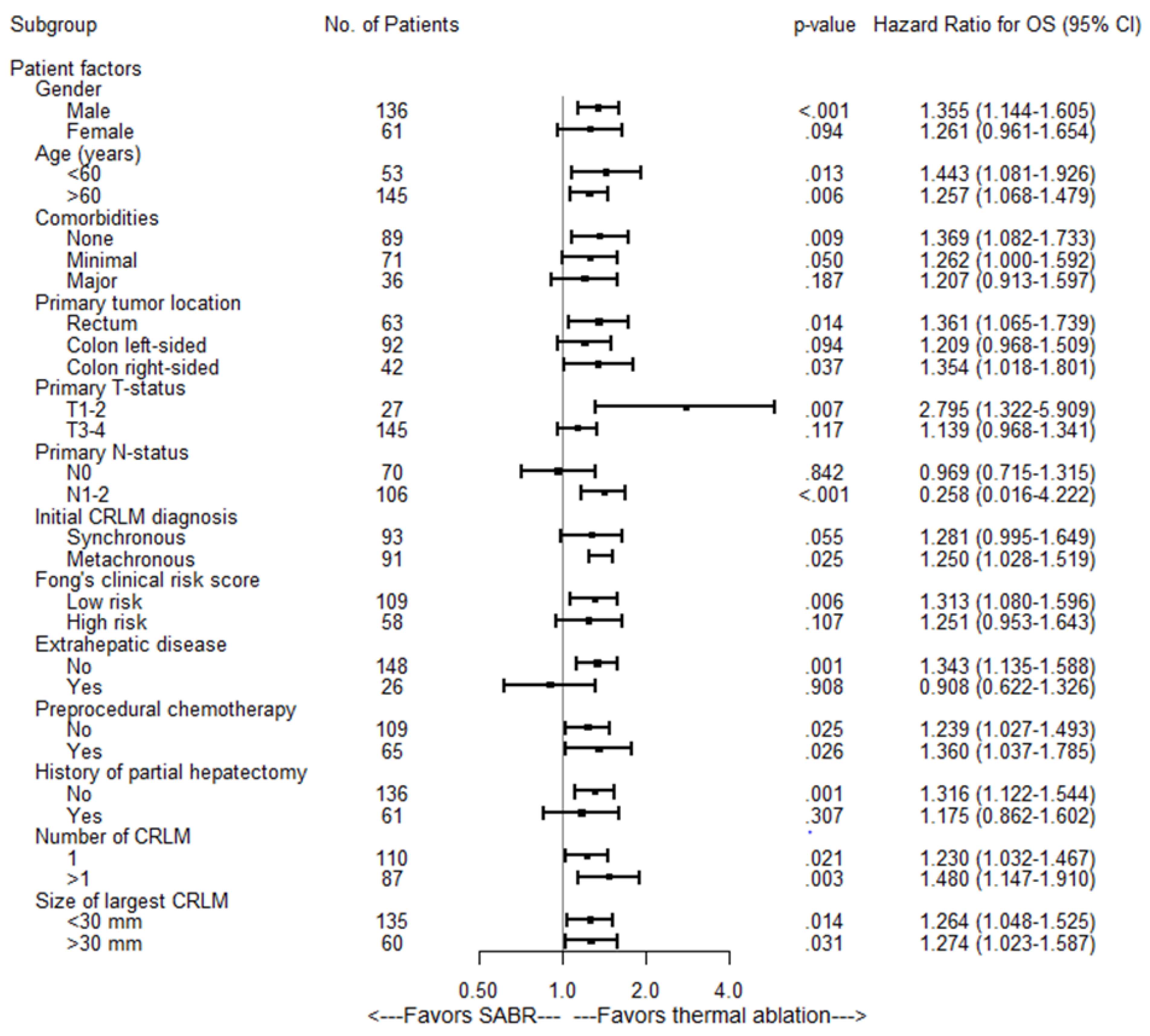
3.3. Overall Survival and Distant Progression-Free Survival

Baseline characteristics associated with an inferior OS in univariate assessment were major comorbidities (HR 1.92; 95% CI: 1.16–3.17; p = 0.03), node-positive primary tumor (HR 1.51; 95% CI: 0.96–2.38; p = 0.07), history of liver resection (HR 0.64; 95% CI: 0.41–1.02; p = 0.06), diameter of largest CRLM treated >3 cm (HR 1.81; 95% CI: 1.21–2.70; p = 0.004) and SABR treatment (HR 1.29; 95% CI: 1.12–1.49; p = 0.003) (see Table 2).
After correcting for potential confounders in multivariate analysis, SABR was still associated with a worse OS (HR of 1.26; 95% CI: 1.10–1.45; p = 0.001). Median OS for the entire cohort was 43.6 months (95% CI 36.3–50.9): 53.0 months (95% CI 45.2–60.8 months) for thermal ablation versus 27.4 months (95% CI 22.1–32.7 months) for SABR (p = 0.0002) (Figure 3A). Overall survival at one, two, three and five years was 94%, 80%, 65% and 41% respectively for thermal ablation and 84%, 61%, 37% and 19% for SABR. Subgroup analyses revealed no heterogeneous treatment effects (Figure 4).
Two subgroup analyses, including (a) only treatment naïve patients and (b) only patients with CRLM < 3 cm, did not change the differential outcomes with a median OS of 50.3 months vs. 26.0 months (p = 0.0005) and 54.8 months vs. 30.7 months (p = 0.01) for thermal ablation versus SABR.
Baseline variables associated with a worse DPFS in univariate analysis were female sex, higher age, higher comorbidity scores, node-positive disease, disease synchronicity, Fong’s clinical risk score and larger size of treated CRLMs. In both uni- and multivariate analysis there was no significant difference between thermal ablation and SABR with regards to the DPFS (HR = 1.07; 95% CI 0.93–1.22; p = 0.35) (Figure 3B).

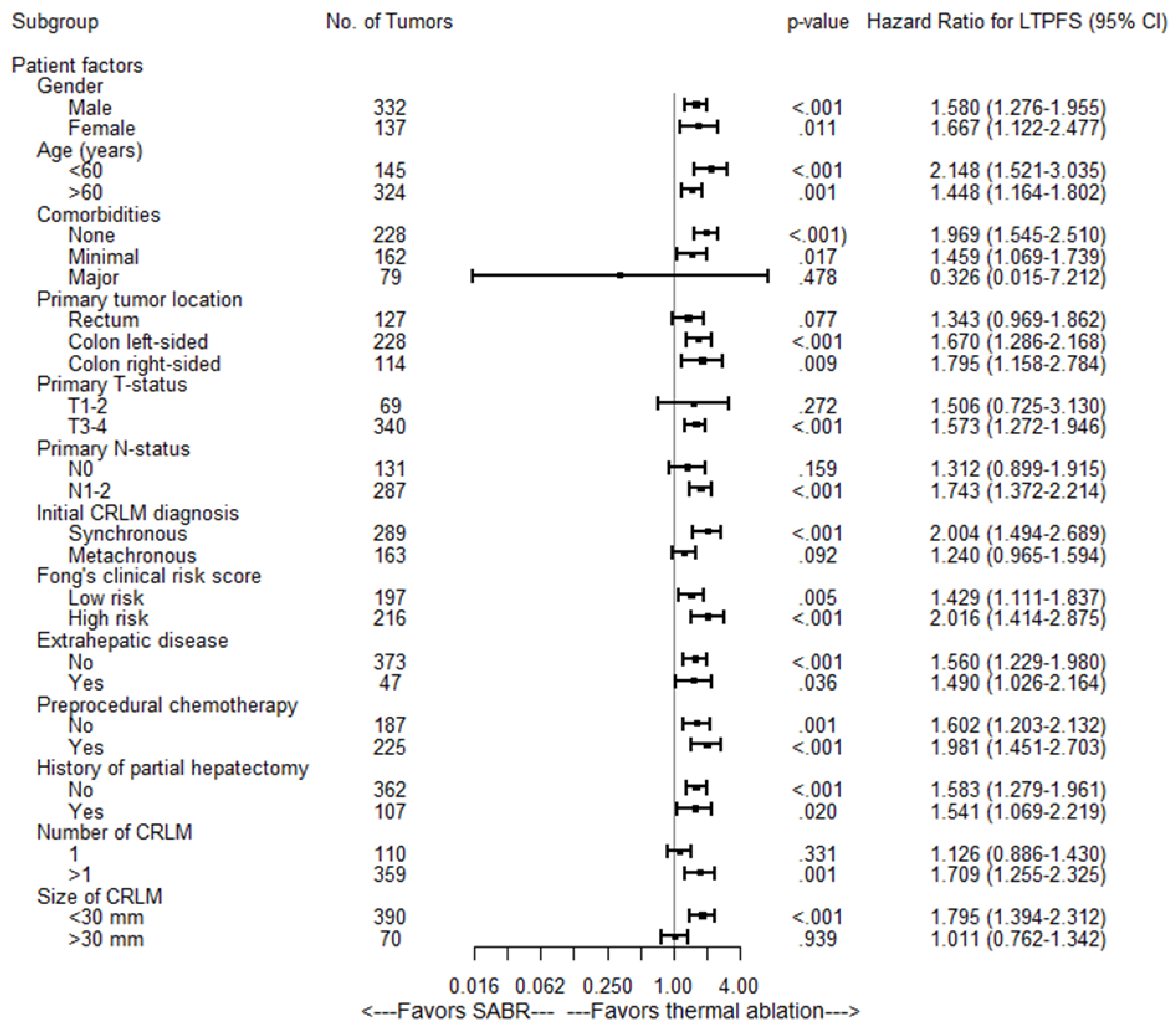
3.4. Local Tumor Progression-Free Survival and Eventual Local Control

Baseline variables associated with a worse LTPFS were synchronous disease, high Fong’s clinical risk score, extrahepatic disease, higher number of CRLMs treated, larger size of treated CRLMs and treatment with SABR (Table 3). Median LTPFS was not reached in both groups. LTP was detected in 31 out of 144 patients (21.5%) and in 34 out of 400 CRLM (8.5%) in the thermal ablation group versus in 18 out of 55 patients (32.7%) and 20 out of 69 CRLM (29%) in the SABR group. In the thermal ablation group there were 45 CRLM >3 cm and LTP occurred in 16 of these CRLM, compared to 9/26 CRLM >3 cm in the SABR group. For patients with LTP, the median time to the detection of LTP per-tumor was 6.6 months (95% CI: 5.7–7.6) for thermal ablation and 9.0 months (95% CI: 7.5–10.5) for SABR. LTPFS per-tumor (HR = 1.35, 95% CI 1.11–1.65; p = 0.003) was worse in the SABR group and there was no significant difference between the SABR and thermal ablation groups in LTPFS per-patient (HR = 1.12, 95% CI 0.92–1.36; p = 0.278) (Figure 3C,D).
The independent diagnostic radiologists’ re-evaluation (JHvW, IN) of suspected LTPs concurred in 80% (n = 16/20) of SABR treated LTPs and in all (n = 34/34) thermally ablated LTPs. The four discrepant cases were resolved in consensus between both diagnostic radiologists and the treating radiation oncologist (NH). For SABR the recurrences were described as solid and new or enlarging marginal lobulated incomplete-ring enhancing masses on ceCT in 14/20 LTPs (see Figure 5 for an example), as a new or augmenting marginal 18F-FDG PET avid mass abutting the irradiated CRLM in 5/20 LTPs and/or as a new or enlarging solid mass with diffusion restriction on ceMRI in 1/20 LTPs. For thermal ablation the detected recurrences were defined as new or enlarging, solid, marginal, lobulated incomplete-ring enhancing masses on contrast enhanced CT in 15/34 LTPs (see Figure 6 for an example), as new or enlarging marginal 18F-FDG PET avid mass abutting the ablated CRLM in 17/34 LTPs and/or as a new or enlarging solid mass with diffusion restriction on MRI in 2/34 LTPs.
Of 31 thermally ablated patients with LTP, 23 (23/34 tumors) were retreated using repeat thermal ablation (n = 20), partial hepatectomy (n = 1), IRE (n = 1) and SABR (n = 1). In 8 thermally ablated patients (11/34 tumors) local retreatment was deemed biologically futile due to widespread distant disease progression at first diagnosis of the LTP (competing risks). After a minimum follow-up of one year following the last local treatment, 15 patients (15/23 tumors) were alive and locally controlled. Three of the irradiated patients with LTP were locally retreated using partial hepatectomy (n = 1) and IRE (n = 2). In 15 irradiated patients (17/20 tumors) simultaneous detection of distant disease progression at first diagnosis of LTP prohibited retreatment (competing risks). After a minimum follow-up of one year following the last local treatment, two retreated patients (2/3 tumors) were alive and locally controlled. Local control per-tumor (HR = 1.90, 95% CI 1.44–2.50; p < 0.001) and per-patient (HR = 1.60, 95% CI 1.23–2.08; p = 0.001) was significantly worse in the SABR group (Figure 3E,F). Subgroup analyses revealed no heterogeneous treatment effects regarding LTPFS per-tumor (Figure 7).
Log-rank test analysis of two subgroups, including (a) only treatment naïve patients and (b) only CRLMs <3 cm, did not change the outcome: thermal ablation resulted in a superior per-tumor LTPFS (p < 0.001). Thermal ablation of intermediate-size CRLM (3–5 cm) was associated with a significantly worse LTPFS (p < 0.001) when compared to small-size CRLM (<3 cm), whereas after SABR no significant differences were detected for intermediate-size versus small-size CRLM (p = 0.361), implying that SABR may be less susceptible to larger tumor sizes (Figure 8A). Univariate Cox regression showed that with regard to the per-tumor LTPFS, MWA was not superior to RFA (p = 0.167), and selective MR-guided ablative radiotherapy was not superior to conventional SABR (p = 0.317) (Figure 8B).

4. Discussion

For the local treatment of unresectable CRLM, in this study thermal ablation was superior to SABR with regard to OS, LTPFS per-tumor and eventual local control when including repeat treatments. Correcting for potential confounders and adding subgroup analyses for local treatment-naïve patients, for patients with only CRLM < 3 cm and for RFA versus MWA and conventional SABR versus MR-guided SABR, did not change overall outcomes. Though size of the CRLM was the strongest predictor for LTP in the thermal ablation group, and though size did not predict LTP in the SABR group, suggesting this technique to be less susceptible to changes in size, thermal ablation was still superior for tumors > 3 cm in size with regard to OS, DPFS, LTPFS and local control. The overall complication rate was low in both groups, with no 90-day mortality and no acute life-threatening complications. Though the total number of adverse events did not differ between the treatment groups, patients in the SABR group more often had lower grade events (CTCAE grade 1 or 2), whereas patients in the thermal ablation group more often experienced events requiring an intervention. The rate of serious but not life-threatening (CTCAE grade 3) adverse events was 0% (0/55 patients) for SABR versus 6.3% (9/144 patients) of patients treated with percutaneous thermal ablation.
Two previous series, both retrospective cohorts, compared SABR to thermal ablation in patients with CRLM [22,23]. Stintzing and colleagues showed results comparable to the present series with a median OS of 52.3 months for thermal ablation vs. 34.4 months for SABR, though not reaching significance in their series of 60 patients (p = 0.06). In contrast to our results, the probability of LTP in their series was high, with hardly any CRLM eventually being locally controlled with either treatment method. Although Franzese and colleagues claim a superior local control for SABR, the justification is difficult to follow given the fact that 18.9% (n = 21 out 111 CRLM) of MWA treated CRLM recurred versus 19.4% (n = 20 out of 103 CRLM) in the SABR group and the attached freedom from local progression curve only shows 8 of these 20 events in the SABR group and 17 of the 21 events in the thermal ablation group because the displayed follow-up stops at 18 months. Differences in OS were not reported. Neither of these earlier series robustly define how to confirm LTP on cross-sectional imaging and neither describe eventual local control following repeat local treatments.
Several studies on SABR for both colorectal and non-colorectal liver metastases reported three-year local control rates of 48.3–91% and a three-year OS of 23–44% [24,25,26,27,28,29,30,31]. The largest series to date, a recent Dutch–Belgian registry covering 515 patients and 668 metastases, showed a three-year local control rate of 68% and three-year OS of 44% [31]. These outcomes are consistent with the present series, with a 64% three-year local control rate and a three-year OS of 35%. As thermal ablation is a repeatable technique, authors more often disclose the LTPFS following the first procedure and local control including repeat sessions whenever necessary. According to a recent meta-analysis that included 48 series, longer-term LTPFS ranges between 56–98% and eventual local control ranges between 86–100% [9]. Again our results reflect earlier series with a three-year LTPFS of 90% and a three-year local control of 97%.
The sensitivity and specificity of ceCT, 18F-FDG PET-CT and ceMRI to detect LTP following thermal ablation are well established, ranging between 83–92% and 95–100% respectively [14]. Though it can be difficult to assess routine cross-sectional imaging of liver tumors following SABR due to radiation-induced liver fibrosis and tissue reorganization within the irradiated zone [32,33], the accuracy in detecting LTP actually seems to compare well with follow-up after thermal ablation [32,33,34]. Jarraya and colleagues have described that the occurrence of a lobulated enhancement predicted LTP with a sensitivity of 89%, a specificity of 100% and an overall concordance rate of 97.9% [33].
Strengths of this comparative analysis are the relatively high number of patients and tumors assessed, the long-term follow-up, the baseline data accessed from a prospective registry and most importantly the multidisciplinary approach with image reassessments done by two abdominal radiologist referees, independently both from each other and from the treating physicians. Multivariate analysis with Cox regression could be used to reduce the impact of selection bias and several subgroup analyses were added to further reduce confounding and substantiate results.
Key limitations are the non-randomized nature and the unclearly defined criteria when to opt for thermal ablation versus SABR. Although anatomically the large majority of irradiated tumors would have also been eligible for thermal ablation and vice versa (the majority of thermally ablated tumors would have qualified for SABR), the exact reasons to opt for a certain treatment method are multifactorial and difficult to recollect by retrospectively searching the hospital’s electronic patient database. Another limitation is the long period of inclusion and its consequent gradual change in specific treatment methods, where for example MR-guided SABR is currently often favored over conventional SABR and where MWA seems to largely have replaced RFA. Though these limitations make our analysis at high risk of residual bias, the superior local effectiveness and repeatability of thermal ablation over SABR does show that the two techniques are not in equipoise. Further refinements to improve the efficacy of SABR seem necessary before randomized controlled trials can be considered for smaller tumors. However, given that differences in local effectiveness between the two treatment methods seem to shrink with increasing tumor size and given the excellent safety profile of SABR, the authors do believe a comparative study for tumors 3–5 cm in size is justified and warranted. Several ongoing randomized controlled trials will hopefully further elucidate the roles of thermal ablation and SABR to treat larger-size or difficult-to-reach CRLM (NCT04081168, NCT03654131 and NCT02820194).
To conclude, for the treatment of CRLM, in this study thermal ablation was superior to SABR with regard to OS, LTPFS and local control, albeit at the cost of a limited risk of serious adverse events. Future work, preferably in the form of a randomized clinical trial, should assess whether the worse outcomes following SABR were the effect of true differences in ablative treatment or merely a result of residual confounding.

Author Contributions

Conceptualization, S.N., M.P.v.d.T. and M.R.M.; methodology, S.N., M.D., F.E.F.T. and B.I.L.-W.; software, S.N. and B.I.L.-W.; validation, S.N., M.D., R.S.P., F.E.F.T., I.M.N., J.O., B.v.d.B., B.G., A.H.R., E.A.C.S., H.J.S., J.J.J.d.V., A.M.v.G., J.H.T.M.v.W., R.-J.S., K.S.V., B.I.L.-W., M.P.v.d.T., C.J.A.H. and M.R.M.; formal analysis, S.N., M.D., F.E.F.T. and B.I.L.-W.; investigation, S.N. and M.D.; data curation, M.D., S.N., M.D., R.S.P., B.v.d.B., E.A.C.S., J.O., M.P.v.d.T. and M.R.M.; writing—original draft preparation, S.N.; writing—review and editing, S.N., M.D., R.S.P., F.E.F.T., I.M.N., J.O., B.v.d.B., B.G., A.H.R., E.A.C.S., H.J.S., J.J.J.d.V., A.M.v.G., J.H.T.M.v.W., R.-J.S., K.S.V., B.I.L.-W., M.P.v.d.T., C.J.A.H. and M.R.M.; visualization, S.N., M.D., F.E.F.T., M.P.v.d.T. and M.R.M.; supervision, M.P.v.d.T., C.J.A.H. and M.R.M.; project administration, S.N.; funding acquisition, not applicable. All authors have read and agreed to the published version of the manuscript.

Funding

This research received no external funding.

Institutional Review Board Statement

The study was approved by the affiliated Institutional Review Board (METc VUmc: 2021.0121).

Informed Consent Statement

Patient consent was waived as the study was not subject to the Medical Research Involving Human Subjects Act judged by the affiliated Institutional Review Board (METc VUmc: 2020.454).

Data Availability Statement

The data presented in this study are available on request from the corresponding author.

Conflicts of Interest

The authors declare no conflict of interest.

References

  1. WHO. Estimated Age-Standardized Incidence Rates (World) in 2018, All Cancers, Both Sexes, All Ages. 2018. Available online: http://gco.iarc.fr/today/online-analysis-map (accessed on 1 April 2021).
  2. Hackl, C.; Neumann, P.; Gerken, M.; Loss, M.; Klinkhammer-Schalke, M.; Schlitt, H.J. Treatment of colorectal liver metastases in Germany: A ten-year population-based analysis of 5772 cases of primary colorectal adenocarcinoma. BMC Cancer 2014, 14, 810. [Google Scholar] [CrossRef] [PubMed] [Green Version]
  3. Engstrand, J.; Nilsson, H.; Stromberg, C.; Jonas, E.; Freedman, J. Colorectal cancer liver metastases—A population-based study on incidence, management and survival. BMC Cancer 2018, 18, 78. [Google Scholar] [CrossRef]
  4. Leporrier, J.; Maurel, J.; Chiche, L.; Bara, S.; Segol, P.; Launoy, G. A population-based study of the incidence, management and prognosis of hepatic metastases from colorectal cancer. BJS 2006, 93, 465–474. [Google Scholar] [CrossRef]
  5. Taylor, A.; Primrose, J.N.; Langeberg, W.; Kelsh, M.; Mowat, F.; Alexander, D.; Choti, M.; Poston, G.; Kanas, G. Survival after liver resection in metastatic colorectal cancer: Review and meta-analysis of prognostic factors. Clin. Epidemiol. 2012, 4, 283–301. [Google Scholar] [CrossRef] [PubMed] [Green Version]
  6. Kopetz, S.; Chang, G.J.; Overman, M.J.; Eng, C.; Sargent, D.; Larson, D.W.; Grothey, A.; Vauthey, J.-N.; Nagorney, D.M.; McWilliams, R.R. Improved Survival in Metastatic Colorectal Cancer Is Associated With Adoption of Hepatic Resection and Improved Chemotherapy. J. Clin. Oncol. 2009, 27, 3677–3683. [Google Scholar] [CrossRef] [PubMed]
  7. House, M.G.; Ito, H.; Gönen, M.; Fong, Y.; Allen, P.J.; DeMatteo, R.P.; Brennan, M.; Blumgart, L.H.; Jarnagin, W.R.; D’Angelica, M.I. Survival after Hepatic Resection for Metastatic Colorectal Cancer: Trends in Outcomes for 1,600 Patients during Two Decades at a Single Institution. J. Am. Coll. Surg. 2010, 210, 744–752. [Google Scholar] [CrossRef]
  8. Adam, R.; Avisar, E.; Ariche, A.; Giachetti, S.; Azoulay, D.; Castaing, D.; Kunstlinger, F.; Levi, F.; Bismuth, F. Five-year survival following hepatic resection after neoadjuvant therapy for nonresectable colorectal. Ann. Surg. Oncol. 2001, 8, 347–353. [Google Scholar] [CrossRef]
  9. Meijerink, M.R.; Puijk, R.S.; Van Tilborg, A.A.; Henningsen, K.H.; Fernandez, L.G.; Neyt, M.; Heymans, J.; Frankema, J.S.; De Jong, K.P.; Richel, D.J.; et al. Radiofrequency and Microwave Ablation Compared to Systemic Chemotherapy and to Partial Hepatectomy in the Treatment of Colorectal Liver Metastases: A Systematic Review and Meta-Analysis. Cardiovasc. Interv. Radiol. 2018, 41, 1189–1204. [Google Scholar] [CrossRef] [Green Version]
  10. van Amerongen, M.J.; Jenniskens, S.F.M.; van den Boezem, P.B.; Fütterer, J.J.; de Wilt, J.H.W. Radiofrequency ablation compared to surgical resection for curative treatment of patients with colorectal liver metastases—A meta-analysis. HPB 2017, 19, 749–756. [Google Scholar] [CrossRef] [PubMed] [Green Version]
  11. Nieuwenhuizen, S.; Puijk, R.S.; van den Bemd, B.; Aldrighetti, L.; Arntz, M.; van den Boezem, P.B.; Bruynzeel, A.M.; Burgmans, M.C.; De Cobelli, F.; Coolsen, M.M.; et al. Resectability and Ablatability Criteria for the Treatment of Liver Only Colorectal Metastases: Multidisciplinary Consensus Document from the COLLISION Trial Group. Cancers 2020, 12, 1779. [Google Scholar] [CrossRef]
  12. Mohamed, F.; Kallioinen, M.; Braun, M.; Fenwick, S.; Shackcloth, M.; Davies, R.J.; Bradbury, J.; Burgess, G.; Chew, C.; Dawson, C.; et al. Management of colorectal cancer metastases to the liver, lung or peritoneum suitable for curative intent: Summary of NICE guidance. Br. J. Surg. 2020, 107, 943–945. [Google Scholar] [CrossRef]
  13. Vera, R.; González-Flores, E.; Rubio, C.; Urbano, J.; Camps, M.V.; Ciampi-Dopazo, J.J.; Rincón, J.O.; Macías, V.M.; Braco, M.A.G.; Suarez-Artacho, G. Multidisciplinary management of liver metastases in patients with colorectal cancer: A consensus of SEOM, AEC, SEOR, SERVEI, and SEMNIM. Clin. Transl. Oncol. 2020, 22, 647–662. [Google Scholar] [CrossRef] [Green Version]
  14. Nielsen, K.; van Tilborg, A.A.; Meijerink, M.R.; Macintosh, M.O.; Zonderhuis, B.M.; de Lange, E.S.; Comans, E.F.I.; Meijer, S.; van den Tol, M.P. Incidence and treatment of local site recurrences following RFA of colorectal liver metastases. World J. Surg. 2013, 37, 1340–1347. [Google Scholar] [CrossRef] [PubMed]
  15. van Tilborg, A.A.; Scheffer, H.J.; de Jong, M.C.; Vroomen, L.G.; Nielsen, K.; van Kuijk, C.; van den Tol, P.M.P.; Meijerink, M.R. MWA Versus RFA for Perivascular and Peribiliary CRLM: A Retrospective Patient- and Lesion-Based Analysis of Two Historical Cohorts. Cardiovasc. Intervent. Radiol. 2016, 39, 1438–1446. [Google Scholar] [CrossRef] [Green Version]
  16. Tanis, E.; Nordlinger, B.; Mauer, M.; Sorbye, H.; van Coevorden, F.; Gruenberger, T.; Schlag, P.; Punt, C.; Ledermann, J.; Ruers, T. Local recurrence rates after radiofrequency ablation or resection of colorectal liver metastases. Analysis of the European Organisation for Research and Treatment of Cancer #40004 and #40983. Eur. J. Cancer 2014, 50, 912–919. [Google Scholar] [PubMed]
  17. Solbiati, L.; Livraghi, T.; Goldberg, S.N.; Ierace, T.; Meloni, F.; Dellanoce, M.; Cova, L.; Halpern, E.F.; Gazelle, G.S. Percutaneous radio-frequency ablation of hepatic metastases from colorectal cancer: Long-term results in 117 patients. Radiology 2001, 221, 159–166. [Google Scholar] [CrossRef] [PubMed]
  18. Berber, E.; Siperstein, A. Local Recurrence after Laparoscopic Radiofrequency Ablation of Liver Tumors: An Analysis of 1032 Tumors. Ann. Surg. Oncol. 2008, 15, 2757–2764. [Google Scholar] [CrossRef]
  19. Ayav, A.; Germain, A.; Marchal, F.; Tierris, I.; Laurent, V.; Bazin, C.; Yuan, Y.; Robert, L.; Brunaud, L.; Bresler, L. Radiofrequency ablation of unresectable liver tumors: Factors associated with incomplete ablation or local recurrence. Am. J. Surg. 2010, 200, 435–439. [Google Scholar] [CrossRef] [PubMed]
  20. Scorsetti, M.; Comito, T.; Tozzi, A.; Navarria, P.; Fogliata, A.; Clerici, E.; Mancosu, P.; Reggiori, G.; Rimassa, L.; Torzilli, G.; et al. Final results of a phase II trial for stereotactic body radiation therapy for patients with inoperable liver metastases from colorectal cancer. J. Cancer Res. Clin. Oncol. 2015, 141, 543–553. [Google Scholar] [CrossRef] [PubMed]
  21. Puijk RSA, M.; Adam, A.; Arai, Y.; Arellano, R. Consensus guidelines for the definition of time-to-event endpoints in image guided tumor ablation: Results of the Society for Interventional Oncology (SIO) and DATECAN (Definition for the Assessment of Time-to-event End-points in CANcer trials) initiative. Radiology 2021. not yet published. [Google Scholar]
  22. Franzese, C.; Comito, T.; Clerici, E.; Di Brina, L.; Tomatis, S.; Navarria, P.; Reggiori, G.; Viganò, L.; Poretti, D.; Pedicini, V.; et al. Liver metastases from colorectal cancer: Propensity score-based comparison of stereotactic body radiation therapy vs. microwave ablation. J. Cancer Res. Clin. Oncol. 2018, 144, 1777–1783. [Google Scholar] [CrossRef]
  23. Stintzing, S.; Grothe, A.; Hendrich, S.; Hoffmann, R.-T.; Heinemann, V.; Rentsch, M.; Fuerweger, C.; Muacevic, A.; Trumm, C.G. Percutaneous radiofrequency ablation (RFA) or robotic radiosurgery (RRS) for salvage treatment of colorectal liver metastases. Acta Oncol. 2013, 52, 971–977. [Google Scholar] [CrossRef] [PubMed] [Green Version]
  24. Lee, M.T.; Kim, J.J.; Dinniwell, R.; Brierley, J.; Lockwood, G.; Wong, R.; Cummings, B.; Ringash, J.; Tse, R.V.; Knox, J.J.; et al. Phase I Study of Individualized Stereotactic Body Radiotherapy of Liver Metastases. J. Clin. Oncol. 2009, 27, 1585–1591. [Google Scholar] [CrossRef] [PubMed] [Green Version]
  25. Andratschke, N.H.; Nieder, C.; Heppt, F.; Molls, M.; Zimmermann, F. Stereotactic radiation therapy for liver metastases: Factors affecting local control and survival. Radiat. Oncol. 2015, 10, 69. [Google Scholar] [CrossRef] [Green Version]
  26. Goodman, B.D.; Mannina, E.M.; Althouse, S.K.; Maluccio, M.A.; Cárdenes, H.R. Long-term safety and efficacy of stereotactic body radiation therapy for hepatic oligometastases. Pr. Radiat. Oncol. 2016, 6, 86–95. [Google Scholar] [CrossRef]
  27. Joo, J.H.; Park, J.-H.; Kim, J.C.; Yu, C.S.; Lim, S.-B.; Park, I.J.; Kim, T.W.; Hong, Y.S.; Kim, K.-P.; Yoon, S.M.; et al. Local Control Outcomes Using Stereotactic Body Radiation Therapy for Liver Metastases from Colorectal Cancer. Int. J. Radiat. Oncol. Biol. Phys. 2017, 99, 876–883. [Google Scholar] [CrossRef]
  28. Mahadevan, A.; Blanck, O.; Lanciano, R.; Peddada, A.; Sundararaman, S.; D’Ambrosio, D.; Sharma, S.; Perry, D.; Kolker, J.; Davis, J. Stereotactic Body Radiotherapy (SBRT) for liver metastasis—Clinical outcomes from the international multi-institutional RSSearch® Patient Registry. Radiat. Oncol. 2018, 13, 1–11. [Google Scholar] [CrossRef] [PubMed]
  29. Andratschke, N.; Alheid, H.; Allgäuer, M.; Becker, G.; Blanck, O.; Boda-Heggemann, J.; Brunner, T.; Duma, M.; Gerum, S.; Guckenberger, M.; et al. The SBRT database initiative of the German Society for Radiation Oncology (DEGRO): Patterns of care and outcome analysis of stereotactic body radiotherapy (SBRT) for liver oligometastases in 474 patients with 623 metastases. BMC Cancer 2018, 18, 283. [Google Scholar] [CrossRef] [Green Version]
  30. Clerici, E.; Comito, T.; Franzese, C.; Di Brina, L.; Tozzi, A.; Iftode, C.; Navarria, P.; Mancosu, P.; Reggiori, G.; Tomatis, S.; et al. Role of stereotactic body radiation therapy in the treatment of liver metastases: Clinical results and prognostic factors. Strahlenther. Onkol. 2020, 196, 325–333. [Google Scholar] [CrossRef]
  31. Mendez Romero, A.; Schillemans, W.; van Os, R.; Koppe, F.; Haasbeek, C.J.; Hendriksen, E.M. The Dutch-Belgian Registry of Stereotactic Body Radiation Therapy for Liver Metastases: Clinical Outcomes of 515 Patients and 668 Metastases. Int. J. Radiat. Oncol. Biol. Phys. 2021, 109, 1377–1386. [Google Scholar] [CrossRef] [PubMed]
  32. Tetreau, R.; Llacer, C.; Riou, O.; Deshayes, E. Evaluation of response after SBRT for liver tumors. Reports of practical oncology and radiotherapy. J. Greatpoland Cancer Cent. Pozn. Pol. Soc. Radiat. Oncol. 2017, 22, 170–175. [Google Scholar]
  33. Jarraya, H.; Borde, P.; Mirabel, X.; Ernst, O.; Boulanger, T.; Lartigau, E.; Ceugnart, L.; Kramar, A.; Taieb, S. Lobulated Enhancement Evaluation in the Follow-Up of Liver Metastases Treated by Stereotactic Body Radiation Therapy. Int. J. Radiat. Oncol. 2015, 92, 292–298. [Google Scholar] [CrossRef] [PubMed] [Green Version]
  34. Yip, C.; Cook, G.J.R.; Owczarczyk, K.; Goh, V. Challenges in imaging assessment following liver stereotactic body radiotherapy: Pitfalls to avoid in clinical practice. Chin. Clin Oncol. 2017, 6 (Suppl. 2), S11. [Google Scholar] [CrossRef] [PubMed] [Green Version]
Figure 1. Flowchart of selected patients from the prospective Amsterdam Colorectal Liver Metastases Registry (AmCORE). CRLM = colorectal liver metastases, RFA = radiofrequency ablation, MWA = microwave ablation, SABR = stereotactic ablative radiotherapy.
Cancers 13 04303 g001
Figure 2. Patients treated with SABR (blue) were older, had larger tumors and higher rates of extrahepatic disease. Patients treated with thermal ablation (red) had higher numbers of CRLMs, higher Fong’s clinical risk scores and higher rates of synchronous disease. All other variables were evenly distributed.
Cancers 13 04303 g002
Figure 3. Survival curves following thermal ablation (red) and stereotactic ablative radiotherapy (SABR) (blue) for unresectable colorectal liver metastases: overall survival (A), distant progression-free survival (B), local tumor progression free survival, per-patient and per-tumor (C,D) and local control allowing repeat treatment, per-patient and per-tumor (E,F).
Cancers 13 04303 g003
Figure 4. Univariate Subgroup Cox Regression Analyses of SABR versus thermal ablation associated with OS.
Cancers 13 04303 g004
Figure 5. Follow-up cross-sectional ceCT images of a segment IVa-VIII CRLM treated with SABR. At three months ceCT showed a typical halo sign of hypoattenuation directly surrounding the irradiated, largely necrotic tumor (white arrowheads) and characteristic hyperattenuation of the perilesional liver parenchyma, which correlates with radiation induced hepatic fibrosis (A). At six months a solid ring-enhancing nodular lesion (white arrow) can be appreciated at the right-lateral margin of the irradiated tumor (B). This appearance is highly specific for the presence of local tumor progression post-SABR. Follow-up ceCT at six and 12 months (C,D) further confirmed the local tumor progression.
Cancers 13 04303 g005
Figure 6. Follow-up cross-sectional ceCT images of a CRLM treated with thermal ablation. At six months a typical sharply demarcated hypoattenuating scar lesion (white arrowheads) is visible (A). At 12 months a characteristic “incomplete ring sign” (white arrow) at the left margin of the ablation zone can be appreciated (B), which, especially when combined with 18F-FDG avidity, is highly specific for local tumor progression following thermal ablation. Follow-up ceCT at 15 and 18 months (C,D) further confirmed local tumor progression.
Cancers 13 04303 g006
Figure 7. Univariate Subgroup Cox Regression Analyses of SABR versus thermal ablation associated with LTPFS per tumor.
Cancers 13 04303 g007
Figure 8. Local tumor progression-free survival curves (LTPFS) following thermal ablation (red) and stereotactic ablative radiotherapy (blue) for unresectable colorectal liver metastases according to subgroup analyses: stratified by CRLM < 3 cm versus 3–5 cm (A) and by specific treatment method radiofrequency ablation (RFA), microwave ablation (MWA), conventional stereotactic ablative radiotherapy (SABR) and selective MR-guided ablative radiotherapy (SMART) (B).
Cancers 13 04303 g008
Table 1. Baseline characteristics of CRLM patients treated with thermal ablation or stereotactic ablative radiotherapy (SABR).
Factor Total CohortThermal AblationSABRp-Value
NNN
Total 19914455
 Patient characteristics
 Sex 0.010
Male137 (68.8%)107 (74.3%)30 (54.5%)
Female62 (31.2%)37 (25.7%)25 (45.5%)
 Age (years)Median (IQR)68.6 (15.9)66.8 (16.5)71.5 (13.0)0.006
 Charlson’s Comorbidity Index (CCI) 0.281
Low CCI 0–590 (45.2%)70 (48.6%)20 (36.4%)
Moderate CCI 5–871 (35.7%)49 (34%)22 (40%)
High CCI >838 (19.1%)25 (17.4%)13 (23.6%)
 Location primary tumor 0.857
Rectum64 (32.2%)45 (31.3%)19 (34.5%)
Left-sided colon93 (46.7%)69 (47.9%)24 (43.6%)
Right-sided colon42 (21.1%)30 (20.8%)12 (21.8%)
 Primary T-status 0.813
T1-227 (13.6%)21 (16.4%)6 (13%)
T3-4147 (73.9%)107 (83.6%)40 (87%)
Unknown25 (12.6%)169
 Primary N-status 0.605
N070 (35.2%)53 (40.8%)17 (35.4%)
N1-2108 (54.3%)77 (59.2%)31 (64.6%)
Unknown21 (10.6%)147
 Primary M-status 0.005
synchronous94 (47.2%)78 (56.9%)16 (32.7%)
metachronous92 (46.2%)59 (43.1%)33 (67.3%)
Unknown13 (6.5%)76
 Fong’s clinical risk score 0.030
Low risk110 (55.3%)73 (59.8%)37 (78.8%)
High risk59 (29.6%)49 (40.2%)10 (21.3%)
 Extrahepatic disease at diagnosis 0.004
No147 (73.9%)112 (90.3%)35 (71.4%)
Yes26 (13.1%)12 (9.7%)14 (28.6%)
 History of preprocedural chemotherapy 0.083
No109 (54.8%)74 (58.7%)35 (72.9%)
Yes66 (33.2%)53 (41.7%)13 (27.1%)
 History of partial hepatectomy 0.174
No137 (68.8%)95 (66%)42 (76.4%)
Yes62 (31.2%)49 (34%)13 (23.6%)
 Number of CRLM treated within procedure
Median2 (1–18) 2 (1–18)1 (1–5)0.0004
 Diameter of CRLM at procedure (mm) <0.0005
Median21 (2–68)14 (2–65) 29 (6–68)
Note: IQR = Interquartile range, p-values in bold are p-values ≤ 0.1.
Table 2. Variables associated with OS in univariate Cox regression analysis and potential confounders in multivariate Cox regression analysis for OS when comparing SABR to thermal ablation (bold type significant confounders).
Univariate Analysis OSMultivariate Analysis OS
VariableHazard Ratio (95% CI)p ValueHazard Ratiop Value
 Treatment
 Thermal ablation1 [reference]<0.0011 [reference]0.001
 SABR1.29 (1.12–1.49) 1.27 (1.10–1.46)
 Sex
 Male1 [reference]0.374
 Female0.83 (0.53–1.29)
 Age in years
 <68 years1 [reference]0.150
 >68 years1.35 (0.90–2.01)
 Comorbidities
 None1 [reference]0.0341 [reference]0.204
 Minor1.08 (0.68–1.70) 0.98 (0.62–1.56)
 Major1.92 (1.16–3.17) 1.54 (0.90–2.64)
 Location primary tumor
 Rectal1 [reference]0.282
 Left sided colon0.89 (0.57–1.39)
 Right sided colon1.36 (0.80–2.32)
 Primary T category
 T1-21 [reference]0.469
 T3-41.28 (0.66–2.48)
 Primary N category
 N01 [reference]0.0771 [reference]0.068
 N1-21.51 (0.96–2.38) 1.54 (0.97–2.46)
 Timing of CRLM
 Metachronous1 [reference]0.722
 Synchronous1.09 (0.72–1.64)
 Fong CRS
 Low risk1 [reference]0.180
 High risk1.38 (0.88–2.16)
 Extrahepatic disease at diagnosis
 No1 [reference]0.844
 Yes0.93 (0.51–1.73)
 Preprocedural chemotherapy
 No1 [reference]0.315
 Yes0.80 (0.52–1.25)
 History of liver resection
 No1 [reference]0.0641 [reference]0.193
 Yes0.64 (0.41–1.02) 0.73 (0.45–1.17)
 Number of CRLM during procedure
 1 CRLM1 [reference]0.541
 >1 CRLM0.892 (0.60–1.33)
 Diameter CRLM at procedure
 ≤3 cm1 [reference]0.0041 [reference]0.014
 3 cm1.81 (1.21–2.70) 1.67 (1.11–2.51)
Note: p-values in bold are p-values ≤ 0.1.
Table 3. Variables associated with LTPFS per-tumor in univariate Cox regression analysis and potential confounders in multivariate Cox regression analysis for LTPFS when comparing SABR to thermal ablation (bold type significant confounders).
Univariate Analysis LTPFSMultivariate Analysis LTPFS
Per-Tumor
VariableHazard Ratio (95% CI)pValueHazard Ratio (95% CI)pValue
 Treatment
 Thermal ablation1 [reference]<0.0011 [reference]0.003
 SABR1.58 (1.31–1.90) 1.35 (1.11–1.65)
 Sex
 Male1 [reference]0.398
 Female0.75 (0.39–1.46)
 Age in years
 <68.6 years1 [reference]0.363
 >68.6 years1.28 (0,75–2.19)
 Comorbidities
 None1 [reference]0.331
 Minor0.78 (0.44–1.39)
 Major0.51 (0.20–1.31)
 Location primary tumor
 Rectal1 [reference]0.496
 Left-sided colon0.95 (0.53–1.73)
 Right-sided colon0.63 (0.28–1.41)
 Primary T category
 T1-21 [reference]0.220
 T3-41.79 (0.71–4.56)
 Primary N category
 N01 [reference]0.914
 N1-20.97 (0.53–1.78)
 Timing of CRLM
 Metachronous1 [reference]<0.0011 [reference]0.103
 Synchronous0.36 (0.21–0.63) 0.61 (0.34–1.11)
 Fong CRS
 Low risk1 [reference]0.0191 [reference]0.141
 High risk0.46 (0.25–0.88) 0.61 (0.32–1.18)
 Extrahepatic disease at diagnosis
 No1 [reference]0.0021 [reference]0.137
 Yes2.80 (1.48–5.30) 1.67 (0.85–3.29)
 Preprocedural chemotherapy
 No1 [reference]0.234
 Yes0.69 (0.37–1.27)
 History of liver resection
 No1 [reference]0.576
 Yes1.19 (0.65–2.19)
 Diameter CRLM at procedure
 ≤3 cm1 [reference]<0.0011 [reference]<0.001
 >3 cm6.30 (3.67–10.83) 4.73 (2.65–8.45)
Note: p-values in bold are p-values ≤ 0.1.
Publisher’s Note: MDPI stays neutral with regard to jurisdictional claims in published maps and institutional affiliations.

