Article

Temporal Observation of Adipocyte Microfiber Using Anchoring Device


1
Center for International Research on Integrative Biomedical Systems (CIBiS), Institute of Industrial Science (IIS), The University of Tokyo, 4-6-1 Komaba, Meguro-ku, Tokyo 153-8505, Japan
2
Department of Mechano-Informatics, Graduate School of Information Science and Technology, The University of Tokyo, 7-3-1 Hongo, Bunkyo-ku, Tokyo 113-8656, Japan
3
Department of Life Sciences, Graduate School of Arts and Sciences, The University of Tokyo, 7-3-1 Hongo, Bunkyo-ku, Tokyo 113-8656, Japan
4
International Research Center for Neurointelligence (WPI-IRCN), The University of Tokyo Institutes for Advanced Study (UTIAS), The University of Tokyo, 7-3-1 Hongo, Bunkyo-ku, Tokyo 113-8656, Japan
*
Author to whom correspondence should be addressed.
Micromachines 2019, 10(6), 358; https://doi.org/10.3390/mi10060358
Submission received: 11 May 2019 / Revised: 27 May 2019 / Accepted: 28 May 2019 / Published: 29 May 2019

Abstract

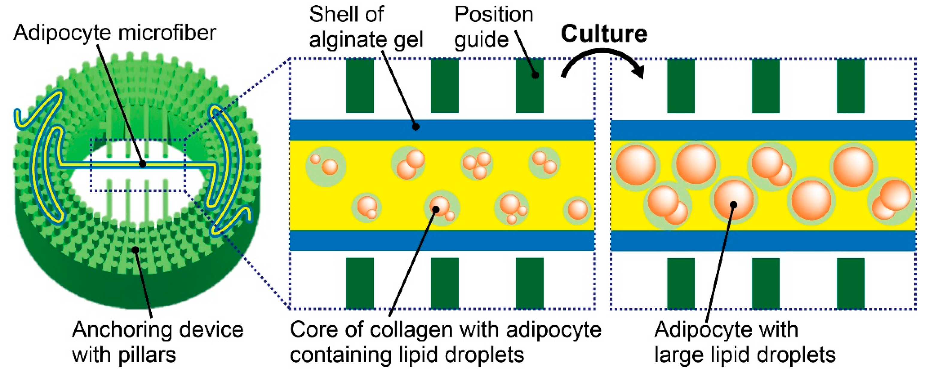
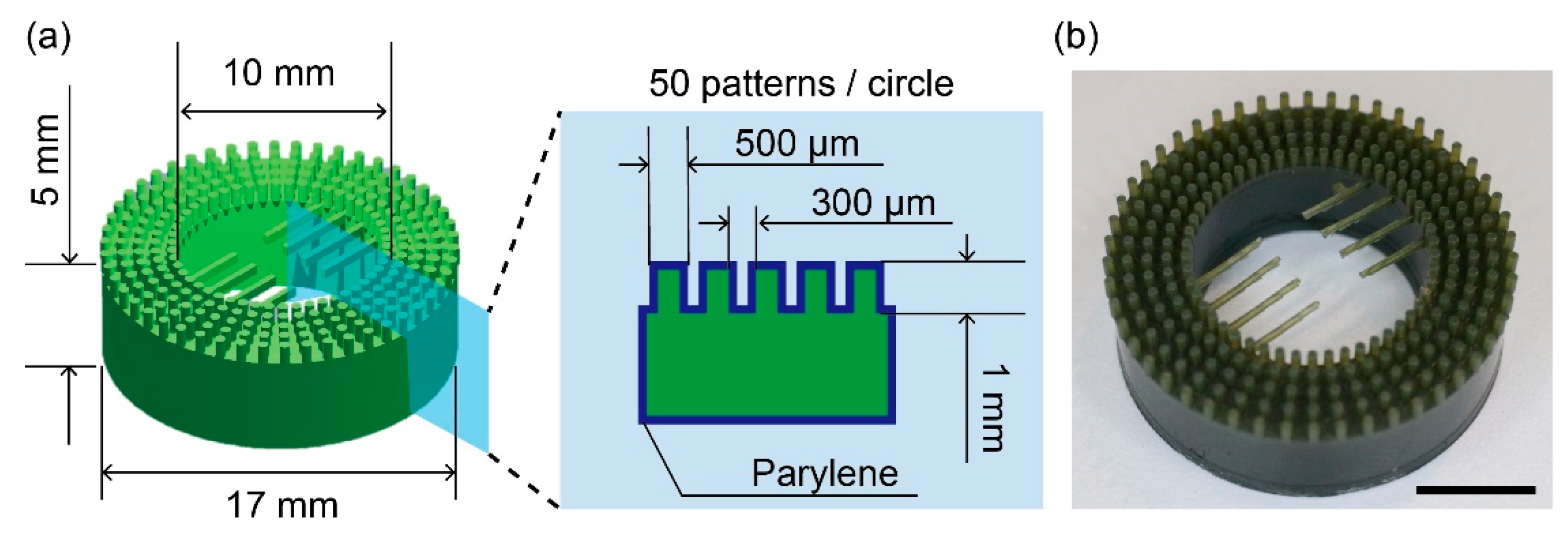
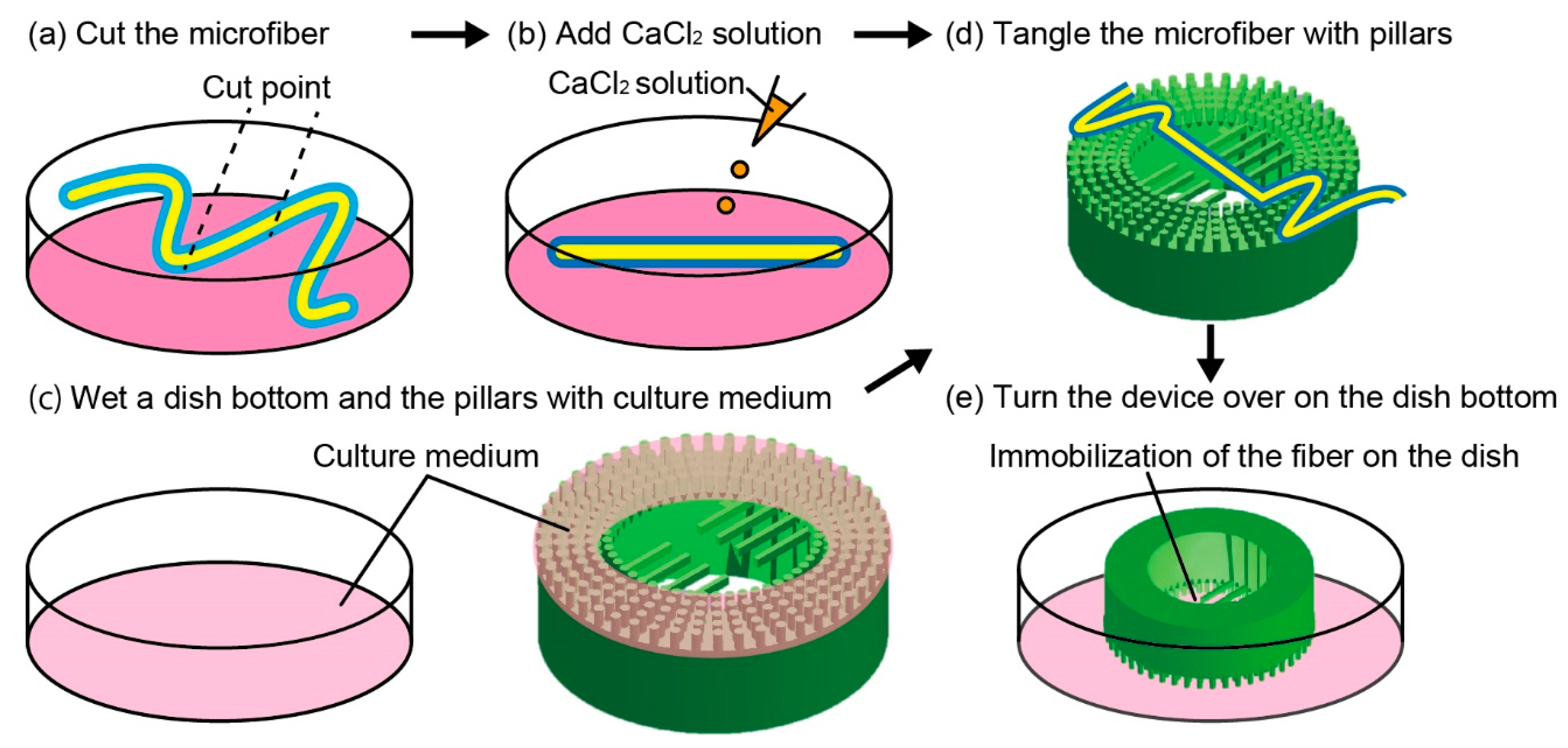
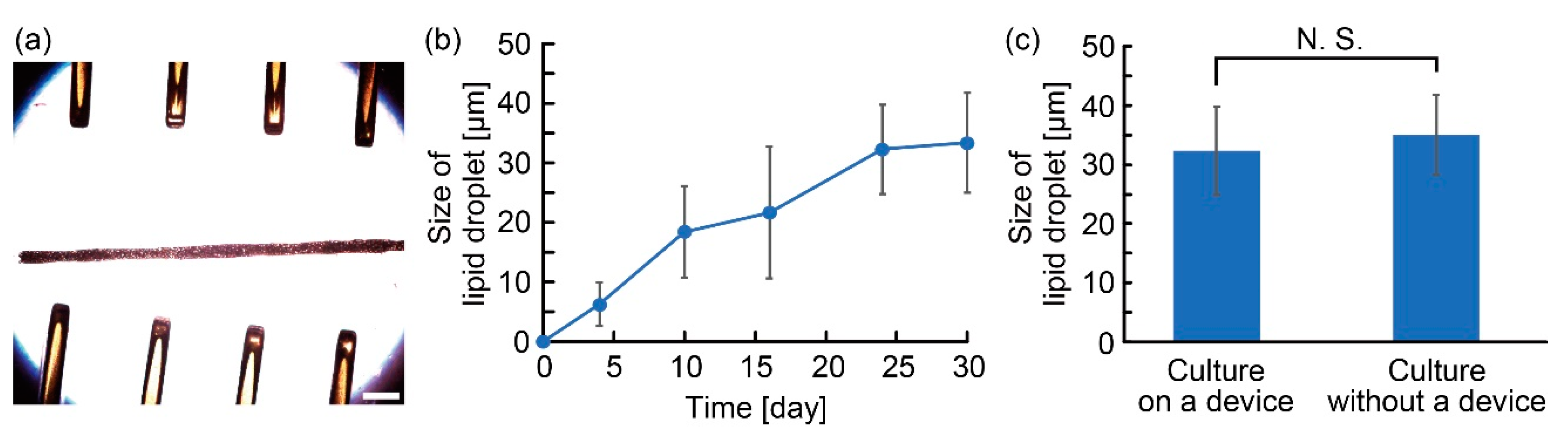
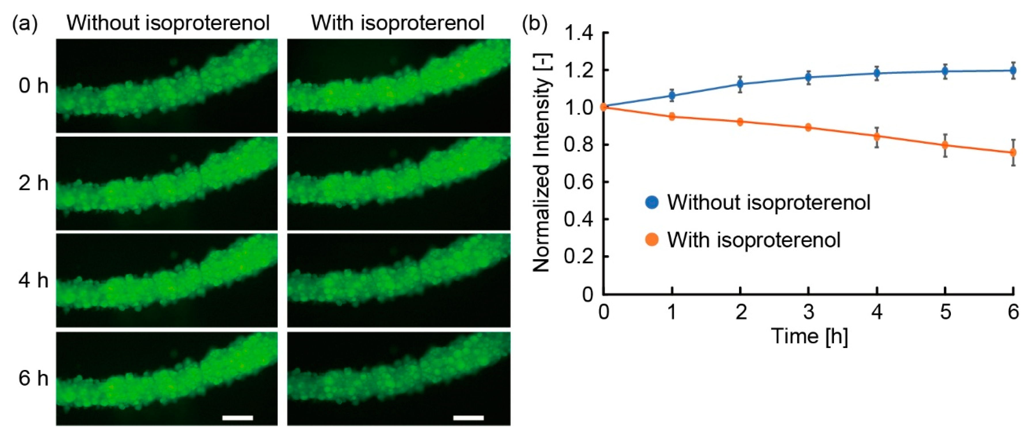
In this paper, we propose an anchoring device with pillars to immobilize an adipocyte microfiber that has a fiber-shaped adipocyte tissue covered by an alginate gel shell. Because the device enabled the immobilization of the microfiber in a culture dish even after its transportation and the exchange of the culture medium, we can easily track the specific positions of the microfiber for a long period. Owing to the characteristics of the anchoring device, we successfully performed temporal observations of the microfiber on the device for a month to investigate the function and morphology of three-dimensional cultured adipocytes. Furthermore, to demonstrate the applicability of the anchoring device to drug testing, we evaluated the lipolysis of the microfiber’s adipocytes by applying reagents with an anti-obesity effect. Therefore, we believe that the anchoring device with the microfiber will be a useful tool for temporal biochemical analyses.
Keywords: microfluidics; biofabrication; adipose tissue; lipolysis microfluidics; biofabrication; adipose tissue; lipolysis