Uremic Toxins and Hemodiafiltration: From Molecular Mechanisms to Clinical Outcomes

 ,
,  , and
, and
Abstract
1. Introduction
2. Mechanisms of Uremic Toxin Accumulation
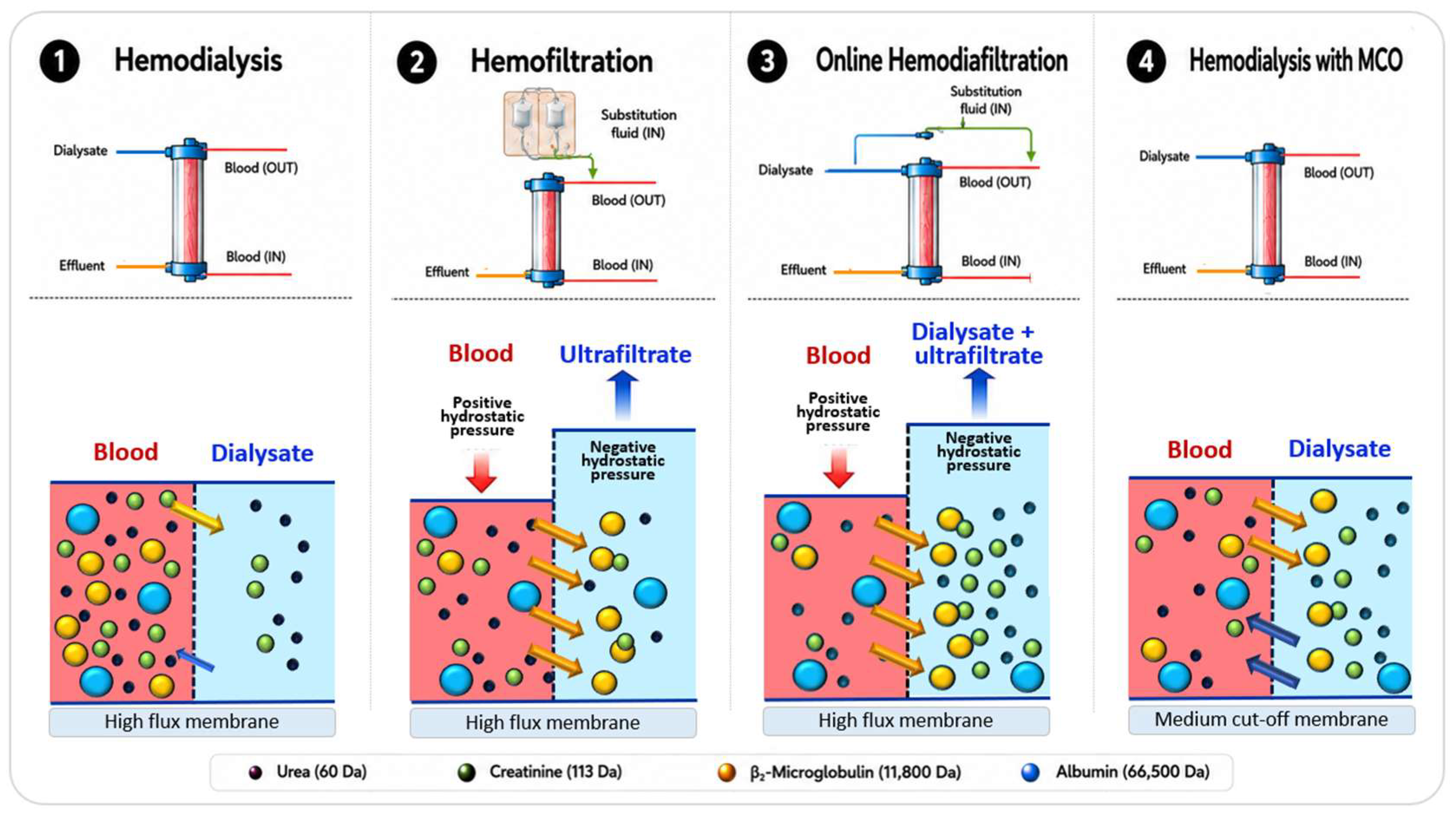
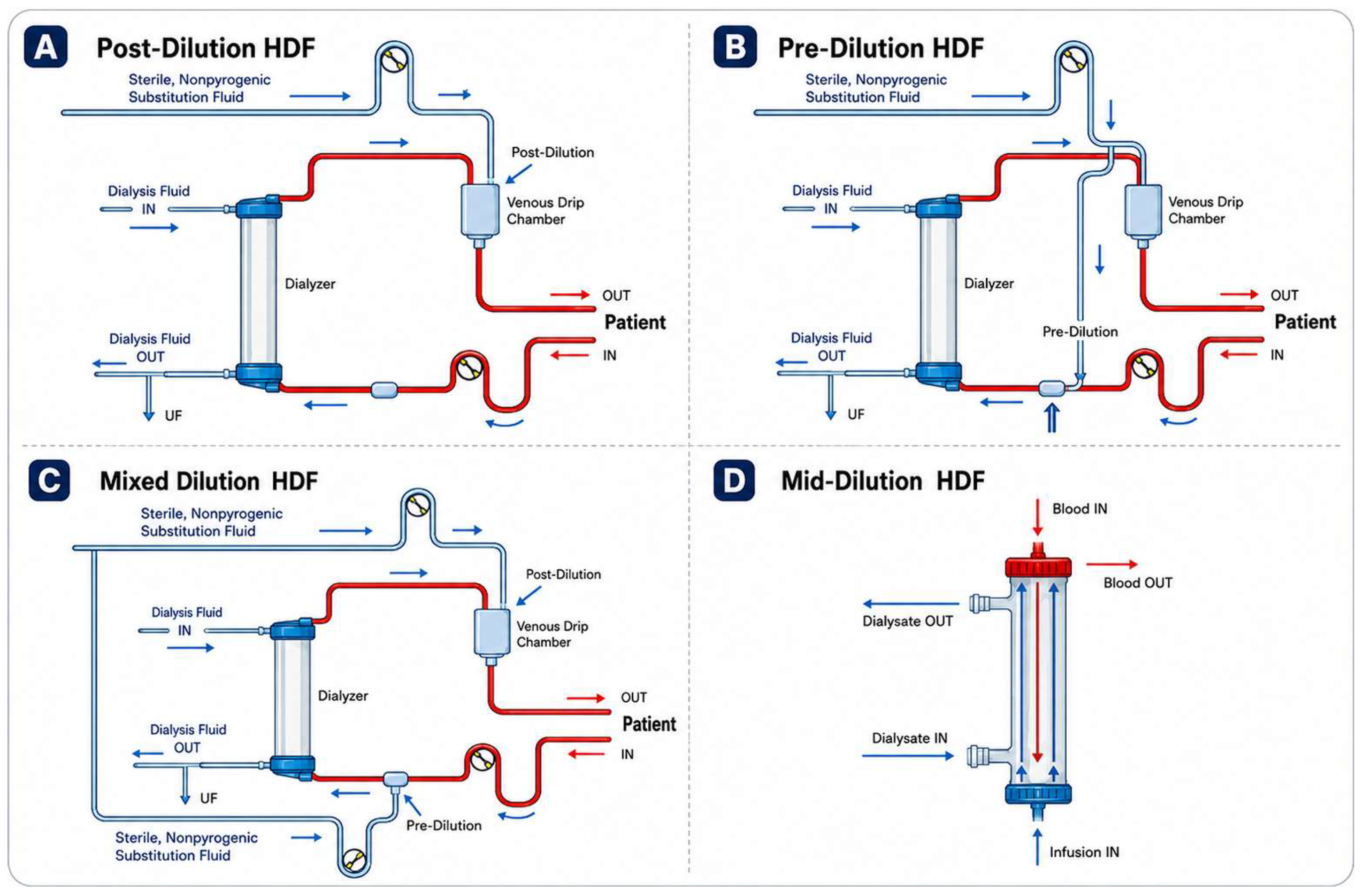
3. Dialysis Modalities and Uremic Toxin Removal
4. Quantification of Solute Removal in Hemodiafiltration
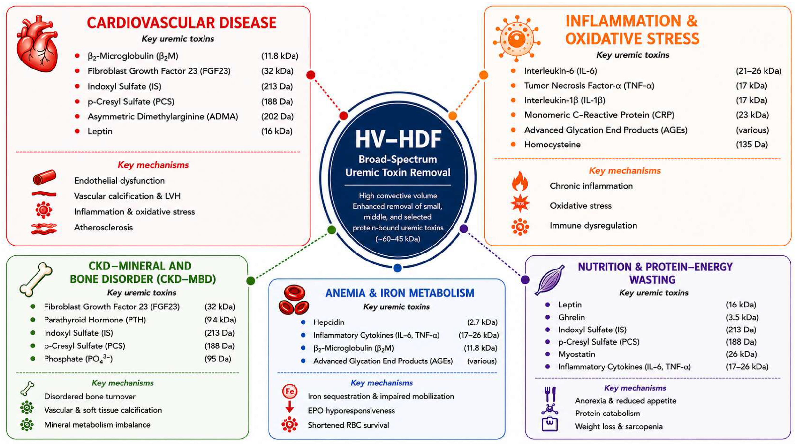
5. Clinical Consequences of Uremic Toxin Accumulation
6. Inflammation and Oxidative Stress
7. CKD–Mineral and Bone Disorder
8. Cardiovascular Health and Intradialytic Hemodynamic Tolerance
9. Anemia and Nutritional Abnormalities
10. Clinical Outcomes: From Uremic Toxin Removal to Patient Benefit
11. Conclusions
Author Contributions
Funding
Institutional Review Board Statement
Informed Consent Statement
Data Availability Statement
Conflicts of Interest
References
- Vanholder, R.; De Smet, R.; Glorieux, G.; Argilés, A.; Baurmeister, U.; Brunet, P.; Clark, W.; Cohen, G.; De Deyn, P.P.; Deppisch, R.; et al. Review on uremic toxins: Classification, concentration, and interindividual variability. Kidney Int. 2003, 63, 1934–1943. [Google Scholar] [CrossRef] [Scilit] [PubMed]
- EUTOX. European Uremic Toxins Working Group of the European Society of Artificial Organs Database. Available online: https://www.uremic-toxins.org/eutox-database/ (accessed on 4 July 2026).
- Vanholder, R.; Glorieux, G.; Argiles, A.; Burtey, S.; Cohen, G.; Duranton, F.; Koppe, L.; Massy, Z.A.; Ortiz, A.; Masereeuw, R.; et al. Metabolomics to Identify Unclassified Uremic Toxins: A Comprehensive Literature Review. Kidney Med. 2025, 7, 100955. [Google Scholar] [CrossRef] [Scilit] [PubMed]
- Cozzolino, M.; Magagnoli, L.; Ciceri, P. From Physicochemical Classification to Multidimensional Insights: A Comprehensive Review of Uremic Toxin Research. Toxins 2025, 17, 295. [Google Scholar] [CrossRef] [Scilit] [PubMed]
- Neirynck, N.; Vanholder, R.; Schepers, E.; Eloot, S.; Pletinck, A.; Glorieux, G. An update on uremic toxins. Int. Urol. Nephrol. 2013, 45, 139–150. [Google Scholar] [CrossRef] [Scilit] [PubMed]
- Rosner, M.H.; Reis, T.; Husain-Syed, F.; Vanholder, R.; Hutchison, C.; Stenvinkel, P.; Blankestijn, P.J.; Cozzolino, M.; Juillard, L.; Kashani, K.; et al. Classification of Uremic Toxins and Their Role in Kidney Failure. Clin. J. Am. Soc. Nephrol. 2021, 16, 1918–1928. [Google Scholar] [CrossRef] [Scilit] [PubMed]
- Sanchez-Ospina, D.; Mas-Fontao, S.; Gracia-Iguacel, C.; Avello, A.; Gonzalez de Rivera, M.; Mujika-Marticorena, M.; Gonzalez-Parra, E. Displacing the Burden: A Review of Protein-Bound Uremic Toxin Clearance Strategies in Chronic Kidney Disease. J. Clin. Med. 2024, 13, 1428. [Google Scholar] [CrossRef] [Scilit] [PubMed]
- Grooteman, M.P.; van den Dorpel, M.A.; Bots, M.L.; Penne, E.L.; van der Weerd, N.C.; Mazairac, A.H.; den Hoedt, C.H.; van der Tweel, I.; Lévesque, R.; Nubé, M.J.; et al. Effect of online hemodiafiltration on all-cause mortality and cardiovascular outcomes. J. Am. Soc. Nephrol. 2012, 23, 1087–1096. [Google Scholar] [CrossRef] [Scilit] [PubMed]
- Ok, E.; Asci, G.; Toz, H.; Ok, E.S.; Kircelli, F.; Yilmaz, M.; Hur, E.; Demirci, M.S.; Demirci, C.; Duman, S.; et al. Mortality and cardiovascular events in online haemodiafiltration (OL-HDF) compared with high-flux dialysis: Results from the Turkish OL-HDF Study. Nephrol. Dial. Transpl. 2013, 28, 192–202. [Google Scholar] [CrossRef] [Scilit] [PubMed]
- Maduell, F.; Moreso, F.; Pons, M.; Ramos, R.; Mora-Macia, J.; Carreras, J.; Soler, J.; Torres, F.; Campistol, J.M.; Martinez-Castelao, A.; et al. High-efficiency postdilution online hemodiafiltration reduces all-cause mortality in hemodialysis patients. J. Am. Soc. Nephrol. 2013, 24, 487–497. [Google Scholar] [CrossRef] [Scilit] [PubMed]
- Morena, M.; Jaussent, A.; Chalabi, L.; Leray-Moragues, H.; Chenine, L.; Debure, A.; Thibaudin, D.; Azzouz, L.; Patrier, L.; Maurice, F.; et al. Treatment tolerance and patient-reported outcomes favor online hemodiafiltration compared to high-flux hemodialysis in the elderly. Kidney Int. 2017, 91, 1495–1509. [Google Scholar] [CrossRef] [Scilit] [PubMed]
- Blankestijn, P.J.; Vernooij, R.W.M.; Hockham, C.; Strippoli, G.F.M.; Canaud, B.; Hegbrant, J.; Barth, C.; Covic, A.; Cromm, K.; Cucui, A.; et al. Effect of Hemodiafiltration or Hemodialysis on Mortality in Kidney Failure. N. Engl. J. Med. 2023, 389, 700–709. [Google Scholar] [CrossRef] [Scilit] [PubMed]
- Peters, S.A.; Bots, M.L.; Canaud, B.; Davenport, A.; Grooteman, M.P.; Kircelli, F.; Locatelli, F.; Maduell, F.; Morena, M.; Nubé, M.J.; et al. Haemodiafiltration and mortality in end-stage kidney disease patients: A pooled individual participant data analysis from four randomized controlled trials. Nephrol. Dial. Transplant. 2016, 31, 978–984. [Google Scholar] [CrossRef] [Scilit] [PubMed]
- Vernooij, R.W.M.; Hockham, C.; Strippoli, G.; Green, S.; Hegbrant, J.; Davenport, A.; Barth, C.; Canaud, B.; Woodward, M.; Blankestijn, P.J.; et al. Haemodiafiltration versus haemodialysis for kidney failure: An individual patient data meta-analysis of randomised controlled trials. Lancet 2024, 404, 1742–1749. [Google Scholar] [CrossRef] [Scilit] [PubMed]
- Neri, L.; Gurevich, K.; Zarya, Y.; Plavinskii, S.; Bellocchio, F.; Stuard, S.; Barbieri, C.; Canaud, B. Practice Patterns and Outcomes of Online Hemodiafiltration: A Real-World Evidence Study in a Russian Dialysis Network. Blood Purif. 2021, 50, 309–318. [Google Scholar] [CrossRef] [Scilit] [PubMed]
- Mercadal, L.; Franck, J.E.; Metzger, M.; Urena Torres, P.; de Cornelissen, F.; Edet, S.; Béchade, C.; Vigneau, C.; Drüeke, T.; Jacquelinet, C.; et al. Hemodiafiltration Versus Hemodialysis and Survival in Patients With ESRD: The French Renal Epidemiology and Information Network (REIN) Registry. Am. J. Kidney Dis. 2016, 68, 247–255. [Google Scholar] [CrossRef] [Scilit] [PubMed]
- See, E.J.; Hedley, J.; Agar, J.W.M.; Hawley, C.M.; Johnson, D.W.; Kelly, P.J.; Lee, V.W.; Mac, K.; Polkinghorne, K.R.; Rabindranath, K.S.; et al. Patient survival on haemodiafiltration and haemodialysis: A cohort study using the Australia and New Zealand Dialysis and Transplant Registry. Nephrol. Dial. Transplant. 2019, 34, 326–338. [Google Scholar] [CrossRef] [Scilit] [PubMed]
- Valderrama, L.A.; Barrera, L.; Cantor, E.J.; Muñoz, J.; Arango, J.; Tobon, C.; Canaud, B. Mortality in High-Flux Hemodialysis vs. High-Volume Hemodiafiltration in Colombian Clinical Practice: A Propensity Score Matching Study. Kidney Dial. 2022, 2, 209–220. [Google Scholar] [CrossRef] [Scilit]
- da Rocha, E.P.; Kojima, C.A.; Modelli de Andrade, L.G.; Costa, D.M.; Magalhaes, A.O.; Rocha, W.F.; de Vasconcelos Junior, L.N.; Rosa, M.G.; Wagner Martins, C.S. Comparing Survival Outcomes between Hemodialysis and Hemodiafiltration Using Real-World Data from Brazil. J. Clin. Med. 2024, 13, 594. [Google Scholar] [CrossRef] [Scilit] [PubMed]
- Zhang, Y.; Winter, A.; Ferreras, B.A.; Carioni, P.; Arkossy, O.; Anger, M.; Kossmann, R.; Usvyat, L.A.; Stuard, S.; Maddux, F.W. Real-world effectiveness of hemodialysis modalities: A retrospective cohort study. BMC Nephrol. 2025, 26, 9. [Google Scholar] [CrossRef] [Scilit] [PubMed]
- Strogoff-de-Matos, J.P.; Canziani, M.E.F.; Barra, A.B.L. Mortality on Hemodiafiltration Compared to High-Flux Hemodialysis: A Brazilian Cohort Study. Am. J. Kidney Dis. 2025, 86, 335–342.e1. [Google Scholar] [CrossRef] [Scilit] [PubMed]
- Zhang, Y.; Winter, A.; Ficociello, L.H.; Ferrera, B.A.; Carioni, P.; Apel, C.; Arkossy, O.; Anger, M.; Kossmann, R.; Usvyat, L.A.; et al. Real-World Hospitalization Outcomes with On-Line Hemodiafiltration Versus High-Flux Hemodialysis: A Retrospective, International Cohort Study. Clin. J. Am. Soc. Nephrol. 2025, 21, 852–859. [Google Scholar] [CrossRef] [Scilit] [PubMed]
- Zhang, Y.; Winter, A.; Ficociello, L.H.; Arya, S.; Stuard, S.; Usvyat, L.A.; Kalantar-Zadeh, K. Hemodialysis Modality and Mortality Outcomes among Incident Dialysis Patients: An International Cohort Study Comparing High-Volume Hemodiafiltration and Hemodialysis. Clin. J. Am. Soc. Nephrol. 2026, 21, 1198–1206. [Google Scholar] [CrossRef] [Scilit] [PubMed]
- Maduell, F.; Broseta, J.J.; Rodas, L.; Montagud-Marrahi, E.; Rodriguez-Espinosa, D.; Hermida, E.; Arias-Guillén, M.; Fontseré, N.; Vera, M.; Gómez, M.; et al. Comparison of Solute Removal Properties Between High-Efficient Dialysis Modalities in Low Blood Flow Rate. Ther. Apher. Dial. 2020, 24, 387–392. [Google Scholar] [CrossRef] [Scilit] [PubMed]
- Maduell, F.; Rodas, L.; Broseta, J.J.; Gomez, M.; Xipell, M.; Guillen, E.; Montagud-Marrahi, E.; Arias-Guillén, M.; Fontseré, N.; Vera, M.; et al. Medium Cut-Off Dialyzer versus Eight Hemodiafiltration Dialyzers: Comparison Using a Global Removal Score. Blood Purif. 2019, 48, 167–174. [Google Scholar] [CrossRef] [Scilit] [PubMed]
- Canaud, B.; Davenport, A.; Morena-Carrere, M.; Amico, M.; Molinari, N.; Cristol, J.P. Continuous beta-2 microglobulin-based clearance highlights superiority of high-Dose HDF over high-flux HD in predicting outcomes. Sci. Rep. 2025, 15, 20421. [Google Scholar] [CrossRef] [Scilit] [PubMed]
- Wathanavasin, W.; Jaturapisanukul, S.; Janwetchasil, P.; Thongprayoon, C.; Cheungpasitporn, W.; Fulop, T. Effects of Hemodiafiltration Versus Hemodialysis on Uremic Toxins, Inflammatory Markers, Anemia, and Nutritional Parameters: A Systematic Review and Meta-Analysis. Toxins 2026, 18, 86. [Google Scholar] [CrossRef] [Scilit] [PubMed]
- Abad, S.; Vega, A.; Quiroga, B.; Arroyo, D.; Panizo, N.; Reque, J.E.; Lopez-Gomez, J.M. Protein-bound toxins: Added value in their removal with high convective volumes. Nefrologia 2016, 36, 637–642. [Google Scholar] [CrossRef] [Scilit] [PubMed]
- Thammathiwat, T.; Tiranathanagul, K.; Limjariyakul, M.; Chariyavilaskul, P.; Takkavatakarn, K.; Susantitaphong, P.; Meesangnin, S.; Wittayalertpanya, S.; Praditpornsilpa, K.; Eiam-Ong, S. Super high-flux hemodialysis provides comparable effectiveness with high-volume postdilution online hemodiafiltration in removing protein-bound and middle-molecule uremic toxins: A prospective cross-over randomized controlled trial. Ther. Apher. Dial. 2021, 25, 73–81. [Google Scholar] [CrossRef] [Scilit] [PubMed]
- Lima, J.D.; Guedes, M.; Rodrigues, S.D.; Flórido, A.C.S.; Moreno-Amaral, A.N.; Barra, A.B.; Canziani, M.E.; Cuvello-Neto, A.; Poli-de-Figueiredo, C.E.; Pecoits-Filho, R.; et al. High-volume hemodiafiltration decreases the pre-dialysis concentrations of indoxyl sulfate and p-cresyl sulfate compared to hemodialysis: A post-hoc analysis from the HDFit randomized controlled trial. J. Nephrol. 2022, 35, 1449–1456. [Google Scholar] [CrossRef] [Scilit] [PubMed]
- Allen, D.A.; Breen, C.; Yaqoob, M.M.; Macdougall, I.C. Inhibition of CFU-E colony formation in uremic patients with inflammatory disease: Role of IFN-gamma and TNF-alpha. J. Investig. Med. 1999, 47, 204–211. [Google Scholar] [CrossRef] [Scilit] [PubMed]
- Panichi, V.; Rizza, G.M.; Paoletti, S.; Bigazzi, R.; Aloisi, M.; Barsotti, G.; Rindi, P.; Donati, G.; Antonelli, A.; Panicucci, E.; et al. Chronic inflammation and mortality in haemodialysis: Effect of different renal replacement therapies. Results from the RISCAVID study. Nephrol. Dial. Transplant. 2008, 23, 2337–2343. [Google Scholar] [CrossRef] [Scilit] [PubMed]
- Panichi, V.; Scatena, A.; Rosati, A.; Giusti, R.; Ferro, G.; Malagnino, E.; Capitanini, A.; Piluso, A.; Conti, P.; Bernabini, G.; et al. High-volume online haemodiafiltration improves erythropoiesis-stimulating agent (ESA) resistance in comparison with low-flux bicarbonate dialysis: Results of the REDERT study. Nephrol. Dial. Transplant. 2015, 30, 682–689. [Google Scholar] [CrossRef] [Scilit] [PubMed]
- Marcelli, D.; Bayh, I.; Merello, J.I.; Ponce, P.; Heaton, A.; Kircelli, F.; Chazot, C.; Di Benedetto, A.; Marelli, C.; Ladanyi, E.; et al. Dynamics of the erythropoiesis stimulating agent resistance index in incident hemodiafiltration and high-flux hemodialysis patients. Kidney Int. 2016, 90, 192–202. [Google Scholar] [CrossRef] [Scilit] [PubMed]
- Malyszko, J.; Malyszko, J.S.; Kozminski, P.; Mysliwiec, M. Type of renal replacement therapy and residual renal function may affect prohepcidin and hepcidin. Ren. Fail. 2009, 31, 876–883. [Google Scholar] [CrossRef] [Scilit] [PubMed]
- Bolasco, P.G.; Ghezzi, P.M.; Serra, A.; Corazza, L.; Murtas, S.; Mascia, M.; Cossu, M.; Ferrara, R.; Cogoni, G.; Cadinu, F.; et al. Hemodiafiltration with endogenous reinfusion with and without acetate-free dialysis solutions: Effect on ESA requirement. Blood Purif. 2011, 31, 235–242. [Google Scholar] [CrossRef] [Scilit] [PubMed]
- Jørstad, S.; Smeby, L.C.; Balstad, T.; Widerøe, T.E. Generation and removal of anaphylatoxins during hemofiltration with five different membranes. Blood Purif. 1988, 6, 325–335. [Google Scholar] [CrossRef] [Scilit] [PubMed]
- Pedreros-Rosales, C.; Jara, A.; Lorca, E.; Mezzano, S.; Pecoits-Filho, R.; Herrera, P. Unveiling the Clinical Benefits of High-Volume Hemodiafiltration: Optimizing the Removal of Medium-Weight Uremic Toxins and Beyond. Toxins 2023, 15, 531. [Google Scholar] [CrossRef] [Scilit] [PubMed]
- Nishizawa, Y.; Hosoda, Y.; Horimoto, A.; Omae, K.; Ito, K.; Higuchi, C.; Sakura, H.; Nitta, K.; Ogawa, T. Fibroblast growth factor 23 (FGF23) level is associated with ultrafiltration rate in patients on hemodialysis. Heart Vessel. 2021, 36, 414–423. [Google Scholar] [CrossRef] [Scilit] [PubMed]
- Fischer, D.C.; Smith, C.; De Zan, F.; Bacchetta, J.; Bakkaloglu, S.A.; Agbas, A.; Anarat, A.; Aoun, B.; Askiti, V.; Azukaitis, K.; et al. Hemodiafiltration Is Associated With Reduced Inflammation and Increased Bone Formation Compared With Conventional Hemodialysis in Children: The HDF, Hearts and Heights (3H) Study. Kidney Int. Rep. 2021, 6, 2358–2370. [Google Scholar] [CrossRef] [Scilit] [PubMed]
- Avbersek-Luznik, I.; Malesic, I.; Rus, I.; Marc, J. Increased levels of osteoprotegerin in hemodialysis patients. Clin. Chem. Lab. Med. 2002, 40, 1019–1023. [Google Scholar] [CrossRef] [Scilit] [PubMed]
- Chen, H.; Han, X.; Cui, Y.; Ye, Y.; Purrunsing, Y.; Wang, N. Parathyroid Hormone Fragments: New Targets for the Diagnosis and Treatment of Chronic Kidney Disease-Mineral and Bone Disorder. Biomed. Res. Int. 2018, 2018, 9619253. [Google Scholar] [CrossRef] [Scilit] [PubMed]
- Patrier, L.; Dupuy, A.M.; Granger Vallée, A.; Chalabi, L.; Morena, M.; Canaud, B.; Cristol, J.P. FGF-23 removal is improved by on-line high-efficiency hemodiafiltration compared to conventional high flux hemodialysis. J. Nephrol. 2013, 26, 342–349. [Google Scholar] [CrossRef] [Scilit] [PubMed]
- Canaud, B. The early years of on-line HDF: How did it all start? How did we get here? Contrib. Nephrol. 2011, 175, 93–109. [Google Scholar] [CrossRef] [Scilit] [PubMed]
- Stein, G.; Franke, S.; Mahiout, A.; Schneider, S.; Sperschneider, H.; Borst, S.; Vienken, J. Influence of dialysis modalities on serum AGE levels in end-stage renal disease patients. Nephrol. Dial. Transplant. 2001, 16, 999–1008. [Google Scholar] [CrossRef] [Scilit] [PubMed]
- Cheung, A.K.; Rocco, M.V.; Yan, G.; Leypoldt, J.K.; Levin, N.W.; Greene, T.; Agodoa, L.; Bailey, J.; Beck, G.J.; Clark, W.; et al. Serum beta-2 microglobulin levels predict mortality in dialysis patients: Results of the HEMO study. J. Am. Soc. Nephrol. 2006, 17, 546–555. [Google Scholar] [CrossRef] [Scilit] [PubMed]
- Eknoyan, G.; Beck, G.J.; Cheung, A.K.; Daugirdas, J.T.; Greene, T.; Kusek, J.W.; Allon, M.; Bailey, J.; Delmez, J.A.; Depner, T.A.; et al. Effect of dialysis dose and membrane flux in maintenance hemodialysis. N. Engl. J. Med. 2002, 347, 2010–2019. [Google Scholar] [CrossRef] [Scilit] [PubMed]
- Padrini, R.; Canova, C.; Conz, P.; Mancini, E.; Rizzioli, E.; Santoro, A. Convective and adsorptive removal of beta2-microglobulin during predilutional and postdilutional hemofiltration. Kidney Int. 2005, 68, 2331–2337. [Google Scholar] [CrossRef] [Scilit] [PubMed]
- Lornoy, W.; Becaus, I.; Billiouw, J.M.; Sierens, L.; Van Malderen, P.; D’Haenens, P. On-line haemodiafiltration. Remarkable removal of beta2-microglobulin. Long-term clinical observations. Nephrol. Dial. Transplant. 2000, 15, 49–54. [Google Scholar] [CrossRef] [Scilit] [PubMed]
- Maduell, F.; del Pozo, C.; Garcia, H.; Sanchez, L.; Hdez-Jaras, J.; Albero, M.D.; Calvo, C.; Torregrosa, I.; Navarro, V. Change from conventional haemodiafiltration to on-line haemodiafiltration. Nephrol. Dial. Transplant. 1999, 14, 1202–1207. [Google Scholar] [CrossRef] [Scilit] [PubMed]
- Roumelioti, M.E.; Trietley, G.; Nolin, T.D.; Ng, Y.H.; Xu, Z.; Alaini, A.; Figueroa, R.; Unruh, M.L.; Argyropoulos, C.P. Beta-2 microglobulin clearance in high-flux dialysis and convective dialysis modalities: A meta-analysis of published studies. Nephrol. Dial. Transplant. 2018, 33, 1025–1039. [Google Scholar] [CrossRef] [Scilit] [PubMed]
- Locatelli, F.; Mastrangelo, F.; Redaelli, B.; Ronco, C.; Marcelli, D.; La Greca, G.; Orlandini, G. Effects of different membranes and dialysis technologies on patient treatment tolerance and nutritional parameters. The Italian Cooperative Dialysis Study Group. Kidney Int. 1996, 50, 1293–1302. [Google Scholar] [CrossRef] [Scilit] [PubMed]
- Susantitaphong, P.; Tiranathanagul, K.; Katavetin, P.; Townamchai, N.; Praditpornsilpa, K.; Tungsanga, K.; Eiam-Ong, S. Efficacy of convective-controlled double high-flux hemodiafiltration versus on-line hemodiafiltration: 1-year prospective study. Blood Purif. 2010, 29, 35–43. [Google Scholar] [CrossRef] [Scilit] [PubMed]
- Widjaja, A.; Kielstein, J.T.; Horn, R.; von zur Muhlen, A.; Kliem, V.; Brabant, G. Free serum leptin but not bound leptin concentrations are elevated in patients with end-stage renal disease. Nephrol. Dial. Transplant. 2000, 15, 846–850. [Google Scholar] [CrossRef] [Scilit] [PubMed]
- Kim, S.; Oh, K.H.; Chin, H.J.; Na, K.Y.; Kim, Y.S.; Chae, D.W.; Ahn, C.; Han, J.S.; Kim, S.; Joo, K.W. Effective removal of leptin via hemodiafiltration with on-line endogenous reinfusion therapy. Clin. Nephrol. 2009, 72, 442–448. [Google Scholar] [CrossRef] [Scilit] [PubMed]
- Mandolfo, S.; Borlandelli, S.; Imbasciati, E. Leptin and beta2-microglobulin kinetics with three different dialysis modalities. Int. J. Artif. Organs 2006, 29, 949–955. [Google Scholar] [CrossRef] [Scilit] [PubMed]
- Canaud, B.; Wizemann, V.; Pizzarelli, F.; Greenwood, R.; Schultze, G.; Weber, C.; Falkenhagen, D. Cellular interleukin-1 receptor antagonist production in patients receiving on-line haemodiafiltration therapy. Nephrol. Dial. Transplant. 2001, 16, 2181–2187. [Google Scholar] [CrossRef] [Scilit] [PubMed]
- Kuo, H.L.; Chou, C.Y.; Liu, Y.L.; Yang, Y.F.; Huang, C.C.; Lin, H.H. Reduction of pro-inflammatory cytokines through hemodiafiltration. Ren. Fail. 2008, 30, 796–800. [Google Scholar] [CrossRef] [Scilit] [PubMed]
- Blankenberg, S.; Tiret, L.; Bickel, C.; Peetz, D.; Cambien, F.; Meyer, J.; Rupprecht, H.J. Interleukin-18 is a strong predictor of cardiovascular death in stable and unstable angina. Circulation 2002, 106, 24–30. [Google Scholar] [CrossRef] [Scilit] [PubMed]
- Mallat, Z.; Henry, P.; Fressonnet, R.; Alouani, S.; Scoazec, A.; Beaufils, P.; Chvatchko, Y.; Tedgui, A. Increased plasma concentrations of interleukin-18 in acute coronary syndromes. Heart 2002, 88, 467–469. [Google Scholar] [CrossRef] [Scilit] [PubMed]
- Gangemi, S.; Mallamace, A.; Minciullo, P.L.; Santoro, D.; Merendino, R.A.; Savica, V.; Bellinghieri, G. Involvement of interleukin-18 in patients on maintenance haemodialysis. Am. J. Nephrol. 2002, 22, 417–421. [Google Scholar] [CrossRef] [Scilit] [PubMed]
- Mallat, Z.; Corbaz, A.; Scoazec, A.; Besnard, S.; Lesèche, G.; Chvatchko, Y.; Tedgui, A. Expression of interleukin-18 in human atherosclerotic plaques and relation to plaque instability. Circulation 2001, 104, 1598–1603. [Google Scholar] [CrossRef] [Scilit] [PubMed]
- Martins, T.B.; Anderson, J.L.; Muhlestein, J.B.; Horne, B.D.; Carlquist, J.F.; Roberts, W.L.; Carlquist, J.F. Risk factor analysis of plasma cytokines in patients with coronary artery disease by a multiplexed fluorescent immunoassay. Am. J. Clin. Pathol. 2006, 125, 906–913. [Google Scholar] [CrossRef] [Scilit] [PubMed]
- Mallat, Z.; Corbaz, A.; Scoazec, A.; Graber, P.; Alouani, S.; Esposito, B.; Humbert, Y.; Chvatchko, Y.; Tedgui, A. Interleukin-18/interleukin-18 binding protein signaling modulates atherosclerotic lesion development and stability. Circ. Res. 2001, 89, E41–E45. [Google Scholar] [CrossRef] [Scilit] [PubMed]
- Huang, P.Y.; Hsu, B.G.; Wang, C.H.; Tsai, J.P. The Prognostic Role of Serum β-Trace Protein Levels among Patients on Maintenance Hemodialysis. Diagnostics 2024, 14, 974. [Google Scholar] [CrossRef] [Scilit] [PubMed]
- Lotz, M. Interleukin-6. Cancer Investig. 1993, 11, 732–742. [Google Scholar] [CrossRef] [Scilit] [PubMed]
- Rus, H.G.; Vlaicu, R.; Niculescu, F. Interleukin-6 and interleukin-8 protein and gene expression in human arterial atherosclerotic wall. Atherosclerosis 1996, 127, 263–271. [Google Scholar] [CrossRef] [Scilit] [PubMed]
- Ghani, R.A.; Zainudin, S.; Ctkong, N.; Rahman, A.F.; Wafa, S.R.; Mohamad, M.; Manaf, M.R.; Ismail, R. Serum IL-6 and IL-1-ra with sequential organ failure assessment scores in septic patients receiving high-volume haemofiltration and continuous venovenous haemofiltration. Nephrology 2006, 11, 386–393. [Google Scholar] [CrossRef] [Scilit] [PubMed]
- Gudowska-Sawczuk, M.; Mroczko, B. Free Light Chains κ and λ as New Biomarkers of Selected Diseases. Int. J. Mol. Sci. 2023, 24, 9531. [Google Scholar] [CrossRef] [Scilit] [PubMed]
- Morena, M.; Creput, C.; Bouzernidj, M.; Rodriguez, A.; Chalabi, L.; Seigneuric, B.; Lauret, C.; Bargnoux, A.S.; Dupuy, A.M.; Cristol, J.P. Randomised trial on clinical performances and biocompatibility of four high-flux hemodialyzers in two mode treatments: Hemodialysis vs post dilution hemodiafiltration. Sci. Rep. 2019, 9, 18265. [Google Scholar] [CrossRef] [Scilit] [PubMed]
- Bourguignon, C.; Chenine, L.; Bargnoux, A.S.; Leray-Moragues, H.; Canaud, B.; Cristol, J.P.; Morena, M. Hemodiafiltration improves free light chain removal and normalizes kappa/lambda ratio in hemodialysis patients. J. Nephrol. 2016, 29, 251–257. [Google Scholar] [CrossRef] [Scilit] [PubMed]
- Gutiérrez, O.M.; Mannstadt, M.; Isakova, T.; Rauh-Hain, J.A.; Tamez, H.; Shah, A.; Smith, K.; Lee, H.; Thadhani, R.; Jüppner, H.; et al. Fibroblast growth factor 23 and mortality among patients undergoing hemodialysis. N. Engl. J. Med. 2008, 359, 584–592. [Google Scholar] [CrossRef] [Scilit] [PubMed]
- Nielsen, T.L.; Plesner, L.L.; Warming, P.E.; Pallisgaard, J.L.; Dalsgaard, M.; Schou, M.; Høst, U.; Rydahl, C.; Brandi, L.; Køber, L.; et al. YKL-40 in patients with end-stage renal disease receiving haemodialysis. Biomarkers 2018, 23, 357–363. [Google Scholar] [CrossRef] [Scilit] [PubMed]
- Biedunkiewicz, J.; Zakrzewska, A.; Małgorzewicz, S.; Komorniczak, M.; Jasiulewicz, K.; Płonka, N.; Tarasewicz, A.; Jankowska, M.; Biedunkiewicz, B.; Dębska-Ślizień, A.; et al. Comparison of uremic toxin removal between expanded hemodialysis and high volume online hemodiafiltrations in different modes. Acta Biochim. Pol. 2024, 71, 13715. [Google Scholar] [CrossRef] [Scilit] [PubMed]
- Kaysen, G.A.; Dubin, J.A.; Müller, H.G.; Mitch, W.E.; Levin, N.W. Levels of alpha1 acid glycoprotein and ceruloplasmin predict future albumin levels in hemodialysis patients. Kidney Int. 2001, 60, 2360–2366. [Google Scholar] [CrossRef] [Scilit] [PubMed]
- Meyer, T.W.; Hostetter, T.H. Uremia. N. Engl. J. Med. 2007, 357, 1316–1325. [Google Scholar] [CrossRef] [Scilit] [PubMed]
- Vanholder, R.; Fouque, D.; Glorieux, G.; Heine, G.H.; Kanbay, M.; Mallamaci, F.; Massy, Z.A.; Ortiz, A.; Rossignol, P.; Wiecek, A.; et al. Clinical management of the uraemic syndrome in chronic kidney disease. Lancet Diabetes Endocrinol. 2016, 4, 360–373. [Google Scholar] [CrossRef] [Scilit] [PubMed]
- Himmelfarb, J.; Vanholder, R.; Mehrotra, R.; Tonelli, M. The current and future landscape of dialysis. Nat. Rev. Nephrol. 2020, 16, 573–585. [Google Scholar] [CrossRef] [Scilit] [PubMed]
- Okazaki, M.; Obi, Y.; Shafi, T.; Rhee, C.M.; Kovesdy, C.P.; Kalantar-Zadeh, K. Residual Kidney Function and Cause-Specific Mortality Among Incident Hemodialysis Patients. Kidney Int. Rep. 2023, 8, 1989–2000. [Google Scholar] [CrossRef] [Scilit] [PubMed]
- Wang, A.Y.; Lai, K.N. The importance of residual renal function in dialysis patients. Kidney Int. 2006, 69, 1726–1732. [Google Scholar] [CrossRef] [Scilit] [PubMed]
- Duranton, F.; Cohen, G.; De Smet, R.; Rodriguez, M.; Jankowski, J.; Vanholder, R.; Argiles, A.; European Uremic Toxin Work, G. Normal and pathologic concentrations of uremic toxins. J. Am. Soc. Nephrol. 2012, 23, 1258–1270. [Google Scholar] [CrossRef] [Scilit] [PubMed]
- Vanholder, R.; Pletinck, A.; Schepers, E.; Glorieux, G. Biochemical and Clinical Impact of Organic Uremic Retention Solutes: A Comprehensive Update. Toxins 2018, 10, 33. [Google Scholar] [CrossRef] [Scilit] [PubMed]
- Eloot, S.; Torremans, A.; De Smet, R.; Marescau, B.; De Deyn, P.P.; Verdonck, P.; Vanholder, R. Complex compartmental behavior of small water-soluble uremic retention solutes: Evaluation by direct measurements in plasma and erythrocytes. Am. J. Kidney Dis. 2007, 50, 279–288. [Google Scholar] [CrossRef] [Scilit] [PubMed]
- Nigam, S.K.; Bush, K.T.; Martovetsky, G.; Ahn, S.Y.; Liu, H.C.; Richard, E.; Bhatnagar, V.; Wu, W. The organic anion transporter (OAT) family: A systems biology perspective. Physiol. Rev. 2015, 95, 83–123. [Google Scholar] [CrossRef] [Scilit] [PubMed]
- Krieter, D.H.; Hackl, A.; Rodriguez, A.; Chenine, L.; Moragues, H.L.; Lemke, H.D.; Wanner, C.; Canaud, B. Protein-bound uraemic toxin removal in haemodialysis and post-dilution haemodiafiltration. Nephrol. Dial. Transplant. 2010, 25, 212–218. [Google Scholar] [CrossRef] [Scilit] [PubMed]
- Ramezani, A.; Massy, Z.A.; Meijers, B.; Evenepoel, P.; Vanholder, R.; Raj, D.S. Role of the Gut Microbiome in Uremia: A Potential Therapeutic Target. Am. J. Kidney Dis. 2016, 67, 483–498. [Google Scholar] [CrossRef] [Scilit] [PubMed]
- Joossens, M.; Faust, K.; Gryp, T.; Nguyen, A.T.L.; Wang, J.; Eloot, S.; Schepers, E.; Dhondt, A.; Pletinck, A.; Vieira-Silva, S.; et al. Gut microbiota dynamics and uraemic toxins: One size does not fit all. Gut 2019, 68, 2257–2260. [Google Scholar] [CrossRef] [Scilit] [PubMed]
- Vaziri, N.D.; Wong, J.; Pahl, M.; Piceno, Y.M.; Yuan, J.; DeSantis, T.Z.; Ni, Z.; Nguyen, T.H.; Andersen, G.L. Chronic kidney disease alters intestinal microbial flora. Kidney Int. 2013, 83, 308–315. [Google Scholar] [CrossRef] [Scilit] [PubMed]
- Liabeuf, S.; Barreto, D.V.; Barreto, F.C.; Meert, N.; Glorieux, G.; Schepers, E.; Temmar, M.; Choukroun, G.; Vanholder, R.; Massy, Z.A. Free p-cresylsulphate is a predictor of mortality in patients at different stages of chronic kidney disease. Nephrol. Dial. Transplant. 2010, 25, 1183–1191. [Google Scholar] [CrossRef] [Scilit] [PubMed]
- Barreto, F.C.; Barreto, D.V.; Liabeuf, S.; Meert, N.; Glorieux, G.; Temmar, M.; Choukroun, G.; Vanholder, R.; Massy, Z.A. Serum indoxyl sulfate is associated with vascular disease and mortality in chronic kidney disease patients. Clin. J. Am. Soc. Nephrol. 2009, 4, 1551–1558. [Google Scholar] [CrossRef] [Scilit] [PubMed]
- Speer, T.; Owala, F.O.; Holy, E.W.; Zewinger, S.; Frenzel, F.L.; Stahli, B.E.; Razavi, M.; Triem, S.; Cvija, H.; Rohrer, L.; et al. Carbamylated low-density lipoprotein induces endothelial dysfunction. Eur. Heart J. 2014, 35, 3021–3032. [Google Scholar] [CrossRef] [Scilit] [PubMed]
- Zoccali, C.; Vanholder, R.; Massy, Z.A.; Ortiz, A.; Sarafidis, P.; Dekker, F.W.; Fliser, D.; Fouque, D.; Heine, G.H.; Jager, K.J.; et al. The systemic nature of CKD. Nat. Rev. Nephrol. 2017, 13, 344–358. [Google Scholar] [CrossRef] [Scilit] [PubMed]
- Eloot, S.; Schneditz, D.; Cornelis, T.; Van Biesen, W.; Glorieux, G.; Dhondt, A.; Kooman, J.; Vanholder, R. Protein-Bound Uremic Toxin Profiling as a Tool to Optimize Hemodialysis. PLoS ONE 2016, 11, e0147159. [Google Scholar] [CrossRef] [Scilit] [PubMed]
- Maduell, F.; Varas, J.; Ramos, R.; Martin-Malo, A.; Perez-Garcia, R.; Berdud, I.; Moreso, F.; Canaud, B.; Stuard, S.; Gauly, A. Hemodiafiltration reduces all-cause and cardiovascular mortality in incident hemodialysis patients: A propensity-matched cohort study. Am. J. Nephrol. 2017, 46, 288–297. [Google Scholar] [CrossRef] [Scilit] [PubMed]
- Canaud, B.; Barbieri, C.; Marcelli, D.; Bellocchio, F.; Bowry, S.; Mari, F.; Amato, C.; Gatti, E. Optimal convection volume for improving patient outcomes in an international incident dialysis cohort treated with online hemodiafiltration. Kidney Int. 2015, 88, 1108–1116. [Google Scholar] [CrossRef] [Scilit] [PubMed]
- Stuard, S.; Maddux, F.W.; Canaud, B. Why High-Volume Post-Dilution Hemodiafiltration Should Be the New Standard in Dialysis Care: A Comprehensive Review of Clinical Outcomes and Mechanisms. J. Clin. Med. 2025, 14, 4860. [Google Scholar] [CrossRef] [Scilit] [PubMed]
- Stuard, S.; Ridel, C.; Cioffi, M.; Trost-Rupnik, A.; Gurevich, K.; Bojic, M.; Karibayev, Y.; Mohebbi, N.; Marcinkowski, W.; Kupres, V.; et al. Hemodialysis Procedures for Stable Incident and Prevalent Patients Optimize Hemodynamic Stability, Dialysis Dose, Electrolytes, and Fluid Balance. J. Clin. Med. 2024, 13, 3211. [Google Scholar] [CrossRef] [Scilit] [PubMed]
- Canaud, B.; Gagel, A.; Peters, A.; Maierhofer, A.; Stuard, S. Does online high-volume hemodiafiltration offer greater efficiency and sustainability compared with high-flux hemodialysis? A detailed simulation analysis anchored in real-world data. Clin. Kidney J. 2024, 17, sfae147. [Google Scholar] [CrossRef] [Scilit] [PubMed]
- Garcia-Prieto, A.; de la Flor, J.C.; Coll, E.; Iglesias, E.; Reque, J.; Valga, F. Expanded hemodialysis: What’s up, Doc? Clin. Kidney J. 2023, 16, 1071–1080. [Google Scholar] [CrossRef] [Scilit] [PubMed]
- Kirsch, A.H.; Lyko, R.; Nilsson, L.G.; Beck, W.; Amdahl, M.; Lechner, P.; Schneider, A.; Wanner, C.; Rosenkranz, A.R.; Krieter, D.H. Performance of hemodialysis with novel medium cut-off dialyzers. Nephrol. Dial. Transplant. 2017, 32, 165–172. [Google Scholar] [CrossRef] [Scilit] [PubMed]
- Zhang, Z.; Yang, T.; Li, Y.; Li, J.; Yang, Q.; Wang, L.; Jiang, L.; Su, B. Effects of Expanded Hemodialysis with Medium Cut-Off Membranes on Maintenance Hemodialysis Patients: A Review. Membranes 2022, 12, 253. [Google Scholar] [CrossRef] [Scilit] [PubMed]
- Macias, N.; Vega, A.; Abad, S.; Aragoncillo, I.; Garcia-Prieto, A.M.; Santos, A.; Torres, E.; Luno, J. Middle molecule elimination in expanded haemodialysis: Only convective transport? Clin. Kidney J. 2019, 12, 447–455. [Google Scholar] [CrossRef] [Scilit] [PubMed]
- Ronco, C.; Brendolan, A.; Lupi, A.; Metry, G.; Levin, N.W. Effects of a reduced inner diameter of hollow fibers in hemodialyzers. Kidney Int. 2000, 58, 809–817. [Google Scholar] [CrossRef] [Scilit] [PubMed]
- Canaud, B. Recent advances in dialysis membranes. Curr. Opin. Nephrol. Hypertens. 2021, 30, 613–622. [Google Scholar] [CrossRef] [Scilit] [PubMed]
- Wanner, C.; Vanholder, R.; Ortiz, A.; Davenport, A.; Canaud, B.; Blankestijn, P.J.; Masereeuw, R.; Kooman, J.P.; Castellano, G.; Stamatialis, D.; et al. Proceedings of a membrane update symposium: Advancements, scientific insights, and future trends for dialysis membranes for enhanced clinical outcomes in end stage kidney disease patients. Front. Nephrol. 2024, 4, 1455260. [Google Scholar] [CrossRef] [Scilit] [PubMed]
- Ronco, C.; Brendolan, A.; Feriani, M.; Milan, M.; Conz, P.; Lupi, A.; Berto, P.; Bettini, M.; La Greca, G. A new scintigraphic method to characterize ultrafiltration in hollow fiber dialyzers. Kidney Int. 1992, 41, 1383–1393. [Google Scholar] [CrossRef] [Scilit] [PubMed]
- Lorenzin, A.; Neri, M.; Lupi, A.; Todesco, M.; Santimaria, M.; Alghisi, A.; Brendolan, A.; Ronco, C. Quantification of Internal Filtration in Hollow Fiber Hemodialyzers with Medium Cut-Off Membrane. Blood Purif. 2018, 46, 196–204. [Google Scholar] [CrossRef] [Scilit] [PubMed]
- Masakane, I.; Sakurai, K. Current approaches to middle molecule removal: Room for innovation. Nephrol. Dial. Transplant. 2018, 33, iii12–iii21. [Google Scholar] [CrossRef] [Scilit] [PubMed]
- Lindgren, A.; Fjellstedt, E.; Christensson, A. Comparison of Hemodialysis Using a Medium Cutoff Dialyzer versus Hemodiafiltration: A Controlled Cross-Over Study. Int. J. Nephrol. Renov. Dis. 2020, 13, 273–280. [Google Scholar] [CrossRef] [Scilit] [PubMed]
- Garcia-Prieto, A.; Vega, A.; Linares, T.; Abad, S.; Macias, N.; Aragoncillo, I.; Torres, E.; Hernandez, A.; Barbieri, D.; Luno, J. Evaluation of the efficacy of a medium cut-off dialyser and comparison with other high-flux dialysers in conventional haemodialysis and online haemodiafiltration. Clin. Kidney J. 2018, 11, 742–746. [Google Scholar] [CrossRef] [Scilit] [PubMed]
- Armenta-Alvarez, A.; Lopez-Gil, S.; Osuna, I.; Grobe, N.; Tao, X.; Ferreira Dias, G.; Wang, X.; Chao, J.; Raimann, J.G.; Thijssen, S.; et al. Removal of Middle Molecules and Dialytic Albumin Loss: A Cross-over Study of Medium Cutoff and High-Flux Membranes with Hemodialysis and Hemodiafiltration. Kidney360 2023, 4, 1095–1102. [Google Scholar] [CrossRef] [Scilit] [PubMed]
- Lukkanalikitkul, E.; Kidkaem, H.; Phonrat, M.; Prathompong, P.; Anutrakulchai, S. A randomized trial comparing medium cut-off membrane dialyzers with online hemodiafiltration for uremic toxins clearance in hemodialysis patients. Sci. Rep. 2025, 15, 5467. [Google Scholar] [CrossRef] [Scilit] [PubMed]
- Maduell, F.; Broseta, J.J.; Rodríguez-Espinosa, D.; Del Risco-Zevallos, J.; Gómez, M.; Rodas, L.M.; Arias-Guillén, M.; Vera, M.; Fontseré, N.; Salgado, M.D.C.; et al. Efficacy and Safety of the Medium Cut-Off Elisio HX Dialyzer. Blood Purif. 2023, 52, 68–74. [Google Scholar] [CrossRef] [Scilit] [PubMed]
- Krieter, D.H.; Rüth, M.; Lemke, H.D.; Wanner, C. Clinical performance comparison of two medium cut-off dialyzers. Ther. Apher. Dial. 2023, 27, 284–292. [Google Scholar] [CrossRef] [Scilit] [PubMed]
- Tiong, M.K.; Krishnasamy, R.; Smith, E.R.; Hutchison, C.A.; Ryan, E.G.; Pascoe, E.M.; Hawley, C.M.; Hewitson, T.D.; Jardine, M.J.; Roberts, M.A.; et al. Effect of a medium cut-off dialyzer on protein-bound uremic toxins and mineral metabolism markers in patients on hemodialysis. Hemodial. Int. 2021, 25, 322–332. [Google Scholar] [CrossRef] [Scilit] [PubMed]
- Bunch, A.; Sanchez, R.; Nilsson, L.G.; Bernardo, A.A.; Vesga, J.I.; Ardila, F.; Guerrero, I.M.; Sanabria, R.M.; Rivera, A.S.; Colombian Registry of Expanded Hemodialysis Investigators. Medium cut-off dialyzers in a large population of hemodialysis patients in Colombia: COREXH registry. Ther. Apher. Dial. 2021, 25, 33–43. [Google Scholar] [CrossRef] [Scilit] [PubMed]
- de Sequera, P.; Perez-Garcia, R.; Vega, A.; Martinez-Vaquera, S.; Acosta, J.G.; Perez Del Valle, K.; Fernandez-Lucas, M.; Garcia-Rubiales, M.A.; Garcia-Herrera, A.L.; Coll, E.; et al. Trial design of the MOTheR HDx study: A multicenter, open-label, prospective, randomized study to explore the morbidity and mortality in patients dialyzed with the Theranova HDx in comparison with online hemodiafiltration. Clin. Kidney J. 2023, 16, 2254–2261. [Google Scholar] [CrossRef] [Scilit] [PubMed]
- Tattersall, J.E.; Ward, R.A. Online haemodiafiltration: Definition, dose quantification and safety revisited. Nephrol. Dial. Transplant. 2013, 28, 542–550. [Google Scholar] [CrossRef] [Scilit] [PubMed]
- Yao, Q.; Pecoits-Filho, R.; Lindholm, B.; Stenvinkel, P. Traditional and non-traditional risk factors as contributors to atherosclerotic cardiovascular disease in end-stage renal disease. Scand. J. Urol. Nephrol. 2004, 38, 405–416. [Google Scholar] [CrossRef] [Scilit] [PubMed]
- Ağbaş, A.; Canpolat, N.; Çalışkan, S.; Yılmaz, A.; Ekmekçi, H.; Mayes, M.; Aitkenhead, H.; Schaefer, F.; Sever, L.; Shroff, R. Hemodiafiltration is associated with reduced inflammation, oxidative stress and improved endothelial risk profile compared to high-flux hemodialysis in children. PLoS ONE 2018, 13, e0198320. [Google Scholar] [CrossRef] [Scilit] [PubMed]
- Santoro, A.; Mancini, E. Is hemodiafiltration the technical solution to chronic inflammation affecting hemodialysis patients? Kidney Int. 2014, 86, 235–237. [Google Scholar] [CrossRef] [Scilit] [PubMed]
- Cavallari, C.; Dellepiane, S.; Fonsato, V.; Medica, D.; Marengo, M.; Migliori, M.; Quercia, A.D.; Pitino, A.; Formica, M.; Panichi, V.; et al. Online Hemodiafiltration Inhibits Inflammation-Related Endothelial Dysfunction and Vascular Calcification of Uremic Patients Modulating miR-223 Expression in Plasma Extracellular Vesicles. J. Immunol. 2019, 202, 2372–2383. [Google Scholar] [CrossRef] [Scilit] [PubMed]
- Carracedo, J.; Merino, A.; Nogueras, S.; Carretero, D.; Berdud, I.; Ramírez, R.; Tetta, C.; Rodríguez, M.; Martín-Malo, A.; Aljama, P. On-line hemodiafiltration reduces the proinflammatory CD14+CD16+ monocyte-derived dendritic cells: A prospective, crossover study. J. Am. Soc. Nephrol. 2006, 17, 2315–2321. [Google Scholar] [CrossRef] [Scilit] [PubMed]
- den Hoedt, C.H.; Bots, M.L.; Grooteman, M.P.; van der Weerd, N.C.; Mazairac, A.H.; Penne, E.L.; Levesque, R.; ter Wee, P.M.; Nube, M.J.; Blankestijn, P.J.; et al. Online hemodiafiltration reduces systemic inflammation compared to low-flux hemodialysis. Kidney Int. 2014, 86, 423–432. [Google Scholar] [CrossRef] [Scilit] [PubMed]
- Filiopoulos, V.; Hadjiyannakos, D.; Metaxaki, P.; Sideris, V.; Takouli, L.; Anogiati, A.; Vlassopoulos, D. Inflammation and oxidative stress in patients on hemodiafiltration. Am. J. Nephrol. 2008, 28, 949–957. [Google Scholar] [CrossRef] [Scilit] [PubMed]
- Dinarello, C.A. Interleukin-1--its multiple biological effects and its association with hemodialysis. Blood Purif. 1988, 6, 164–172. [Google Scholar] [CrossRef] [Scilit] [PubMed]
- Barath, P.; Fishbein, M.C.; Cao, J.; Berenson, J.; Helfant, R.H.; Forrester, J.S. Detection and localization of tumor necrosis factor in human atheroma. Am. J. Cardiol. 1990, 65, 297–302. [Google Scholar] [CrossRef] [Scilit] [PubMed]
- Maury, C.P.; Teppo, A.M. Circulating tumour necrosis factor-alpha (cachectin) in myocardial infarction. J. Intern. Med. 1989, 225, 333–336. [Google Scholar] [CrossRef] [Scilit] [PubMed]
- Ronco, C.; Canaud, B.; Aljama, P. Hemodiafiltration; Karger, Ed.; Karger: Basel, Switzerland, 2007. [Google Scholar]
- Lang, T.; Zawada, A.M.; Theis, L.; Braun, J.; Ottillinger, B.; Kopperschmidt, P.; Gagel, A.; Kotanko, P.; Stauss-Grabo, M.; Kennedy, J.P.; et al. Hemodiafiltration: Technical and Medical Insights. Bioengineering 2023, 10, 145. [Google Scholar] [CrossRef] [Scilit] [PubMed]
- Gagel, A.; Wiesen, G.; Stuard, S.; Canaud, B. Advances in Membrane, Dialyzer Design, and Related Monitoring Technologies for Hemodiafiltration: Translating Bench-Side Innovations to Bedside Applications. J. Clin. Med. 2026, 15, 1921. [Google Scholar] [CrossRef] [Scilit] [PubMed]
- Tielemans, C.; Husson, C.; Schurmans, T.; Gastaldello, K.; Madhoun, P.; Delville, J.P.; Marchant, A.; Goldman, M.; Vanherweghem, J.L. Effects of ultrapure and non-sterile dialysate on the inflammatory response during in vitro hemodialysis. Kidney Int. 1996, 49, 236–243. [Google Scholar] [CrossRef] [Scilit] [PubMed]
- Canaud, B.; Granger-Vallee, A. Should ultrapure dialysate be part of standard therapy in hemodialysis? Semin. Dial. 2011, 24, 426–427. [Google Scholar] [CrossRef] [Scilit] [PubMed]
- de Francisco, A.L.; Perez-Garcia, R. Ultrapure dialysate and its effect on patients outcome. Saudi J. Kidney Dis. Transpl. 2001, 12, 406–412. [Google Scholar] [PubMed]
- Kwan, B.C.; Chow, K.M.; Ma, T.K.; Cheng, P.M.; Leung, C.B.; Li, P.K.; Szeto, C.C. Effect of using ultrapure dialysate for hemodialysis on the level of circulating bacterial fragment in renal failure patients. Nephron. Clin. Pract. 2013, 123, 246–253. [Google Scholar] [CrossRef] [Scilit] [PubMed]
- Lonnemann, G. Chronic inflammation in hemodialysis: The role of contaminated dialysate. Blood Purif. 2000, 18, 214–223. [Google Scholar] [CrossRef] [Scilit] [PubMed]
- Schiffl, H.; Lang, S.M.; Stratakis, D.; Fischer, R. Effects of ultrapure dialysis fluid on nutritional status and inflammatory parameters. Nephrol. Dial. Transplant. 2001, 16, 1863–1869. [Google Scholar] [CrossRef] [Scilit] [PubMed]
- Susantitaphong, P.; Riella, C.; Jaber, B.L. Effect of ultrapure dialysate on markers of inflammation, oxidative stress, nutrition and anemia parameters: A meta-analysis. Nephrol. Dial. Transplant. 2013, 28, 438–446. [Google Scholar] [CrossRef] [Scilit] [PubMed]
- Ward, R.A. Ultrapure dialysate. Semin. Dial. 2004, 17, 489–497. [Google Scholar] [CrossRef] [Scilit] [PubMed]
- Laursen, S.H.; Vestergaard, P.; Hejlesen, O.K. Phosphate Kinetic Models in Hemodialysis: A Systematic Review. Am. J. Kidney Dis. 2018, 71, 75–90. [Google Scholar] [CrossRef] [Scilit] [PubMed]
- Daugirdas, J.T. Quantifying the Contribution of Nondialytic Factors Affecting Predialysis Serum Phosphate Level When Comparing Hemodiafiltration with Hemodialysis. Toxins 2026, 18, 179. [Google Scholar] [CrossRef] [Scilit] [PubMed]
- Lornoy, W.; De Meester, J.; Becaus, I.; Billiouw, J.M.; Van Malderen, P.A.; Van Pottelberge, M. Impact of convective flow on phosphorus removal in maintenance hemodialysis patients. J. Ren. Nutr. 2006, 16, 47–53. [Google Scholar] [CrossRef] [Scilit] [PubMed]
- Zehnder, C.; Gutzwiller, J.P.; Renggli, K. Hemodiafiltration--a new treatment option for hyperphosphatemia in hemodialysis patients. Clin. Nephrol. 1999, 52, 152–159. [Google Scholar] [PubMed]
- Penne, E.L.; van der Weerd, N.C.; van den Dorpel, M.A.; Grooteman, M.P.; Levesque, R.; Nube, M.J.; Bots, M.L.; Blankestijn, P.J.; ter Wee, P.M.; Investigators, C. Short-term effects of online hemodiafiltration on phosphate control: A result from the randomized controlled Convective Transport Study (CONTRAST). Am. J. Kidney Dis. 2010, 55, 77–87. [Google Scholar] [CrossRef] [Scilit] [PubMed]
- Davenport, A.; Gardner, C.; Delaney, M.; Pan Thames Renal Audit, G. The effect of dialysis modality on phosphate control: Haemodialysis compared to haemodiafiltration. The Pan Thames Renal Audit. Nephrol. Dial. Transplant. 2010, 25, 897–901. [Google Scholar] [CrossRef] [Scilit] [PubMed]
- Daugirdas, J.T. Comparison of measured vs kinetic-model predicted phosphate removal during hemodialysis and hemodiafiltration. Nephrol. Dial. Transplant. 2022, 37, 2522–2527. [Google Scholar] [CrossRef] [Scilit] [PubMed]
- Movilli, E.; Camerini, C.; Gaggia, P.; Poiatti, P.; Pola, A.; Viola, B.F.; Zubani, R.; Jeannin, G.; Cancarini, G. Effect of post-dilutional on-line haemodiafiltration on serum calcium, phosphate and parathyroid hormone concentrations in uraemic patients. Nephrol. Dial. Transplant. 2011, 26, 4032–4037. [Google Scholar] [CrossRef] [Scilit] [PubMed]
- Kuhlmann, M.K. Phosphate elimination in modalities of hemodialysis and peritoneal dialysis. Blood Purif. 2010, 29, 137–144. [Google Scholar] [CrossRef] [Scilit] [PubMed]
- Haas, T.; Hillion, D.; Dongradi, G. Phosphate kinetics in dialysis patients. Nephrol. Dial. Transplant. 1991, 6, 108–113. [Google Scholar] [PubMed]
- Minutolo, R.; Bellizzi, V.; Cioffi, M.; Iodice, C.; Giannattasio, P.; Andreucci, M.; Terracciano, V.; Di Iorio, B.R.; Conte, G.; De Nicola, L. Postdialytic rebound of serum phosphorus: Pathogenetic and clinical insights. J. Am. Soc. Nephrol. 2002, 13, 1046–1054. [Google Scholar] [CrossRef] [Scilit] [PubMed]
- Zhang, P.; Fu, X.; Sawashita, J.; Yao, J.; Zhang, B.; Qian, J.; Tomozawa, H.; Mori, M.; Ando, Y.; Naiki, H.; et al. Mouse model to study human A beta2M amyloidosis: Generation of a transgenic mouse with excessive expression of human beta2-microglobulin. Amyloid 2010, 17, 50–62. [Google Scholar] [CrossRef] [Scilit] [PubMed]
- Portales-Castillo, I.; Yee, J.; Tanaka, H.; Fenves, A.Z. Beta-2 Microglobulin Amyloidosis: Past, Present, and Future. Kidney360 2020, 1, 1447–1455. [Google Scholar] [CrossRef] [Scilit] [PubMed]
- Yamamoto, S. Molecular mechanisms underlying uremic toxin-related systemic disorders in chronic kidney disease: Focused on β(2)-microglobulin-related amyloidosis and indoxyl sulfate-induced atherosclerosis-Oshima Award Address 2016. Clin. Exp. Nephrol. 2019, 23, 151–157. [Google Scholar] [CrossRef] [Scilit] [PubMed]
- Locatelli, F.; Martin-Malo, A.; Hannedouche, T.; Loureiro, A.; Papadimitriou, M.; Wizemann, V.; Jacobson, S.H.; Czekalski, S.; Ronco, C.; Vanholder, R.; et al. Effect of membrane permeability on survival of hemodialysis patients. J. Am. Soc. Nephrol. 2009, 20, 645–654. [Google Scholar] [CrossRef] [Scilit] [PubMed]
- European Best Practice Guidelines Expert Group on Hemodialysis, E.R.A. Section II. Haemodialysis adequacy. Nephrol. Dial. Transplant. 2002, 17, 16–31. [CrossRef] [Scilit] [PubMed]
- Watanabe, Y.; Yamagata, K.; Nishi, S.; Hirakata, H.; Hanafusa, N.; Saito, C.; Hattori, M.; Itami, N.; Komatsu, Y.; Kawaguchi, Y.; et al. Japanese society for dialysis therapy clinical guideline for “hemodialysis initiation for maintenance hemodialysis”. Ther. Apher. Dial. 2015, 19, 93–107. [Google Scholar] [CrossRef] [Scilit] [PubMed]
- Maduell, F.; Ojeda, R.; Arias-Guillén, M.; Bazan, G.; Vera, M.; Fontseré, N.; Massó, E.; Gómez, M.; Rodas, L.; Jiménez-Hernández, M.; et al. Assessment of dialyzer surface in online hemodiafiltration; objective choice of dialyzer surface area. Nefrologia 2015, 35, 280–286. [Google Scholar] [CrossRef] [Scilit] [PubMed]
- Ward, R.A.; Greene, T.; Hartmann, B.; Samtleben, W. Resistance to intercompartmental mass transfer limits beta2-microglobulin removal by post-dilution hemodiafiltration. Kidney Int. 2006, 69, 1431–1437. [Google Scholar] [CrossRef] [Scilit] [PubMed]
- Leypoldt, J.K.; Cheung, A.K.; Deeter, R.B. Rebound kinetics of beta2-microglobulin after hemodialysis. Kidney Int. 1999, 56, 1571–1577. [Google Scholar] [CrossRef] [Scilit] [PubMed]
- Leypoldt, J.K.; Holmes, C.J.; Rutherford, P. Clearance of middle molecules during haemodialysis and haemodiafiltration: New insights. Nephrol. Dial. Transplant. 2012, 27, 4245–4247. [Google Scholar] [CrossRef] [Scilit] [PubMed]
- Leypoldt, J.K.; Storr, M.; Agar, B.U.; Boschetti-de-Fierro, A.; Bernardo, A.A.; Kirsch, A.H.; Rosenkranz, A.R.; Krieter, D.H.; Krause, B. Intradialytic kinetics of middle molecules during hemodialysis and hemodiafiltration. Nephrol. Dial. Transplant. 2019, 34, 870–877. [Google Scholar] [CrossRef] [Scilit] [PubMed]
- Guedes, M.; Vernooij, R.W.M.; Davenport, A.; Kuhlmann, M.K.; Aregger, F.; Pecoits-Filho, R. Clinical performance, intermediate and long-term outcomes of high-volume hemodiafiltration in patients with kidney failure. Semin. Dial. 2022, 35, 420–426. [Google Scholar] [CrossRef] [Scilit] [PubMed]
- Lornoy, W.; Becaus, I.; Billiouw, J.M.; Sierens, L.; van Malderen, P. Remarkable removal of beta-2-microglobulin by on-line hemodiafiltration. Am. J. Nephrol. 1998, 18, 105–108. [Google Scholar] [CrossRef] [Scilit] [PubMed]
- Battaglia, Y.; Shroff, R.; Meijers, B.; Nistor, I.; Alfano, G.; Franssen, C.; Luyckx, V.; Liakopoulos, V.; Mantovani, A.; Baciga, F.; et al. Haemodiafiltration versus high-flux haemodialysis—A Consensus Statement from the EuDial Working Group of the ERA. Nephrol. Dial. Transplant. 2025, 40, 1590–1614. [Google Scholar] [CrossRef] [Scilit] [PubMed]
- Ward, R.A.; Daugirdas, J.T. Kinetics of beta-2-Microglobulin with Hemodiafiltration and High-Flux Hemodialysis. Clin. J. Am. Soc. Nephrol. 2024, 19, 869–876. [Google Scholar] [CrossRef] [Scilit] [PubMed]
- Canaud, B.; Morena, M.; Cristol, J.P.; Krieter, D. Beta2-microglobulin, a uremic toxin with a double meaning. Kidney Int. 2006, 69, 1297–1299. [Google Scholar] [CrossRef] [Scilit] [PubMed]
- Canaud, B.; Blankestijn, P.J.; Grooteman, M.P.C.; Davenport, A. Why and how high volume hemodiafiltration may reduce cardiovascular mortality in stage 5 chronic kidney disease dialysis patients? A comprehensive literature review on mechanisms involved. Semin. Dial. 2022, 35, 117–128. [Google Scholar] [CrossRef] [Scilit] [PubMed]
- Raj, D.S.; Vincent, B.; Simpson, K.; Sato, E.; Jones, K.L.; Welbourne, T.C.; Levi, M.; Shah, V.; Blandon, P.; Zager, P.; et al. Hemodynamic changes during hemodialysis: Role of nitric oxide and endothelin. Kidney Int. 2002, 61, 697–704. [Google Scholar] [CrossRef] [Scilit] [PubMed]
- Hamrahian, S.M.; Vilayet, S.; Herberth, J.; Fülöp, T. Prevention of Intradialytic Hypotension in Hemodialysis Patients: Current Challenges and Future Prospects. Int. J. Nephrol. Renov. Dis. 2023, 16, 173–181. [Google Scholar] [CrossRef] [Scilit] [PubMed]
- Stenvinkel, P. Inflammation in end-stage renal disease: The hidden enemy. Nephrology 2006, 11, 36–41. [Google Scholar] [CrossRef] [Scilit] [PubMed]
- Kalantar-Zadeh, K.; Block, G.; McAllister, C.J.; Humphreys, M.H.; Kopple, J.D. Appetite and inflammation, nutrition, anemia, and clinical outcome in hemodialysis patients. Am. J. Clin. Nutr. 2004, 80, 299–307. [Google Scholar] [CrossRef] [Scilit] [PubMed]
- Dhaun, N.; Goddard, J.; Webb, D.J. The endothelin system and its antagonism in chronic kidney disease. J. Am. Soc. Nephrol. 2006, 17, 943–955. [Google Scholar] [CrossRef] [Scilit] [PubMed]
- Faul, C.; Amaral, A.P.; Oskouei, B.; Hu, M.C.; Sloan, A.; Isakova, T.; Gutiérrez, O.M.; Aguillon-Prada, R.; Lincoln, J.; Hare, J.M.; et al. FGF23 induces left ventricular hypertrophy. J. Clin. Investig. 2011, 121, 4393–4408. [Google Scholar] [CrossRef] [Scilit] [PubMed]
- Jean, G.; Terrat, J.C.; Vanel, T.; Hurot, J.M.; Lorriaux, C.; Mayor, B.; Chazot, C. High levels of serum fibroblast growth factor (FGF)-23 are associated with increased mortality in long haemodialysis patients. Nephrol. Dial. Transplant. 2009, 24, 2792–2796. [Google Scholar] [CrossRef] [Scilit] [PubMed]
- Chen, M.C.; Kuo, C.H.; Lin, Y.L.; Hsu, B.G. Gut-derived uremic toxins and cardiovascular health in chronic kidney disease. Tzu Chi Med. J. 2025, 37, 264–274. [Google Scholar] [CrossRef] [Scilit] [PubMed]
- Kawanishi, H. Is There Enough Evidence to Prove That Hemodiafiltration Is Superior? Blood Purif. 2018, 46, 3–6. [Google Scholar] [CrossRef] [Scilit] [PubMed]
- Gross, M.; Gagel, A.; Maierhofer, A. The Donnan equilibrium is still valid in high-volume HDF. Int. J. Artif. Organs 2024, 47, 867–875. [Google Scholar] [CrossRef] [Scilit] [PubMed]
- Waniewski, J.; Pietribiasi, M.; Pstras, L. Calculation of the Gibbs-Donnan factors for multi-ion solutions with non-permeating charge on both sides of a permselective membrane. Sci. Rep. 2021, 11, 22150. [Google Scholar] [CrossRef] [Scilit] [PubMed]
- Rodriguez, A.; Morena, M.; Bargnoux, A.S.; Chenine, L.; Leray-Moragues, H.; Cristol, J.P.; Canaud, B. Quantitative assessment of sodium mass removal using ionic dialysance and sodium gradient as a proxy tool: Comparison of high-flux hemodialysis versus online hemodiafiltration. Artif. Organs 2021, 45, E280–E292. [Google Scholar] [CrossRef] [Scilit] [PubMed]
- Chazot, C.; Deleuze, S.; Fadel, B.; Hebibi, H.; Jean, G.; Levannier, M.; Puyoo, O.; Attaf, D.; Stuard, S.; Canaud, B. Is high-volume post-dilution haemodiafiltration associated with risk of fluid volume imbalance? A national multicentre cross-sectional cohort study. Nephrol. Dial. Transplant. 2019, 34, 2089–2095. [Google Scholar] [CrossRef] [Scilit] [PubMed]
- Myburgh, J.A.; Mythen, M.G. Resuscitation fluids. N. Engl. J. Med. 2013, 369, 2462–2463. [Google Scholar] [CrossRef] [Scilit] [PubMed]
- Oberleithner, H. Vascular endothelium: A vulnerable transit zone for merciless sodium. Nephrol. Dial. Transplant. 2014, 29, 240–246. [Google Scholar] [CrossRef] [Scilit] [PubMed]
- Sande, F.M.V.; Kooman, J.P.; Konings, C.J.; Leunissen, K.M.L. Thermal effects and blood pressure response during postdilution hemodiafiltration and hemodialysis: The effect of amount of replacement fluid and dialysate temperature. J. Am. Soc. Nephrol. 2001, 12, 1916–1920. [Google Scholar] [CrossRef] [Scilit] [PubMed]
- Daugirdas, J.T. Lower cardiovascular mortality with high-volume hemodiafiltration: A cool effect? Nephrol. Dial. Transplant. 2016, 31, 853–856. [Google Scholar] [CrossRef] [Scilit] [PubMed]
- Bowry, S.K.; Gatti, E. Impact of hemodialysis therapy on anemia of chronic kidney disease: The potential mechanisms. Blood Purif. 2011, 32, 210–219. [Google Scholar] [CrossRef] [Scilit] [PubMed]
- Bonforte, G.; Grillo, P.; Zerbi, S.; Surian, M. Improvement of anemia in hemodialysis patients treated by hemodiafiltration with high-volume on-line-prepared substitution fluid. Blood Purif. 2002, 20, 357–363. [Google Scholar] [CrossRef] [Scilit] [PubMed]
- Pedrini, L.A.; Comelli, M.; Ruggiero, P.; Feliciani, A.; Manfrini, V.; Cozzi, G.; Castellano, A.; Pezzotta, M.; Gatti, G.; Arazzi, M.; et al. Mixed hemodiafiltration reduces erythropoiesis stimulating agents requirement in dialysis patients: A prospective randomized study. J. Nephrol. 2020, 33, 1037–1048. [Google Scholar] [CrossRef] [Scilit] [PubMed]
- Pedrini, L.A.; De Cristofaro, V.; Comelli, M.; Casino, F.G.; Prencipe, M.; Baroni, A.; Campolo, G.; Manzoni, C.; Coli, L.; Ruggiero, P.; et al. Long-term effects of high-efficiency on-line haemodiafiltration on uraemic toxicity. A multicentre prospective randomized study. Nephrol. Dial. Transplant. 2011, 26, 2617–2624. [Google Scholar] [CrossRef] [Scilit] [PubMed]
- Stuard, S.; Maddux, F.W. High-Volume Hemodiafiltration: Expanding the Evidence Beyond Randomized Trials-A Critical Perspective on the 2025 EuDial Consensus. J. Clin. Med. 2025, 14, 3174. [Google Scholar] [CrossRef] [Scilit] [PubMed]
- Aucella, F.; Scalzulli, R.P.; Vigilante, M.; Stallone, C. The hemodiafiltration with endogenous reinfusion reduces the erythroid progenitor inhibition by uremic serum. G. Ital. Nefrol. 2004, 21, S128–S132. [Google Scholar] [PubMed]
- Horl, W.H. The clinical consequences of secondary hyperparathyroidism: Focus on clinical outcomes. Nephrol. Dial. Transplant. 2004, 19, V2–V8. [Google Scholar] [CrossRef] [Scilit] [PubMed]
- Molina, P.; Vizcaino, B.; Molina, M.D.; Beltran, S.; Gonzalez-Moya, M.; Mora, A.; Castro-Alonso, C.; Kanter, J.; Avila, A.I.; Gorriz, J.L.; et al. The effect of high-volume online haemodiafiltration on nutritional status and body composition: The ProtEin Stores prEservaTion (PESET) study. Nephrol. Dial. Transplant. 2018, 33, 1223–1235. [Google Scholar] [CrossRef] [Scilit] [PubMed]
- Aichi, M.; Kuragano, T.; Iwasaki, T.; Ookawa, S.; Masumoto, M.; Mizusaki, K.; Yahiro, M.; Kida, A.; Nanami, M. Hemodiafiltration Improves Low Levels of Health-Related Quality Of Life (Qol) and Nutritional Conditions of Hemodialysis Patients. ASAIO J. 2022, 68, 297–302. [Google Scholar] [CrossRef] [Scilit] [PubMed]
- Paglialonga, F.; Monzani, A.; Prodam, F.; Smith, C.; De Zan, F.; Canpolat, N.; Agbas, A.; Bayazit, A.; Anarat, A.; Bakkaloglu, S.A.; et al. Nutritional and Anthropometric Indices in Children Receiving Haemodiafiltration vs Conventional Haemodialysis—The HDF, Heart and Height (3H) Study. J. Ren. Nutr. 2023, 33, 17–28. [Google Scholar] [CrossRef] [Scilit] [PubMed]
- Canaud, B.; Strippoli, G.; Davenport, A. High-Volume Hemodiafiltration Versus High-Flux Hemodialysis: A Narrative Review for the Clinician. J. Clin. Med. 2025, 14, 2614. [Google Scholar] [CrossRef] [Scilit] [PubMed]
- Kalantar-Zadeh, K.; Kopple, J.D.; Humphreys, M.H.; Block, G. Comparing outcome predictability of markers of malnutrition-inflammation complex syndrome in haemodialysis patients. Nephrol. Dial. Transplant. 2004, 19, 1507–1519. [Google Scholar] [CrossRef] [Scilit] [PubMed]
- Wolley, M.; Jardine, M.; Hutchison, C.A. Exploring the Clinical Relevance of Providing Increased Removal of Large Middle Molecules. Clin. J. Am. Soc. Nephrol. 2018, 13, 805–814. [Google Scholar] [CrossRef] [Scilit] [PubMed]
- Haag-Weber, M.; Cohen, G.; Horl, W.H. Clinical significance of granulocyte-inhibiting proteins. Nephrol. Dial. Transplant. 2000, 15, 15–16. [Google Scholar] [CrossRef] [Scilit] [PubMed]



| Uremic Toxin | MW | Main Biological/Clinical Effects in ESKD | Relative Removal by HDF |
|---|---|---|---|
| Phosphate | 95 Da | CKD-MBD, SHPT, VC | Improved vs. HD |
| PB p-Cresyl sulfate [28,29,30] | 187 Da | Chronic INF, vascular toxicity, CV mortality | Improved with HVHDF |
| PB Indoxyl sulfate [28,29,30] | 212 Da | Oxidative stress, VC, endothelial dysfunction | Improved with HVHDF |
| Hepcidin [31,32,33,34,35,36] | 2.8 kDa | ESA resistance, impaired iron mobilization | Good |
| Complement products [37] | ~8–24 kDa | Chronic INF, immune dysregulation | Good |
| PTH fragments [38,39,40,41,42,43] | 9.2 kDa | CKD-MBD, bone disease, VC | Good |
| AGEs [44,45] | >10 kDa | Endothelial dysfunction, vascular injury, INF | Moderate to good |
| β2-microglobgulin [26,46,47,48,49,50,51,52,53] | 11.8 kDa | DRA, CV disease, mortality | Excellent |
| Leptin [54,55,56] | 16 kDa | Appetite suppression, PEW, INF | Good |
| Interleukin-1α [57] | 17.5 kDa | Endothelial dysfunction, ESA resistance | Moderate |
| Interleukin-18 [58,59,60,61,62,63,64] | 20 kDa | Oxidative stress, immune dysregulation | Moderate |
| β-trace protein [65] | ~23–29 kDa | CV risk, endothelial dysfunction | Good |
| Interleukin-6 [66,67,68] | 24.5 kDa | Systemic INF, CV mortality | Moderate |
| Free lambda light chains [69,70,71] | 25 kDa | Chronic INF, endothelial dysfunction | Good with HVHDF |
| Free kappa light chains [69,70,71] | 25 kDa | INF, VC | Good |
| TNF-α [58] | 26 kDa | Vascular INF, thrombosis, endothelial injury | Moderate |
| FGF 23 [39,72] | 26–32 kDa | LV hypertrophy, myocardial fibrosis, VC | Good |
| YKL-40 [73,74] | 40 kDa | Vascular INF, atherosclerosis | Moderate to good |
| α-1-acid glycoprotein [70,75] | 43 kDa | INF, malnutrition, hypoalbuminemia | Moderate |
| Parameter | Formula | Description | |
|---|---|---|---|
| 1. Plasma flow (Qp) | Qp = Qb × (1 − hematocrit/100) | (1) | Plasma flow is derived from blood flow and hematocrit |
| 2. Plasma CL (Kp) | Kp = (Cart × Qp.art − Cven × Qp.ven)/Cart | (2) | Total clearance calculated from plasma concentrations and flows across the dialyzer |
| 3. Dialysate CL (Kd) | Kd = Cdial out × Qd,out/Cart | (3) | Total clearance calculated from the dialysate side |
| 4. Theoretical Diffusive CL (Kdiff) | (4) | Clearance in HDF is modeled in 2 consecutive steps: 1. step: Diffusion. Diffusive solute clearance from the intrinsic characteristics of the dialyzer and flow rates | |
| 5. Theoretical Convective CL (Kconv) | Kconv = [(Qb,art − Kdiff)/Qb,art] × Qf × S | (5) | 2. step: Convection. Convective solute clearance from the intrinsic sieving characteristics of the dialyzer and flow rates |
Disclaimer/Publisher’s Note: The statements, opinions and data contained in all publications are solely those of the individual author(s) and contributor(s) and not of MDPI and/or the editor(s). MDPI and/or the editor(s) disclaim responsibility for any injury to people or property resulting from any ideas, methods, instructions or products referred to in the content. |
© 2026 by the authors. Licensee MDPI, Basel, Switzerland. This article is an open access article distributed under the terms and conditions of the Creative Commons Attribution (CC BY) license.
Share and Cite
Stuard, S.; Hugh-Jones, C.; Chatoth, D.; Anger, M.; Gagel, A.; Canaud, B. Uremic Toxins and Hemodiafiltration: From Molecular Mechanisms to Clinical Outcomes. Toxins 2026, 18, 312. https://doi.org/10.3390/toxins18070312
Stuard S, Hugh-Jones C, Chatoth D, Anger M, Gagel A, Canaud B. Uremic Toxins and Hemodiafiltration: From Molecular Mechanisms to Clinical Outcomes. Toxins. 2026; 18(7):312. https://doi.org/10.3390/toxins18070312
Chicago/Turabian StyleStuard, Stefano, Charles Hugh-Jones, Dinesh Chatoth, Michael Anger, Alfred Gagel, and Bernard Canaud. 2026. "Uremic Toxins and Hemodiafiltration: From Molecular Mechanisms to Clinical Outcomes" Toxins 18, no. 7: 312. https://doi.org/10.3390/toxins18070312
APA StyleStuard, S., Hugh-Jones, C., Chatoth, D., Anger, M., Gagel, A., & Canaud, B. (2026). Uremic Toxins and Hemodiafiltration: From Molecular Mechanisms to Clinical Outcomes. Toxins, 18(7), 312. https://doi.org/10.3390/toxins18070312