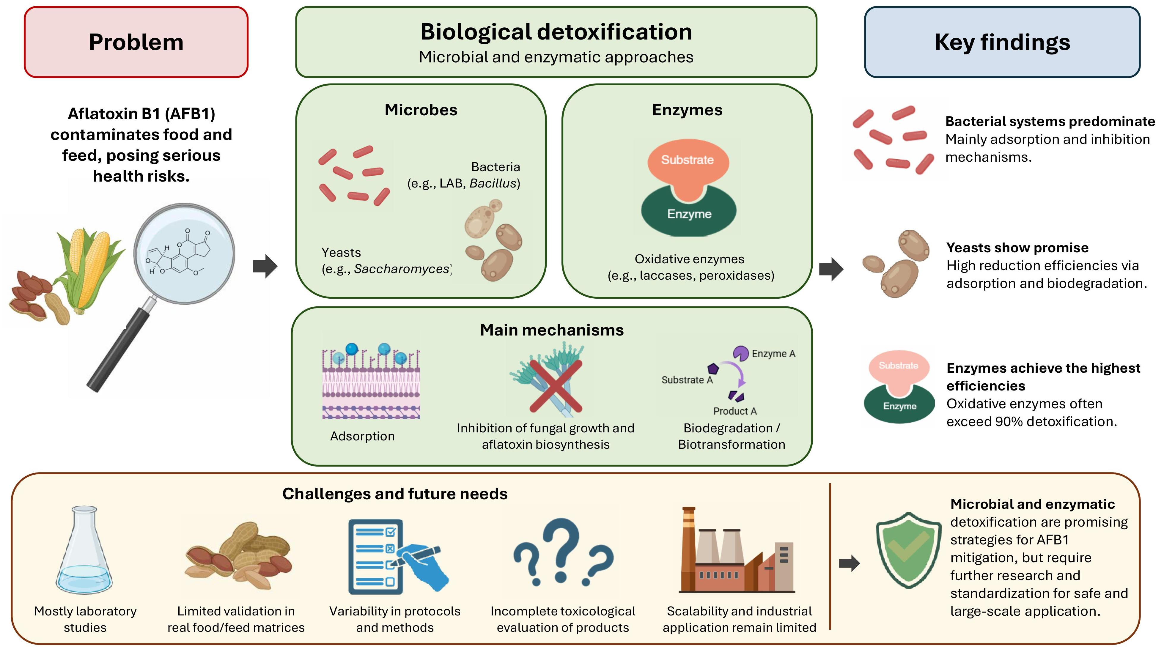
Biological Detoxification of Aflatoxin B1: A Systematic Review of Microbial and Enzymatic Strategies, Mechanisms, and Applications in Food and Feed Systems




Abstract
1. Introduction
2. Results
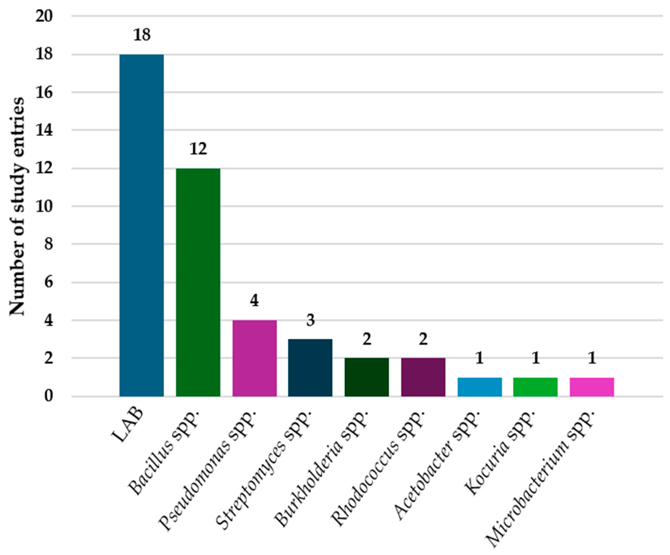
2.1. Bacterial Systems Involved in AFB1 Detoxification by Adsorption, Binding, or Inhibition of Toxin Biosynthesis
2.2. Bacterial and Selected Fungal Systems Involved in AFB1 Biodegradation and Biotransformation
2.3. Yeast-Based Systems Involved in AFB1 Detoxification
2.4. Enzymatic Detoxification of AFB1
2.5. Experimental Application Models for AFB1 Detoxification
3. Discussion
4. Conclusions
5. Materials and Methods
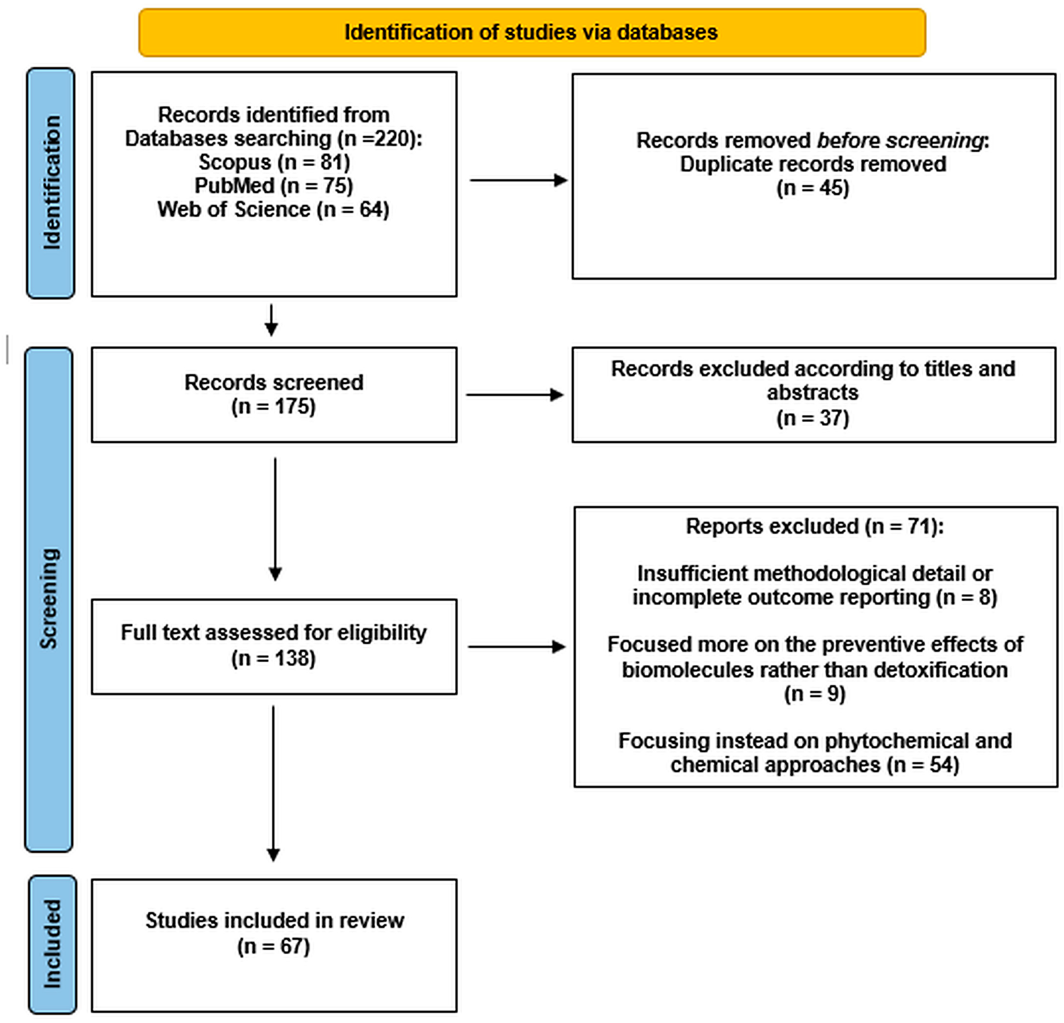
5.1. Search Strategy and Eligibility Criteria
5.2. Systematic Review Process
5.3. Bacterial Nomenclature Standardization
Supplementary Materials
Author Contributions
Funding
Institutional Review Board Statement
Informed Consent Statement
Data Availability Statement
Acknowledgments
Conflicts of Interest
Abbreviations
| AFB1 | Aflatoxin B1 |
| AFB2a | Aflatoxin B2a |
| AFQ1 | aflatoxin Q1 |
| DyP | Dye-decolorizing peroxidase |
| ELISA | Enzyme-linked immunosorbent assay |
| EPS | Exopolysaccharide |
| HPLC | High-performance liquid chromatography |
| HPLC-FLD | High-performance liquid chromatography with fluorescence detection |
| HPLC-HRMS | High-performance liquid chromatography high-resolution mass spectrometry |
| HPLC-MS | High-performance liquid chromatography–mass spectrometry |
| HPLC-QTOF-MS | High-performance liquid chromatography quadrupole time-of-flight mass spectrometry |
| HPTLC | High-performance thin-layer chromatography |
| LC-HRMS | Liquid chromatography high-resolution mass spectrometry |
| LC-MS | Liquid chromatography–mass spectrometry |
| LC-QTOF-MS | Liquid chromatography quadrupole time-of-flight mass spectrometry |
| PRISMA | Preferred Reporting Items for Systematic Reviews and Meta-Analyses |
| UHPLC | Ultra-high-performance liquid chromatography |
| UHPLC-MS/MS | Ultra-high-performance liquid chromatography tandem mass spectrometry |
| UPLC | Ultra-performance liquid chromatography |
| UPLC-QTOF-MS | Ultra-performance liquid chromatography quadrupole time-of-flight mass spectrometry |
References
- Manyes, L.; Font, G. Mycotoxins: Toxicity, Occurrence, Risk Assessment and Prevention. In Encyclopedia of Human Nutrition; Elsevier: Amsterdam, The Netherlands, 2023; pp. 492–500. [Google Scholar]
- Bunny, S.M.; Umar, A.; Bhatti, H.S.; Honey, S.F. Aflatoxin Risk in the Era of Climatic Change-a Comprehensive Review. CABI Agric. Biosci. 2024, 5, 105. [Google Scholar] [CrossRef] [Scilit]
- Marín, S.; Aldars-García, L.; Molino, F.; Ramos, A.J.; Sanchis, V. Aflatoxin B1 Production: A Time–Water Activity–Temperature Model. Fungal Biol. 2024, 128, 2399–2407. [Google Scholar] [CrossRef] [Scilit] [PubMed]
- Leggieri, M.C.; Toscano, P.; Battilani, P. Predicted Aflatoxin B1 Increase in Europe Due to Climate Change: Actions and Reactions at Global Level. Toxins 2021, 13, 292. [Google Scholar] [CrossRef] [Scilit] [PubMed]
- Cao, W.; Yu, P.; Yang, K.; Cao, D. Aflatoxin B1: Metabolism, Toxicology, and Its Involvement in Oxidative Stress and Cancer Development. Toxicol. Mech. Methods 2022, 32, 395–419. [Google Scholar] [CrossRef] [Scilit] [PubMed]
- Ostry, V.; Malir, F.; Toman, J.; Grosse, Y. Mycotoxins as Human Carcinogens—The IARC Monographs Classification. Mycotoxin Res. 2017, 33, 65–73. [Google Scholar] [CrossRef] [Scilit] [PubMed]
- Pożarska, A.; Karpiesiuk, K.; Kozera, W.; Czarnik, U.; Dąbrowski, M.; Zielonka, Ł. AFB1 Toxicity in Human Food and Animal Feed Consumption: A Review of Experimental Treatments and Preventive Measures. Int. J. Mol. Sci. 2024, 25, 5305. [Google Scholar] [CrossRef] [Scilit] [PubMed]
- Syraji, Y.; Jeyaramraja, P.R.; Mada, T.; Gobikanila, K. Comprehensive Review of Aflatoxin Contamination, Its Occurrence, Effects, Management, and Future Perspectives. Discov. Food 2025, 5, 377. [Google Scholar] [CrossRef] [Scilit]
- Zhang, K.; Flannery, B.; Zhang, L. Challenges and Future State for Mycotoxin Analysis: A Review From a Regulatory Perspective. J. Agric. Food Chem. 2024, 72, 8380–8388. [Google Scholar] [CrossRef] [Scilit] [PubMed]
- Song, C.; Yang, J.; Wang, Y.; Ding, G.; Guo, L.; Qin, J. Mechanisms and Transformed Products of Aflatoxin B1 Degradation under Multiple Treatments: A Review. Crit. Rev. Food Sci. Nutr. 2024, 64, 2263–2275. [Google Scholar] [CrossRef] [Scilit] [PubMed]
- Kinyoro, I.S.; Kaale, L. Technologies to Decontaminate Aflatoxins in Foods: A Review. Int. J. Food Sci. Technol. 2024, 59, 6783–6796. [Google Scholar] [CrossRef] [Scilit]
- Ciegler, A.; Lillehoj, E.B.; Peterson, R.E.; Hall, H.H. Microbial Detoxification of Aflatoxin. Appl. Microbiol. 1966, 14, 934–939. [Google Scholar] [CrossRef] [PubMed]
- Detroy, R.W.; Hesseltine, C.W. Isolation and Biological Activity of a Microbial Conversion Product of Aflatoxin B1. Nature 1968, 219, 967. [Google Scholar] [CrossRef] [Scilit] [PubMed]
- Macit, A.; Sevim, S.; Kizil, M. Aflatoxin B1 and M1 Detoxification in Foodstuffs: Examining the Efficacy of Probiotics with and without Prebiotics—A Systematic Review. Food Biosci. 2024, 58, 103724. [Google Scholar] [CrossRef] [Scilit]
- Kumar, V.; Bahuguna, A.; Ramalingam, S.; Dhakal, G.; Shim, J.-J.; Kim, M. Recent Technological Advances in Mechanism, Toxicity, and Food Perspectives of Enzyme-Mediated Aflatoxin Degradation. Crit. Rev. Food Sci. Nutr. 2022, 62, 5395–5412. [Google Scholar] [CrossRef] [Scilit] [PubMed]
- Ouyang, B.; Xu, W.; Ni, D.; Zhang, W.; Ding, J.; Mu, W. Microbial and Enzymatic Strategies for Aflatoxin Control: Integrating Intelligent Detection and Computational Design. Food Chem. 2025, 492, 145584. [Google Scholar] [CrossRef] [Scilit] [PubMed]
- Zhang, A.; Yang, J. A Review of Research Progress on the Microbial or Enzymatic Degradation and Mechanism of Aflatoxin B1. J. Microbiol. Biotechnol. 2025, 35, e2504044. [Google Scholar] [CrossRef] [Scilit] [PubMed]
- Chen, G.; Fang, Q.; Liao, Z.; Xu, C.; Liang, Z.; Liu, T.; Zhong, Q.; Wang, L.; Fang, X.; Wang, J. Detoxification of Aflatoxin B1 by a Potential Probiotic Bacillus amyloliquefaciens WF2020. Front. Microbiol. 2022, 13, 891091. [Google Scholar] [CrossRef] [Scilit] [PubMed]
- Tang, Y.; Liu, X.; Tang, L.; Dong, J. Investigating the Mechanism of Bacillus amyloliquefaciens YUAD7 Degrading Aflatoxin B1 in Alfalfa Silage Using Isotope Tracing and Nuclear Magnetic Resonance Methods. Chem. Biol. Technol. Agric. 2024, 11, 102. [Google Scholar] [CrossRef] [Scilit]
- Tang, Y.; Liu, X.; Dong, L.; He, S. Screening and Identification of an Aflatoxin B(1)-Degrading Strain from the Qinghai-Tibet Plateau and Biodegradation Products Analysis. Front. Microbiol. 2024, 15, 1367297. [Google Scholar] [CrossRef] [Scilit] [PubMed]
- Abdolmaleki, M.; Saki, A.; Alikhani, M. Detoxification of Aflatoxin B1 by Isolating and Screening Bacillus Species from the Gastrointestinal Tract of Broilers. Poult. Sci. J. 2025, 13, 267–276. [Google Scholar] [CrossRef]
- Yuan, S.; Wu, Y.; Jin, J.; Tong, S.; Zhang, L.; Cai, Y. Biocontrol Capabilities of Bacillus subtilis E11 against Aspergillus flavus In Vitro and for Dried Red Chili (Capsicum annuum L.). Toxins 2023, 15, 308. [Google Scholar] [CrossRef] [Scilit] [PubMed]
- Al-Saadi, H.; Al-Sadi, A.; Al-Wahaibi, A.; Al-Raeesi, A.; Al-Kindi, M.; Pandian, S.; Al-Harrasi, M.; Al-Mahmooli, I.; Velazhahan, R. Rice Weevil (Sitophilus oryzae L.) Gut Bacteria Inhibit Growth of Aspergillus flavus and Degrade Aflatoxin B1. J. Fungi 2024, 10, 377. [Google Scholar] [CrossRef] [Scilit] [PubMed]
- Adácsi, C.; Kovács, S.; Pócsi, I.; Pusztahelyi, T. Elimination of Deoxynivalenol, Aflatoxin B1, and Zearalenone by Gram-Positive Microbes (Firmicutes). Toxins 2022, 14, 591. [Google Scholar] [CrossRef] [Scilit] [PubMed]
- Badji, T.; Durand, N.; Bendali, F.; Piro-Metayer, I.; Zinedine, A.; Ben Salah-Abbés, J.; Abbés, S.; Montet, D.; Riba, A.; Brabet, C. In Vitro Detoxification of Aflatoxin B1 and Ochratoxin A by Lactic Acid Bacteria Isolated from Algerian Fermented Foods. Biol. Control 2023, 179, 105181. [Google Scholar] [CrossRef] [Scilit]
- Kosztik, J.; Mörtl, M.; Székács, A.; Kukolya, J.; Bata-Vidács, I. Aflatoxin B1 and Sterigmatocystin Binding Potential of Lactobacilli. Toxins 2020, 12, 756. [Google Scholar] [CrossRef] [Scilit] [PubMed]
- Ondiek, W.; Wang, Y.; Sun, L.; Zhou, L.; On, S.L.W.; Zheng, H.; Ravi, G. Removal of Aflatoxin B1 and T-2 Toxin by Bacteria Isolated from Commercially Available Probiotic Dairy Foods. Food Sci. Technol. Int. 2022, 28, 15–25. [Google Scholar] [CrossRef] [Scilit] [PubMed]
- Møller, C.O.d.A.; Freire, L.; Rosim, R.E.; Margalho, L.P.; Balthazar, C.F.; Franco, L.T.; Sant’Ana, A.d.S.; Corassin, C.H.; Rattray, F.P.; de Oliveira, C.A.F. Effect of Lactic Acid Bacteria Strains on the Growth and Aflatoxin Production Potential of Aspergillus parasiticus, and Their Ability to Bind Aflatoxin B(1), Ochratoxin A, and Zearalenone In Vitro. Front. Microbiol. 2021, 12, 655386. [Google Scholar] [CrossRef] [Scilit] [PubMed]
- Simões, L.; Fernandes, N.; Teixeira, J.; Abrunhosa, L.; Dias, D.R. Brazilian Table Olives: A Source of Lactic Acid Bacteria with Antimycotoxigenic and Antifungal Activity. Toxins 2023, 15, 71. [Google Scholar] [CrossRef] [Scilit] [PubMed]
- Tajik, H.; Sayadi, M. Effects of Probiotic Bacteria of Lactobacillus acidophilus and Lactobacillus casei on Aflatoxin B1 Detoxification within a Simulated Gastrointestinal Tract Model. Toxin Rev. 2022, 41, 92–99. [Google Scholar] [CrossRef] [Scilit]
- Wang, X.; Wang, S.; Xu, J.; Wu, B.; Hu, Z.; Niu, H. Isolation, Characterization, and Biopreservation of Lactobacillus brevis DN-1 to Inhibit Mold and Remove Aflatoxin B1 in Peanut and Sunflower Cakes. Agriculture 2024, 14, 698. [Google Scholar] [CrossRef] [Scilit]
- Abedi, E.; Pourmohammadi, K.; Mousavifard, M.; Sayadi, M. Comparison between Surface Hydrophobicity of Heated and Thermosonicated Cells to Detoxify Aflatoxin B1 by Co-Culture Lactobacillus plantarum and Lactobacillus rhamnosus in Sourdough: Modeling Studies. LWT 2022, 154, 112616. [Google Scholar] [CrossRef] [Scilit]
- Escrivá, L.; Calpe, J.; Lafuente, C.; Moreno, A.; Musto, L.; Meca, G.; Luz, C. Aflatoxin B1 and Ochratoxin A Reduction by Lactobacillus Spp. during Bread Making. J. Sci. Food Agric. 2023, 103, 7095–7103. [Google Scholar] [CrossRef] [Scilit] [PubMed]
- Zolfaghari, H.; Khezerlou, A.; Ehsani, A.; Yari-Khosroushahi, A.Y. Detoxification of Aflatoxin B1 by Probiotic Yeasts and Bacteria Isolated from Dairy Products of Iran. Adv. Pharm. Bull. 2020, 10, 482–487. [Google Scholar] [CrossRef] [Scilit] [PubMed]
- Balsini, M.; Dovom, M.; Kadkhodaee, R.; Najafi, M.; Yavarmanesh, M. Effect of Digestion and Thermal Processing on the Stability of Microbial Cell-Aflatoxin B1 Complex. LWT 2021, 142, 110994. [Google Scholar] [CrossRef] [Scilit]
- Lemmetty, J.; Lee, Y.; Laitila, T.; Bredehorst, S.; Coda, R.; Katina, K.; Maina, N.H. Sequestration of Aflatoxin B1 by Lactic Acid Bacteria: Role of Binding and Biotransformation. Food Res. Int. 2025, 199, 115351. [Google Scholar] [CrossRef] [Scilit] [PubMed]
- Baltazar, C.S.; Brodith, A.M.P.; Catipay, K.E.B.; Montilde, E.G.; Padilla, B.L.G.; Ranches, K.J.R.; Hinay, A.A. Molecular Detection, Phylogenetic Analysis, and Aflatoxin B1 Binding Capacity of Limosilactobacillus fermentum Isolated from Fermented Coconut Toddy. Food Sci. Preserv. 2025, 32, 458–465. [Google Scholar] [CrossRef] [Scilit]
- Bata-Vidács, I.; Kosztik, J.; Mörtl, M.; Székács, A.; Kukolya, J. Aflatoxin B1 and Sterigmatocystin Binding Potential of Non-Lactobacillus LAB Strains. Toxins 2020, 12, 799. [Google Scholar] [CrossRef] [Scilit] [PubMed]
- Yun, J.; Kim, T.; Cho, C.; Lee, J. Antifungal Mechanisms Investigation of Lactic Acid Bacteria against Aspergillus flavus: Through Combining Microbial Metabolomics and Co-Culture System. J. Appl. Microbiol. 2024, 135, lxae112. [Google Scholar] [CrossRef] [Scilit] [PubMed]
- Asurmendi, P.; Gerbaldo, G.; Pascual, L.; Barberis, L. Lactic Acid Bacteria with Promising AFB1 Binding Properties as an Alternative Strategy to Mitigate Contamination on Brewers’ Grains. J. Environ. Sci. Health B 2020, 55, 1002–1008. [Google Scholar] [CrossRef] [Scilit] [PubMed]
- Papp, D.A.; Kocsubé, S.; Farkas, Z.; Szekeres, A.; Vágvölgyi, C.; Hamari, Z.; Varga, M. Aflatoxin B1 Control by Various Pseudomonas Isolates. Toxins 2024, 16, 367. [Google Scholar] [CrossRef] [Scilit] [PubMed]
- Liu, H.; Tang, Y.; Si, W.; Yin, J.; Xu, Y.; Yang, J. Rhodococcus turbidus PD630 Enables Efficient Biodegradation of Aflatoxin B1. LWT 2023, 186, 115225. [Google Scholar] [CrossRef] [Scilit]
- El-Shanshoury, A.E.-R.R.; Metwally, M.A.; El-Sabbagh, S.M.; Emara, H.A.; Saba, H.A.E. Biocontrol of Aspergillus flavus Producing Aflatoxin B1 by Streptomyces Exfoliatus. Egypt. J. Bot. 2022, 62, 457–473. [Google Scholar] [CrossRef] [Scilit]
- Campos-Avelar, I.; de la Noue, A.; Durand, N.; Cazals, G.; Martinez, V.; Strub, C.; Fontana, A.; Schorr-Galindo, S. Aspergillus flavus Growth Inhibition and Aflatoxin B1 Decontamination by Streptomyces Isolates and Their Metabolites. Toxins 2021, 13, 340. [Google Scholar] [CrossRef] [Scilit] [PubMed]
- Kavitake, D.; Singh, S.P.; Kandasamy, S.; Bruntha Devi, P.B.; Shetty, P.H. Report on Aflatoxin-Binding Activity of Galactan Exopolysaccharide Produced by Weissella Confusa KR780676. 3 Biotech 2020, 10, 181. [Google Scholar] [CrossRef] [Scilit] [PubMed]
- Bao, Y.; Dong, T.; Shao, T.; Wang, W.; Varzakas, T.; Agriopoulou, S.; Yuan, X. Antifungal and Mycotoxin Detoxification Potentials of Acetobacter Tropicalis AT7 and Lactiplantibacillus plantarum LP64 in Whole-Plant Corn Silage. Anim. Feed Sci. Technol. 2024, 313, 115987. [Google Scholar] [CrossRef] [Scilit]
- Zhao, W.; Wu, Y.; Wang, H.; Yan, Z. Degradation of Aflatoxin B1 by Recombinant Laccase AnLI from Aspergillus Niger SF951 Expressed in Escherichia Coli BL21: A Mechanism Assessment in Silico and In Vitro. Food Biosci. 2025, 71, 107082. [Google Scholar] [CrossRef] [Scilit]
- Kumar, V.; Bahuguna, A.; Lee, J.; Sood, A.; Han, S.; Chun, H.; Kim, M. Degradation Mechanism of Aflatoxin B1 and Aflatoxin G1 by Salt Tolerant Bacillus albus YUN5 Isolated from “Doenjang”, a Traditional Korean Food. Food Res. Int. 2023, 165, 112479. [Google Scholar] [CrossRef] [Scilit] [PubMed]
- Dong, W.; Liu, M.; Liu, B.; Xiao, Y.; Liu, X.; Yang, M.; Yuan, X.; Li, G.; Meng, K. Isolation of Bacillus licheniformis and Its Protective Effect on Liver Oxidative Stress and Apoptosis Induced by Aflatoxin B1. Poult. Sci. 2024, 103, 104079. [Google Scholar] [CrossRef] [Scilit] [PubMed]
- Hua, L.; Ye, P.; Li, X.; Xu, H.; Lin, F. Anti-Aflatoxigenic Burkholderia contaminans BC11-1 Exhibits Mycotoxin Detoxification, Phosphate Solubilization, and Cytokinin Production. Microorganisms 2024, 12, 1754. [Google Scholar] [CrossRef] [Scilit] [PubMed]
- Yang, X.; Chen, X.; Song, Z.; Zhang, X.; Zhang, J.; Mei, S. Antifungal, Plant Growth-Promoting, and Mycotoxin Detoxication Activities of Burkholderia Sp. Strain XHY-12. 3 Biotech 2020, 10, 158. [Google Scholar] [CrossRef] [Scilit] [PubMed]
- Feng, J.; Cao, L.; Du, X.; Zhang, Y.; Cong, Y.; He, J.; Zhang, W. Biological Detoxification of Aflatoxin B1 by Enterococcus faecium HB2-2. Foods 2024, 13, 1887. [Google Scholar] [CrossRef] [Scilit] [PubMed]
- Wang, J.; Nan, J.; Chen, Q.; Zhou, Y.; Gao, X.; Li, Y. Exploration of Aflatoxin B1 Degradation Products via Kocuria rosea: Structure Elucidation and Toxicity Analysis. Appl. Sci. 2024, 14, 11024. [Google Scholar] [CrossRef] [Scilit]
- Wang, J.; Chen, Q.; Yan, P.; Dong, C.; Shao, Z. Isolation and Optimization of Aflatoxin B1 Degradation by Uniform Design and Complete Genome Sequencing of Novel Deep-Sea Kocuria Rosea Strain 13. Toxins 2023, 15, 520. [Google Scholar] [CrossRef] [Scilit] [PubMed]
- Rafai, S.; Moreno, A.; Cimbalo, A.; Vila-Donat, P.; Manyes, L.; Meca, G. In Vitro Evaluation of Aflatoxin B1 Detoxification by Lactobacillus, Pediococcus, and Bacillus Strains. Toxins 2025, 17, 403. [Google Scholar] [CrossRef] [Scilit] [PubMed]
- Zhang, Y.; Wang, P.; Kong, Q.; Cotty, P.J. Biotransformation of Aflatoxin B1 by Lactobacillus helviticus FAM22155 in Wheat Bran by Solid-State Fermentation. Food Chem. 2021, 341, 128180. [Google Scholar] [CrossRef] [Scilit] [PubMed]
- Yan, Y.; Zhang, X.; Chen, H.; Huang, W.; Jiang, H.; Wang, C.; Xiao, Z.; Zhang, Y.; Xu, J. Isolation and Aflatoxin B1-Degradation Characteristics of a Microbacterium Proteolyticum B204 Strain from Bovine Faeces. Toxins 2022, 14, 525. [Google Scholar] [CrossRef] [Scilit] [PubMed]
- Zapasnik, A.; Bryla, M.; Wojtczak, A.; Sokolowska, B. In Vivo Effectiveness of Pleurotus ostreatus in Degradation of Toxic Metabolites of Filamentous Fungi Such as Aflatoxin B1 and Zearalenone. Metabolites 2025, 15, 20. [Google Scholar] [CrossRef] [Scilit] [PubMed]
- Ali, S.; Hassan, M.; Essam, T.; Ibrahim, M.; Al-Amry, K. Biodegradation of Aflatoxin by Bacterial Species Isolated from Poultry Farms. Toxicon 2021, 195, 7–16. [Google Scholar] [CrossRef] [Scilit] [PubMed]
- Maneeboon, T.; Roopkham, C.; Mahakarnchanakul, W.; Chuaysrinule, C. Exploration of Pseudomonas knackmussii AD02 for the Biological Mitigation of Post-Harvest Aflatoxin Contamination: Characterization and Degradation Mechanism. J. Stored Prod. Res. 2024, 109, 102470. [Google Scholar] [CrossRef] [Scilit]
- Aghamohseni, Z.; Rezaie, S.; Khaniki, G.; Alimohammadi, M.; Alikord, M.; Noorbakhsh, F.; Kouchesfahani, M.; Molaee-aghaee, E. Antifungal Activity and Detoxification by Candida albicans against Aspergillus parasiticus and Aflatoxin Production. J. Consum. Prot. Food Saf. 2022, 17, 377–386. [Google Scholar] [CrossRef] [Scilit]
- Yang, G.; Li, B.; Chen, K.; Du, M.; Zalán, Z.; Hegyi, F.; Kan, J. Isolation and Evaluation of Probiotics from Traditional Chinese Foods for Aflatoxin B1 Detoxification: Geotrichum candidum XG1 (Yeast) and Mechanistic Insights. Food Chem. 2024, 452, 139541. [Google Scholar] [CrossRef] [Scilit] [PubMed]
- Gómez-Albarrán, C.; Melguizo, C.; Patiño, B.; Vázquez, C.; Gil-Serna, J. Diversity of Mycobiota in Spanish Grape Berries and Selection of Hanseniaspora Uvarum U1 to Prevent Mycotoxin Contamination. Toxins 2021, 13, 649. [Google Scholar] [CrossRef] [Scilit] [PubMed]
- Hamad, G.M.; Amer, A.; El-Nogoumy, B.; Ibrahim, M.; Hassan, S.; Siddiqui, S.A.; Elgazzar, A.M.; Khalifa, E.; Omar, S.A.; Abd-Elmohsen Abou-Alella, S.; et al. Evaluation of the Effectiveness of Charcoal, Lactobacillus rhamnosus, and Saccharomyces cerevisiae as Aflatoxin Adsorbents in Chocolate. Toxins 2023, 15, 21. [Google Scholar] [CrossRef] [Scilit] [PubMed]
- Huang, H.; Li, Z.; Qi, Z.; Ma, L.; Hu, G.; Zou, C.; Chen, T. Engineered S. Cerevisiae-pYD1-ScFv-AFB1 Mitigates Aflatoxin B1 Toxicity via Bio-Binding and Intestinal Microenvironment Repair. Food Chem. Toxicol. 2025, 196, 115232. [Google Scholar] [CrossRef] [Scilit] [PubMed]
- Tapingkae, W.; Srinual, O.; Lumsangkul, C.; Doan, H.V.; Chiang, H.-I.; Manowattana, A.; Boonchuay, P.; Chaiyaso, T. Industrial-Scale Production of Mycotoxin Binder from the Red Yeast Sporidiobolus pararoseus KM281507. J. Fungi 2022, 8, 353. [Google Scholar] [CrossRef] [Scilit] [PubMed]
- Zhang, W.; Chang, X.; Han, Y.; Li, T.; Dou, J.; Du, W.; Wu, W.; Wang, W.; Zhang, Z.; Sun, C. Biodegradation of Aflatoxin B1 by a Novel Mined Aldo-Keto Reductase from Meyerozyma guilliermondii AF01. Biol. Control 2025, 200, 105676. [Google Scholar] [CrossRef] [Scilit]
- Zhou, Z.; Li, R.; Ng, T.B.; Huang, F.; Ye, X. Considerations Regarding Affinity Determinants for Aflatoxin B(1) in Binding Cavity of Fungal Laccase Based on in Silico Mutational and In Vitro Verification Studies. Ecotoxicol. Environ. Saf. 2022, 234, 113412. [Google Scholar] [CrossRef] [Scilit] [PubMed]
- Subagia, R.; Schweiger, W.; Kunz-Vekiru, E.; Wolfsberger, D.; Schatzmayr, G.; Ribitsch, D.; Guebitz, G. Detoxification of Aflatoxin B1 by a Bacillus subtilis Spore Coat Protein through Formation of the Main Metabolites AFQ1 and Epi-AFQ1. Front. Microbiol. 2024, 15, 1406707. [Google Scholar] [CrossRef] [Scilit] [PubMed]
- Zhang, C.; Wang, D.; Wang, C.; Yu, H.; Zhong, P.; Dang, W.; Yang, Y.; Wang, Y.; Yan, X. Developing a Ni-Grafted Magnetic Nanoparticle for Direct CotA Capture in Rapid Detoxification of Aflatoxin B1. J. Hazard. Mater. 2025, 485, 136829. [Google Scholar] [CrossRef] [Scilit] [PubMed]
- Mangini, V.; Rosini, E.; Caliandro, R.; Mangiatordi, G.F.; Delre, P.; Sciancalepore, A.G.; Pollegioni, L.; Haidukowski, M.; Mazzorana, M.; Sumarah, M.W.; et al. DypB Peroxidase for Aflatoxin Removal: New Insights into the Toxin Degradation Process. Chemosphere 2024, 349, 140826. [Google Scholar] [CrossRef] [Scilit] [PubMed]
- Bian, L.; Chang, T.; Zhang, J.; Xu, Y.; Wang, T.; Zhu, X.; Zhang, C. Engineering of Bacillus laccase frL103 for Highly Efficient Degradation of Aflatoxin B1. J. Agric. Food Chem. 2025, 73, 14641–14650. [Google Scholar] [CrossRef] [Scilit] [PubMed]
- Qin, X.; Su, X.; Tu, T.; Zhang, J.; Wang, X.; Wang, Y.; Wang, Y.; Bai, Y.; Yao, B.; Luo, H.; et al. Enzymatic Degradation of Multiple Major Mycotoxins by Dye-Decolorizing Peroxidase from Bacillus subtilis. Toxins 2021, 13, 429. [Google Scholar] [CrossRef] [Scilit] [PubMed]
- Zaccaria, M.; Dawson, W.; Russel Kish, D.; Reverberi, M.; Bonaccorsi di Patti, M.C.; Domin, M.; Cristiglio, V.; Chan, B.; Dellafiora, L.; Gabel, F.; et al. Experimental-Theoretical Study of Laccase as a Detoxifier of Aflatoxins. Sci. Rep. 2023, 13, 860. [Google Scholar] [CrossRef] [Scilit] [PubMed]
- Al-Rajhi, A.; Ganash, M.; Alshammari, A.; Alsalamah, S.; Abdelghany, T. In Vitro and Molecular Docking Evaluation of Target Proteins of Lipase and Protease for the Degradation of Aflatoxins. Bioresources 2024, 19, 2701–2713. [Google Scholar] [CrossRef] [Scilit]
- Hao, W.-B.; Gu, X.; Yu, X.; Zhao, Y.; Li, C.; Jia, M.; Du, X.-D. Laccase Lac-W Detoxifies Aflatoxin B1 and Degrades Five Other Major Mycotoxins in the Absence of Redox Mediators. Environ. Pollut. 2023, 338, 122581. [Google Scholar] [CrossRef] [Scilit] [PubMed]
- Liu, Y.; Mao, H.; Hu, C.; Tron, T.; Lin, J.; Wang, J.; Sun, B. Molecular Docking Studies and In Vitro Degradation of Four Aflatoxins (AFB1, AFB2, AFG1, and AFG2) by a Recombinant Laccase from Saccharomyces cerevisiae. J. Food Sci. 2020, 85, 1353–1360. [Google Scholar] [CrossRef] [Scilit] [PubMed]
- Rasheed, U.; Ul Ain, Q.; Ali, A.; Liu, B. One Stone Two Birds: Recycling of an Agri-Waste to Synthesize Laccase-Immobilized Hierarchically Porous Magnetic Biochar for Efficient Degradation of Aflatoxin B1 in Aqueous Solutions and Corn Oil. Int. J. Biol. Macromol. 2024, 273, 133115. [Google Scholar] [CrossRef] [Scilit] [PubMed]
- Shao, H.; Su, X.; Wang, Y.; Zhang, J.; Tu, T.; Wang, X.; Huang, H.; Yao, B.; Luo, H.; Qin, X. Oxidative Degradation and Detoxification of Multiple Mycotoxins Using a Dye-Decolorizing Peroxidase from the White-Rot Fungus Bjerkandera adusta. LWTY 2024, 206, 116597. [Google Scholar] [CrossRef] [Scilit]
- Wang, Y.; Zhang, Q.; Pei, J.; Su, Y.; Adegoke, T.V.; Wang, Y. Rational Screening of Four Peroxidases with High Aflatoxin B1 Degradation Efficiency via Integrated Computational Simulations. J. Agric. Food Chem. 2025, 73, 24354–24368. [Google Scholar] [CrossRef] [Scilit] [PubMed]
- Hu, S.; Xu, C.; Lu, P.; Wu, M.; Chen, A.; Zhang, M.; Xie, Y.; Han, G. Widespread Distribution of the DyP-Carrying Bacteria Involved in the Aflatoxin B1 Biotransformation in Proteobacteria and Actinobacteria. J. Hazard. Mater. 2024, 478, 135493. [Google Scholar] [CrossRef] [Scilit] [PubMed]
- Zahra, N.; Jamil, N.; Ahmad, S.; Rahman, S.; Ullah, N.; Sarwar, A.; ul Haq, T.; Khan, A.; Alwutayd, K.; Al-Asmari, F.; et al. Elucidating Aflatoxins Profile and Recommended Detoxification Procedures in Corn, Rice, and Wheat. Ital. J. Food Sci. 2025, 37, 478–487. [Google Scholar] [CrossRef] [Scilit]
- Ouyang, W.; Liao, Z.; Yang, X.; Zhang, X.; Zhu, X.; Zhong, Q.; Wang, L.; Fang, X.; Wang, J. Microbial Composition of Water Kefir Grains and Their Application for the Detoxification of Aflatoxin B1. Toxins 2024, 16, 107. [Google Scholar] [CrossRef] [Scilit] [PubMed]
- Ouyang, B.; Zhang, W.; Guang, C.; Xu, W.; Mu, W. Opportunities and Challenges in Developing Promising Mycotoxin-Degrading Enzymes with High Thermostability and Strong pH Activity/Stability. Food Biosci. 2024, 62, 105239. [Google Scholar] [CrossRef] [Scilit]
- Wang, Y.; Chen, Y.; Jiang, L.; Huang, H. Improvement of the Enzymatic Detoxification Activity towards Mycotoxins through Structure-Based Engineering. Biotechnol. Adv. 2022, 56, 107927. [Google Scholar] [CrossRef] [Scilit] [PubMed]
- Aasa, A.; Govender, S.; Malgas, S.; Thantsha, M. Microbial and Enzymatic Biodegradation of Aflatoxins and Ochratoxins: Mechanisms, Applications, and Emerging Innovations. Arch. Microbiol. 2026, 208, 144. [Google Scholar] [CrossRef] [Scilit] [PubMed]
- Page, M.J.; McKenzie, J.E.; Bossuyt, P.M.; Boutron, I.; Hoffmann, T.C.; Mulrow, C.D.; Shamseer, L.; Tetzlaff, J.M.; Akl, E.A.; Brennan, S.E.; et al. The PRISMA 2020 Statement: An Updated Guideline for Reporting Systematic Reviews. BMJ 2021, 372, n71. [Google Scholar] [CrossRef] [Scilit] [PubMed]



| (a) | |||||||
| Organism (Strain) | Source/Origin | Experimental Conditions | Key Results | Mechanism of Detoxification | Metabolites Detected | Analytical Quantification Method | Reference |
| Bacillus amyloliquefaciens WF2020 | Naturally fermented pickles | LB liquid culture with AFB1 (1–8 µg/mL), 37–45 °C, 72–96 h | Complete inhibition of AFB1 production in co-culture | Inhibition of Aspergillus flavus growth and aflatoxin biosynthesis | - | HPLC; HPLC–QTOF-MS | (Chen et al., 2022) [18] |
| Bacillus amyloliquefaciens YUAD7 | Yak manure | Co-culture in PDB with Aspergillus flavus conidia (1 × 10⁸ conidia/mL) and bacterial cells (1 × 10⁷ CFU/mL), 30 °C, 48 h, 200 rpm | Significant inhibition of A. flavus growth and total inhibition of AFB1 production compared with control | Inhibition of A. flavus growth and AFB1 biosynthesis | - | UPLC–Q-TOF/MS | (Tang et al., 2024) [19] |
| Bacillus amyloliquefaciens YUAD7 | Yak manure, Qinghai–Tibet Plateau | Liquid culture with AFB1 (10 µg/mL), 37 °C, 72 h | AFB1 detoxification reached 91.7% in liquid medium and >85% in food matrices | Binding and degradation via extracellular excretions | C12H14O4, C5H12N2O2, C10H14O2, C4H12N2O | UPLC-Q-Orbitrap HRMS, NMR | (Tang et al., 2024) [20] |
| Bacillus sp. MA82 | Broiler gastrointestinal tract and fecal samples | Whole-cell binding assay in PBS with AFB1 (5 µg/mL), 37 °C, 0–24 h | AFB1 adsorption increased from 45% at 0 h to 75% after 24 h | Cell wall adsorption/binding | - | HPLC | (Abdolmaleki et al., 2025) [21] |
| Bacillus subtilis (E11; compared with V1J1 and 9932) | Isolated from fermented foods (China) | In vitro confrontation with A. flavus; liquid culture assays in LB, PDA, and PDB media, 28–37 °C, 24–120 h | Growth inhibition of A. flavus (~64%); AFB1 removal up to 81.34% after 24 h | Inhibition of fungal growth; inhibition of AFB1 biosynthesis | - | ELISA | (Yuan et al., 2023) [22] |
| Bacillus subtilis RWGB1, Bacillus oceanisediminis RWGB2, Bacillus firmus RWGB3, Pseudomonas aeruginosa RWGB4 | Rice weevil gut | In vitro incubation with AFB1, 30 °C, 72 h | AFB1 detoxification ranged from 48.9% to 84.2%, depending on strain | Binding to AFB1 | - | LC-MS | (Al-Saadi et al., 2024) [23] |
| Bacillus thuringiensis AMK10/1; Lysinibacillus boronitolerans AMK9/1; Lysinibacillus fusiformis AMK10/2; Rummeliibacillus suwonensis AMK9/2 | Fermented forages (Hungary) | PBS system with AFB1 (24 µg/L), viable cells or cell wall fractions, 25 °C, 1 h | AFB1 elimination was generally <20%; maximum removal reached 64% with the S-layer fraction of B. thuringiensis AMK10/1 | Cell wall adsorption mediated by S-layer proteins | - | HPLC-FLD | (Adácsi et al., 2022) [24] |
| Enterococcus faecium (Lab-L4/al), Enterococcus durans (Lab-L1), Lactiplantibacillus plantarum R1096 | Algerian fermented wheat (El-Hammoum) and fermented milk | CPB buffer with AFB1 (40 ng/mL), pH 5–6, 1010 CFU/mL, viable or heat-inactivated cells, 25 °C, 24 h | AFB1 adsorption ranged from 25% to 80%, with highest activity observed for nonviable cells | Cell wall adsorption | - | HPLC-FLD | (Badji et al., 2023) [25] |
| Lactiplantibacillus pentosus TV3 | Animal fecal samples | Cell suspension (108 CFU/mL) with AFB1, 37 °C, 10 min | AFB1 adsorption reached 11.5% | Binding- Cell wall adsorption | - | HPLC-UV | (Kosztik et al., 2020) [26] |
| Lactic acid bacteria | probiotic dairy foods (fermented milk drinks, probiotic yogurt) | Incubation with AFB1, pH 5.5, 37 °C, 72 h | AFB1 detoxification reached 46% with live cells and 62% with denatured cells | Adsorption to bacterial cell walls | AFB1-8,9-dihydrodiol | LC-MS/MS | (Ondiek et al., 2022) [27] |
| Lactic acid bacteria | Dairy and food-associated lactic acid bacteria collections | Co-culture with Aspergillus parasiticus NRRL 2999 in YES medium, 25 °C, 7 days; adsorption assay in phosphate buffer with AFB1 (1 µg/mL) | Inhibition of AFB1 production reached 100% depending on strain; AFB1 adsorption ranged from 40% to 70% | Inhibition of fungal growth and aflatoxin biosynthesis; cell wall adsorption | - | HPLC–FLD | (Møller et al., 2021) [28] |
| Lacticaseibacillus paracasei subsp. paracasei CCMA 1764; Levilactobacillus brevis CCMA 1762; Lactiplantibacillus pentosus CCMA 1768 | Naturally fermented Brazilian table olives | Dual-culture overlay assay and cell-free supernatant assay against Aspergillus flavus; CFS tested at 300–500 µL/mL, 25 °C, 7 days; additional MRS broth assays for 12 days | Complete inhibition of A. flavus growth; 100% AFB1 detoxification at 500 µL/mL CFS; AFB1 adsorption reached 48–51% | Growth inhibition of Aspergillus spp.; cell wall adsorption | Aflatoxin B2a | HPLC–FLD for AFB1 quantification UHPLC for AFB2a confirmation | (Simões et al., 2023) [29] |
| Lactobacillus acidophilus ATCC 4356 Lacticaseibacillus casei ATCC 39392 | Probiotic reference strain | Simulated gastrointestinal model with AFB1 (5 µg/mL); oral, gastric (pH 2.5), and intestinal (pH 7.5) phases, ~4 h | AFB1 detoxification ranged from 24.3% to 70.0% | Cell wall adsorption | - | HPLC–FLD | (Tajik and Sayadi, 2020) [30] |
| Levilactobacillus brevis DN-1 | Moldy feed samples | MRS medium with AFB1 (5 µg/L), anaerobic incubation, 37 °C, 24–48 h | AFB1 reduction 71.38% in liquid culture | Cell wall adsorption; inhibition of Aspergillus growth and AFB1 biosynthesis | - | HPLC-FLD | (Wang et al., 2024) [31] |
| Lactiplantibacillus plantarum ATCC 8014 Lacticaseibacillus rhamnosus ATCC 7469 | Iranian Research Organization for Science and Technology | AFB1-contaminated sourdough (10 µg/kg), thermosonicated co-culture, 27–37 °C, 8–24 h | Maximum AFB1 adsorption reached 8.04 µg/kg under thermosonicated co-culture conditions (37 °C, 24 h) | Cell wall adsorption mainly driven by hydrophobic interactions | - | HPLC-FLD | (Abedi et al., 2022) [32] |
| Lactiplantibacillus plantarum B3, Lacticaseibacillus paracasei B10 | Goat milk whey | MRS broth with AFB1, 37 °C, 72 h | AFB1 detoxification ranged from 27% to 55% | Cell wall adsorption | HPLC-QTOF-MS | (Escrivá et al., 2023) [33] | |
| Lacticaseibacillus rhamnosus | Traditional dairy products | Simulated gastrointestinal model with AFB1 (10 ppb), 1 × 10¹⁰ CFU/mL, gastric phase pH 2.5, intestinal phase pH 7.5, 37 °C, 4 h | AFB1 detoxification reached 31.14% | Cell wall adsorption | - | ELISA | (Zolfaghari et al., 2020) [34] |
| Lacticaseibacillus rhamnosus GG | Commercial probiotic | PBS system with AFB1 (1 µg/mL), 2 × 10⁹ CFU, viable or heat-treated cells, NaCl 0–1.8%, 25–37 °C, 1–2 h | Maximum AFB1 adsorption reached 97.74% with heat-treated cells (1% NaCl, 1 h); desorption remained ≤3% after in vitro digestion | Non-covalent cell wall adsorption | - | HPLC–FLD | (Balsini et al., 2021) [35] |
| Levilactobacillus brevis, Lactobacillus helveticus, Lactoplantibacillus plantarum, Leuconostoc pseudomesenteroides, Weissella confusa, Weissella cibaria | Food-derived strains (maize porridge, dairy products) | Incubation with AFB1 at pH 3 or pH 7, 25–37 °C, 24–48 h | AFB1 detoxification ranged from 16% to 71%, depending on strain | Surface adsorption to lactic acid bacteria cells | - | LC-QTOF-MS, UPLC-FLD | (Lemmetty et al., 2025) [36] |
| Limosilactobacillus fermentum | Fermented coconut toddy (“tuba”), Philippines | Incubation with AFB1 (1.64 ppb), live or heat-treated cells, 37 °C, 1 h | AFB1 adsorption reached 87.30 ± 7.29% with live cells and 70.49 ± 9.59% with heat-treated cells | Cell wall adsorption | - | ELISA | (Baltazar et al., 2025) [37] |
| Pediococcus acidilactici OR83 | Animal fecal samples | Cell suspension (108 CFU/mL) with AFB1, 37 °C, 10 min | AFB1 adsorption reached 7.6% | Cell wall adsorption | - | HPLC-UV | (Bata-Vidács et al., 2020) [38] |
| Pediococcus pentosaceus (N17-02, N19-37, N57-15, N57-24) Weissella paramesenteroides (N33-01, N44-02); Companilactobacillus crustorum RL48-10 | Korean Nuruk (fermentation starter) | Transwell co-culture with Aspergillus flavus, 25 °C, 3–8 days | Significant inhibition of AFB1 production | Inhibition of AFB1 biosynthesis; cell wall adsorption; production of antifungal metabolites | Lactic acid, 4-hydroxybenzaldehyde, adenine, 2,3-cAMP | LC-QTOF-MS, LC-MS/MS | (Yun et al., 2024) [39] |
| Pediococcus pentosaceus L6; Lactiplantibacillus plantarum L12; Leuconostoc mesenteroides L18, L19; Loigolactobacillus coryniformis subsp. coryniformis L47; Levilactobacillus brevis L52 | Brewer’s grains (Argentina) | PBS system with AFB1 (150 ng/mL), viable or heat-treated cells, 37 °C, 4 h | AFB1 adsorption ranged from 37.6% to 70.7% | Cell wall adsorption | - | HPLC-FLD | (Asurmendi et al., 2020) [40] |
| Pseudomonas (Ps-4, 66, 68) | Corn rhizosphere | Solid co-culture with Aspergillus flavus, 25 °C, 7 days; liquid co-culture, 28 °C, 3 days, 130 rpm | Complete inhibition of AFB1 production (>99%) | Inhibition of AFB1 biosynthesis | - | HPLC-HRMS | (Papp et al., 2024) [41] |
| Rhodococcus turbidus PD630 | Soil | Co-culture system with AFB1, 30 °C, 72 h | AFB1 detoxification reached 93.04% after 72 h | Cell wall adsorption | - | HPLC, UV | (Liu et al., 2023) [42] |
| Streptomyces exfoliatus Agricultural soils (Egypt) | Agricultural soils (Egypt) | Cell-free culture filtrate produced in starch nitrate broth, 30 °C, 7 days; tested against Aspergillus flavus and AFB1 | Complete inhibition of AFB1 production at ≥20% (v/v) culture filtrate | Inhibition of fungal growth and sporulation; inhibition of aflatoxin B1 biosynthesis | - | Thin-layer chromatography with fluorodensitometric quantification | (El-Shanshoury et al., 2022) [43] |
| Streptomyces spp. (59 soil isolates including IX20, IX45) and Streptomyces griseoviridis (Mycostop®) | Organic amendments and soil samples | Dual culture with Aspergillus flavus and AFB1 degradation assays in solid (CYA) and liquid (CYB) media, 25 °C | Strong inhibition of A. flavus growth; marked reduction in AFB1 accumulation; residual AFB1 decreased to 6% for selected isolates | Inhibition of fungal growth and AFB1 biosynthesis | - | HPLC–MS | (Campos-Avelar et al., 2021) [44] |
| Weissella confusa KR780676 | Indian traditional fermented food | Exopolysaccharide (EPS) extracted from MRS broth, 30 °C, 48 h | AFB1 detoxification reached 32.40% at 50 mg/mL and 34.79% at 100 mg/mL | Binding to galactan exopolysaccharide | - | HPTLC, PSA | (Kavitake et al., 2020) [45] |
| (b) | |||||||
| Organism (Strain) | Source/Origin | Experimental Conditions | Key Results | Mechanism of Detoxification | Metabolites Detected | Analytical Quantification Method | Reference |
| Acetobacter tropicalis AT7/and Lactiplantibacillus plantarum LP64 | Mold-contaminated silages | Incubation with AFB1 (50 µg/L), 37 °C, 72 h | AFB1 detoxification reached 47.8% and 57.0%, respectively | Biodegradation | - | UPLC–MS/MS | (Bao et al., 2024) [46] |
| Aspergillus niger SF951 | Cultured microbiome associated with Salvia miltiorrhiza (strain screened by metagenomic laccase-gene mining) | Liquid incubation with AFB1 (0.5 µg/mL), 37 °C, 3 days | AFB1 detoxification reached 55.67% | Biodegradation mainly by extracellular fractions | C16H22O4, C16H35O2N, C24H30O6, C18H39O2N | HPLC–MS (AFB1 quantification); UHPLC–MS/MS | (Zhao et al., 2025) [47] |
| Bacillus albus YUN5 | Doenjang (Korean fermented soybean paste) | Cell-free supernatant with AFB1 (100 ng/mL), 35 °C, 7 days | AFB1 detoxification reached 76.28% | Biodegradation mediated by non-proteinaceous metabolites present in the supernatant | Six degradation products (m/z 316, 286, 361, 317, 302, 244) | HPLC-FLD; LC–MS | (Kumar et al., 2023) [48] |
| Bacillus amyloliquefaciens WF2020 | Naturally fermented pickles | LB liquid culture with AFB1 (1–8 µg/mL), 37–45 °C, 72–96 h | AFB1 detoxification exceeded 80% after 72 h | Extracellular enzyme-mediated biodegradation | C15H11O, C15H15O2, C15H19O4 | HPLC; HPLC–QTOF-MS | (Chen et al., 2022) [18] |
| Bacillus amyloliquefaciens YUAD7 | Yak manure | Artificially contaminated alfalfa silage with AFB1 (100 µg/kg), inoculated at 108 CFU/mL, 20 °C, 48 days | AFB1 detoxification reached 99.7%, with a final concentration of 1.7 µg/kg | Biodegradation | C12H14O4, C5H12N2O2, C10H14O2, C4H12N2O, | UPLC–Q-TOF/MS | (Tang et al., 2024) [19] |
| Bacillus licheniformis QT338 (strain S51) | Chicken intestine | Liquid culture with AFB1 (100 ng/mL), 30 °C, 72 h | AFB1 detoxification reached 61.03% | Biodegradation | - | ELISA | (Dong et al., 2024) [49] |
| Burkholderia contaminans BC11-1 | Forest rhizosphere soil, Luzhou, China | Incubation at 28 °C, 7 days, physically separated from AFB1 by a 0.22 µm membrane filter | AFB1 detoxification reached 90% without direct contact | Extracellular metabolite-mediated biodegradation | - | ELISA | (Hua et al., 2024) [50] |
| Burkholderia sp. XHY-12 | Corn soil | Liquid fermentation with AFB1 (2.5 µg/mL), 37 °C, 60 h | AFB1 detoxification reached 85.2% | Biodegradation by extracellular enzymes | - | HPLC | (Yang et al., 2020) [51] |
| Enterococcus faecium HB2-2 | Soil (China) | Nutrient broth with AFB1 under optimized alkaline conditions, 32 °C, up to 96 h | AFB1 detoxification reached 90.0%; fermentation supernatant showed highest activity | Biodegradation mediated mainly by extracellular proteinaceous components | Degradation products with(m/z 331, 287, and 249) | HPLC with fluorescence detection; LC–MS | (Feng et al., 2024) [52] |
| Kocuria rosea (strain 13) | Deep-sea origin | Liquid culture in M2 medium; AFB1 incubation up to 5 days | Efficient AFB1 degradation with reduced cytotoxicity of degradation products | Biotransformation | Degradation products including aflatoxicol, aflatoxin D1 and aflatoxin D2 | HPLC–HRMS | (Wang et al., 2024) [53] |
| Kocuria rosea strain 13 | Deep-sea environment (West Pacific Ocean) | Liquid culture with AFB1; optimized conditions: 30 °C, 2 days, pH 7.11, seawater 100% | AFB1 detoxification reached 88.0% | Biodegradation | - | HPLC-UV | (Wang et al., 2023) [54] |
| Latilactobacillus curvatus 14; Pediococcus pentosaceus 4; Bacillus firmus 6 | Culture collection (CECT, Spain) | Fermentation of contaminated maize flour extract, 30–37 °C, 12–48 h | AFB1 reduction up to 41.1% (L. curvatus 14), 25.4% (P. pentosaceus 4), 25.1% (B. firmus 6) | Biotransformation | Aflatoxicol; aflatoxin D1; aflatoxin P2; aflatoxin Q1; aflatoxin B2a | LC–QTOF–MS | (Rafai et al., 2025) [55] |
| Lactobacillus helveticus FAM22155 | Laboratory culture collection (China) | Solid-state fermentation of wheat bran contaminated with AFB1, 37 °C, 48 h, compared with liquid fermentation | AFB1 detoxification reached 86–89% during solid-state fermentation | Protein-mediated biotransformation during solid-state fermentation | AFP1, AFP2, AFP3 and AFP4 | ELISA for aflatoxin B1 quantification; UHPLC–QTOF–MS for degradation product analysis | (Zhang et al., 2021) [56] |
| Microbacterium proteolyticum B204 | Bovine faeces | Liquid culture with AFB1 (10 µg/mL), 30 °C, 24 h; whole culture and cell-free supernatant evaluated | AFB1 detoxification reached 77.0% in whole culture and 80.1% in supernatant | Extracellular proteinaceous enzyme-mediated biodegradation | - | HPLC-FLD | (Yan et al., 2022) [57] |
| Pleurotus ostreatus | Commercial mushroom strain | Cultivation on contaminated substrate (commercial substrate + maize, 1:1), 25 °C, 3 weeks of mycelial growth | AFB1 detoxification ranged from 53% to 87% | Biodegradation | - | HPLC–FLD | (Zapaśnik et al., 2025) [58] |
| Pseudomonas fluorescens (SZ1) | Soil, (Egypt) | Nutrient broth supplemented with AFB1 (100 ppb), 37 °C, 72 h | AFB1 detoxification reached up to 99% | Extracellular proteinaceous component-mediated biodegradation | - | HPLC with fluorescence detection | (Ali et al., 2021) [59] |
| Pseudomonas knackmussii AD02 | Peanut-growing soil (Thailand) | Nutrient broth with AFB1 (100 ng/mL), pH 7.0, 25 °C, 24 h | AFB1 detoxification ranged from 88.85% to 90.0% across 20–500 ng/mL | Extracellular enzyme-mediated biodegradation | - | HPLC-FLD | (Maneeboon et al., 2024) [60] |
| Organism (Strain) | Source/Origin | Experimental Conditions | Key Results | Mechanism of Detoxification | Metabolites Detected | Analytical Quantification Method | Reference |
|---|---|---|---|---|---|---|---|
| Candida albicans ATCC14053 | - | Wheat grains asynchronously inoculated with yeast (107 CFU/mL) and Aspergillus parasiticus spores (104 spores/g), 28–30 °C, 12 days | Strong inhibition of A. parasiticus growth; AFB1 detoxification reached 75.55% in wheat grains | Inhibition of AFB1 biosynthesis | - | HPLC-FLD | (Aghamohseni et al., 2022) [61] |
| Geotrichum candidum XG1 | Traditional Chinese fermented foods | Liquid culture with AFB1, 30 °C, 48 h | AFB1 detoxification reached 99.1–100% | Biodegradation | C17H14O8, C17H16O8 | HPLC-FLD; UPLC-QTOF-MS/MS | (Yang et al., 2024) [62] |
| Hanseniaspora uvarum U1 | Spanish grape berries | PDB medium with AFB1 (1 µg/L), viable or heat-inactivated cells, pH 3.0–7.0, 30 °C, 48 h | AFB1 detoxification ranged from 93.7% to 99.1%, with maximum activity at pH 5.5 | Cell wall adsorption; possible active degradation | - | ELISA | (Gómez-Albarrán et al., 2021) [63] |
| Saccharomyces cerevisiae | Traditional dairy products | Simulated gastrointestinal model with AFB1 (10 ppb), 2 × 10⁸ cells/mL, 37 °C | AFB1 detoxification reached 30.46% | Cell wall adsorption | - | ELISA | (Zolfaghari et al., 2020) [34] |
| Saccharomyces cerevisiae | Commercial strains | In vitro PBS model simulating gastrointestinal conditions, pH 3.0 and 6.8, 2–4 h | AFB1 adsorption ranged from 52% to 99.7%, with highest activity in tri-mix formulation | Cell wall adsorption | - | HPLC | (Hamad et al., 2023) [64] |
| Saccharomyces cerevisiae-pYD1-ScFv-AFB1 (engineered strain) | Engineered from S. cerevisiae ATCC 9763 | In vitro binding assay, in vivo mouse exposure model with AFB1 (0.3 mg/kg/day) for 4 weeks; oral administration of 1 × 10⁹ CFU/day | AFB1 binding capacity was 1.7-fold higher than wild-type yeast, fecal AFB1 excretion increased | Specific bio-binding mediated by surface-displayed anti-AFB1 single-chain antibody | - | HPLC (feces) | (Huang et al., 2025) [65] |
| Sporidiobolus pararoseus KM281507 | Red yeast cells (spray-dried, encapsulated) | Incubation with AFB1 (1–5 µg/mL), 25–37 °C, 48 h, anaerobic conditions | AFB1 detoxification reached up to 93% at low dose under poultry gastrointestinal model conditions | Cell wall adsorption mediated by β-glucan-rich biomass | - | ELISA, HPLC-FLD | (Tapingkae et al., 2022) [66] |
| Enzyme Name | Source/Origin | Reaction Conditions | Time/Dose | % AFB1 Reduction | Detected Metabolites | Analytical Method | Reference |
|---|---|---|---|---|---|---|---|
| Aldo–keto reductase (MgAKR, gene MG2-4) | Meyerozyma guilliermondii AF01 | In vitro phosphate buffer system (pH 5.0–7.0), 30–37 °C, NADPH-dependent | 1350–1620 µg/mL; NADPH up to 4.8 mM, 72 h | AFB1 detoxification exceeded 90% under optimal conditions | Aflatoxicol | LC–MS | (Zhang et al., 2025) [67] |
| Recombinant fungal laccase (rCuL) | Cerrena unicolor 6884 | Buffer system at pH 7.0–8.0, 45–65 °C, initial AFB1 concentration 2.0 µg/mL | 1 U/mL purified enzyme, 24 h | AFB1 detoxification reached 94% | - | (Zhou et al., 2022) [68] | |
| Recombinant laccase rAnLI | Laccase gene AnLI from A. niger SF951 purified enzyme | pH 5.0, 35 °C, 1 mM Cu2+, AFB1 (1 µg/mL) | 0.1 µg/mL enzyme, 48 h | AFB1 detoxification reached 94.72% | C16H22O4, C16H35O2N, C24H30O6, C18H39O2N | UHPLC–MS/MS | (Zhao et al., 2025) [47] |
| CotA laccase (BsCotA, recombinant) | Bacillus subtilis spore coat protein | 100 mM phosphate buffer (pH 7.0), 37 °C, Cu2+ incorporated, no mediator | 0.2 µM, 48–72 h | AFB1 detoxification reached ~80% within 48 h | AFQ1, epi-AFQ1 | LC-MS/MS | (Subagia et al., 2024) [69] |
| CotA laccase (free form) | Bacillus subtilis (recombinant expression in E. coli) | PBS buffer (pH 7.4–8.0), 50–70 °C | 20 µg, 1–4 h | AFB1 detoxification reached ~74.4% | AFQ1; AP347; AP331; AP317; AP301; AP259; AP235; AP223; AP155; AP141 | UPLC–QTOF–MS | (Zhang et al., 2025) [70] |
| DypB (WT) | Rhodococcus jostii; recombinant expression in E. coli | Sodium malonate buffer (pH 6.0), 0.1 mM H2O2, 2 mM Mn2+, 25 °C | 0.1 U/mL enzyme, up to 72 h | AFB1 detoxification reached 95 after 72 h | C17H14O6, C16H14O6, C16H14O7, C17H14O7 | LC–HRMS | (Mangini et al., 2024) [71] |
| Laccase frL103 (wild type; T418A, T418S) | Bacillus vallismortis; recombinant expression in E. coli | Tris–HCl buffer (50 mM, pH 7.0), 30 °C, 300 rpm | 2.5 µM, 24 h | AFB1 detoxification ranged from 45.7% to 56.7% | - | - | (Bian et al., 2025) [72] |
| Dye-decolorizing peroxidase (BsDyP) | Bacillus subtilis SCK6; recombinant expression in Escherichia coli | Malonate buffer (50 mM, pH 4.0), 30 °C | 1.25 U/mL, 48 h | AFB1 detoxification ranged from 50.0% to 76.93% | AFB1-diol | LC–MS/MS | (Qin et al., 2021) [73] |
| Laccase | Trametes versicolor (commercial enzyme) | Acetate buffer (100 mM, pH 6.5), 28 °C, shaking; no mediator | 25 U/mL; up to 96 h | AFB1 reduction of approximately 12% after 96 h | Ring-opened AFB1 products, epoxide and dihydroxylated derivatives | LC-MS | (Zaccaria et al., 2023) [74] |
| Lipase /Protease (commercial) | Humicola lanuginosa | Fungal culture filtrate containing aflatoxins, 30 °C | 25–200 U/mL, 12 h | AFB1 detoxification ranged from 35.8% to 81.3% | - | ELISA | (Al-Rajhi et al., 2024) [75] |
| Lac-W (laccase, multicopper oxidase) | Weizmannia coagulans 36D1 (recombinant, expressed in E. coli) | pH 9.0, 30 °C, static incubation, no redox mediator | 3 U/µg, 24 h | AFB1 detoxification reached 88% in standard system and 92% in feed matrix | AFQ1 | HPLC-FLD; UHPLC-MS/MS | (Hao et al., 2023) [76] |
| Laccase (LAC3, recombinant) | Saccharomyces cerevisiae | pH 5.7, 30 °C | 3 U/mL, 12–60 h | AFB1 detoxification reached 90.33% | - | HPLC-MS | (Liu et al., 2020) [77] |
| Laccase (immobilized on BF-NH2) | Bacillus amyloliquefaciens | pH 5, 30 °C | 0.2 U, 5 h | AFB1 detoxification reached 90% in corn oil | AFQ1 | HPLC | (Rasheed et al., 2024) [78] |
| Dye-decolorizing peroxidase (BaDyP) | Bjerkandera adusta | pH 4.0, 30 °C, 1 mM Mn2+ or 1-HBT | 1 U, 48 h | AFB1 detoxification reached 86.68% | AFB1-diol, AFQ1, 15-OH-ZEN, HZEN, C15H18O8 | UPLC-MS/MS | (Shao et al., 2024) [79] |
| Laccase (ApeLip), Dye-decolorizing peroxidase (KpDyp), Lignin peroxidase (TrcLip), Versatile peroxidase (VPL2) | Agrocybe pediades, Klebsiella pneumoniae, Trametopsis cervina, Pleurotus eryngii | pH 9.0, 37 °C | 24 h | AFB1 detoxification ranged from 90.06% to 92.91% | AFB1-8,9-dihydrodiol | UPLC-QTOF-MS | (Wang et al., 2025) [80] |
| DyP (Dye-decolorizing peroxidase) | Paracoccus sp. XF-30 | 30 °C | 48 h | AFB1 detoxification reached 71.63% | AFQ1 | HPLC | (Hu et al., 2024) [81] |
| Experimental Application | Biological Agent/Enzyme | Tested Matrix | Outcomes | Reference |
|---|---|---|---|---|
| Bread making (fermentation + baking) | Lactiplantibacillus plantarum B3 lyophilized | AFB1-contaminated maize flour | AFB1 detoxification reached 55.0% compared with contaminated control flour | (Escrivá et al., 2023) [33] |
| Biocontrol during storage | Bacillus subtilis E11 | Dried red chili (Capsicum annuum L.) artificially inoculated with A. flavus | Almost complete inhibition of fungal growth; significant reduction in AFB1 after 10 days compared with control | (Yuan et al., 2023) [22] |
| Biocontrol during grain storage | Streptomyces exfoliatus | Wheat grains | Significant suppression of Aspergillus flavus growth and sporulation; AFB1 reduced to undetectable or very low levels compared with untreated controls | (El-Shanshoury et al., 2022) [43] |
| Fermentation-based detoxification | Enterococcus faecium HB2-2 | AFB1-contaminated peanut meal | AFB1 detoxification ranged from 47.7% to 82.9%, depending on the solid-to-liquid ratio | (Feng et al., 2024) [52] |
| Solid-state fermentation | Lactobacillus helveticus FAM22155 | Wheat bran | AFB1 detoxification reached 86–89% after 48 h; activity mainly attributed to proteinaceous metabolites produced during fermentation | (Zhang et al., 2021) [56] |
| Sourdough fermentation | L. plantarum ATCC 8014 and L. rhamnosus ATCC 7469 | AFB1-spiked sourdough (10 µg/kg) | Maximum AFB1 adsorption occurred with thermosonication-treated co-culture during proofing (37 °C, 24 h) | (Abedi et al., 2022) [32] |
| Traditional fermentation (1 year) | Bacillus albus YUN5 | Doenjang | Total aflatoxins decreased to 6.04 ± 3.17 µg/kg (B. albus) and 16.91 ± 0.00 µg/kg (cell-free supernatant) after 12 months | (Kumar et al., 2023) [48] |
| Washing wastewater treatment | Immobilized CotA laccase | Wastewater generated from washing food matrices (rice, red ginseng, etc.) and traditional Chinese medicinal materials | Complete removal of AFB1 after 6 h | (Zhang et al., 2025) [70] |
| Post-harvest treatment | Lactic acid bacteria probiotics (yogurt-derived) | Naturally contaminated corn, rice and wheat | AFB1 detoxification reached 52.28% (corn), 83.03% (rice), and 77.22% (wheat) after 7 days | (Zahra et al., 2025) [82] |
| Chocolate fortification | Activated charcoal + Lacticaseibacillus rhamnosus + Saccharomyces cerevisiae (tri-mix) | Dark chocolate | AFB1 detoxification ranged from 90.2% to 96.8%, depending on pH and incubation time | (Hamad et al., 2023) [64] |
| In vitro digestion (gastric, duodenal and colonic phases) | L. curvatus 14, P. pentosaceus 4, B. firmus 6 | Extract from naturally contaminated maize flour | AFB1 detoxification reached 72.3% (P. pentosaceus 4) and 69.7% (L. curvatus 14) during the colonic phase | (Rafai et al., 2025) [55] |
| Mycoremediation during mushroom cultivation | Pleurotus ostreatus | Commercial mushroom substrate supplemented with maize (1:1) | AFB1 detoxification ranged from 53% to 87% in spent substrate, negligible AFB1 detected in fruiting bodies | (Zapaśnik et al., 2025) [58] |
| In vitro gastrointestinal digestion | Indigenous probiotic bacteria and yeasts | AFB1 solution under gastric and intestinal simulated fluid | AFB1 detoxification reached up to 31.14% during digestion | (Zolfaghari et al., 2020) [34] |
| Silage fermentation | Bacillus amyloliquefaciens YUAD7 | Alfalfa silage artificially contaminated with AFB1 | AFB1 concentration decreased from 100 µg/kg to 1.7 µg/kg after 48 days | (Tang et al., 2024) [19] |
| Feed detoxification (GI simulation) | Sporidiobolus pararoseus KM281507 | Poultry feed (GI simulation) | AFB1 detoxification reached 93% | (Tapingkae et al., 2022) [66] |
| Food matrix treatment | Microbacterium proteolyticum B204 (cell-free supernatant) | Peanuts, corn, cheese | AFB1 detoxification reached78.0% (peanuts), 83.3% (corn), 58.7% (cheese) after 24 h | (Yan et al., 2022) [57] |
| Food fermentation model | Geotrichum candidum XG1 | Red pepper (Capsicum annuum) | AFB1 detoxification reached 83.0% after treatment | (Yang et al., 2024) [62] |
| Feed biopreservation | Levilactobacillus brevis DN-1 | Artificially contaminated peanut cake and sunflower cake | Complete inhibition of AFB1 production after 5 days, 96.4% detoxification after 10 days in peanut cake, complete inhibition after 10 days and 92.7% detoxification after 30 days in sunflower cake | (Wang et al., 2024) [31] |
| Enzymatic treatment of contaminated feed | Lac-W laccase | Naturally contaminated corn cob | AFB1 concentration decreased from 63 ppb to 5 ppb (92% detoxification) | (Hao et al., 2023) [76] |
| Food model (co-incubation) | Water kefir grains | Cow milk, Longjing tea infusion, Tieguanyin tea infusion, black tea infusion | AFB1 detoxification ranged from 54.90% to 58.85% | (Ouyang et al., 2024) [83] |
| Solid-state fermentation | Rhodococcus turbidus PD630 | Contaminated corn, wheat, and peanut flour | AFB1 detoxification reached 64.17–67.66% in wheat and corn, and 56.74% in peanuts | (Liu et al., 2023) [42] |
Disclaimer/Publisher’s Note: The statements, opinions and data contained in all publications are solely those of the individual author(s) and contributor(s) and not of MDPI and/or the editor(s). MDPI and/or the editor(s) disclaim responsibility for any injury to people or property resulting from any ideas, methods, instructions or products referred to in the content. |
© 2026 by the authors. Licensee MDPI, Basel, Switzerland. This article is an open access article distributed under the terms and conditions of the Creative Commons Attribution (CC BY) license.
Share and Cite
Rafai, S.; Manyes, L.; Moreno, A.; Cimbalo, A.; Meca, G.; Dopazo, V. Biological Detoxification of Aflatoxin B1: A Systematic Review of Microbial and Enzymatic Strategies, Mechanisms, and Applications in Food and Feed Systems. Toxins 2026, 18, 313. https://doi.org/10.3390/toxins18070313
Rafai S, Manyes L, Moreno A, Cimbalo A, Meca G, Dopazo V. Biological Detoxification of Aflatoxin B1: A Systematic Review of Microbial and Enzymatic Strategies, Mechanisms, and Applications in Food and Feed Systems. Toxins. 2026; 18(7):313. https://doi.org/10.3390/toxins18070313
Chicago/Turabian StyleRafai, Sarra, Lara Manyes, Ana Moreno, Alessandra Cimbalo, Giuseppe Meca, and Victor Dopazo. 2026. "Biological Detoxification of Aflatoxin B1: A Systematic Review of Microbial and Enzymatic Strategies, Mechanisms, and Applications in Food and Feed Systems" Toxins 18, no. 7: 313. https://doi.org/10.3390/toxins18070313
APA StyleRafai, S., Manyes, L., Moreno, A., Cimbalo, A., Meca, G., & Dopazo, V. (2026). Biological Detoxification of Aflatoxin B1: A Systematic Review of Microbial and Enzymatic Strategies, Mechanisms, and Applications in Food and Feed Systems. Toxins, 18(7), 313. https://doi.org/10.3390/toxins18070313